Embed Size (px)
Citation preview

Visualization of Borna disease virus andhost-virus protein-protein interactions by insitu proximity ligation assay
Jiting Yan
Degree project in applied biotechnology, Master of Science (2 years), 2010Examensarbete i tillämpad bioteknik 30 hp till masterexamen, 2010Biology Education Centre, Uppsala University, and Department of Biomedical Sciences and VeterinaryPublic Health, Swedish University of Agricultural Science (SLU)Supervisor: Jonas J Wensman

1
Abstract
Borna disease virus (BDV) is a noncytolytic and neurotropic RNA virus that causes persistent infection in the central nervous system in a wide range of animal species. Previous research revealed that BDV phosphoprotein (P) and nucleoprotein (N) can
interact with several host proteins, which might be associated with viral persistent
infection and neurotransmitter pathways. In situ proximity ligation assay (in situ PLA) is a novel detection method used to detect protein-protein interactions in cultured cells and sectioned tissues. In this study, in situ PLA was used to detect BDV P in persistently infected C6 cells (C6BDV), and infected animal tissues. BDV-specific antibodies were also detected in serum from an experimentally infected cat. Furthermore, host-virus protein-protein interactions (i.e. BDV-P/-N with HMGB1, BDV-P /-N with Cdc2, BDV-P/-N with DLC8) and post-translational modification of BDV P, in the form of phosphorylation at serine residues, were studied. Our results indicate that in situ PLA could be useful to detect BDV P in infected cells and animal tissues, and BDV-specific antibodies in animal serum. Furthermore, both the earlier proven host-virus protein-protein interactions (i.e. BDV-P with HMGB1, BDV-P/–N with Cdc2) and interactions previously not reported (i.e. BDV-N with HMGB1, BDV-P with DLC8) were visualized in C6BDV. Moreover, phosphorylated BDV P
was visualized in C6BDV as well. In general, in situ PLA has shown great strength to study protein-protein interactions. This method gives the possibility to study these interactions in tissue samples of natural hosts, and improves the way to detect how BDV interacts with the host and causes disease.

2
Contents
Introduction…………………………………………………………………………..3
Epidemiology, pathogenesis and pathology…………….……………………….…3
BDV genome organization and proteins……………..………………………….….4
Implications for host-virus protein-protein interactions............................................5
Materials and Methods…………………………........................................................8
Cell culture and tissue section………………...…...…………………………….…8
In situ Proximity Ligation Assay (in situ PLA)……..…...…………………….……8
Detection of BDV P specific antibodies in serum......................................................9
Immunofluorescence..………………………….…..………………………….…...10
Immunohistochemistry..............................................................................................10
Imaging…………………….......................………………………………………..11
Results………………………………………………………………………….……11
Using in situ PLA for detection of BDV P in brain tissue from infected animals….11
Using in situ PLA for detection of BDV P specific antibodies in serum from infected
cat …………………………………………………………………………………13
Using in situ PLA for detection of host-virus protein-protein interactions in
persistently infected cells………………………………………….………….……14
Discussion………………………………………………………………………...….17
Detection of BDV P in brain tissue from infected animals……...………….……..17
Detection of BDV P specific antibodies in serum from infected cat……….....…...17
Detection of host-virus protein-protein interactions in persistently infected cells...17
Acknowledgements………………………………………………………………….19
References…………………………………………………………………………...19

3
Introduction
Epidemiology, pathogenesis and pathology Borna disease virus (BDV), a non-segmented negative stranded (NNS) RNA virus belonging to the family Bornaviridae in the order Mononegavirales, is the causative agent of Borna disease (BD). BDV was first described in Germany in the 18th century. When animals are infected naturally or experimentally, BDV causes a non-cytolytic persistent infection of cells in the central nervous system (CNS). The natural hosts of BDV include various animal species ranging from rodents to primates (e.g. sheep, cattle, donkey, dog, cat, rabbit, and bird) (Jordan and Lipkin, 2001; Hagiwara et al.,
2008). The clinical manifestation between experimentally and naturally infected animals is similar (Jordan and Lipkin, 2001). Recently, avian bornavirus (ABV) was found in parrots suffering from proventricular dilatation disease (PDD), characterized by gastrointestinal dysfunction accompanied with encephalomyelitis (Staeheli et al.,
2010). However, there still has a controversy about whether BDV can infect humans. BDV antibodies have been detected in patients with neurological and psychiatric diseases such as schizophrenia, which suggests that BDV might be associated with human psychiatric disorders (Bode et al., 1995). BDV is a strictly neurotropic virus that probably enters to the CNS via olfactory, oropharyngeal or gastrointestinal mucosae (Rott and Becht, 1995). It spreads within the CNS by intraaxonal transport in the form of ribonucleoprotein (RNP) complexes (Gosztonyi et al., 1993). Previous studies using experimentally infected rats showed that the pathogenesis of BDV is immune mediated and the immune response of antiviral T cells incurs neurological disorder (Gosztonyi and Ludwig, 1995). BDV infection can lead to non-purulent encephalomyelitis in the grey matter of the olfactory bulb, cerebral cortex, hippocampus, basal ganglia, and brain stem (Gosztonyi and Ludwig 1995). Furthermore, accompanying with neuronal degeneration, BDV can be found in perivascular mononuclear cells consisting of lymphocytes and macrophages (Gosztonyi and Ludwig 1995). Since BDV can cause persistent infection in the CNS, it is difficult to obtain confidently clinical manifestations in living animals. In the early stage of infection, only non-specific signs can be observed including slight hyperthermia, anorexia, constipation, jaundice, colic and difficulties in swallowing (Rott and Becht, 1995). In the acute phase of disease, some neurological signs resulting from the non-purulent encephalomyelitis can be observed (such as depression, ataxia, circular movements, collapsing, running into obstacles and paralysis) (Rott and Becht, 1995).

4
BDV genome organization and proteins The genome of BDV is around 8.9 kb long and encodes six open reading frames (ORFs) in the three transcription units framed by complementary termini similar to those of other NNS RNA viruses (Fig 1) (Briese et al., 1994; Wehner et al., 1997). Molecular biological analysis indicated that polypeptides encoded by these ORFs are nucleoprotein (N), phosphoprotein transcription activator (P), matrix protein (M), surface glycoprotein (G), RNA-dependent RNA polymerase (L) polypeptides which were found in other NNS RNA viruses, and a small non-structural protein called X or p10 (Perez et al., 2005; Schneider, 2005).
Figure 1. Genomic organization and transcriptional map of BDV. The boxes shown the following ORFs: G, glycoprotein; L, RNA-dependent RNA polymerase; M, matrix protein; N, nucleoprotein; P, phosphoprotein; X, non-structural protein. S1-S3, transcription initiation sites; T1-T4, transcription termination sites. Three transcription units are indicated. The X protein is readily detected in BDV infected cells, but does not present in virus particles (Wehner et al., 1997). Previous studies indicated that the combination of X and P can inhibit nuclear distribution of P, and work as a cofactor/regulator of the viral polymerase (Kobayashi et al., 2003). Furthermore, X protein associated with mitochondria can inhibit virus-induced apoptosis of persistently infected cells (Poenisch et al., 2009). M protein is present on the surface of the virion envelope and mediates the attachment of the virion to cellular receptor (Kliche et al., 1994, Hatalski et al., 1995). G protein which is expressed from the second ORF of the third transcription unit is probably mediate early stage event in infection including virus attachment and entry. The L protein which comes from the last ORF is a RNA-dependent RNA polymerase that can interact with P protein (Jordan and Lipkin, 2001). N protein which is encoded by the first ORF exists either as 40 kDa or 38 kDa form. Previous studies exposed that p40 is primarily present in the nucleus and p38 is primarily present in the cytoplasm (Pyper and Gartner 1997; Kobayashi et al., 1998). It is likely that p38 enter the nucleus by interacting with P (Jordan and Lipkin 2001). However, the significance of the two N isoforms in vivo is unknown. Previous research suggested that N protein is important for nuclear targeting (Jordan and Lipkin, 2001).

5
P protein, which is abundantly expressed from the second ORF of the second transcription unit of the BDV genome in infected cells, associates and cooperates with the polymerase. It plays an important role in virus transcription and replication (Lipkin et al., 1990; Thierer et al., 1992). Moreover, P protein is phosphorylated by the protein cellular kinases protein kinase C (PKC) and casein kinase II (CKII) (Schwemmle et al., 1997.). The P phosphorylation could be associated with conformational changes that influence the ability of P protein to form homo-multimer, to bind other viral proteins and to serve as transcriptional activator (Barik and Banerjee, 1992). In addition, previous research suggests that the regulation of P phosphorylation could associate with viral spread efficiency (Schmid et al., 2007 and 2010 ). Implications for host-virus protein-protein interactions Previous research revealed that there are several host-virus protein-protein interactions between BDV and its host. BDV phosphoprotein directly binds to amphoterin-HMGB1 and inhibits the function of amphoterin-HMGB1 by decreasing p53- mediated transcription activation of cyclin G promoter (Kamitani et al., 2001). P protein also can interact with the gamma-aminobutyric acid receptor-associated protein (GABARAP), which may disrupt the trafficking of GABARs (Peng et al.,
2008). In addition, P protein can associate with TBK-1 and inhibit the latter's kinase activity, which counteracts TBK-1-dependent IFN expression (Unterstab et al., 2005). The interactions between nucleoprotein and Cdc2-Cyclin B1 complex which regulates the cell cycle transition from G2 to M phase decelerated proliferation of infected host cells due to a delayed G2-to-M transition (Planz et al., 2003). This study focused on the interactions between viral proteins and host proteins (i.e. HMGB1, DLC8 and Cdc2), and also concerned post-translational modifications of BDV P (i.e. phosphorylation of serine residues in situ). HMGB1, a multifunctional protein, is a nonhistone architectural protein that is involved in many biological processes including chromatin remodeling, transcription, DNA damage repair etc. Moreover, HMGB1 also has been shown as an inflammatory signal transducer to participate in inflammation. Recently, HMGB1 has been recognized as an alertor which can release danger signals when occurring infection and/or tissue damage, or binding to Toll-like receptor 4 (TLR4) as a ligand or advanced glycation end-products (RAGE) as a receptor (Yang et al., 2010). The binding of HMGB1 to RAGE leads to activation of the MAPK and NF-kB, which in turn induces a pro-inflammatory response. In non-activated monocytes/macrophages, HMGB1 is a nuclear protein (Yang et al., 2010). Upon activation by an infection, HMGB1 is acetylated and can thereby leave the nucleus to reach the cell surface and be released (Yang et al., 2010). Then the released HMGB1 can bind to RAGE and TLR4 of neighboring cells, which will lead a pro-inflammatory response and release of pro-inflammatory cytokines. Therefore, the interactions between BDV P and

6
HMGB1 can be the way for BDV to reduce the inflammatory response to evade the host immune response. DLC8, a dynein light chain protein, was identified as a transport molecule in the cytoplasm. Several viruses use DLC8 in different stages of their infective cycle inside the host cell. For example, the interactions between Rabies virus P protein and DLC8 were important to the transport of the virus from peripheral neurons to CNS, which is in accordance with the fact that DLC8 is a subunit of the dynein motor complex involved in retrograde cargo transport (Greber and Way 2006). Similarly, the ebolavirus viral protein VP35, which can bind to DLC8 and form complex with N and L, is essential for transcription and replication in artificial minigenome system (Kubota et al., 2009). Therefore, EBOV transcription and viral replication might be relative to VP35-DLC8 interactions (Kubota et al., 2009). Furthermore, DLC8 can bind to the p35-binding protein and take part in the regulation of apoptosis (Lo et al., 2005). It also can bind to IĸBα and possibly can regulate the activity of NFĸB which is a transcription factor involved in inflammation and apoptosis (Crepieux et al., 1997). So far, there is no proof indicates that there are any interactions between DLC8 and BDV protein. Since N protein is important for nuclear targeting and P is thought to be an essential cofactor for viral polymerase, the interactions between DLC8 and P/N could reveal how BDV enter to CNS and lead a persistent infection. Cdc2, also known as cyclin-dependent kinase 1 (Cdk1), can form complex with Cyclin B1. During the G2 phase of the cell cycle, this complex is activated through dephosphorylation of Thr14 and Tyr15, and phosphorylation of Thr161 of Cdc2 (Castedo et al., 2002). When Cyclin B1 is phosphorylated, the Cdc2-Cyclin B1 complex is translocated into the nucleus. This translocation is needed for cells to enter the M phase. If the activation of complex Cdc2-Cyclin B1 is not functional, the cells can be driven into apoptosis. Moreover, BDV N previously has been shown that it can interact with both phosphorylated and non-phosphorylated Cdc2, as well as with Cyclin B1 (Planz et al., 2003). However, BDV P shows that it only can interact with the non-phosphorylated form of Cdc2. Furthermore, BDV N can reduce the proliferation rate of transfected rat fibroblast cells, which has not been shown in BDV P transfected cells, but in BDV-infected cells. This might be the way for persistent infection of BDV. Phosphorylation of BDV P is mainly carried out by protein kinase C (PKC) at the serine residues 26 and 28 (Schmid et al., 2007). It has been shown that PKC phosphorylation of BDV P is essential for optimal viral spread in neurons. However, BDV P might competitively interfere with normal PKC substrates, which can affect synaptic plasticity (Prat et al., 2009). The study of protein-protein interactions in situ in cell or tissue is always cumbersome. The in situ proximity ligation assay (in situ PLA) as a novel detection method was recently introduced to detect protein-protein interactions in cultured cells and

7
sectioned tissues (Söderberg et al., 2006). First, two or more primary antibodies are used to recognize the target protein or protein complex. After removing the antibodies that have failed to bind their target, two secondary antibodies conjugated with oligonucleotides are added to bind the primary antibodies. If the proteins are in close proximity, the oligonucleotides on the antibodies can be used as template to form a DNA circle consisting of two linear oligonucleotides by a DNA ligase. Then this circle can be amplified by rolling circle amplification (RCA) to form a long single stranded concatemer of repeated product. The repeated product is visualized by hybridization with fluorophore-labeled oligonucleotides. In situ PLA requires dual recognition by the secondary antibodies, which greatly enhances the selectivity. However, this approach has never been used for detection of virus in tissue and host-virus protein-protein interactions. Given the absence of a satisfactory antemortem diagnostic test, it is very difficult to certify BD in living animal. However, several serological methods can be used as guideline for clinician. Nested RT–PCR is usually used to detect BDV RNA in brain tissue or peripheral blood monocytes of naturally infected animals, and is highly sensitive which has been considered as a great advantage (Sauder and de la Torre, 1998). However, high risk of laboratory contamination of nested RT-PCR is another problem to be concerned. Therefore, real-time RT-PCR has recently been introduced, combining the need for high sensitivity and specificity, together with decreased risk of contamination (Schindler et al., 2007; Wensman et al., 2007). However, serology is still considered to be important and the most reliable tests employed are IFA, ELISA and Western blot assays (Villanueva et al., 2010). As the most common method used in BDV-serology, IFA is considered to be specific, but not sensitive enough. ELISA is commonly used in serology, since it is easy to standardize and do not need highly trained staff compared to IFA, but it is limited for detecting low-affinity, low-titer anti-BDV antibodies. However, there is no consensus that using this method for routine diagnostics. Some Western blot assays have been developed and used to detect viral antibodies, but it is considered to be cumbersome and insensitive (Flower et al., 2008; Villanueva et al., 2010). In addition, serums from mammals with confirmed BDV infection by serological methods usually recognize the viral N, and P antigens (Villanueva et al., 2010). Nevertheless, in situ PLA has never been used for detection of BDV-specific antibodies in serum from infected animals. This approach might be a new way for BDV diagnosis in serology. In this study, in situ PLA was used to detect BDV in infected cells, tissues from experimentally and naturally infected animals, and to detect BDV-specific antibodies in serum from an experimentally infected cat. Moreover, the visualization of the host-virus protein-protein interactions (i.e. BDV-P/-N with HMGB1; BDV-P /-N with Cdc2; BDV-P /-N with DLC8) and post-translational modifications of the viral phosphoprotein were performed as well.

8
Materials and Methods
Cell Culture and Tissue Section
C6 (rat glioma) cells infected with BDV He/80 (Cubitt et al., 1994) were used in immunofluorescence assay (IFA) and in situ PLA. Vero monkey cells infected with BDV He/80 were used in the serological assay. Non-infected and persistently infected cells were cultured in DMEM containing 5% FBS, 1% L-Glutamine and 1% penicillin-streptomycin solution and grown in a humidified 5% CO2 incubator at 37°C. For IFA and in situ PLA, cells were seeded in LabTek® II Chamber slides (Nunc, Rochester, NY). Cells were washed three-times in PBS and fixed for 10 minutes in acetone and then air dried before use. Brain tissue samples were taken from an experimentally infected rat, a German horse with classic Borna disease and a Swedish cat with staggering disease using non-infected rat, horses and cats as negative controls. Formalin-fixed paraffin-embedded (FFPE) sections of brain tissue were deparaffinized and rehydrated in xylene and alcohol. Then heat-induced epitope retrieval (HIER) in citrate buffer at pH 6.0 (Dako, Glostrup, Denmark) was performed. In situ proximity ligation assay (in situ PLA)
The primary antibodies were used as follow: Antibody Source Working dilution Rabbit polyclonal anti-BDV P (p23)
Johansson et al., 2002 1:50000 in cells 1:10000 in tissue
Rabbit polyclonal anti-BDV N (p40)
Johansson et al., 2002 1:25000 in cells
Mouse monoclonal anti-BDV P (p23)
Ludwig et al., 1993 1:50 in cells and tissue
Mouse monoclonal anti-BDV N (p40)
Ludwig et al., 1993 1:50 in cells
Rabbit polyclonal anti-human HMGB1
ab18256, Abcam, Cambridge, UK
1:16000 in cells
Rabbit polyclonal anti-human DLC8
ab87283, Abcam, Cambridge, UK
1:4000 in cells
Mouse monoclonal [A17] anti-xenopus Cdc2
ab18, Abcam, Cambridge, UK
1:500 in cells
Mouse monoclonal [3C171] anti-phosphoserine
ab17465, Abcam, Cambridge, UK
1:2500 in cells

9
PLA reagents were purchased as Duolink kit (Olink Bioscience, Uppsala, Sweden). Acetone fixed C6 cells and HIER treated sections were blocked by a blocking buffer (TBS with 5% goat serum) for 60 minutes at room temperature, to reduce unspecific binding of the antibodies. Primary antibodies were diluted (as above) in blocking buffer, added to cells or tissues and incubated overnight at 4°C. Afterwards, the primary antibodies were removed and the cells or tissues were washed three times with TBS-T. The secondary antibodies or proximity probes (anti-rabbit PLUS and anti-mouse MINUS for cells, anti-mouse PLUS and anti-rabbit MINUS for tissue) were diluted 1:5 in blocking buffer, added to the cells or tissues and incubated in a humid chamber for 120 minutes at 37°C. Then the cells or tissues were washed three times with TBS-T and incubated with hybridization stock diluted 1:5 in ddH2O, and incubated 15 minutes at 37°C. After washing extensively, the ligation stock was diluted 1:5 and ligase was added to a final dilution of 1:40, followed by incubation same as before for 15 minutes at 37°C. Then the polymerase was diluted 1:80 in the amplification stock diluted 1:5, which was added to cells or tissues and incubated for 90 minutes at 37°C. Thereafter, the cells or tissues were washed two times for 3 minutes with TBS-T. Then detection of the proximity probes was performed with the Duolink detection kit 613 (tissue) or 563 (cells). The detection stocks were diluted 1:5 in ddH2O, added to the cells or tissue and incubated 60 minutes at 37°C. Finally, the cells and tissue were washed by TBS-T and TBS. To gain morphological information and relieve background autofluorescence problems on the FFPE tissue slides, the fluorescent signals were converted to chromogenic signals by using DuoCISH kit (Dako, Glostrup,Denmark). Tissue slides were manually stained according to the manufacturer's manual. Detection of BDV P using in situ PLA in serum First, anti-cat IgG PLUS probe was prepared using the ProbeMaker kit (Olink Bioscience). Briefly, the goat anti-cat IgG (Jackson Immunoresearch, West Grove, PA) was diluted from 40 mg/ml to 1 mg/ml. The total volume was 20 μl. Then 2 μl Conjugation Buffer was added to 20 μl of the anti-cat IgG to be conjugated. The anti-cat IgG solution was added to the vial of activated oligonucleotide and incubated at room temperature overnight. Then, 2 μl Stop Buffer was added to the reaction and incubated at room temperature for 30 mins. At last, 24 μl of Storage solution was added for stabilizing the antibody. BDV-infected and non-infected Vero cells were used. The fixation and block step were same as above (see In situ PLA).
10
The primary antibodies and PLA probes were used as follow: Primary antibodies
Souce Working dilution of primary antibodies
Proximity probes
Souce Workingdilution
Experimenta-lly infected cat serum
Cat no.8 (Lundgren et al., 1997)
1:20, 1:40, 1:80, 1:160
Anti-cat PLUS Probem-aker (Olink)
1:25
Anti-BDV p23 MINUS (Johansson et
al.,) *
1:100
Non-infected cat serum
Cat no.12 (Lundgren et al., 1997)
1:20 Anti-cat PLUS Probem-aker (Olink)
1:25
Anti-BDV p23 MINUS *
1:100
Rabbit serum by p23 immunized
Johansson et al., 2002
1:20, 1:40, 1:80, 1:160
Anti-Rabbit PLUS
Probem-aker (Olink)
1:5
Anti-BDV p23 MINUS *
1:100
* Anti-BDV p23 MINUS was conjugated by K. -J. Leuchowius. The remaining steps are the same as in in situ PLA described above.
Immunofluorescence
To evaluate antibodies dilution, C6 cells were processed by IFA using same treatments as used in in situ PLA. TRITC-conjugated goat anti-rabbit (Jackson Immunoresearch, West Grove, PA) and FITC-conjugated swine anti-mouse IgG (Dako, Glostrup, Denmark) were used as secondary antibodies at a 1:100 dilution. Hoechst was added to visualize nuclei. Immunohistochemistry To evaluate workability of antibodies in tissue section, Vector ABC Elite (Vector Laboratories, Burlingame, CA) was used on HIER-treated tissue slides according to the manufacturer's manual. Slides were incubated in 0.3% H2O2 for 30 minutes to quench endogenous peroxidase. After blocking, primary antibody and biotinylated secondary antibody incubation steps, slides were incubated with VectaStain ABC reagent for 30 minutes at 37°C. DAB Peroxidase Substrate Kit (Vector Laboratories, Burlingame, CA) was used to visualise the antibody binding sites.

11
Imaging
The slides were checked by Nikon immunofluorescence microscope. The images were captured with Coolpix using NIS elements software. Brightfield imaging was performed on Leica SCN400 slide scanner. Subsequently, images were processed with Adobe Photoshop CS4 (Adobe System, Inc.). The images were analyzed with the Duolink ImageTool (Olink Bioscience) to quantify the signals in in situ PLA.
Results
Using in situ PLA for detection of BDV P in brain tissue from infected animals
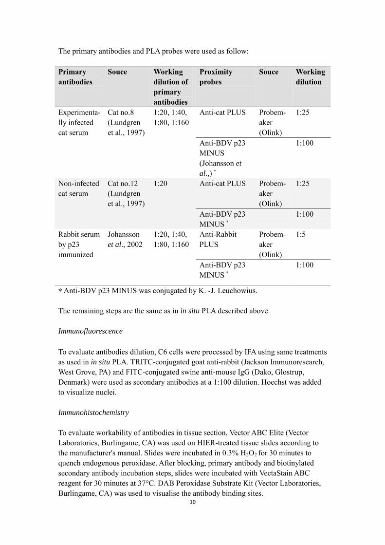
Initially, BDV P was used in persistently infected C6 cells (C6BDV) and non-infected C6 cells (C6) to evaluate the in situ PLA (Leuchowius and Wensman, unpublished data). Then IHC was displayed in experimentally infected rat to evaluate BDV P (Leuchowius and Wensman, unpublished data). In my study, brain tissue from an experimentally infected rat was investigated for BDV P in in situ PLA. High amounts of specific signals could be observed on the cerebral cortex of the infected rat (Figure 2a). At higher magnification, the specific signals exhibited as red spots in the infected rat (Figure 2b). However, some general staining could be observed in the granular cell layer of the cerebellum of the non-infected rat (Figure 2c and 2d). Based on these results, a naturally infected cat previously BDV-positive by real-time RT-PCR (Wensman et al., 2007), was investigated. Further, a naturally infected horse, confirmed to have classic BD, also was investigated. As shown in Figure 3a, BDV P showed as red staining could be detected in the naturally infected cat. However, some red staining was also observed in the non-infected cat (Figure 3b). In the brain tissue of the German horse with classic Borna disease, specific signals of high intensity could be seen as red spots (Figure 4a).
12
a b
c d
Figure 2. Detection of BDV P using in situ PLA in brain tissue of the same experimentally infected rat and non-infected rat as in IHC. a-b. In situ PLA of BDV P in experimentally infected rat using rabbit polyclonal and mouse monoclonal anti-BDV P (p23) antibodies. The red staining is BDV P. (a) 1X magnification. (b) 40X magnification. c-d. In situ PLA of BDV P in non-infected rat using the same rabbit polyclonal and mouse monoclonal anti-BDV P (p23) antibodies. (c) 1X magnification. (d) 20X magnification.
a b
Figure 3. Detection of BDV P using in situ PLA in brain tissue of the naturally infected cat and non-infected cat. a. in situ PLA of BDV P in naturally infected cat using rabbit polyclonal and mouse monoclonal anti-BDV P (p23) antibodies. The red staining indicated by arrow is BDV P. 20X magnification. b. in situ PLA of BDV P in non-infected cat using the same rabbit polyclonal and mouse monoclonal anti-BDV P (p23) antibodies. 10X magnification.

13
a b
Figure 4. Detection of BDV P using in situ PLA in brain tissue of the naturally infected horse and non-infected horse. a. in situ PLA of BDV P in naturally infected horse using rabbit polyclonal and mouse monoclonal anti-BDV P (p23) antibodies. The red staining is BDV P. 20X magnification. b. Non-infected horse. 20X magnification. Photos by Anna-Lena Berg, Astrazeneca, Södertälje, Sweden
Using in situ PLA for detection of BDV P specific antibodies in serum from infected
cat
To be able to detect BDV-specific antibodies, serum from an experimentally infected cat was investigated using in situ PLA. First, anti-cat IgG was directly conjugated with PLA oligonucleotide arm to make the anti-cat PLUS. To evaluate the anti-BDV p23 MINUS, serum from a BDV p23 immunized rabbit (Johansson et al., 2002) was determined in infected Vero cells using anti-rabbit PLUS from the Duolink kit (Olink Bioscience). As shown in Figure 5a, specific signal as red spots were detected in nuclei. Next, the titre of BDV-specific antibodies in serum from an experimentally infected cat was determined by IFA using two-fold dilutions from 1:20-1:160). Except for the highest dilution (1:160), signals could be observed (data not shown). Serum from the same cat was then analyzed by in situ PLA. BDV P was detected in the nuclei and with high intensity at all dilution levels, thereby indicating presence of BDV P-specific antibodies (Figure 5b). However, it was hard to find clearly specific signal when the same serum was incubated on non-infected Vero cells (Figure 5d). Serum from a non-infected cat was used as a negative control. No signals were found in the non-infected cat serum performed on infected and non-infected Vero cells.

14
a b
c d
Figure 5. Detection of BDV P using in situ PLA in serum. a. Positive control, BDV P in experimentally infected rabbit serum by vero cells infected with BDV. The red spots are BDV P, nuclei are blue. 20X magnification. b. in situ PLA of BDV P in naturally infected cat serum by vero cells infected with BDV. c. Negative control, BDV P in non-infected cat serum by vero cells infected with BDV. d. Negative control, BDV P in naturally infected cat serum by normal vero cells. Using in situ PLA for detection of host-virus protein-protein interactions in
persistently infected cells
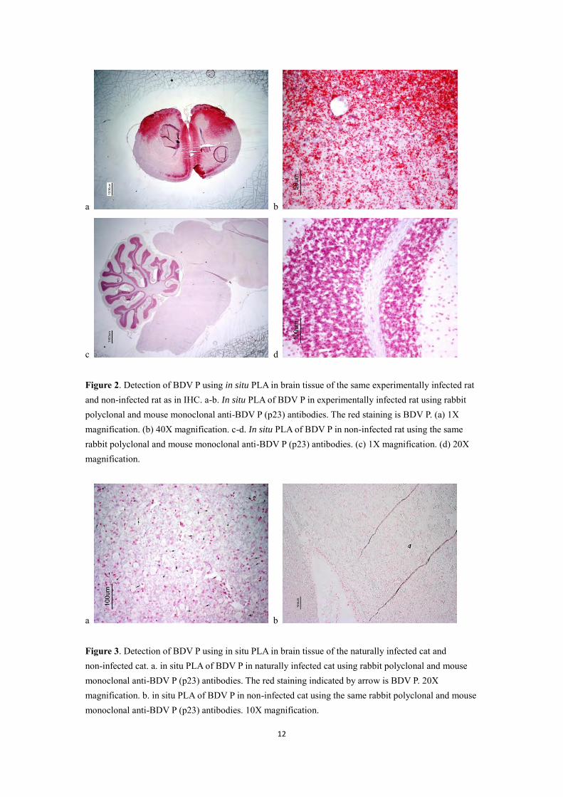
First, the primary antibodies were evaluated in C6 cells using IFA (not shown). Then host-virus protein-protein interactions (BDV-P and -N, with HMGB1; BDV-P and –N, with Cdc2; BDV-P and -N, with DLC8) and post-translational modifications of the viral phosphoprotein were demonstrated in C6BDV and C6 cells using in situ PLA. As shown in Figure 6, there is a remarkable difference in the number of signals between C6BDV and C6 cells. As shown in Figure 7, the positive control, BDV N showed an oversaturation of signals in nuclei of C6BDV cells. The interactions between HMGB1 and BDV P, HMGB1 and BDV N, Cdc2 and BDV P, and Cdc2 and BDV N were detected in nuclei more than in cytoplasm. The interactions between DLC8 and BDV-P were detected both in nuclei and cytoplasm. For DLC8 and BDV N interactions, there was a quite high background. The post-translational modifications of BDV P were delivered by the interactions of BDV P with phosphoserine. Most of the interactions between BDV P and phosphoserine could be observed in the nuclei. Two negative controls, when the primary antibodies were left out, were definitely negative.

15
Figure 6. Signal quantification of protein-protein interactions. Seven protein-protein interactions was analyzed in C6BDV and C6 cells: BDV P and DLC8, BDV N and DLC8, BDV P and HMGB1, BDV N and HMGB1, BDV P and Cdc2, BDV N and Cdc2, and post-translational modifications of BDV P (BDV P and Phosphoserine); BDV N detection was used as positive control; The samples with no primary antibody and just added probes were used as negative control. The signals were quantified using Duolink Image Tool. Signals quantification by Karl-Johan Leuchowius, Uppsala University
16
Figure 7. Visualization of host-virus protein-protein interactions in C6BDV and C6 cells: BDV P and DLC8, BDV N and DLC8, BDV P and HMGB1, BDV N and HMGB1, BDV P and Cdc2, BDV N and Cdc2, and post-translational modifications of BDV P (BDV P and Phosphoserine); BDV N detection was used as positive control; The samples with no primary antibody and just added probes were used as negative control. The red spots are interactions, nuclei are blue. 20X magnification. Photo by Karl-Johan Leuchowius, Uppsala University

17
Discussion
BDV P in brain tissue from infected animals In this study, some general staining which was probably caused by endogenous peroxidase was found in the cerebellum of non-infected rat by in situ PLA. The general staining can be defined as background compared to the spot-like signals in the infected rat. Previous studies suggest that the inflammatory reaction in the CNS caused by BDV is characterized as a nonsuppurative meningoencephalitis of the gray matter of the olfactory bulb, cerebral cortex, hippocampus, basal ganglia, and brainstem (Villanueva et al., 2010). Compared with the rat stereotaxic map (Paxinos and Watson, 1997), most of the red staining in the infected cat was found at the olfactory tubercle, piriform cortex, anterior olfactory nucleus and cerebral cortex as well. The results from naturally infected horse indicate that in situ PLA could be a useful tool for posthumous diagnosis in infectious diseases, especially when viral antigen is scarce. Since the amount of BDV antigen in infected cat is lower than in the other hosts, the signal from infected cat is less than in infected horse. Further, some background was observed in the non-infected cat. Taken together, this suggests that further evaluation of BDV P in situ PLA should be performed on infected cats. BDV P specific antibodies in serum from infected cat
Serology has been widely used in the diagnosis of mammalian BDV infection, even if there has controversy around its interpretation (Ludwig and Bode, 2000). In this study, serum from infected cat and immunized rabbit reacted strongly with P protein of infected Vero cells in in situ PLA at the highest titre. However, IFA as the most common method used in serology cannot detect BDV P at the same titre. This indicates that in situ PLA for detection of antibodies is more specific and sensitive than IFA. Nevertheless, in situ PLA is considered more complex than IFA since it has several different incubation and washing steps. To summarize, since the present of BDV antibodies in serum largely corresponded with the development of clinical BD, it’s hard to detect BDV specific antibodies from serum in the early stage of infection. All the serologic results obtained from the early stage of infection are probably low false negative reactions. Therefore, most of serological methods need to combine with nested RT-PCR to diagnose BDV infection. Host-virus protein-protein interactions in persistently infected cells
Previous studies suggested that BDV P can directly bind to HMGB1 and inhibit its function to repress p53-mediated transcriptional activity, which may be involved in

18
unique features of BDV infection in CNS cells, such as noncytolytic replication and persistent infection (Zhang et al., 2003). In this study, the results indicate that HMGB1 might interact with BDV N as well, which has never been reported. However, BDV P and BDV N can interact with each other (Berg et al., 1998). If the proteins studied are close enough (approximately up to 40 nm), the in situ PLA can generate a signal. Therefore, the interactions between BDV N and HMGB1 might be the result of indirect interactions via BDV P. Previous studies have shown that BDV N can interact with both phosphorylated and non-phosphorylated Cdc2, and with Cyclin B1 as well (Planz et al., 2003). BDV P was shown only to interact with the non-phosphorylated form of Cdc2. In this study, the results confirm that Cdc2 can interact with both BDV P and BDV N. Furthermore, previous studies indicated that interactions between Cdc2 and BDV N can reduce the proliferation rate in transfected and infected cells, which might be involved in unique features of BDV persistence (Planz et al., 2003). However, the effect of the interaction between Cdc2 and BDV P is still unknown. The interaction between DLC8 and Rabies virus P protein has been reported in previous studies (Greber and Way 2006). Since DLC8 is a subunit of the dynein motor complex involved in minus end-directed movement of organelles along microtubules, the DLC8-P interaction might explain how virus transport from the infected site to the CNS (Greber and Way 2006). In addition, the Ebola (EBOV) viral protein VP35, which can bind to DLC8 and form complex with N and L, is essential for transcription and replication in an artificial minigenome system (Kubota et al., 2009). Therefore, EBOV transcription and viral replication might be related to VP35-DLC8 interactions (Kubota et al., 2009). Interestingly, Rabies virus, Ebola virus and BDV all belong to the order Mononegavirales. The result of this study suggests that there might be potential interactions between BDV P and DLC8, which could reveal how BDV transport from the peripheral neurons to the CNS. However, these interactions still need to be further investigated and confirmed by other methods. Due to the interference of high background, it is hard to identify the interactions between BDV N and DLC8.
Since normal cells also have a lot of proteins phosphorylated at serine residues, and the polyclonal BDV P antiserum has a certain degree of cross-reactivity, some background was observed for BDV P and phosphoserine interactions in the non-infected C6 cells. Because of in situ PLA generating signal when two proteins are close enough, it is hard to distinguish whether this interaction happened in post-translational modifications of BDV P or between phosphorylated host proteins and BDV P and/or BDV N. Previous study using minireplicon assay indicated that BDV P is phosphorylated by PKCɛ at serine residues 26 and 28 and by CKII at serine residues 70 and 86. Moreover, the phosphorylation of BDV P negatively regulates its cofactor activity that improves the viral polymerase activity and the viral spread efficiency (Schmid et al., 2007). Furthermore, P phosphorylation might prevent

19
uncontrolled polymerase activity and subsequent lysis of cells, which might be the reason for persistently infection of BDV in cells without causing a cytopathic effect (Schmid et al., 2007). Recent research suggested that BDV P also acts as a kinase decoy which can competitively interfere with normal PKC substrates that affects synaptic plasticity (Prat et al., 2009). To summarize, this was the first time in situ PLA was used to detect BDV protein in tissue and BDV-specific antibodies in serum, and to visualize earlier proven host-virus protein-protein interactions (i.e. the interactions between BDV-P and HMGB1, BDV-N and Cdc2, and BDV-P and Cdc2). Furthermore, some interactions (i.e. the interactions between BDV-N and HMGB1, BDV-P and DLC8) which have never been reported were also detected in this study. However, these interactions still need further investigation to be confirmed. In most methods used to detect protein-protein interactions, it is always hard to distinguish direct interactions and indirect interactions in protein complexes. The same problem also occurs in in situ PLA. To overcome this problem, cells transfected respectively with BDV P and BDV N should be used in host-virus protein-protein interactions research in future. In general, in situ PLA shows great strength to study protein-protein interactions. It gives new possibilities to study these interactions in tissue samples of natural hosts, and improves the way to detect how BDV interacts with the host and causes disease. Acknowledgements I would like to thank my supervisor Jonas Wensman for giving me the opportunity to work in this fantastic group and with this interesting project. Thank you for always having your door open to answer my questions. I also would like to thank Prof. Mikael Berg and Prof. Sándor Belák for your useful advices on my project. Special thanks to Dr. Karl-Johan Leuchowius for your help with image analysis and probe conjugation, to Erik Nyström (at Olink Bioscience) for your technical advice and supplying reagents, and to Inga-Lena Öström-Örde for your help with the serology work. I really appreciate to Dr. Claudia Baule, Muhammad Munir and Paidikondala Maruthibabu, for your kind support and interesting discussion. I want to give the deep gratitude to Gong Pan for your comments on manuscript. Special thanks everyone in the V.I.P. group for creating the small academic world with great freedom and opportunity. The last but most important, I want to thanks my beloved parents and friends. Without your endless love and supports, I will never go this far.
Refferences
Barik, S., and AK. Banerjee. 1992. Sequential phosphorylation of the phosphoprotein of vesicular stomatitis virus by cellular and viral protein kinases is essential for transcription activation. J Virol. 66:1109-1118.

20
Berg, M., C. Ehrenborg, J. Blomberg, R. Pipkorn and AL. Berg. 1998. Two domains of the Borna disease virus p40 protein are required for interaction with the p23 protein. J Gen Virol. 79(12):2957-2963. Bode, L., W. Zimmermann, R. Ferszt, F. Steinbach, and H. Ludwig. 1995. Borna disease virus genome transcribed and expressed in psychiatric patients. Nat Med,
1:232–236. Briese, T., A. Schneemann, AJ. Lewis, et al. 1994. Genomic organization of Borna disease virus. Proc Natl Acad Sci USA. 91:4362-4366. Castedo, M., J. L. Perfettini, T. Roumier, and G. Kroemer. 2002. Cyclin-dependent kinase-1: linking apoptosis to cell cycle and mitotic catastrophe. Cell Death Differ.
9:1287-1293. Crepieux, P., H. Kwon, N. Leclerc, W. Spencer, S. Richard, R. Lin, and J. Hiscott. 1997. IĸBα physically interacts with a cytoskeleton-associated protein through its signal response domain. Mol. Cell. Biol. 17:7375–7385. Cubitt, B., C. Oldstone, and J. C. de la Torre. 1994. Sequence and genome organization of Borna disease virus. J. Virol. 68, 1382–1396. Flower. R.L.P., S. Kamhieh, L. McLean, L. Bode, H. Ludwig, C.M. Ward. 2008. Human Borna disease virus infection in Australia: Serological markers of infection in multi-transfused patients. APMIS. 116:89-93 Gosztonyi, G., and H. Ludwig. 1995. Borna Disease-neuropathology and pathogenesis. Curr. Top. Microbiol. Immunol. 190:39-69 Gosztonyi, G., B. Dietzschold, M. Kao, C.E. Rupprecht, H. Ludwig, and H. Koprowski.1993. Rabies and Borna disease: a comparative pathogenetic study of two neurovirulent agents. Laboratory Investigation. 68:285-295. Greber, U. F., and M. Way. 2006. A superhighway to virus infection. Cell. 124:741–754. Hagiwara, K., Y. Tsuge, M. Asakawa, et al.2008. Borna disease virus RNA detected in Japanese macaques. Primates. 2008; 49:57–64 Hatalski, C. G., S. Kliche, L. Stitz, and W. I. Lipkin. 1995. Neutralizing antibodies in Borna disease virus-infected rats. J Virol. 69:741-747. Hauk, O., I. Johnsrude, and F. Pulvermuller. 2004. Somatotopic representation of
21
action words in human motor and premotor cortex. Neuron. 41 (Jan. 22):301–307. Jordan, I. and W.I. Lipkin. 2001. Borna disease virus. Reviews in Medical Virology, 11:37-57. Kamitani, W., Y. Shoya, T. Kobayashi, M. Watanabe, B.J. Lee, G. Zhang, K. Tomonaga, and K. Ikuta. 2001. Borna disease virus phosphoprotein binds a neurite outgrowth factor, amphoterin/HMG-1. J Virol. 75:8742-8751. Kobayashi, T., Y. Shoya, and T. Koda, et al. 1998. Nuclear targeting activity associated with the amino terminal region of the Borna disease virus nucleoprotein. Virology. 243:188-197. Kobayashi, T., G. Zhang, B-J. Lee, S. Baba, M. Yamashita, W. Kamitani, H. Yanai, K. Tomonaga, and K. Ikuta. 2003. Modulation of Borna Disease Virus Phosphoprotein Nuclear Localization by thr Viral Protein X Encoded in the Overlapping Open Reading Frame. J. Virol. 77:8099-8107. Kliche, S., T. Briese, A. H. Henschen, L. Stitz, and W. I. Lipkin. 1994. Characterization of a Borna disease virus glycoprotein, gp18. J Virol. 68:6918-6923. Kubota, T., M. Matsuoka, T.H. Chang, et al. 2009. Ebolavirus VP35 Interacts with the Cytoplasmic Dynein Light Chain 8. J. Virol. 83(13):6952-6956. Lipkin, W. I., G. H. Travis, K. M. Carbone, and M. C. Wilson. 1990. Isolation and characterization of Borna disease agent cDNA clones. Proc. Natl. Acad. Sci. USA 87:4184–4188. Lo, K. W., H. M. Kan, L. N. Chan, W. G. Xu, K. P. Wang, Z. Wu, M. Sheng, and M. Zhang. 2005. The 8-kDa dynein light chain binds to p53-binding protein 1 and mediates DNA damage-induced p53 nuclear accumulation. J. Biol. Chem. 280:8172–8179. Nowotny, N., J. Kolodziejek, C. O. Jehle, A. Suchy, P. Staeheli, and M. Schwemmle, 2000. Isolation and characterization of a new subtype of Borna disease virus. J.
Virol. 74:5655–5658. Peng, G., Y. Yan, C. Zhu, S. Wang, X. Yan, L. Lu, W. Li, J. Hu, W. Wei, Y. Mu, et al.
2008. Borna disease virus P protein affects neural transmission through interactions with gamma-aminobutyric acid receptor-associated protein. J Virol. 82:12487-12497. Perez, M. and J. C. de la Torre. 2005. Identification of the Borna disease virus

22
(BDV) proteins required for the formation of BDV-like particles. J. Gen. Virol. 86:1891-1895. Planz, O., S. Pleschka, K. Oesterle, F. Berberich-Siebelt, C. Ehrhardt, L. Stitz, and S. Ludwig. 2003. Borna disease virus nucleoprotein interacts with the CDC2-cyclin B1 complex. J Virol. 77:11186-11192. Poenisch, M., N. Burger, P. Staeheli, G. Bauer, and U. Schneider. 2009. Protein X of Borna disease virus inhibits apoptosis and promotes viral persistence in the CNS of newborn-infected rats. J Virol. 83, 4297–4307. Paxinos G., and C. Watson. 1997. The Rat Brain in Stereotaxic Coordinates. Academic Press, Inc. Prat, CM., S. Schmid, F. Farrugia, N. Cenac, G. Le Masson, M. Schwemmle, and D. Gonzalez-Dunia. 2009. Mutation of the protein kinase C site in borna disease virus phosphoprotein abrogates viral interference with neuronal signaling and restores normal synaptic activity. PLoS Pathog. 5:e1000425. Pyper, J. M. and A. E. Gartner. 1997. Molecular basis for the differential subcellular localization of the 38- and 39-kilodalton structural proteins of Borna disease virus. J Virol. 71:5133-5139. Rott, R. and H. Becht. 1995. Natural and experimental Borna disease in animals. Curr. Top. Microbiol. Immunol. 190:17-30. Sauder, C. and J.C. de la Torre, 1998. Sensitivity and reproducibility of RT-PCR to detect Borna disease virus (BDV) RNA in blood: implications for BDV epidemiology. J Virol Mehtods. 71(2):229-45. Schmid, S., D. Mayer, U. Schneider, and M. Schwemmle. 2007. Functional characterization of the major and minor phosphorylation sites of the P protein of Borna disease virus. J Virol. 81:5497-5507. Schmid, S., P. Metz, C.M. Prat, D. Gonzalez-Dunia, M. Schwemmle. 2010. Protein kinase C-dependent phosphorylation of Borna disease virus P protein is required for efficient viral spread. Arch Virol. 155(5):789-93 Schindler A.R., A. Vögtlin, M. Hilbe, M. Puorger, K. Zlinszky, M. Ackermann, et al. Reverse transcription real-time PCR assays for detection and quantification of Borna disease virus in diseased hosts. Mol Cell Probes. 2007;21:47–55 Schneider, U. 2005. Novel insights into the regulation of the viral polymerase complex of neurotropic Borna disease virus. Virus Res. 111:148-160.

23
Schwemmle, M., B. De, L. Shi, A. Banerjee, W. I. Lipkin. 1997. Borna Disease Virus P-protein Is Phosphorylated by Protein Kinase Cepsilon and Casein Kinase II. J.
Biol. Chem. 272:21818-21823 Staeheli, P., M. Rinder, B. Kaspers. 2010. Avian bornavirus associated with fatal disease in psittacine birds. J Virol. 84:6269-6275. Thierer, J., H. Riehle, O. Grebenstein, T. Binz, S. Herzog, N. Thiedemann, L. Stitz, R. Rott, F. Lottspeich, and H. Niemann. 1992. The 24K protein of Borna disease virus. J. Gen. Virol. 73:413–416. Unterstab, G., S. Ludwig, A. Anton, O. Planz, B. Dauber, D. Krappmann, G. Heins, C. Ehrhardt, and T. Wolff. 2005. Viral targeting of the interferon-ß-inducing Traf family member-associated NF-ĸB activator (TANK)-binding kinase-1. Proc Natl
Acad Sci U S A. 102:13640-13645. Villanueva, I., P. Gray, N. Mirhosseini, S. Payne, S. Hoppes, K. S. Honkavuori, T. Briese, D. Turner, and I. Tizard. 2010. The diagnosis of proventricular dilatation disease: Use of a Western blot assay to detect antibodies against avian Borna virus. Veterinary Microbiology. 143 (2010):196-201. Wehner, T., A. Ruppert, C. Herden, K. Frese, H. Becht, and JA. Richt. 1997. Detection of a novel Borna disease virusencoded 10 kDa protein in infected cells and tissues. J Gen Virol. 78:2459-2466. Wensman, JJ., P. Thorén, M. Hakhverdyan, S. Belák, M. Berg. Development of a real-time RT-PCR assay for improved detection of Borna disease virus. J Virol
Methods. 2007;143:1–10. Yang, D., P. Tewary, G. de la Rosa, F. Wei, and J. J. Oppenheim. 2010. The alarmin functions of high-mobility group proteins. Biochim Biophys Acta. 1799:157-163. Zhang, G., T. Kobayashi, et al. 2003. Borna Disease Virus Phosphoprotein Represses p53-Mediated Transcriptional Activity by Interference with HMGB1. J. Virol. 77 (22):12243-12251.