Embed Size (px)
Citation preview

longer lifetime and cause lower morbidity than implantedIV catheters.3 Variable rate telemetry-controlled pumpswere first tested in the 1980’s. These pumps measure 8 3 2cm and weigh 150 to 300 g. They are surgically implanted ina subcutaneous pocket in the lower abdomen, usually un-der general anesthesia. Insulin is placed into the pump byneedle injection every 1 to 3 months.
Clinical experience with the implantable insulin pump hasdemonstrated good glucose control. A randomized trial of21 patients with type I diabetes assigned to treatment withthe pump, as opposed to multiple daily insulin injections,showed no significant difference in hemoglobin-A1c con-centrations or frequency of hypoglycemia between the 2groups.4 Subsequent long-term studies demonstrated dra-matic and statistically significant decreases in hypoglycemicepisodes with implantable insulin pumps compared withsubcutaneous insulin injections. In type II diabetes, there isstrong evidence that the pump reduces the incidence ofmild hypoglycemia and allows the same control of hypo-glycemia as multiple daily injections of insulin.
Complications of the insulin pump include catheter block-age, events at the pump implantation site, and mechanicalpump failure. Catheter blockage may occur from adhesionsoccurring at the tip of the catheter or by precipitation ofinsulin. In studies of catheters occluded by fibrin clot orencapsulated inflammatory tissue, amyloid deposits immu-noreactive to insulin antibodies were detected.5 It has beenspeculated that modified products of insulin may activatethe immune response and stimulate the growth of local cellsto produce fibrosis around the catheter. Microprecipitationof insulin is the most common cause, however, of pumpcatheter blockage. As indicated in the reviewed studies, thisevent usually occurs after 8 months, with the largest inci-dence after 24 months. Catheter blockage has been cor-rected by flushing the catheter with a buffer such as sodiumhydroxide, release of the obstruction at the catheter tip bylaparoscopic surgery, or by replacement of the pump orcatheter.
Pump implantation site problems are the second most com-mon reason for removal of an implantable insulin pump.
Problems may include infection, skin inflammation, atrophyor erosion, and local fluid accumulation. The occurrence ofthese problems varies widely with studies. Physical activitywas the only identifiable risk factor for pump pocket events.
Mechanical failures of the pump are the third most commonreason for removal. Mechanical failures are becoming in-creasingly rare because of advances in the technology ofthese subcutaneous pumps.
In summary, the implantable insulin pump has a place inthe management of both type I and type II diabetes. Ad-vances need to be made in developing a more stable formof insulin and a catheter more compatible with less occur-rence of precipitation of insulin. Recent development ofoptical glucose-sensing systems will be a significant steptoward development of an artificial pancreas.6
E. CHRISTOPHER ELLISON, MDDepartment of Surgery
Ohio State UniversityColumbus, Ohio
References
1. Diabetes Control and Complications Research Group. The effect ofintensive treatment of diabetes on the development and progressionof long term complications in insulin dependent diabetes mellitus.N Engl J Med 1993;329:977–986.
2. American Diabetes Association. Continuous subcutaneous insulininfusion (position statement). Diabetes Care 1997;21(suppl):50.
3. Scavini M, Galli L, Reich S, et al. Catheter survival during long terminsulin therapy with the implanted, programmable pump. DiabetesCare 1997;20:610–613.
4. Selam JL, Raccah D, Jean-Didier N, et al. Randomized comparison ofmetabolic control achieved by intraperitoneal insulin infusion withimplantable pumps versus intensive subcutaneous insulin therapyin type 1 diabetic patients. Diabetes Care 1992;15:53–58.
5. Renard E, Baldet P, Picot MC, et al. Catheter complications associ-ated with implantable systems for peritoneal insulin delivery. Dia-betes Care 1995;18:300–306.
6. Jaremko J, Rorstad O. Advances toward the implantable artificialpancreas for treatment of diabetes. Diabetes Care 1998;21:444–450.
General
Virtual Reality Surgery: Toy or Tool for the Future?Guest Reviewer: James E. Barone, MD, FACS, FCCM
VIRTUAL REALITY FLEXIBLE SIGMOIDOSCOPY SIMULATOR TRAINING: IMPACTON RESIDENT PERFORMANCE.Tuggy ML. J Am Board Fam Pract 1998;11:426–433.
Objective To determine the utility of a virtual reality flexible sigmoidoscopy colon simulator in residenttraining.
Design A randomized, prospective trial of the use of the simulator to train residents in sigmoidoscopy.
194 CURRENT SURGERY • Volume 56 / Numbers 4/5 • May/June 1999
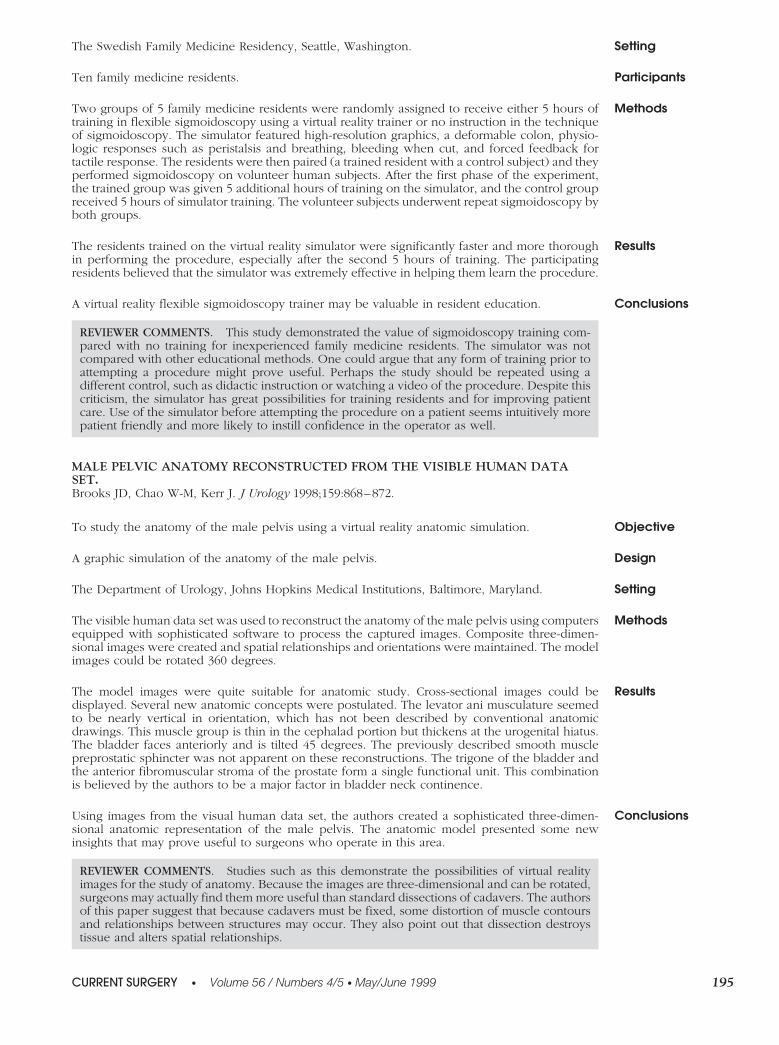
SettingThe Swedish Family Medicine Residency, Seattle, Washington.
ParticipantsTen family medicine residents.
MethodsTwo groups of 5 family medicine residents were randomly assigned to receive either 5 hours oftraining in flexible sigmoidoscopy using a virtual reality trainer or no instruction in the techniqueof sigmoidoscopy. The simulator featured high-resolution graphics, a deformable colon, physio-logic responses such as peristalsis and breathing, bleeding when cut, and forced feedback fortactile response. The residents were then paired (a trained resident with a control subject) and theyperformed sigmoidoscopy on volunteer human subjects. After the first phase of the experiment,the trained group was given 5 additional hours of training on the simulator, and the control groupreceived 5 hours of simulator training. The volunteer subjects underwent repeat sigmoidoscopy byboth groups.
ResultsThe residents trained on the virtual reality simulator were significantly faster and more thoroughin performing the procedure, especially after the second 5 hours of training. The participatingresidents believed that the simulator was extremely effective in helping them learn the procedure.
ConclusionsA virtual reality flexible sigmoidoscopy trainer may be valuable in resident education.
REVIEWER COMMENTS. This study demonstrated the value of sigmoidoscopy training com-pared with no training for inexperienced family medicine residents. The simulator was notcompared with other educational methods. One could argue that any form of training prior toattempting a procedure might prove useful. Perhaps the study should be repeated using adifferent control, such as didactic instruction or watching a video of the procedure. Despite thiscriticism, the simulator has great possibilities for training residents and for improving patientcare. Use of the simulator before attempting the procedure on a patient seems intuitively morepatient friendly and more likely to instill confidence in the operator as well.
MALE PELVIC ANATOMY RECONSTRUCTED FROM THE VISIBLE HUMAN DATASET.Brooks JD, Chao W-M, Kerr J. J Urology 1998;159:868–872.
ObjectiveTo study the anatomy of the male pelvis using a virtual reality anatomic simulation.
DesignA graphic simulation of the anatomy of the male pelvis.
SettingThe Department of Urology, Johns Hopkins Medical Institutions, Baltimore, Maryland.
MethodsThe visible human data set was used to reconstruct the anatomy of the male pelvis using computersequipped with sophisticated software to process the captured images. Composite three-dimen-sional images were created and spatial relationships and orientations were maintained. The modelimages could be rotated 360 degrees.
ResultsThe model images were quite suitable for anatomic study. Cross-sectional images could bedisplayed. Several new anatomic concepts were postulated. The levator ani musculature seemedto be nearly vertical in orientation, which has not been described by conventional anatomicdrawings. This muscle group is thin in the cephalad portion but thickens at the urogenital hiatus.The bladder faces anteriorly and is tilted 45 degrees. The previously described smooth musclepreprostatic sphincter was not apparent on these reconstructions. The trigone of the bladder andthe anterior fibromuscular stroma of the prostate form a single functional unit. This combinationis believed by the authors to be a major factor in bladder neck continence.
ConclusionsUsing images from the visual human data set, the authors created a sophisticated three-dimen-sional anatomic representation of the male pelvis. The anatomic model presented some newinsights that may prove useful to surgeons who operate in this area.
REVIEWER COMMENTS. Studies such as this demonstrate the possibilities of virtual realityimages for the study of anatomy. Because the images are three-dimensional and can be rotated,surgeons may actually find them more useful than standard dissections of cadavers. The authorsof this paper suggest that because cadavers must be fixed, some distortion of muscle contoursand relationships between structures may occur. They also point out that dissection destroystissue and alters spatial relationships.
CURRENT SURGERY • Volume 56 / Numbers 4/5 • May/June 1999 195
VIRTUAL REALITY APPLIED TO HEPATIC SURGERY SIMULATION: THE NEXTREVOLUTION.Marescaux J, Clement J-M, Tassetti V, et al. Ann Surg 1998;228:627–634.
Objective To describe the preliminary findings in the development of a virtual reality liver simulation.
Design A basic yet highly sophisticated, three-dimensional, computer-generated graphic model of theliver.
Methods Using a powerful computer workstation, virtual reality software, and images from the VisualHuman Project of the National Library of Medicine, a three-dimensional model of the external andinternal anatomy of the liver was created. The software was also used to enable the model liver tobe sectioned and to react to simulated externally applied forces as would be utilized in laparo-scopic surgery. A robotic device was utilized to apply external force to the model.
Results A remarkably realistic liver image was developed. The liver model deformed appropriately whenmanipulated with laparoscopic instruments (Fig. 1). With the three-dimensional rotational capa-bility of the design, any surface of the model could be viewed. A plane of resection could beplanned and carried out using the model. Figure 2 demonstrates the placement of a simulatedtumor in the liver.
Conclusions This graphic model of the liver is a useful tool for the study of the anatomy of the liver. The modelalso represents a step toward the use of virtual reality images in the planning and possibleexecution of hepatic surgery. The logical progression of this technology would be to incorporatea set of CT scan or MRI images from a real patient with a hepatic tumor into the simulation. Thevariations of the anatomy and the precise location of the lesion could be considered in the designof the operative procedure. The procedure could then be practiced until perfected before thepatient is even admitted to the hospital.
REVIEWER COMMENTS. With great effort and substantial computer resources, the authorshave produced elegant three-dimensional images of the liver and its complex anatomy. Thediscussion section of this paper represents a primer on virtual reality. The authors describe thedifferent types of three-dimensional rendering and the technical problems involved in creatingrealistic simulations. The description of their method of working to provide interactivity isconcise and easy to understand. The authors point out that many obstacles still exist before trulyrealistic virtual reality simulations of surgical procedures can be created. The authors proposemany extensions of their work and can be expected to produce more fascinating material in thefuture. The educational possibilities of this type of virtual reality simulation are limited only bythe imagination. In an accompanying editorial,1 Krummel suggests that this type of virtualreality modeling is a first step toward the refinement of robotic surgery. Krummel also postu-lates that multiple “takes” of practice robotic procedures could be combined so that an idealoperative procedure could be assembled in the same way that movies are created.
SUMMARY
When hearing the term “virtual reality surgery,” most peo-ple picture a surgeon in Washington operating on a patientwho is in a battlefield hospital in a remote and hostileenvironment. As we learned at this year’s meeting of theAssociation of Program Directors and Surgery, this scenariois unlikely to become practical in the near future due toproblems of cost, bandwidth capacity, and many otherissues. The 3 articles reviewed in this section illustrate thestatus of virtual reality in 1999.
The first paper shows what can be done to educate resi-dents without the need for them to practice on live patients.The colon simulator is so realistic that it serves as a suitablemodel that can help a resident refine his or her technicalskills before performing the procedure on a real patient.The second paper explores the possibilities of advancedanatomical dissection using virtual reality. As strange as itmay seem, the virtual reality model of the pelvis may actu-ally reflect the reality of pelvic anatomy better than a humancadaver is able to. The third paper takes the anatomic
model even further. With virtual reality, a hepatic tumorresection can be planned and practiced well in advance ofthe actual operative procedure. In many centers, hepaticsurgery is infrequently performed. This paper may well beregarded as a landmark because it is one of the first tosuggest that a procedure as complex as hepatic surgery canbe planned and practiced without the need for cadavers.
For a further update on virtual reality, I recommend a newsreport by Vanchieri in the Journal of the National CancerInstitute.2 For medical uses, virtual reality simulations arebeing created in 4 areas of education: the interactive teach-ing of material previously delivered as lectures, training intechnical skills, research on minimally invasive procedures,and determination of the response of the human body toapplied forces. At the University of California in San Diego,an experiment has been planned that will test the ability ofmedical students to learn head and neck anatomy usingvirtual reality simulations. These students will be testedagainst control subjects who study the same material usingconventional cadaver dissections. Other educators are us-ing Internet-based programs to teach dermatology.
196 CURRENT SURGERY • Volume 56 / Numbers 4/5 • May/June 1999

Technical skills are being taught using such tools as a virtualreality hysteroscope at the University of Pennsylvania. Thisadvanced simulator can be adjusted so medical studentscan be given less difficult procedures to perform. Otherinstitutions are utilizing virtual reality to teach techniquessuch as intravenous cannulation, laparoscopy, and the con-duct of anesthesia.
Surprisingly few articles on virtual reality can be foundwhen searching databases of the surgical literature. The 3papers reviewed for this section are among the better recentworks on this subject. The future promises an exponential
increase in the number and diversity of articles on virtualreality surgery.
JAMES E. BARONE, MD, FACS, FCCMColumbia University College of
Physicians and SurgeonsThe Stamford HospitalStamford, Connecticut
References
1. Krummel TM. Surgical simulation and virtual reality: the comingrevolution. Ann Surg 1998;228:635–637.
2. Vanchieri C. Virtual reality: will practice make perfect? J Nat CancerInst 1999;9:207–209.
General
Empyema: Causes, Standard Approaches, and New Techniquesfor TreatmentGuest Reviewer: L. Beaty Pemberton, MD
EVALUATION OF TREATMENT MODALITIES FOR THORACIC EMPYEMA: A COST-EFFECTIVENESSANALYSIS.Thourani VH, Brady KM, Mansour KA, et al. Ann Thorac Surg 1998;66:1121–1127.
DesignA retrospective review of treatment modalities for all patients with discharge diagnosis of thoracicempyema at 1 institution between 1990 and 1997.
ObjectiveReview of causes, treatment modalities, and hospital charges in patients with a diagnosis ofthoracic empyema.
FIGURE 2. The white, oval area in the right lobe of the liverrepresents a simulated tumor. The lower pictures show a sim-ulated line of resection.
FIGURE 1. Three-dimensional model of the surface and por-tions of the internal anatomy of the liver.
CURRENT SURGERY • Volume 56 / Numbers 4/5 • May/June 1999 197


![arXiv:2009.07432v1 [eess.IV] 16 Sep 2020 · Reality ·Computer Assisted Interventions ·Image Guided Surgery 1 Introduction In endoscopic surgery visualising blood vessels is a common](https://img.dokumen.tips/doc/110x75/600bdd56d43bc745e87a2fe2/arxiv200907432v1-eessiv-16-sep-2020-reality-computer-assisted-interventions.jpg)