Embed Size (px)
Citation preview
ARTICLE IN PRESS
www.fems-microbiology.org
FEMS Microbiology Reviews xxx (2005) xxx–xxx
Viral zoonoses in Europe
Hannimari Kallio-Kokko a,b,*, Nathalie Uzcategui a, Olli Vapalahti a,b,c, Antti Vaheri a,b
a Haartman Institute, Department of Virology, University of Helsinki, POB 21, 00014 Helsinki, Finlandb HUSLAB, Department of Virology, Helsinki University Hospital, Finland
c Department of Basic Veterinary Sciences, Division of Microbiology and Epidemiology, Faculty of Veterinary Medicine,
University of Helsinki, Finland
Received 11 October 2004; received in revised form 11 April 2005; accepted 19 April 2005
First published online 29 June 2005
Abstract
A number of new virus infections have emerged or re-emerged during the past 15 years. Some viruses are spreading to new areas
along with climate and environmental changes. The majority of these infections are transmitted from animals to humans, and thus
called zoonoses. Zoonotic viruses are, as compared to human-only viruses, much more difficult to eradicate. Infections by several of
these viruses may lead to high mortality and also attract attention because they are potential bioweapons. This review will focus on
zoonotic virus infections occurring in Europe.
� 2005 Federation of European Microbiological Societies. Published by Elsevier B.V. All rights reserved.
Keywords: Viral zoonoses; Europe; Emerging infections; Arbovirus; Vertebrate-borne viruses; Vector-borne viruses
Contents
1. Introduction . . . . . . . . . . . . . . . . . . . . . . . . . . . . . . . . . . . . . . . . . . . . . . . . . . . . . . . . . . . . . . . . . . . . . . . . . . . . 00
2. Vertebrate-borne viruses . . . . . . . . . . . . . . . . . . . . . . . . . . . . . . . . . . . . . . . . . . . . . . . . . . . . . . . . . . . . . . . . . . . . 00
0168-6
doi:10.
* Co
E-m
2.1. Hantaviruses: a prime example of emerging and re-emerging infections . . . . . . . . . . . . . . . . . . . . . . . . . . . . . . 00
2.2. Lyssaviruses . . . . . . . . . . . . . . . . . . . . . . . . . . . . . . . . . . . . . . . . . . . . . . . . . . . . . . . . . . . . . . . . . . . . . . . . 00
2.3. Arenaviruses . . . . . . . . . . . . . . . . . . . . . . . . . . . . . . . . . . . . . . . . . . . . . . . . . . . . . . . . . . . . . . . . . . . . . . . . 00
2.4. Orthopoxviruses . . . . . . . . . . . . . . . . . . . . . . . . . . . . . . . . . . . . . . . . . . . . . . . . . . . . . . . . . . . . . . . . . . . . . 00
2.5. Orthomyxoviruses . . . . . . . . . . . . . . . . . . . . . . . . . . . . . . . . . . . . . . . . . . . . . . . . . . . . . . . . . . . . . . . . . . . . 00
3. Vector-borne viruses . . . . . . . . . . . . . . . . . . . . . . . . . . . . . . . . . . . . . . . . . . . . . . . . . . . . . . . . . . . . . . . . . . . . . . 00
3.1. Alphaviruses . . . . . . . . . . . . . . . . . . . . . . . . . . . . . . . . . . . . . . . . . . . . . . . . . . . . . . . . . . . . . . . . . . . . . . . . 00
3.2. Flaviviruses . . . . . . . . . . . . . . . . . . . . . . . . . . . . . . . . . . . . . . . . . . . . . . . . . . . . . . . . . . . . . . . . . . . . . . . . 00
3.3. Nairoviruses: Crimean-Congo hemorrhagic fever virus . . . . . . . . . . . . . . . . . . . . . . . . . . . . . . . . . . . . . . . . . . 00
3.4. Orthobunyaviruses: Tahyna and Inkoo viruses. . . . . . . . . . . . . . . . . . . . . . . . . . . . . . . . . . . . . . . . . . . . . . . . 00
3.5. Phleboviruses . . . . . . . . . . . . . . . . . . . . . . . . . . . . . . . . . . . . . . . . . . . . . . . . . . . . . . . . . . . . . . . . . . . . . . . 00
4. Concluding remarks . . . . . . . . . . . . . . . . . . . . . . . . . . . . . . . . . . . . . . . . . . . . . . . . . . . . . . . . . . . . . . . . . . . . . . . 00
Acknowledgements . . . . . . . . . . . . . . . . . . . . . . . . . . . . . . . . . . . . . . . . . . . . . . . . . . . . . . . . . . . . . . . . . . . . . . . 00
References. . . . . . . . . . . . . . . . . . . . . . . . . . . . . . . . . . . . . . . . . . . . . . . . . . . . . . . . . . . . . . . . . . . . . . . . . . . . . . 00
445/$22.00 � 2005 Federation of European Microbiological Societies. Published by Elsevier B.V. All rights reserved.
1016/j.femsre.2005.04.012
rresponding author. Tel.: +358 9 191 26890; fax: +358 9 191 26491.
ail address: [email protected] (H. Kallio-Kokko).

2 H. Kallio-Kokko et al. / FEMS Microbiology Reviews xxx (2005) xxx–xxx
ARTICLE IN PRESS
1. Introduction
During the past 15 years a number of new virus infec-
tions have emerged or re-emerged. Most of them, such
as Sin Nombre and Andes hantaviruses, SARS corona-
virus, avian influenza, Nipah and Hendra viruses, haveappeared in subtropical or tropical regions. Dengue is
spreading to new areas and West Nile virus has reached
the New World. Infections by several of these viruses
may lead to high mortality and also attract attention be-
cause they are potential bioweapons. Some viruses such
as tick-borne encephalitis virus are spreading to new
areas along with climate and environmental changes.
Most of these infections are zoonoses and clearly virusesshared by animals and humans are, unlike human-only
viruses, much more difficult to eradicate. Here, we re-
view zoonotic virus infections occurring in Europe.
The infections like Lassa fever and dengue that are im-
ported to Europe but are not indigenous to European
nature will not be discussed in detail in the review. We
have divided the virus infections into two categories,
those that are transmitted to humans directly fromvertebrate animals (like rodents, foxes, bats and birds)
and those that are primarily transmitted by arthropods
(mosquitoes, ticks, sandflies). The latter class is formed
by arboviruses but notably they have vertebrate hosts
in nature.
2. Vertebrate-borne viruses
2.1. Hantaviruses: a prime example of emerging and
re-emerging infections
2.1.1. Virology
Hantaviruses are enveloped viruses with a tri-seg-
mented negative-stranded genome and belong to the
family Bunyaviridae [1,2]. The 6.4 kb L (large) segmentRNA encodes the �250 kDa RNA polymerase, the
�3.6 kb M (medium) segment the two glycoproteins
Table 1
Viral structure
Genus (Family) Genome Genome
size (kb)
Gen
segm
Hantavirus (Bunyaviridae) ssRNA, neg 11–12 3
Lyssavirus (Rhabdoviridae) ssRNA, neg 12 1
Arenavirus (Arenaviridae) ssRNA, neg 10–14 2
Orthopoxvirus (Poxviridae) dsDNA 160–220 1
Orthomyxovirus (Orthomyxoviridae) ssRNA, neg 10–14 6–8
Alphavirus (Togaviridae) ssRNA, pos 8–12 1
Flavivirus (Flaviviridae) ssRNA, pos 10–11 1
Nairovirus (Bunyaviridae) ssRNA, neg 18–19 3
Orthobunyavirus (Bunyaviridae) ssRNA, neg 11–21 3
Phlebovirus (Bunyaviridae) ssRNA, neg 11–12 3
68–76 kDa Gn and 52–58 kDa Gc, – formerly known
as G1 and G2; and the �1.7 kb S (small) segment the
50–54 kDa nucleocapsid protein (N) (Table 1, Fig. 1).
In addition, the S segment of some hantaviruses has an-
other open reading frame named Ns but its product or
function remains to be discovered. Viral messengerRNAs of the members of the Bunyaviridae are not poly-
adenylated and are truncated relative to the genome
RNAs at the 3 0 termini. Messenger RNAs have 5 0-meth-
ylated caps and 10–18 nontemplated nucleotides which
are derived from host cell mRNAs. The termini of all
three segments are conserved and complementary to
each other, a feature that has assisted in cloning and
discovery of new hantaviruses. Unlike most otherBunyaviridae, hantaviruses are not arthropod-borne
(arboviruses), but are ROdent-BOrne, roboviruses.
Each hantavirus is primarily carried by a distinct
rodent/insectivore species although a few host switches
seem to have occurred during the tens of millions of
years of their co-evolution with their carrier animals
[3]. We now know that the genetic diversity of hantavi-
ruses is generated partly by (i) genetic drift (accumula-tion of point mutations and insertions/deletions)
leading to quasispecies [4,5], (ii) genetic shifts (reassort-
ments of genome fragments within the same virus
genotype/species), and (iii) according to recent findings
[6,7], by homologous recombination, a mechanism not
previously observed for negative-strand RNA viruses.
2.1.2. Ecology and epidemiology
Hantaviruses, which cause hemorrhagic fevers with
renal syndrome (HFRS) in Eurasia and hantavirus car-
diopulmonary syndrome (HCPS) in the Americas, are
prime examples of emerging and re-emerging infectious
agents. Like most of these infections hantaviral
diseases are zoonoses. With the exception of the
South-American Andes virus, which can be transmitted
directly from human to human, hantavirus infec-tions are thought to be transmitted to humans
primarily from aerosols of rodent excreta. Only some
ome
ents
Lipid
envelope
Virion size
(nm)
Proteins
+ 80–120 L, Gn/G1, Gc/G2, N, (Ns)
+ 200 · 60 N, P, M, G, L
+ 50–300 N, G1, G2, L, Z
+ 220–450 Appr. 200 ORF�s+ 80–120 PB1, PB2, PA, HA, NP, NA,
NB, M1, M2, BM2, NS1, NS2
+ 70 NSP1–4, C, E1, E2
+ 40–60 C, M, E, NS1, NS2A, NS2B,
NS3, NS4A, NS4B, NS5
+ 80–120 L, Gn/G1, Gc/G2, N
+ 80–120 N, NSs, G1, G2, NSm, L
+ 80–120 L, Gn/G1, Gc/G2, N, Ns

FamilyPolymerase/Nonstructural proteins
NucleoproteinPhosphoprotein Membrane proteins
RhabdoviridaeN MP G L
Arenaviridae
GPC (G1, G2) N L
BunyaviridaeN, NSs Gn/G1, Gc/G2, NSm L
Orthomyxoviridae*
M1M2 NA NP HA PA PB1 PB2
Togaviridae
NSP1, NSP2, NSP3, NSP4 C
FlaviviridaeC
E3, E2, E1
M, E NS1, NS2A, NS2B, NS3, NS4A, NS4B, NS5
Z
NS1NS2
Fig. 1. Schematic representation of genome structures and expression strategies of the RNA virus families described in this review. (*) Segments and
proteins according to Influenza A virus.
H. Kallio-Kokko et al. / FEMS Microbiology Reviews xxx (2005) xxx–xxx 3
ARTICLE IN PRESS
hantaviruses cause disease in humans. In Europe there
are three major hantaviral pathogens [8] (Table 2, Fig.
2): Puumala virus carried by the bank vole (Clethrion-
omys glareolus) causes a relatively mild disease, alsoknown as nephropathia epidemica (NE); Dobrava
virus carried by the yellow necked mouse, Apodemus
flavicollis, causes severe HFRS; and the genetically
and antigenically closely related Saaremaa virus carried
by the field mouse, Apodemus agrarius, causes mild
NE-like disease. There are also reports of Seoul virus
carried by rats (Rattus norvegicus and Rattus rattus)
which causes HFRS of intermediate severity. In addi-tion, European common voles (Microtus arvalis and
Microtus rossiaemeridionalis) carry Tula hantavirus,
which can infect humans but apparently asymptomati-
cally [9,10]. Topografov hantavirus isolated from Sibe-
rian lemmings (Lemmus sibiricus) has not been detected
in North European lemmings (Lemmus lemmus)
although it can grow in them [11]. Infections in rodents
are mainly asymptomatic and persistent.Hantavirus infections are quite common in Europe
[8] (Table 3), Puumala virus is common in Northern
Europe, European Russia and parts of Central-Western
Europe (Fig. 2). Dobrava virus is found mainly in the
Balkans (Fig. 2). Saaremaa virus has been detected in
Eastern and Central Europe but its epidemiology is
not well defined (Fig. 2). Apart from laboratory infec-
tions [12,13] Seoul virus has been detected in wild rats,only in France [14]. It is also apparent that many parts
of Europe, such as Britain, Poland and Byelorussia, re-
main ‘‘white’’ on the European hantavirus map [8]. This
means either that HFRS is rare or nonexistent in these
regions or is not widely recognized and diagnosed bythe biomedical community.
In Northern Europe HFRS as well as the carrier
rodents exhibit peaks in 3–4 year cycles [15] while in
Central Europe the HFRS incidence follows the fluc-
tuations of ‘‘mast years’’, i.e. the availability of beech
and oak seeds for the hantavirus-carrying rodents. In
Central Europe HFRS peaks in the summer while in
Northern Europe most cases occur in late autumnand early winter, from November to January. Risk
factors to catch hantavirus infections and HFRS in-
clude professions such as forestry, farming, and mili-
tary, or activities such as camping, and the use of
summer cottages. Males are more likely to be exposed
than females [15,16].
2.1.3. Clinical picture and pathogenesis
Puumala and Dobrava viruses both cause HFRS but
the infections differ considerably in severity [17]: both
are characterized by acute-onset fever, headache,
abdominal pains, backache, temporary renal insuffi-
ciency – first oliguria, proteinuria and increase in serum
creatinine and then polyuria – and thrombocytopenia
but the extent of hemorrhages (hematuria, petecchiae,
internal hemorrhages), requirement for dialysis treat-ment, hypotension and mortality are much higher in

Table 2
Zoonotic viruses circulating in Europe
Genus (Family) Virus Carrier (host/vector) Disease in humans Mortality
Hantavirus (Bunyaviridae) Puumala Clethrionomys glareolus
(bank vole)
HFRS (mild, NE) 0.1%
Dobrava Apodemus flavicollis
(yellow-necked mouse)
HFRS (severe) 10%
Saaremaa Apodemus agrarius
(striped field mouse)
HFRS (NE-like) Low
Seoul Rattus norvegicus and
Rattus rattus (rat)
HFRS (intermediate) 1–2%
Tula Microtus arvalis
(European common vole)
Apathogenic?
Lyssavirus (Rhabdoviridae) Classical rabies Dog, fox, raccon dog,
North American bats
Rabies (encephalitis) 100%
EBLV 1a, b Eptisicus sp
(insectivorous bat)
Rabies (encephalitis) 100%
EBLV 2a, b Myotis sp
(insectivorous bat)
Rabies (encephalitis) 100%
Arenavirus (Arenaviridae) LCMV Mus musculus
(house mouse)
Meningoencephalitis Low
Orthopoxvirus (Poxviridae) Cowpox Apodemus, Clethrionomys,
Microtus rodents
Skin eruptions 1/ca.70
Orthomyxovirus
(Orthomyxoviridae)
Influenza A/H7N7 Wild aquatic birds Conjunctivitis,
respiratory infection
1/85
Alphavirus (Togaviridae) Sindbis Birds/Culex, Culiseta
(mosquitoes)
Rash, arthritis/arthralgia None reported
Flavivirus (Flaviviridae) TBE Ixodes spp. (ticks) Encephalitis 0.5%
Louping Ill Ixodes spp. (ticks) Encephalitis Low
West Nile Culex spp. (mosquitoes) Encephalitis Low
Usutu Culex spp. (mosquitoes) Rash, flu-like illness None reported
Nairovirus (Bunyaviridae) CCHF Hyalomma, Rhipicephalus,
Dermatocento (ticks)
HF 20–35%
Orthobunyavirus (Bunyaviridae) Inkoo Aedes sp (mosquitoes) Meningitis, encephalitis Not reported
Tahyna Aedes sp (mosquitoes) Meningitis, encephalitis Occasionally
Phlebovirus (Phleboviridae) Toscana Fever Phlebotomus perniciosus
(sandfly)
Meningitis, encephalitis Not reported
Sandfly Fever Phlebotomus papatasii
(sandfly)
Meningitis, encephalitis Not reported
4 H. Kallio-Kokko et al. / FEMS Microbiology Reviews xxx (2005) xxx–xxx
ARTICLE IN PRESS
Dobrava HFRS than in NE. About a third of NE
patients experience temporary visual disturbances (myo-
pia), which is a very characteristic, if not pathognomonic
sign of the disease. Notably, the clinical consequences of
all of the hantaviral pathogens in humans vary from
asymptomatic to lethal. Severe NE is associated with a
certain haplotype, HLA-B8, DR3, DQ2 alleles [18].
However, although Puumala virus infection is generallyassociated with mild HFRS, NE may have significant
long-term consequences. A 5-year follow-up study dem-
onstrated that 20% of the patients show an increase in
systolic blood pressure and proteinuria [19]. This is
important since the infection is so common in many
areas of Europe [8] (Table 3, Fig. 2). In addition, in
some patients Puumala virus infection may invade
the pituitary gland and lead to mortality or at least
hypophyseal insufficiency requiring hormone-replace-
ment therapy [20].
The pathogenesis of HFRS is poorly understood
[17,21]. However, it is known that b3 integrins can medi-
ate the entry of pathogenic hantaviruses [22] and that
hantaviruses can regulate apoptosis [23–26,28]. Also
there is evidence [17,21] that increased capillary perme-
ability is an essential component in the pathogenesis ofboth HFRS and HCPS, although different target tissues,
kidneys and lungs are affected in the two diseases.
HFRS patients show locally increased levels of TNF-ain the plasma and kidneys [27,28] and high levels of uri-
nary secretion of the proinflammatory cytokine IL-6
[29]. Studies with a monkey model mimicking human
Puumala virus infection [30] may assist in elucidating
the mechanism of pathogenesis.

Table 3
Human hantavirus infections in Europe
Region Puumala Dobrava Saaremaa Cases/yeara Seroprevalence
European Russia + + 3000 6%
Finland + 1000 5%, 20% in areas
Sweden + 300 8% in Northern part
Germany + + 100 1–3%
France + 100
Belgium + 100 1.5%
Norway + 100
Slovenia + + + 15 2%
Netherlands + 10 1%
Denmark + + 10 1% in some areas
Slovakia + + 10
Bosnia-Herzegovina + + 10
Greece + + 5 4%
Estonia + + 9%
Latvia + + 4%
Austria + 1.2%
Czech Republic 1–2%
Hungary + +
Portugal 1%
Albania +
Yugoslavia + +
a Number of cases diagnosed serologically. The numbers are estimations.
Fig. 2. Geographic distribution of hantaviruses pathogenic to humans in Europe.
H. Kallio-Kokko et al. / FEMS Microbiology Reviews xxx (2005) xxx–xxx 5
ARTICLE IN PRESS
Of the four structural proteins, both in humoral and
cellular immunity, the N protein appears to be the prin-
cipal immunogen [31]. Cytotoxic T-lymphocyte (CTL)
responses are seen [32] and may be important both for
protective immunity and pathogenesis of hantavirus
infections [21].
2.1.4. Diagnostics and prevention
The diagnosis of acute HFRS is primarily based on
serology, since viral RNA cannot be regularly detected
in the blood or urine of patients [33,34]. Both immuno-
fluorescence tests and enzyme immunoassays are widely
used for detection of specific IgM or low-avidity IgG
antibodies, characteristic of acute infection [35–37]. In
addition, immunochromatographic 5-min IgM-anti-
body tests [38,39] have been developed. Vaccines against
hantavirus infections have been used for years in China
and Korea, but not in Europe or the Americas [40]. Nospecific therapy is used in Europe, although both ribavi-
rin and interferon-a have been successfully used in trials
6 H. Kallio-Kokko et al. / FEMS Microbiology Reviews xxx (2005) xxx–xxx
ARTICLE IN PRESS
in China [41,42]. A major problem is that at the time
HFRS patients are hospitalized, virus replication is al-
ready disappearing.
2.2. Lyssaviruses
2.2.1. Virology
Members of the genus Lyssavirus within the family
Rhabdoviridae are bullet-shaped, enveloped viruses
approximately 60 nm in diameter and 200 nm in length.
The �12 kb non-segmented negative-strand genome
encodes five proteins (starting from the 3 0 end): the
58–62 kDa nucleoprotein (N), the 35–40 kDa phospho-
protein (P), the 22–25 kDa matrix protein (M), thetrimeric 65–80 kDa glycoprotein (G) and the 190 kDa
polymerase protein (L) (Table 1, Fig. 1). Proteins are
separately transcribed in cascade by a special mechanism
from a single 3 0-end promoter which results in a decreas-
ing transcription and expression gradient for proteins
encoded from the 3 0 to 5 0 end.
The major antigen with neutralizing epitopes and
pathogenetic determinants is the glycoprotein, which
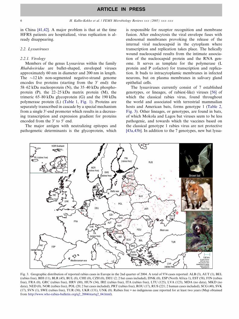
Fig. 3. Geographic distribution of reported rabies cases in Europe in the 2nd
(rabies free), BIH (11), BLR (45), BUL (8), CHE (0), CZH (0), DEU (2; 2 bat
free), FRA (0), GRC (rabies free), HRV (80), HUN (34), IRE (rabies free),
data), NED (0), NOR (rabies free), POL (20; 2 bat cases included), PRT (rabi
(17), SVN (1), SWE (rabies free), TUR (30), UKR (131), UNK (0). Rabies fr
from http://www.who-rabies-bulletin.org/q2_2004/startq2_04.html).
is responsible for receptor recognition and membrane
fusion. After endocytosis the viral envelope fuses with
endosomal membranes provoking the release of the
internal viral nucleocapsid in the cytoplasm where
transcription and replication takes place. The helically
wound nucleocapsid results from the intimate associa-tion of the nucleocapsid protein and the RNA gen-
ome. It serves as template for the polymerase (L
protein and P cofactor) for transcription and replica-
tion. It buds to intracytoplamic membranes in infected
neurons, but on plasma membranes in salivary gland
epithelial cells.
The lyssaviruses currently consist of 7 established
genotypes, or lineages, of rabies(-like) viruses [56] ofwhich the classical rabies virus, found throughout
the world and associated with terrestrial mammalian
hosts and American bats, forms genotype 1 (Table 2,
Fig. 3). Other lineages, or genotypes, are found in bats,
of which Mokola and Lagos bat viruses seem to be less
pathogenic, and towards which the vaccines based on
the classical genotype 1 rabies virus are not protective
[43a,43b]. In addition to the 7 genotypes, new bat lyssa-
quarter of 2004. A total of 974 cases reported: ALB (3), AUT (1), BEL
cases included), DNK (0), ESP (North Africa 1), EST (58), FIN (rabies
ITA (rabies free), LTU (125), LVA (123), MDA (no data), MKD (no
es free), ROU (17), RUS (221; 2 human cases included), SCG (40), SVK
ee = no indigenous case reported for at least two years (Map obtained
H. Kallio-Kokko et al. / FEMS Microbiology Reviews xxx (2005) xxx–xxx 7
ARTICLE IN PRESS
virus genotypes have been recently found, e.g. from
Russia [43c].
2.2.2. Epidemiology and ecology
During the past 100 years or more in Europe, clas-
sical rabies virus has made two major host shifts, firstlyfrom the dog to red foxes, and then to racoon dogs
brought from East Asia to be raised for fur; this
species then became widely established in the wild
(Fig. 3). Phylogenetic data also suggest that west-
and southward spread of rabies virus occurred during
the last century [44a]. Although in Europe the most
important carriers of classical rabies virus are foxes
and racoon dogs, the virus can be transmitted to sec-ondary hosts such as domestic animals (dog, cat, cattle,
horse, sheep) or e.g. deer – practically any mammal
species could be a potential carrier. Bats are a special
case; lyssaviruses are maintained in bats, even in the
absence of classical rabies in carnivores; thus in coun-
tries where classical rabies has been eliminated, bat ra-
bies has become the dominating or only source of
rabies virus and retains the potential for host-switchinginto the carnivore reservoir which constitutes a more
direct threat for public health [44b].
As many bats are protected species, the detection or
‘‘absence’’ of bat rabies is dependent also on the
intensity of screening efforts [45,46]. As of the beginning
Table 4
Classical rabies virus infections in Europe (Year 2003)
Country No. cases/wildlife No. cases/domestic anim
Albania 2 0
Austria 1 0
Belarus 761 316
Bosnia Hercegovina 63 17
Bulgaria 15 4
Croatia 590 43
Estonia 697 117
Finland 0 1
France 0 0
Germany 24 0
Hungary 129 43
Latvia 828 135
Lithuania 796 312
Moldova 13 20
Poland 310 72
Romania 67 28
Russian Federation 1360 1502
Serbia a Montenegro 207 54
Slovak Republic 284 42
Slovenia 8 0
Switzerland & Liec. 0 1
Turkey 17 139
Ukraine 924 1104
Total 7095 3951
Rabies free European countries:
Belgium, Finland, Iceland, Ireland, Italy, Luxembourg, Norway, Sweden
Data obtained from Rabies Bulletin Europe, vol. 27, no. 4, Quarter 4 (2003
of 2004, the following countries in Europe were declared
‘‘rabies-free’’ by WHO (meaning no indigenous cases
occurred during the last two years): Belgium, Cyprus,
Finland, Greece, Iceland, Ireland, Italy, Luxembourg,
Norway, Portugal and Sweden (Fig. 3). In 2003, rabies
virus was detected in Europe in 7095 wild animals(excluding bats), 3951 domestic animals; 33 bat and 6
human rabies cases were diagnosed (see Table 4). The
countries where rabies was circulating include (in dimin-
ishing order of cases) Russia, Ukraine, Lithuania,
Belarus, Latvia, Estonia, Croatia, Poland, Slovakia,
Serbia-Montenegro and Turkey with hundred(s)
to thousands of (animal) cases, Romania, Bosnia-
Herzegovina, Germany, Moldova and Bulgaria withdozens of cases, and individual cases were registered
(some imported and not affecting the rabies-free status)
in Slovenia, the Netherlands, Denmark, France,
Albania, Finland, Austria and Switzerland (Table 4,
Fig. 3). All the rabies cases (2003) detected in Denmark,
France and the Netherlands were bat rabies, in addition,
bat rabies was found in Germany, Poland, Russia and
Ukraine, most commonly EBL-1 carried by Eptesicus
serotinus bats [47–51a], http://www.who-rabies-bulle-
tin.org. The first case of bat rabies in a human in the
UK for decades, was recorded in 2002. The etiological
agent was EBL-2 virus, transmitted by an autochtonous
Daubenton bat (Myotis sp.) [51b].
als No. cases/bats No. cases/humans No. cases/total
0 0 2
0 0 1
0 0 1077
0 0 80
0 0 19
0 0 633
0 0 814
0 0 1
2 0 2
13 0 37
0 0 172
0 1 964
0 0 1108
0 0 33
6 0 388
0 0 95
1 3 2866
0 0 261
0 0 326
0 0 8
0 0 1
0 0 156
1 2 2031
33 6 2031
).

8 H. Kallio-Kokko et al. / FEMS Microbiology Reviews xxx (2005) xxx–xxx
ARTICLE IN PRESS
2.2.3. Clinical picture in man and pathogenesis
Members of 6 out of the 7 lyssavirus genotypes (ex-
cept Lagos bat virus, i.e. genotype 2) have caused rabies
disease in man. The infection is inevitably fatal in hu-
mans or other mammals unless immune intervention
(vaccination and administration of antibodies) is used.Transmission may occur though the bite of an animal
delivering the virus deep in to striated muscle or connec-
tive tissue, but infection may also occur after abrasion of
the skin or licking of mucous membranes. The bite of a
bat with small teeth may go unnoticed; on the other
hand the bat lyssaviruses may infect human skin more
easily than has been recognised. The incubation period
is 20–60 days, but has been shown in some cases to bemonths, even years. The classical picture of rabies in-
cludes prodromal illness with fever and non-specific
symptoms, as well as itching and local paresthesia. This
is followed by neurological signs, consisting of either
encephalitic ‘‘furious rabies’’ or paralytic ‘‘dumb ra-
bies’’. In the former, episodic hyperactivity and excita-
tion of the CNS manifest as, e.g. hydrophobia,
hypersalivation and convulsions. The patient finallydevelops paralysis, coma and cardiorespiratory failure.
In the paralytic form, no excitation is seen, but paralytic
disease develops to coma and death [45,52a,52b].
After entry of the virus to the body, the virus must
gain access to peripheral nerves and to be transported
towards the CNS. Rabies virus components are attached
either directly or by encapsulated vesicles to the dynein
motor carrying the ‘‘cargo’’ along the axonal microtu-bular system towards the cell stroma, approximately at
a speed of 25 mm/day [45,53,54]. It is transmitted after
replication on neuronal membranes transsynaptically
to adjacent neurons and finally invades the CNS where
it first disturbs the limbic system associated with the
excitation, and later neocortex, with little histopatholo-
gical changes in neurons. Centrifugal spread of the virus
from the CNS to many tissues through somatic andautonomic nerves also occurs, where the salivary glands
are especially important for the spread of the virus to the
next victim. Immunological responses do not occur be-
fore CNS involvement.
All the viruses in phylogroup I are also pathogenic to
mice by i.m. injection, and can cross-neutralize each
other. This has practical implications, as fortunately the
vaccine strains (of genotype 1) also appear to protectagainst the EBVL viruses [55]. A pathogenic determinant
common to phylogroup I viruses seems to be amino acid
R333 in the glycoprotein [56]; substitution of this amino
acid also abolishes the retrograde transport [57].
When symptoms develop, no cure is available. How-
ever, after exposure to a bite or scratch of a potentially
rabid animal, rabies must and can effectively be pre-
vented by post-exposure prophylaxis, originally devel-oped by Louis Pasteur. The treatment includes in a
non-vaccinated person (a) washing (with water and
detergent) and disinfecting the wound to minimize the
amount of cell-free virus (this alone can increase survival
50%), (b) starting a post-exposure vaccine regimen
which includes (in Europe) five doses of cell-culture de-
rived vaccine intramuscularly into the deltoid muscle on
days 0, 3, 7, 14, 28 and (c) in case of severe or deep in-jury additional passive anti-rabies immunoglobulin,
which should be administered principally to the wound
area. If the person was pre-vaccinated or the animal
can be caught and studied for the presence of rabies,
the protocol can be adjusted accordingly [45,52a].
2.2.4. Diagnosis and prevention
Clinical suspicion of rabies in a case of encephalitisof unknown origin is the starting point. Ante mortem
diagnostics can be achieved most easily with RT-PCR
from saliva. In addition, rabies antigen can be detected
in brain or nuchal skin biopsies, and in some cases
antibodies in serum or CSF may be found. Post-mor-
tem diagnostics is most rapid with antigen detection
or RT-PCR from the brain, virus isolation is also
possible.In addition to post-exposure prophylaxis, vaccines in
humans can be used for pre-exposure prophylaxis (3
doses) in risk groups (veterinarians, wildlife workers,
travellers to endemic areas; especially small children
who may be unable to explain a potential exposure to
a rabid animal) [45,52a,58].
The primary method for control of rabies is vaccina-
tion of dogs (and cats). This practice was established atthe beginning of the last century in most of Europe, but
has not yet been achieved in many developing countries,
where annually 60 000 people still die of rabies. In most
of Europe (excluding Eastern Europe) rabies has been
eradicated from terrestrial wildlife species (carnivores)
by aerial distribution of vaccine baits across the coun-
tryside. The vaccines used to protect wildlife species in-
clude attenuated vaccines as well as a recombinantvaccinia virus carrying the rabies virus glycoprotein
(the latter has in one case caused a skin infection in
man). Although rabies in terrestrial animals can be con-
trolled efficiently, eradication from the bat reservoir is
not currently feasible.
2.3. Arenaviruses
2.3.1. Virology
Arenaviruses are the only members of the RNA virus
family Arenaviridae. These viruses are enveloped, lipid
solvent-sensitive, pleomorphic particles with a mean
diameter of 120 nm (ranging between 50 and 300 nm).
Host cell-derived ribosomes are present in the virions,
and give the virus particles a ‘‘sandy’’ appearance under
the electron microscope, hence the name arenavirus(arena: sand in Latin). The virion structure of arenavi-
ruses is quite simple, virus particles contain two RNA
H. Kallio-Kokko et al. / FEMS Microbiology Reviews xxx (2005) xxx–xxx 9
ARTICLE IN PRESS
segments (S and L) linked to nucleocapsid proteins and
viral polymerase molecules, and these nucleocapsid-
polymerase complexes are surrounded by a lipid enve-
lope into which two glycoproteins (G1 and G2) are
linked protruding on the outside of the virion. Arenav-
iral RNA segments have an ambisense coding arrange-ment, the S segment (�3.5 kb) encoding a 63-kDa
nucleocapsid protein (N) in the viral complementary se-
quence, and in the viral-sense 5 0-end sequence a 75-kDa
glycoprotein precursor (GPC) which is posttranslation-
ally cleaved to two glycosylated proteins 34–44 kDa
G1 and G2; and the L segment (�7.2 kb) encoding a
180-kDa viral polymerase (L) in the viral complemen-
tary sequence, and in the viral-sense 5 0-end of thesequence a 10–14 kDa RING finger Z protein, which
has been shown to have a role in arenavirus budding
[59a] (Table 1, Fig. 1). Initiation of transcription may in-
volve cap-snatching, although the transcription mecha-
nism is not yet fully elucidated.
2.3.2. Ecology and epidemiology
Arenaviruses include 23 viruses all carried by differ-ent rodent hosts (except Tacaribe virus which has been
isolated only from fruit bats) [59b]. Arenaviruses are
capable of causing chronic infections in their rodent
hosts, and infectious virus is present in the blood and
is also secreted into body fluids (saliva, urine, semen),
which is presumably the route of transmission to hu-
mans. The appearance and incidence of arenaviral infec-
tions are closely associated with the distribution of therodent host species and the rodent population dynamics.
Arenaviruses have co-evolved with their specific host
species during millions of years, and have been divided
into OldWorld andNewWorld groups first on a serolog-
ical bass, and later into evolutionary lineages (NewWorld
group) using genetic analysis [60a,60b,60c]. Both groups
contain viruses that are included in the Category A path-
ogen list (defined by CDC, USA), which means that thepropagation of these agents is allowed only in Biosafety
Level 4 laboratories. These highly pathogenic arenavi-
ruses include the South American Junin, Machupo,
Guanarito, and Sabia viruses from theNewWorld group,
and the African Lassa virus from the Old World group.
All these viruses can cause hemorrhagic fevers in humans,
and are considered potential bioterrorism agents being
thus included in biohazard preparedness programs. InEurope, these viruses occur only rarely as imported cases.
The only arenavirus endemic in Europe is lymphocytic
choriomeningitis virus (LCMV), shown to circulate in
Mus musculus populations (Table 2), and associated with
pet hamster derived epidemics [59b,61a]. Relatively few
epidemiological data are available concerning the actual
distribution of LCMV in Europe, but in addition to sero-
logical evidence from Mus sp. material from Spain [61b],antibodies against LCMV have been detected in rodent
species other thanMus musculus (our unpublished data),
which indicates that other yet unknown arenaviruses may
circulate in Europe.
2.3.3. Clinical picture and pathogenesis
At least ten arenaviruses have been reported to be
able to cause disease in humans. As mentioned above,five arenaviruses (Lassa, Junin, Machupo, Guanarito,
and Sabia viruses) are even capable of causing a life-
threatening viral hemorrhagic fever [62a,62b]. None
of these five viruses are endemic in Europe, but a few
imported Lassa virus infections have been diagnosed
in Germany and Great Britain during the last few years
with no secondary infections detected [63–65]. In-
creased travelling increases also the risk for transmis-sion of exotic arenaviruses to non-endemic areas such
as Europe.
LCMV is thus far the only known endemic arenavirus
in Europe [62b]. In humans LCMV infections are mostly
either asymptomatic or influenza-like diseases. In some
cases aseptic meningitis or meningoencephalomyelitis is
seen. LCMV is also capable of causing congenital
infections manifested by hydrocephalus, microcephalus,chorioretinitis, and mental or psychomotor retardation
[66,67a,67b]. LCMV infections are rarely fatal for
humans.
2.3.4. Diagnostics and prevention
The diagnosis of arenaviral infections is based on
serology and/or direct detection of the virus [68]. For
serodiagnosis methods using immunofluorescence assay(IFA) as well as enzyme immunoassay (EIA) have been
described. Either a four-fold rise in IgG antibody titers
or presence of IgM antibodies is considered indicative
of acute infection. The antibodies that appear first in
the acute phase of infection are directed against the
nucleocapsid protein; neutralizing antibodies against
the glycoproteins appear later in the convalescent
phase (if at all). This means that typing of the causa-tive agent is difficult on a serological basis at the early
stage of infection, and is actually possible only in the
convalescent phase due to the slow rise of virus type-
specific neutralizing antibodies. For direct detection
of the virus, antigen detection assays are useful in the
early diagnosis of Lassa fever especially. Also reverse
transcriptase (RT)-PCR tests have been developed
to detect arenaviral RNA in patient samples[69a,69b,70a,70b,70c]. Virus isolation attempts can also
be successful.
The supportive treatment of arenaviral infections in-
cludes ensuring the fluid, electrolyte and osmotic bal-
ance. In severe hemorrhagic fever-cases early diagnosis
is important because the use of ribavirin has been found
effective if the treatment commences within the first six
days after onset of symptoms [71]. Immune plasma con-taining neutralizing antibodies has also been useful in
some cases.
10 H. Kallio-Kokko et al. / FEMS Microbiology Reviews xxx (2005) xxx–xxx
ARTICLE IN PRESS
For prevention of arenaviral diseases, several at-
tempts to develop vaccines have been made [72a]. One
vaccine, Candid 1, has been successfully used in the pre-
vention of Argentine hemorrhagic fever caused by Junin
virus with a clear reduction in the number of infections
observed in humans [72b,73].
2.4. Orthopoxviruses
2.4.1. Virology
The genus Orthopoxvirus in the family Poxviridae
consists of large 220–450 nm brick-shaped viruses, with
a double-stranded DNA genome (160–220 kb) (Table 1),
that are serologically cross-reactive and -protective. Themiddle part of the genome is very conserved among
orthopoxviruses encoding structural proteins and repli-
cation machinery whereas the ends are more variable
comprising genes involved with host-specificity and
counteracting the immune response [74]. Replication
of orthopoxviruses occurs in the cytoplasm and includes
translation of early mRNAs (such as DNA polymerases
and immune defense molecules), DNA replication,translation of intermediate mRNAs (for late transcrip-
tion factors), and late mRNAs encoding structural pro-
teins and late enzymes, respectively. Altogether, e.g. the
cowpox virus genome encodes nearly 200 open reading
frames. Intracellular non-enveloped virions are first
formed, comprising the majority of the infectious viral
progeny; some particles develop in ER/Golgi into envel-
oped, either cell-attached or extracellular viral particles,often motile due to attached actin tails [75]. Homolo-
gous recombination occurs readily between orthopoxvi-
rus sequences which has raised some concerns about the
use of vaccinia virus-based vaccines in wild animals [76].
2.4.2. Epidemiology and ecology
Some orthopoxviruses are host-specific, whereas
some are more promiscuous but have a distinct reser-voir. Their nomenclature may be misleading; e.g. mon-
keypox is not carried by monkeys, neither is cowpox
carried by cows. Smallpox or variola virus, now eradi-
cated and historically the cause of one of the most feared
human diseases, was specific to man. Many other ortho-
poxviruses circulate in wildlife species and are often zoo-
notic. Examples include the vaccinia virus, the modern
smallpox vaccine, the origins of which are unclear butwas originally described as cowpox by Edward Jenner
[77] in late 18th century England, and which later has
also re-escaped to nature in other parts of the world
[78]; monkeypox virus, pathogenic to primates, includ-
ing humans, causing a smallpox-like disease with sec-
ondary transmission and with a likely reservoir in
small rodents in Central Africa; cowpox virus, which
is the main orthopoxvirus in Europe and may be trans-mitted to man either directly from rodents or from a sec-
ondary carrier, typically cat. Cowpox virus has been
detected in Western Eurasia and in Europe, voles of
Clethrionomys and Microtus species and Apodemus mice
are the main reservoir hosts [79] (Table 2). Shedding of
the virus from rodents is apparently transient. Infection
of cattle is rare; domestic cats relatively frequently pres-
ent with clinical disease, but infection of zoo animals,e.g. elephants, has also been reported [80]. Following
the cessation of smallpox vaccination, more than 25
years ago, the number of humans susceptible to cowpox
has increased. More than 60 human cowpox cases have
been reported in the literature since 1969 [81,82].
2.4.3. Clinical picture and pathogenesis of cowpox
All age groups may acquire cowpox, but most caseshave been in girls under 12 years of age, who have
had a cat or e.g. a field mouse as a pet. Infection prob-
ably occurs through abrasions in the skin; persons with
atopic eczema are more prone to the infection. [81–84].
The incubation period is 5–7 days, after which papules
develop into lesions 1–3 cm in diameter which proceed
through pustular, ulceral and eschar stages over a period
of a about two weeks. They may be painful and vary innumber, size and severity. Local lymphadenopathy,
pyrexia and nausea may occur; secondary bacterial
infections are common. Typically, solitary lesions
are found, located mainly in fingers, hands or face
(e.g. eyelid) [81,85]. In 6–8 weeks the lesions heal gradu-
ally, some residual scars may remain. In some cases
severe generalized skin infection occurs [83], especially
in atopic and in immunocompromised individuals andmay in extreme cases lead to death [86]. Cidofovir
(a phosphorylated nucleoside analog of cytosine) may
have potential as an antiviral against cowpox virus
[87]. Man-to-man transmission of cowpox virus (unlike
for monkeypox) has not been reported.
2.4.4. Diagnostics and prevention
It is usually possible to detect orthopoxvirus particlesdirectly from the skin lesions by electron microscopy.
The virus can also be readily isolated in e.g. Vero cells
or chorioallantoic membrane of chicken embryos from
the lesions and subsequently characterised [80]. Several
sensitive PCR approaches have been described, some
related to the bioterrorism (smallpox) preparedness
[88–90]; in each case further typing at the species level
is needed. In addition, during acute cowpox infection,IgM antibodies and low-avidity IgG antibodies have
been detected [83].
Following the cessation of smallpox vaccination
approximately 30 years ago, the younger age groups
are the most susceptible population, both to smallpox
and to cowpox, which is more closely related to vaccinia
virus. Recent estimates indicate as low as 40% protec-
tion levels among Europeans. For instance in Finland,in the age group over 50, everybody had orthopoxvirus
antibodies as measured by immunofluorescence assay.

H. Kallio-Kokko et al. / FEMS Microbiology Reviews xxx (2005) xxx–xxx 11
ARTICLE IN PRESS
The seroprevalence decreased gradually towards youn-
ger age groups reflecting the gradual cessation of small-
pox vaccination, with the last vaccinations in Finland
occurring in 1977 [83]. Smallpox was finally declared
to be eradicated from the world in 1980 [91] after which
few people in Europe have received the vaccine. In addi-tion to wild rodents carrying cowpox, import of exotic
pets may also pose a risk for orthropoxvirus transmis-
sion, as was seen in a recent outbreak of monkeypox
virus in the USA [92].
2.5. Orthomyxoviruses
2.5.1. Virology
Orthomyxoviruses are enveloped, negative-strand
RNA viruses with 6–8 genome segments, of which avian
influenza, (i.e. influenza A) viruses may cause severe dis-
ease in domestic poultry and cause zoonotic infections.
The influenza viruses in wild aquatic birds are the source
of these epidemics in chickens as well as providing a
gene pool for reassortants with human influenza A
viruses which then may become established in human-to-human transmission resulting in influenza pandemics.
In addition, influenza A viruses are known pathogens of
pigs, horses, mink, seals and whales [93,94].
Influenza A virus is 80–120 nm in diameter and has 8
genome segments varying in size from 0.89 to 2.3 kb.
The three largest segments 1–3 encode the polymerase
subunits PA (83 kDa), PB1 (87 kDa) and PB2
(84 kDa), respectively; whereas the segments 4–6 eachencode one viral protein, namely the 63 kDa hemagglu-
tinin (HA), the 56 kDa nucleoprotein (NP), and the
50 kDa neuraminidase (NA), respectively (Table 1,
Fig. 1). The two smallest segments, 7 and 8, encode each
two proteins, the 28 kDa matrix protein (M1) and the
11 kDa membrane protein (M2), and the 27 kDa NS1
and the 14 kDa NS2 proteins, respectively (Table 1,
Fig. 1). In common with the Bunyaviridae, the genomicRNA is packed in the nucleoprotein which carries poly-
merase subunits, and the 5 0 and 3 0-ends are conserved
and complementary to each other and thus able to form
panhandle structures in which the promoter regions re-
side. ‘‘Cap-snatching’’ from cellular mRNAs and an oli-
go-U motif are used to create viral mRNAs starting with
a cap structure and ending in a poly-A region. The virus
enters the cells after binding to the sialic acid receptorsby endocytosis, which is followed by acidification of
the endocytic vesicle and, mediated by the ion channel
forming M2 protein, of the interior of the virus, which
leads then to fusion of the viral and endosomal mem-
brane and release of the viral nucleocapsids to the cyto-
plasm, respectively. However, untypically for an RNA
virus, the replication, transcription and nucleprotein
assembly occur in the nucleus. The envelope proteinsare processed to the plasma membrane, where budding
of the virions finally occurs. The envelope proteins are
also the most important antigenic determinants. The
homotrimeric hemagglutinin (HA), defines the ‘‘H type’’
which is responsible for the binding to the sialic-acid
containing host cell receptors and membrane fusion
properties. The neuraminidase (NA) defines the ‘‘N
type’’, and cleaves terminal sialic acid residues from gly-coconjugates enabling the virus to reach target cells in
the mucin-rich epithelium and facilitating release of
the virus from the cells [93,94]. The catalytic site of the
neuraminidase is a target for antivirals oseltamivir and
zanamivir, and the M2 protein is a target for amanta-
dine and rimantadine.
To become active, the hemagglutinin protein needs to
be cleaved by trypsin-like proteases found in respiratoryand gastrointestinal epithelia. Human-adapted influenza
viruses replicate in the respiratory tract, whereas in
avians the virus replicates primarily in the gut. When
transmitted to and within poultry, the normal hemag-
glutinin of influenza A virus H5 or H7 of wild aquatic
birds of the ‘‘low pathogenic type (LPAI) may be mu-
tated. Accumulation of basic residues at the cleavage site
makes the HA cleavable by most proteases of cells, suchas furin, and results in ‘‘highly-pathogenic avian influ-
enza’’ (HPAI) virus able to replicate in most tissues kill-
ing rapidly up to 100% of chickens [93–95]. HPAI, also
known as ‘‘fowl plague’’ was first described in 1878 and
the virus was first isolated in 1901 suggesting that hu-
mans have been directly exposed to avian influenza A
viruses for centuries [96].
2.5.2. Epidemiology, clinical aspects and treatment
Influenza A virus gene pools reside in wild aquatic
birds where at least 15 HA types and 9 NA types are
found, as compared to 3 HA (H1–H3) and 2 NA (N1,
N2) types circulating in man. Also, the genes in aquatic
birds are in evolutionary stasis without undergoing
changes due to selective pressures. Influenza A viruses
in man are under constant selective pressure imposedby population immunity causing the hemagglutinin of
influenza A virus to change its antigenic properties by
accumulation of mutations (genetic drift). However,
through double infection and reassortment (genetic
shift), novel (e.g. hemagglutinin) genes from aquatic
birds may become established in human influenza
viruses giving rise to pandemics due to lack of adapta-
tion to and immunity in, humans.Previously, it was thought that the different receptor
specificity – favoring different side chains of sialic acid
– of human and avian influenza viruses would make di-
rect transmission of avian viruses to humans unlikely,
but both could be expected to infect swine, which carry
receptors for both. Thus pigs are considered to be poten-
tial reservoirs for generating new influenza virus vari-
ants. It has only recently been discovered that virusesconsidered unique to avian species may also infect
man, although this may involve change in receptor
12 H. Kallio-Kokko et al. / FEMS Microbiology Reviews xxx (2005) xxx–xxx
ARTICLE IN PRESS
specificities [97]. In 1996 conjunctivitis in UK caused by
avian influenza viruses (and previously, by seal influenza
A viruses) was reported [98,99] (Table 2). Direct zoo-
notic transmission of avian influenza viruses to man
resulting in human respiratory illness, was not known
to occur or had not been diagnosed before the outbreakof H5N1 avian influenza virus in Hong Kong in 1997
where 6/18 patients died of lower respiratory tract infec-
tion [100]. After this, in Europe, an outbreak of H7N1
HPAI avian influenza was encountered in Italy (1999–
2000) without reported transmission to humans [101].
In 2003, a major H7N7 HPAI avian influenza outbreak
occurred in the Netherlands [102,103]. During this epi-
demic, veterinarians and people who culled infectedpoultry were at greatest risk of infection. It was noted
by active surveillance that human H7 infections (and
simultaneous H3 infections) were occurring, and conse-
quently prophylactic treatment with oseltamivir was
started. The H7 virus was confirmed to be transmitted
to 85 humans of which the majority had conjunctivitis,
seven had an influenza-like illness; one veterinarian
(who did not receive prophylactic oseltamivir) died.His symptoms started with high fever and headache
two days after visiting a farm with infected chickens.
One week later, he was admitted to hospital with pneu-
monia, where his status deteriorated to multi-organ fail-
ure, with death due to respiratory insufficiency 2 weeks
after onset of symptoms. In addition, in three cases
household primary contacts were shown to have the dis-
ease by human-to-human transmission [102].Both NA inhibitors, zanamivir and oseltamivir inhib-
ited virus obtained from humans during this outbreak
and a significant difference was found in avian influenza
virus detection between oseltamivir users (1/38) and
those who had not taken prophylactic medication
(5/52) [102].
Outbreaks of avian influenza have continued to occur
in other parts of the world, especially the devastatingongoing H5N1 epidemic in South-East Asia since late
2003 with as of June 2005 a total of 54 deaths/107 cases
in Viet Nam, Thailand and Cambodia. The epidemic has
had a dramatic impact on poultry farming and industry
with million birds dying or being destroyed to reduce
virus dispersal. This ongoing epidemic clearly has
‘‘pandemic potential’’.
2.5.3. Diagnosis and prevention
Avian influenza viruses may be detected by virus iso-
lation (in cell culture or embryonated eggs) or RT-PCR
which may be targeted at the specific HA subtype or
may be generic to influenza A viruses (e.g. targeting
the matrix protein gene) [104]. Also, serological tests
such as. hemagglutination inhibition may be used.
Typing may be based on serological methods (such ashemagglutination inhibition), specific primers/probes or
sequencing. Many commercial influenza A antigen tests
detect the nucleoprotein or NA activity and should be
applicable to the avian viruses, although this is poorly
studied and documented. Furthermore, it should be
noted that RT-PCR from throat swabs of the lethal case
in the Netherlands were negative [102].
Avian influenza, or ‘‘fowl plague’’ outbreaks usuallyarise following contact with wild aquatic birds, such as
mallards and ducks, in which the virus replicates as a
rule without causing symptoms. The virus that is ex-
creted in the gut can typically be isolated (or detected
by RT-PCR) from cloacal swabs. Hinshaw et al. [105]
studied over nine thousand birds and detected 30% prev-
alence in young and 11% prevalence in adult birds.
Influenza virus is readily transmitted in cold environ-ments and is stable for 126–207 days at +17 �C, or in
wet faeces (as shown for H5N1 virus) for at least 4 days
at 25 �C and more than 40 days at +4 �C [106,107].
The main preventive and control measures include
proofing the chicken breeding facilities against wild
birds. Raising chickens and turkeys in the open is a risk,
minimizing secondary spread of outbreaks by stamping
out the infected poultry, followed by cleaning, disinfec-tion and controlling movements of humans and animals,
trade embargoes and reporting the outbreaks (‘‘fowl
plague’’ is in the top priority ‘‘list A’’ of the Interna-
tional Animal Health Code of the Office International
des Epizooties). Selling poultry live, a common practice
in South-East Asia, is a definite risk factor. Vaccination
of poultry has been used as an additional control mea-
sure but may lead to undetected shedding and transmis-sion of mutant virus selected under the pressure imposed
by the vaccine [94].
To prevent further transmission to humans, rapid
measures are needed due to the short incubation period.
The Dutch experience suggests that results can be
achieved by personal protection (e.g. protective eye
glasses, masks) for all workers who screen and cull poul-
try, vaccination with regular inactivated influenza virusvaccine and prophylactic oseltamivir for those handling
potentially infected poultry, to be continued for 2 days
after last exposure [102]. For humans, specific vaccines
containing H5 and H7 antigens would be welcome.
3. Vector-borne viruses
3.1. Alphaviruses
3.1.1. Virology
The only known zoonotic agent of the Togaviridae
family to cause human disease in Europe is the
mosquito-borne Sindbis virus (SINV), in the genus
Alphavirus. SINV is distributed throughout the Old
World and Australia and causes rashes and polyarthritisoutbreaks in Northern Europe, similar to Chikungunya,
O�nyong-nyong virus and Ross River virus in Far East

H. Kallio-Kokko et al. / FEMS Microbiology Reviews xxx (2005) xxx–xxx 13
ARTICLE IN PRESS
Asia, Africa and Australia, respectively, whereas other
alphaviruses causing encephalitic infections in man
(Venezuelan, Western and Eastern equine encephalitis
virus) are found in the New World.
Alphaviruses are enveloped, positive-strand RNA
viruses with an �11 kb genome. The non-structural pro-teins (NSP1–4) are encoded from the 5 0-terminus of the
genome, and a separate subgenomic 26S RNA from the
3 0-end is used as a messenger for the structural proteins:
the 30–33 kDa capsid protein (C), and the 45–58 kDa
envelope glycoproteins (E1 and E2) [94] (Table 1,
Fig. 1).
3.1.2. Epidemiology and ecology
SINV was first isolated in the Nile Delta in the 1950s
from a pool of mosquitoes without knowledge of any
disease association. It is now known to be the causative
agent of mosquito-borne epidemic polyarthritis with
accompanying rashes and when described in Northern
Europe it was given the names Ockelbo disease, Pogosta
disease, or Karelian fever when found in Sweden, Fin-
land and Russia, respectively. Most infections occurduring August-September, and larger outbreaks tend
to appear with a seven-year interval (e.g. in Finland,
1282 serologically verified cases occurred in 1995, 600
cases in 2002; and in Sweden 50–65 cases are reported
during peak years) [108,109], with a peak incidence
(56%) in 50-year old females. The association of SINV
with Pogosta disease was first discovered in the early
1960s and it is believed that the virus may have been dis-tributed throughout Northern Europe around this time.
This conclusion is based on the evidence that thousands
of human and bird sera collected during the early 1960s
in Finland, were negative for SINV antibodies, whereas
in the 1990s 2–5% of the population were SINV-anti-
body positive [108]. Antibodies to SINV without polyar-
thritis outbreaks have been recorded in Italy, Romania,
Greece and the former Yugoslavia [109]. However, hu-man disease due to SINV has been recorded in South
Africa, from where it may have originated. SINV was
isolated from mosquitoes in Sweden, Norway and Rus-
sia in the early 1980s [109–111] and SINV antibodies are
found in wild tetraonic and migratory birds, most com-
monly (in Sweden) from Passeriformes such as redwing
(Turdus iliacus), fieldfare (Turdus pilaris), blue tit (Parus
caeluleus), chaffinch (Frinigilla coelebs), songthrush(Turdus philomelos); thrushes have been suggested to
be a major candidate as an amplifying host [109,112].
In addition, antibodies are commonly found in Gallifor-
mes [109,108]. Phylogenetically, SINV strains are similar
in Northern Europe and South Africa (in the north to
south dimension), but differ considerably from strains
circulating in Asia and Australia, where SINV-associ-
ated rash-arthritis is not recorded. This is consistentwith the notion of a north-south dispersal of SINV
strains by migratory birds [113]. Several bird species
can be infected experimentally [114]. It is evident that
SINV cycles between birds and ornithophilic (Culex
and Culiseta) mosquitoes (Table 2). However, it may
spill over to other hosts (evidence from many verte-
brates from a frog to a bear) and vectors (including ticks
and Aedes mosquitoes) [109].Recently, during an outbreak in Finland in 2002, the
causative agent of Pogosta disease was isolated for the
first time in Europe from skin biopsies and a blood sam-
ple of patients [115]; the virus strains were most closely
related to SINV strains isolated from mosquitoes in
Sweden and Russia 20 years previously.
3.1.3. Clinical picture and pathogenesis
The incubation period for the disease is about one
week and the onset is accompanied by arthritis/arthral-
gia and itchy rash as the dominant symptoms, and also
fatigue, mild fever, headache and muscle pain. Hemato-
logical laboratory parameters are within the normal
range and levels of C-reactive protein (CRP) are not ele-
vated. The rash is usually located on the trunk and
thighs and lasts for a couple of days. One third to a halfof patients, suffer from joint pains for more than 12
months [116,117]. Usually several joints are affected,
the most common being the ankle, wrists, knee, and fin-
ger joints (50% or more of patients), as well as hip,
shoulder and elbow joints [118,119a,119b].
3.1.4. Diagnostics and prevention
Diagnosis is based on serology using enzymeimmunoassay, immunofluorescence assay or hemagglu-
tination-inhibition, and detection of seroconversion or
a 4-fold rise in titre between two samples, or positive
IgM in a single sample. The first sample is usually taken
during the first week after onset of illness, another sam-
ple is required to diagnose or exclude SINV infection;
IgM antibodies are detectable until approximately 6
days post-onset, and IgG antibodies can be detectedapproximately 10 days after onset of illness [119b,120].
In some cases persisting IgM antibody can be detected
years after infection [117]. For research purposes, the
virus can be detected by RT-PCR [119b,121] or isolated
from skin biopsies [115]. No specific preventive mea-
sures are available, apart from avoiding mosquito bites.
3.2. Flaviviruses
3.2.1. Virology
Flaviviruses comprise a diverse group of pathogens
that have been traditionally classified as arthropod-
borne viruses. They are linear positive single-stranded
RNA viruses with a monopartite genome (10–11 kb)
that encodes 3 structural proteins: the 13 kDa capsid
protein (C), the 51–59 kDa major envelope protein (E)and the 8.5 kDa glycoprotein M (in mature virions);
and 7 non-structural proteins (NS1, NS2A, NS2B,
Table 5
Tick-borne enchephalitis viral infections in Europe per country
through time
Country Year 1990 Year 1995 Year 2000 Year 2002
Austria 89 109 60 60
Belarus –a 66 23 18
Croatia 23 59 18 30
Czech R. 193 744 719 647
Denmark – – 3 1
Estonia 37 175 272 90
Finland 9 23 41 38
France 2 6 0 2
Germany – 226 133 226
Hungary 222 226 133 226
Italy – 6 15 6
Latvia 122 134 544 153
Lithuania 9 426 419 168
Poland 8 267 170 126
Slovakia 14 89 92 62
Slovenia 235 260 190 262
Sweden 54 68 133 105
Switzerland 26 60 91 59
Ukraine – – – 12
Total 1043 2944 3056 2291
a No data available. Source: www.tbe-info.com/reports/index.html.
14 H. Kallio-Kokko et al. / FEMS Microbiology Reviews xxx (2005) xxx–xxx
ARTICLE IN PRESS
NS3, NS4A, NS4B and NS5) (Table 1, Fig. 1). Non-
coding or untranslated regions flank the infectious
RNA genome. When seen in the electron microscope
flaviviruses appear as uniform spherical particles,
40–60 nm in diameter. The virus particles consist of a
lipid envelope that has a surface covered by protrusionsthat contain envelope (E) and membrane (M) structural
proteins, organized as dimers. This envelope surrounds
an isometric capsid protein of approximately 30 nm in
diameter [122–124].
3.2.2. Ecology and epidemiology
There are currently about 70 members in the family
Flaviviridae [125] which have been found infecting awide variety of organisms including mammals, arthro-
pods, avian and amphibians. Many of these viruses are
major pathogens of humans, domestic and farmed ani-
mals as well as wildlife species. With the possible excep-
tion of the dengue viruses, the flaviviruses are zoonotic,
depending almost entirely for their existence on wildlife
vertebrate and in many cases, invertebrate species. The
type species of the genus is yellow fever virus (YFV),hence the term ‘‘flavi’’ from the Latin word flavus, which
in turn describes the yellowish color of the skin in yellow
fever infections [126].
The classification, and serological and phylogenetic
studies of flaviviruses reflect the importance of the vec-
tor on the biology and evolution of this genus. There
are essentially three groups of flaviviruses: tick-borne,
mosquito-borne and non-vectored flaviviruses, althoughthis grouping is, to some extent, arbitrary since some
mosquito-borne viruses are also transmitted by ticks
and vice versa [127] (Table 2). Phylogenetic analysis also
shows very strong correlations between genetic relation-
ships, epidemiology and ecology of these viruses
[128,129].
Some flaviviruses are responsible for a significant pro-
portion of the morbidity and mortality that is registeredannually worldwide. They cause epidemic outbreaks that
involve encephalitis and/or haemorrhagic fever, often
fatal and involving millions of humans or in some case
birds or mammals. Important flaviviruses affecting hu-
mans are the dengue viruses, yellow fever virus,West Nile
virus (WNV), tick-borne encephalitis virus (TBEV), Jap-
anese encephalitis virus, Saint Louis encephalitis virus,
andMurray Valley encephalitis virus among others. Den-gue virus alone causes more than 50–100 million cases
worldwide each year and some 2500 million people are
now at risk from dengue infections [130].
The most important flavivirus in Europe is TBEV,
which is endemic in many European countries, and also
in Russia, (Table 5, Fig. 4) Northern China and North-
ern Japan [126,131a]. It affects thousands of people
annually and has a significant impact on public health.The virus is transmitted to humans mainly through a
tick bite, however, the infection has also been reported
to occur by drinking unpasteurised goat milk from
viraemic animals [131b,131c]. The virus is maintainedin nature in a cycle involving ticks and wild vertebrate
hosts and also by transovarial and transstadial transmis-
sion in its vector [126,132]. Serological evidence and vir-
al isolations, as well as sequence similarities have
suggested that migratory birds could also play a role
in the transmission of TBEV from central Europe to
Scandinavian countries; moreover, endemic areas of
TBEV are regions of high migratory bird activity[133]. TBEV is classified taxonomically into three sub-
types: European subtype, Far Eastern subtype, and
Siberian subtype. The first subtype is transmitted mainly
by Ixodes ricinus, and the last two by Ixodes persulcatus
[134,135]. The distribution of TBEV is well coordinated
with the distribution of its vector, furthermore, different
genotypes are located in distinct geographical areas and
associated with specific vector hosts. Recent data haveshown the co-circulation of all three subtypes of TBEV
in the same geographical region, specifically in Latvia,
where the two vector Ixodes species habitats meet
[136]. However, the endemic region in Europe is patchy
and covers only part of the geographical range of e.g.
Ixodes ricinus. There has been an increase of the inci-
dence of TBEV in many of its endemic areas but not
in Austria where a countrywide successful vaccinationcampaign was established reducing the disease incidence
to lower levels [131c,131d,137a].
3.2.3. Clinical picture and pathogenesis of TBEV
TBEV affects principally the nervous system and can
cause several clinical features of different severity
Fig. 4. Geographic distribution of flaviviruses in Europe based on the virus isolation from arthropods or vertebrates. WNV = West Nile virus,
TBEV = tick borne encephalitis virus, LIV = louping ill virus, USUV = Usutu virus. WNV isolation from humans (black dots), laboratory-
confirmed human or equine cases of WNV (black squares), presence of antibodies in vertebrates (circles and hatched areas). TBEV is distributed
within the area enclosed by the dashed line and continues to expand to Russia, Siberia and Japan. LIV has been isolated mainly from sheep and ticks.
USUV has been isolated from birds and humans in Austria (Figure adapted from Hubalek and Halouzka [145b]).
H. Kallio-Kokko et al. / FEMS Microbiology Reviews xxx (2005) xxx–xxx 15
ARTICLE IN PRESS
including meningitis, meningoencephalitis, meningoen-
cephalomyelitis and meningoradiculoneuritis. Hospital-
ization varies from days to months and in some cases
years of treatment and rehabilitation are necessary as se-
quelae occur in approximately 1/3 of the patients. The
incubation period of TBE is between 7 and 14 days.
The disease is characteristically biphasic. The first phase
(day 2–4 of onset of symptoms) is viraemic and can beeither asymptomatic or fever, malaise, headache, anor-
exia, nausea, and muscle pains may be present. The sec-
ond phase occurs in up to 30% of the patients after
about 8 days lag period (approximately 21 days after
the tick-bite) as a neurological disease of which about
0.5% has a fatal outcome [137b,137c,137d]. The neuro-
logical symptoms and severity vary: the clinical picture
includes meningitis (in about half of the patients),meningoencephalitis, meningoencephalomyelitis and
meningoradiculoneuritis. Hospitalization varies between
days and months and in some cases years of treatment
and rehabilitation are necessary in case of e.g. paresis.
Altogether, neuropsychiatric sequelae occur in approxi-
mately 1/3 of the patients.
3.2.4. Description of other important flaviviruses in
Europe
Louping ill virus (LIV) is endemic in Ireland, in the
northern region of Great Britain and in Norway
(Fig. 4) affecting principally sheep with a disease that
is known as ovine encephalomyelitis, infectious
encephalomyelitis of sheep, or trembling-ill. LIV is
transmitted to sheep by the tick vector Ixodes ricinus.The natural life cycle of LIV resembles that of TBEV,
it can be sustained in the natural environment through
non-systemic transmission of virus between ticks co-
feeding on rodents and other wild animals, which in
turn infect grazing animals such as sheep and goat
as a zoonotic disease. Louping ill virus has also been
observed in a bird-tick-bird cycle involving the red
grouse (Lagopus lagopus scoticus), ptarmigan birdspecies and the Ixodes tick [138].
Infection with LIV in sheep is characterised by a bi-
phasic fever, depression, ataxia, muscular in-coordina-
tion, tremors, posterior paralysis, coma, and death.
There is evidence for infection by LIV in other domestic
species and wildlife, i.e. cattle, horses, pigs, dogs, deer,

16 H. Kallio-Kokko et al. / FEMS Microbiology Reviews xxx (2005) xxx–xxx
ARTICLE IN PRESS
shrews, wood-mice, voles, and hares [139,140a]. It is
generally believed that the majority of avian infections
occur through a tick bite, however laboratory experi-
ments and field observations have demonstrated that
the red grouse can also become infected by feeding on
infected ticks indicating that the vector bite is not theonly route of infection [140b].
Humans are also susceptible to infection with LIV.
However, the majority of the cases reported are acciden-
tally acquired infections in laboratory workers. The sec-
ond most frequent infection results from handling
infected animal carcasses. Infection with LIV in humans
can cause a neurological disease resembling the clinical
picture observed for TBEV infections, i.e. biphasicencephalitis, influenza-type illness, fever, articular pain,
meningitis, myagia and poliomyelitis-like illness [141].
Other possible transmission routes for LIV infection to
humans include drinking contaminated milk from goat
or sheep in an acute phase of infection [142], tick-bite,
exposure to infective material, or through skin abrasions
or wounds. Antigenic and phylogenetic studies have
shown that LIV is most closely related to strains ofthe Western European subtype of TBEV and it is esti-
mated to have emerged from this lineage approximately
800 years ago [143].
West Nile virus (WNV) was first discovered in 1937
in Uganda [144]. It occurs throughout Africa, the
Middle East, Europe, Russia, India and Indonesia
and was recently introduced into North America
(New York) [126,145]. In the Old World WNV isprimarily considered to be an African virus which
annually disperses northwards out of Africa when birds
migrate to Europe, the Middle East and Asia [145]. In
humans, the majority of WNV infections cause a non-
symptomatic or mild febrile illness; however some
infections can cause encephalitis and in most severe
cases can lead to death, particularly in elderly pati-
ents. The incubation period is between 3 and 15 daysafter a mosquito bite (http://www.cdc.gov/ncidod/
dvbid/westnile/wnv_factsheet.htm). West Nile virus is
an illustrative example of the human impact on the dis-
persal and evolution of flaviviruses. The virus appeared
for the first time in the USA, in New York in 1999
causing sixty-two confirmed human infections and se-
ven deaths [146]. The virus successfully over-wintered
and during the next years dispersed widely throughoutNorth America and now more recently also to Central
America and the Caribbean. In North America the virus
infects a very wide range of mosquito and animal species.
The exact mechanism of introduction into North
America is not known [146–148a]. To date, more than
14 000 human cases and 586 deaths from WNV have
been reported in the United Sates of America (http://
www.cdc.gov/ncidod/dvbid/westnile). In Europe, out-breaks caused by WNV have been recorded since the
early 1950s, especially in Mediterranean countries,
Romania and Southern Russia (Fig. 4). Larger out-
breaks (over 800 cases) have been reported since the
mid 1990s in urban settings, especially a large outbreak
in Bucharest, Romania in 1996 [145a,145b,145c]. In
Southern Russia, specifically in Volgograd, Astrakhan
and Krasnodar regions, WNV has caused large epidem-ics of approximately 1000 human cases, with 4% mortal-
ity rate of reported cases. Molecular epidemiological
studies have shown that the latest large outbreaks in
Volgograd were caused by strains genetically similar to
that of Romania-1996, Kenya-1998 and NewYork-
1999 reflecting the widespread distribution capacity of
these epidemic viral strains [149b,151b]. The incidence
of WNV in Europe is largely unknown. Phylogeneticstudies have showed that WNV has diverged into two
main lineages, which form internal clusters or clades
[149c,150a,150b,150c]. More recently, a novel virus
(Rabensburg virus), antigenically and genetically closely
related to WNV was isolated from a Culex pipens mos-
quito pool in Czech Republic. This new virus could rep-
resent a third lineage of WNV, however more studies are
needed to confirm this hypothesis or to determinewhether or not this could represent a new member of
the Flaviviridae family [151a].
Usutu virus (USUV) was first isolated from a mos-
quito in Africa in 1959 [148c]. It was characterised
serologically and classified within the Japanese enceph-
alitis virus serocomplex. Usutu virus had rarely been
isolated since then, with only one reported human
case and being present only in two regions of Africa.However, in the late summer of 2001, USUV was
found in Vienna in Austria during a study of an avian
epidemic characterized by fatal encephalitis that was
thought to be caused by WNV. The virus caused
die-off among the bird population, especially black-
birds, in Vienna (Fig. 4) and when isolated from birds
and mosquitoes it showed 97% genetic identity to the
African USUV [148b]. This is the first time USUV hasbeen observed outside Africa and also the first time it
has been associated with fatal disease in animals. The
virus re-appeared in the late summer of 2002 in the
same region of Vienna [149a]. Most recently, in 2003
the virus has been isolated from humans (associated
with rash in one patient) and from birds in Austria
and evidence exists as to the virus being able to over-
winter establishing itself in this geographical area(Nowotny, N. Second European Congress of Virology,
Eurovirology 2004).
3.2.5. Imported flaviviruses in Europe
As mentioned above the distribution of flaviviruses
is worldwide, and we have described the zoonotic flavi-
viruses endemic to Europe. However, it is important to
note the increasing incidence of imported flavivirusestotaling over 500 cases mainly due to changes in
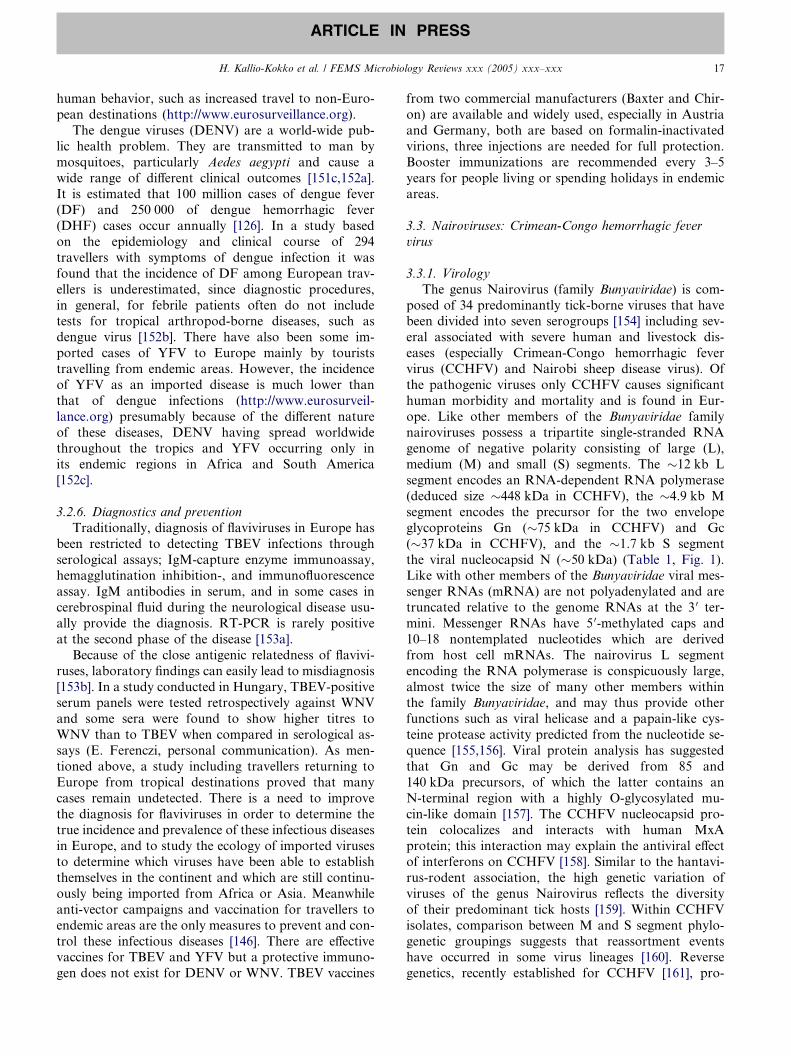
H. Kallio-Kokko et al. / FEMS Microbiology Reviews xxx (2005) xxx–xxx 17
ARTICLE IN PRESS
human behavior, such as increased travel to non-Euro-
pean destinations (http://www.eurosurveillance.org).
The dengue viruses (DENV) are a world-wide pub-
lic health problem. They are transmitted to man by
mosquitoes, particularly Aedes aegypti and cause a
wide range of different clinical outcomes [151c,152a].It is estimated that 100 million cases of dengue fever
(DF) and 250 000 of dengue hemorrhagic fever
(DHF) cases occur annually [126]. In a study based
on the epidemiology and clinical course of 294
travellers with symptoms of dengue infection it was
found that the incidence of DF among European trav-
ellers is underestimated, since diagnostic procedures,
in general, for febrile patients often do not includetests for tropical arthropod-borne diseases, such as
dengue virus [152b]. There have also been some im-
ported cases of YFV to Europe mainly by tourists
travelling from endemic areas. However, the incidence
of YFV as an imported disease is much lower than
that of dengue infections (http://www.eurosurveil-
lance.org) presumably because of the different nature
of these diseases, DENV having spread worldwidethroughout the tropics and YFV occurring only in
its endemic regions in Africa and South America
[152c].
3.2.6. Diagnostics and prevention
Traditionally, diagnosis of flaviviruses in Europe has
been restricted to detecting TBEV infections through
serological assays; IgM-capture enzyme immunoassay,hemagglutination inhibition-, and immunofluorescence
assay. IgM antibodies in serum, and in some cases in
cerebrospinal fluid during the neurological disease usu-
ally provide the diagnosis. RT-PCR is rarely positive
at the second phase of the disease [153a].
Because of the close antigenic relatedness of flavivi-
ruses, laboratory findings can easily lead to misdiagnosis
[153b]. In a study conducted in Hungary, TBEV-positiveserum panels were tested retrospectively against WNV
and some sera were found to show higher titres to
WNV than to TBEV when compared in serological as-
says (E. Ferenczi, personal communication). As men-
tioned above, a study including travellers returning to
Europe from tropical destinations proved that many
cases remain undetected. There is a need to improve
the diagnosis for flaviviruses in order to determine thetrue incidence and prevalence of these infectious diseases
in Europe, and to study the ecology of imported viruses
to determine which viruses have been able to establish
themselves in the continent and which are still continu-
ously being imported from Africa or Asia. Meanwhile
anti-vector campaigns and vaccination for travellers to
endemic areas are the only measures to prevent and con-
trol these infectious diseases [146]. There are effectivevaccines for TBEV and YFV but a protective immuno-
gen does not exist for DENV or WNV. TBEV vaccines
from two commercial manufacturers (Baxter and Chir-
on) are available and widely used, especially in Austria
and Germany, both are based on formalin-inactivated
virions, three injections are needed for full protection.
Booster immunizations are recommended every 3–5
years for people living or spending holidays in endemicareas.
3.3. Nairoviruses: Crimean-Congo hemorrhagic fever
virus
3.3.1. Virology
The genus Nairovirus (family Bunyaviridae) is com-
posed of 34 predominantly tick-borne viruses that havebeen divided into seven serogroups [154] including sev-
eral associated with severe human and livestock dis-
eases (especially Crimean-Congo hemorrhagic fever
virus (CCHFV) and Nairobi sheep disease virus). Of
the pathogenic viruses only CCHFV causes significant
human morbidity and mortality and is found in Eur-
ope. Like other members of the Bunyaviridae family
nairoviruses possess a tripartite single-stranded RNAgenome of negative polarity consisting of large (L),
medium (M) and small (S) segments. The �12 kb L
segment encodes an RNA-dependent RNA polymerase
(deduced size �448 kDa in CCHFV), the �4.9 kb M
segment encodes the precursor for the two envelope
glycoproteins Gn (�75 kDa in CCHFV) and Gc
(�37 kDa in CCHFV), and the �1.7 kb S segment
the viral nucleocapsid N (�50 kDa) (Table 1, Fig. 1).Like with other members of the Bunyaviridae viral mes-
senger RNAs (mRNA) are not polyadenylated and are
truncated relative to the genome RNAs at the 3 0 ter-
mini. Messenger RNAs have 5 0-methylated caps and
10–18 nontemplated nucleotides which are derived
from host cell mRNAs. The nairovirus L segment
encoding the RNA polymerase is conspicuously large,
almost twice the size of many other members withinthe family Bunyaviridae, and may thus provide other
functions such as viral helicase and a papain-like cys-
teine protease activity predicted from the nucleotide se-
quence [155,156]. Viral protein analysis has suggested
that Gn and Gc may be derived from 85 and
140 kDa precursors, of which the latter contains an
N-terminal region with a highly O-glycosylated mu-
cin-like domain [157]. The CCHFV nucleocapsid pro-tein colocalizes and interacts with human MxA
protein; this interaction may explain the antiviral effect
of interferons on CCHFV [158]. Similar to the hantavi-
rus-rodent association, the high genetic variation of
viruses of the genus Nairovirus reflects the diversity
of their predominant tick hosts [159]. Within CCHFV
isolates, comparison between M and S segment phylo-
genetic groupings suggests that reassortment eventshave occurred in some virus lineages [160]. Reverse
genetics, recently established for CCHFV [161], pro-
18 H. Kallio-Kokko et al. / FEMS Microbiology Reviews xxx (2005) xxx–xxx
ARTICLE IN PRESS
vides a unique opportunity to study the biology of nai-
roviruses and tailor optimal therapeutic and prophylac-
tic measures against CCHFV infections.
3.3.2. Ecology and epidemiology
CCHFV is transmitted most efficiently by Hya-
lomma ticks, followed by Rhipicephalus, Dermatocentor
spp and many other species of ixodid (hard) and some
argasid (soft) ticks (Table 2). Among ticks CCHFV is
capable of transmitting infection both transovarially
and transstadially. The life cycle of CCHFV also in-
cludes a tick-vertebrate host cycle involving both wild
and domestic animals. The virus or antibodies against
it have been detected in rodents, hares, hedgehogs andsome birds but human infections seem to be princi-
pally from contacts with livestock (mainly cattle,
sheep, goats) including in Africa farmed ostriches
and less often ticks [162,163]. Thus CCHFV is more
commonly seen in persons exposed to blood and tis-
sues of infected animals during occupational activities,
such as farming, herding, veterinary examination and
abattoir work. Nosocomial infection is common andoften results in small outbreaks. Yet, outdoor and rec-
reational activities also represent a risk factor. In
addition, horizontal transmission of CCHFV from
mother to child may occur [164].
The global distribution of CCHF follows closely
that of Hyalomma spp. in the Middle East, Asia,
Africa and Southeast Europe [162,163]. Sequence
analysis of CCHFV S RNA segments gives patternsfollowing links between different geographic locations,
in some cases suggesting links originating from trade
in livestock and long-distance carriage of virus or in-
fected ticks during bird migration [165]. In Europe,
CCHF occurs in the Balkan Peninsula (Albania, Bul-
garia, Greece, Turkey, Yugoslavia) but also in South-
ern Russia, Hungary, France and Portugal
[162,166,167].
3.3.3. Clinical picture and pathogenesis
The symptoms and signs of Crimean-Congo hemor-
rhagic fever are similar to those of other viral hemor-
rhagic fevers [162,163]. The incubation period is
generally short ranging usually from 2 to 9 days.
There is typically a very sudden onset of illness with
fever, rigors, chills, intense headache and backacheor leg pains, myalgia, nausea, and vomiting. Patients
may also present with photophobia, somnolence and
menigism with confusion or aggression. Hemorrhages
in the form of petecchiae, ecchymoses, epistaxis, mele-
na and bleeding from various organs usually begin a
few days later. Tachycardia is common and lymphad-
enopathy is seen occasionally. The case-fatality rate is
20–35%. The pathology consists of hemorrhagic andnecrotic lesions in various organs as well as fibrin
deposits.
3.3.4. Diagnostics and prevention
The methods used for detection of CCHFV infection
in humans or livestock include indirect immunofluores-
cence on virus-infected or N-expressing transfected cells.
Both IgM and IgG antibodies reactive for CCHF ap-
pear within a week in patients [162,168]. Alternativelya recombinant nucleocapsid protein-based enzyme
immunoassay [169–171] may be used. Rapid detection
and quantification of CCHFV RNA is possible by
real-time reverse transcription PCR [172].
Ribavirin is clearly effective in treatment of Crimean-
Congo hemorrhagic fever [173,174] but supportive treat-
ment (blood, thrombocytes, coagulation factors) is
equally important. Immune plasma from recovered pa-tients has also been used to treat patients but not in con-
trolled studies with a follow-up of virus-neutralizing
activities [162]. The most effective method of preventing
infections is to take measure to avoid exposure to ticks
(protective clothing, tick repellent, frequent body
searches to remove ticks). Inactivated virus prepared
in mouse brains has been used on a limited scale as a
vaccine in Southeastern Europe and the former USSRbut the sporadic and unpredictable occurrence of the
disease renders it difficult to identify target populations
and has slowed development of a safe and modern
vaccine [162].
3.4. Orthobunyaviruses: Tahyna and Inkoo viruses
3.4.1. Virology
Orthobunyaviruses belong to the family Bunyaviri-
dae. These enveloped viruses have a three-segmented
negative-strand RNA genome. The 0.97 kb S segment
encodes the 19–25 kDa nucleocapsid protein (N)
and a 10–13 kDa nonstructural protein (Ns); the
�4.5 kb M segment encodes the polyprotein precursor
of two glycoproteins, G1 and G2 (108–210 kDa and
29–41 kDa, respectively), and a nonstructural proteinNSm (15–18 kDa); and the �6.9 kb L segment encodes
the RNA polymerase (260 kDa) (Table 1, Fig. 1). Viral
messenger RNAs are not polyadenylated and are trun-
cated relative to the genome RNAs at the 3 0 termini.
Messenger RNAs have 5 0-methylated caps and some
nontemplated nucleotides which are derived from host
cell mRNAs.
3.4.2. Ecology and epidemiology
Orthobunyaviruses are transmitted to humans by a
variety of mosquito species, and in Europe two known
human pathogenic representatives of the genus circulate,
Tahyna virus and Inkoo virus (mostly transmitted by
Aedes spp.) [109,175,176] (Table 2). These viruses belong
to the California serogroup, and their incidence is asso-
ciated with the distribution of the carrier mosquitoes.The natural cycle of these viruses involve also mammal
hosts. Tahyna virus circulates throughout most of
H. Kallio-Kokko et al. / FEMS Microbiology Reviews xxx (2005) xxx–xxx 19
ARTICLE IN PRESS
Europe, whereas Inkoo virus is mainly reported from
northern Europe [109,177a].
3.4.3. Clinical picture and diagnosis
Tahyna virus causes an influenza-like disease which
may also present as a meningitis. The infection causedby Inkoo virus is mostly sub-clinical or a mild disease,
although encephalitis has been reported. The most typ-
ical symptoms associated with encephalitis or
meningitis caused by Californian serogroup viruses
include fever, headache, nausea, vomiting, convulsions
and confusion. In Europe the seroprevalence for
Californian serogroup viruses varies from 1% to 69%,
depending on the geographic area [109]. In Finland theseroprevalence against Inkoo virus varies from 2% to
6% (Aland islands) to 61–69% (Finnish Lapland) [109,
Putkuri et al, unpublished results]. In disease-endemic
areas the seropositivity of the population increases with
age [177b]. The diagnosis of orthobunyaviruses is based
on serology, either as a rise in IgG-antibody titers, or the
presence of IgM antibodies. Also RT-PCR methods are
under development to detect viral RNA in cerebrospinalfluid samples of patients with encephalitis.
3.5. Phleboviruses
3.5.1. Virology
Phleboviruses are one of the five groups within
the family Bunyaviridae. They have a negative single-
stranded RNA tripartite genome, namely: L (6.4 kb),M (3.2–4.2 kb) and S (1.7–1.9 kb). The L segment
encodes a single protein of 240 kDa (RNA polymer-
ase) in the complementary sense. The M segment
encodes a protein precursor of �130 kDa in the comple-
mentary sense which is cleaved into two glycoproteins
G1 (�56 kDa) and G2 (�59 kDa). The S segment has
an ambisense coding arrangement, and it encodes the
nucleocapsid protein (N) of �27 kDa in the complemen-tary sense and a non-structural protein (NSs) of
�30 kDa in the virion sense [178,179] (Table 1, Fig. 1).
Viral messenger RNAs (mRNA) are not polyadenylated
and are truncated relative to the genome RNAs at the 3 0
termini. mRNAs have 5 0-methylated caps and some
nontemplated nucleotides which are derived from host
cell mRNAs.
When seen under the electron microscope the viri-ons are spherical and enveloped and approximately
80–110 nm in diameter. Glycoprotein projections of
5–10 nm are also seen embedded in the membrane.
There are between 200 and 1500 spikes per virion
[178,179].
3.5.2. Ecology and epidemiology
Phleboviruses are arthropod-borne viruses and aretransmitted mainly by phlebotomine sandflies, hence
the derivation of its name ‘‘phlebo’’ (Table 2). How-
ever, mosquitoes and ticks can also transmit phlebovi-
ruses. Phleboviruses are distributed throughout most
of the world but have not been reported in Australia
[180]. The first documented record of a disease resem-
bling those caused by phleboviruses dates back to the1900s in the Mediterranean countries of Europe [178].
However, the first virus isolation occurred for Rift
Valley Fever virus in 1930 in Kenya [181]. Retrospec-
tive serological studies have indicated that outbreaks
caused by phleboviruses have occurred since 1912 in
the Sub-Saharan Africa region [182]. Phleboviruses
are major causal agents of encephalitis in humans,
especially among children, in Mediterranean countries.They also have an important impact in livestock espe-
cially in African endemic areas. Infection of livestock
is characterised by a high rate of abortions and
pathologies associated with hemorrhagic illness (leuko-
penia, thrombocytopenia, fibrin thrombi, intravascular
coagulopathy, etc.) [183–185]. The distribution of the
disease follows that of its vector. However the natural
history of phleboviruses is largely unknown and formany species the amplifying host has not been identi-
fied [185–187].
3.5.3. Clinical picture and pathogenesis
In general, phlebovirus infections cause a 2–4 days ill-
ness characterised by an incubation period of 2–6 days
followed by fever, general malaise, headache, photopho-
bia, and back and joint pain [188]. The illness occursduring the summer months when the activity of phlebot-
omine flies is high [178].
There are three recognised members of the genus
Phlebovirus, associated with disease in humans. Rift
Valley fever virus (RVFV), which is the type species of
the genus and is transmitted by mosquitoes, causing
an influenza-like disease that affects domestic animals
and humans. The majority of human infections areasymptomatic; however, in 0.5% of cases it can cause se-
vere haemorrhagic fever, encephalitis and death. The
geographical distribution of RVFV includes Africa,
Egypt, Saudi Arabia and Yemen and occurs as epizoot-
ics involving Aedes spp. mosquitoes and domestic ani-
mals [180].
Toscana virus (TSV) causes a mild influenza-like
disease that can occasionally develop into an acuteneurological disease, such as meningitis or meningoen-
cephalitis in Italy and possibly other Mediterranean
countries. A similar virus (Granada virus) was recently
isolated in Spain [193]. TSV is the number one cause
of encephalitis among children in Italy. The distribution
of the virus follows that of its vector, Phlebotomus
perniciosus, found in Italy, Spain, Portugal, and Cyprus.
Human cases have also been reported from southernFrance and Greece. The virus replicates in the sandfly
vector population; however, experimental evidence
20 H. Kallio-Kokko et al. / FEMS Microbiology Reviews xxx (2005) xxx–xxx
ARTICLE IN PRESS
suggests that an amplifying vertebrate host is needed to
sustain the virus in the arthropod population [189–194].
Sandfly fever virus (SFV) Sicilian and Naples viruses
can cause a mild-influenza like disease. The distribution
of SFV also follows that of its vector, Phlebotomus
papatasii, found in the Mediterranean basin extendingto the Middle East and Arabian Peninsula, the Caucasus
mountains, Pakistan and India [185–187].
3.5.4. Diagnosis and prevention
Diagnostics of Phlebovirus is performed mainly by
IgM-capture EIA and IFA. The virus can be isolated
using intracranial inoculation in suckling mice or using
a susceptible tissue culture cell line (Vero, LLC-MK2,BHK-21) [178,195a]. Toscana virus and Naples Sandfly
fever virus group together and are distinct antigenically
and phylogenetically from Sicilian Sandfly fever virus
[195b].
Prevention of infections by phleboviruses varies
depending on the type species. Immunising livestock
with the formalin-inactivated or live-attenuated vac-
cines can prevent RVFV by avoiding epizootics andhence human infections [178,196]. In the case of
TSV and SFV, prevention is limited to controlling
the vector population through insecticides and repel-
lents [197,198].
4. Concluding remarks
What are the reasons for the appearance of all these
emerging and re-emerging viruses? Have changes taken
place in the viruses themselves? Apparently not. The
principal reasons are changes in the environment and
human activity that have created new attacks with nat-
ure. Factors such as climate change, increasing intensity
of agriculture, the building of dams and introduction of
other irrigation measures which generate new breedingsites for mosquitoes, crises and wars which bring rodents
and arthropods closer to humans, population growth
and especially in suburban slums, and global air traffic
are important factors generating such new contacts, or
may affect the ecological cycles of these viruses. Many
epidemics may be unexpected outcomes of such changes.
Moreover, the new molecular biology techniques facili-
tate detection of new viral agents.Many prevention and control measures, such as the
anti-Aedes campaigns, have been abandoned in tropi-
cal countries. The yellow-fever vaccination campaigns
have been re-implemented just recently with the new
outbreaks. This negligence has increased the chance
of acquiring new infections from overseas and the
chances of broadening the ecological niches for many
diseases. The new vector of many diseases is humantransportation, which introduces the artificial migra-
tion (or imports) of the vector (mosquitoes) usually
as larvae.
This review could not comprehensively cover all zoo-
notic agents in Europe, and new viruses with zoonotic
transmission are continuously suggested, such as hepati-
tis E virus, found in rats and connected e.g. to eatingdeer meat [199]. It is easy to predict that we will continue
to see many more emerging and re-emerging viruses dur-
ing the coming years.
Acknowledgements
Work in Finland has been supported by the Euro-pean Union grant EU-QLR2-CT-2002-01358.
References
[1] Plyusnin, A., Vapalahti, O. and Vaheri, A. (1996) Hantaviruses:
genome structure, expression and evolution. J. Gen. Virol. 77,
2677–2687.
[2] Jonsson, C.B. and Schmaljohn, C.S. (2001) Replication of
hantaviruses. Volume ‘‘Hantaviruses’’ (Schmaljohn, C.S.,
Nichol, S.T., Eds.). Curr. Top. Microbiol. Immunol. 256,
15–32.
[3] Plyusnin, A. and Morzunov, S.P. (2001) Virus evolution and
genetic diversity of hantaviruses and their rodent hosts. Volume
‘‘Hantaviruses’’ (Schmaljohn, C.S., Nichol, S.T., Eds.). Curr.
Top. Microbiol. Immunol. 256, 47–75.
[4] Plyusnin, A., Vapalahti, O., Lehvaslaiho, H., Apekina, N.,
Mikhailova, T., Gavrilovskaya, I., Laakkonen, J., Niemimaa, J.,
Henttonen, H., Brummer-Korvenkontio, M. and Vaheri, A.
(1995) Genetic variation of wild Puumala viruses within the
serotype, local rodent populations and individual animal. Virus
Res. 38, 25–41.
[5] Plyusnin, A., Cheng, Y., Lehvaslaiho, H. and Vaheri, A. (1996)
Quasispecies in wild-type Tula hantavirus populations. J. Virol.
70, 9060–9063.
[6] Sibold, C., Meisel, H., Kruger, D.H., Labuda, M., Lysy, J.,
Kozuch, O., Pejcoch, M., Vaheri, A. and Plyusnin, A. (1999)
Recombination in Tula hantavirus evolution: analysis of genetic
lineages from Slovakia. J. Virol. 73, 667–675.
[7] Plyusnin, A., Kukkonen, S.K.J., Plyusnina, A., Vapalahti, O.
and Vaheri, A. (2002) Transfection-mediated generation of
functionally competent Tula hantavirus with recombinant S
RNA segment. EMBO J. 21, 1497–1503.
[8] Vapalahti, O., Mustonen, J., Lundkvist, A., Henttonen, H.,
Plyusnin, A. and Vaheri, A. (2003) Hantavirus infections in
Europe. Lancet Infect. Dis. 3, 653–661.
[9] Plyusnin, A., Vapalahti, O., Lankinen, H., Lehvaslaiho, H.,
Apekina, N., Myasnikov, Y., Kallio-Kokko, H., Henttonen,
H., Lundkvist, A., Brummer-Korvenkontio, M., Gavrilovs-
kaya, I. and Vaheri, A. (1994) Tula virus: a newly detected
hantavirus carried by European common voles. J. Virol. 68,
7833–7839.
[10] Vapalahti, O., Lundkvist, A., Kukkonen, S.K.J., Cheng, Y.,
Gilljam, M., Kanerva, M., Manni, T., Pejcoch, M., Niemimaa,
J., Kaikusalo, A., Henttonen, H., Vaheri, A. and Plyusnin, A.
(1996) Isolation and characterization of Tula hantavirus, a
distinct serotype in the genus Hantavirus, family Bunyaviridae. J.
Gen. Virol. 77, 3063–3067.
H. Kallio-Kokko et al. / FEMS Microbiology Reviews xxx (2005) xxx–xxx 21
ARTICLE IN PRESS
[11] Vapalahti, O., Lundkvist, A., Fedorov, V., Conroy, C.J.,
Hirvonen, S., Plyusnina, A., Nemirov, K., Fredga, K., Cook,
J.A., Niemimaa, J., Kaikusalo, A., Henttonen, H., Vaheri, A.
and Plyusnin, A. (1999) Isolation and characterization of a
hantavirus from Lemmus sibiricus: evidence for host-switch
during hantavirus evolution. J. Virol. 73, 5586–5592.
[12] Desmyter, J., LeDuc, J.W., Johnson, K.M., Brasseur, F.,
Deckers, C. and van Ypersele de Strihou, C. (1983) Laboratory
rat associated outbreak of haemorrhagic fever with renal
syndrome due to Hantaan-like virus in Belgium. Lancet 2,
1445–1448.
[13] Lloyd, G., Bowen, E.T., Jones, N. and Pendry, A. (1984) HFRS
outbreak associated with laboratory rats in UK. Lancet 1, 1175–
1176.
[14] Heyman, P., Plyusnina, A., Berny, P., Cochez, C., Artois, M.,
Zizi, M., Pirnay, J.P. and Plyusnin, A. (2004) Seoul hantavirus in
Europe: first demonstration of the virus genome in wild Rattus
norvegicus captured in France. Eur. J. Clin. Microbiol. Infect.
Dis. 23, 711–717.
[15] Brummer-Korvenkontio, M., Vapalahti, O., Henttonen, H.,
Koskela, P., Kuusisto, P. and Vaheri, A. (1999) Epidemiological
study of nephropathia epidemica in Finland 1989–1996. Scand.
J. Infect. Dis. 31, 427–435.
[16] Vapalahti, K., Paunio, M., Vaheri, A. and Vapalahti, O. (1999)
Puumala virus infections in Finland: increased occupational risk
for farmers. Am. J. Epidemiol. 149, 1142–1151.
[17] Kanerva, M., Mustonen, J. and Vaheri, A. (1998) Pathogenesis
of Puumala and other hantavirus infections. Rev. Med. Virol. 8,
67–86.
[18] Mustonen, J., Partanen, J., Kanerva, M., Pietila, K., Vapalahti,
O., Pasternack, A. and Vaheri, A. (1996) Genetic susceptibility
to severe course of nephropathia epidemica caused by Puumala
hantavirus. Kidney Int. 49, 217–221.
[19] Makela, S., Ala-Houhala, I., Mustonen, J., Koivisto, A.-M.,
Kouri, T., Turjanmaa, V., Vapalahti, O., Vaheri, A. and
Pasternack, A. (2000) Renal function and blood pressure five
years after Puumala virus-induced nephropathy. Kidney Int. 58,
1711–1718.
[20] Hautala, T., Sironen, T., Vapalahti, O., Paakko, E., Sarkioja, T.,
Salmela, P.I., Vaheri, A., Plyusnin, A. and Kauma, H. (2002)
Hypophyseal hemorrhage and panhypopituitarism during Puu-
mala virus infection: magnetic resonance imaging and detection
of viral antigen in the hypophysis. Clin. Infect. Dis. 35, 96–101.
[21] Terajima, M., Vapalahti, O., Van Epps, H., Vaheri, A. and
Ennis, F.A. (2004) Immune responses to Puumala virus infection
and the pathogenesis of nephropathia epidemica. Microbes
Infect. 6, 238–245.
[22] Mackow, E.R. and Gavrilovskaya, I.N. (2001) Cellular patho-
genesis and hantavirus pathogenesis. Volume ‘‘Hantaviruses’’
(Schmaljohn, C.S., Nichol, S.T., Eds.). Curr. Top. Microbiol.
Immunol. 256, 91–115.
[23] Kang, J.I., Park, S.H., Lee, P.W. and Ahn, B.Y. (1999)
Apoptosis is induced by hantaviruses in cultured cells. Virology
264, 99–105.
[24] Akhmatova, N.K., Yusupova, R.S., Khaiboullina, S.F. and
Sibiryak, S.V. (2003) Lymphocyte apoptosis during Hemor-
rhagic Fever with Renal Syndrome. Russ. J. Immunol. 8, 37–46.
[25] Markotic, A., Hensley, L., Geisbert, T., Spik, K. and Schmal-
john, C. (2003) Hantaviruses induce cytopathic effects and
apoptosis in continuous human embryonic kidney cells. J. Gen.
Virol. 84, 2197–2202.
[26] Li, X.D., Kukkonen, S., Vapalahti, O., Plyusnin, A., Lankinen,
H. and Vaheri, A. (2004) Tula hantavirus infection of Vero E6
cells induces apoptosis involving caspase 8 activation. J. Gen.
Virol. 85, 3261–3268.
[27] Linderholm, M., Ahlm, C., Settergren, B., Waage, A. and
Tarnvik, A. (1996) Elevated plasma levels of tumor necrosis
factor (TNF)-alpha, soluble TNF receptors, interleukin (IL)-6,
and IL-10 in patients with hemorrhagic fever with renal
syndrome. J. Infect. Dis. 173, 38–43.
[28] Temonen, M., Mustonen, J., Helin, H., Pasternack, A., Vaheri,
A. and Holthofer, H. (1996) Cytokines, adhesion molecules, and
cellular infiltration in nephropathia epidemica kidneys: an
immunohistochemical study. Clin Immunol Immunopathol. 78,
47–55.
[29] Makela, S., Mustonen, J., Ala-Houhala, I., Hurme, M., Koiv-
isto, A.-M., Vaheri, A. and Pasternack, A. (2004) Urinary
excretion of interleukin-6 correlates with proteinuria in acute
Puumala hantavirus-induced nephritis. Am. J. Kidney Dis. 43,
809–816.
[30] Klingstrom, J., Plyusnin, A., Vaheri, A. and Lundkvist, A.
(2002) Wild-type Puumala hantavirus infection induces cyto-
kines, C-reactive protein, creatinine and nitric oxide in Cyno-
molgus macaques. J. Virol. 76, 444–449.
[31] Vapalahti, O., Lundkvist, A. and Vaheri, A. (2001). Human
immune response, host genetics and severity of disease. Volume
‘‘Hantaviruses’’ (Schmaljohn, C.S., Nichol, S.T., Eds.). Curr.
Top. Microbiol. Immunol. 256, 153–169.
[32] Van Epps, H.L., Terajima, M., Mustonen, J., Arstila, T.P.,
Corey, E.A., Vaheri, A. and Ennis, F.A. (2002) Long-lived
memory T lymphocytes after hantavirus infection. J. Exp. Med.
196, 579–588.
[33] Plyusnin, A., Horling, J., Kanerva, M., Mustonen, J., Cheng, Y.,
Partanen, J., Vapalahti, O., Kukkonen, S.K.J., Niemimaa, J.,
Henttonen, H., Niklasson, B., Lundkvist, A. and Vaheri, A.
(1997) Puumala hantavirus genome in patients with nephropa-
thia epidemica: correlation of PCR positivity with HLA haplo-
type and link to viral sequences in local rodents. J. Clin.
Microbiol. 35, 1090–1096.
[34] Plyusnin, A., Mustonen, J., Asikainen, K., Plyusnina, A.,
Niemimaa, J., Henttonen, H. and Vaheri, A. (1999) Analysis
of Puumala hantavirus genome in patients with nephropathia
epidemica and rodent carriers from the sites of infection. J. Med.
Virol. 59, 397–405.
[35] Hedman, K., Vaheri, A. and Brummer-Korvenkontio, M. (1991)
Rapid diagnosis of hantavirus disease with an IgG-avidity assay.
Lancet 338, 1353–1356.
[36] Kallio-Kokko, H., Vapalahti, O., Hedman, K., Brummer-
Korvenkontio, M. and Vaheri, A. (1993) Puumala virus
antibody and immunoglobulin G avidity assays based on
recombinant nucleocapsid antigen. J. Clin. Microbiol. 31,
677–680.
[37] Brus Sjolander, K., Elgh, F., Kallio-Kokko, H., Vapalahti, O.,
Hagglund, M., Palmcrantz, V., Juto, P., Vaheri, A., Niklasson,
B. and Lundkvist, A. (1997) Evaluation of serological methods
for diagnosis of Puumala hantavirus infection (nephropathia
epidemica). J. Clin. Microbiol. 35, 3264–3268.
[38] Hujakka, H., Koistinen, V., Eerikainen, P., Kuronen, I.,
Mononen, I., Parviainen, M., Lundkvist, A., Vaheri, A.,
Narvanen, A. and Vapalahti, O. (2001) New immunochromato-
graphic rapid test for diagnosis of acute Puumala virus infection.
J. Clin. Microbiol. 39, 2146–2150.
[39] Hujakka, H., Koistinen, V., Kuronen, I., Eerikainen, P.,
Parviainen, M., Lundkvist, A., Vaheri, A., Vapalahti, O. and
Narvanen, A. (2003) Diagnostic rapid tests for acute hantavirus
infections: specific tests for Hantaan, Dobrava and Puumala
viruses versus a hantavirus combination test. J. Virol. Methods
108, 117–122.
[40] Hooper, J.W. and Li, D. (2001) Vaccines against hantaviruses.
Volume ‘‘Hantaviruses’’ (Schmaljohn, C.S., Nichol, S.T., Eds.).
Curr. Top. Microbiol. Immunol. 256, 171–191.
[41] Huggins, J.W., Hsiang, C.M., Cosgriff, T.M., Guang, M.Y.,
Smith, J.I., Wu, Z.O., Le Duc, J.W., Zheng, Z.M., Meegan, J.M.
and Wang, Q.N., et al. (1991) Prospective, double-blind, con-

22 H. Kallio-Kokko et al. / FEMS Microbiology Reviews xxx (2005) xxx–xxx
ARTICLE IN PRESS
current, placebo-controlled clinical trial of intravenous ribavirin
therapy of hemorrhagic fever with renal syndrome. J. Infect. Dis.
164, 1119–1127.
[42] Bai, J., Zhu, K. and Zhou, G. (1997) The therapeutic effect of
purified human leucocytic interferon-alpha on hemorrhagic fever
with renal syndrome. Zhonghua Nei Ke Za Zhi 36, 90–93.
[43] (a) Bahloul, C., Jacob, Y., Tordo, N. and Perrin, P. (1998)
DNA-based immunisation for exploring the enlargement of
immunological cross-reactivity against the lyssaviruses. Vaccine
16, 417–425;
(b) Jallet, C., Jacob, Y., Bahloul, C., Drings, A., Desmezie res,
E., Tordo, N. and Perrin, P. (1999) Chimeric lyssavirus
glycoproteins with increased immunological potential. J. Virol.
73, 225–233;
(c) Botvinkin, A.D., Poleschuk, E.M., Kuzmin, I.V., Borisova,
T.I-, Gazaryan, S.V., Yager, P. and Rupprecht, C.E. (2003)
Novel lyssaviruses isolated from bats in Russia. Emerg. Infect.
Dis. 9, 1623–1625.
[44] (a) Bourhy, H., Kissi, B., Audry, L., Smreczak, M., Sadkowska-
Todys, M., Kulonen, K., Tordo, N., Zmudzinski, J.F. and
Holmes, E.C. (1999) Ecology and evolution of rabies virus in
Europe. J. Gen. Virol. 180, 2545–2557;
(b) Badrane, H. and Tordo, N. (2001) Host-switching in
lyssavirus history from chiroptera to carnivora orders. J. Virol.
75, 8096–8104.
[45] Warrrell, M.J. and Warrell, D.A. (2004) Rabies and other
lyssavirus disesases. Lancet 363, 959–969.
[46] Echevarria, J.E., Avellon, A., Juste, J., Vera, M. and Ibanez, C.
(2001) Screening of active lyssavirus infection in wild bat
populations by viral RNA detection on oropharyngeal swabs.
J. Clin. Microbiol. 39, 3678–3683.
[47] Amengual, B., Whitby, J.E., King, A., Cobo, J.S. and Bourhy,
H. (1997) Evolution of European bat lyssaviruses. J. Gen. Virol.
78, 2319–2328.
[48] Whitby, J.E., Heaton, P.R., Black, E.M., Wooldridge, M.,
McElhinney, L.M. and Johnstone, P. (2000) First isolation of a
rabies-related virus from a Daubenton�s bat in the United
Kingdom. Vet. Rec. 147, 385–388.
[49] Nathwani, D., McIntyre, P.G., White, K., Shearer, A.J.,
Reynolds, N., Walker, D., Orange, G.V. and Fooks, A. (2003)
Fatal human rabies caused by European bat Lyssavirus type 2a
infection in Scotland. Clin. Infect. Dis. 37, 598–601.
[50] Grauballe, P.C., Baagøe, H.J., Fekadu, M., Westergaard, J.M.
and Zoffmann, H. (1987) Bat rabies in Denmark. Lancet 1, 379–
380.
[51] (a) Serra-Cobo, J., Amengual, B., Abellan, C. and Bourhy, H.
(2002) European bat lyssavirus infection in Spanish bat popu-
lations. Emerg. Infect. Dis. 8, 413–420;
(b) Nathwani, D., McIntyre, P.G., White, K., Shearer, A.J.,
Reynolds, N., Walker, D., Orange, G.V. and Fooks, A.R. (2003)
Fatal human rabies caused by European bat Lyssavirus type 2a
infection in Scotland. Clin Infect Dis. 37, 598–601.
[52] (a) Plotkin, S. (2000) Rabies. Clin. Infect. Dis. 30, 4–12;
(b) Hemachudha, T., Laothamatas, J. and Rupprecht, C.E.
(2002) Human rabies: a disease of complex neuropathogenetic
mechanisms and diagnostic challenges. Lancet Neurol. 1,
101–109.
[53] Raux, H., Flamand, A. and Blondel, D. (2000) Interaction of the
rabies virus P protein with the LC8 dynein light chain. J. Virol.
74, 10212–10216.
[54] Jacob, Y., Badrane, H., Ceccaldi, P.E. and Tordo, N. (2000)
Cytoplasmic dynein LC8 interacts with lyssavirus phosphopro-
tein. J. Virol. 74, 10217–10222.
[55] Lafon, M., Bourhy, H. and Sureau, P. (1988) Immunity
against the European bat rabies (Duvenhage) virus induced
by rabies vaccines: an experimental study in mice. Vaccine 6,
362–368.
[56] Badrane, H., Bahloul, C., Perrin, P. and Tordo, N. (2001)
Evidence of two Lyssavirus phylogroups with distinct pathoge-
nicity and immunogenicity. J. Virol. 75, 3268–3276.
[57] Mebatsion, T. (2001) Extensive attenuation of rabies virus by
simultaneously modifying the dynein light chain binding site in
the P protein and replacing Arg333 in the G protein. J. Virol. 75,
11496–11502.
[58] Jackson, A.C., Warrell, M.J., Rupprecht, C.E., Ertl, H.C.,
Dietzschold, B., O�Reilly, M., Leach, R.P., Fu, Z.F., Wunner,
W.H., Bleck, T.P. and Wilde, H. (2003) Management of rabies in
humans. Clin. Infect. Dis. 36, 60–63.
[59] (a) Perez, M., Craven, R.C. and de la Torre, J.C. (2003) The
small RING finger protein Z drives arenavirus budding: impli-
cations for antiviral strategies. Proc. Natl. Acad. Sci. USA 100,
12978–12983;
(b) Salazar-Bravo, J., Ruedas, L.A. and Yates, T.L. (2002) In:
Arenaviruses I (Oldstone, M.B.A., Ed.), pp. 25–64. Springer-
Verlag, Berlin, Heidelberg, New York.
[60] (a) Bowen, M.D., Peters, C.J. and Nichol, S.T. (1997) Phyloge-
netic analysis of the Arenaviridae: patterns of virus evolution
and evidence for cospeciation between arenaviruses and their
rodent hosts. Mol. Phylogenet. Evol. 8, 301–316;
(b) Clegg, J.S.C. (2002) In: Arenaviruses I (Oldstone, M.B.A.,
Ed.), pp. 1–24. Springer-Verlag, Berlin, Heidelberg, New York;
(c) Charrel, R.N., Feldmann, H., Fulhorst, C.F., Khelifa, R., de
Chesse, R. and de Lamballerie, X. (2002) Phylogeny of New
World arenaviruses based on the complete coding sequences of
the small genomic segment identified an evolutionary lineage
produced by intrasegmental recombination. Biochem. Biophys.
Res. Commun. 296, 1118–1124.
[61] (a) Rousseau, M.C., Saron, M.F., Broqui, P. and Bourgeade, A.
(1997) Lymphocytic choriomeningitis virus in southern France:
four case reports and a review of the literature. Eur. J.
Epidemiol. 13, 817–823;
(b) Lledo, L., Gegundez, M.I., Saz, J.V., Bahamontes, N. and
Beltran, M. (2003) Lymphocytic choriomeningitis virus infection
in a province of Spain: analysis of sera from the general
population and wild rodents. J. Med. Virol. 70, 273–275.
[62] (a) Peters, J.C. (2002) In: Arenaviruses I (Oldstone, M.B.A.,
Ed.), pp. 65–74. Springer-Verlag, Berlin, Heidelberg, New York;
(b) McGormick, J.B. and Fisher-Hoch, S.P. (2002) In: Arenav-
iruses I (Oldstone, M.B.A., Ed.), pp. 75–110. Springer-Verlag,
Berlin, Heidelberg, New York.
[63] Gunther, S., Emmerich, P., Laue, T., Kuhle, O., Asper, M.,
Jung, A., Grewing, T., ter Meulen, J. and Schmitz, H.
(2000) Imported lassa fever in Germany: molecular charac-
terization of a new lassa virus strain. Emerg. Infect. Dis. 6,
466–476.
[64] Schmitz, H., Kohler, B., Laue, T., Drosten, C., Veldkamp, P.J.,
Gunther, S., Emmerich, P., Geisen, H.P., Fleischer, K., Beersma,
M.F. and Hoerauf, A. (2002) Monitoring of clinical and
laboratory data in two cases of imported Lassa fever. Microbes
Infect. 4, 43–50.
[65] Haas, W.H., Breuer, T., Pfaff, G., Schmitz, H., Kohler, P.,
Asper, M., Emmerich, P., Drosten, C., Golnitz, U., Fleischer, K.
and Gunther, S. (2003) Imported Lassa fever in Germany:
surveillance and management of contact persons. Clin. Infect.
Dis. 36, 1254–1258.
[66] Mets, M.B. (1999) Childhood blindness and visual loss: an
assessment at two institutions including a ‘‘new’’ cause. Trans.
Am. Ophthalmol. Soc. 97, 653–696.
[67] (a) Wright, R., Johnson, D., Neumann, M., Ksiazek, T.G.,
Rollin, P., Keech, R.V., Bonthius, D.J., Hitchon, P., Grose,
C.F., Bell, W.E. and Bale Jr., J.F. (1997) Congenital lymphocytic
choriomeningitis virus syndrome: a disease that mimics congen-
ital toxoplasmosis or Cytomegalovirus infection. Pediatrics 100,
E9;

H. Kallio-Kokko et al. / FEMS Microbiology Reviews xxx (2005) xxx–xxx 23
ARTICLE IN PRESS
(b) Barton, L.L., Mets, M.B. and Beauchamp, C.L. (2002)
Lymphocytic choriomeningitis virus: emerging fetal teratogen.
Am. J. Obstet. Gynecol. 187, 1715–1716.
[68] Charrel, R.N. and de Lamballerie, X. (2003) Arenaviruses other
than Lassa virus. Antiviral Res. 57, 89–100.
[69] (a) Park, J.Y., Peters, C.J., Rollin, P.E., Ksiazek, T.G., Gray,
B., Waites, K.B. and Stephensen, C.B. (1997) Development of
a reverse transcription-polymerase chain reaction assay for
diagnosis of lymphocytic choriomeningitis virus infection and
its use in a prospective surveillance study. J. Med. Virol. 51,
107–114;
(b) Drosten, C., Gottig, S., Schilling, S., Asper, M., Panning,
M., Schmitz, H. and Gunther, S. (2002) Rapid detection and
quantification of RNA of Ebola and Marburg viruses, Lassa
virus, Crimean-Congo hemorrhagic fever virus, Rift Valley
fever virus, dengue virus, and yellow fever virus by real-time
reverse transcription-PCR. J. Clin. Microbiol. 40, 2323–2330.
[70] (a) Drosten, C., Gottig, S., Schilling, S., Asper, M., Panning, M.,
Schmitz, H. and Gunther, S. (2002) Rapid detection and
quantification of RNA of Ebola and Marburg viruses, Lassa
virus, Crimean-Congo hemorrhagic fever virus, Rift Valley fever
virus, dengue virus, and yellow fever virus by real-time reverse
transcription-PCR. J. Clin. Microbiol. 40, 2323–2330;
(b) Drosten, C., Kummerer, B.M., Schmitz, H. and Gunther
(2003) Molecular diagnostics of viral hemorrhagic fevers. Anti-
viral Res 57, 61–87;
(c) Gunther, S., Asper, M., Roser, C., Luna, L.K., Drosten, C.,
Becker-Ziaja, B., Borowski, P., Chen, H.M. and Hosmane, R.
(2004) Application of real-time PCR for testing antiviral
compounds against Lassa virus, SARS coronavirus and Ebola
virus in vitro. Antiviral Res. 63, 209–215.
[71] McCormick, J.B., King, I.J., Webb, P.A., Scribner, C.L.,
Craven, R.B., Johnson, K.M., Elliott, L.H. and Belmont-
Williams, R. (1986) Lassa fever. Effective therapy with ribavirin.
N. Engl. J. Med. 314, 20–26.
[72] (a) Fisher-Hoch, S.P. and McCormick, J.B. (2001) Towards a
human Lassa fever vaccine. Rev. Med. Virol. 11, 331–341;
(b) McKee Jr., K.T., Oro, J.G., Kuehne, A.I., Spisso, J.A. and
Mahlandt, B.G. (1993) Safety and immunogenicity of a live-
attenuated Junin (Argentine hemorrhagic fever) vaccine in
rhesus macaques. Am. J. Trop. Med. Hyg. 48, 403–411.
[73] del Carmen Saavedra, M., Sottosanti, J.M., Riera, L. and
Ambrosio, A.M. (2003) IgG subclasses in human immune
response to wild and attenuated (vaccine) Junin virus infection.
J. Med. Virol. 69 (3), 447–450.
[74] Esposito, J.J. and Fenner, F. (2001) Poxviruses. In: Fields
Virology (Knipe, D.M. and Howley, P.M., Eds.), 4th ed, pp.
2885–2921. Lippincott Williams & Wilkins, Philadelphia.
[75] Smith, G.L, Vanderplasschena, A. and ja Law, M. (2002) The
formation and function of extracellular enveloped vaccinia virus.
J. Gen. Virol. 83, 2915–2931.
[76] Sandvik, T., Tryland, M., Hansen, H., Mehl, M., Moens, M.,
Olsvik, Ø. and ja Traavik, T. (1998) Naturally occurring
orthopoxviruses: potential for recombination with vaccine
vectors. J. Clin. Microbiol. 36, 2542–2547.
[77] Jenner, E. (1798) An Inquiry into the Causes and Effects of the
Variolae Vaccinae, a Disease Discovered in some of the Western
Counties of England, Particularly Gloucestershire, and known
by the Name of the Cow Pox. Sampson Low, London.
[78] Damaso, C.R.A., Esposito, J.J., Condit, R.C. and Moussatche,
N. (2000) An emergent poxvirus from humans and cattle in Rio
de Janeiro state: Cantagalo virus may derive from Brazilian
smallpox vaccine. Virology 277, 439–449.
[79] Chantrey, J., Meyer, H., Baxby, D., Begon, M., Bown, K.J.,
Hazel, S.M., Jones, T., Montgomery, W.I. and Bennett, M.
(1999) Cowpox: reservoir hosts and geographic range. Epidem-
iol. Infect. 122, 455–460.
[80] Meyer, H., Schay, C., Mahnel, H. and Pfeffer, M. (1999)
Characterization of orthopoxviruses isolated from man and
animals in Germany. Arch. Virol. 144, 491–501.
[81] Baxby, D., Bennett, M. and Getty, B. (1994) Human cowpox
1969–93: a review based on 54 cases. Br. J. Dermatol. 131, 598–
607.
[82] Wienecke, R., Wolff, H., Schaller, M., Meyer, H. and Plewig, G.
(2000) Cowpox virus infection in an 11-year-old girl. J. Am.
Acad. Dermatol. 42, 892–894.
[83] Pelkonen, P.M., Tarvainen, K., Hynninen, A., Kallio, E.R.K.,
Henttonen, H., Palva, A., Vaheri, A. and ja Vapalahti, O. (2003)
Cowpox with severe generalized eruption, Finland. Emerg.
Infect. Dis. 9, 1458–1461.
[84] Schupp, P., Pfeffer, M., Meyer, H., Burck, G., Kolmel, K. and
Neumann, C. (2001) Cowpox virus in a 12-year-old boy: rapid
identification by an orthopoxvirus-specific polymerase chain
reaction. Br. J. Dermatol. 145, 146–150.
[85] Tryland, M., Myrmel, H., Holtet, L., Haukenes, G. and Traavik,
T. (1998) Clinical cowpox cases in Norway. Scan J. Infect. Dis.
30, 301–303.
[86] Czerny, C.P., Eis-Hubinger, A.M., Mayr, A., Schneweis,
K.E. and Pfeiff, B. (1991) Animal poxviruses transmitted
from cat to man: current event with lethal end. J. Vet. Med.
B38, 421–431.
[87] Smee, D.F., Bailey, K.W., Wong, M. and Sidwell, R.W. (2000)
Intranasal treatment of cowpox virus respiratory infection in
mice with cidofovir. Antiviral Res. 47, 171–177.
[88] Ropp, S.L., Jin, Q., Knight, J.C., Massung, R.F. and ja
Esposito, J.J. (1995) PCR Strategy for Identification and
Differentiation of Smallpox and Other Orthopoxviruses. J. Clin.
Microbiol. 33, 2069–2076.
[89] Espy, M.J., Cockerill III, F.R., Meyer, R.F., Bowen, M.D.,
Poland, G.A., Hadfield, T.L. and ja Smith, T.F. (2002) Detection
of Smallpox Virus DNA by LightCycler PCR. J. Clin. Micro-
biol. 40, 1985–1988.
[90] Niedrig, M., Schmitz, H., Becker, S., Gunther, S., ter Meulen, J.,
Meyer, H., Ellerbrok, H., Nitsche, A., Gelderblom, H.R. and ja
Drosten, C. (2004) First international quality assurance study on
the rapid detection of viral agents of bioterrorism. J. Clin.
Microbiol. 42, 1753–1755.
[91] Fenner, F, Henderson, D.A., Arita, I., Jezek, Z. and ja Ladnyi,
I.D. (1988) Smallpox and its Eradication. Office of publications,
WHO, Geneve, Switzerland.
[92] Di Giulio, D.B. and Eckbur, P.B. (2004) Human monkeypox: an
emerging zoonosis. Lancet Infect. Dis. 4, 15–25.
[93] Baigent, S.J. and McCauley, J.W. (2003) Influenza type A in
humans,mammals and birds: determinants of virus virulence,
host-range and interspecies transmission. BioEssays 25, 657–
671.
[94] Murphy, F.A., Gibbs, E.P.J., Horzinek, M.C. and Studdert,
M.J. (1999) Veterinary Virology, 3rd ed. Academic Press.
[95] Perdue, M.L. and Suarez, D.L. (2000) Structural features of
avian influenza virus hemagglutinin that influence virulence. Vet.
Microbiol. 74, 77–86.
[96] Alexander, D.J. (2000) A review of avian influenza in different
bird species. Vet. Microbiol. 74, 3–13.
[97] Iwatsuki-Horimoto, K., Kanazawa, R., Sugii, S., Kawaoka, Y.
and Horimoto, T. (2004) The index influenza A virus subtype
H5N1 isolated from a human in 1997 differs in its receptor-
binding properties from a virulent avian influenza virus. J. Gen.
Virol. 85, 1001–1005.
[98] Webster, R.G., Geraci, J. and Petursson, G. (1981) Conjuncti-
vitis in human beings caused by influenza A virus of seals. N.
Engl. J. Med. 304, 911.
[99] Kurtz, J., Manvell, R.J. and Banks, J. (1996) Avian influenza
virus isolated from a woman with conjunctivitis. Lancet 348,
901–902.

24 H. Kallio-Kokko et al. / FEMS Microbiology Reviews xxx (2005) xxx–xxx
ARTICLE IN PRESS
[100] Chan, P.K.S. (2002) Outbreak of Avian Influenza A(H5N1)
Virus infection in Hong Kong in 1997. Clin. Infect. Dis. 34, S58–
S64.
[101] Capua, I., Mutinelli, F., Marangon, S. and Alexander, D.J.
(2000) H7N1 avian influenza in Italy (1999 to 2000) in
intensively reared chickens and turkeys. Avian Pathol. 29,
537–543.
[102] Koopmans, M., Wilbrink, B., Conyn, M., Natrop, G., van der
Nat, H., Vennema, H., Meijer, A., van Steenbergen, J., Fouchier,
R., Osterhaus, A. and Bosman, A. (2004) Transmission of H7N7
avian influenza A virus to human beings during a large outbreak
in commercial poultry farms in the Netherlands. Lancet 363,
587–593.
[103] Fouchier, R.A., Schneeberger, P.M., Rozendaal, F.W., Broek-
man, J.M., Kemink, S.A., Munster, V., Kuiken, T., Rim-
melzwaan, G.F., Schutten, M., Van Doornum, G.J., Koch, G.,
Bosman, A., Koopmans, M. and Osterhaus, A.D. (2004) Avian
influenza A virus (H7N7) associated with human conjunctivitis
and a fatal case of acute respiratory distress syndrome. Proc.
Natl. Acad. Sci. USA. 101, 1356–1361.
[104] Fouchier, R.A., Bestebroer, T.M., Herfst, S., Van Der Kemp, L.,
Rimmelzwaan, G.F. and Osterhaus, A.D. (2000) Detection of
influenza A viruses from different species by PCR amplification
of conserved sequences in the matrix gene. J. Clin. Microbiol. 38,
4096–4101.
[105] Hinshaw, V.S., Wood, J.M., Webster, R., Deibel, R. and Turner,
B. (1985). Bull World Health Organ. 63, 711–719.
[106] Stallknecht, D.E., Shane, S.M., Kearney, M.T. and Zwank, P.J.
(1990) Persistence of Avian Influenza Viruses in Water. Avian
Dis. 34, 406–411.
[107] Shortridge, K.F., Zhou, N.N., Guan, Y., Gao, P., Ito, T.,
Kawaoka, Y., Kodihalli, S., Krauss, S., Markwell, D.,
Murti, K.G., Norwood, M., Senne, D., Sims, L., Takada,
A. and Webster, R.G. (1998) Characterization of avian
H5N1 influenza viruses from poultry in Hong Kong.
Virology 252, 331–342.
[108] Brummer-Korvenkontio, M., Vapalahti, O., Kuusisto, P.,
Saikku, P., Manni, T., Koskela, P., Nygren, T., Brummer-
Korvenkontio, H. and Vaheri, A. (2002) Epidemiology of
Sindbis virus infections in Finland 1981–96: possible factors
explaining a peculiar disease pattern. Epidemiol. Infect. 129,
335–345.
[109] Lundstrom, J.O. (1999) Mosquito-borne viruses in western
Europe: a review. J Vector Ecol. 24, 1–39.
[110] Niklasson, B., Espmark, A., LeDuc, J.W., Gargan, T.P., Ennis,
W.A., Tesh, R.B. and Main Jr., A.J. (1984) Association of a
Sindbis-like virus with Ockelbo disease in Sweden. Am. J. Trop.
Med. Hyg. 33, 1212–1217.
[111] Lvov, D.K., Skvortsova, T.M., Berezina, L.K., Gromashevsky,
V.L., Yakovlev, B.I., Gushchin, B.V., Aristova, V.A., Sidorova,
G.A., Gushchina, E.L. and Klimenko, S.M. (1984) Isolation of
Karelian fever agent from Aedes communis mosquitoes. Lancet
2, 399–400.
[112] Lundstrom, J.O., Lindstrom, K.M., Olsen, B., Dufva, R. and
Krakower, D.S. (2001) Prevalence of Sindbis virus neutralizing
antibodies among Swedish passerines indicates that thrushes are
the main amplifying hosts. J. Med. Entomol. 38, 289–297.
[113] Norder, H., Lundstrom, J.O., Kozuch, O. and Magnius, L.O.
(1996) Genetic relatedness of Sindbis virus strains from Europe,
Middle East, and Africa. Virology 222, 440–445.
[114] Lundstrom, J.O., Turell, M.J. and Niklasson, B. (1993) Viremia
in three orders of birds (Anseriformes, Galliformes and Passer-
iformes) inoculated with Ockelbo virus. J. Wildl. Dis. 29, 189–
195.
[115] Kurkela, S., Manni, T., Vaheri, A. and Vapalahti, O. (2004)
Causative agent of Pogosta disease isolated from blood and skin
lesions. Emerg. Infect. Dis. 10, 889–894.
[116] Niklasson, B. and Espmark, A. (1986) Ockelbo disease: arthral-
gia 3–4 years after infection with a Sindbis virus related agent.
Lancet 1, 1039–1040.
[117] Niklasson, B., Espmark, A. and Lundstrom, J. (1988) Occur-
rence of arthralgia and specific IgM antibodies three to four
years after Ockelbo disease. J. Infect. Dis. 157, 832–835.
[118] Skogh, M. and Espmark, A. (1982) Ockelbo disease: epidemic
arthritis-exanthema syndrome in Sweden caused by Sindbis-virus
like agent. Lancet 1, 795–796.
[119] (a) Turunen, M., Kuusisto, P., Uggeldahl, P.E. and Toivanen, A.
(1998) Pogosta disease: clinical observations during an outbreak
in the province of North Karelia, Finland. Br. J. Rheumatol. 37,
1177–1180;
(b) Kurkela, S., Manni, T., Myllynen, J., Vaheri, A. and
Vapalahti, O. (2005) Clinical and laboratory manifestations of
Sindbis virus infection: Prospective study, Finland 2002–2003. J
Infect. Dis. 191, 1820–1829.
[120] Vene, S., Franzen, C. and Niklasson, B. (1994) Development of
specific antibody patterns and clinical symptoms following
Ockelbo virus infection. Arch. Virol. 134, 61–71.
[121] Horling, J., Vene, S., Franzen, C. and Niklasson, B. (1993)
Detection of Ockelbo virus RNA in skin biopsies by polymerase
chain reaction. J. Clin. Microbiol. 31, 2004–2009.
[122] Chastel, C., Main, A.J., Guiguen, C., le Lay, G., Quillien, M.C.,
Monnat, J.Y. and Beaucournu, J.C. (1985) The isolation of
Meaban virus, a new Flavivirus from the seabird tick Ornithod-
oros (Alectorobius) maritimus in France. Arch. Virol. 83, 129–
140.
[123] Henchal, E.A. and Putnak, J.R. (1990) The dengue viruses. Clin.
Microbiol. Rev. 3, 376–396.
[124] Lindenbach, B. and Rice, C. (2001) Flaviviridae: The viruses and
their replication. In: Knipe, D., Howley, P. (Eds.), Virology,
Fourth edition, vols. 1 and 2. Lippincott Williams & Wilkins,
Philadelphia, pp. 991–1041 [Chapter 32].
[125] Heinz, F.X., Collett, M.S., Purcell, R.H., Gould, E.A., Howard,
C.R., Houghton, M., Moorman, R.J.M., Rice, C.M. and Thiel,
H.J. (2000) Family Flaviviridae. In: Virus Taxonomy. 7th
International committee for the Taxonomy of Viruses (Regen-
mortel, M.H.V., Fauquet, C.M., Bishop, D.H.L., Carstens, E.,
Estes, M.K., Lemon, S., Maniloff, J., Mayo, M.A., McGeogch,
C.R., Pringle, C.R. and Wickner, R.B., Eds.), pp. 859–878.
Academic Press, San Diego.
[126] Burke, M. (2001) Flaviviruses. In: Knipe, D., Howley, P. (Eds.),
Virology, Fourth edition ed., vols. 1 and 2. Lippincott Williams
& Wilkins, Philadelphia, pp. 1043–1125 [Chapter 33].
[127] Theiler, M. and Downs, W.G. (1973) The Arthropod-borne
Viruses of Vertebrates. An Account of the Rockefeller Founda-
tion Virus Program (1951–1970). Yale University Press, London.
[128] Gaunt, M.W., Sall, A.A., Lamballerie, X., Falconar, A.K.,
Dzhivanian, T.I. and Gould, E.A. (2001) Phylogenetic relation-
ships of flaviviruses correlate with their epidemiology, disease
association and biogeography. J. Gen. Virol. 82, 1867–1876.
[129] Gould, E.A., de Lamballerie, X., Zanotto, P.M. and Holmes,
E.C. (2001) Evolution, epidemiology, and dispersal of flavivi-
ruses revealed by molecular phylogenies. Adv. Virus Res. 57, 71–
103.
[130] WHO. (2002) Dengue and dengue haemorrhagic fever. Fact
sheet No. 117.
[131] (a) Takashima, I., Morita, K., Chiba, M., Hayasaka, D., Sato,
T., Takezawa, C., Igarashi, A., Kariwa, H., Yoshimatsu, K.,
Arikawa, J. and Hashimoto, N. (1997) A case of tick-borne
encephalitis in Japan and isolation of the virus. J. Clin.
Microbiol. 35, 1943–1947;
(b) Kohl, I., Kozuch, O., Eleckova, E., Labuda, M. and
Zaludko, J. (1996) Family outbreak of alimentary tick-borne
encephalitis in Slovakia associated with a natural focus of
infection. Eur J Epidemiol. 12 (4), 373–375;

H. Kallio-Kokko et al. / FEMS Microbiology Reviews xxx (2005) xxx–xxx 25
ARTICLE IN PRESS
(c) Gritsun, T.S., Lashkevich, V.A. and Gould, E.A. (2003) Tick-
borne encephalitis. Antiviral Res. 57, 129–146;
(d) Gritsun, T.S., Nuttall, P.A. and Gould, E.A. (2003) Tick-
borne flaviviruses. Adv. Virus Res. 61, 17–71.
[132] Gresikova, M. and Calisher, C.H. (1988) Tick-borne Encepha-
litis. In: The Arboviviruses: Ecology and Epidemiology (Monath,
T.P., Ed.), Vol. IV, pp. 177–203. CRC Press, Boca Raton, FL.
[133] Han, X., Aho, M., Vene, S., Brummer-Korvenkontio, M.,
Juceviciene, A., Leinikki, P., Vaheri, A. and Vapalahti, O. (2002)
Studies on TBE epidemiology in Finland (and Lithuania). Int. J.
Med. Microbiol. 291, 48–49.
[134] Ecker, M., Allison, S.L., Meixner, T. and Heinz, F.X. (1999)
Sequence analysis and genetic classification of tick-borne
encephalitis viruses from Europe and Asia. J. Gen. Virol. 80,
179–185.
[135] Maytchoutko, V., Vene, S., Haglund, M., Forsgren, M., Duks,
A., Kalnina, V., Horling, J. and Lundkvist, A. (2000) Charac-
terization of tick-borne encephalitis virus from Latvia. J. Med.
Virol. 60, 216–222.
[136] Lundkvist, A, Vene, S., Golovljova, I., Mavtchoutko, V., Fors-
gren,M., Kalnina, V. and Plyusnin, A. (2001) Characterization of
tick-borne encephalitis virus from Latvia: evidence for co-circu-
lation of three distinct subtypes. J. Med. Virol. 65, 730–735.
[137] (a) Kunz, C. (1992) Tick borne encephalitis in Europe. Acta
Leidensia 60–62, 1–14;
(b) Haglund, M., Forsgren, M., Lindh, G. and Lindquist, L.
(1996) A 10-year Follow-up study of TBE in the Stockholm Area
and a review of the litterature: Need for a vaccination strategy
Scand. J. Infect. Dis. 28, 217–224;
(c) Gunther, G., Haglund, M., Lindquist, L., Forsgren, M. and
Skoldenberg, B. (1997) Tick-bone encephalitis in Sweden in
relation to aseptic meningo-encephalitis of other etiology: a
prospective study of clinical course and outcome. J. Neurol. 244,
230–238;
(d) Kaiser, R. (1999) The clinical and epidemiological profile of
tick-borne encephalitis in southern Germany 1994–98: a pro-
spective study of 656 patients. Brain 122, 2067–2078.
[138] Gilbert, L., Jones, L.D., Hudson, P.J., Gould, E.A. and Reid,
H.W. (2000) Role of small mammals in the persistence of
Louping-ill virus: field survey and tick co-feeding studies. Med.
Vet. Entomol. 14, 277–282.
[139] Smith, C.E.G. and Varma, M.G.R. (1981) Louping ill. In:
CRC Handbook Series in Zoonoses, section B: Viral Zoono-
ses (Beran, C.W., Ed.), vol I, pp. 191–200. CRC Press, Boca
Raton, FL.
[140] (a) Webb, H.E., Connolly, J.H., Kane, F.F., O�Reilly, K.J. and
Simpson, D.I. (1968) Laboratory infections with Louping ill with
associated encephalitis. Lancet 3, 255–258;
(b) Gilbert, L., Jones, L.D., Laurenson, M.K., Gould, E.A.,
Reid, H.W. and Hudson, P.J. (2004) Ticks need not bite their red
grouse hosts to infect them with louping ill virus. Proc R Soc
Lond B Biol Sci. May 7;271 (Suppl. 4):S202–5.
[141] Davidson, M.M., Williams, H. and MacLeod, J.A. (1991)
Louping ill in man: A forgotten disease. J. Infect. 23, 241–249.
[142] Reid, H. (1987) Controlling tick-borne diseases of sheep in
Britain. Practice 9, 189–191.
[143] McGuire, K., Holmes, E.C, Gao, G.F., Reid, H.W. and Gould,
E.A. (1998) Tracing the origins of Louping ill virus by molecular
phylogenetic analysis. J. Gen. Virol. 79, 981–988.
[144] Smithburn, K.C., Hughes, T.P., Burke, A.W. and Paul, J.H.
(1940) A neurotropic virus isolated from the blood of a native of
Uganda. Am. J. Trop. Med. Hyg. 20, 471.
[145] (a) Murgue, B., Zeller, H. and Deubel, V. (2002) The ecology
and epidemiology of West Nile virus in Africa, Europe and Asia.
Curr. Top. Microbiol. Immunol. 267, 195–221;
(b) Hubalek, Z. and Halouzka, J. (1999) West Nile Fever–a
Reemerging Mosquito-Borne Viral Disease in Europe. Emerg.
Infect. Dis. 5, 643–650;
(c) Tsai, T.F., Popovici, F., Cernescu, C., Campbell, G.L. and
Nedelcu, NI. (1998) West Nile encephalitis epidemic in south-
eastern Romania. Lancet 352, 767–771.
[146] Roehrig, J.T., Layton, M., Smith, P., Campbell, G.L., Nasci, R.
and Lanciotti, R.S. (2002) The emergence of West Nile virus in
North America: ecology, epidemiology, and surveillance. Curr.
Top. Microbiol. Immunol. 267, 223–240.
[147] Burt, F.J., Grobbelaar, A.A., Leman, P.A., Anthony, F.S.,
Gibson, G.V. and Swanepoel, R. (2002) Phylogenetic relation-
ships of southern African West Nile virus isolates. Emerg. Infect.
Dis. 8, 820–826.
[148] (a) Lanziotti, R.S., Roehrig, J.T., Deubel, V., Smith, J., Parker,
M., Steele, K., Crise, B., Volpe, K.E., Crabtree, M.B., Scherret,
J.H., Hall, R.A., MacKenzie, J.S., Cropp, C.B., Panigrahy, B.,
Ostlund, E., Schmitt, B., Malkinson, M., Banet, C., Weissman,
J., Komar, N., Savage, H.M., Stone, W., McNamara, T. and
Gubler, D.J. (1999) Origin of the West Nile virus responsible for
an outbreak of encephalitis in the northeastern United States.
Science 286, 2333–2337;
(b) Weissenbock, H., Kolodziejek, J., Url, A., Lussy, H., Rebel-
Bauder, B. and Nowotny, N. (2002) Emergence of Usutu virus,
an African mosquito-borne flavivirus of the Japanese enceph-
alitis virus group, central Europe. Emerg. Infect. Dis. 8, 652–
656;
(c) Woodhall, J.P. (1964) The viruses isolated from arthropods
at the East African Virus Research Institute in the 26 years
ending December 1963. Proc. E African Acad. 2, 141–146.
[149] (a) Weissenbock, H., Kolodziejek, J., Fragner, K., Kuhn, R.,
Pfeffer, M. and Nowotny, N. (2003) Usutu virus activity in
Austria, 2001–2002. Microbes. Infect. 5, 1132–1136;
(b) Lvov, D.K., Butenko, A.M., Gromashevsky, V.L., Larichev,
V.P., Gaidamovich, S.Y., Vyshemirsky, O.I., Zhukov, A.N.,
Lazorenko, V.V., Salko, V.N., Kovtunov, A.I., Galimzyanov,
K.M., Platonov, A.E., Morozova, T.N., Khutoretskaya, N.V.,
Shishkina, E.O. and Skvortsova, T.M. (2000) Isolation of two
strains of West Nile virus during an outbreak in southern Russia,
1999. Emerg. Infect. Dis. 6, 373–376;
(c) Berthet, F., Zeller, H.G., Drouetm, M., Rauzier, J., Digoutte,
J. and Deubel, V. (1997) Extensive nucleotide changes and
deletions within the envelope gene of Euro-African West Nile
viruses. J. Gen. Virol. 78, 2293–2297.
[150] (a) Savage, H.M., Ceianu, C., Nicolescu, G., Karabatsos, N.,
Lanciotti, R.S., Vladimirescu, A., Laiv, L., Ungureanu, A.,
Romanca, C. and Tsai, T.F. (1999) Entomologic and avian
investigations of an epidemic of West Nile fever in Romania,
1996, with serologic and molecular characterization of a virus
from mosquitoes. Am. J. Trop. Med. Hyg. 61, 600–611;
(b) Lanciotti, R.S., Roehrig, J.T., Deubel, V., Smith, J., Parker,
M., Steele, K., Crise, B., Volpe, K.E., Crabtree, M.B., Scherret,
J.H., Hall, R.A., MacKenzie, J.S., Cropp, C.B., Panigrahy, B.,
Ostlund, E., Schmitt, B., Malkinson, M., Banet, C., Weissman,
J., Komar, N., Savage, H.M., Stone, W., McNamara, T. and
Gubler, D.J. (1999) Origin of the West Nile virus responsible for
an outbreak of encephalitis in the northeastern US. Science 286,
2333–2337;
(c) Charrel, R.N., Brault, A.C., Gallian, P., Lemasson, J.J.,
Murgue, B., Murri, S., Pastorino, B., Zeller, H., de Chesse, R.,
de Micco, P. and de Lamballerie, X. (2003) Evolutionary
relationship between Old World West Nile virus strains evidence
for viral gene flow between Africa, the Middle East and Europe.
Virology 315, 381–388.
[151] (a) Bakonyi, T., Huba�lek, Z., Rudolf, I. and Nowotny, N.
(2004) Novel Flavivirus or new lineage of West Nile virus,
Central Europe. Emerg. Infect. Dis. 11, 225–231;
(b) Platonov, A.E., Shipulin, G.A., Shipulina, O.Y., Tyutyunnik,
E.N., Frolochkina, T.I., Lanciotti, R.S., Yazyshina, S., Plato-

26 H. Kallio-Kokko et al. / FEMS Microbiology Reviews xxx (2005) xxx–xxx
ARTICLE IN PRESS
nova, V.I., Obukhov, I.L., Zhukov, A.N., Vengerov, Y.Y. and
Pokrovskii, V.I. (2001) Outbreak of West Nile virus infection,
Volgograd Region, Russia, 1999. Emerg Infect Dis. Jan-Feb 7
(1), 128–132;
(c) George, R. and Lam, S.K. (1997) Dengue virus infection–the
Malaysian experience. Ann. Acad. Med. Singapore 26, 815–819.
[152] (a) Rico-Hesse, R. (1990) Molecular evolution and distribution
of dengue viruses type 1 and 2 in nature. Virology 174, 479–493;
(b) Jelinek, T., Muhlberger, N., Harms, G., Corachan, M.,
Grobusch, M.P., Knobloch, J., Bronner, U., Laferl, H., Kapaun,
A., Bisoffi, Z., Clerinx, J., Puente, S., Fry, G., Schulze, M.,
Hellgren, U., Gjorup, I., Chalupa, P., Hatz, C., Matteelli, A.,
Schmid, M., Nielsen, L.N., da Cunha, S., Atouguia, J., Myrv-
ang, B. and Fleischer, K. (2002) European Network on Imported
Infectious Disease Surveillance. 2002. Epidemiology and clinical
features of imported dengue fever in Europe: sentinel surveil-
lance data from TropNetEurop. Clin. Infect. Dis. 35, 1047–1052;
(c) Gould, E.A., de Lamballerie, X., Zanotto, P.M. and Holmes,
E.C. (2003) Origins, evolution, and vector/host coadaptations
within the genus Flavivirus. Adv Virus Res. 59, 277–314.
[153] (a) Holzmann, H. (2003) Diagnosis of tick-borne encephalitis.
Vaccine 21, S1/36–S1/40;
(b) Gould, L.H. and Fikrig, E. (2004) West Nile virus: a growing
concern? J. Clin. Invest. 113, 1102–1107.
[154] Nichol, S.T. (2001) Bunyaviruses. In: Fields Virology (Knipe,
D.M. and Howley, P.M., Eds.), vol. 2, pp. 1603–1634. Lippin-
cott Williams & Wilkins, Philadelphia.
[155] Kinsella, E., Martin, S.G., Grolla, A., Czub, M., Feldmann, H.
and Flick, R. (2004) Sequence determination of the Crimean-
Congo hemorrhagic fever virus L segment. Virology 321, 23–28.
[156] Honig, J.E., Osborne, J.C. and Nichol, S.T. (2004) Crimean-
Congo hemorrhagic fever virus genome L RNA segment and
encoded protein. Virology 321, 29–35.
[157] Sanchez, A.J., Vincent, M.J. and Nichol, S.T. (2002) Character-
ization of the glycoproteins of Crimean-Congo hemorrhagic
fever virus. J. Virol. 76, 7263–7275.
[158] Andersson, I., Bladh, L., Mousavi-Jazi, M., Magnusson, K.-E.,
Lundkvist, A., Haller, O. and Mirazimi, A. (2004) Human MxA
protein inhibits the replication of Crimean-Congo hemorrhagic
fever virus. J. Virol. 78, 4323–4329.
[159] Honig, J.E., Osborne, J.C. and Nichol, S.T. (2004) Crimean-
Congo hemorrhagic fever virus genome L RNA segment and
encoded protein. Virology 321, 29–35.
[160] Hewson, R., Gmyl, A., Gmyl, L., Smirnova, S.E., Karganova,
G., Jamil, B., Hasan, R., Chamberlain, J. and Clegg, C. (2004)
Evidence of segment reassortment in Crimean-Congo hemor-
rhagic fever virus. J. Gen. Virol. 85, 3059–3070.
[161] Flick, R., Flick, K., Feldmann, H. and Elgh, F. (2003) Reverse
genetics for Crimean-Congo hemorrhagic fever virus. J. Virol.
77, 5997–6000.
[162] Swanepoel, R. (1998) In: Crimean-Congo Hemorrhagic Fever
(Palmer, S.R., Soulsby, L. and Simpson, D.I.H., Eds.), pp. 311–
317. Oxford University Press, Oxford, New York, Tokyo.
[163] Isaacson, M. (2001) Viral hemorrhagic fever hazards for
travelers in Africa. Clin. Infect. Dis. 33, 1707–1712.
[164] Saijo, M., Tang, Q., Shimay, B., Han, L., Zhang, Y., Asiguma,
M., Tianshu, D., Maeda, A., Kurane, I. and Morikawa, S.
(2004) Possible horizontal transmission of Crimean-Congo
hemorrhagic fever virus from mother to her child. Jpn. J. Infect.
Dis. 57, 55–57.
[165] Hewson, R., Chamberlain, J., Mioulet, V., Lloyd, G., Jamil, B.,
Hasan, R., Gmyl, A., Gmyl, L., Smirnova, S.E., Lukashev, A.,
Karganova, G. and Clegg, C. (2004) Crimean-Congo hemor-
rhagic fever virus: sequence analysis of the small RNA segments
from a collection of viruses world wide. Virus Res. 102, 185–189.
[166] Papa, A., Bozov, B., Pavlidou, V., Papadimitriou, E., Pelemis,
M. and Antoniadis, A. (2002) Genetic detection and isolation of
Crimean-Congo hemorrhagic fever virus, Kosovo, Yugoslavia.
Emerg. Infect. Dis. 8, 852–854.
[167] Papa, A., Bino, S., Llagami, A., Brahimaj, B., Papadimitriou, E.,
Pavlidou, V., Velo, E., Cahani, G., Hajdini, M., Pilaca, A.,
Harxhi, A. and Antoniadis, A. (2002) Crimean-Congo hemor-
rhagic fever in Albania, 2001. Eur. J. Microbiol. Infect. Dis. 21,
603–606.
[168] Saijo, M., Qing, T., Niikura, M., Maeda, A., Ikegami, T., Sakai,
K., Prehaud, C., Kurane, I. and Morikawa, S. (2002) Immuno-
fluorescence technique using HeLa cells expressing recombinant
nucleoprotein for detection of immunoglobulin G antibodies to
Crimean-Congo hemorrhagic fever virus. J. Clin. Microbiol. 40,
372–375.
[169] Saijo, M., Qing, T., Niikura, M., Maeda, A., Ikegami, T.,
Prehaud, C., Kurane, I. and Morikawa, S. (2002) Recombinant
nucleoprotein-based enzyme-linked immunosorbent assay for
detection of immunoglobulin G antibodies to Crimean-Congo
hemorrhagic fever virus. J. Clin. Microbiol. 40, 1587–1591.
[170] Qing, T., Saijo, M., Lei, H., Niikura, M., Maeda, A., Ikegami,
T., Xinjung, W., Kurane, I. and Morikawa, S. (2003) Detection
of immunoglobulin G to Crimean-Congo hemorrhagic fever
virus in sheep sera by recombinant nucleoprotein-based enzyme-
linked immunosorbent and immuno-fluorescence assays. J.
Virol. Methods 108, 111–116.
[171] Tang, Q., Saijo, M., Zhang, V., Asiguma, M., Tianshu, D., Han,
L., Shimay, B., Maeda, A., Kurane, I. and Morikawa, S. (2003)
A patient with Crimean-Congo hemorrhagic fever serologically
diagnosed by recombinant nucleoprotein-based antibody detec-
tion systems. Clin. Diagn. Lab. Immunol. 10, 489–491.
[172] Drosten, C., Gottig, S., Schilling, S., Asper, M., Panning, M.,
Schmitz, H. and Gunther, S. (2002) Rapid detection and
quantification of RNA of Ebola and Marburg viruses, Lassa
virus, Crimean-Congo hemorrhagic fever virus, dengue virus,
and yellow fever virus by real-time reverse transcription-PCR. J.
Clin. Microbiol. 40, 2323–2330.
[173] Fisher-Hoch, S.P., Khan, J.A., Rehman, S., Mirza, S., Khurshid,
M. and McCormick, J.B. (1995) Crimean-Congo hemorrhagic
fever treated with oral Ribavirin. Lancet 346, 472–475.
[174] Ergonui, O., Celikbas, A., Dokuzoguz, B., Eren, S., Baykam,
N. and Esener, H. (2004) Characteristics of patients with
Crimean-Congo hemorrhagic fever in a recent outbreak in
Turkey and impact of oral Ribavirin therapy. Clin. Infect.
Dis. 39, 284–287.
[175] Lundstrom, J. (1994) Vector competence of Western European
mosquitoes for arboviruses: A review of field and experimental
studies. Bull. Soc. Vector Ecol. 19, 23–36.
[176] Jaenson, T. (1990) Vector roles of Fennoscandian mosquitoes
attracted to mammals, birds, and dogs. Med. Vet. Entomol. 4,
221–226.
[177] (a) Brummer-Korvenkontio, M., Saikku, P., Korhonen, P.,
Ulmanen, I., Rennala, T. and Karvonen, J. (1973) Arboviruses
in Finland. Am. J. Trop. Med. Hyg. 22, 404–413;
(b) Hubalek, Z., Zeman, P., Halouzka, J., Juricova, Z.,
Stovickova, E., Balkova, H., Sikutova, S. and Rudolf (2005)
Mosquitoborne viruses, Czech Republic, 2002. Emerg. Infect.
Dis. 11, 116–118.
[178] Stuart, T. (2001). Bunyaviruses. In: Knipe, D., Howley, P.
(Eds.), Virology, Fourth edition ed., vol. 2. Lippincott Williams
& Wilkins, Philadelphia, pp. 1603–1633 [Chapter 49].
[179] Elliott, R.M., Bouloy, M., Calisher, C.H., Goldbach, R., Moyer,
J.T., Nichol, S.T., Petersson, R., Plyusnin, A. and Schmaljohn,
C.S. (2000) Family Bunyaviridae. In: Virus Taxonomy. 7th
International committee for the Taxonomy of Viruses (Regen-
mortel, M.H.V., Fauquet, C.M., Bishop, D.H.L., Carstens, E.,
Estes, M.K., Lemon, S., Maniloff, J., Mayo, M.A., McGeogch,
C.R., Pringle, C.R. and Wickner, R.B, Eds.), pp. 859–878.
Academic Press, San Diego.
H. Kallio-Kokko et al. / FEMS Microbiology Reviews xxx (2005) xxx–xxx 27
ARTICLE IN PRESS
[180] Swanepoel, R. and Coetzer, J.A.W. (1994) Rift Valley fever. In:
Infectious Diseases of Livestocks with Special Reference to
Southern Africa (Coetzer, J.A.W., Thomson, G.R. and Tustin,
R.C., Eds.), pp. 668–717. Oxford University Press, Cape Town,
South Africa.
[181] Daubney, R., Hudson, J.R. and Garnham, P.C. (1931) Enzootic
hepatitis or Rift Valley fever: an undescribed virus disease of
sheep, cattle and man from East Africa. J. Pathol. Bacteriol. 34,
545–579.
[182] Eddy, G.A. and Peters, C.J. (1980) The extended horizons of
Rift Valley fever: current and projected immunogens. Prog. Clin.
Biol. Res. 47, 179–191.
[183] Cosgriff, T.M., Morrill, J.C., Jennings, G.B., Hodgson, L.A.,
Slayter, M.V., Gibbs, P.H. and Peters, C.J. (1989) Hemostatic
derangement produced by Rift Valley fever virus in rhesus
monkeys. Rev. Infect. Dis. 11, S807–814.
[184] Swanepoel, R., Struthers, J.K., Erasmus, M.J., Shepherd, S.P.,
McGillivray, G.M., Shepherd, A.J., Hummitzsch, D.E., Eras-
mus, B.J. and Barnard, B.J. (1986) Comparative pathogenicity
and antigenic cross-reactivity of Rift Valley fever and other
African phleboviruses in sheep. J. Hyg. 97, 331–346.
[185] Goverdhan, M.K., Dhanda, V. and Modi, G.B. (1976) Isolation
of phlebotomus (sandfly) fever virus from sandflies and humans
during the same season in Aurangabad district, maharashtra
State, India. Indian J. Med. Res. 64, 57–63.
[186] Schmidt, J.R., Schmidt, M.L. and Said, M.I. (1971) Phleboto-
mus fever in Egypt: Isolation of phlebotomus fever viruses from
Phlebotomus papatasi. Am. J. Trop. Med Hyg. 20, 483–490.
[187] Tesh, R.B. (1988) The genus Phlebovirus and its vectors. Ann.
Rev. Entomol. 33, 169–181.
[188] Bartelloni, P.J. and Tesh, R.B. (1976) Clinical and serologic
responses of volunteers infected with phlebotomus fever virus
(Sicilian type). Am. J. Trop. Med. Hyg. 25, 456–462.
[189] Braito, A., Ciufolini, M.G., Pippi, L., Corbisiero, R., Fioren-
tini, C., Gistri, A. and Toscano, L. (1998) Phlebotomus-
transmitted toscana virus infections of the central nervous
system: a seven-year experience in Tuscany. Scand. J. Infect.
Dis. 30, 505–508.
[190] Braito, A., Corbisiero, R., Corradini, S., Fiorentini, C. and
Ciufolini, M.G. (1998) Toscana virus infections of the central
nervous system in children: a report of 14 cases. J. Pediatr. 132,
144–148.
[191] Braito, A., Corbisiero, R., Corradini, S., Marchi, B., Sanca-
sciani, N., Fiorentini, C. and Ciufolini, M.G. (1997) Evidence
of Toscana virus infections without central nervous system
involvement: a serological study. Eur. J. Epidemiol. 13, 761–
764.
[192] Eitrem, R., Stylianou, M. and Niklasson, B. (1991) High
prevalence rates of antibody to three sandfly fever viruses
(Sicilian, Naples, and Toscana) among Cypriots. Epidemiol.
Infect. 107, 685–691.
[193] Mendoza-Montero, J., Gamez-Rueda, M.I., Navarro-Mari,
J.M., de la Rosa-Fraile, M. and Oyonarte-Gomez, S. (1998)
Infections due to sandfly fever virus serotype Toscana in Spain.
Clin. Infect. Dis. 27, 434–436.
[194] Nicoletti, L., Verani, P., Caciolli, S., Ciufolini, M.G., Renzi, A.,
Bartolozzi, D., Paci, P., Leoncini, F., Padovani, P. and Traini, E.
(1991) Central nervous system involvement during infection by
Phlebovirus toscana of residents in natural foci in central Italy
(1977–1988). Am. J. Trop. Med. Hyg. 45, 429–434.
[195] (a) Martin, M.L., Lindsey-Regnery, H., Sasso, D.R., McCor-
mick, J.B. and Palmer, E. (1985) Distinction between Bunyavir-
idae genera by surface structure and comparison with Hantaan
virus using negative stain electron microscopy. Arch. Virol. 86,
17–28;
(b) Liu, D.Y., Tesh, R.B., Travassos Da Rosa, A.P., Peters, C.J.,
Yang, Z., Guzman, H. and Xiao, S.Y. (2003) Phylogenetic
relationships among members of the genus Phlebovirus (Bunya-
viridae) based on partial M segment sequence analyses. J. Gen.
Virol. 84, 465–473.
[196] Linthicum, K.J., Anyamba, A., Tucker, C.J., Kelley, P.W.,
Myers, M.F. and Peters, C.J. (1999) Climate and satellite
indicators to forecast Rift Valley fever epidemics in Kenya.
Science 285, 397–400.
[197] Meadors 3rd, G.F., Gibbs, P.H. and Peters, C.J. (1986)
Evaluation of a new Rift Valley fever vaccine: safety and
immunogenicity trials. Vaccine 4, 179–184.
[198] Randall, R., Binn, L.N. and Harrison, V.R. (1964) Immuniza-
tion against Rift Valley Fever Virus. Studies of the immunoge-
nicity of lyophilized formalin-inactivated vaccine. J. Immunol.
93, 293–299.
[199] Shuchin, T., Naoto, K., Kazuaki, T. and Shunji, M. (2003)
Zoonotic transmission of hepatitis E virus from deer to human
beings. Lancet 362, 371–373.


![Viral zoonoses (Rabies and VHFs)- 2013 (FN) [Compatibility Mode].pdf](https://img.dokumen.tips/doc/110x75/577cdd231a28ab9e78ac47ae/viral-zoonoses-rabies-and-vhfs-2013-fn-compatibility-modepdf.jpg)