Embed Size (px)
Citation preview
2/19/2018
1
Clinical Nutrition: Dispelling Myths,
Embracing Realities
Sharon Hangliter MS, RD, CNSC, LDN
LMC Clinical Nutrition Manager, ICU Dietitian
February 23, 2018
TODAY’s TOPICS
► Identifying/defining malnutrition–NO protein marker!!
► Vitamin deficiencies – what will you really see??
► Enteral nutrition (tube feedings) ● ICU when/why/where?● bowel sounds● gastric residuals● diarrhea
► Parenteral nutrition (TPN)● more is NOT better● who/when?
2
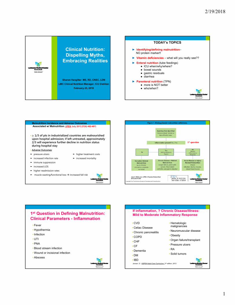
Malnutrition Incidence and Adverse OutcomesAssociated w/ Malnutrition (JPEN July 2013;37(4):482-497)
• > 1/3 of pts in industrialized countries are malnourished upon hospital admission; if left untreated, approximately 2/3 will experience further decline in nutrition status during hospital stay
• Adverse Outcomes:
► pressure ulcers ► higher treatment costs
► increased infection rate ► increased mortality
► immune suppression
► increased LOS
► higher readmission rates
► muscle wasting/functional loss increased fall risk
Figure 1. Etiology-based malnutrition definitions.
Jane V. White et al. JPEN J Parenter Enteral Nutr 2012;36:275-283
Copyright © by The American Society for Parenteral and Enteral Nutrition
1st question
1st Question in Defining Malnutrition:Clinical Parameters - Inflammation
• Fever
• Hypothermia
• Infection
• UTI
• PNA
• Blood stream infection
• Wound or incisional infection
• Abscess
If inflammation, ? Chronic Disease/Illness: Mild to Moderate Inflammatory Response
• CVD
• Celiac Disease
• Chronic pancreatitis
• COPD
• CHF
• CF
• Dementia
• DM
• IBD
• Hematologic malignancies
• Neuromuscular disease
• Obesity
• Organ failure/transplant
• Pressure ulcers
• RA
• Solid tumors
Jensen, G. ASPEN Adult Core Curriculum, 3rd edition, 2012
2/19/2018
2
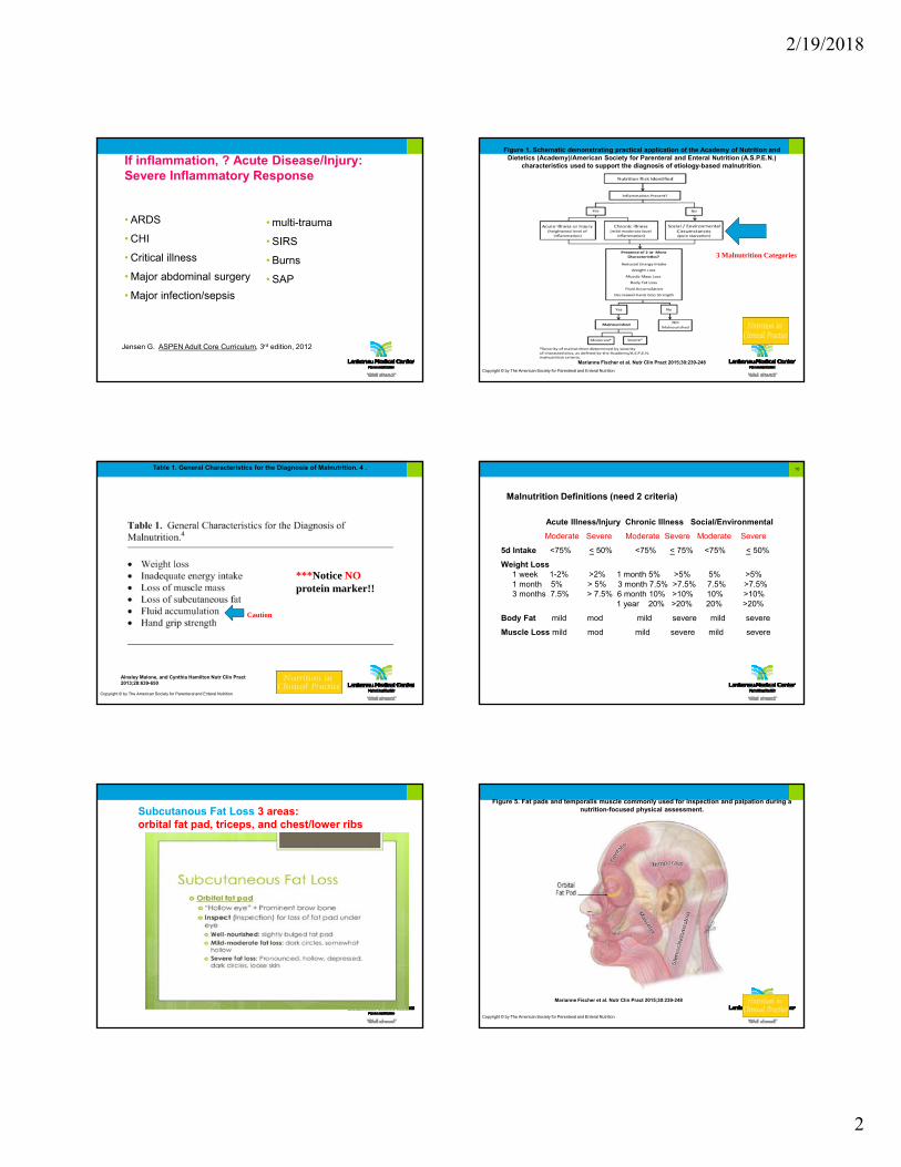
If inflammation, ? Acute Disease/Injury: Severe Inflammatory Response
• ARDS
• CHI
• Critical illness
• Major abdominal surgery
• Major infection/sepsis
• multi-trauma
• SIRS
• Burns
• SAP
Jensen G. ASPEN Adult Core Curriculum, 3rd edition, 2012
Figure 1. Schematic demonstrating practical application of the Academy of Nutrition and Dietetics (Academy)/American Society for Parenteral and Enteral Nutrition (A.S.P.E.N.)
characteristics used to support the diagnosis of etiology-based malnutrition.
Marianne Fischer et al. Nutr Clin Pract 2015;30:239-248
Copyright © by The American Society for Parenteral and Enteral Nutrition
3 Malnutrition Categories
Table 1. General Characteristics for the Diagnosis of Malnutrition. 4 .
Ainsley Malone, and Cynthia Hamilton Nutr Clin Pract 2013;28:639-650
Copyright © by The American Society for Parenteral and Enteral Nutrition
***Notice NOprotein marker!!
Caution
Malnutrition Definitions (need 2 criteria)
Acute Illness/Injury Chronic Illness Social/Environmental
Moderate Severe Moderate Severe Moderate Severe
5d Intake <75% < 50% <75% < 75% <75% < 50%
Weight Loss 1 week 1-2% >2% 1 month 5% >5% 5% >5%1 month 5% > 5% 3 month 7.5% >7.5% 7.5% >7.5%3 months 7.5% > 7.5% 6 month 10% >10% 10% >10%
1 year 20% >20% 20% >20%
Body Fat mild mod mild severe mild severe
Muscle Loss mild mod mild severe mild severe
10
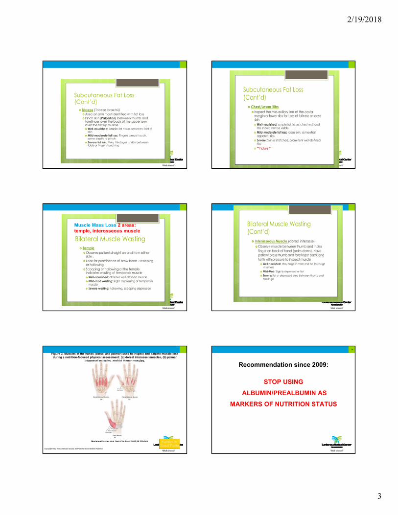
Subcutanous Fat Loss 3 areas: orbital fat pad, triceps, and chest/lower ribs
Figure 5. Fat pads and temporalis muscle commonly used for inspection and palpation during a nutrition-focused physical assessment.
Marianne Fischer et al. Nutr Clin Pract 2015;30:239-248
Copyright © by The American Society for Parenteral and Enteral Nutrition
2/19/2018
3
Muscle Mass Loss 2 areas: temple, interosseous muscle
Figure 3. Muscles of the hands (dorsal and palmar) used to inspect and palpate muscle loss during a nutrition-focused physical assessment: (a) dorsal interossei muscles, (b) palmar
interossei muscles, and (c) thenar muscles.
Marianne Fischer et al. Nutr Clin Pract 2015;30:239-248
Copyright © by The American Society for Parenteral and Enteral Nutrition
Recommendation since 2009:
STOP USING
ALBUMIN/PREALBUMIN AS
MARKERS OF NUTRITION STATUS
18
2/19/2018
4
23
How Can We Monitor Response to Nutrition Support?Postop Albumin/Prealbumin/C-reactive protein
• SCCM/ASPEN 2016 Traditional nutrition assessment tools (albumin, prealbumin, transferrin) are not validated in critical care and should not be used as markers of nutrition status.
• “Albumin, prealbumin, transferrin, and RBP reflect the acute phase response (increases in vascular permeability and reprioritization of hepatic protein synthesis) and do not accurately represent nutrition status in the ICU setting.” “….serum albumin concentrations would not be expected to change through the course of management until the stress metabolism abates. Thus, serum protein concentrations have no use postoperatively to measure adequacy of nutrition therapy”.
• North American Surgical Nutrition Summit, 2013:
• “Hypoalbuminemia is a valid prognosticator of preop risk, correlating significantly with increased LOS, infection, and mortality. However, it should not be followed over time in hospitalized patients. Use of any marker (albumin, prealbumin, or transferrin) for nutrition status is controversial, since they represent “negative acute phase proteins” with levels altered by any stress, injury, infection, organ failure, or acute phase response. Such proteins are poor indicators of actual nutrition state.”.
20Postop: do we care about Albumin/Prealbumin? NO!!
Prealbumin is better marker of severity of illness or “nutritional risk” than nutritional status/malnutrition or adequacy of nutrition support (except in late illness)
Bauer. Intensive Care Medicine 2000.Davis. JPEN 2012.Jenson. JPEN 2009.Raguso. Curr Opin Clin Nutr Metab Care 2003.
28Postop: do we care about Albumin/Prealbumin? NO!!
• Little value in assessment of nutritional status in critical illness/infection/postop due to:
• increased transcapillary escape of albumin into interstitial/intercellular fluid
• decreased synthesis with critical illness/surgery when positive acute phase production increases
Intravascular Space
Interstitial/intercellularspace
Table 1. General Characteristics for the Diagnosis of Malnutrition. 4 .
Ainsley Malone, and Cynthia Hamilton Nutr Clin Pract 2013;28:639-650
Copyright © by The American Society for Parenteral and Enteral Nutrition
***Notice NOprotein marker!!
Caution
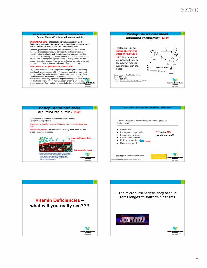
Vitamin Deficiencies –what will you really see??!!
23
The micronutrient deficiency seen in some long-term Metformin patients
24
2/19/2018
5
Answer: What is Vitamin B12
What other disease states require B12 supplementation?
25
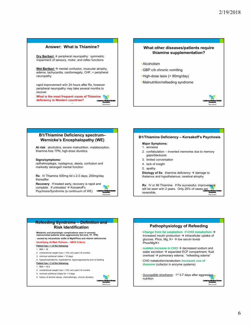
GI Tract – Sites of Nutrient Absorption
26
Source: Shikora SA et al. “Nutrition and Gastrointestinal Complications of Bariatric Surgery” Nutrition in Clinical Practice Feb. 2007;22:29-40.
What Disease States and Patients May Require Vitamin B12 Supplements?
• IBD resections or ileal involvement
•Pernicious anemia
•Atrophic gastritis
•Total/partial gastrectomy
•Gastric Bypass, Sleeve Gastrectomy
•Vegans
27
Lack of Intrinsic Factor
Lap Roux-n-Y Lap Vertical SleeveGastrectomy
Micronutrient Supplements Required by Bariatric Surgery Patients
Gastric Bypass (GBP) Lap Adjustable Gastric Sleeve Gastrectomy (need 5 supplements) Band (LAGB) (need 5 supplements)
chewable MVI bid chewable MVI 1x/day chewable MVI bid
chewable iron 18mg bid chewable iron 18mg/day
Chewable Calcium Citrate/D tid chewable Ca Citrate/D tid chewable Ca Citrate/D tid(or Calcium Citrate lozenge tidwith 5000units liquid Vit D/day)
B12 1000mcg oral/day or B12 1000mcg oral 3x/week IM 1000mcg/month or IM 1000mcg/month
The micronutrient deficiency with neurologic (peripheral neuropathy, confusion) and cardiovascular (tachycardia, cardiomegaly, CHF)
implications.
30
2/19/2018
6
31
Answer: What is Thiamine?
• Dry Beriberi peripheral neuropathy: symmetric impairment of sensory, motor, and reflex functions
• Wet Beriberi mental confusion, muscular atrophy, edema, tachycardia, cardiomegaly, CHF, + peripheral neuropathy
• rapid improvement w/in 24 hours after Rx, however peripheral neuropathy may take several months to recover
• What is the most frequent cause of Thiamine deficiency in Western countries?
What other diseases/patients require thiamine supplementation?
•Alcoholism
•GBP c/b chronic vomiting
•High-dose lasix (> 80mg/day)
•Malnutrition/refeeding syndrome
B1/Thiamine Deficiency spectrum–Wernicke’s Encephalopathy (WE)
• At risk: alcoholics, severe malnutrition, malabsorption, thiamine-free TPN, high-dose diuretics
• Signs/symptoms:opthalmoplegia, nystagmus, ataxia, confusion and markedly deranged mental function
• Rx: IV Thiamine 500mg tid x 2-3 days; 250mg/day thereafter
• Recovery: If treated early, recovery is rapid and complete. If untreated Korsakoff’s Psychosis/Syndrome (a continuum of WE)
B1/Thiamine Deficiency – Korsakoff’s Psychosis
• Major Symptoms:1. amnesia
2. confabulation – invented memories due to memory gaps/blackouts
3. limited conversation
4. lack of insight
5. apathy
• Etiology of Sx: thiamine deficiency damage to thalamus and hypothalamus; cerebral atrophy
• Rx: IV or IM Thiamine. If Rx successful, improvement will be seen w/in 2 years. Only 20% of cases are reversible.
Refeeding Syndrome – Definition and Risk Identification
• Metabolic and physiologic complications seen in severely malnourished patients when aggressively fed (oral, TF, TPN)
• caused by intracellular shifts of Mg/K/Phos and vitamin deficiencies
• Identifying At-Risk Patients – NICE Criteria
• Patient has > 1 of the following:
1. BMI < 16
2. unintentional weight loss > 15% w/in past 3-6 months
3. minimal nutritional intake > 10 days
4. hypophosphatemia, hypokalemia, hypomagnesemia prior to feeding
• Patient has > 2 of the following:
1. BMI < 18.5
2. unintentional weight loss > 10% w/in past 3-6 months
3. minimal nutritional intake for > 5 days
4. history of alcohol abuse, chemotherapy, chronic diuretics
Pathophysiology of Refeeding
• Change from fat catabolism CHO metabolism increased insulin production intracellular uptake of glucose, Phos, Mg, K+ low serum levels Phos/Mg/K+
• sudden increase in CHO decreased sodium and water excretion expanded ECF compartment, fluid overload pulmonary edema; “refeeding edema”
• CHO metabolism/anabolism increases use of thiamine (cofactor in enzyme systems)
• Susceptible timeframe: 1st 3-7 days after aggressive nutrition
2/19/2018
7
Pathophysiology of Refeeding Syndrome
• Thiamine Functions: cofactor in CHO metabolism (glycolysis); In deficiency state: 1) pyruvate converted to lactate instead of acetyl CoA lactic acidosis and death due to wet beriberi in patients receiving thiamine-free TPN
• Phosphorus Functions: required for ATP production, cofactor in enzyme systems. Lack of RBC phosphorus hemolysis, anemia, inadequate tissue oxygenation hyperventilationSevere hypophosphatemia (<1.5mg/dl) 1) neuromuscular -confusion, seizures, coma; weakness, rhabdomyolysis; 2) cardiac - decreased MAP; 3) respiratory - hypoxia, impaired diaphragmatic contractility
Management Guidelines – IV Phosphate Replacement **
• Mild hypophosphatemia, asymptomatic
2.3-2.7mg/dl 0.08-0.16mmol/kg
• Moderate hypophosphatemia, asymptomatic
1.5-2.2mg/dl 0.16-0.32mmol/kg
• Severe, symptomatic
< 1.5mg/dl 0.32-0.64mmol/kg
** For normal renal function. Patients with renal insufficiency: < 50% standard dose. Use adjusted BW for BMI > 30 or > 130% IBW.
Pathophysiology of Refeeding Syndrome
● Potassium Functions: cellular metabolism; glycogen and protein synthesis
Severe hypokalemia (< 2.5mEq/L) 1) neurologic – paralysis2) cardiac – altered myocardial contraction and signal conduction; arrhythmias, cardiac arrest
• Magnesium Functions: cofactor in many enzyme systems including ATP production and oxidative phosphorylationModerate to severe hypomagnesemia < 1.0mg/dl)
1) cardiac – EKG changes, arrhythmias
2) neuromuscular – tremor, seizures, coma
3) hypomagnesemia-induced hypokalemia
4) hypomagnesemia-induced hypocalcemia
Management Guidelines – IV Potassium Replacement *
Serum K+
• 2.5-3.4 mEq/L 20-40mEq (10-20mEq/h)**
• < 2.5 mEq/L or
if symptomatic 40-80mEq
* For normal renal function. Patients with renal insufficiency: < 50% standard dose.
** Continuous cardiac monitoring and infusion via CVC for infusion rate > 10mEq K+/hr.
Management Guidelines – IV Magnesium Replacement
• Mild/moderate hypomagnesemia, asymptomatic(serum Mg 1-1.5 mg/dl)
1-4g MagSulfate (8-32mEq magnesium), < 1 mEq/kg*
• Severe or symptomatic hypomagnesemia
(serum Mg < 1 mg/dl)
4-8g MagSulfate (32-64mEq magnesium), < 1.5 mEq/kg*
*For normal renal function. Patients w/ renal insufficiency: < 50% standard dose. Use adjusted BW for BMI > 30 or > 130% IBW.
The micronutrient deficiency associated with prolonged diarrhea in
Crohn’s/Ulcerative Colitis patients
2/19/2018
8
Answer: What is zinc?
► zinc deficiency diarrhea, anorexia, dysgeusia
► Active diarrhea: Rx 220mg Zinc Sulfate/day
► What other micronutrient deficiencies are common in Crohn’s/Ulcerative Colitis patients?
43
Potential Micronutrient Deficiencies in IBD
•Folate
•B12 – ileal involvment/resection
•Ca/Vit D – malabsorption, poor calcium intake
• Iron – poor intake, bloody diarrhea (UC)
Summary: Reality of Micronutrient Deficiencies
• Alcoholics: folate 1mg, thiamine 100mg, Vit B6 (50mg or MVI)
• Metformin: Vitamin B12
• IBD: Calcium, Vit D, iron, B12, folate, zinc (active diarrhea)
• Refeeding syndrome – thiamine, folate
• Weight loss surgeries:► GBP: MVI, iron bid, Ca, Vit D, B12 oral daily► Sleeve Gastrectomy: MVI, iron 1/day, Ca, Vit D,
B12 oral 3x/week ► Lap Band: MVI, Ca, Vit D
45
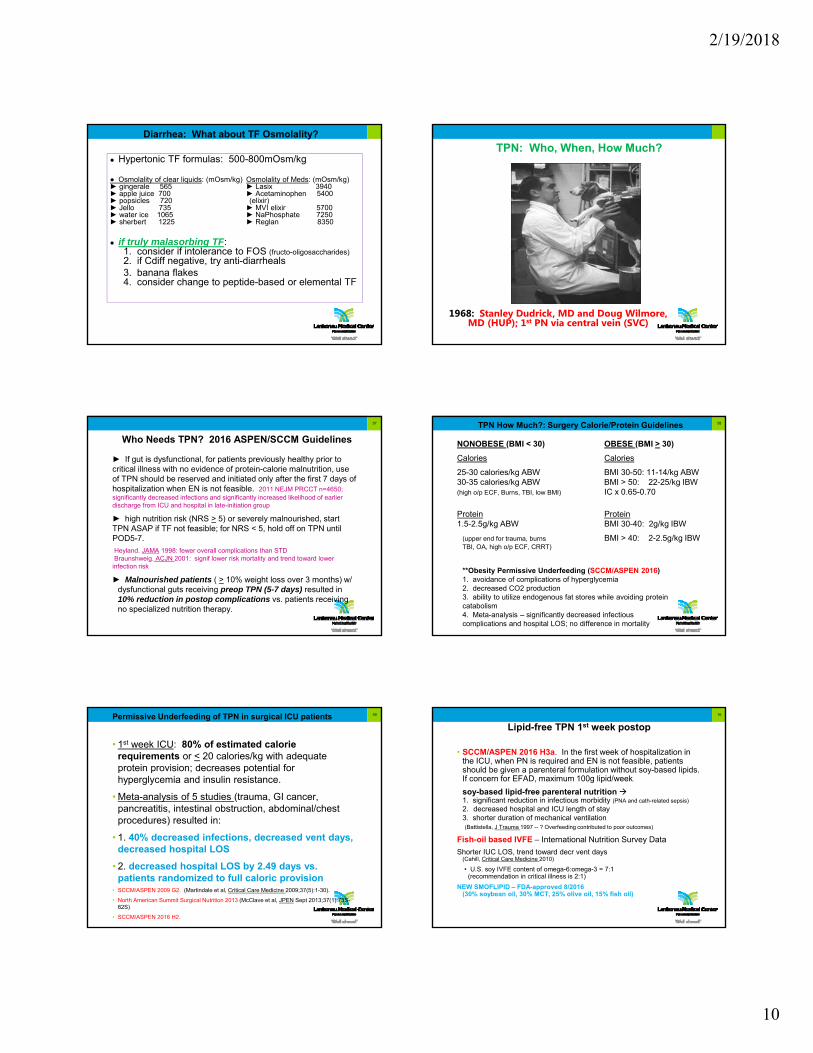
Enteral Nutrition: ICU when/where/why?, bowel sounds, gastric residuals, diarrhea
Enteral Nutrition/Tube Feeds – When/Why?
• No bowel sounds/flatus/stool required to start TF(SCCM/ASPEN 2016 B3)► “While GI factors should be evaluated when initiating EN, overt signs of contractility should not be required prior to initiation of EN”.
► Bowel sounds are indicative only of contractility and do not necessarily relate to mucosal integrity, barrier function, or absorptive capacity
● TF w/in 24-48h in critically ill unable to eat (SCCM/ASPEN 2016 B1)► supports functional integrity by maintaining tight junctions b/t intraepithelial cells, stimulating blood flow, and triggering release of trophic agents (CCK, gastrin, bile salts)► maintains structural integrity – villous height► stimulates production of immunocytes composing GALT► contributes to organ mucosal-associated lymphoid tissue (lungs, liver, kidneys)
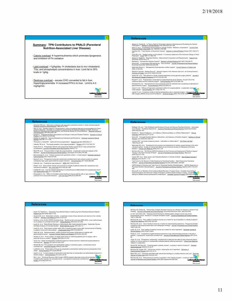
Does the gut play a role in MSOF? YES!!GALT = Gut-associated lymphoid tissue
Fed gut: gut produces B/T lymphocytes lymph nodes systemic
circulation
•Unfed gut/decr contractility bacterial overgrowth increased cytokines increased gut permeability macrophage activation lungs, liver, kidneys

2/19/2018
9
Do We Need Bowel Sounds? NO!!
• SCCM/ASPEN 2016 In the ICU population, neither the presence or absence of bowel sounds nor evidence of passage of flatus and stool is required for the initiation of enteral feeding.
• ►Bowel sounds only indicative of contractility; don’t relate to integrity of GI mucosa or absorptive capacity
• SCCM/ASPEN 2016 In the ICU setting, evidence of bowel motility (resolution of clinical ileus) is not required in order to initiate EN in the ICU. ► As long as the patient remains hemodynamically stable, it is safe and appropriate to feed through mild to moderate ileus.
Is TF Contraindicated in Pressor-Dependent Patients? NO!!
• Some pressors may increase splanchnic blood flow:
• ►Dopamine (<10mcg/kg/min)►Levophed (<3mcg/min; 0.5-3ml/min)
• Intestinal Vasoconstrictive Effects:
• ► Dopamine > 10mcg/kg/min► Levophed >4mcg/min► Phenylephrine/Neosynephrine► Vasopressin
Summary: TF Recommendations with Hypotension/Vasopressive Agents
• EN when hemodynamically stable (fluid resuscitated, stable pressor doses, MAP > 60 mmHg)
STOP TF if:
•► sustained MAP < 60► increasing doses pressors► increased vent support (increasing PEEP, FiO2)► signs of GI intolerance (abd distention/pain, increased NGT output if nasoenteric feeds, cessation of stooling, abd Xray/CT significant small bowel or colon dilation)
• Isotonic, fiber-free formula; Fiber (preferred bacterial substrate) in setting of decreased gut motility incr bowel distention, bacterial overgrowth stretched bowel wall more susceptible to decreased integrity
Enteral Nutrition – Where?
• Gastric feeds for most critically ill patients (SCCM/ASPEN 2016 B4b)
► technically easier, decreases time to EN start► largest multicenter RCT gastric vs SB TF: no difference in clinical outcomes including LOS, mortality, nutrient delivery, incidence of PNA (Davies, Critical Care Medicine 2012)
► 13 RCTs do demonstrate lower rates PNA w/ SB TF
• Divert TF to SB for those at high-risk aspiration or intolerant to gastric feeds (SCCM/ASPEN B4a, D4a)
• SURGERY PATIENTS:
It is appropriate to attempt to provide postop EN judiciously in a patient above a gut anastomosis, in one with an open abdomen, in a setting of bowel wall edema, in a stable patient on vasopressor therapy, or in one with hypoactive bowel sounds and postop ileus. (sCCM/ASPEN 2016 O4; 2013 N. American Surgical Nutrition Summit)Barlow, Clin Nutr 2011; Collier, JPEN 2007; Khalid, Am J Crit Care 2010; Caddell, Curr Gastroenterol Rep 2011; Dissanaike, J Am Coll Surg 2008)
Gastric Residual Volumes (GRVs)
Gastric residual volumes should NOT be used as part of routine care. If protocol calls for gastric residuals, avoid holding TF for GRVs < 500ml in absence of other signs of intolerance. GRVs do not correlate w/ incidence of PNA, regurgitation, or aspiration.(SCCM/ASPEN 2016 D2a, D2b)
*** DO NOT CHECK GASTRIC RESIDUALS IN JTUBES!!! ***
•Flaws in the GRV Rationale
► Daily volume of gastric (3000ml) and salivary (1500ml) secretions averages an hourly rate approximately 188ml/hr in a normally-fed adult. Gastric capacity averages 1500-1900ml.
► Most GRVs < 150ml and no significant difference in pattern of GRVs in critically ill patients vs. healthy volunteers.
► 4 RCTs: increasing GRV from 50-150 to 250-500 does not increase the incidence of regurgitation, aspiration, or PNA.(Taylor, Critical Care Medicine 1999; Montego, Intensive Care Medicine 2010; Pinilla, JPEN 2001; McClave, JPEN 2002; McClave, Critical Care Medicine 2005)
What About Diarrhea?
• Diarrhea Defined: Frequent watery, loose bowel movements; > 500ml every 8 hours OR > 3 stools/day for > 2 consecutive days
• Questions to ask: 1) does it meet the definition of diarrhea? 2) Cdiff or infectious cause?3) antibiotic/med-induced diarrhea?
• What about Osmolality? NO! NEVER dilute formulas.► Saliva, pancreatic enzymes, bile salts, neutralize pH in first 10-45cm of small bowel ► Infused gastrically, formulas achieve isotonicity (250-
300mOsm/kg) by the Ligament of Treitz; infused into Ligament of Treitz, formulas achieve isotonicity by the jejunum
2/19/2018
10
Diarrhea: What about TF Osmolality?
● Hypertonic TF formulas: 500-800mOsm/kg
● Osmolality of clear liquids: (mOsm/kg) Osmolality of Meds: (mOsm/kg)► gingerale 565 ► Lasix 3940► apple juice 700 ► Acetaminophen 5400► popsicles 720 (elixir)► Jello 735 ► MVI elixir 5700► water ice 1065 ► NaPhosphate 7250► sherbert 1225 ► Reglan 8350
● if truly malasorbing TF:1. consider if intolerance to FOS (fructo-oligosaccharides)2. if Cdiff negative, try anti-diarrheals3. banana flakes4. consider change to peptide-based or elemental TF
1968: Stanley Dudrick, MD and Doug Wilmore, MD (HUP); 1st PN via central vein (SVC)
TPN: Who, When, How Much?
57
Who Needs TPN? 2016 ASPEN/SCCM Guidelines
► If gut is dysfunctional, for patients previously healthy prior to critical illness with no evidence of protein-calorie malnutrition, use of TPN should be reserved and initiated only after the first 7 days of hospitalization when EN is not feasible. 2011 NEJM PRCCT n=4650; significantly decreased infections and significantly increased likelihood of earlier discharge from ICU and hospital in late-initiation group
► high nutrition risk (NRS > 5) or severely malnourished, start TPN ASAP if TF not feasible; for NRS < 5, hold off on TPN until POD5-7.
Heyland. JAMA 1998: fewer overall complications than STDBraunshweig. ACJN 2001: signif lower risk mortality and trend toward lower
infection risk
► Malnourished patients ( > 10% weight loss over 3 months) w/ dysfunctional guts receiving preop TPN (5-7 days) resulted in 10% reduction in postop complications vs. patients receiving no specialized nutrition therapy.
58TPN How Much?: Surgery Calorie/Protein Guidelines
NONOBESE (BMI < 30) OBESE (BMI > 30)
Calories Calories
25-30 calories/kg ABW BMI 30-50: 11-14/kg ABW30-35 calories/kg ABW BMI > 50: 22-25/kg IBW(high o/p ECF, Burns, TBI, low BMI) IC x 0.65-0.70
Protein Protein1.5-2.5g/kg ABW BMI 30-40: 2g/kg IBW
(upper end for trauma, burns BMI > 40: 2-2.5g/kg IBWTBI, OA, high o/p ECF, CRRT)
**Obesity Permissive Underfeeding (SCCM/ASPEN 2016)1. avoidance of complications of hyperglycemia2. decreased CO2 production3. ability to utilize endogenous fat stores while avoiding protein catabolism 4. Meta-analysis – significantly decreased infectious complications and hospital LOS; no difference in mortality
Permissive Underfeeding of TPN in surgical ICU patients
• 1st week ICU: 80% of estimated calorie requirements or < 20 calories/kg with adequate protein provision; decreases potential for hyperglycemia and insulin resistance.
• Meta-analysis of 5 studies (trauma, GI cancer, pancreatitis, intestinal obstruction, abdominal/chest procedures) resulted in:
• 1. 40% decreased infections, decreased vent days, decreased hospital LOS
• 2. decreased hospital LOS by 2.49 days vs. patients randomized to full caloric provision
• SCCM/ASPEN 2009 G2. (Martindale et al, Critical Care Medicine 2009;37(5):1-30).
• North American Summit Surgical Nutrition 2013 (McClave et al, JPEN Sept 2013;37(1):73S-82S)
• SCCM/ASPEN 2016 H2.
59 16
Lipid-free TPN 1st week postop
• SCCM/ASPEN 2016 H3a. In the first week of hospitalization in the ICU, when PN is required and EN is not feasible, patients should be given a parenteral formulation without soy-based lipids. If concern for EFAD, maximum 100g lipid/week.
soy-based lipid-free parenteral nutrition 1. significant reduction in infectious morbidity (PNA and cath-related sepsis)
2. decreased hospital and ICU length of stay3. shorter duration of mechanical ventilation(Battistella, J Trauma 1997 -- ? Overfeeding contributed to poor outcomes)
Fish-oil based IVFE – International Nutrition Survey Data
Shorter IUC LOS, trend toward decr vent days(Cahill, Critical Care Medicine 2010)
• U.S. soy IVFE content of omega-6:omega-3 = 7:1(recommendation in critical illness is 2:1)
NEW SMOFLIPID – FDA-approved 8/2016(30% soybean oil, 30% MCT, 25% olive oil, 15% fish oil)
2/19/2018
11
61
Summary: TPN Contributors to PNALD (Parenteral Nutrition-Associated Liver Disease)
• Calorie overload hyperinsulinemia which promotes lipogenesis and inhibition of FA oxidation
• Lipid overload: >1g/kg/day cholestasis due to incr cholesterol, TGs, and phospholipid concentrations in liver. Limit fat to 30% kcals or 1g/kg.
• Dextrose overload – excess CHO converted to fat in liver. Hypertriglyceremidia increased FFA’s to liver. Limit to 4-5 mg/kg/min.
References
• Skipper A, Tupesis N. “Is There a Role for Nonprotein Calories in Developing and Evaluating the Nutrient Prescription?”. Nutrition in Clinical Practice June 2005;20(3):321-324.
• Klein CJ, et al. “Overfeeding macronutrients to critically ill adults: Metabolic complications”. Journal of the American Dietetic Association July 1998;98(7):795-806.
• VanWay CW. “Total Calories vs Nonprotein Calories”. Nutrition in Clinical Practice October 2001;16(5):271-272.
• Cerra FB, et al. “Applied nutrition in ICU patients: A consensus statement of the American College of Chest Physicians”. Chest 1997;111:769-778.
• Gasser E, Parekh N. “Parenteral Nutrition: Macronutrient Composition and Requirements”. Support Line December 2005;27(6):6-12.
• Maxfield D. “Perioperative Nutrition Support”. Nutrition in Clinical Practice April 2001;16(2):69-73.
• Driscoll DF. “Compounding TPN Admixtures: Then and Now”. Journal of Parenteral and Enteral Nutrition November/December 2003;27(6):433-438.
• Martindale Robert G. “Management of perioperative nutrition support”. Current Opinion in Critical Care 2006;12:290-294.
• Blackburn George L, Bistrian Bruce R. “Nutrition Support in the Intensive Care Unit: An Evolving Science”. Archives of Surgery June 2010;145(6):533-538.
• Kassin MT, et al. “Risk factors for 30-day hospital readmission among general surgery patients”. Journal of the American College of Surgeons 2012;215(3):322-330.
• Bozzetti F, et al. “Postoperative complications in gastrointestinal cancer patients: the joint role of the nutritional status and the nutritional support”. Clinical Nutrition 2007;27(4):698-709.
• Schiesser M, et al. “Assessment of a novel screening score for nutritional risk in predicting complications in gastrointestinal surgery”. Clinical Nutrition 2008;27(4):565-570.
• Jiang H, et al. “Efficacy of hypocaloric parenteral nutrition for surgical patients: a systematic review and meta-analysis”. Clinical Nutrition 2011;30(6):730-737.
• Lassen K, et al. Consensus review of optimal perioperative care in colorectal surgery: Enhanced Recovery After Surgery (ERAS) Group recommendations”. Archives Surgery 2009;144(10):961-969.
36References• Dickerson RN et al. “Net protein anabolism with hypocaloric parenteral nutrition in obese stressed patients”.
American Journal of Clinical Nutrition 1986;44:747-755.
• Klein S, et al. “Nutrition support in clinical practice: review of published data and recommendations for future research directions. Summary of a conference sponsored by the National Institutes of Health, American Society for Parenteral and Enteral Nutrition, and American Society for Clinical Nutrition”. American Journal of Clinical Nutrition 1997;66(3):683-706.
• Kumpf VJ. “Parenteral Nutrition-Associated Liver Disease in Adult and Pediatric Patients”. Nutrition in Clinical Practice June 2006;21(3):279-290.
• Wanten G, Calder PC. “Immune Modulation by Parenteral Lipid Emulsions”. American Journal of Clinical Nutrition 2007;85:1171-1184
• Thompson C, Russell M. “Lipids and Immunosuppression”. Support Line April 2008;30(2):3-7.
• Valentijn TM, et al. “The obesity paradox in the surgical population”. Surgeon 2013;11(3):169-176.
• Kudsk KA et al. “Preoperative albumin and surgical site identify surgical risk for major postoperative complications” Journal of Parenteral and Enteral Nutrition 2003;27:1-9.
• Marik PE, et al. “Immunonutrition in high-risk surgical patients: a systematic review and analysis of the literature”. Journal of Parenteral and Enteral Nutrition July/August 2010;34(4):378-386.
• Braunschweig CL, et al. “Enteral compared with parenteral nutrition: a meta-analysis”. American Journal of Clinical Nutrition 2001;74(4):534-542.
• Barlow R, et al. “Prospective multicenter randomized controlled trial of early enteral nutrition for patients undergoing major upper gastrointestinal surgical resection”. Clinical Nutrition 2011;30(5):560-566.
• Collier B, et al. “Feeding the open abdomen”. JPEN 2007;31(5):410-415.
• Khalid I, et al. “Early enteral nutrition and outcomes of critically ill patients treated with vasopressors and mechanical ventilation”. Am J Crit Care 2010;19(3):261-268.
• Caddell KA, et al. “Can the intestinal dysfunction of critical illness be differentiated from postoperative ileus?” Curr Gastroenterol Rep 2011;13(4):358-367.
• Dissanaike S, et al. “Effect of immediate enteral feeding on trauma patients with an open abdomen: protection from nosocomial infections”. J Am Coll Surg 2008;207(5):690-697.
References
• DeMeijer VE, et al. “Fish Oil-Based Lipid Emulsions Prevent and Reverse Parenteral Nutrition – Associated Liver Disease: The Boston Experience”. Journal of Parenteral and Enteral Nutrition September/October 2009;33(5):541-547.
• Shenkin A. “Serum Prealbumin: Is It a Marker of Nutritional Status or of Risk of Malnutrition?”. ClinicalChemistry 2006;52(12):2177-2179.
• Seres DS. “Surrogate Nutrition Markers, Malnutrition, and Adequacy of Nutrition Support”. Nutrition in Clinical Practice June 2005;20(3):308-313.
• Johnson AM. “Low levels of plasma proteins: malnutrition or inflammation?”. Clin Chem Lab Med 1999;37(2):91-96
• Martindale RG, et al. “Guidelines for the provision and assessment of nutrition support therapy in the adult critically ill patient: Society of Critical Care Medicine and American Society for Parenteral and Enteral Nutrition”. Critical Care Medicine 2009;37(5):1-30.
• McClave SA, et al. “SCCM and ASPEN Guidelines for the Provision and Assessment of Nutrition Support Therapy in the Adult Critically Ill Patient”. Journal of Parenteral and Enteral Nutrition, May/June 2009;33(2):277-316.
• Casaer MP, et al. “Early versus Late Parenteral Nutrition in Critically Ill Adults”. New England Journal of Medicine June 29,2011, 1-12.
• Task Force for the Revision of Safe Practices for Parenteral Nutrition. “Safe Practices for Parenteral Nutrition”. Journal of Parenteral and Enteral Nutrition Nov/Dec 2004;26(6): S39-S69.
• ASPEN Board of Directors and Task Force on Parenteral Nutrition Standardization. “ASPEN Statement on Parenteral Nutrition Standardization”. Journal of Parenteral and Enteral Nutrition Sept/Oct 2007;31(5):441-448.
• McLeod R, et al. Members of the Evidence Based Reviews in Surgery Group. Canadian Association of General Surgeons and American College of Surgeons evidence based reviews in surgery: preoperative fasting for adultsto prevent perioperative complications”. Canadian Journal of Surgery 2005;48(5):409-411.
37
References
• Austin TA, Hamilton C. “Assessing Trace Element Status in Adult Long-Term Parenteral Nutrition Patients”. Support Line April 2008;30(2):17-22.
• Heyland DK, et al. “Antioxidant nutrients: a systematic review of trace elements and vitamins in the critically ill patient”. Intensive Care Medicine 2005;31:327-337.
• Kondrup J et al, Ad Hoc ESPEN Working Group. “Nutritional risk screening (NRS 2002): a new method based on an analysis of controlled clinical trials”. Clinical Nutrition 2003;22(3):321-336.
• Lewis SJ, et al. “Early Enteral Feeding vs. Nil By Mouth After Gastrointestinal Surgery: Systematic Review and Meta-Analysis of Controlled Studies”. British Medical Journal, 2001;323:1-5.
• Lewis SJ, et al. “Early enteral nutrition within 24h of intestinal surgery versus later commencement of feeding: a systemic review and meta analysis”. J Gastrointest Surg 2009;13(3):569-575.
• Garth AK, et al. “Nutritional status, nutrition practices and postop complications in patients with gastrointestinal cancer”. Journal of Human Nutrition and Dietetics 2010;23(4):393-401.
• Windsor A, et al. “Fit for surgery: an expert panel review on optimizing patients prior to surgery, with a particular focus on nutrition”. Surgeon 2004;2(6):315-319.
• Jie B, et al. Impact of preoperative nutritional support on clinical outcome in abdominal surgical patients at nutritional risk”. Nutrition 2012;28(10):1022-1027.
• Noblett SE et al. “Pre-operative oral carbohydrate loading in colorectal surgery: a randomized clinical controlled trial”. Colorectal Disease 2006;8(7):563-569.
• Luttikhold J et al. “Preservation of the gut by preoperative carbohydrate loading improves postoperative food intake”. Clinical Nutrition 2013;32(4):556-561.
• Awad S, et al. “A meta-analysis of randomized controlled trials on preoperative oral carbohydrate treatment in elective surgery”. Clinical Nutrition 2013;32(1):34-44.
• Ren L et al. “Enhanced Recovery After Surgery (ERAS) program attenuates stress and accelerates recovery in patients after resection for colorectal cancer: a prospective randomized controlled trial”. World Journal of Surgery 2012;36(2):407-414.
38
References
• McClave SA, Snider HL. “Clinical Use of Gastric Residual Volumes as a Monitor for Patients on Enteral Tube Feeding”. Journal of Parenteral and Enteral Nutrition, November/December 2002; 26(6):S43-S50.
• Lin HC, Van Citters GW. “Stopping enteral feeding for arbitrary gastric residual volume may not be physiologically sound: Results of a computer simulation model”. Journal of Parenteral and Enteral Nutrition1997;21:286-289.
• McClave SA, et al. “Poor validity of residual volumes as a marker for risk of aspiration in critically ill patients”. Critical Care Medicine 2005;33(2):324-330.
• McClave SA, et al. “Use of residual volume as a marker for enteral feeding intolerance: Prospective blinded comparison with physical examination and radiographic findings”. Journal of Parenteral and Enteral Nutrition1992;16:99-105.
• Lukan JK, et al. “Poor validity of residual volumes as a marker for risk of aspiration”. American Journal of Clinical Nutrition 2002;75:S417-S418.
• Pinilla JC, et al. “Comparison of gastrointestinal intolerance to two enteral feeding protocols in critically ill patients: a prospective, randomized controlled trial”. Journal of Parenteral and Enteral Nutrition 2001;25:81-86.
• Taylor SJ, et al. “Prospective, randomized, controlled trial to determine the effect of early enhanced enteral nutrition on clinical outcome in mechanically ventilated patients suffering head injury”. Critical Care Medicine1999;27(11):2525-2531.
• Parrish RP, McClave SA. “Checking gastric residual volumes: a practice in search of science?”. Practical Gastroenterology 2008;32(10):33-47.
• McClave SA, Dryden GW. “Critical care nutrition: reducing the risk of aspiration”. Seminars inGastrointestinal Disease 2003;14(1):2-10.
• Elpern EH, et al. “Outcomes associated with enteral tube feedings in a medical intensive care unit”. American Journal of Critical Care 2004;13(3):221-227.
• McClave SA, et al. “North American summit on aspiration in the critically ill patient: consensus statement”. Journal of Parenteral and Enteral Nutrition 2002;26(6 Supplement):S80-S85.
66
2/19/2018
12
References
• Robinson MR, et al. “The relationship between obesity, nutritional status, and mortality in the critically ill”. Critical Care Medicine 2015;43(1):87-100.
• Battistella FD et al. “A prospective, randomized trial of intravenous fat emulsion administration in trauma victims requiring total parenteral nutrition”. Journal of Trauma 1997;43(1)52-58.
• Cahill NE, et al. “Nutrition therapy in the critical care setting: what is “best achievable practice? An international multicenter observational study”. Critical Care Medicine 2010;38(2):395-401.
• Heyland DK et al. “Total parenteral nutrition in the critically ill patient: a meta-analysis. JAMA1998;280(23):2013-2019.
• Doig GS et al. “Early enteral nutrition reduces mortality in trauma patients requiring intensive care: a meta-analysis of reandomized controlled trials”. Injury 2011;42(1):50-56.
• O’Keefe GE et al. “Inflammation and the host response to inury, a large-scale collaborative project: patient-oriented research core – standard operating procedures for clinical care VIII – nutritional support of the trauma patient”. Journal of Trauma 2008;65(6):1520-1528.
• Heyland DK, et al. “Identifying critically ill patients who benefit the most from nutrition therapy: the development and initial validation of a novel risk assessment tool”. Critical Care, 2011;15(6):R268.
• Jabbar A, et al. “Gut immunology and the differential response to feeding and starvation”. Nutrition in Clinical Practice, 2003;18(6):461-482.
• Hiesmayr M, et al. “Decreased food intake is a risk factor for mortality in hospitalized patients: the Nutrition Day Survey 2006”. Clinical Nutrition 2009;28(5):484-491.
• Heyland DK, et al. “The success of enteral nutrition and ICU-acquired infections: a multicenter observational study”. Clinical Nutrition 2011;30(2):148-155.
• McClave SA et al. “Nutrition Support in Acute Pancreatitis: a systematic review of the literature”. JPEN 2006;30(2):143-156.
• Pupelis G et al. “Randomized trial of safety and efficacy of postoperative enteral feeding in patients w/ severe pancreatitis: preliminary report”. Eur J Surg 2000;166(5):383-387.
67
References• Pupelis G, et al. “Jejunal feeding, even when instituted late, improves outcomes in patients with severe pancreatitis
and peritonitis”. Nutrition 2001;17(2):91-94.
• Sun JK et al. “Early enteral nutrition prevents intra-abdominal hypertension and reduces the severity of severe acute pancreatitis compared with delayed enteral nutrition: a prospective pilot study”. World J Surgery 2013:37(9):2053-2060.
• Wereszczynska-Siemiatkowska U et al. “Early enteral nutrition is superior to delayed enteral nutrition for the prevention of infected necrosis and mortality in acute pancreatitis”. Pancreas 2013:42(4):640-646.
• Eatock FC et al. “A randomized study of early nasogastric versus nasojejnunal feeding in severe acute pancreatitis”. Am J Gastroenterology 2005;100(2):432-439.
• Kumar A et al. “Early enteral nutrition in severe acute pancreatitis: a prospective randomized controlled trial comparing nasojejunal and nasogastric routes”. J Clin Gastroenterology 2006;40(5):431-434.
• Singh A, et al. “Parenteral nutrition combined with enteral nutrition for severe acute pancreatitis”. Gastroenterology2012.
• Burlew CC, et al. “Who should we feed? Western Trauma Association multi-institutional study of enteral nutrition in the open abdomen after injury? J Trauma Acute Care Surgery 2012;73(6):1380-1387.
• Heyland DK et al. “Enhanced protein-energy provision via the enteral route feeding protocol in critically ill patients: results of a cluster randomized trial”. Critical Care Medicine 2013;41(12): 2743-2753.
• Davies AR et al. “A multicenter, randomized controlled trial comparing early nasojejunal with nasogastric nutrition in critical illness”. Critical Care Medicine 2012;40(8):2342-2348.
• Wu et al. “Randomized Clinical Trial of New Intravenous Lipid (SMOFlipid 20%) Versus Medium-Chain Triglycerides/Long-Chain Triglycerides in Adult Patients Undergoing Gastrointestinal Surgery”. JPEN September 2014;38(7):80-808.
• Wind J, et al. “Laparoscopy and/or fast track multimodal management versus standard care (LAFA) study group: Enhanced Recovery After Surgery (ERAS) group. Systematic review of enhanced recovery programmes in colonic surgery”. Br J. Surgery 2006;93:800-809.
• Gusfafsson UO et al. “Enhanced Recovery After Surgery study group. Adherence to the enhanced recovery after surgery protocol and outcomes after colorectal cancer surgery”. Archives of Surgery 2011;146:571-577.
68