Embed Size (px)
Citation preview
Dr. Kaan Yücel http://yeditepeanatomy1.wordpress.com Yeditepe Anatomy
13. January.2012 Friday
Branches of the rootsroots of the brachial plexusIn addition to small segmental branches from C5 to C8 to muscles of the neck and a contribution of C5 to the phrenic nerve, the roots of the brachial plexus give rise to the dorsal scapular and long thoracic nerves.
Dorsal scapular nerve (C5)Dorsal scapular nerve (C5) Innervates rhomboid major & minor muscles.
Long thoracic nerve (C5-C7)Long thoracic nerve (C5-C7) Innervates serratus anterior muscle.
Branches of the (superior) trunk(superior) trunk of the brachial plexusSuprascapular nerve (C5 & C6)
The suprascapular nerve originates in the base of the neck from the superior trunk of the brachial plexus. It passes through the suprascapular foramen to reach the posterior scapular region, where it lies in the plane between bone and muscle. It innervates the supraspinatus muscle, then terminates in and innervates the infraspinatus muscle. Generally, the suprascapular nerve has no cutaneous branches.
The nerve to subclavius muscle (C5 & C6) Innervates subclavius muscle.
Three nerves originate entirely or partly from the lateral cord. 1. Lateral pectoral nerve Innervates pectoralis major muscle.2. Musculocutaneous nerve (C5, C6, C7)2. Musculocutaneous nerve (C5, C6, C7)
http://home.comcast.net/~wnor/lesson4nervesofant&postarm.htm
The medial cord has five branches.1.Medial pectoral nerveThe medial pectoral nerve is the most proximal branch. It receives a communicating branch from the lateral pectoral nerve. Branches of the nerve penetrate and supply the pectoralis minor muscle. Some of these branches pass through the muscle to reach and supply the pectoralis major muscle. Innervates pectoralis major and pectoralis minor.
http://www.youtube.com/yeditepeanatomy 1
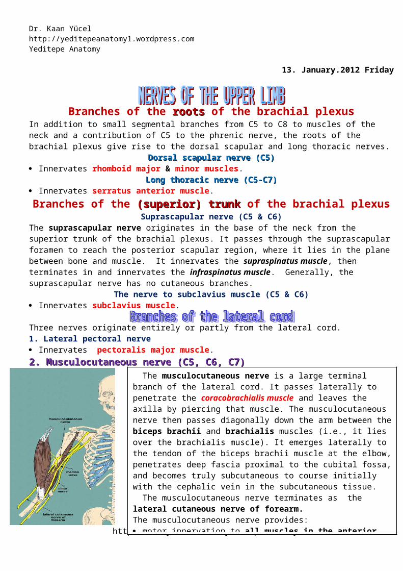
The musculocutaneous nerve is a large terminal branch of the lateral cord. It passes laterally to penetrate the coracobrachialis muscle and leaves the axilla by piercing that muscle. The musculocutaneous nerve then passes diagonally down the arm between the biceps brachii and brachialis muscles (i.e., it lies over the brachialis muscle). It emerges laterally to the tendon of the biceps brachii muscle at the elbow, penetrates deep fascia proximal to the cubital fossa, and becomes truly subcutaneous to course initially with the cephalic vein in the subcutaneous tissue.
The musculocutaneous nerve terminates as the lateral cutaneous nerve of forearm. The musculocutaneous nerve provides: motor innervation to all muscles in the anterior compartment of the arm; sensory innervation to skin on the lateral surface of the forearm. 3. Lateral root of median nerveThe lateral root of median nerve is the largest terminal branch of the lateral cord and passes medially to join a similar branch from the medial cord to form the median nerve.
Dr. Kaan Yücel http://yeditepeanatomy.wordpress.com Yeditepe Anatomy
2. Medial cutaneous nerve of arm (medial brachial cutaneous nerve)The medial cutaneous nerve of arm passes through the axilla and into the arm where supplies skin over the medial side of the distal third of the arm. In the axilla, the nerve communicates with the intercostobrachial nerve of T2. 3. Medial cutaneous nerve of forearm (medial antebrachial cutaneous nerve)The medial cutaneous nerve of forearm (medial antebrachial cutaneous nerve) originates just distal to the origin of the medial cutaneous nerve of arm. It passes out of the axilla and into the arm and supplies the skin over the biceps brachii muscle. It then innervates the skin over the medial surface of the forearm down to the wrist.
The medial cutaneous nerve of the forearm (medial antebrachial cutaneous nerve) is an independent branch of the medial cord of the brachial plexus. With the posterior cutaneous nerve of the forearm from the radial nerve, and the lateral cutaneous nerve of the forearm which is the continuation of the musculocutaneous nerve, each supplying the area of skin indicated by its name, these three nerves provide all the cutaneous innervation of the forearm. There is no “anterior cutaneous nerve of the forearm.” (Memory device: This is similar to the brachial plexus, which has lateral, medial, and posterior cords but no anterior cord.)4. Ulnar nerve (C8-T1)Ulnar nerve (C8-T1)
The ulnar nerve is a large terminal branch of the medial cord. The ulnar nerve passes through the arm and forearm into the hand.
The ulnar nerve enters the arm with the median nerve and axillary artery. The ulnar nerve in the arm passes distally from the axilla anterior to the insertion of the teres major and to the long head of the triceps, on the medial side of the brachial artery. In the middle of the arm, the ulnar nerve penetrates the medial intermuscular septum and enters the posterior compartment. It then passes into the anterior compartment of the forearm. Posterior to the medial epicondyle, where the ulnar nerve is referred to in lay terms as the “funny bone,” it is superficial, easily palpable, and vulnerable to injury. Like the median nerve, the ulnar nerve has no branches in the arm, but it also supplies articular branches to the elbow joint.
The ulnar nerve enters the anterior compartment of the forearm by passing posteriorly around the medial epicondyle of the humerus and between the humeral and ulnar heads of the flexor carpi ulnaris muscle.
In the forearm the ulnar nerve gives rise to: 1) Muscular branches to the flexor carpi ulnaris and to the medial half of the flexor digitorum profundus.2) Two small cutaneous branches; palmar branch passes into the hand to supply skin on the medial side of the palm; larger dorsal cutaneous branch of the ulnar nerve innervates skin on the posteromedial medial half of the dorsum of the hand, the 5th finger, and the medial half of the 4th finger. The ulnar nerve ends at the distal border of the flexor retinaculum by dividing into superficial (mainly sensory) and deep (mainly motor) branches.
The superficial branch of the ulnar nerve supplies the anterior surfaces of the medial one and a half digits. The deep branch of the ulnar nerve supplies the hypothenar muscles, the medial two lumbricals, the adductor pollicis, the deep head of the flexor pollicis brevis, and all the interossei.
As the deep branch of the ulnar nerve passes across the palm, it lies in a fibro-osseous tunnel (Guyon's canal) between the hook of the hamate and the flexor tendons. Occasionally, small outpouchings of synovial membrane (ganglia) from the joints of the carpus compress the nerve within this canal, producing sensory and motor symptoms. 5. Median nerve (C5-T1)Median nerve (C5-T1)
The median nerve is formed anterior to the third part of the axillary artery by the union of lateral and medial roots. The lateral root of the median nerve is the direct continuation of the lateral cord of the brachial plexus. It is joined by the medial root to form the median nerve trunk, and this passes downward on the lateral side of the axillary artery.
The median nerve enters the arm from the axilla at the inferior margin of the teres major muscle. It passes vertically down the medial side of the arm in the anterior compartment and is related to the brachial artery throughout its course: The median nerve has no major branches in the arm or in the axilla.
The median nerve is the principal nerve of the anterior compartment of the forearm. It supplies muscular branches directly to the muscles of the superficial and intermediate layers of forearm flexors (except the flexor
http://www.youtube.com/yeditepeanatomy 2
Dr. Kaan Yücel http://yeditepeanatomy1.wordpress.com Yeditepe Anatomy
carpi ulnaris), and deep muscles (except for the medial [ulnar] half of the flexor digitorum profundus; ring and little fingers) via its branch, the anterior interosseous nerve.
It leaves the cubital fossa by passing between the two heads of the pronator teres muscle and passing between the humero-ulnar and radial heads of the flexor digitorum superficialis muscle. It leaves the forearm and enters the palm of the hand by passing through the carpal tunnel deep to the flexor retinaculum.
The median nerve has no branches in the arm other than small twigs to the brachial artery. Its major branch in the forearm is the anterior interosseous nerve.1) Articular branches: These branches pass to the elbow joint as the median nerve passes it.2) Muscular branches: The nerve to the pronator teres usually arises at the elbow. A broad bundle of nerves pierces the superficial flexor group of muscles and innervates the flexor carpi radialis, palmaris longus, and flexor digitorum superficialis.3) Anterior interosseous nerve: The largest branch of the median nerve in the forearm is the anterior interosseous nerve innervates the muscles in the deep layer (flexor pollicis longus, the lateral half of flexor digitorum profundus, and pronator quadratus).4) Palmar cutaneous branch of the median nerve: A small palmar branch passes superficially into the hand and innervates the skin over the base and central palm. This palmar branch is spared in carpal tunnel syndrome because it passes into the hand superficial to the flexor retinaculum of the wrist.
The median nerve is the most important sensory nerve in the hand because it innervates skin on the thumb, index and middle fingers, and lateral side of the ring finger. The nervous system, using touch, gathers information about the environment from this area, particularly from the skin on the thumb and index finger. In addition, sensory information from the lateral three and one-half digits enables the fingers to be positioned with the appropriate amount of force when using precision grip. The median nerve also innervates the thenar muscles that are responsible for opposition of the thumb to the other digits.
The median nerve enters the hand by passing through the carpal tunnel and divides into a recurrent branch and palmar digital branches. The recurrent branch of the median nerve innervates the three thenar muscles. The palmar digital nerves innervate skin on the palmar surfaces of the lateral three and one-half digits and cutaneous regions over the dorsal aspects of the distal phalanges (nail beds) of the same digits. In addition to skin, the digital nerves supply the lateral two lumbrical muscles. The median nerve continues into the hand to innervate: • three thenar muscles associated with the thumb;• two lateral lumbrical muscles associated with movement of the index and middle fingers; and• skin over the palmar surface of the lateral three and one-half digits and over the lateral side of the palm and middle of the wrist.
Five nerves originate from the posterior cord of the brachial plexus: 1. Superior subscapular nerveSuperior subscapular nerve is short and passes into and supplies the subscapularis muscle. 2. Thoracodorsal nerveThoracodorsal nerve is passes vertically along the posterior axillary wall. It penetrates and innervates the latissimus dorsi muscle. 3.Inferior subscapular nerveInferior subscapular nerve also passes inferiorly along the posterior axillary wall and innervates the subscapularis and teres major muscles. 4.Axillary nerve (C5-C6)
The axillary nerve originates from the posterior cord of the brachial plexus. It exits the axilla by passing through the quadrangular space in the posterior wall of the axilla, and enters the posterior scapular region. Together with the posterior circumflex humeral artery and vein, it is directly related to the posterior surface of the surgical neck of the humerus. It innervates both the deltoid and teres minor muscles. A superior lateral cutaneous nerve of arm originates from the axillary nerve after passing through the quadrangular space and innervates the skin in the inferior part of the deltoid muscle. 5. Radial nerve (C5-T1)
http://www.youtube.com/yeditepeanatomy 3

Dr. Kaan Yücel http://yeditepeanatomy.wordpress.com Yeditepe Anatomy
Radial nerve is the largest terminal branch of the posterior cord. It passes out of the axilla and into the posterior compartment of the arm by passing through the triangular interval between the inferior border of the teres major muscle, the long head of the triceps brachii muscle, and the shaft of the humerus. It is accompanied through the triangular interval by the profunda brachii artery, which originates from the brachial artery in the anterior compartment of the arm.
As the radial nerve passes diagonally, from medial to lateral, through the posterior compartment, it lies in the radial groove directly on bone. On the lateral side of the arm, it enters the anterior compartment where it lies between the brachialis muscle and a muscle of the posterior compartment of the forearm-the brachioradialis muscle. The radial nerve enters the forearm just deep to the brachioradialis muscle. In the arm, the radial nerve has muscular and cutaneous branches. Muscular branches include those to the triceps brachii, brachioradialis, and extensor carpi radialis longus muscles. In addition, the radial nerve contributes to the innervation of the lateral part of the brachialis muscle. Cutaneous branches of the radial nerve that originate in the posterior compartment of the arm are the inferior lateral cutaneous nerve of arm and the posterior cutaneous nerve of forearm, both of which penetrate through the lateral head of the triceps brachii muscle and the overlying deep fascia to become subcutaneous.The inferior lateral cutaneous nerve of the arm supplies the skin over the lateral and anterior aspects of the lower part of the arm. The posterior cutaneous nerve of the forearm runs down the middle of the back of the forearm as far as the wrist.Unlike the medial and ulnar nerves, the radial nerve serves motor and sensory functions in both the arm and the forearm (but only sensory functions in the hand). However, its sensory and motor fibers are distributed in the forearm by two separate branches, the superficial (sensory or cutaneous) and deep radial/posterior interosseous nerve (motor). The radial nerve bifurcates into deep and superficial branches anterior to the lateral epicondyle of the humerus, between the brachialis and the brachioradialis, in the lateral border of the cubital fossa.
The deep branch is predominantly motor and passes between the two heads of the supinator muscle to access and supply muscles in the posterior compartment of the forearm.
The superficial branch of the radial nerve is sensory. It passes down the anterolateral aspect of the forearm deep to the brachioradialis muscle. The nerve continues into the hand where it innervates skin on the posterolateral surface.The radial nerve and its branches innervate: • all muscles in the posterior compartments of the arm and forearm• skin on the posterior aspect of the arm and forearm • lower lateral surface of the arm • dorsal lateral surface of the hand.
The only part of the radial nerve that enters the hand is the superficial branch. It enters the hand by passing over the anatomical snuffbox on the dorsolateral side of the wrist. Terminal branches of the nerve can be palpated or "rolled" against the tendon of the extensor pollicis longus as they cross the anatomical snuffbox. The superficial branch of the radial nerve innervates skin over the dorsolateral aspect of the palm and the dorsal aspects of the lateral three and one-half digits distally to approximately the terminal interphalangeal joints.
Motor innervation of the handThe hand is supplied by the ulnar, median, and radial nerves. All three nerves contribute to cutaneous or
general sensory innervation. The ulnar nerve innervates all intrinsic muscles of the hand except for the three thenar muscles and the two lateral lumbricals, which are innervated by the median nerve. The radial nerve only innervates skin on the dorsolateral side of the hand.
Sensory innervation of the handUlnar nerve medial side of the palm, medial half of the dorsum of the hand, the 5th finger, and the medial half of the 4th finger, anterior surfaces of the medial one and a half digits,Median nerve thumb,index,middle fingers,lateral side of the ring [distal parts on the dorsum of the hand]Radial nerve dorsolateral side.
http://www.youtube.com/yeditepeanatomy 4
Dr. Kaan Yücel http://yeditepeanatomy1.wordpress.com Yeditepe Anatomy
http://meds.queensu.ca/courses/assets/modules/clerk_acutehand/sensory_innervation1.html
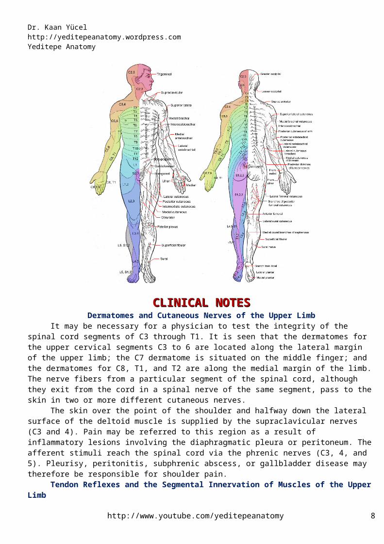
DermatomesA dermatome is an area of skin that is mainly supplied by a single spinal nerve. There are eight cervical nerves (C1 being an exception with no dermatome), twelve thoracic nerves, five lumbar nerves and five sacral nerves. Each of these nerves relays sensation (including pain) from a particular region of skin to the brain.
CLINICAL NOTESCLINICAL NOTESDermatomes and Cutaneous Nerves of the Upper Limb
It may be necessary for a physician to test the integrity of the spinal cord segments of C3 through T1. It is seen that the dermatomes for the upper cervical segments C3 to 6 are located along the lateral margin of the upper limb; the C7 dermatome is situated on the middle finger; and the dermatomes for C8, T1, and T2 are along the medial margin of the limb. The nerve fibers from a particular segment of the spinal cord, although they exit from the cord in a spinal nerve of the same segment, pass to the skin in two or more different cutaneous nerves.
The skin over the point of the shoulder and halfway down the lateral surface of the deltoid muscle is supplied by the supraclavicular nerves (C3 and 4). Pain may be referred to this region as a result of
http://www.youtube.com/yeditepeanatomy 5
Dr. Kaan Yücel http://yeditepeanatomy.wordpress.com Yeditepe Anatomy
inflammatory lesions involving the diaphragmatic pleura or peritoneum. The afferent stimuli reach the spinal cord via the phrenic nerves (C3, 4, and 5). Pleurisy, peritonitis, subphrenic abscess, or gallbladder disease may therefore be responsible for shoulder pain.
Tendon Reflexes and the Segmental Innervation of Muscles of the Upper Limb
The skeletal muscle receives a segmental innervation. Most muscles are innervated by several spinal nerves and therefore by several segments of the spinal cord. A physician should know the segmental innervation of the following muscles because it is possible to test them by eliciting simple muscle reflexes in the patient: Biceps brachii tendon reflex: C5 and 6 (flexion of the elbow joint by tapping the biceps tendon) Triceps tendon reflex: C6, 7, and 8 (extension of the elbow joint by tapping the triceps tendon) Brachioradialis tendon reflex: C5, 6, and 7 (supination of the radioulnar joints by tapping the insertion of
the brachioradialis tendon)Brachial Plexus InjuriesBrachial Plexus Injuries
The roots, trunks, and divisions of the brachial plexus reside in the lower part of the posterior triangle of the neck, whereas the cords and most of the branches of the plexus lie in the axilla. Complete lesions involving all the roots of the plexus are rare. Incomplete injuries are common and are usually caused by traction or pressure; individual nerves can be divided by stab wounds.
Upper Lesions of the Brachial Plexus (Erb-Duchenne Palsy)Upper lesions of the brachial plexus are injuries resulting from excessive displacement of the head to the
opposite side and depression of the shoulder on the same side. This causes excessive traction or even tearing of C5 and 6 roots of the plexus. It occurs in infants during a difficult delivery or in adults after a blow to or fall on the shoulder. The suprascapular nerve, the nerve to the subclavius, and the musculocutaneous and axillary nerves all possess nerve fibers derived from C5 and 6 roots and will therefore be functionless. The following muscles will consequently be paralyzed: supraspinatus (abductor of the shoulder) and infraspinatus (lateral rotator of the shoulder); subclavius (depresses the clavicle); biceps brachii (supinator of the forearm, flexor of the elbow, weak flexor of the shoulder), and greater part of the brachialis (flexor of the elbow), and the coracobrachialis (flexor of the shoulder); and the deltoid (abductor of the shoulder) and the teres minor (lateral rotator of the shoulder). Thus, the limb will hang limply by the side, medially rotated by the unopposedsternocostal part of the pectoralis major; the forearm will be pronated because of loss of the action of the biceps. The position of the upper limb in this condition has been likened to that of a porter or waiter hinting for a tip (waiter’s tip position). In addition, there will be a loss of sensation down the lateral side of the arm.
http://www.birthinjury.org/brachial-plexus-types-of-injuries.htmlLower Lesions of the Brachial Plexus (Klumpke Palsy)
http://www.youtube.com/yeditepeanatomy 6

Dr. Kaan Yücel http://yeditepeanatomy1.wordpress.com Yeditepe Anatomy
Lower lesions of the brachial plexus are usually traction injuries caused by excessive abduction of the arm, as occurs in the case of a person falling from a height clutching at an object to save himself or herself. The first thoracic nerve is usually torn. The nerve fibers from this segment run in the ulnar and median nerves to supply all the small muscles of the hand. The hand has a clawed appearance caused by hyperextensionof the metacarpophalangeal joints and flexion of the interphalangeal joints. The extensor digitorum is unopposed by the lumbricals and interossei and extends the metacarpophalangeal joints; the flexor digitorum superficialis and profundus are unopposed by the lumbricals and interossei and flex the middle and terminal phalanges, respectively.In addition, loss of sensation will occur along the medial side of the arm. If the eighth cervical nerve is also damaged, the extent of anesthesia will be greater and will involve the medial side of the forearm, hand, and medial two fingers.
Lower lesions of the brachial plexus can also be produced by the presence of a cervical rib or malignantmetastases from the lungs in the lower deep cervical lymph nodes.
Characteristic upper extremity position in Klumpke’s palsy.http://pediatric-orthopedics.org/orthopedic-conditions-from-birth-to-walking/24-birth-palsies-brachial-plexus-injuries.htmlCompression of the Brachial Plexus, Subclavian Artery, and Subclavian Vein by
the ClavicleThe interval between the clavicle and the first rib in some patients may become narrowed and thus is
responsible for compression of nerves and blood vessels.Long Thoracic Nerve Injuries
The long thoracic nerve, which arises from C5, 6, and 7 and supplies the serratus anterior muscle, can be injured by blows to or pressure on the posterior triangle of the neck or during the surgical procedure of radical mastectomy. Paralysis of the serratus anterior results in the inability to rotate the scapula during the movement of abduction of the arm above a right angle. The patient therefore experiences difficulty in raising the arm above the head. The vertebral border and inferior angle of the scapula will no longer be kept closely applied to the chest wall and will protrude posteriorly, a condition known as “winged scapula”.
http://www.coreconcepts.com.sg/mcr/scapula-winging/There is also a video in the link.
Axillary Nerve InjuriesThe axillary nerve, which arises from the posterior cord of the brachial plexus (C5 and 6), can be
http://www.youtube.com/yeditepeanatomy 7
Dr. Kaan Yücel http://yeditepeanatomy.wordpress.com Yeditepe Anatomy
injured by the pressure of a badly adjusted crutch pressing upward into the armpit. The passage of the axillary nerve backward from the axilla through the quadrangular space makes it particularly vulnerable here to downward displacement of the humeral head in shoulder dislocations or fractures of the surgical neck of the humerus. Paralysis of the deltoid and teres minor muscles results. The cutaneous branches of the axillary nerve, including the upper lateral cutaneous nerve of the arm, are functionless, and consequently there is a loss of skin sensation over the lower half of the deltoid muscle. The paralyzed deltoid wastes rapidly and the underlying greater tuberosity can be readily palpated. Because the supraspinatus is the only other abductor of the shoulder, this movement is much impaired. Paralysis of the teres minor is not recognizable clinically.
Radial Nerve InjuriesThe radial nerve is commonly damaged in the axilla and in the spiral groove.
Injuries to the Radial Nerve in the AxillaIn the axilla the nerve can be injured by the pressure of the upper end of a badly fitting crutch pressing
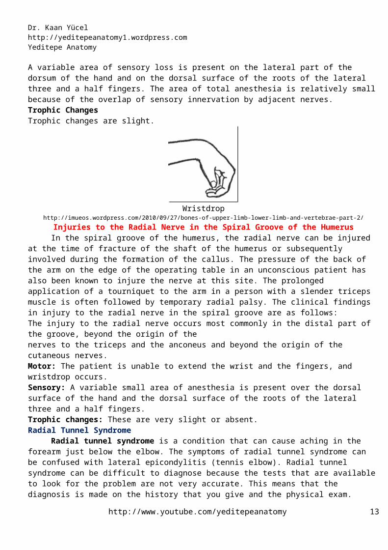
up into the armpit or by a drunkard falling asleep with one arm over the back of a chair. It can also be badly damaged in the axilla by fractures and dislocations of the proximal end of the humerus. When the humerus is displaced downward in dislocations of the shoulder, the radial nerve, which is wrapped around the back of the shaft of the bone, is pulled downward, stretching the nerve in the axilla excessively. The clinical findings in injury to the radial nerve in the axilla are as follows.MotorThe triceps, the anconeus, and the long extensors of the wrist are paralyzed. The patient is unable to extend the elbow joint, the wrist joint, and the fingers. Wristdrop, or flexion of the wrist, occurs as a result of the action of the unopposed flexor muscles of the wrist. Wristdrop is very disabling because one is unable to flex the fingers strongly for the purpose of firmly gripping an object with the wrist fully flexed. (Try it on yourself.) If the wrist and proximal phalanges are passively extended by holding them in position with the opposite hand, the middle and distal phalanges of the fingers can be extended by the action of the lumbricals and interossei, which are inserted into the extensor expansions. The brachioradialis and supinator muscles are also paralyzed, but supination is still performed well by the biceps brachii.SensoryA small loss of skin sensation occurs down the posterior surface of the lower part of the arm and down a narrow strip on the back of the forearm. A variable area of sensory loss is present on the lateral part of the dorsum of the hand and on the dorsal surface of the roots of the lateral three and a half fingers. The area of total anesthesia is relatively small because of the overlap of sensory innervation by adjacent nerves. Trophic ChangesTrophic changes are slight.
Wristdrophttp://imueos.wordpress.com/2010/09/27/bones-of-upper-limb-lower-limb-and-vertebrae-part-2/
Injuries to the Radial Nerve in the Spiral Groove of the HumerusIn the spiral groove of the humerus, the radial nerve can be injured at the time of fracture of the shaft of
the humerus or subsequently involved during the formation of the callus. The pressure of the back of the arm on the edge of the operating table in an unconscious patient has also been known to injure the nerve at this site. The prolonged application of a tourniquet to the arm in a person with a slender triceps muscle is often followed by temporary radial palsy. The clinical findings in injury to the radial nerve in the spiral groove are as follows:The injury to the radial nerve occurs most commonly in the distal part of the groove, beyond the origin of thenerves to the triceps and the anconeus and beyond the origin of the cutaneous nerves.Motor: The patient is unable to extend the wrist and the fingers, and wristdrop occurs.
http://www.youtube.com/yeditepeanatomy 8
Dr. Kaan Yücel http://yeditepeanatomy1.wordpress.com Yeditepe Anatomy
Sensory: A variable small area of anesthesia is present over the dorsal surface of the hand and the dorsal surface of the roots of the lateral three and a half fingers.Trophic changes: These are very slight or absent.Radial Tunnel Syndrome
Radial tunnel syndrome is a condition that can cause aching in the forearm just below the elbow. The symptoms of radial tunnel syndrome can be confused with lateral epicondylitis (tennis elbow). Radial tunnel syndrome can be difficult to diagnose because the tests that are available to look for the problem are not very accurate. This means that the diagnosis is made on the history that you give and the physical exam.The radial nerve actually starts at the side of the neck, where the individual nerve roots exit the spine through small openings between the vertebra called foramen. The nerve roots then join together to form three main nerves that travel down the arm to the hand. The radial nerve is one of those nerves. The radial nerve runs behind the arm and crosses the elbow on the outside as it travels down the forearm into the hand. At the outside (lateral) portion of the elbow, the radial nerve travels in a tunnel that is formed by the surrounding muscles and bone. The nerve actually runs below the muscle that that allows you to twist the hand clockwise, like when you try to use a screwdriver to tighten a screw. This muscle is called the supinator muscle.
The symptoms of radial tunnel syndrome include tenderness and pain at the lateral side (outside) of the elbow. Although the cause is different, the symptoms of radial tunnel syndrome are very similar to lateral epicondylitis, or tennis elbow. The symptoms of radial tunnel syndrome get worse with using the arm - just like tennis elbow. The pain is on the outside of the elbow - just like tennis elbow. The one difference is thatthe place where the elbow is most tender is slightly different. More @ http://www.kwoc.net/radial%20tunnel%20syndrome.pdfTennis Elbow“Tennis elbow” is a misnomer for a condition that occurs frequently on the lateral condyle of the elbow. There are many causes for this condition, and it is generally accepted that tennis accounts for only 5% of these painful elbows. “Tennis elbow” is now a well-established diagnosis, and its causative pathology has been well defined. Its incidence is common and it has many causes, among which only occasionally is the game of tennis.The basic anatomical cause is sudden and often repeated use of the forearm extensor muscles, which previously had not been much used. Anatomically the muscle involved is the extensor carpi radialis brevis (ECRB) and not the pronator teres, as was suggested in the late 1800s. ECRB arises from the lateral epicondyle of the humerus, passing distally with its neighbor extensor carpi radialis longus. The longus inserts into the base of the second metacarpal, while the brevis largely inserts into the base of the third or central metacarpal of the hand. It is thus the powerful midline extensor of the hand.
Whatever the cause of this overuse injury, tennis elbow usually presents as a small area of chronic pain on the lateral aspect of the elbow. Other characteristic symptoms are pain on wrist extension, pain when shaking hands, and frequently a weakened grip. Even lifting a cup of coffee can precipitate pain. Often there is not one specific incident that produces the symptoms; repeated use of the hand will maintain and often increase the discomfort. An abrupt onset of symptoms is uncommon. In a first occurrence, the pain usually gets worse for several weeks and even months; it may even radiate down the forearm. Work or recreation may commence the condition. Although it can occur at any age, tennis elbow is said to be more common between 30 and 50 years of age.More @ Flatt AE. Tennis elbow. Proc (Bayl Univ Med Cent). 2008 Oct;21(4):400-2.http://www.ncbi.nlm.nih.gov/pmc/articles/PMC2566914/?tool=pubmedIs it Tennis Elbow or Radial Tunnel?http://www.dynamicchiropractic.com/mpacms/dc/article.php?id=38691
Injuries to the Deep Branch of the Radial Nerve
http://www.youtube.com/yeditepeanatomy 9

Dr. Kaan Yücel http://yeditepeanatomy.wordpress.com Yeditepe Anatomy
The deep branch of the radial nerve is a motor nerve to the extensor muscles in the posterior compartment of the forearm. It can be damaged in fractures of the proximal end of the radius or during dislocation of the radial head. The nerve supply to the supinator and the extensor carpi radialis longus will be undamaged, and because the latter muscle is powerful, it will keep the wrist joint extended, and wristdrop will not occur. No sensory loss occurs because this is a motor nerve.
Injuries to the Superficial Radial NerveDivision of the superficial radial nerve, which is sensory, as in a stab wound, results in a variable small area of anesthesia over the dorsum of the hand and the dorsal surface of the roots of the lateral three and a half fingers.
Musculocutaneous Nerve InjuriesThe musculocutaneous nerve is rarely injured because of its protected position beneath the biceps brachii muscle. If it is injured high up in the arm, the biceps and coracobrachialis are paralyzed and the brachialis muscle is weakened (the latter muscle is also supplied by the radial nerve). Flexion of the forearm at the elbow joint is then produced by the remainder of the brachialis muscle and the flexors of the forearm. When the forearm is in the prone position, the extensor carpi radialis longus and the brachioradialis muscles assist in flexion of the forearm. There is also sensory loss along the lateral side of the forearm. Wounds or cuts of the forearm can sever the lateral cutaneous nerve of the forearm, a continuation of the musculocutaneous nerve beyond the cubital fossa, resulting in sensory loss along the lateral side of the forearm.
Median Nerve InjuriesFrom a clinical standpoint, the median nerve is injured occasionally in the elbow region in supracondylar fractures of the humerus. It is most commonly injured by stab wounds or broken glass just proximal to the flexor retinaculum;here it lies in the interval between the tendons of the flexor carpi radialis and flexor digitorum superficialis, overlapped by the palmaris longus. The clinical findings in injury to the median nerve are as follows.
Injuries to the Median Nerve at the ElbowMotor
The pronator muscles of the forearm and the long flexor muscles of the wrist and fingers, with the exception of the flexor carpi ulnaris and the medial half of the flexor digitorum profundus, will be paralyzed. As a result, the forearm is kept in the supine position; wrist flexion is weak and is accompanied by adduction. The latter deviation is caused by the paralysis of the flexor carpi radialis and the strength of the flexor carpi ulnaris and the medial half of the flexor digitorum profundus. No flexion is possible at the interphalangeal joints of the index and middle fingers, although weak flexion of the metacarpophalangeal joints of these fingers is attempted by the interossei. When the patient tries to make a fist, the index and to a lesser extent the middle fingers tend to remain straight, whereas the ring and little fingers flex . The latter two fingers are, however, weakened by the loss of the flexor digitorum superficialis.
Flexion of the terminal phalanx of the thumb is lost because of paralysis of the flexor pollicis longus. The muscles of the thenar eminence are paralyzed and wasted so that the eminence is flattened. The thumb is laterally rotated and adducted. The hand looks flattened and “ape-like.”SensorySkin sensation is lost on the lateral half or less of the palm of the hand and the palmar aspect of the lateral three and a half fingers. Sensory loss also occurs on the skin of the distal part of the dorsal surfaces of the lateral three and a half fingers. The area of total anesthesia is considerably less because of the overlap of adjacent nerves.Vasomotor ChangesThe skin areas involved in sensory loss are warmer and drier than normal because of the arteriolar dilatation and absence of sweating resulting from loss of sympathetic control.Trophic ChangesIn long-standing cases, changes are found in the hand and fingers. The skin is dry and scaly, the nails crack easily, and atrophy of the pulp of the fingers is present.
Injuries to the Median Nerve at the WristMotor: The muscles of the thenar eminence are paralyzed and wasted so that the eminence becomes flattened. The thumb is laterally rotated and adducted. The hand looks flattened and “ape-like.” Opposition movement of the thumb is impossible. The first two lumbricals are paralyzed, which can be recognized clinically when the
http://www.youtube.com/yeditepeanatomy 10

Dr. Kaan Yücel http://yeditepeanatomy1.wordpress.com Yeditepe Anatomy
patient is asked to make a fist slowly, and the index and middle fingers tend to lag behind the ring and little fingers. Sensory, vasomotor, and trophic changes: These changes are identical to those found in the elbow lesions.
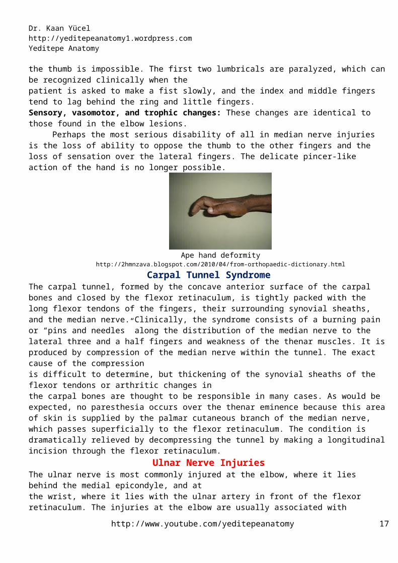
Perhaps the most serious disability of all in median nerve injuries is the loss of ability to oppose the thumb to the other fingers and the loss of sensation over the lateral fingers. The delicate pincer-like action of the hand is no longer possible.
Ape hand deformityhttp://2hmnzava.blogspot.com/2010/04/from-orthopaedic-dictionary.html
Carpal Tunnel SyndromeThe carpal tunnel, formed by the concave anterior surface of the carpal bones and closed by the flexor retinaculum, is tightly packed with the long flexor tendons of the fingers, their surrounding synovial sheaths, and the median nerve. Clinically, the syndrome consists of a burning pain or “pins and needles” along the distribution of the median nerve to the lateral three and a half fingers and weakness of the thenar muscles. It is produced by compression of the median nerve within the tunnel. The exact cause of the compressionis difficult to determine, but thickening of the synovial sheaths of the flexor tendons or arthritic changes inthe carpal bones are thought to be responsible in many cases. As would be expected, no paresthesia occurs over the thenar eminence because this area of skin is supplied by the palmar cutaneous branch of the median nerve, which passes superficially to the flexor retinaculum. The condition is dramatically relieved by decompressing the tunnel by making a longitudinal incision through the flexor retinaculum.
Ulnar Nerve InjuriesThe ulnar nerve is most commonly injured at the elbow, where it lies behind the medial epicondyle, and atthe wrist, where it lies with the ulnar artery in front of the flexor retinaculum. The injuries at the elbow are usually associated with fractures of the medial epicondyle. The superficial position of the nerve at the wrist makes it vulnerable to damage from cuts and stab wounds. The clinical findings in injury to the ulnar nerve are as follows.
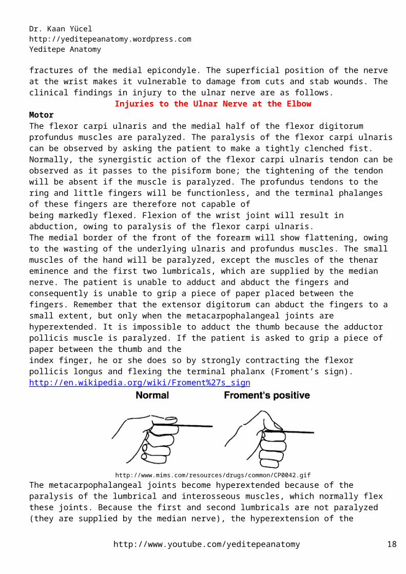
Injuries to the Ulnar Nerve at the ElbowMotorThe flexor carpi ulnaris and the medial half of the flexor digitorum profundus muscles are paralyzed. The paralysis of the flexor carpi ulnaris can be observed by asking the patient to make a tightly clenched fist. Normally, the synergistic action of the flexor carpi ulnaris tendon can be observed as it passes to the pisiform bone; the tightening of the tendon will be absent if the muscle is paralyzed. The profundus tendons to the ring and little fingers will be functionless, and the terminal phalanges of these fingers are therefore not capable ofbeing markedly flexed. Flexion of the wrist joint will result in abduction, owing to paralysis of the flexor carpi ulnaris. The medial border of the front of the forearm will show flattening, owing to the wasting of the underlying ulnaris and profundus muscles. The small muscles of the hand will be paralyzed, except the muscles of the thenar eminence and the first two lumbricals, which are supplied by the median nerve. The patient is unable to adduct and abduct the fingers and consequently is unable to grip a piece of paper placed between the fingers. Remember that the extensor digitorum can abduct the fingers to a small extent, but only when the metacarpophalangeal joints are hyperextended. It is impossible to adduct the thumb because the adductor pollicis muscle is paralyzed. If the patient is asked to grip a piece of paper between the thumb and theindex finger, he or she does so by strongly contracting the flexor pollicis longus and flexing the terminal phalanx (Froment’s sign). http://en.wikipedia.org/wiki/Froment%27s_sign
http://www.youtube.com/yeditepeanatomy 11
Dr. Kaan Yücel http://yeditepeanatomy.wordpress.com Yeditepe Anatomy
http://www.mims.com/resources/drugs/common/CP0042.gifThe metacarpophalangeal joints become hyperextended because of the paralysis of the lumbrical and interosseous muscles, which normally flex these joints. Because the first and second lumbricals are not paralyzed (they are supplied by the median nerve), the hyperextension of the metacarpophalangeal joints is most prominent in the fourth and fifth fingers. The interphalangeal joints areflexed, owing again to the paralysis of the lumbrical and interosseous muscles, which normally extend these joints through the extensor expansion. The flexion deformity at the interphalangeal joints of the fourth and fifth fingers is obvious because the first and second lumbrical muscles of the index and middle fingers are not paralyzed. In longstanding cases the hand assumes the characteristic “claw” deformity (main en griffe). Wasting of the paralyzed muscles results in flattening of the hypothenar eminence and loss of the convex curve to the medial border of the hand. Examination of the dorsum of the hand will show hollowing between the metacarpal bones caused by wasting of the dorsal interosseous muscles.SensoryLoss of skin sensation will be observed over the anterior and posterior surfaces of the medial third of the hand and the medial one and a half fingers.Vasomotor ChangesThe skin areas involved in sensory loss are warmer and drier than normal because of the arteriolar dilatation and absence of sweating resulting from loss of sympathetic control. Injuries to the Ulnar Nerve at the WristMotor: The small muscles of the hand will be paralyzed and show wasting, except for the muscles of the thenareminence and the first two lumbricals. The clawhand is much more obvious in wrist lesions because the flexor digitorum profundus muscle is not paralyzed, and marked flexion of the terminal phalanges occurs.Sensory: The main ulnar nerve and its palmar cutaneous branch are usually severed; the posterior cutaneousbranch, which arises from the ulnar nerve trunk about 2.5 in. (6.25 cm) above the pisiform bone, is usuallyunaffected. The sensory loss will therefore be confined to the palmar surface of the medial third of the hand and the medial one and a half fingers and to the dorsal aspects of the middle and distal phalanges ofthe same fingers.Vasomotor and trophic changes: These are the same as those described for injuries at the elbow. It is important to remember that with ulnar nerve injuries, the higher the lesion is the less obvious is the clawing deformity of the hand.
Unlike median nerve injuries, lesions of the ulnar nerve leave a relatively efficient hand. The sensation over the lateral part of the hand is intact, and the pincer-like action of the thumb and index finger is reasonably good, although there is some weakness, owing to loss of the adductor pollicis.
Claw hand deformity
http://2hmnzava.blogspot.com/2010/04/from-orthopaedic-dictionary.html http://www.pathopedia-india.com/clawhand.JPG
http://www.youtube.com/yeditepeanatomy 12
Dr. Kaan Yücel http://yeditepeanatomy1.wordpress.com Yeditepe Anatomy
CLINICAL ANATOMY OF THE BRACHIAL PLEXUS BLOCKCLINICAL ANATOMY OF THE BRACHIAL PLEXUS BLOCKIn the neck, the brachial plexus occupies the lower part of the posterior triangle. It lies below and
anterior to a line connecting the cricoid cartilage of the larynx to the midpoint of the clavicle. In the axilla, thebrachial plexus and its branches are arranged withinthe axillary sheath around the axillary artery, which can bepalpated. Four techniques can be used—interscalene block, supraclavicular block, infraclavicular block, and axillary block.Interscalene BlockProcedure: With the head turned laterally and upward from the side of the block, the palpating finger can feel the groove between the scalenus anterior and the scalenus medius muscles just lateral to the sternocleidomastoid muscle. The blocking needle is inserted into the interval between the scalene muscles, and the roots of the upper part of the brachial plexus can be blocked.Supraclavicular BlockProcedure: The trunks of the brachial plexus can be blocked as they cross the first rib and enter the axilla. A blocking needle is inserted between the scalenus anterior and the scalenus medius muscles anddirected caudally behind the subclavian artery toward the upper surface of the first rib. It is here that the brachial plexus is very compact, consisting of the upper middle and lower trunks. Infraclavicular BlockProcedure: The middle of the clavicle is identified. A blocking needle is inserted 1 in. (2.5 cm) inferior to it.Anatomy of complications: The close relationship of the axillary vessels to the brachial plexus within the axillary sheath means that vessel puncture and hematoma formation may occur. Axillary BlockProcedure: With the arm abducted to an angle greater than 90°, the axillary artery within the axillary sheathmay be palpated high up in the axilla. The artery is compressed, and a blocking needle is inserted just proximal to the point of compression into the axillary sheath. The disadvantage of this approach is the difficulty in blocking the musculocutaneous nerve. The object of compressing the artery distal to the point of injection is to close off the axillary sheath distally so that the anesthetic agent may rise in the sheath to the musculocutaneous nerve.Anatomy of complications: The close relationship of the axillary vessels to the brachial plexus within the axillary sheath means that vessel puncture and hematoma formation may occur.
Musculocutaneous Nerve BlockIndications: Repair of lacerations on the lateral border of the forearmProcedure: These include the following:Brachial plexus approach: The musculocutaneous nerve trunk may be blocked with the rest of the brachialplexus. The infraclavicular or axillary approach is used; in the axillary approach great care has to be taken to ensure that the anesthetic agent rises sufficiently high in the axillary sheath to block the musculocutaneous nerve.Lateral cutaneous nerve of the forearm approach: The musculocutaneous nerve may also be blocked as itemerges between the biceps and the brachialis muscles just above the lateral epicondyle of the humerus, whereit becomes the lateral cutaneous nerve of the forearm. The needle is inserted just lateral to the tendon of the biceps muscle on a line between the two epicondyles of the humerus.
Median Nerve BlockArea of anesthesia: The skin on the lateral half of the palm, the palmar aspect of the lateral three and a halffingers, including the nail beds on the dorsum. Indications: Repair of lacerations of the palm and fingersProcedures: These include the following:Block at the elbow: With the elbow joint extended, the brachial artery can easily be palpated in the cubital fossa on the medial side of the tendon of the biceps muscle. The needle is inserted on the medial side of the brachial artery.Block at the wrist: Here the median nerve lies on the medial side of the tendons of the flexor carpi radialisand to the lateral side of the flexor digitorum superficialis; it usually lies posterior to the tendon of the palmaris longus muscle (sometimes absent).
http://www.youtube.com/yeditepeanatomy 13
Dr. Kaan Yücel http://yeditepeanatomy.wordpress.com Yeditepe Anatomy
Ulnar Nerve BlockIndications: Repair of lacerations of the hand and fingersProcedures: These involve the following:Block at the elbow: At the elbow, the ulnar nerve enters the forearm between the olecranon process of the ulnaand the medial epicondyle of the humerus. Here the nerve may be palpated and infiltrated with an anesthetic agent.Block at the wrist: At the wrist, the ulnar nerve enters the hand anterior to the flexor retinaculum and lateral to the tendons of the flexor carpi ulnaris muscle and the pisiform bone. The ulnarartery lies on the lateral side of the ulnar nerve. The needle is inserted just lateral to the flexor carpi ulnaristendon at the level of the distal transverse crease of the wrist.
Radial Nerve BlockIndications: Repair of lacerations of the hand:Procedures: These involve the following:Block at the elbow: At the elbow, the radial nerve descends anterior to the lateral epicondyle of the humerusin the interval between the brachialis and the brachioradialis muscles. With the elbow joint extended, the lateral edge of the biceps tendon is easily palpated. The needle is inserted halfway between the tendon and the tip of the lateral epicondyle, and the local anesthetic is injected at this point.Block at the wrist: Just proximal to the wrist, the superficial branch of the radial nerve lies lateral to the radialartery. The nerve leaves the artery and passes laterally and backward under the tendon of brachioradialis to reach the posterior surface of the wrist. At the level of the proximal transverse flexor crease on the lateral side of the radial artery, the nerve may be infiltrated with an anesthetic solution. Suggested links:http://download.videohelp.com/vitualis/med/uppnn.htmhttp://anatomy.uams.edu/anatomyhtml/nerves_upperlimb.htmlhttp://anatomyguy.com/path-tracing-vessels-and-nerves-of-the-upper-limbhttp://www.scrubnotes.com/2008/02/popes-blessing-vs-claw-hand.html
Axillary arteryThe axillary artery supplies the walls of the axilla and related regions. The axillary artery begins at the lateral border of the 1st rib as the continuation of the subclavian artery and ends at the inferior border of the teres major where it continues as the brachial artery. Throughout its course, the artery is closely related to the cords of the brachial plexus and their branches and is enclosed with them in a connective tissue sheath called the axillary sheath. If this sheath is traced upward into the root of the neck, it is seen to be continuous with the prevertebral fascia.The axillary artery is separated into three parts by the pectoralis minor muscle, which crosses anteriorly to the vessel: first part is proximal to pectoralis minor; medial part of pectoralis minor & lateral part of first rib second part is posterior to pectoralis minor; third part is distal to pectoralis minor;from lateral part of pectoralis minor to inferior border of teres minor. Generally, six branches arise from the axillary artery: 1 branch, the superior thoracic artery, originates from the first part;2 branches, the thoraco-acromial artery and the lateral thoracic artery, originate from the second part;3 branches, the subscapular artery, the anterior circumflex humeral artery, and the posterior circumflex humeral artery, originate from the third part.In women, branches of the lateral thoracic artery contribute to the vascular supply of the breast.
The subscapular artery, the largest branch of the axillary artery, terminates by dividing into the circumflex scapular and thoracodorsal arteries. The anterior circumflex humeral artery anastomoses with the posterior circumflex humeral artery. Along with these two arteries, the circumflex scapular artery and throcadorsal artery participate in the anastomoses around the scapula.
http://www.youtube.com/yeditepeanatomy 14

Dr. Kaan Yücel http://yeditepeanatomy1.wordpress.com Yeditepe Anatomy
http://upload.wikimedia.org/wikipedia/commons/thumb/4/4c/Axillary_branches.PNG/350px-Axillary_branches.PNGBrachial artery
The major artery of the arm, the brachial artery, is found in the anterior compartment. Beginning as a continuation of the axillary artery at the lower border of the teres major muscle, it terminates just distal to the elbow joint, opposite to the neck of the radius where it divides into the radial and ulnar arteries. The brachial artery, relatively superficial and palpable throughout its course, lies anterior to the triceps and brachialis. As it passes inferolaterally, the brachial artery accompanies the median nerve.
In the proximal arm, the brachial artery lies on the medial side. In the distal arm, it moves laterally. It crosses anteriorly to the elbow joint. In proximal regions, the brachial artery can be compressed against the medial side of the humerus. Branches of the brachial artery in the arm include those to adjacent muscles and two ulnar collateral vessels (superior and inferior ulnar collateral arteries), which contribute to a network of arteries around the elbow joint. Additional branches are the profunda brachii artery and nutrient arteries to the humerus, which pass through a foramen in the anteromedial surface of the humeral shaft. The deep artery of the arm (L. arteria profunda brachii) is the largest branch of the brachial artery and has the most superior origin. It accompanies the radial nerve along the radial groove as it passes posteriorly. The deep artery terminates by dividing into middle and radial collateral arteries, which participate in the periarticular arterial anastomoses around the elbow.
Of the named branches of the brachial artery; the profunda brachii accompanies the radial nerve, whereas the superior ulnar collateral artery accompanies the ulnar nerve.
http://www.youtube.com/yeditepeanatomy 15
Dr. Kaan Yücel http://yeditepeanatomy.wordpress.com Yeditepe Anatomy
http://en.wikipedia.org/wiki/File:Gray525.png http://en.wikipedia.org/wiki/File:Gray526.pngRadial artery
The radial artery originates from the brachial artery at approximately the neck of the radius and passes along the lateral aspect of the forearm. The radial artery is the smaller of the terminal branches of the brachial artery.
In the distal forearm, the radial artery lies immediately lateral to the large tendon of the flexor carpi radialis muscle and directly anterior to the pronator quadratus muscle and the distal end of the radius. In the distal forearm, the radial artery can be located using the flexor carpi radialis muscle as a landmark. The radial pulse can be felt by gently palpating the radial artery against the underlying muscle and bone. When the brachioradialis is pulled laterally, the entire length of the artery is visible.
Branches of the radial artery originating in the forearm include: radial recurrent artery, which contributes to an anastomotic network around the elbow joint small palmar carpal branch superficial palmar branch enters the hand by passing through, or superficial to, the thenar muscles at the base of the thumb, which anastomoses with the superficial palmar arch formed by the ulnar artery. The blood supply to the hand is by the radial and ulnar arteries, which form two interconnected vascular arches (superficial and deep) in the palm. Vessels to the digits, muscles, and joints originate from the two arches and the parent arteries.
The radial artery curves around the lateral side of the wrist, passes over the floor of the anatomical snuffbox and into the deep plane of the palm by penetrating anteriorly through the back of the hand. It accesses the deep plane of the palm and forms the deep palmar arch.
The blood supply to the posterior compartment of the forearm occurs predominantly through branches of the radial, posterior interosseous, and anterior interosseous arteries. The radial artery has muscular branches, which contribute to the supply of the extensor muscles on the radial side of the forearm.The deep palmar arch passes medially through the palm between the metacarpal bones and the long flexor tendons of the digits. On the medial side of the palm, it communicates with the deep palmar branch of the ulnar artery. Before penetrating the back of the hand, the radial artery gives rise to two vessels: a dorsal carpal branch, gives rise to dorsal metacarpal arteries and the first dorsal metacarpal artery.Two vessels, the princeps pollicis artery and the radialis indicis artery, arise from the radial artery.The deep palmar arch gives rise to: three palmar metacarpal arteries
http://www.youtube.com/yeditepeanatomy 16
Dr. Kaan Yücel http://yeditepeanatomy1.wordpress.com Yeditepe Anatomy
three perforating branchesThe blood supply to the hand is by the radial and ulnar arteries, which form two interconnected vascular arches (superficial and deep) in the palm. Vessels to the digits, muscles, and joints originate from the two arches and the parent arteries.
Ulnar arteryThe ulnar artery is larger than the radial artery and passes down the medial side of the forearm. It
leaves the cubital fossa by passing deep to the pronator teres muscle, and then passes through the forearm in the fascial plane between the flexor carpi ulnaris and flexor digitorum profundus muscles. In distal regions of the forearm, the ulnar nerve is immediately medial to the ulnar artery. The ulnar artery leaves the forearm, enters the hand by passing lateral to the pisiform bone and superficial to the flexor retinaculum of the wrist, and arches over the palm. It is often the major blood supply to the medial three and one-half digits.
Pulsations of the ulnar artery can be palpated on the lateral side of the flexor carpi ulnaris tendon, where it lies anterior to the ulnar head.Branches of the ulnar artery that arise in the forearm include: 1) ulnar recurrent artery with anterior and posterior branches, which contribute to an anastomotic network of vessels around the elbow joint (The anterior and posterior ulnar recurrent arteries anastomose with the inferior and superior ulnar collateral arteries, respectively, thereby participating in the periarticular arterial anastomoses of the elbow)2) numerous muscular arteries, which supply surrounding muscles3) common interosseous artery, which divides into anterior and posterior interosseous arteries4) two small carpal arteries (dorsal carpal branch and palmar carpal branch) Perforating the interosseous membrane in the distal forearm, the anterior interosseous artery terminates by joining the posterior interosseous artery.The blood supply to the posterior compartment of the forearm occurs predominantly through branches of the radial, posterior interosseous, and anterior interosseous arteries.
Posterior interosseous arteryThe posterior interosseous artery originates in the anterior compartment from the common interosseous branch of the ulnar artery and passes into the posterior compartment of the forearm. It contributes a branch, the recurrent interosseous artery, to the vascular network around the elbow joint. The posterior interosseous artery terminates by joining the dorsal carpal arch of the wrist.
Anterior interosseous arteryThe anterior interosseous artery, also a branch of the common interosseous branch of the ulnar artery, is situated in the anterior compartment of the forearm on the interosseous membrane. The terminal end of the anterior interosseous artery joins the posterior interosseous artery.
The blood supply to the hand is by the radial and ulnar arteries, which form two interconnected vascular arches (superficial and deep) in the palm. Vessels to the digits, muscles, and joints originate from the two arches and the parent arteries.The ulnar artery and ulnar nerve enter the hand on the medial side of the wrist. Distally, the ulnar artery swings laterally across the palm, forming the superficial palmar arch, which is superficial to the long flexor tendons of the digits and just deep to the palmar aponeurosis. On the lateral side of the palm, the arch communicates with a palmar branch of the radial artery. One branch of the ulnar artery in the hand is the deep palmar branch. It anastomoses with the deep palmar arch derived from the radial artery. Branches from the superficial palmar arch include: a palmar digital artery three large, common palmar digital arteries
Axillary veinThe axillary vein begins at the lower margin of the teres major muscle and is the continuation of the
basilic vein, which is a superficial vein that drains the posteromedial surface of the hand and forearm and penetrates the deep fascia in the middle of the arm. The veins of the axilla are more abundant than the arteries,
http://www.youtube.com/yeditepeanatomy 17
Dr. Kaan Yücel http://yeditepeanatomy.wordpress.com Yeditepe Anatomy
are highly variable, and frequently anastomose. Tributaries of the axillary vein generally follow the branches of the axillary artery. Other tributaries include brachial veins that follow the brachial artery, and the cephalic vein which is a superficial vein that drains the lateral and posterior parts of the hand, the forearm, and the arm.
Two sets of veins of the arm, superficial and deep, anastomose freely with each other. The superficial veins are in the subcutaneous tissue, and the deep veins accompany the arteries. Both sets of veins have valves, but they are more numerous in the deep veins than in the superficial veins.The two main superficial veins of the arm, the cephalic and basilic veins. The cephalic vein ascends in the superficial fascia on the lateral side of the biceps and drains into the axillary vein.Paired deep veins, collectively constituting the brachial vein, accompany the brachial artery. Their frequent connections encompass the artery, forming an anastomotic network within a common vascular sheath. The pulsations of the brachial artery help move the blood through this venous network.The brachial vein begins at the elbow by union of the accompanying veins of the ulnar and radial arteries and ends by merging with the basilic vein to form the axillary vein. Not uncommonly, the deep veins join to form one brachial vein during part of their course.
The superficial veins of the forearm lie in the superficial fascia. The cephalic vein arises from the lateral side of the dorsal venous arch on the back of the hand and winds around the lateral border of the forearm; it then ascends into the cubital fossa and up the front of the arm on the lateral side of the biceps. It terminates in the axillary vein in the deltopectoral triangle.
The basilic vein arises from the medial side of the dorsal venous arch on the back of the hand and winds around the medial border of the forearm; it then ascends into the cubital fossa and up the front of the arm on the medial side of the biceps. Its terminates, by joining the venae comitantes of the brachial artery to form the axillary vein. The median cubital vein, a branch of the cephalic vein in the cubital fossa, runs upward and medially and joins the basilic vein. The basilic vein also receives a variable number of tributaries from the medial and posterior surfaces of the upper limb.
Deep veins accompanying arteries are plentiful in the forearm. These accompanying veins (L. venae comitantes) arise from the anastomosing deep venous palmar arch in the hand. From the lateral side of the arch, paired radial veins arise and accompany the radial artery; from the medial side, paired ulnar veins arise and accompany the ulnar artery. The veins accompanying each artery anastomose freely with each other. The radial and ulnar veins drain the forearm but carry relatively little blood from the hand.Deep veins of the anterior compartment drain into brachial veins associated with the brachial artery in the cubital fossa.
Deep veins of the posterior compartment generally accompany the arteries. They ultimately drain into brachial veins associated with the brachial artery in the cubital fossa. As generally found in the upper limb, the hand contains interconnected networks of deep and superficial veins. The deep veins follow the arteries; the superficial veins drain into a dorsal venous network on the back of the hand over the metacarpal bones.
CLINICAL NOTESCLINICAL NOTESArterial Innervation and Raynaud’s Disease
The arteries of the upper limb are innervated by sympathetic nerves. The preganglionic fibers originate from cell bodies in the second to eighth thoracic segments of the spinal cord. They ascend in the sympathetic trunk and synapse in the middle cervical, inferior cervical, first thoracic,or stellate ganglia. The postganglionic fibers join the nerves that form the brachial plexus and are distributed to the arteries within the branches of the plexus. For example, the digital arteries of the fingers are supplied by postgan glionic sympathetic fibers that run in the digital nerves. Vasospastic diseases involving digital arterioles, such as Raynaud’s disease, may require a cervicodorsal preganglionic sympathectomy to prevent necrosis of the fingers. The operation is followed by arterial vasodilatation, with consequent increased blood flow to the upper limb.
Aneurysm of Axillary ArteryThe first part of the axillary artery may enlarge (aneurysm of the axillary artery) and compress the trunks of the brachial plexus, causing pain and anesthesia (loss of sensation) in the areas of the skin supplied by the affected nerves.
Spontaneous Thrombosis of the Axillary VeinSpontaneous thrombosis of the axillary vein occasionally occurs after excessive and unaccustomed movements of the arm at the shoulder joint.
http://www.youtube.com/yeditepeanatomy 18


![Muscle Innervation Chart II[1]](https://img.dokumen.tips/doc/110x75/55241db64a7959da488b45f0/muscle-innervation-chart-ii1.jpg)