Embed Size (px)
Citation preview

STUDIES IN THE DYNAMICS OF HISTOGENESIS.
II. TENSION OF DIFFERENTIAL GROWTII AS A STIMULUS TO
MYOGENESIS IN TIKE ESOPHAGUS.
BY EBEN J. CAREY.
(From the Department of Anatomy, College of Medicine, Marq~e~ University Milwaukee.)
(Received for publication, April 3, 1920.)
INTRODUCTION.
Facts presented, for the first time, in a previous publication by the writer (1919-20) prove that the developing descending colon of the pig embryo possesses two zones of differential growth which by their interaction mutually influence each other during the formation period. The inner epithelial tube is the dominant, most active re- gion of growth. I t presents numerous mitotic figures which pursue a path cephalad, primarily, in the manner of a left-handed helix. The outer mesenchymal zone is less active in growth and early in develop- ment is composed of a uniform mass of undifferentiated cells.
The inner, rapidly growing, epithelial tube practically revolves due to the rapid spiral growth of its cells. With subsequent growth an apparent ring (this is qualified apparent for in reality a close spiral is formed) of smooth muscle myoblasts appears gradually near the periph- ery of the vortex.
This position taken by the inner, close spiral, smooth muscle coat at some distance from the epithelial tube is dependent upon an op- timum tension. The attitude heretofore taken by embryologists eliminated the search for the underlying cause of the first formed, inner, muscle coat The theory of self-differentiation excluded in- terpretation as regards myogenesis. A certain mesenchymal cell, re- gardless of position, was considered as destined to become a myoblast.
This highly differentiated tissue is now considered to self-dextelop, for so called muscle-forming elements have been identified in the
61
The Journal of General Physiology
on January 13, 2019jgp.rupress.org Downloaded from http://doi.org/10.1085/jgp.3.1.61Published Online: 20 September, 1920 | Supp Info:

62 DYNAMICS OF HISTOGENESIS. I I
o ~ m (Conklin; Wilson). B y the exclusion of this element in sectioning experiments the subsequent, positional, environmental relation is as much destroyed as the absence of the myoplasm. I t may well be that the yellow pigmented zone is destined to assume a certain subsequent relation in development. This position may necessitate subjection to an optimum tensional stress stimulus due to the differential growth. The conclusion of self-differentiation is consequently unwarranted and too broad. All the work tending to support the generalization that muscle self-differentiates excludes the inner, environmental stimulus--the stretching or tensional stimulus of differential growth. Has the isolated myoplasm been cultured and found to form muscle? Only the affirmative answer to this question will warrant the assertion that muscles self-develop and then only providing the exclusion of surrounding germ plasm has been accom- plished so as to exclude totally tensional stresses of differential growth of relational parts.
The last statement is made due to the fact that W. H. Lewis came to the conclusion that muscle self-differentiates from experiments in which transplantation of tissues around the otic capsule of tad- poles was performed. That musculature subsequently appears is not to be wondered at, for the potencies or actualities of differential growth were also misplaced with the transplant. If this piece re- mained viable it was bound to reveal subsequently the same tissues as in its normal location for the resultants of differential growth and the potential, mechanical stimulus due to spa(e relation were left intact.
Consequently, the potencies of a blastomere are as much a function of its position as of its material substances. The material substances receive and react to the stimulus. The stimulus is a function of posi- tion. To elicit the response of meselichymal cells in the formation of muscle tissue the proper optimum tensional stress stimulus must be applied. In tissue differentiation, therefore, the stimulus as well as the reception and response must be taken into consideration.
Tensional stresses are of various kinds and degrees. The quantity as well as the quality of stretching is important. The connective tissues are resultants of certain degrees of stresses. Muscular tissues, on the other hand, are responses to still different types of stresses.
EBEN J. CAREY 63
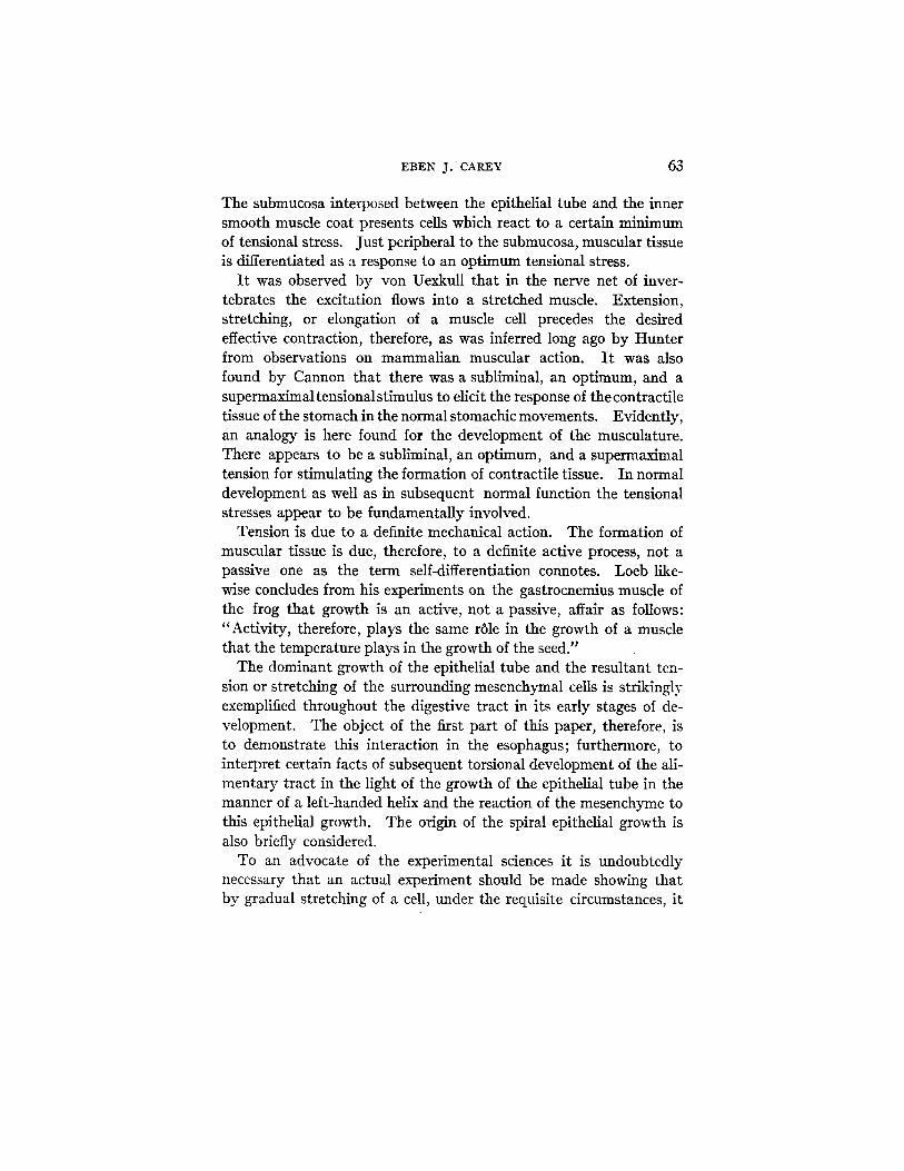
The submucosa interposed between the epithelial tube and the inner smooth muscle coat presents cells which react to a certain minimum of tensional stress. Just peripheral to the submucosa, muscular tissue is differentiated as a response to an optimum tensional stress.
I t was observed by von UexkuU that in the nerve net of inver- tebrates the excitation flows into a stretched muscle. Extension, stretching, or elongation of a muscle cell precedes the desired effective contraction, therefore, as was inferred long ago by Hunter from observations on mammalian muscular action. I t was also found by Cannon that there was a subliminal, an optimum, and a supermaximal tensionalstimulus to elicit the response of the contractile tissue of the stomach in the normal stomachic movements. Evidently, an analogy is here found for the development of the musculature. There appears to be a subliminal, an optimum, and a supermaximal tension for stimulating the formation of contractile tissue. In normal development as well as in subsequent normal function the tensional stresses appear to be fundamentally involved.
Tension is due to a definite mechanical action. The formation of muscular tissue is due, therefore, to a definite active process, not a passive one as the term self-differentiation connotes. Loeb like- wise concludes from his experiments on the gastrocnemius muscle of the frog that growth is an active, not a passive, affair as follows: "Activity, therefore, plays the same r61e in the growth of a muscle that the temperature plays in the growth of the seed."
The dominant growth of the epithelial tube and the resultant ten- sion or stretching of the surrounding mesenchymal cells is strikingly exemplified throughout the digestive tract in its early stages of de- velopment. The object of the first part of this paper, therefore, is to demonstrate this interaction in the esophagus; furthermore, to interpret certain facts of subsequent torsional development of the ali- mentary tract in the light of the growth of the epithelial tube in the manner of a left-handed helix and the reaction of the mesenchyme to this epithelial growth. The origin of the spiral epithelial growth is also briefly considered.
To an advocate of the experimental sciences it is undoubtedly necessary that an actual experiment should be made showing that by gradual stretching of a cell, under the requisite circumstances, it

64 DYNAMICS OF HISTOGENESIS. I I
is transformed into a muscle cell. To this end the writer is directing his attention. I t must not be forgotten, however, that valuable sug- gestions pointing to a tensional stimulus as a factor in myogenesis is derived from a study of the origin of this tisue in a closely graded and advancing series of embryos. In the latter case direct observation reveals what is actually going on in nature's own laboratory.
Observations on the Early Development of the Esophagus of the Pig.
Hitherto, descriptions of esophageal development were written from a view-point regarding histogenesis as passive. No correlation of the developing epithelial tube and the surrounding mesenchyme has been presented. That one element could influence the other during the critical genetic steps has been overlooked. In order to comprehend clearly the development of the esophagus, or any forma- tive structure for that matter, the active, dynamic point of view must be possessed by the observer and not the purely passive one. The interaction and interdependence of integers united in a common structure must be considered together and not as isolated, non-related entities. I t is from the dynamic aspect then that the following ob- servations differ from previous accounts of esophageal genesis. I t is also to be noted that certain developmental gaps are herein filled, thereby making the sequence of histogenetic events objectively evident.
Corresponding portions of the middle of the esophagus from an ascending series of embryos were selected for comparative study. This region was primarily chosen due to the fact that the lower cer- vical and upper thoracic portions have become narrowed due to the elongation of the esophagus concomitant with the descent of the stomach. The diminution in the diameter of this zone, leads to a more rapid revolution of the epithelial mitotic figures on account of the decrease in circumference of the epithelial tube. This more rapid rate of rotation causes a corresponding greater vortical agitation in the less active fluid like mesenchyme. The reacting maelstrom is consequently more clearly seen and the path of the activating mitotic figures more easily followed. The muscular rim bordering the mes- enchymal vortex is also more clearly defined.
EBEN J. CAGEY 65
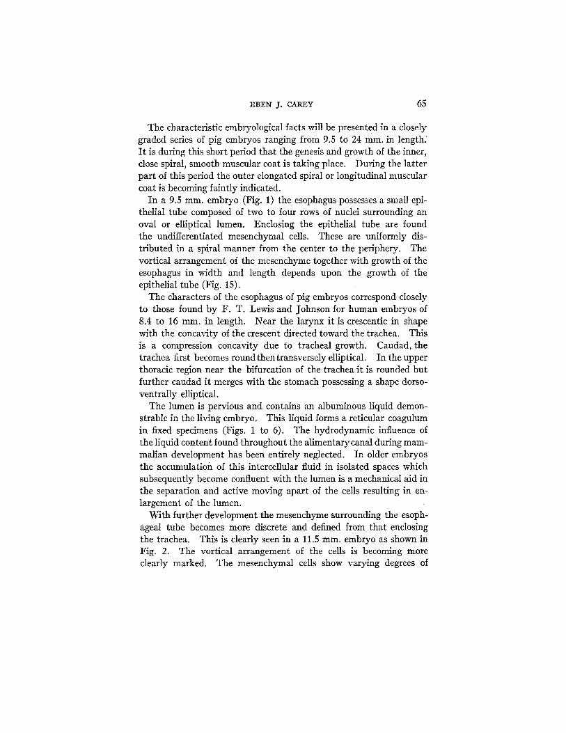
The characteristic embryological facts will be presented in a closely graded series of pig embryos ranging from 9.5 to 24 ram. in length. I t is during this short period that the genesis and growth of the inner, close spiral, smooth muscular coat is taking place. During the latter part of this period the outer elongated spiral or longitudinal muscular coat is becoming faintly indicated.
In a 9.5 ram. embryo (Fig. 1) the esophagus possesses a smal] epi- thelial tube composed of two to four rows of nuclei surrounding an oval or elliptical lumen. Enclosing the epithelial tube are found the undifferentiated mesenchymal cells. These are uniformly dis- tributed in a spiral manner from the center to the periphery. The vortical arrangement of the mesenchyme together with growth of the esophagus in width and length depends upon the growth of the epithelial tube (Fig. 15).
The characters of the esophagus of pig embryos correspond closely to those found by F. T. Lewis and Johnson for human embryos of 8.4 to 16 ram. in length. Near the larynx it is crescentic in shape with the concavity of the crescent directed toward the trachea. This is a compression concavity due to tracheal growth. Caudad, the trachea first becomes round then transversely elliptical. In the upper thoracic region near the bifurcation of the trachea it is rounded but further caudad it merges with the stomach possessing a shape dorso- ventrally elliptical.
The lumen is pervious and contains an albuminous liquid demon- strable in the living embryo. This liquid forms a zeticular coagulum in fixed specimens (Figs. 1 to 6). The hydrodynamic influence of the liquid content found throughout the alimentary canal during mam- malian development has been entirely neglected. In older embryos the accumulation of this intercellular fluid in isolated spaces which subsequently become confluent with the lumen is a mechanical aid in the separation and active moving apart of the cells resulting in en- largement of the lumen.
With further development the mesenchyme surrounding the esoph- ageal tube becomes more discrete and defined from that enclosing the trachea. This is clearly seen in a 11.5 ram. embryo as shown in Fig. 2. The vortical arrangement of the Cells is becoming more clearly marked. The mesenchymal cells show varying degrees of

66 DYNAMICS OF HISTOGENESIS. II
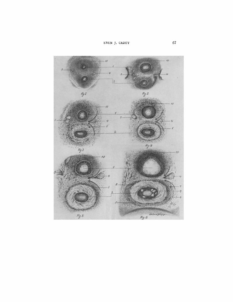
FIGS. 1 to 6. Development of the esophagus and trachea at corresponding levels through the lower cervical region of pig embryos ranging in length from 9.5 to 24 ram. (1) trachea surrounded by a nebular mass of undifferentiated mesenchyme; (2) esophagus, surrounded by a nebular mass of undifferentiated mesenchyme; lumen contains reticular coagulum; (3) left vagus nerve; (4) right vagus nerve; (5) inner close spiral muscle; (6) outer elongated spiral muscle; (7) ganglionic cells of myenteric plexus (Auerbach's plexus); (8) ganglionic cells of submucous plexus (Meissner's plexus); (9) intercellular vacuoles of esophageal epithelium; (10) mesenchyme and embryonic cartilage of the ventral aspect of the trachea.
FIG. 1. Transverse section through the lower cervical region of a 9.5 ram. pig embryo.
FIG. 2. Transverse section through the lower cervical region of an 11.5 ram. pig embryo.
FIG. 3. Transverse section through the lower cervical region of a 14 mm. pig embryo.
FIG. 4. Transverse section through the lower cervical region of a 15 ram. pig embryo.
FIG. 5. Transverse section through the lower cervical region of an 18 mm. pig embryo.
FIG. 6. Transverse section through the lower cervical region of a 24 ram. pig embryo.
The more rapid growth of the tracheal epithelial tube over that of the esopha- gus is to be especially noted. The mesenchyme around the trachea also grows at a more rapid rate than that around the esophagus. Consequently the less rapidly growing esophagea.l mesenchyme is thrown into a veritable vortex. This vortex represents a centrifugal reaction of the mesenchyme to the centripetal action of the esophageal epithelial tube. This tube is rapidly growing in the manner of a left-handed helix. At the periphery of the mesenchymal whirlpool the optimum tensional stimulus is preser~ted resulting in the histogenesis of the inner, close spiral, smooth muscle coat. The outer muscle coat is faintly detected in the esophagus of a 24 ram. pig embryo.
The mesenchyme around the trachea, due to its rapid growth is compressed. This compression results in the formation of cartilage tissue. The embryonic cartilage is becoming evident in the trachea of a 24 ram. pig embryo (Fig. 6).
The reacting mesenchyme tends to converge between the trachea and the esophagus on the right and diverge on the left. The right vagus nerve appears to be drawn between the two tubes by this action.
EBEN J. CAREY 67

68 DYNAMICS OF HISTOGENESIS. II
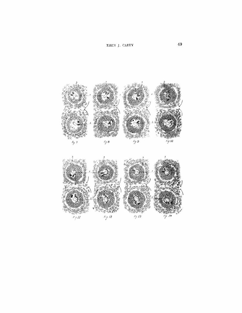
FIGS. 7 to 14. Cross-sections of the trachea and esophagus of a 9.5 ram. pig embryo. Note particularly that the mitotic figures of the trachea follow the path of a right-handed helix, whereas those of the esophagus follow the path of a left-handed helix. These two tubes, therefore, tend to rotate in opposite direc- tions in development. This is comparable to the cylindrical rollers of a printing press. The mesenchymal vortices converge on the right side, represented by arrows, and diverge on the left. This is more evident in older specimens (see Fig. 6). (1) trachea; (2) esophagus; (3) proliferation bud of the trachea. These epithelial cells appear to be thrown off by an eruption and lost in the mesenchyme. At this location the basement membrane of the epithelial tube is absent. The rapid rate of growth of the tracheal epithelial tube probably causes this eruption by reacting centrifugal force.
EBEN J. CAREY 69

70 DYNAMICS OF HISTOGENESIS. II
7388
1.338
1.288 /,~ 3 8
1./$8 1.168
. @88
.938 ,888
.788
.LSg ~ .b38
.5~8
.5M
~ .4'88
.6'Z8
.388
. ~ 8 .2,88
.2 ~8
./88
I / I /
¢"
d I I /
/ I /
o~ /
/
/ /
/
L~
I S / : / , .... ?
I
.~'" I/ :! ~¢,:~ i : i .,eol ..." / : \ , ~: / < :~.:,..-- ~;,o~
' t , . .i, i . ~ ~e"~s "
6 7 8 9 1o /1 /2 13 #/ /~ 1o 17 /8 19 20 21 ,2,Z 23.24 < Ze~o/~ a~ em$~o~,a /~ millz~nelee.~
FIO. 15. Curves of differential growth of the epithelial tube and mesen- chyme of the esophagus. Since there is no restricting peritoneal membrane in the lower cervical region of the esophagus the more rapid rate of growth of the epithelial tube over that of the mesenchyme is not as definite as that seen in the descending colon. The great extent of mesenchymal area is due to the centri- petal force of the epithelial tube. The centrifugal reaction tends to throw the cells off tangentially. Consequently, the greater area of the mesenchyme is not due to an intrinsically more rapid rate of mitosis over that found in the epithelial tube but to the radial force (centrifugal force) of the mesenchyme in reaction to the centripetal force of the epithelial tube.
EB~N J. C A ~ Y 71
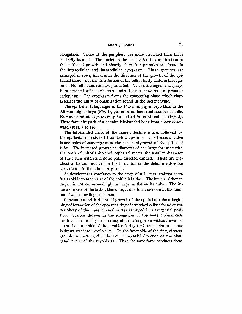
elongation. Those at the periphery are more stretched than those centrally located. The nuclei are first elongated in the direction of the epithelial growth and shortly thereafter granules are found in the intercellular and intracellular cytoplasm. These granules are arranged in rows, likewise in the direction of the growth of the epi- thelial tube. Yet the distribution of the cellsis fairly uniform through- out. No cell boundaries are presented. The entire region is a syncy- tiurn studded with nuclei surrounded by a narrow zone of granular endoplasrn. The ectoplasm forms the connecting phase which char- acterizes the unity of organization found in the mesenchyme.
The epithelial tube, larger in the 11.5 ram. pig embryo than in the 9.5 ram. pig embryo (Fig. 1), possesses an increased number of cells. Numerous mitotic figures may be plotted in serial sections (Fig. 5). These form the path of a definite left-handed helix from above down- ward (Figs. 7 to 14).
The left-handed helix of the large intestine is also followed by the epithelial mitosis but from below upwards. The ileocecal valve is one point of convergence of the helicoidal growth of the epithelial tube. The increased growth in diameter of the large intestine with the path of mitosis directed cephalad meets the smaller diameter of the ileum with its mitotic path directed caudad. These are me- chanical factors involved in the formation of the definite valve-like constrictors in the alimentary tract.
As development continues to the stage of a 14 ram. embryo there is a rapid increase in size of the epithelial tube. The lumen, although larger, is not correspondingly as large as the entire tube. The in- crease in size of the latter, therefore, is due to an increase in the num- ber of cells crowding the lumen.
Concomitant with the rapid growth of the epithelial tube a begin- ning of formation of the apparent ring of stretched cells is found at the periphery of the mesehchymal vortex arranged in a tangential posi- tion. Various degrees in the elongation of the mesenchy)nal cells are found decreasing in intensity of stretching from without inwards.
On the outer side of the myoblastic ring the intercellular substance is drawn out into myofibrill~e. On the inner side of the ring, discrete granules are arranged in the same tangential direction as the elon- gated nuclei of the myoblasts. That the same force produces these
72 DYNAMICS OF HISTOGENESIS. I I
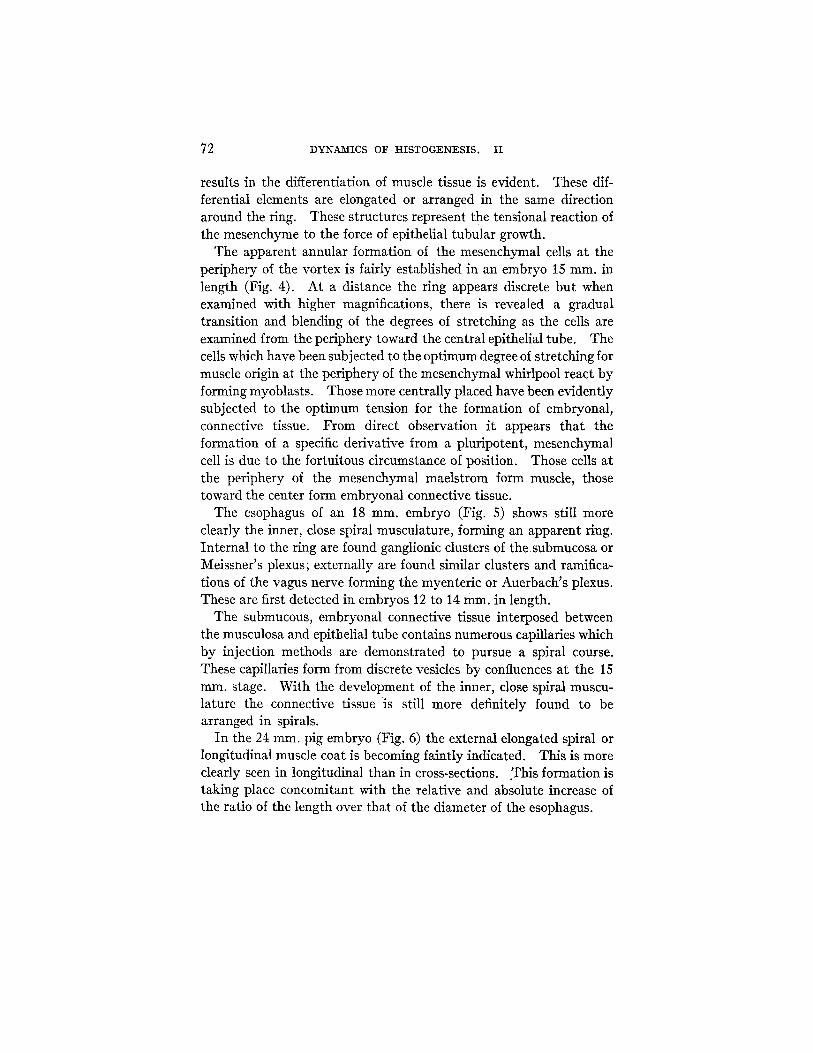
results in the differentiation of muscle tissue is evident. These dif- ferential elements are elongated or arranged in the same direction around the ring. These structures represent the tensional reaction of the mesenchyme to the force of epithelial tubular growth.
The apparent annular formation of the mesenchymal cells at the periphery of the vortex is fairly established in an embryo 15 ram. in length (Fig. 4). At a distance the ring appears discrete but when examined with higher magnifications, there is revealed a gradual transition and blending of the degrees of stretching as the cells are examined from the periphery toward the central epithelial tube. The cells which have been subjected to the opt imum degree of stretching for muscle origin at the periphery of the mesenchymal whirlpool react by forming myoblasts. Those more centrally placed have been evidently subjected to the opt imum tension for the formation of embryonal, connective tissue. From direct observation it appears that the formation of a specific derivative from a pluripotent, mesenchymal cell is due to the fortuitous circumstance of position. Those cells at the periphery of the mesenchymal maelstrom form muscle, those toward the center form embryonal connective tissue.
The esophagus of an 18 ram. embryo (Fig. 5) shows still more clearly the inner, close spiral musculature, forming an apparent ring. Internal to the ring are found ganglionic clusters of the submucosa or Meissner's plexus; externally are found similar clusters and ramifica- tions of the vagus nerve forming the myenteric or Auerbach's plexus. These are first detected in embryos 12 to 14 turn. in length.
The submucous, embryonal connective tissue interposed between the musculosa and epithelial tube contains numerous capillaries which by injection methods are demonstrated to pursue a spiral course. These capillaries form from discrete vesicles by confluences at the 15 ram. stage. With the development of the inner, close spiral muscu- lature the connective tissue is still more definitely found to be arranged in spirals.
In the 24 ram. pig embryo (Fig. 6) the external elongated spiral or longitudinal muscle coat is becoming faintly indicated. This is more clearly seen in longitudinal than in cross-sections. .This formation is taking place concomitant with the relative and absolute increase of the ratio of the length over that of the diameter of the esophagus.
EBEN J. CAREY 73
The precocious segregation of the esophageal epithelial cells would appear to be dist inctly disadvantageous, for here a much larger pro- port ion of each cell is in contact with others than in the looser texture of the mesenchyme. The intercellular vacuoles filled with fluid seen in the epithelium of the esophagus of a 24 ram. pig embryo (Fig. 6) is a provision for aiding excretion. These intercellular vacuoles are comparable to the periodic appearance of intercellular cavities ob- served by Kofoid in L i m a x .
F.'60
1 . . 5 5 l
t
_ _ , ; ' ,
~3s! .':
..... . . . . "
. . . . ,
~ I.~o . . . . . . - - ~[~ "
1,1o
6 z 8 g fo 111.~ ~3 Itp-15 16 N 18 1 9 , z o 2 1 2 , ~ 23 2~ kenya, o/ e,~},<, irl ~tili~<ole.,.5
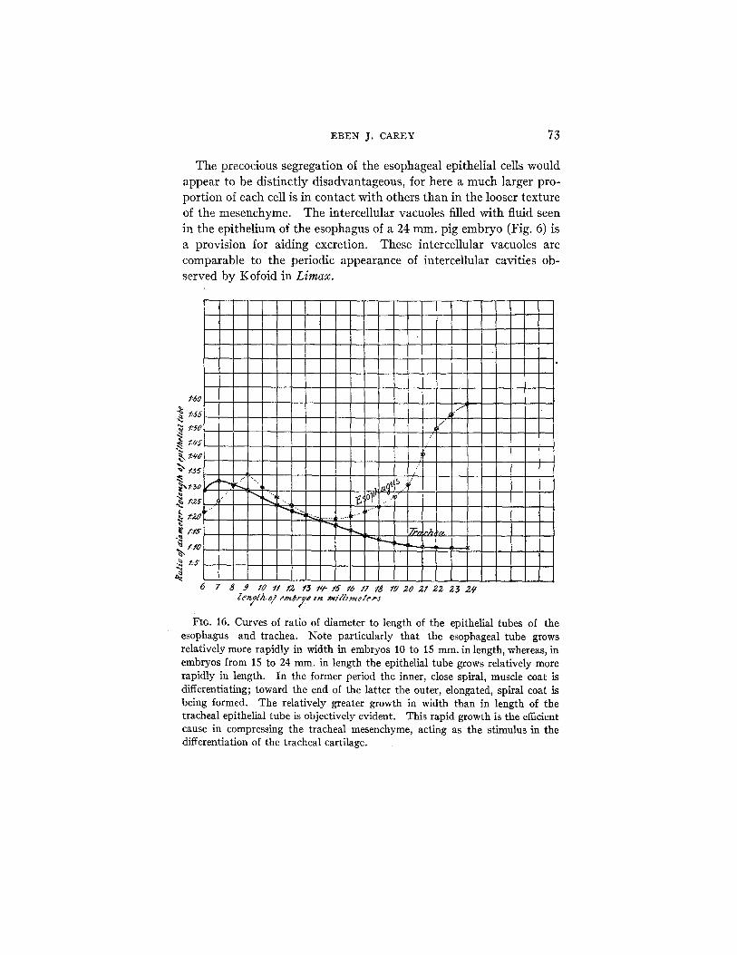
FIO. 16. Curves of ratio of diameter to length of the epithelial tubes of the esophagus and trachea. Note particularly that the esophageal tube grows relatively more rapidly in width in embryos 10 to 15 ram. in length, whereas, in embryos from 15 to 24 mm. in length the epithelial tube grows relatively more rapidly in length. In the former period the inner, close spiral, muscle coat is differentiating; toward the end of the latter the outer, elongated, spiral coat is being formed. The relatively greater growth in width than in length of the tracheal epithelial tube is objectively evident. This rapid growth is the efficient cause in compressing the tracheal mesenchyme, acting as the stimulus in the differentiation of the tracheal cartilage.

74 DYNAMICS OF HISTOGENESIS. II
I506 ~1 /
tNOO
1300 ~ /
,i
o(,t " .950 ^J 1 .gu ~ /
• -~ .g~-o .~! ~,"i
-~ 7,, ~ / / o ' 2
a ~¢ ",1" . , < ~" • 550 /I .5~0 , ~, .Oql] ~ ,'" ,' ;?¢" ~t'"
Oi • ~ ,' /.."#]..,~
• 300 "" "
.g50 t "~ /
• 2 , 00 ¢
6 7
I/ l
I | i l
/e,~/,',~ o/ e,,ta~o / n ,,,n,,,~t,.,...,
FIG. 17. Curves of differential growth of the tracheal epithelial tube and mesenchyme. The greater growth of the epithelial tube over that of the mesenchyme after the embryo has reached a length of 11 mm. is shown graphi- cally. This rapid growth causes a progressive compression of the mesenchyme acting, therefore, as the efficient stimulus in the differentiation of cartilage tissue. The tracheal embryonic cartilage appears in embryos between 18 and 24 ram. in length. Note particularly that during this period the curve of epithelial tubular growth in area rapidly ascends•

EBEN 3. CAREY 75
In the earlier stages the esophageal epithelial tube grows relatively more rapidly in diameter than in length (Figs. 16 and 17). In the 9.5 ram. embryo the esophageal epithelial tube measures 0.04 ram. in diameter and 1.3 ram. in length; in the 14 ram. embryo the diameter is 1 ram. and the length 2.1 ram. Therefore, the esophageal epithelial tube is growing relatively more rapidly in width than in length,!from the 9.5 to 14 ram. embryonic stages.
1~"o
~ .700 ,d¢O
.600
.550
~ .50o
.zeO o
~ . .Z 5 ¢
./SO
FIO. 18.
i /
y . /
/ /
/
J
. / /
/ /
)
" l r~ ' / " / , ,~ ,,,,,v ,./
/J °-
~! or" I _.~ ....
6 z 8 9 lo tl t~ ~ ~ ' 5 16 lz la m ~ o e z e ~ s e ~ I e n f h ~ e ~ t y 8 1~ ~t/J~,~ter, s
Curves of differential growth in area of the tracheal and esophageal epithelial tubes. These curves show strikingly the more rapid growth in the same unit of time of the tracheal tube over that of the esophageal tube. Note that between embryos 6 to 10 m m . in length there is an absolute decrease in area of the tube. This decrease is concomitant with the rapid descent of the stomach. This decrease in area is due to stretching as the result of traction caused by the descent of the stomach. After the stage of the 10 ram. embryo, growth in area of the esophagus rapidly progresses.
76 DYNAMICS OF HISTOGENESIS. II
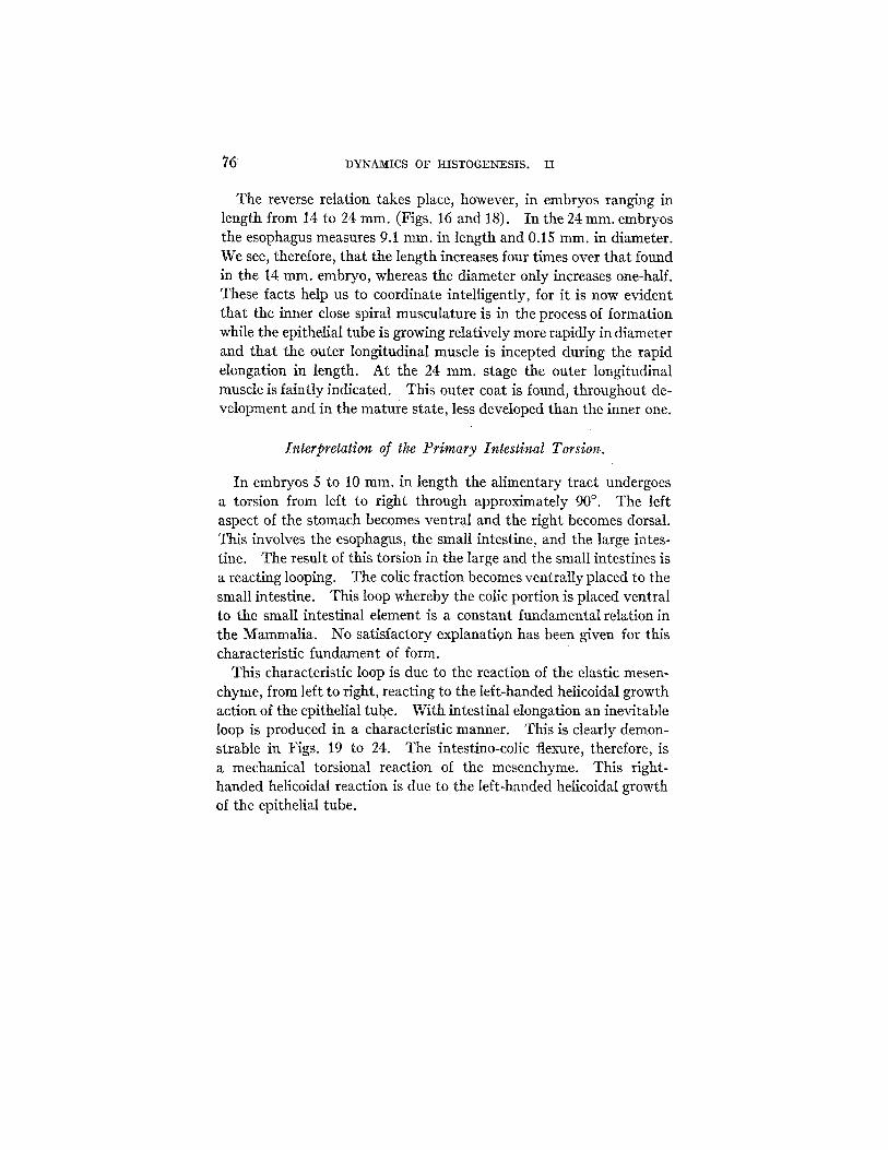
The reverse relation takes place, however, in embryos ranging in length from 14 to 24 ram. (Figs, 16 and 18). In the 24 ram. embryos the esophagus measures 9.1 ram. in length and 0.15 rain. in diameter. We see, therefore, that the length increases four times over that found in the 14 ram. embryo, whereas the diameter only increases one-half. These facts help us to coordinate intelligently, for it is now evident that the inner close spiral musculature is in the process of formation while the epithelial tube is growing relatively more rapidly in diameter and that the outer longitudinal muscle is incepted during the rapid elongation in length. At the 24 ram. stage the outer longitudinal muscle is faintly indicated. This outer coat is found, throughout de- velopment and in the mature state, less developed than the inner one.
Interpretation of the Primary Intestinal Torsion.
In embryos 5 to 10 ram. in length the alimentary tract undergoes a torsion from left to right through approximately 90 °. The left aspect of the stomach becomes ventral and the right becomes dorsal. This involves the esophagus, the small intestine, and the large intes- fine. The result of this torsion in the large and the small intestines is a reacting looping. The colic fraction becomes ventrally placed to the small intestine. This loop whereby the colic portion is placed ventral to the small intestinal element is a constant fundamental relation in the Mammalia. No satisfactory explanatign has been given for this characteristic fundament of form.
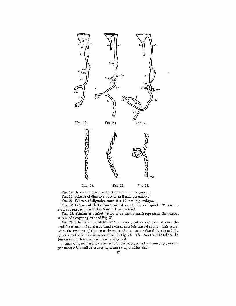
This characteristic loop is due to the reaction of the elastic mesen- chyme, from left to right, reacting to the left-handed helicoidal growth action of the epithelial tube. With intestinal elongation an inevitable loop is produced in a characteristic manner. This is clearly demon- strable in Figs. 19 to 24. The intestino-colic flexure, therefore, is a mechanical torsional reaction of the mesenchynle. This right- handed helicoidal reaction is due to the left-handed helicoidal growth of the epithelial tube.
2.. .
J Z.l •
FIc. 19. FIc. 20. FIG. 21.
- - - £ L
I 9
FIG. 22. FIc. 23. Fio. 24.
FIG. 19. Schema of digestive tract of a 6 ram. pig embryo. FIG. 20. Schema of digestive tract of an 8 mm. pig embryo. FIc. 21. Schema of digestive tract of a 10 mm. pig embryo. FIG. 22. Schema of elastic band twisted as a left-handed spiral. This repre-
sents the mesenchyme of the straight digestive tract. Fro. 23. Schema of ventral flexure of an elastic band; represents the ventral
flexure of elongating tract of Fig. 20. FIG. 24 Schema of inevitable ventral looping of caudal element over the
cephalic element of an elastic band twisted as a left-handed spiral. This repre- sents the reaction of the mesenchyme to the torsion produced by the spirally growing epithelial tube as schemafized in Fig. 21. The loop tends to relieve the torsion to which the mesenchyme is subjected.
t, trachea; e, esophagus; s, stomach; l, liver; d. p., dorsal pancreas; v.p., ventral pancreas; s.i., small intestine; c., cecum; v.d., vitelline duct.
77

I . .
a .
. .
N
FIG. 25. Diagram of the tracheM bud from the ventral aspect of the foregut. The former presents mitotic figures following the path of a right-handed helix. The latter presents mitotic figures following the path of a left-handed helix. The tracheal or lung bud is an outgrowth from the ventral aspect of the cephalic aspect of the entodermal foregut. The cause of the mitosis pur- suing a path of a right-handed helix in the trachea is due to the deflection of the left-handed, helicoidal, mitotic path of the foregut. This deflection occurs at the cephalic orifice of the tracheal epithelial tube where it arises from the floor of the entodermal tube of the foregut.
78
EBEN j. CAI~EY 79
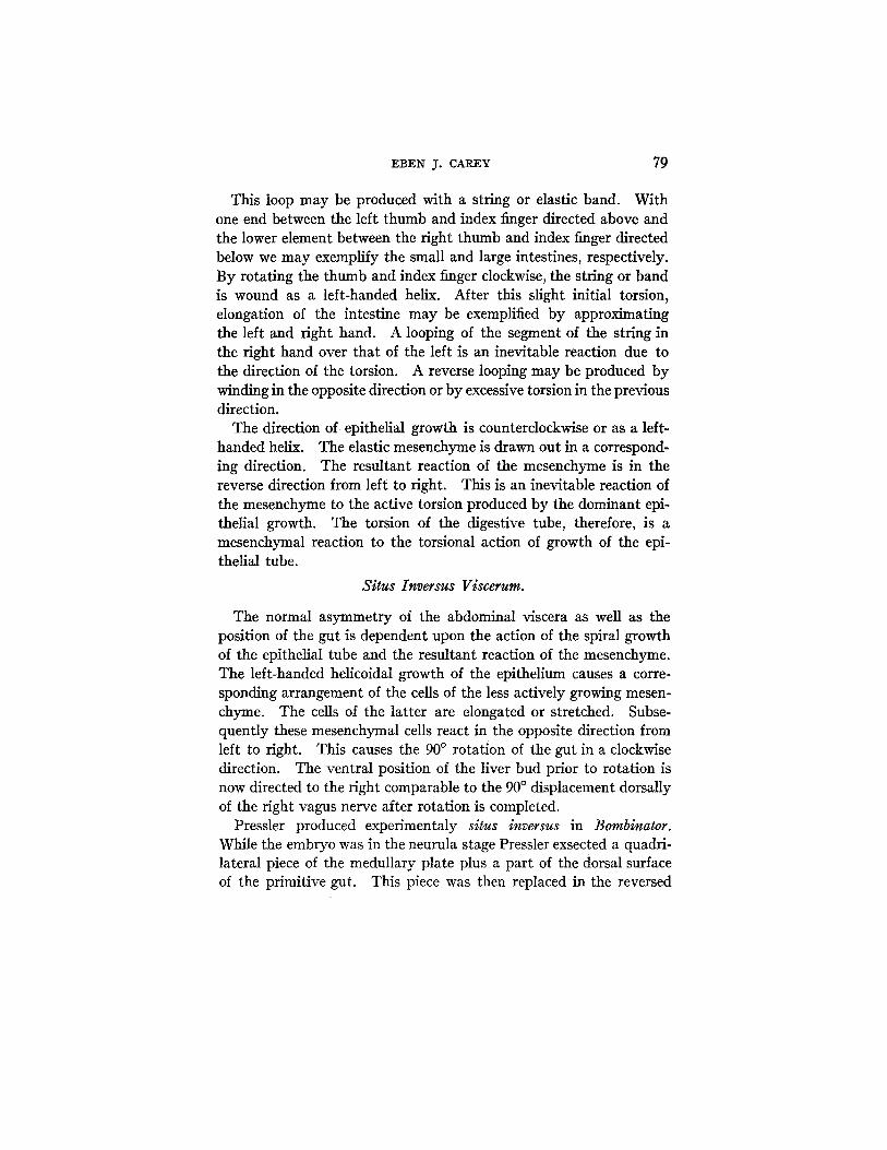
This loop may be produced with a string or elastic band. With one end between the left thumb and index finger directed above and the lower element between the right thumb and index finger directed below we may exemplify the small and large intestines, respectively. By rotating the thumb and index finger clockwise, the string or band is wound as a left-handed helix. After this slight initial torsion, elongation of the intestine may be exemplified by approximating the left and right hand. A looping of the segment of the string in the right hand over that of the left is an inevitable reaction due to the direction of the torsion. A reverse looping may be produced by winding in the opposite direction or by excessive torsion in the previous direction.
The direction of epithelial growth is counterclockwise or as a left- handed helix. The elastic mesenchyme is drawn out in a correspond- ing direction. The resultant reaction of the mesenchyme is in the reverse direction from left to right. This is an inevitable reaction of the mesenchyme to the active torsion produced by the dominant epi- thelial growth. The torsion of the digestive tube, therefore, is a mesenchymal reaction to the torsional action of growth of the epi- thelial tube.
Situs Inversus Viscerum.
The normal asymmetry of the abdominal viscera as well as the position of the gut is dependent upon the action of the spiral growth of the epithelial tube and the resultant reaction of the mesenchyme. The left-handed helicoidal growth of the epithelium causes a corre- sponding arrangement of the cells of the less actively growing mesen- chyme. The cells of the latter are elongated or stretched. Subse- quently these mesenchymal cells react in the opposite direction from left to right. This causes the 90 ° rotation of the gut in a clockwise direction. The ventral position of the liver bud prior to rotation is now directed to the right comparable to the 90 ° displacement dorsally of the right vagus nerve after rotation is completed.
Pressler produced experimentaly situs inversus in Bombinator. While the embryo was in the neurula stage Pressler exsected a quadri- lateral piece of the medullary plate plus a part of the dorsal surface of the primitive gut. This piece was then replaced in the reversed
80 DYNAI~ICS OF HISTOGENESIS. II
position so that the cephalic end was directed caudad and the caudal end was directed cephalad. Tadpoles were reared from these experi- mental embryos and in many cases a complete situs inversus viscerum was presented.
The transposition of the viscera is due to the fact that the reversed segment presents a screw-like growth action of the epithelium oppo- site to that normally present. The mesenchymal cells form a fight- handed spiral arrangement and in reacting to stretching cause a 90 ° rotation of the gut from right to left, or counterclockwise. This reversed rotation due to the torsional reaction of the mesenchyme would cause the ventrally placed liver bud to be directed to the left instead of to the right.
An excellent discussion of the views of Wilder, Bateson, and New- man as regards mirror imaging and symmetry reversal in monsters will be found in a paper by Morrill. That Morrill had a clear-cut perception of the location in which solution of the problem of nor- mal asymmetry of the viscera was to be found is seen in the follow- ing statement: " the factors controlling asymmetry are located in the primitive gut and become operative before the liver bud has developed."
Crampton suggested that asymmetry in the viscera may depend upon spiral cleavage during the first stages of development. This rested on the fact that the majority of snails possess a dextral shell associated with a right-handed spiral cleavage. Certain snails have a sinistral shell and reversed asymmetry in the viscera. This rever- sal of the viscera was associated with a left-handed spiral cleavage.
From a study of vertebrate monsters, however, Morrill correctly comes to the following conclusion: "From the evidence at hand, it seems probable that the primary cause of visceral asymmetry in verte- brates is to be sought for at the completion of cleavage rather than in the period of cleavage itself."
Origin of Spiral Growth.
The explanation of spiral epithelial growth is under investigation. Spiral growth involves two motions: a motion of rotation, circular, and a motion of straight progression, rectilinear. The latter is evi- dently imparted from growing points of mitosis. Growth would
EBEN J. CARE¥ 81
tend to pursue a straight line if the factors of resistance due to differ- ential growth did not interfere. An exception to this would be found in the spiral type of cleavage observed by Child in Arenicola cristata, and Wilson in Nerds. In these forms the rotation of the cells appears to be predetermined in the parent cells, as is proved by the position of the spindles and by the form of division. In the intestine the inner curved surface of the mesenchymal wall constantly deflects the mitotic path in a circular manner. This would impart the rotary element to the compound spiral motion of growth.
The ~elation of the pharynx to the esophagus and of the rectum to the colon is comparable to that of the wide and narrow components of a funnel. A stream running down the inclined wall of the wide ele- ment would be deflected in a rotary manner so that its progression through the narrow integer would be in the manner of a helix. The determination of a left- or right-handed helix would depend upon the initial direction of impelling the stream.
The direction of epithelial growth in a left-handed helix has a defi- nite mechanical basis in the intestine, but as yet is undetermined. The epithelial growth is roughly comparable to the stream exemplified above; i.e., the glass of the funnel to the mesenchyme. The initial impulse of the water pressure is comparable to that of growth. This causes the rectilinear motion of progression. The circular resistance of the glass is analogous to that of the mesenchyme. This causes a deflection of the rectilinear motion in a rotary manner, the two char- acterizing the helicoidal motion of the intestine.
CONCLUSIONS.
Esophageal Development.
1. The region of most active mitosis per mm. of cross-section in the esophagus is the entodermal epithelial tube. The mitotic figures follow a spiral path in the manner of a left-handed helix from the cephalic to the caudal direction.
2. The region of least active growth per ram. of cross-section in the esophagus is the mesenchyrne surrounding the epithelial tube.
3. The helicoidal activity of the epithelial tube causes a vortical reaction in the surrounding mesenchyme. The mesenchymal whirl- pool represents a reaction to the spirally growing epithelial tube.

82 DYNAMICS OF HISTOGENESIS. I I
4. In embryos 9.5 to 14 mm. in length the esophageal epithelial tube grows relatively more rapidly in width than in length. During th/s period the myoblasts which form the inner, close spiral, muscle coat of the esophagus are becoming rapidly differentiated in the outer condensed margin of the mesenchymal maelstrom.
5. The nuclei, first spherical then oval, and finally rod shaped with rounded ends, are drawn out in the direction of the circumference of the mesenchymal rim which is directed tangentially.
6. The cytoplasm is also drawn out in the direction of the mesen- chymal rim of the vortex. The elongated rows of isolated granules appear which subsequently, by confluence, form the myofibriUa~. These cytoplasmic derivatives are elongated in the direction of the circumference of the vortex.
7. Between the epithelial tube and the myoblastic rim at the peri- phery of the mesenchymal whorl is found the embryonic connective tissue. From this direct observation the conclusion is made that an optimum tensional stress stimulus is necessary to elicit the formation of muscular tissue at the circumference of the mesenchymal vortex. Consequently, the formation of a specific derivative from a pluripo- tent mesenchymal cell is due to the fortuitous circumstance of position.
8. In embryos from 14 to 24 mm. in length, the esophagus grows relatively more rapidly in length than in width. This elongation is due to two factors; first, the descent of the stomach, and, second, the resistance to diametrical growth presented by the inner close spiral musculature. The epithelial tube, still the dominant zone of mitotic activity, pursues the lines of least resistance, and consequently growth in length takes place. This is due to the shifting of the planes of cell division on account of the compression of the inner, close spiral, muscle coat.
9. The undifferentiated mesenchyme peripherad to the inner, close spiral musculature is elongated and the histogenetic changes in mus- cular formation are gradually taking place between 14 and 24 mm. A very attenuated, outer, elongated, spiral, or longitudinal muscle coat is detected in the esophagus of a 24 mm. pig embryo.
10. The characteristic intesfino-colic flexure is a torsional reaction of the mesenchyme. The mesenchymal cells are thrown into a left- handed helicoidal series, corresponding to the activity in the epithelial
~EBEN J. CAREY 83
tube. The right-handed helicoidal reaction of the mesenchyme, therefore, is due to the left-handed helicoidal growth of the epithelial tube.
11. The normal asymmetry of the abdominal viscera as well as the position of the gut is dependent upon the clockwise reaction of the stretched mesenchymal cell. These cells are stretched by the left- handed helicoidal growth of the epithelial tube. One factor producing situs inversus viscerum could be the reversal of the spiral growth of the epithelial tube resulting in a reaction of the mesenchyme in a direction opposite, namely counterclockwise, to that which occurs normally.
BIBLIOGRAPHY.
Bateson, W., Problems of genetics, New Haven, 1916. Cannon, W. B., Am. J. Physiol., 1911-12, xxix, 255. Carey, E. J., J. Gen. Physiol., 1919-20, ii, 357. Child, C. M., Zool. Bull., 1897, i, 125. Conldin, E. G., J. Exp. Zool., 1905, ii, 145. Hunter, J., The works of John Hunter, London, 1837, iii, 150. Johnson, F. P., Am. J. Anat. 1910, x, 521. Kofoid, C. A., Bull. Mus. Comp. Zool., Harvard College, 1895, xxvii, 145. Lewis, F. T., in Keibel, F., and Mall, F. P., Human embryology, Philadelphia
and London, 1912, ii, 355. Lewis, W. H., Am. Y. Anat., 1906--07, vi, 473. Loeb, J., Mechanistic conception of life, Chicago, 1912, 99. Morrill, C. V., Anat. Rec., 1919, xvi, 265. Newman, H. H., Biol. Bull., 1916, xxx, 173. Pressler, K., Arch. Entwcklngsmechn. Organ., 1911, xxxii, 189. yon Uexkull, Ergebn. Physiol., 1904, iii, 4. Wilder, H. H., Biol. Bull., 1916, xxx, 150. Wilson, E. B., Y. Morphol., 1892, vi, 1.