Embed Size (px)
Citation preview

Vidal, E. and Tolosa, E. and Espinar, S. and Pérez de Va, B. and Nofrarías, M. and Alba, A. and Allepuz, A. and Grau-Roma, Llorenc and López-Soria, S and Martínez, J. and Abarca, M.L. and Castellà, J. and Manteca, X. and Casanova, M.I. and Isidoro-Ayza, M. and Galindo-Cardiel, I. and Soto, S. and Dolz, R. and Majó, N. and Ramis, A. and Segalés, J. and Mas, L. and Chacón, C. and Picart, L. and Marco, A. and Domingo, M. (2016) Six-year follow-up of slaughterhouse surveillance (2008-2013): the Catalan Slaughterhouse Support Network (SESC). Veterinary Pathology, 53 (3). pp. 532-544. ISSN 1544-2217
Access from the University of Nottingham repository: http://eprints.nottingham.ac.uk/37928/1/Vidal%20et%20al.%2C%20revised.pdf
Copyright and reuse:
The Nottingham ePrints service makes this work by researchers of the University of Nottingham available open access under the following conditions.
This article is made available under the University of Nottingham End User licence and may be reused according to the conditions of the licence. For more details see: http://eprints.nottingham.ac.uk/end_user_agreement.pdf
A note on versions:
The version presented here may differ from the published version or from the version of record. If you wish to cite this item you are advised to consult the publisher’s version. Please see the repository url above for details on accessing the published version and note that access may require a subscription.
For more information, please contact [email protected]

For Peer Review
Six-year follow-up of slaughterhouse surveillance (2008-
2013): the Catalan Slaughterhouse Support Network (SESC)
Journal: Veterinary Pathology
Manuscript ID: VET-15-CO-0005.R1
Manuscript Type: Commentary
Date Submitted by the Author: 05-Mar-2015
Complete List of Authors: Vidal, Enric; Centre de Recerca en Sanitat Animal (CReSA), Tolosa, Eva; ASPCAT, Espinar, Sierra; CReSA, Pérez, Bernat; Centre de Recerca en Sanitat Animal, Bacterial and parasitic infections and resistance to antimicrobians Nofrarías, Miquel; Centre de Recerca en Sanitat Animal, Alba, Anna; CReSA, Allepuz, Alberto; CReSA, Grau-Roma, Llorenç López-Soria, Sergio; CReSA, Martínez, Jorge; CReSA, Abarca, Maria Lourdes; UAB, Castellà, Joaquim; UAB, Manteca, Xavier; UAB, Casanova, María; Universitat Autònoma de Barcelona, Servicio de diagnóstico de patología veterinaria Isidoro-Ayza, Marcos; Universitat Autònoma de Barcelona, Sanitat i Anatomia Animal (SDPV) Galindo, Ivan; UAB, Soto, Sara; UAB, Dolz, Roser; Centre de Recerca en Sanitat Animal (CReSA), Majó, Natàlia; Universitat Autònoma de Barcelona, Centre de Recerca en Sanitat Anima (CReSA); Universitat Autònoma de Barcelona, Departament de Sanitat i Anatòmia i Sanitat Animals Ramis, Toni; UAB, Segalés, Joaquim; CReSA, Mas, Lluisa; ASPCAT, Chacón, Carmen; ASPCAT, Picart, Lluis; ASPCAT, Marco, Alberto; Universitat Autònoma de Barcelona, Departament de Sanitat i Anatomia Animal, Facultat de Veterinària Domingo, Mariano; Veterinary School, Universitat Autònoma de Barcelona, Servei de Diagnòstic de Patologia Veterinària; Veterinary School, Universitat Autònoma de Barcelona, Departament de Sanitat i Anatomia Animals; Centre de Recerca en Sanitat Animal (CReSA), UAB-IRTA,
Keywords: slaughterhouse, surveillance, pathology, food inspection, one health, food safety, continuing education
http://mc.manuscriptcentral.com/vetpath
Veterinary Pathology

For Peer ReviewAbstract:
Meat inspection has the ultimate objective of declaring the meat and offal obtained from carcasses of slaughtered animals fit or unfit for human consumption. This safeguards the health of consumers by ensuring that the foodstuff coming out of these establishments poses no risk to public health. Concomitantly, it contributes to animal disease surveillance. The Catalan Public Health Protection Agency (Generalitat de Catalunya) identified the need to provide its meat inspectors with a support structure to improve diagnostic capacity: the Slaughterhouse Support Network (SESC). The main goal of the SESC program was to offer continuing education to meat inspectors in order to improve the diagnostic capacity on the lesions observed at slaughterhouses. With this aim, a web-based application was designed. The system allowed meat inspectors to submit their inquiries, images of the lesions and, if needed, samples to conduct laboratory analysis. In this commentary, a review of the cases from the first six years of SESC operation (2008-2013) is presented and the data are analyzed within the context of the covered geographical region, Catalonia. The program not only provides continuing education to inspectors but, in addition, contributes to the collection of useful information on animal health and welfare. Therefore, SESC complements animal disease surveillance programs, such as tuberculosis, and is a powerful tool for early detection of (re)emergence of animal diseases and zoonosis.
Page 1 of 71
http://mc.manuscriptcentral.com/vetpath
Veterinary Pathology
123456789101112131415161718192021222324252627282930313233343536373839404142434445464748495051525354555657585960

For Peer Review
1
Commentary
Six-year follow-up of slaughterhouse surveillance (2008-2013): the Catalan
Slaughterhouse Support Network (SESC)
1*Enric Vidal, 2Eva Tolosa, 1Sierra Espinar, 1Bernat Pérez de Val, 1Miquel
Nofrarias,1Anna Alba,1Alberto Allepuz, 3Llorenç Grau-Roma, 1Sergio López-Soria,
3Jorge Martínez,3MªLourdes Abarca,3Joaquim Castellà, 3Xavier Manteca, 3Maria
Isabel Casanova, 3Marcos Isidoro-Ayza, 3Iván Galindo-Cardiel, 3Sara Soto, 1Roser
Dolz, 1,3Natàlia Majó, 1,3Antonio Ramis, 1,3Joaquim Segalés, 2Lluïsa Mas, 2Carme
Chacón , 2Lluís Picart, 3Alberto Marco, 1,3Mariano Domingo
1Centre de Recerca en Sanitat Animal (CReSA), UAB-IRTA, Campus de la
Universitat Autònoma de Barcelona (UAB), 08193 Bellaterra, Barcelona,Catalonia,
Spain. (EV, BP, MN, AA, AA, LLG, SL, JM, RD, NM, AR, JS and MD)
2Agència de Salut Pública de Catalunya, Departament de Salut pública, Generalitat
de Catalunya, Barcelona,Catalonia, Spain (ET, LM, CC and LP),
3Departament de Sanitat i Anatomia Animals, Universitat Autònoma de Barcelona
(UAB), 08193 Bellaterra, Barcelona, Catalonia, Spain. (MLA, JC, XM, MIC, MI, IG,
SS; NM, AR, JS, AM and MD)
*Corresponding author email: [email protected]
Centre de Recerca en Sanitat Animal (CReSA), UAB-IRTA, Campus de la Universitat
Autònoma de Barcelona (UAB), 08193 Bellaterra, Barcelona,Catalonia, Spain
Phone: +34 935814526 Fax: +34 935814490
Page 2 of 71
http://mc.manuscriptcentral.com/vetpath
Veterinary Pathology
123456789101112131415161718192021222324252627282930313233343536373839404142434445464748495051525354555657585960

For Peer Review
2
Abstract
Meat inspection has the ultimate objective of declaring the meat and offal obtained
from carcasses of slaughtered animals fit or unfit for human consumption. This
safeguards the health of consumers by ensuring that the foodstuff coming out of
these establishments poses no risk to public health. Concomitantly, it contributes to
animal disease surveillance. The Catalan Public Health Protection Agency
(Generalitat de Catalunya) identified the need to provide its meat inspectors with a
support structure to improve diagnostic capacity: the Slaughterhouse Support
Network (SESC). The main goal of the SESC program was to offer continuing
education to meat inspectors in order to improve the diagnostic capacity on the
lesions observed at slaughterhouses. With this aim, a web-based application was
designed. The system allowed meat inspectors to submit their inquiries, images of
the lesions and, if needed, samples to conduct laboratory analysis. In this
commentary, a review of the cases from the first six years of SESC operation (2008-
2013) is presented and the data are analyzed within the context of the covered
geographical region, Catalonia. The program not only provides continuing education
to inspectors but, in addition, contributes to the collection of useful information on
animal health and welfare. Therefore, SESC complements animal disease
surveillance programs, such as tuberculosis, and is a powerful tool for early detection
of (re)emergence of animal diseases and zoonosis.
Keywords: slaughterhouse, surveillance, pathology, food inspection, one health,
food safety, continuing education
Page 3 of 71
http://mc.manuscriptcentral.com/vetpath
Veterinary Pathology
123456789101112131415161718192021222324252627282930313233343536373839404142434445464748495051525354555657585960

For Peer Review
3
Introduction
Meat inspection, a task traditionally performed in slaughterhouses by veterinarians
(sometimes assisted by meat inspection technicians), has the main objective of
declaring the meat and offal obtained from carcasses of slaughtered animals fit or
unfit for human consumption. Therefore, the safeguard of consumers’ health by
ensuring that the foodstuff coming out of these establishments poses no risk to public
health is the ultimate goal. In addition, the diagnosis of lesions found during meat
inspection might provide useful information regarding animal health and welfare
issues that may have a relatively low relevance for public health but might be of great
importance to farmers and veterinarians.
By mid 2000s, the Catalan Public Health Agency, belonging to the Health
Department of the Catalan Government (Generalitat de Catalunya) identified the
need to provide its meat inspectors with a support structure. In 2007, the Agency
commissioned to Centre de Recerca en Sanitat Animal (CReSA) the organization of
a system to support the meat inspectors: the Slaughterhouse Support Network
(servei de Suport a ESCorxadors, SESC) (http://www.cresa.cat/blogs/sesc/).
In this commentary we intend to introduce this innovative system to the readers along
with a brief analysis of the data gathered during its first 6 years of operation. The
objective is to emphasize the relevant role veterinary pathology has in meat
inspection and, consequently, in improving public health and animal disease
surveillance. Other relevant aspects are the synergy that is created between the
administration’s meat inspection services and both academic and research
pathologists. Also we think it is proof of the benefits of applying new information
technologies to our field. Finally, the data presented on diagnoses is not intended to
Page 4 of 71
http://mc.manuscriptcentral.com/vetpath
Veterinary Pathology
123456789101112131415161718192021222324252627282930313233343536373839404142434445464748495051525354555657585960

For Peer Review
4
provide novel findings but rather to illustrate the challenges meat inspectors are
faced with and how these are managed through the SESC program.
The main goal of the SESC program was to provide meat inspectors (official
veterinarians of the Catalan Public Health Agency) with continuing education in their
ability to diagnose lesions they might come across in slaughterhouses of Catalonia.
With this aim, a web-based application was designed through which meat inspectors
could submit their inquiries along with images of the lesions found and, if needed,
samples to conduct laboratory analysis. The objective was to reach a final diagnosis
of each case and send a final report to the inspector. It is important to note that
condemnation of the carcass, a part of it, or any affected viscera has to be based on
current legislation and the inspector’s criteria, not on the report received form SESC.
However, in some instances, the result of the report can influence or refine the
inspector’s final decision. That would be the case, for example, of Cysticercus bovis
compatible lesions confirmation or if a lesion compatible with tuberculosis (TB) was
found. In many occasions, the query leads to a final diagnosis, thus supporting the
inspectors decision and improving its quality and reliability. Since condemnation is
not to be based on SESC’s report the time delay between the moment an inquiry is
received and when the answer is delivered is not critical. However, as mentioned
above, in some occasions the resolution of the case is urgent. In these cases the
inspector can label the inquiry as urgent in the web-application. Urgent-labeled
inquiries will be answered in 24 hours, unless sample analysis is included, as this
might delay the answer depending on the tests performed.
These inquiries, when received at SESC, are forwarded to a number of veterinary
pathologists and other animal health and welfare professionals of CReSA and the
Universitat Autònoma de Barcelona (UAB) Veterinary Faculty. Occasionally, inquiries
Page 5 of 71
http://mc.manuscriptcentral.com/vetpath
Veterinary Pathology
123456789101112131415161718192021222324252627282930313233343536373839404142434445464748495051525354555657585960

For Peer Review
5
are also forwarded to international collaborators. With the answers obtained from the
different experts, SESC’s technicians elaborate a response that is submitted to the
consulting inspector with copy to the public health authorities. The experts include
mainly pathologists, but also parasitologists, microbiologists, virologists,
immunologists, experts in animal welfare, meat science and food hygiene
professionals and animal anatomists.
The web-based application form includes fields that the inspector has to complete
with information regarding the slaughterhouse of origin, information about the animal
(or animals) affected and a description of the lesions and organs involved. This
information and the images of the lesions uploaded to the application form are used
by the experts to elaborate a report with the most likely diagnosis and/or a list of
possible differentials. Additionally, the meat inspector has the possibility to include
information on tissue samples sent to SESC for laboratory analysis. SESC
technicians process and distribute the samples to the most appropriate laboratories
based on the experts’ assessment of each case. Ideally, samples are to be submitted
within the first 24 hours and kept refrigerated. Alternatively, when the submission is
not done immediately, it is recommended to split the sample in two parts: one half
kept frozen and the other fixed in formalin.
First six years of SESC in numbers
Context in which SESC operates
SESC gives coverage to all slaughterhouses of Catalonia. A total of 254 slaughter
lines were active in this territory at the moment of writing this report; including 45
bovine lines, 76 ovine lines, 17 equines lines, 48 porcine lines, 44 poultry lines (9 of
Page 6 of 71
http://mc.manuscriptcentral.com/vetpath
Veterinary Pathology
123456789101112131415161718192021222324252627282930313233343536373839404142434445464748495051525354555657585960

For Peer Review
6
which cull anseriformes as well) and 24 lagomorph lines. Each slaughter line is
covered by, at least, one official meat inspector.
Catalonia is the Spanish autonomous community with the largest numbers of animals
slaughtered annually. Regarding the number of farms, poultry is the sector most
represented in the region (n=6814 farms) followed by the bovine (n=6579) and
porcine (n=5983) sectors (data from 2012, obtained from the Agriculture department
website: http://www20.gencat.cat/portal/site/DAR). A significant amount of the
livestock culled in Catalonia is imported from other Spanish autonomous
communities and from other countries.
The percentage of carcasses that were fully or partially condemned each year in
Catalonia is presented in Supplemental Table 1. A system, based on the current
legislation, has been implemented to gather condemnation data. However this
database includes broad lesion categories and data on specific relevant diseases
such as zoonoses. For instance, one of the most frequently reported reasons for total
or partial condemnation of carcasses, and indeed of viscera, was “inflammatory
lesions”, indicating that the diagnosis of these cases either was not reported or had
never been established. The scope of this commentary is not to analyze the
condemnation data nor is the objective of the SESC program to analyze every
condemned carcass. However a thorough systematic data gathering and a greater
use of diagnostic tools might be valuable to establish meat inspection as an effective
syndromic surveillance tool for animal diseases including zoonoses.
Implementation of SESC between 2008 and 2013
Page 7 of 71
http://mc.manuscriptcentral.com/vetpath
Veterinary Pathology
123456789101112131415161718192021222324252627282930313233343536373839404142434445464748495051525354555657585960

For Peer Review
7
During the period from 2008 to 2013 a total of 975 cases were managed. The first
year, only 60 cases were submitted since many inspectors were not still aware of the
system. Then, in 2009, a peak of cases was registered, up to 279, but the following
years the number of consultations was stabilized around 150 cases per year
(Supplemental Table 2). Approximately 12% of each year’s inquiries were purely
telematic (telematics: methods of sending information between computers in different
places), but the majority included submission of samples for laboratory analysis
(Supplemental Table 2).
Supplemental table 3 shows a breakdown of each year’s inquiries distributed by
species. Bovine, porcine and poultry together covered more than 85% of the
inquiries. As seen in supplemental Tables 4 (number of animals slaughtered per
year) and 5 (annual weight of the meat produced in Catalan slaughterhouses),
porcine and poultry are the two biggest production sectors in the region. However,
the species with the highest number of inquiries has been the bovine, even though it
is only the third species in the meat production ranking (Supplemental Table 5). The
explanation is straightforward since many of the submissions, as discussed later, are
cases of suspected zoonoses including bovine Cysticercosis (BC), caused by
Cysticercus bovis, and TB, caused by Mycobacterium tuberculosis complex (MTBC)
organisms.
The proportion of cases that elicit inquiries to the support service (Supplemental
Table 6) was very small compared to the number of condemned carcasses, and
would be smaller if data of viscera condemnation would have been considered. This
is to be expected, since the use of SESC is not compulsory for meat inspectors, and
it depends on their criteria if a case is to be submitted for diagnosis or not. Thus, only
cases in which the inspector has doubts regarding the final diagnosis are to be
Page 8 of 71
http://mc.manuscriptcentral.com/vetpath
Veterinary Pathology
123456789101112131415161718192021222324252627282930313233343536373839404142434445464748495051525354555657585960

For Peer Review
8
submitted. The rate of case submission was higher for large animals (i.e. cattle and
horse). This was predictable since the value of a single carcass is comparatively
higher and the need of a solid reason for condemnation might have encouraged
inspectors to submit more cases. Moreover, the occurrence of zoonosis such as
bovine cysticercosis or TB in cattle is also a factor increasing the case submission
rate in this specie.
The diagnostic data presented hereon must be interpreted in the context of SESC
operation discussed above and, thus, it is not necessarily representative of the actual
animal health epidemiological picture of Catalonia but is of interest to illustrate the
value of the program with respect to surveillance and public health. Of particular
relevance is the bias posed by the fact that only those cases that somehow
generated diagnostic uncertainty to the inspector were submitted.
Cases from bovine slaughterhouses
Regarding bovine (Bos taurus) inquiries, 161 out of 537 (29.9%) were bovine TB
suspected cases. From them, 97 out of 161 (60%) were confirmed to be TB by
means of pathological analysis, Mycobacterium tuberculosis complex (MTBC)
detection by direct PCR and/or isolation, and PCR identification of MTBC. Over the
years, a substantial reduction of TB cases has been noticed in Catalonia (Figure 1).
This is in agreement with the decreasing herd prevalence of TB in the region, which
decreased from 0.85% in 2008 to 0.04% in 2013.18 Thus, the proportion of non-
confirmed TB suspected cases should have increased comparatively, as indeed
happened in 2010 and 2011. But, in absolute terms, the number of suspected but not
confirmed TB cases also diminished. The non-TB submission rate per 1000 culled
cattle was calculated and is shown in Table 1. This decreasing rate could be
Page 9 of 71
http://mc.manuscriptcentral.com/vetpath
Veterinary Pathology
123456789101112131415161718192021222324252627282930313233343536373839404142434445464748495051525354555657585960
For Peer Review
9
explained by either (A) a reduced awareness of meat inspectors or (B) an increased
experience towards recognizing non-TB lesions, which were consequently not
submitted for confirmation. At this time point, when the prevalence curve has reached
an asymptotic phase in the TB eradication program, passive surveillance of TB at the
slaughterhouse is crucial to detect and control new outbreaks.6,17 Thus, any
granulomatous-like lesion found at slaughterhouse should be tested to rule out
putative new TB outbreaks (Figure 2 and 3). A summary of the differential diagnoses
of suspected but not confirmed TB cases can be found in Table 2.
Another main reason to submit bovine samples to SESC was to rule out another
zoonosis, bovine cysticercosis (BC), caused by the larval stage of Taenia saginata:
184 out of 537 (34%) (Figure 4 and 5). These submissions included also those
inquiries where the inspectors wanted a differential diagnosis between BC and
eosinophilic myositis caused by Sarcocystis spp.. Although Sarcocystis spp. cysts
were not always observed associated to this type of lesions, this diagnosis was made
based on the inflammatory cell infiltrate, enriched in eosinophils.
Most suspected BC cases were submitted as muscle tissue samples, mainly in the
myocardium, masseter muscles and tongue, but some liver samples were also
submitted under this presumptive diagnosis. Of these cases, 110 out of 184 (60%)
were confirmed to be lesions indicative of BC. As it happened with TB suspects, a
decreasing trend in the numbers of BC suspected cases submitted to SESC (Figure
6) has been noticed over the years. BC prevalence detected through meat inspection
in Catalonia is as low as 0.02% (0.01–0.03, 95% confidence interval, CI) and this is
an underestimation according to serological studies which calculated a prevalence of
1.1% (0.8–1.8, 95% CI).3 The ability of meat inspection to detect BC is considered to
be limited2, particularly in the current epidemiological context where lightly infested
Page 10 of 71
http://mc.manuscriptcentral.com/vetpath
Veterinary Pathology
123456789101112131415161718192021222324252627282930313233343536373839404142434445464748495051525354555657585960

For Peer Review
10
cases are expected.8 Thus, sectioning of target muscles, such as myocardium and
masseters, to detect BC is at debate since it might increase meat contamination. 1,9
But until more sensitive (antemortem serological) methods are validated and
implemented, meat inspection remains the only means for BC prevalence control and
consumer protection. In fact, additional myocardial and other tissue cuts have been
suggested to increase its effectiveness.12
Supplemental Table 7 summarizes all inquiries from bovine carcasses, and a few
examples are shown in figures 2-5 and 7-10. For each inquiry, one single diagnosis
has been recorded in the table. When a final etiologic diagnosis was not established,
a short description of the lesion was given. Also, a few inquiries included more than
one animal, but often with the same lesion: these were recorded once. Additionally, 4
inquiries were submitted regarding lesions found in buffalo carcasses (Bubalus
bubalis). Three of them corresponded to TB suspected lesions, which were not
confirmed (they were: unspecific granulomatous lesions (n=2) and foreign lipidic
material resorption (n=1)). A fourth case was a parasitic lesion compatible with
hydatid cyst
Cases from porcine slaughterhouses
Due to its different processing, pig carcasses are commercialized with its skin on.
Thus, skin alterations gain relevance in the pathologies detected at the
slaughterhouse since these might be cause of carcass condemnation; 47/181 (25%)
of the porcine inquiries involved alteration of the skin (Figures 11 to 16). However,
the scalding process of the carcasses causes important artifacts that alter the
histological appearance of the epidermis and superficial dermis, compromising the
proper histopathological interpretation. In many occasions, only unspecific lesions
Page 11 of 71
http://mc.manuscriptcentral.com/vetpath
Veterinary Pathology
123456789101112131415161718192021222324252627282930313233343536373839404142434445464748495051525354555657585960

For Peer Review
11
can be identified, such as vascular congestion and perivascular dermatitis with
variable presence of eosinophils. These lesions, grossly identified as erythema, are
often accompanied by blood reabsorption in lymph nodes, which might be
generalized (Figure 12). This situation poses a dilemma to the meat inspector since
lymphadenopathy might be interpreted as a sign of generalized disease (which would
require condemnation of the whole carcass). The causes of skin erythema in pig
carcasses are multiple, including incorrect husbandry and/or stress, inappropriate
bleeding, and infectious generalized disease among others. Even though the first one
might not pose a risk for human consumers, it needs to be studied on animal welfare
grounds. The latter demands a careful inspection of the carcass to rule out other
signs of sepsis (such as multiple petechiae) and might benefit from laboratory
confirmation. Table 4 summarizes all skin conditions reported in this period.
The life cycle of Taenia solium is difficult to be completed in current porcine
management systems; however, autochthonous and imported human cases of
Cysticercus cellullosae and taeniosis are still being diagnosed in Europe.30 During
the reported period only two parasitic muscle granulomas in pigs were submitted
compatible with C. cellullosae, but PCR ruled out the diagnosis. The implementation
of the support network allows early detection and confirmation of possible
reemergence of this zoonosis in swine. Cysticercus tenuicollis, however, was not an
unusual finding (n=14). No cases of Trichinella spp. were recorded in domestic pigs
during this period (data obtained from ASPC). Since Trichinella spp. diagnosis is
performed at the slaughterhouse, no samples regarding this infestation where
submitted.
Several cases of widespread granulomatous lesions were detected in pig carcasses
from five different farms in late 2010 and early 2011. A final diagnosis of
Page 12 of 71
http://mc.manuscriptcentral.com/vetpath
Veterinary Pathology
123456789101112131415161718192021222324252627282930313233343536373839404142434445464748495051525354555657585960

For Peer Review
12
mycobacteriosis due to M. avium subsp. avium was attained.23 Interestingly, pigs are
highly susceptible to M. avium complex infections, displaying lesions
undistinguishable from those caused by MTBC.6 Therefore, a rapid diagnosis
becomes crucial in terms of occupational hazards. In that case, the support network
allowed for a rapid identification and management of the outbreak as well as for an
assessment of the public health and occupational risks associated to it.
A considerable number of neoplastic lesions were submitted during the study period.
The most frequent were lymphomas (9/26) (Figure 17) closely followed by
melanomas (5/26) (Figure 14). These figures are in accordance with the published
literature, but nephroblastoma was only diagnosed in one occasion although it is
frequently reported in the literature.11 The systematic analysis of the tumors allowed
the identification of two neoplasms that had not previously described in this species:
a liposarcoma7 and two cases of osteochondromatosis4; additionally a rare
presentation of multiple cutaneous mast cell tumours was described.19
Supplemental table 8 summarizes all inquiries from porcine carcasses. A few
examples of lesions found in porcine carcasses are shown in figures 11 to 18.
Cases from small ruminant slaughterhouses
Supplemental tables 9 and 10 summarize caprine and ovine enquires, respectively.
From the small ruminant cases, zoonoses affecting the skin such as orf (Figure 19),
scabies (Figure 20) or ringworm (Figures 21 and 22), affecting particularly goat kids,
were diagnosed. It is also noteworthy that 5 cases of granulomatous lesions
compatible with TB were detected in adult goat carcasses. Indeed, caprine TB, either
caused by M. caprae or M. bovis, is an emerging disease in a number of European
countries.5,24,26,27 In fact, infected goats may be a source of infection for cattle.21The 5
Page 13 of 71
http://mc.manuscriptcentral.com/vetpath
Veterinary Pathology
123456789101112131415161718192021222324252627282930313233343536373839404142434445464748495051525354555657585960

For Peer Review
13
TB goat cases were detected in adult goats, a population which represents only a
0.18% of the number of goats slaughtered (2011 data published by MAGRAMA),
since adult goats are rarely sent to slaughterhouse compared to goat kids, which are
extensively consumed (Supplemental table 4). Thus, given the small numbers of
slaughtered animals, and considering the rather slow evolution of TB lesions, the
proportion of TB cases detected in this population should not be underestimated. A
few examples of small ruminant cases are illustrated in figures 19 to 24.
Cases from poultry slaughterhouses
The majority of inquiries originated in poultry slaughterhouses were of lesions
compatible with Marek’s disease (MD) (Figure 25). During the study period, 85 cases
of MD were histopathologically confirmed mostly in organic or slow-growing chickens
(n=78) and less often in layers (n=5) and breeders (n=2). Among the differential
diagnoses of MD, the most frequent was squamous cell carcinoma (n=11).
Supplemental table 11 summarizes all confirmed diagnostics in cases submitted from
poultry slaughterhouses. The emergence of the organic food market has broadened
the range of pathologies observed at slaughter. For instance, diseases such as
visceral gout (Figure 26), fungal (Figures 27) or viral dermatitis (Figure 28) were
found, which are rarely seen in intensively reared chicken. Additional examples of
poultry cases submitted are shown in figures 29 and 30.
Cases from rabbit and horse slaughterhouses
Supplemental tables 12 and 13 summarize cases submitted from slaughterhouses
culling rabbits and horses, respectively. Several rabbit carcasses were submitted
with a conspicuous blue discoloration of the rear limb musculature (Figure 31). As the
time progressed, the blue “ink-like” coloration extended, affecting larger areas of the
Page 14 of 71
http://mc.manuscriptcentral.com/vetpath
Veterinary Pathology
123456789101112131415161718192021222324252627282930313233343536373839404142434445464748495051525354555657585960

For Peer Review
14
carcass. Microbiological analysis allowed identification of Pseudomonas fluorescens,
a contaminating bacterium, as the cause of this change in color (Figure 32).13 About
50% of rabbit inquiries involved white liver lesions either of bacterial origin or caused
by parasites such as coccidia, most likely Eimeria stiedae, (Figure 33) and
Cysticercus pisiformis (Figure 34).
Inquiries from horse slaughterhouses were rather sporadic (Figures 35 and 36).
Final remarks
The purpose of this slaughterhouse support network is to provide meat inspectors
with continuing education tools to enhance/complement their diagnostic skills. With
this objective in mind, a selection of cases is regularly published in a trilingual
(Catalan, English, Spanish) free-access blog (www.cresa.cat/blogs/sesc). All the
participating inspectors can benefit from the different cases posted and discuss them.
Divulgation of updates is accomplished through social networks and a mailing list.
Seminars are also organized to update inspectors on how to use the system and to
discuss the cases.
A user satisfaction survey yielded a mean result over 9 on a scale from 0 to 10.
However several organizational aspects of the network could be improved to promote
inspector’s engagement. These include a homogeneous sample transportation
service for all slaughterhouses and a user-friendly smart phone-based application to
obtain images of the lesions and submit information. Another limitation of the system
is the low diagnostic efficiency of those inquiries based only on images, sometimes
due to the low quality of the submitted images, stressing the need to provide
inspectors with appropriate image capture technologies.
Page 15 of 71
http://mc.manuscriptcentral.com/vetpath
Veterinary Pathology
123456789101112131415161718192021222324252627282930313233343536373839404142434445464748495051525354555657585960

For Peer Review
15
The synergies obtained from the existence of this support network are multiple.
Public health inspectors take advantage of the expertise, knowledge and networks of
scientists and academic staff. These, in turn, benefit from a source of updated
information on the animal diseases appearing at slaughterhouses. Altogether, this
system represents a valuable insight to direct research efforts towards the most
relevant needs. And this, in turn, benefits the animal production sector. Also, it is a
pioneering system with few precedents in other countries where sampling programs
at slaughterhouse exist but focused on specific surveillance programs, such as
transmissible spongiform encephalopathies or TB. Of course animal health laboratory
networks exist, such as California Animal Health & Food Safety Laboratory System
(CAHFS) in the USA or Animal and Plant Health Agency (APHA) in the UK (formerly
known as the Animal Health and Veterinary Laboratories Agency -AHVLA-) among
many others, which include pathological and laboratorial investigation of disease
outbreaks, but lack the key focus at the slaughterhouse level.
The “one world, one health” concept, which has been a trending topic over the last
decade, emphasizes the impact of animal diseases on public health.
Slaughterhouses are a key control point in the food chain: veterinary pathology and
post mortem meat inspection has a unique potential of detecting subclinical diseases.
Data obtained from slaughterhouses can be used for syndromic surveillance
purposes if geographical information is available. 10,28,29 An efficient meat inspection
not only helps detecting and controlling some food-borne diseases and zoonoses
affecting the consumers, but also serves as sentinel for animal health and animal
welfare issues. Collaboration between academia, administration and industry is key
to make the most out of the data generated by slaughterhouse surveillance. 15
Page 16 of 71
http://mc.manuscriptcentral.com/vetpath
Veterinary Pathology
123456789101112131415161718192021222324252627282930313233343536373839404142434445464748495051525354555657585960

For Peer Review
16
Indeed, SESC has proven to be a helpful tool to coordinate different administrative
departments of the Catalan government (Public Health and Agriculture) in the
sampling and laboratory diagnosis of animals that tested positive to the tuberculin
skin test and to integrate this with the passive surveillance, i.e. reporting of
slaughterhouse TB suspected cases. The improvement of surveillance and
eradication programs of animal diseases requires holistic strategies. In this regard,
slaughterhouse surveillance can effectively complement the active surveillance
measures and epidemiological investigations (i.e., related to animal movements,
shared pastures, wildlife reservoirs etc). As such, bovine TB control programs in
industrialized countries are mainly based on test and slaughter of positive reactors,
complemented by slaughterhouse surveillance. 25 During the period 2009-2011, 38%
of the new bovine TB outbreaks in Catalonia were detected through the
slaughterhouse surveillance conducted by SESC (unpublished data). Bovine TB
detection through meat inspection has been shown to be an important tool in the
detection of infected herds and, therefore, it should be emphasized.16,17,22 Moreover,
at the final stage of the bovine TB eradication program, there could be a transition
from bovine TB-testing at farm level to slaughterhouse surveillance, and meat
inspection may become the only surveillance component.1 In the future, inclusion of
inspectors supervising game meat for human consumption could also provide
coverage to some of the diseases affecting wildlife. As an example, TB is a
significant issue in wild boars in Spain, where these are extensively consumed.14,20
On the other hand, risk-based assessments of meat inspection procedures by food
safety agencies recommend implementing visual only inspection.1 For instance, in
bovine species, Salmonella spp. and pathogenic verocytotoxin-producing Escherichia
coli (VTEC) have been identified among the higher priority biological hazards. The
Page 17 of 71
http://mc.manuscriptcentral.com/vetpath
Veterinary Pathology
123456789101112131415161718192021222324252627282930313233343536373839404142434445464748495051525354555657585960

For Peer Review
17
manipulations performed to carcasses during actual meat inspection procedures
might enhance spreading and cross-contamination of these food-borne bacteria.1
However, this new approach might reduce the capacity of detecting certain diseases,
such as BC or TB.
In summary, SESC not only provides continuing education to inspectors, but also
useful information regarding animal health and welfare. Therefore, it complements
animal disease surveillance programs, such as bovine TB, and is a powerful tool to
detect the (re)emergence of new or atypical animal diseases and zoonosis.
Acknowledgments
All cases where submitted and documented (gross photographs) by official meat
inspectors of the Catalan Public Health Agency (ASPC) of the Public Health
department of the Generalitat de Catalunya. The authors wish to acknowledge the
excellent technical assistance of Blanca Pérez, Aida Neira of the UAB Veterinary
Faculty’s Veterinary Pathology Diagnostic Service (SDPV), Carolina Gómez from the
Veterinary Mycology group of the UAB and that of Marta Valle, Mariano Moreno,
Maite Martín and Zoraida Cervera from CReSA. A special acknowledgment to Ruben
Cordón and Òscar Grau from CReSA IT department for their proficient management
of SESC’s website.
Figure Legends:
Figure 1. Number of TB suspects submitted per year. The percentage of TB
confirmed cases is indicated in the blue segment of the bar. During 2013 no new
indigenous outbreaks were detected through slaughterhouse surveillance in
Catalonia. However the number of TB diagnosed cases in imported veal calves
increased compared to previous years.
Page 18 of 71
http://mc.manuscriptcentral.com/vetpath
Veterinary Pathology
123456789101112131415161718192021222324252627282930313233343536373839404142434445464748495051525354555657585960

For Peer Review
18
Figures 2-5 Bovine cases (I). An elevated number of bovine cases were submitted to
confirm or rule out zoonoses such as bovine cysticercosis or bovine tuberculosis.
Figure 2. Nocardia sp., granulomatous lymphadenitis, tracheobronchial lymph node,
cattle. On cut section, multifocal to coalescing, whitish areas. Figure 3. Presumptive
Actinomycosis or Actinobacillosis, pyogranulomatous lymphadenitis, tracheobonchial
lymph node, calf. Characteristic Splendore Hoeppli material surrounded by
degenerate and viable neutrophils and necrotic debris. HE Figure 4. Cysticercus
bovis, granulomatous myocarditis, heart, calf. Single, focal well circumscribed
granuloma. Figure 5. Cysticercus bovis, myocardium, calf. Parasite scolex within a
vesicle surrounded by granulomatous inflammatory infiltrate, HE.
Figure 6. Number of BC suspected cases submitted per year. The percentage of BC
confirmed cases is indicated in the blue segment of the bar.
Figures 7-10 Bovine cases.(II) In many occasions sample submission to SESC
allows the inspectors to obtain a final etiological diagnosis of their findings. Figure 7.
Ringworm., cervical skin, calf. Oval raised crusty lesions, Figure 8. Dermatophytosis,
hair, calf. Presence typical spherical arthrochonidia of dermatophytes on the hair
surface taken from figure 7. Direct observation under the microscope. Figure 9.
Besnoitia sp. pannicullitis, subcutaneous tissue, 3 year-old Gasconne cow.
.Ecchymotic haemorrhages and presence of macroscopically visible besnoitia cysts
conferring a “gitty” appearance to the caracass. Figure 10. Besnoitia sp.
Pannicullitis and myiositis, subcutaneous tissue, 3 year-old Gasconne
cow.Panniculitis and myositis associated to the presence of large Besnoitia spp.
cysts within the cytoplasm of multinucleated macrophages, sample form.figure.9. HE.
Page 19 of 71
http://mc.manuscriptcentral.com/vetpath
Veterinary Pathology
123456789101112131415161718192021222324252627282930313233343536373839404142434445464748495051525354555657585960

For Peer Review
19
Figures 11 to 14. Porcine cases (I). Skin conditions, sometimes with involvement of
regional lymph nodes, are the findings most commonly eliciting inquiries from pig
slaughterhouses. Figure 11. Porcine Dermatitits and Nephropathy Syndrome
(PDNS), erytematous-necrotizing lesions, skin, 2 years old sow. Figure 12. Porcine
Dermatitits and Nephropathy Syndrome (PDNS), blood resorption (accumulation of
blood in lymph nodes draining sites of hemorrhage), superficial inguinal lymph nodes
of the carcass shown in Fig. 11, sow. Slightly enlarged and reddened lymph nodes.
Figure 13. Pityriasis rosea, erythematous lesions, skin, 6 months old pig. Typical
swine juvenile psoriasiform pustular dermatitis lesions..Figure 14. Melanoma, skin, 5
months old pig. Bulging black single nodular lesion.
Figures 15 to 18. Porcine cases (II). In some occasions a final etiological diagnosis
could not be reached but those cases are extensively discussed and a list of possible
differentials is sent to the submitting inspector. Regarding neoplasms lymphoma was
the most frequently diagnosed in pig carcasses. Figure 15. Unknown etiology.
dermatitis, skin, 6 months old pig. The lesion showed a peculiar radiating pattern, no
samples were submitted for laboratorial diagnosis but ringworm was discussed
among other possible etiologies. Figure 16. Unknown etiology generalized erythema,
pig carcass. A severe congestion of the dermis was observed. The cause could not
be determined but excessive scalding time and/or incomplete bleeding were
discussed as possible causes. Figure 17. Multicentric lymphoma, liver, 6 months old
pig. Multiple coalescing whitish hepatic nodules.. Figure 18. Unknown etiology,
fibrinous peritonitis,cross bred, 6 months old pig. This lesion is typically associated to
systemic bacterial infections such as Haemophilus parasuis, Streptococcus suis or
Mycoplasma hyorrhinis
Page 20 of 71
http://mc.manuscriptcentral.com/vetpath
Veterinary Pathology
123456789101112131415161718192021222324252627282930313233343536373839404142434445464748495051525354555657585960

For Peer Review
20
Figure 19-26. Small ruminant cases. Zoonotic skin conditions were frequently
submitted for confirmation from small ruminant abattoirs. Figure 19. Orf proliferative
lesions, lips and gums, 1 month old lamb. Figure 20. Sarcoptic mange, facial skin, 3
months old lamb.Crusty skin lesions covering the whole face. Figure 21. Ringworm,
proliferative crusty skin lesions, facial skin, 1 month old kid. Mulitfocal well
circumscribed proliferative-crusty lesions Figure 22. Trichophyton verrucossum,
hair, kid. Samples cultured from lesions in figure 21.Mycosel agar culture. Figure 23.
Mycoplasma ovis, jaundice, lamb carcasses. Figure 24 Ovine cysticercosis, Parasitic
miliary granulomatous hepatitis, liver, 2 months old lamb.Miliar white nodular lesions
in the liver parenchima
Figures 25-30 Poultry cases. Confirmation of Marek’s disease was the most frequent
reason for submission from poultry slaughterhouses. A range of pathologies
associated to organic breeding of poultry were also noticeable. Figure 25. Marek’s
Disease, neoplasic lymphoid nodules, liver, broiler chicken. Multifocal coalescing
white nodules. Figure 26. Renal gout,, kidney, organic bred chicken. Uric acid
crystals deposited on the kidney Insert: Urate crystal eliciting a granulomatous
nephritis. HE Figure 27. Fungi (unidentified) granulomatous dermatitis, skin, organic
bred chicken. Two soft plaque-like skin lesions (arrowheads). Figure 28. Probable
papillomavirus, dermatitis, comb, organic bred chicken. Multifocal crusty lesion in the
comb.. Figure 29. Unknown etiology, haemorrhagic lesions, duodenum wall,
chicken. Lesion typically found in poultry of no pathological significance. Figure 30.
Green muscle disease, deep pectoral muscle, broiler chicken. Greenish
discolouration of the deep pectoral muscle also known as deep pectoral muscle
myopathy.
Page 21 of 71
http://mc.manuscriptcentral.com/vetpath
Veterinary Pathology
123456789101112131415161718192021222324252627282930313233343536373839404142434445464748495051525354555657585960

For Peer Review
21
Figures 31-34 Rabbit cases. Rabbit livers with white lesions were frequently
submitted for etiological diagnosis. Figure 31. Pseudomonas fluorescens, muscle,
rabbit. Two rabbit carcasses presented with a blue discoloration of the muscular
tissue. Figure 32 Pseudomonas fluorescens, the contaminating bacteria that caused
the color alteration. Cetrimide agar culture.. Figure 33. Cysticercus pisiformis,
granulomatous hepatitis, liver, rabbit. White multifocal lesions in the liver
parenchyma due to larval migration tracts.. Figure 34. Coccidiosis, colangioheaptiits,
liver , rabbit. Protozoan structures, compatible with a coccidiosis by Eimeria stiedae.
HE. Figures 35-36 Equine cases Figure 35. Gasterophilus intestinalis, gastritis,
stomach, horse. Multiple arthropod larvae attached to the stomach wall. Figure 36.
Lipodostrophy, epicardial fat, horse. Brownish discoloration of the adipose tissue due
to pigment deposition.
References
1. (BIOHAZ) EP on BH. Scientific Opinion on the public health hazards to be covered by inspection of meat (bovine animals). EFSA J 2013. 2013;11(6).
2. Abuseir S, Kühne M, Schnieder T, Klein G, Epe C. Evaluation of a serological method for the detection of Taenia saginata cysticercosis using serum and meat juice samples. Parasitol. Res. 2007; 101(1):131–7.
3. Allepuz A, Gabriël S, Dorny P, et al.Comparison of bovine cysticercosis prevalence detected by antigen ELISA and visual inspection in the North East of Spain. Res Vet Sci. 2012;92(3):393-5.
4. De Brot S, Grau-Roma L, Vidal E, Segalés J. Occurrence of osteochondromatosis (multiple cartilaginous exostoses) in a domestic pig (Sus scrofa domesticus). J Vet Diagnostic Investig . 2013;25 (5 ):599-602.
5. Crawshaw T, Daniel R, Clifton-Hadley R, et al. TB in goats caused by Mycobacterium bovis. Vet Rec. 2008;163(4):127.
Page 22 of 71
http://mc.manuscriptcentral.com/vetpath
Veterinary Pathology
123456789101112131415161718192021222324252627282930313233343536373839404142434445464748495051525354555657585960

For Peer Review
22
6. Domingo M, Vidal E, Marco A. Pathology of bovine tuberculosis. Res Vet Sci. 2014; 97(Supplement): S20-S29.
7. Doria-Torra G, Martínez J, Domingo M, et al. Liposarcoma in animals: literature review and case report in a domestic pig (S.scrofa). J Vet Diagn Invest. 2015;27(2):In press.
8. Dorny P, Praet N. Taenia saginata in Europe. Vet Parasitol. 2007;149(1-2):22-4
9. Dorny P, Vallée I, Alban L, et al. Development of harmonised schemes for the monitoring and reporting of Cysticercus in animals and foodstuffs in the European Union. EFSA. 2010. Available at: http://www.efsa.europa.eu/en/supporting/doc/34e.pdf. Accessed August 19, 2014.
10. Dupuy C, Demont P, Ducrot C, Calavas D, Gay E. Factors associated with offal, partial and whole carcass condemnation in ten French cattle slaughterhouses. Meat Sci. 2014;97(2):262-9.
11. Edwards M, Mulley R. Genetic, Developmental, and Neoplastic Diseases. In: Straw BE, D’Allaire S, Mengeling WL, Taylor DJ, eds. Diseases of Swine. 8th ed. Ames: Iowa State University Press; 1999:695-712.
12. Eichenberger RM, Stephan R, Deplazes P. Increased sensitivity for the diagnosis of Taenia saginata cysticercus infection by additional heart examination compared to the EU-approved routine meat inspection. Food Control. 2011;22(6):989-992
13. Frazier W, Westhoff D. Food Microbiology. Zaragoza: Editorial Acribia SA; 1993.
14. García-Bocanegra I, de Val BB, Arenas-Montes A, et al. Seroprevalence and risk factors associated to mycobacterium bovis in wild artiodactyl species from southern spain, 2006-2010. PLoS One. 2012;7.
15. Guta S, Casal J, Garcia-Saenz A, et al. Risk factors for bovine tuberculosis persistence in beef herds of Southern and Central Spain. Prev Vet Med. 2014;115(3–4):173-180.
16. Guta S, Casal J, Napp S, et al. Epidemiological investigation of bovine tuberculosis herd breakdowns in Spain 2009/2011. PLoS One. 2014;9(8):e104383.
17. Humphrey HM, Orloski KA, Olea-Popelka FJ. Bovine tuberculosis slaughter surveillance in the United States 2001¿2010: assessment of its traceback investigation function. BMC Vet Res. 2014;10(1):182.
18. MAGRAMA. Programa Nacional de Erradicación de Tuberculosis Bovina presentado por España para el año 2014. 2013.
Page 23 of 71
http://mc.manuscriptcentral.com/vetpath
Veterinary Pathology
123456789101112131415161718192021222324252627282930313233343536373839404142434445464748495051525354555657585960

For Peer Review
23
19. Martínez J, Martínez V, Grau-Roma L, López J, Segalés J. Multiple cutaneous mast cell tumors in a pig. J Vet Diagn Invest. 2011;23(6):1222-5.
20. Mentaberre G, Romero B, De Juan L, et al. Long-term assessment of wild boar harvesting and cattle removal for bovine tuberculosis control in free ranging populations. PLoS One. 2014;9.
21. Napp S, Allepuz A, Mercader I, et al. Evidence of goats acting as domestic reservoirs of bovine tuberculosis. Vet Rec. 2013;172(25):663.
22. Olea-Popelka F, Freeman Z, White P, et al. Relative effectiveness of irish factories in the surveillance of slaughtered cattle for visible lesions of tuberculosis, 2005-2007. Ir Vet J. 2012;65:2.
23. Pérez de Val B, Grau-Roma L, Segalés J, Domingo M, Vidal E. Mycobacteriosis outbreak caused by Mycobacterium avium subsp. avium detected through meat inspection in five porcine fattening farms. Vet Rec . 2014;174 (4 ):96.
24. Quintas H, Reis J, Pires I, Alegria N. Tuberculosis in goats. Vet Rec. 2010;166(14):437-438.
25. Reviriego Gordejo FJ, Vermeersch JP. Towards eradication of bovine tuberculosis in the European Union. 4th Int Conf Mycobacterium bovis. 2006;112(2-4):101-109.
26. Rodriguez S, Bezos J, Romero B, et al. Mycobacterium caprae Infection in Livestock and Wildlife, Spain. Emerg Infect Dis. 2011;17(3):532-535.
27. Sharpe AE, Brady CP, Johnson A, Byrne W, Kenny K, Costello E. Concurrent outbreak of tuberculosis and caseous lymphadenitis in a goat herd. Vet Rec. 2010;166(19):591-592.
28. Thomas-Bachli AL, Pearl DL, Friendship RM, Berke O. Exploring relationships between whole carcass condemnation abattoir data, non-disease factors and disease outbreaks in swine herds in Ontario (2001-2007). BMC Res Notes. 2014;7:185.
29. Vial F, Reist M. Evaluation of Swiss slaughterhouse data for integration in a syndromic surveillance system. BMC Vet Res. 2014;10:33.
30. Zammarchi L, Strohmeyer M, Bartalesi F, et al. Epidemiology and management of cysticercosis and Taenia solium taeniasis in Europe, systematic review 1990-2011. PLoS One. 2013;8(7):e69537.
Page 24 of 71
http://mc.manuscriptcentral.com/vetpath
Veterinary Pathology
123456789101112131415161718192021222324252627282930313233343536373839404142434445464748495051525354555657585960

For Peer Review
1
Commentary
Six-year follow-up of slaughterhouse surveillance (2008-2013): the Catalan
Slaughterhouse Support Network (SESC)
1*Enric Vidal, 2Eva Tolosa, 1Sierra Espinar, 1Bernat Pérez de Val, 1Miquel
Nofrarias,1Anna Alba,1Alberto Allepuz, 3Llorenç Grau-Roma, 1Sergio López-Soria,
3Jorge Martínez,3MªLourdes Abarca,3Joaquim Castellà, 3Xavier Manteca, 3Maria
Isabel Casanova, 3Marcos Isidoro-Ayza, 3Iván Galindo-Cardiel, 3Sara Soto, 1Roser
Dolz, 1,3Natàlia Majó, 1,3Antonio Ramis, 1,3Joaquim Segalés, 2Lluïsa Mas, 2Carme
Chacón , 2Lluís Picart, 3Alberto Marco, 1,3Mariano Domingo
1Centre de Recerca en Sanitat Animal (CReSA), UAB-IRTA, Campus de la
Universitat Autònoma de Barcelona (UAB), 08193 Bellaterra, Barcelona,Catalonia,
Spain. (EV, BP, MN, AA, AA, LLG, SL, JM, RD, NM, AR, JS and MD)
2Agència de Salut Pública de Catalunya, Departament de Salut pública, Generalitat
de Catalunya, Barcelona,Catalonia, Spain (ET, LM, CC and LP),
3Departament de Sanitat i Anatomia Animals, Universitat Autònoma de Barcelona
(UAB), 08193 Bellaterra, Barcelona, Catalonia, Spain. (MLA, JC, XM, MIC, MI, IG,
SS; NM, AR, JS, AM and MD)
*Corresponding author email: [email protected]
Centre de Recerca en Sanitat Animal (CReSA), UAB-IRTA, Campus de la Universitat
Autònoma de Barcelona (UAB), 08193 Bellaterra, Barcelona,Catalonia, Spain
Phone: +34 935814526 Fax: +34 935814490
Formatted: Indent: Left: 0", Hanging: 0.49"
Page 25 of 71
http://mc.manuscriptcentral.com/vetpath
Veterinary Pathology
123456789101112131415161718192021222324252627282930313233343536373839404142434445464748495051525354555657585960

For Peer Review
2
Abstract
Meat inspection has the ultimate objective of declaring the meat and offal obtained
from carcasses of slaughtered animals fit or unfit for human consumption. This
safeguards the health of consumers by ensuring that the foodstuff coming out of
these establishments poses no risk to public health. Concomitantly, it contributes to
animal disease surveillance. The Catalan Public Health Protection Agency
(Generalitat de Catalunya) identified the need to provide its meat inspectors with a
support structure to improve diagnostic capacity: the Slaughterhouse Support
Network (SESC). The main goal of the SESC program was to offer continuing
education to meat inspectors in order to improve the diagnostic capacity on the
lesions observed at slaughterhouses. With this aim, a web-based application was
designed. The system allowed meat inspectors to submit their enquiriesinquiries,
images of the lesions and, if needed, samples to conduct laboratory analysis. In this
commentary, a review of the casuistic cases from the first six years of SESC
operation (2008-2013) is presented and the data are analyzed within the context of
the covered geographical region, Catalonia. The program not only provides
continuing education to inspectors but, in addition, contributes to the collection of
useful information on animal health and welfare. Therefore, SESC complements
animal disease surveillance programs, such as tuberculosis, and is a powerful tool
for early detection of (re)emergence of animal diseases and zoonosis.
Keywords: slaughterhouse, surveillance, pathology, food inspection, one health,
food safety, continuing education
Page 26 of 71
http://mc.manuscriptcentral.com/vetpath
Veterinary Pathology
123456789101112131415161718192021222324252627282930313233343536373839404142434445464748495051525354555657585960

For Peer Review
3
Introduction
Meat inspection, a task traditionally performed in slaughterhouses by veterinarians
(sometimes assisted by meat inspection technicians), has the main objective of
declaring the meat and offal obtained from carcasses of slaughtered animals fit or
unfit for human consumption. Therefore, the safeguard of consumers’ health by
ensuring that the foodstuff coming out of these establishments poses no risk to public
health is the ultimate goal. In addition, the diagnosis of lesions found during meat
inspection might provide useful information regarding animal health and welfare
issues that may have a relatively low relevance for public health but might be of great
importance to farmers and veterinarians.
By mid 2000s, the Catalan Public Health Agency, belonging to the Health
Department of the Catalan Government (Generalitat de Catalunya) identified the
need to provide its meat inspectors with a support structure. In 2007, the Agency
commissioned to Centre de Recerca en Sanitat Animal (CReSA) the organization of
a system to support the meat inspectors: the Slaughterhouse Support Network
(Servei servei de sSupport a escorxadorsESCorxadors, SESC)
(http://www.cresa.cat/blogs/sesc/).
In this commentary we intend to introduce this innovative system to the readers along
with a brief analysis of the data gathered during its first 6 years of operation. The
objective is to emphasize the relevant role veterinary pathology has in meat
inspection and, consequently, in improving public health and animal disease
surveillance. Other relevant aspects are the synergy that is created between the
administration’s meat inspection services and both academic and research
pathologists. Also we think it is proof of the benefits of applying new information
Page 27 of 71
http://mc.manuscriptcentral.com/vetpath
Veterinary Pathology
123456789101112131415161718192021222324252627282930313233343536373839404142434445464748495051525354555657585960

For Peer Review
4
technologies to our field. Finally, the data presented on diagnoses is not intended to
provide novel findings but rather to illustrate the challenges meat inspectors are
faced with and how these are managed through the SESC program.
The main goal of the SESC program was to provide meat inspectors (official
veterinarians of the Catalan Public Health Agency) with continuing education in their
ability to diagnose lesions they might come across in slaughterhouses of Catalonia.
With this aim, a web-based application was designed through which meat inspectors
could submit their enquiriesinquiries along with images of the lesions found and, if
needed, samples to conduct laboratory analysis. The objective was to reach a final
diagnosis of each case and send a final report to the inspector. It is important to note
that condemnation of the carcass, a part of it, or any affected viscera has to be based
on current legislation and the inspector’s criteria, not on the report received form
SESC. However, in some instances, the result of the report can influence or refine
the inspector’s final decision. That would be the case, for example, of Cysticercus
bovis compatible lesions confirmation or if a lesion compatible with tuberculosis (TB)
was found. In many occasions, the query leads to a final diagnosis, thus supporting
the inspectors decision and improving its quality and reliability. Since condemnation
is not to be based on SESC’s report the time delay between the moment an inquiry is
received and when the answer is delivered is not critical. However, as mentioned
above, in some occasions the resolution of the case is urgent. In these cases the
inspector can label the inquiry as urgent in the web-application. Urgent-labeled
inquiries will be answered in 24 hours, unless sample analysis is included, as this
might delay the answer depending on the tests performed.
These enquiriesinquiries, when received at SESC, are forwarded to a number of
veterinary pathologists and other animal health and welfare professionals of CReSA
Page 28 of 71
http://mc.manuscriptcentral.com/vetpath
Veterinary Pathology
123456789101112131415161718192021222324252627282930313233343536373839404142434445464748495051525354555657585960

For Peer Review
5
and the Universitat Autònoma de Barcelona (UAB) Veterinary Faculty. Occasionally,
enquiriesinquiries are also forwarded to international collaborators. With the answers
obtained from the different experts, SESC’s technicians elaborate a response that is
submitted to the consulting inspector with copy to the public health authorities. The
experts include mainly pathologists, but also parasitologists, microbiologists,
virologists, immunologists, experts in animal welfare, meat science and food hygiene
professionals and animal anatomists.
The web-based application form includes fields that the inspector has to complete
with information regarding the slaughterhouse of origin, information about the animal
(or animals) affected and a description of the lesions and organs involved. This
information and the images of the lesions uploaded to the application form are used
by the experts to elaborate a report with the most likely diagnosis and/or a list of
possible differentials. Additionally, the meat inspector has the possibility to include
information on tissue samples sent to SESC for laboratory analysis. SESC
technicians process and distribute the samples to the most appropriate laboratories
based on the experts’ assessment of each case. Ideally, samples are to be submitted
within the first 24 hours and kept refrigerated. Alternatively, when the submission is
not done immediately, it is recommended to split the sample in two parts: one half
kept frozen and the other fixed in formalin.
First six years of SESC in numbers
Context in which SESC operates
SESC gives coverage to all slaughterhouses of Catalonia. A total of 254 slaughter
lines were active in this territory at the moment of writing this report; including 45
Page 29 of 71
http://mc.manuscriptcentral.com/vetpath
Veterinary Pathology
123456789101112131415161718192021222324252627282930313233343536373839404142434445464748495051525354555657585960

For Peer Review
6
bovine lines, 76 ovine lines, 17 equines lines, 48 porcine lines, 44 poultry lines (9 of
which cull anseriformes as well) and 24 lagomorph lines. Each slaughter line is
covered by, at least, one official meat inspector.
Catalonia is the Spanish autonomous community with the largest numbers of animals
slaughtered annually. Regarding the number of farms, poultry is the sector most
represented in the region (n=6814 farms) followed by the bovine (n=6579) and
porcine (n=5983) sectors (data from 2012, obtained from the Agriculture department
website: http://www20.gencat.cat/portal/site/DAR). A significant amount of the
livestock culled in Catalonia is imported from other Spanish autonomous
communities and from other countries.
The percentage of carcasses that were fully or partially condemned each year in
Catalonia is presented in Supplemental Table 1. A system, based on the current
legislation, has been implemented to gather condemnation data. However this
database includes broad lesion categories and data on specific relevant diseases
such as zoonoses. For instance, one of the most frequently reported reasons for total
or partial condemnation of carcasses, and indeed of viscera, was “inflammatory
lesions”, indicating that the diagnosis of these cases either was not reported or had
never been established. The scope of this commentary is not to analyze the
condemnation data nor is the objective of the SESC program to analyze every
condemned carcass. However a thorough systematic data gathering and a greater
use of diagnostic tools might be valuable to establish meat inspection as an effective
syndromic surveillance tool for animal diseases including zoonoses.
Implementation of SESC between 2008 and 2013
Page 30 of 71
http://mc.manuscriptcentral.com/vetpath
Veterinary Pathology
123456789101112131415161718192021222324252627282930313233343536373839404142434445464748495051525354555657585960

For Peer Review
7
During the period from 2008 to 2013 a total of 975 cases were managed. The first
year, only 60 cases were submitted since many inspectors were not still aware of the
system. Then, in 2009, a peak of cases was registered, up to 279, but the following
years the number of consultations was stabilized around 150 cases per year
(Supplemental Table 12). Approximately 12% of each year’s enquiriesinquiries were
purely telematic (telematics: methods of sending information between computers in
different places), but the majority included submission of samples for laboratory
analysis (Supplemental Table 12).
Supplemental Ttable 23 shows a breakdown of each year’s enquiriesinquiries
distributed by species. Bovine, porcine and poultry together covered more than 85%
of the enquiriesinquiries. As seen in supplemental Tables 42 (number of animals
slaughtered per year) and 53 (annual weight of the meat produced in Catalan
slaughterhouses), porcine and poultry are the two biggest production sectors in the
region. However, the species with the highest number of enquiriesinquiries has been
the bovine, even though it is only the third species in the meat production ranking
(Supplemental Table 35). The explanation is straightforward since many of the
submissions, as discussed later, are cases of suspected zoonoses including bovine
Cysticercosis (BC), caused by Cysticercus bovis, and TB, caused by Mycobacterium
tuberculosis complex (MTBC) organisms.
The proportion of cases that elicit enquiriesinquiries to the support service
(Supplemental Table 36) was very small compared to the number of condemned
carcasses, and would be smaller if data of viscera condemnation would have been
considered. This is to be expected, since the use of SESC is not compulsory for meat
inspectors, and it depends on their criteria if a case is to be submitted for diagnosis
or not. Thus, only cases in which the inspector has doubts regarding the final
Page 31 of 71
http://mc.manuscriptcentral.com/vetpath
Veterinary Pathology
123456789101112131415161718192021222324252627282930313233343536373839404142434445464748495051525354555657585960

For Peer Review
8
diagnosis are to be submitted. The rate of case submission was higher for large
animals (i.e. cattle and horse). This was predictable since the value of a single carcass is
comparatively higher and the need of a solid reason for condemnation might have encouraged
inspectors to submit more cases. Moreover, the occurrence of zoonosis such as bovine cysticercosis
or TB in cattle is also a factor increasing the case submission rate in this specie. This was
predictable since a much smaller number of these animals are sacrificed, particularly
in the case of horses, of which small numbers are culled in Catalonia (hence the ratio
of cases submitted per condemned carcass is higher). Also, since the value of a
single carcass is comparatively higher the need of a solid reason for condemnation
might have encouraged inspectors to submit more cases. The occurrence of
zoonosis such as bovine cysticercosis in cattle and TB in cattle and small ruminants
is also a factor increasing the case submission rate in these species.
The diagnostic data presented hereon must be interpreted in the context of SESC
operation discusses discussed above and, thus, it is not necessarily representative of
the actual animal health epidemiological picture of Catalonia but is of interest to
illustrate the value of the program with respect to surveillance and public health. Of
particular relevance is the bias posed by the fact that only those cases that somehow
generated diagnostic uncertainty to the inspector were submitted.
Cases from bovine slaughterhouses
Regarding bovine (Bos taurus) enquiriesinquiries, 161 out of 537 (29.9%) were
bovine TB suspected cases. From them, 97 out of 161 (60%) were confirmed to be
TB by means of pathological analysis, Mycobacterium tuberculosis complex (MTBC)
detection by direct PCR and/or isolation, and PCR identification of MTBC. Over the
years, a substantial reduction of TB cases has been noticed in Catalonia (Figure 1).
Page 32 of 71
http://mc.manuscriptcentral.com/vetpath
Veterinary Pathology
123456789101112131415161718192021222324252627282930313233343536373839404142434445464748495051525354555657585960

For Peer Review
9
This is in agreement with the decreasing herd prevalence of TB in the region, which
decreased from 0.85% in 2008 to 0.04% in 2013.18. Thus, the proportion of non-
confirmed TB suspected cases should have increased comparatively, as indeed
happened in 2010 and 2011. But, in absolute terms, the number of suspected but not
confirmed TB cases also diminished. The non-TB submission rate per 1000 culled
cattle was calculated and is shown in Table 41. This decreasing rate could be
explained by either (A) a reduced awareness of meat inspectors or (B) an increased
experience towards recognizing non-TB lesions, which were consequently not
submitted for confirmation. At this time point, when the prevalence curve has reached
an asymptotic phase in the TB eradication program, passive surveillance of TB at the
slaughterhouse is crucial to detect and control new outbreaks.6,17. Thus, any
granulomatous-like lesion found at slaughterhouse should be tested to rule out
putative new TB outbreaks (Figure 2 and 3). A summary of the differential diagnoses
of suspected but not confirmed TB cases can be found in Table 52.
Another main reason to submit bovine samples to SESC was to rule out another
zoonosis, bovine cysticercosis (BC), caused by the larval stage of Taenia saginata:
184 out of 537 (34%) (Figure 4 and 5). These submissions included also those
enquiriesinquiries where the inspectors wanted a differential diagnosis between BC
and eosinophilic myositis caused by Sarcocystis spp.. Although Sarcocystis spp.
cysts were not always observed associated to this type of lesions, this diagnosis was
made based on the inflammatory cell infiltrate, enriched in eosinophils.
Most suspected BC cases were submitted as muscle tissue samples, mainly in the
myocardium, masseter muscles and tongue, but some liver samples were also
submitted under this presumptive diagnosis. Of these cases, 110 out of 184 (60%)
were confirmed to be lesions indicative of BC. As it happened with TB suspects, a
Page 33 of 71
http://mc.manuscriptcentral.com/vetpath
Veterinary Pathology
123456789101112131415161718192021222324252627282930313233343536373839404142434445464748495051525354555657585960

For Peer Review
10
decreasing trend in the numbers of BC suspected cases submitted to SESC (Figure
6) has been noticed over the years. BC prevalence detected through meat inspection
in Catalonia is as low as 0.02% (0.01–0.03, 95% confidence interval, CI) and this is
an underestimation according to serological studies which calculated a prevalence of
1.1% (0.8–1.8, 95% CI).3. The ability of meat inspection to detect BC is considered to
be limited 2, particularly in the current epidemiological context where lightly infested
cases are expected.8. Thus, sectioning of target muscles, such as myocardium and
masseters, to detect BC is at debate since it might increase meat contamination. 1,9.
But until more sensitive (antemortem serological) methods are validated and
implemented, meat inspection remains the only means for BC prevalence control and
consumer protection. In fact, additional myocardial and other tissue cuts have been
suggested to increase its effectiveness.12.
Supplemental Table 4 7 summarizes all enquiriesinquiries from bovine carcasses,
and a few examples are shown in figures 2-5 and 7-10. For each enquiryinquiry, one
single diagnosis has been recorded in the table. When a final etiologic diagnosis was
not established, a short description of the lesion was given. Also, a few
enquiriesinquiries included more than one animal, but often with the same lesion:
these were recorded once. Additionally, 4 enquiriesinquiries were submitted
regarding lesions found in buffalo carcasses (Bubalus bubalis). Three of them
corresponded to TB suspected lesions, which were not confirmed (they were:
unspecific granulomatous lesions (n=2) and foreign lipidic material resorption (n=1)).
A fourth case was a parasitic lesion compatible with hydatid cyst
Cases from porcine slaughterhouses
Page 34 of 71
http://mc.manuscriptcentral.com/vetpath
Veterinary Pathology
123456789101112131415161718192021222324252627282930313233343536373839404142434445464748495051525354555657585960

For Peer Review
11
Due to its different processing, pig carcasses are commercialized with its skin on.
Thus, skin alterations gain relevance in the pathologies detected at the
slaughterhouse since these might be cause of carcass condemnation; 47/181 (25%)
of the porcine enquiriesinquiries involved alteration of the skin (Figures 11 to 16).
However, the scalding process of the carcasses causes important artifacts that alter
the histological appearance of the epidermis and superficial dermis, compromising
the proper histopathological interpretation. In many occasions, only unspecific lesions
can be identified, such as vascular congestion and perivascular dermatitis with
variable presence of eosinophils. These lesions, grossly identified as erythema, are
often accompanied by blood reabsorption in lymph nodes, which might be
generalized (Figure 12). This situation poses a dilemma to the meat inspector since
lymphadenopathy might be interpreted as a sign of generalized disease (which would
require condemnation of the whole carcass). The causes of skin erythema in pig
carcasses are multiple, including incorrect husbandry and/or stress, inappropriate
bleeding, and infectious generalized disease among others. Even though the first one
might not pose a risk for human consumers, it needs to be studied on animal welfare
grounds. The latter demands a careful inspection of the carcass to rule out other
signs of sepsis (such as multiple petechiae) and might benefit from laboratory
confirmation. Table 7 4 summarizes all skin conditions reported in this period.
The life cycle of Taenia solium is difficult to be completed in current porcine
management systems; however, autochthonous and imported human cases of
Cysticercus cellullosae and taeniosis are still being diagnosed in Europe. 30. During
the reported period only two parasitic muscle granulomas in pigs were submitted
compatible with C. cellullosae, but PCR ruled out the diagnosis. The implementation
of the support network allows early detection and confirmation of possible
Page 35 of 71
http://mc.manuscriptcentral.com/vetpath
Veterinary Pathology
123456789101112131415161718192021222324252627282930313233343536373839404142434445464748495051525354555657585960

For Peer Review
12
reemergence of this zoonosis in swine. Cysticercus tenuicollis, however, was not an
unusual finding (n=14). Conversely, nNo cases of Trichinella spp. were recorded in
domestic pigs during this period (data obtained from ASPC). Since Trichinella spp.
diagnosis is performed at the slaughterhouse, no samples regarding this infestation
where submitted.
Several cases of widespread granulomatous lesions were detected in pig carcasses
from five different farms in late 2010 and early 2011. A final diagnosis of
mycobacteriosis due to M. avium subsp. avium was attained. 23. Interestingly, pigs
are highly susceptible to M. avium complex infections, displaying lesions
undistinguishable from those caused by MTBC.6. Therefore, a rapid diagnosis
becomes crucial in terms of occupational hazards. In that case, the support network
allowed for a rapid identification and management of the outbreak as well as for an
assessment of the public health and occupational risks associated to it.
A considerable amount number of neoplastic lesions were submitted during the study
period. The most frequent were lymphomas (9/26) (Figure 17) closely followed by
melanomas (5/26) (Figure 14). These figures are in accordance with the published
literature, but nephroblastoma was only diagnosed in one occasion although it is
frequently reported in the literature.11. The systematic analysis of the tumors allowed
the identification of two neoplasms that had not previously described in this species:
a liposarcoma7 and a two cases of osteochondromatosis4; additionally a rare
presentation of multiple cutaneous mast cell tumours was described.19.
Supplemental table 5 8 summarizes all enquiriesinquiries from porcine carcasses. A
few examples of lesions found in porcine carcasses are shown in figures 11 to 18.
Cases from small ruminant slaughterhouses
Page 36 of 71
http://mc.manuscriptcentral.com/vetpath
Veterinary Pathology
123456789101112131415161718192021222324252627282930313233343536373839404142434445464748495051525354555657585960

For Peer Review
13
Supplemental tables 6 9 and 7 10 summarize caprine and ovine enquires,
respectively. From the small ruminant cases, zoonosis zoonoses affecting the skin
such as orf (Figure 19), scabies (Figure 20) or ringworm (Figures 21 and 22),
affecting particularly goat kids, were diagnosed. It is also noteworthy that 5 cases of
granulomatous lesions compatible with TB were detected in adult goat carcasses.
Indeed, caprine TB, either caused by M. caprae or M. bovis, is an emerging disease
in a number of European countries.5,24,26,27. In fact, infected goats may be a source of
infection for cattle.21.The 5 TB goat cases were detected in adult goats, a population
which represents only a 0.18% of the number of goats slaughtered (2011 data
published by MAGRAMA), since adult goats are rarely sent to slaughterhouse
compared to goat kids, which are extensively consumed (Supplemental table 24).
Thus, given the small numbers of slaughtered animals, and considering the rather
slow evolution of TB lesions, the proportion of TB cases detected in this population
should not be underestimated. A few examples of small ruminant cases are
illustrated in figures 19 to 2624.
Cases from poultry slaughterhouses
The majority of enquiriesinquiries originated in poultry slaughterhouses were of
lesions compatible with Marek’s disease (MD) (Figure 2725). During the study period,
85 cases of MD were histopathologically confirmed mostly in organic or slow-growing
chickens (n=78) and less often in layers (n=5) and breeders (n=2). Among the
differential diagnosis diagnoses of MD, the most frequent was squamous cell
carcinoma (n=11). Supplemental table 8 11 summarizes all confirmed diagnostics in
cases submitted from poultry slaughterhouses. The emergence of the organic food
market has broadened the range of pathologies observed at slaughter. For instance,
diseases such as visceral gout (Figure 2826), fungal (Figures 29 27and 30) or viral
Page 37 of 71
http://mc.manuscriptcentral.com/vetpath
Veterinary Pathology
123456789101112131415161718192021222324252627282930313233343536373839404142434445464748495051525354555657585960

For Peer Review
14
dermatitis (Figure 3128) were found, which are rarely seen in intensively reared
chicken. Additional examples of poultry cases submitted are shown in figures 32 29
andto 340.
Cases from rabbit and horse slaughterhouses
Supplemental tables 9 12 and 10 13 summarize cases submitted from
slaughterhouses culling rabbits and horses, respectively. Several rabbit carcasses
were submitted with a conspicuous blue discoloration of the rear limb musculature
(Figure 3531). As the time progressed, the blue “ink-like” coloration extended,
affecting larger areas of the carcass. Microbiological analysis allowed identification of
Pseudomonas fluorescens, a contaminating bacterium, as the cause of this change
in color (Figure 3632).13. About 50% of rabbit enquiriesinquiries involved white liver
white lesions either of bacterial origin or caused by parasites such as coccidia, most
likely Eimeria stiedae, (Figures 37 and 383) and Cysticercus pisiformis (Figure 3934).
EnquiriesInquiries from horse slaughterhouses were rather anecdotic sporadic
(Figures 40-4235 and 36).
Final remarks
The purpose of this slaughterhouse support network is to provide meat inspectors
with continuing education tools to enhance/complement their diagnostic skills. With
this objective in mind, a selection of cases is regularly published in a trilingual
(Catalan, English, Spanish) free-access blog (www.cresa.cat/blogs/sesc). All the
participating inspectors can benefit from the different cases posted and discuss them.
Divulgation of updates is accomplished through social networks and a mailing list.
Seminars are also organized to update inspectors on how to use the system and to
discuss the cases.
Page 38 of 71
http://mc.manuscriptcentral.com/vetpath
Veterinary Pathology
123456789101112131415161718192021222324252627282930313233343536373839404142434445464748495051525354555657585960

For Peer Review
15
A user satisfaction survey yielded a mean result over 9 on a scale from 0 to 10.
However several organizational aspects of the network could be improved to promote
inspector’s engagement. These include a homogeneous sample transportation
service for all slaughterhouses and a user-friendly smart phone-based application to
obtain images of the lesions and submit information. Another limitation of the system
is the low diagnostic efficiency of those enquiriesinquiries based only on images,
sometimes due to the low quality of the submitted images, stressing the need to
provide inspectors with appropriate image capture technologies.
The synergies obtained from the existence of this support network are multiple.
Public health inspectors take advantage of the expertise, knowledge and networks of
scientists and academic staff. These, in turn, benefit from a source of updated
information on the animal diseases appearing at slaughterhouses. Altogether, this
system represents a valuable insight to direct research efforts towards the most
relevant needs. And this, in turn, benefits the animal production sector. Also, it is a
pioneering system with few precedents in other countries where sampling programs
at slaughterhouse exist but focused on specific surveillance programs, such as
transmissible spongiform encephalopathies or TB. Of course animal health laboratory
networks exist, such as California Animal Health & Food Safety Laboratory System
(CAHFS) in the USA or Animal and Plant Health Agency (APHA) in the UK (formerly
known as the Animal Health and Veterinary Laboratories Agency -AHVLA-) among
many others, which include pathological and laboratorial investigation of disease
outbreaks, but lack the key focus at the slaughterhouse level.
The “one world, one health” concept, which has been a trending topic over the last
decade, emphasizes the impact of animal diseases on public health.
Slaughterhouses are a key control point in the food chain: veterinary pathology and
Page 39 of 71
http://mc.manuscriptcentral.com/vetpath
Veterinary Pathology
123456789101112131415161718192021222324252627282930313233343536373839404142434445464748495051525354555657585960

For Peer Review
16
post mortem meat inspection has a unique potential of detecting unnoticed
subclinical diseases. Data obtained from slaughterhouses can be used for syndromic
surveillance purposes if geographical information is available. 10,28,29. An efficient
meat inspection not only helps detecting and controlling some food-borne diseases
and zoonoses affecting the consumers, but also serves as sentinel for animal health
and animal welfare issues. Collaboration between academia, administration and
industry is key to make the most out of the data generated by slaughterhouse
surveillance. 15.
Indeed, SESC has proven to be a helpful tool to coordinate different administrative
departments of the Catalan government (Public Health and Agriculture) in the
sampling and laboratory diagnosis of animals that tested positive to the tuberculin
skin test and to integrate this with the passive surveillance, i.e. reporting of
slaughterhouse TB suspected cases. The improvement of surveillance and
eradication programs of animal diseases requires holistic strategies. In this regard,
slaughterhouse surveillance can effectively complement the active surveillance
measures and epidemiological investigations (i.e., related to animal movements,
shared pastures, wildlife reservoirs etc). As such, bovine TB control programs in
industrialized countries are mainly based on test and slaughter of positive reactors,
complemented by slaughterhouse surveillance. 25. During the period 2009-2011, a
38% of the new bovine TB outbreaks in Catalonia were detected through the
slaughterhouse surveillance conducted by SESC (unpublished data). Bovine TB
detection through meat inspection has been shown to be an important tool in the
detection of infected herds herd breakdown detection through meat inspection has
been evidenced to be an important complement for the detection of infected herds
16,17,22 and, therefore, it should be emphasized.16,17,22 Moreover, at the final stage of
Page 40 of 71
http://mc.manuscriptcentral.com/vetpath
Veterinary Pathology
123456789101112131415161718192021222324252627282930313233343536373839404142434445464748495051525354555657585960
For Peer Review
17
the bovine TB eradication program, there could be a transition from bovine TB-testing
at farm level to slaughterhouse surveillance, and meat inspection may become the
only surveillance component. 1. In the future, inclusion of inspectors supervising
game meat for human consumption could also provide coverage to some of the
diseases affecting wildlife. As an example, TB is a significant issue in wild boars in
Spain, where these are extensively consumed. 14,20.
On the other hand, risk-based assessments of meat inspection procedures by food
safety agencies recommend implementing visual only inspection.1. For instance, in
bovine species, Salmonella spp. and pathogenic verocytotoxin-producing Escherichia
coli (VTEC) have been identified among the higher priority biological hazards. The
manipulations performed to carcasses during actual meat inspection procedures
might enhance spreading and cross-contamination of these food-borne bacteria. 1.
However, this new approach might reduce the capacity of detecting certain diseases,
such as BC or TB.
In summary, SESC not only provides continuing education to inspectors, but also
useful information regarding animal health and welfare. Therefore, it complements
animal disease surveillance programs, such as bovine TB, and is a powerful tool to
detect the (re)emergence of new or atypical animal diseases and zoonosis.
Acknowledgments
All cases where submitted and documented (gross photographs) by official meat
inspectors of the Catalan Public Health Agency (ASPC) of the Public Health
department of the Generalitat de Catalunya. The authors wish to acknowledge the
excellent technical assistance of Blanca Pérez, Aida Neira of the UAB Veterinary
Faculty’s Veterinary Pathology Diagnostic Service (SDPV), Carolina Gómez from the
Page 41 of 71
http://mc.manuscriptcentral.com/vetpath
Veterinary Pathology
123456789101112131415161718192021222324252627282930313233343536373839404142434445464748495051525354555657585960
For Peer Review
18
Veterinary Mycology group of the UAB and that of Marta Valle, Mariano Moreno,
Maite Martín and Zoraida Cervera from CReSA. A special acknowledgment to Ruben
Cordón and Òscar Grau from CReSA IT department for their proficient management
of SESC’s website.
Figure Legends:
Figure 1. Number of TB suspects submitted per year. The percentage of TB
confirmed cases is indicated in the blue segment of the bar. During 2013 no new
indigenous outbreaks were detected through slaughterhouse surveillance in
Catalonia. However the number of TB diagnosed cases in imported veal calves
increased compared to previous years.
Figures 2-5 and 7-10. Bovine casuistrycases (I). An elevated number of bovine
cases were submitted to confirm or rule out zoonoses such as bovine cysticercosis or
bovine tuberculosis. Figure 2. Nocardia sp., granulomatous lymphadenitis,
tracheobronchial lymph node, cattle. Granulomatous lesions submitted as a TB
compatible lesion, Nocardia was cultured, tracheobronchial lymph node. On cut
section, multifocal to coalescing, whitish areas. Figure 3. Presumptive Actinomycosis
or Actinobacillosis, pPyogranulomatous lymphadenitis, in a tracheobonchial lymph
node, calf. Characteristic SSplendore Hoeppli material surrounded by degenerate
and viable neutrophils and necrotic debris. , typical of bacterial ethiology (HE)HE
Figure 4. Cysticercus bovis, granulomatous myocarditis, heart, calf. Single, focal well
circumscribed granulomaGranuloma in the myocardium of a 1 year-old Friesian calf.
Figure 5. Cysticercus bovis, myocardium, calf. Parasitic granuloma, myocardium,
suggestive of Cysticercus bovis (HE)Parasite scolex within a vesicle surrounded by
granulomatous inflammatory infiltrate, HE. Figure 7. Oval crusty skin lesions,
Formatted: Font: Not Bold, Highlight
Formatted: Font: Not Bold, Highlight
Formatted: Font: Not Bold, Highlight
Formatted: Font: Not Bold
Formatted: Font: Not Bold
Formatted: Font: Not Bold
Formatted: Font: Not Bold, Italic
Formatted: Font: Not Bold
Formatted: Font: Not Bold
Page 42 of 71
http://mc.manuscriptcentral.com/vetpath
Veterinary Pathology
123456789101112131415161718192021222324252627282930313233343536373839404142434445464748495051525354555657585960

For Peer Review
19
cervical skin of an 11 months old calf submitted as a suspected case of scabies.
Figure 8. Direct observation under the microscope of the hair from the lesion in Fig.7
revealed it was a case of ringworm, confirmed by the presence typical spherical
arthrochonidia of dermatophytes on the hair surface. Figure 9. Gritty appearance of
the subcutaneous tissue in a 3 year-old Gasconne cow carcass. Figure 10.
Panniculitis and myositis associated to the presence of Besnoitia spp. cysts of the
case shown in Fig.9 (HE).
Figure 6. Number of BC suspected cases submitted per year. The percentage of BC
confirmed cases is indicated in the blue segment of the bar.
Figures 7-10 Bovine cases.(II) In many occasions sample submission to SESC
allows the inspectors to obtain a final etiological diagnosis of their findings. Figure 7.
Ringworm., cervical skin, calf. Oval raised crusty skin lesions, cervical skin of an 11
months old calf submitted as a suspected case of scabies. Figure 8.
Dermatophytosis, hair, calf. Presence typical spherical arthrochonidia of
dermatophytes on the hair surface taken from figure 7. Direct observation under the
microscope. of the hair from the lesion in Fig.7 revealed it was a case of ringworm,
confirmed by the presence typical spherical arthrochonidia of dermatophytes on the
hair surface. Figure 9. Besnoitia sp. pannicullitis, Gritty appearance of the
subcutaneous tissue, in a 3 3 year-old Gasconne cow. carcass.Ecchymotic
haemorrhages and presence of macroscopically visible besnoitia cysts conferring a
“gitty” appearance to the caracass. Figure 10. Besnoitia sp. Pannicullitis and
myiositis, subcutaneous tissue, 3 year-old Gasconne cow.Panniculitis and myositis
associated to the presence of large Besnoitia spp. cysts within the cytoplasm of
multinucleated macrophages, sample form. of the case shown in Ffigure.9. (HE).
Formatted: Highlight
Formatted: Highlight
Page 43 of 71
http://mc.manuscriptcentral.com/vetpath
Veterinary Pathology
123456789101112131415161718192021222324252627282930313233343536373839404142434445464748495051525354555657585960
For Peer Review
20
Figures 11 to 1814. Porcine casuistrycases (I). Skin conditions, sometimes with
involvement of regional lymph nodes, are the findings most commonly eliciting
inquiries from pig slaughterhouses. Figure 11. Porcine Dermatitits and Nephropathy
Syndrome (PDNS), Eerytematous-necrotizing lesions, skin, 2 years old sow.,
histopathological examination revealed necrotizing lesions compatible with Porcine
Dermatitits and Nephropathy Syndrome (PDNS) Figure 12. Porcine Dermatitits and
Nephropathy Syndrome (PDNS), Bblood resrorption (accumulation of blood in lymph
nodes draining sites of hemorrhage), superficial inguinal lymph nodes of the carcass
shown in Fig. 11, sow. sSlightly enlarged and reddened lymph nodes. Figure 13.
Pityriasis rosea, Erythematous erythematous lesions, skin, in a 6 months old
porciigne carcass. compatible with aTypical swine juvenile psoriasiform pustular
dermatitis lesions.. also known as Pityriasis rosea Figure 14. Melanoma, skin, 5
months old pig with metastasis in the regional lymph node. Bulging black single
nodular lesion.Figure 15. Unsolved case of a dermatitis in a 6 months old pig
carcass with a peculiar radiating pattern, no samples were submitted for laboratorial
diagnosis but ringworm was discussed among other possible etiologies. Figure 16.
Generalized erythema in a pig carcass due to severe congestion of the dermis. The
cause could not be determined but excessive scalding time and/or incomplete
bleeding were discussed as possible causes. Figure 17. Multicentric lymphoma,
liver, 6 months old pig. This was the most frequently diagnosed neoplasia in swine.
Figure 18. Fibrinous peritonitis in a cross bred, 6 months old, pig carcass. This lesion
is typically associated to systemic bacterial infections such as Haemophilus parasuis,
Streptococcus suis or Mycoplasma hyorrhinis
Formatted: Font: Not Bold, Highlight
Formatted: Font: Not Bold, Highlight
Formatted: Font: Not Bold, Highlight
Formatted: Font: Not Bold, Highlight
Formatted: Font: Not Bold, Highlight
Page 44 of 71
http://mc.manuscriptcentral.com/vetpath
Veterinary Pathology
123456789101112131415161718192021222324252627282930313233343536373839404142434445464748495051525354555657585960

For Peer Review
21
Figures 15 to 18. Porcine cases (II). In some occasions a final etiological diagnosis
could not be reached but those cases are extensively discussed and a list of possible
differentials is sent to the submitting inspector. Regarding neoplasms lymphoma was
the most frequently diagnosed in pig carcasses. Figure 15. Unknown etiology. solved
case of a dermatitis, skin in a, 6 months old pig. carcass The lesion showed awith a p
peculiar radiating pattern, no samples were submitted for laboratorial diagnosis but
ringworm was discussed among other possible etiologies. Figure 16. Unknown
etiology Ggeneralized erythema, in a pig carcass. A due to severe congestion of the
dermis was observed. The cause could not be determined but excessive scalding
time and/or incomplete bleeding were discussed as possible causes. Figure 17.
Multicentric lymphoma, liver, 6 months old pig. This was the most frequently
diagnosed neoplasia in swineMultiple coalescing whitish hepatic nodules.. Figure 18.
Unknown etiology, Ffibrinous peritonitis, in a cross bred, 6 months old, pig carcass.
This lesion is typically associated to systemic bacterial infections such as
Haemophilus parasuis, Streptococcus suis or Mycoplasma hyorrhinis
Figure 19-26. Small ruminant casuistrycases. Zoonotic skin conditions were
frequently submitted for confirmation from small ruminant abattoirs. Figure 19. Orf
proliferative lesions, lips and gums, of a 1 month old lamb. Figure 20. Sarcoptic
mange, facial skin, 3 months old lamb.Crusty skin lesions covering the whole face.
Figure 21. Ringworm, typical proliferative, crusty skin lesions, facial skin, 1 month
old kid. Mulitfocal well circumscribed proliferative-crusty lesions Figure 22. Growth
of Trichophyton verrucossum, hair, kid. from sSamples cultured ofrom lesions in
figure 21. (Mycosel agar culture.) Figure 23. Mycoplasma ovis, jaundice, lamb
carcassesSeveral lamb carcasses with. icterus and splenomegaly Figure 24 Ovine
cysticercosis, Parasitic miliary granulomatous hepatitis, , liver, of a 2 months old
Formatted: Font: Not Bold, Highlight
Formatted: Font: Not Bold, Highlight
Formatted: Font: Not Bold, Highlight
Formatted: Font: Not Bold, Highlight
Formatted: Font: Not Bold, Highlight
Formatted: Font: Not Bold
Formatted: Highlight
Formatted: Highlight
Formatted: Highlight
Page 45 of 71
http://mc.manuscriptcentral.com/vetpath
Veterinary Pathology
123456789101112131415161718192021222324252627282930313233343536373839404142434445464748495051525354555657585960

For Peer Review
22
lamb.Miliar white nodular lesions in the liver parenchima, compatible with ovine
cysticercosis.. Mycoplasma ovis was confirmed by observation of cocoid-shaped
bacteria within erythrocytes in a blood smear (arrowheads) form carcases shown in
figure 23. Figure 25. Parasitic miliary granulomatous hepatitis, liver of a 2 months old
lamb, compatible with ovine cysticercosis. Figure 26. Myocardial granulomas, heart,
3 months old lamb. Another case of ovine cysticercosis which had also lesions in the
diaphragm.
Figures 2725-34 30 Poultry casuistrycases. Confirmation of Marek’s disease was the
most frequent reason for submission from poultry slaughterhouses. A range of
pathologies associated to organic breeding of poultry were also noticeable. Figure
2725. Marek’s Disease, neoplasic lymphoid nodules, liver, broiler chicken. Multifocal
coalescing white nodules. Figure 2826. Renal goutVisceral gout,, kidney, organic
bred chicken. uUric acid crystals deposited on the kidneyviscera of an organic
chicken Insert: Urate crystal eliciting a granulomatous nephritis. (HE) Figure 2927.
Fungi (unidentified)al granulomatous dermatitis, skin, organic bred chicken. carcass
evidenced as tTwo soft plaque-like skin lesions (arrowheads). Figure 30. Fungal
hyphae within the lesions shown in figure 29 (arrowhead) (Groccott stain) Figure
3128. Probable papillomavirus, Multifocal dermatitis, c of the comb, organic bred
chicken., with intranuclear inclusion bodies suggestive of a viral etiology (inclusion
bodies were positive to papillomavirus immunohistochemistry but viral particles could
not be confirmed ultrastructurally) Multifocal crusty lesion in the comb.. Figure 3229.
Unknown etiology, Hhaemorrhagic lesions , duodenum wall, often observed in
poultry,chicken. Lesion typically found in poultry of no pathological significance.
Figure 3330. Green muscle disease, deep pectoral muscle, broiler chicken. Greenish
discolouration of the deep pectoral muscle also known as or deep pectoral muscle
Formatted: Highlight
Formatted: Font: Not Bold
Formatted: Font: Not Bold
Formatted: Font: Not Bold
Page 46 of 71
http://mc.manuscriptcentral.com/vetpath
Veterinary Pathology
123456789101112131415161718192021222324252627282930313233343536373839404142434445464748495051525354555657585960

For Peer Review
23
myopathy in a broiler. Figure 34. Hypopion secondary to corneal ulceration in an
ostrich.
Figures 3531-39 34 Rabbit casuistry andcases Figures 40-42 Equine casuistry.
Rabbit livers with white lesions were frequently submitted for etiological diagnosis.
Figure 3531. Pseudomonas fluorescens, muscle, rabbit. Two rabbit carcasses
presented with a blue discoloration of the muscular tissue. Figure 362 Pseudomonas
fluorescens, the contaminating bacteria that caused the color alteration. (Cetrimide
agar culture.) Figure 37. White lesions in two rabbit livers. Figure 383. Cysticercus
pisiformis, granulomatous hepatitis, liver, rabbit. White multifocal lesions in the liver
parenchyma due to larval migration tracts.Another common cause of white
granulomatous lesions in rabbit livers is Cysticercus pisiformis migration. Figure 394.
Coccidiosis, colangioheaptiits, liver , rabbit. Protozoan structures, compatible with a
coccidiosis by Eimeria stiedae. , as the cause of the colangiohepatitis in the liver of
figure 37 (HE). Figures 35-36 Equine cases Figure 4035. Multiple Gasterophilus
intestinalis, gastritis, stomach, horse. Multiple arthropod larvae attached to the
stomach wall, Horse. Figure 4136. Lipodostrophy, epicardial fat, horse. B Brownish
discoloration of the adipose tissuepericardial fat in a horse due to pigment deposition.
Figure 42. Multiple pigment deposits of varying coloration among adypocites,
probably within fagocytic cells. The lesion was suggestive of a wear-and-tear
pigment, such as lipofuscin or ceroid, which could be a consequence of adipose
tissue degeneration. Thus, the lesion was classified as a lipodystrophy.(HE)
Formatted: Font: Not Bold, Highlight
Formatted: Font: Not Italic
Formatted: Font: Not Italic
Formatted: Font: Not Italic
Formatted: Font: Not Italic
Formatted: Font: Not Bold
Page 47 of 71
http://mc.manuscriptcentral.com/vetpath
Veterinary Pathology
123456789101112131415161718192021222324252627282930313233343536373839404142434445464748495051525354555657585960

For Peer Review
24
References
1. (BIOHAZ) EP on BH. Scientific Opinion on the public health hazards to be covered by inspection of meat (bovine animals). EFSA J 2013. 2013;11(6).:
2. Abuseir S, Kühne M, Schnieder T, Klein G, Epe C. Evaluation of a serological method for the detection of Taenia saginata cysticercosis using serum and meat juice samples. Parasitol. Res. 2007; 101(1):131–7.
3. Allepuz A, Gabriël S, Dorny P, et al.Comparison of bovine cysticercosis prevalence detected by antigen ELISA and visual inspection in the North East of Spain. Res Vet Sci. 2012;92(3):393-5.
4. De Brot S, Grau-Roma L, Vidal E, Segalés J. Occurrence of osteochondromatosis (multiple cartilaginous exostoses) in a domestic pig (Sus scrofa domesticus). J Vet Diagnostic Investig . 2013;25 (5 ):599-602.
5. Crawshaw T, Daniel R, Clifton-Hadley R, et al. TB in goats caused by Mycobacterium bovis. Vet Rec. 2008;163(4):127.
6. Domingo M, Vidal E, Marco A. Pathology of bovine tuberculosis. Res Vet Sci. 2014; 97(Supplement): S20-S29.
7. Doria-Torra G, Martínez J, Domingo M, et al. Liposarcoma in animals: literature review and case report in a domestic pig (S.scrofa). J Vet Diagn Invest. 2015;27(2):In press.
8. Dorny P, Praet N. Taenia saginata in Europe. Vet Parasitol. 2007;149(1-2):22-4
9. Dorny P, Vallée I, Alban L, et al. Development of harmonised schemes for the monitoring and reporting of Cysticercus in animals and foodstuffs in the European Union. EFSA. 2010. Available at: http://www.efsa.europa.eu/en/supporting/doc/34e.pdf. Accessed August 19, 2014.
10. Dupuy C, Demont P, Ducrot C, Calavas D, Gay E. Factors associated with offal, partial and whole carcass condemnation in ten French cattle slaughterhouses. Meat Sci. 2014;97(2):262-9.
11. Edwards M, Mulley R. Genetic, Developmental, and Neoplastic Diseases. In: Straw BE, D’Allaire S, Mengeling WL, Taylor DJ, eds. Diseases of Swine. 8th ed. Ames: Iowa State University Press; 1999:695-712.
12. Eichenberger RM, Stephan R, Deplazes P. Increased sensitivity for the diagnosis of Taenia saginata cysticercus infection by additional heart examination compared to the EU-approved routine meat inspection. Food Control. 2011;22(6):989-992
Page 48 of 71
http://mc.manuscriptcentral.com/vetpath
Veterinary Pathology
123456789101112131415161718192021222324252627282930313233343536373839404142434445464748495051525354555657585960

For Peer Review
25
13. Frazier W, Westhoff D. Food Microbiology. Zaragoza: Editorial Acribia SA; 1993.
14. García-Bocanegra I, de Val BB, Arenas-Montes A, et al. Seroprevalence and risk factors associated to mycobacterium bovis in wild artiodactyl species from southern spain, 2006-2010. PLoS One. 2012;7.
15. Guta S, Casal J, Garcia-Saenz A, et al. Risk factors for bovine tuberculosis persistence in beef herds of Southern and Central Spain. Prev Vet Med. 2014;115(3–4):173-180.
16. Guta S, Casal J, Napp S, et al. Epidemiological investigation of bovine tuberculosis herd breakdowns in Spain 2009/2011. PLoS One. 2014;9(8):e104383.
17. Humphrey HM, Orloski KA, Olea-Popelka FJ. Bovine tuberculosis slaughter surveillance in the United States 2001¿2010: assessment of its traceback investigation function. BMC Vet Res. 2014;10(1):182.
18. MAGRAMA. Programa Nacional de Erradicación de Tuberculosis Bovina presentado por España para el año 2014. 2013.
19. Martínez J, Martínez V, Grau-Roma L, López J, Segalés J. Multiple cutaneous mast cell tumors in a pig. J Vet Diagn Invest. 2011;23(6):1222-5.
20. Mentaberre G, Romero B, De Juan L, et al. Long-term assessment of wild boar harvesting and cattle removal for bovine tuberculosis control in free ranging populations. PLoS One. 2014;9.
21. Napp S, Allepuz A, Mercader I, et al. Evidence of goats acting as domestic reservoirs of bovine tuberculosis. Vet Rec. 2013;172(25):663.
22. Olea-Popelka F, Freeman Z, White P, et al. Relative effectiveness of irish factories in the surveillance of slaughtered cattle for visible lesions of tuberculosis, 2005-2007. Ir Vet J. 2012;65:2.
23. Pérez de Val B, Grau-Roma L, Segalés J, Domingo M, Vidal E. Mycobacteriosis outbreak caused by Mycobacterium avium subsp. avium detected through meat inspection in five porcine fattening farms. Vet Rec . 2014;174 (4 ):96.
24. Quintas H, Reis J, Pires I, Alegria N. Tuberculosis in goats. Vet Rec. 2010;166(14):437-438.
25. Reviriego Gordejo FJ, Vermeersch JP. Towards eradication of bovine tuberculosis in the European Union. 4th Int Conf Mycobacterium bovis. 2006;112(2-4):101-109.
26. Rodriguez S, Bezos J, Romero B, et al. Mycobacterium caprae Infection in Livestock and Wildlife, Spain. Emerg Infect Dis. 2011;17(3):532-535.
Page 49 of 71
http://mc.manuscriptcentral.com/vetpath
Veterinary Pathology
123456789101112131415161718192021222324252627282930313233343536373839404142434445464748495051525354555657585960
For Peer Review
26
27. Sharpe AE, Brady CP, Johnson A, Byrne W, Kenny K, Costello E. Concurrent outbreak of tuberculosis and caseous lymphadenitis in a goat herd. Vet Rec. 2010;166(19):591-592.
28. Thomas-Bachli AL, Pearl DL, Friendship RM, Berke O. Exploring relationships between whole carcass condemnation abattoir data, non-disease factors and disease outbreaks in swine herds in Ontario (2001-2007). BMC Res Notes. 2014;7:185.
29. Vial F, Reist M. Evaluation of Swiss slaughterhouse data for integration in a syndromic surveillance system. BMC Vet Res. 2014;10:33.
30. Zammarchi L, Strohmeyer M, Bartalesi F, et al. Epidemiology and management of cysticercosis and Taenia solium taeniasis in Europe, systematic review 1990-2011. PLoS One. 2013;8(7):e69537.
Page 50 of 71
http://mc.manuscriptcentral.com/vetpath
Veterinary Pathology
123456789101112131415161718192021222324252627282930313233343536373839404142434445464748495051525354555657585960

For Peer Review
Table 1: Non-TB granuloma submission rate
Year Suspected TB cases
TB confirmed Non-TB
Nº of culled cattle
Non TB granuloma submission rate (1000)
2009 54 37 17 473824 0.04 2010 42 29 13 480685 0.03
2011 24 11 13 477388 0.03 2012 18 8 10 477549 0.02 2013 15 9 6 479812 0.01
TB Tuberculosis
Page 51 of 71
http://mc.manuscriptcentral.com/vetpath
Veterinary Pathology
123456789101112131415161718192021222324252627282930313233343536373839404142434445464748495051525354555657585960

For Peer Review
Table 2: Bovine TB differential diagnoses (N=64).
Infectious Inflammatory lesions of known origin (n=4)
Rhodococcus equi (1) Pasteurella multocida (1) Nocardia spp. (1) (Figure 2) Trueperella pyogenes +Pasteurella multocida (1)
Inflammatory Inflammatory lesions of which an etiological diagnosis
was not established (n=25) Bacterial granulomatous lymphadenitis (16) (Figure 3) Suppurative bronchopneumonia (2) Tracheobronchial lymph node foreing body granuloma(1) Hyperplasic/reactive lymph node (1) Interstitial pneumonia (1) Chronic proliferative peritonitis (1) Chronic proliferative polyserositis (1) Traumatic pericarditis (1) Unspecific granulomatous pneumonia (1)
Fungal (n=13)
Fungal granulomatous lymphadenitis (13)
Parasitic (n=5)
Parasitic granulomatous lesions (liver, lymph nodes) (3) Hidatidosis(liver and lung)(1) Bovine cysticercosis (myocardium) (1)
Neoplasia (n=8)
Mesothelioma (4) Carcinoma (3) Histiocytic sarcoma (1)
Other (n=9)
Foreign lipid material resorption (prescapular and cervical lymph nodes) (2) Ectopic splenic tissue (1) No diagnosis reached (n=6)
Page 52 of 71
http://mc.manuscriptcentral.com/vetpath
Veterinary Pathology
123456789101112131415161718192021222324252627282930313233343536373839404142434445464748495051525354555657585960

For Peer Review
Table 3: Bovine cysticercosis differential diagnoses (n=74)
Parasitic (n=36)
Eosinophylic myositis (34) due to Sarcocystis spp. Cysticercus tenuicollis1 (2)
Inflammatory Inflammatory lesions of which an etiological
diagnosis was not established. (n=7)
Bacterial lingual granuloma (2) Unspecific myositis (3) Suppurative myocarditis (embolic metastatic) (1) Necrotic multifocal myocarditis (1)
Neoplasia (n=8)
Myocardial adenomatoid tumour (3) Fibrovascular benign proliferation (3) Spindle cell neoplasia (1) Nerve sheath tumour (1)
Other (n=23)
Epitelial cyst (7) either endocardial or biliary2.
No apparent lesions found (3) Ectopic lymphoid tissue in myocardium (1) Focal area of fibrosis and masseter degeneration (1) Unspecific myocardial degeneration (1) Unspecific multifocal necrosis (1) No diagnosis reached (n=9)
1Parasitic cysts found in the liver serosa and diagnosed morphologically as C.tenuicollis.
2Biliary serous cysts submitted as
suspected parasitic vesicles.
Page 53 of 71
http://mc.manuscriptcentral.com/vetpath
Veterinary Pathology
123456789101112131415161718192021222324252627282930313233343536373839404142434445464748495051525354555657585960

For Peer Review
Table 4: Summary of porcine enquiries involving the skin (n=47)
Inflammatory Includes inflammatory lesions of which an etiological
diagnosis was not established (n=27)
Perivascular eosinophilic dermatitis (10) PDNS (Porcine dermatitis and nephopathy syndrome, Figure 11) (8) Pityriasis rosea (Figure 13) (5) Actinic dermatitis (2) Pimples (1) Skin necrosis and scar tissue (1)
Infectious Includes inflammatory lesions of known origin (n=1)
Erysipelas (1)
Neoplasia (n=7)
Melanoma (5) Mastocytoma (1) Lymphoid cell neoplasia (1)
Other (n=12)
Skin erythema/ congestion (2) Multifocal petechiae (2) Melanosis (1) Generalized erythema (red pig, Figure 16 ) (1) Traumatic skin injuries (1) Undiagnosed dermatitis (rare multifocal dermatitis with a peculiar radial pattern, Figure 15) (1) No diagnosis reached (n=4)
1
1Mostly consisting of telematic enquiries in which the images did not
provide enough information to obtain a diagnosis, in such cases a differential diagnostic list was provided to the submitting inspector
Page 54 of 71
http://mc.manuscriptcentral.com/vetpath
Veterinary Pathology
123456789101112131415161718192021222324252627282930313233343536373839404142434445464748495051525354555657585960

For Peer Review
Supplemental Table 1: Carcass condemnation data in Catalonia in the period 2008-2013.
2008 2009 2010 2011 2012 2013
Bovine Total 825(0.15) 567 (0.11) 625 (0.13) 892 (0.19) 544 (0.11) 457 (0.10)
Partial 13858 (2.58) 10805 (2.28) 10470 (2.18) 12096 (2.53) 11352 (2.38) 11114 (2.32)
Porcine Total 33206 (0.20) 26283 (0.16) 27867 (0.16) 30890 (0.18) 29111 (0.16) 26283 (0.14)
Partial 555606 (3.34) 508001 (3.02) 308318 (1.81) 481342 (2.76) 610614 (3.38) 609842 (3.28)
Poultry Total 2485204 (1.15)
2091014 (1.00)
1910962 (0.88)
1836412 (0.87)
1688858 (0.82)
1721279 (0.85)
Partial 10190740 (4.73)
11606971 (5.56)
12818807 (5.92)
13172624 (6.21)
14522735 (7.03)
14455851 (7.13)
Ovine &caprine
Total 1612 (0.09) 1568 (0.09) 1402 (0.07) 1796 (0.11) 1401 (0.09) 1222 (0.09)
Partial 13246 (0.75) 3702 (0.22) 3337 (0.16) 5358 (0.33) 4104 (0.27) 4464 (0.33)
Equine Total 14 (0.21) 6 (0.09) 18 (0.26) 28 (0.34) 48 (0.66) 67 (1.01)
Partial 77 (1.13) 125 (1.94) 106 (1.53) 276 (3.38) 112 (1.54) 133 (2.01)
Rabbit Total 38936 (0.27) 35674 (0.26) 36940 (0.26) 48812 (0.35) 54270 (0.33) 43958 (0.28)
Partial 178609 (1.24) 188539 (1.38) 276826 (1.98) 424755 (3.02) 341195 (2.08) 270854 (1.71)
Data provided by the Agència de Protecció de la Salut de Catalunya. This table does not include data on viscera
condemnation. Total: number of carcasses totally condemned. Partial: number of carcasses of which only a part has
been condemned. In brackets: percentage over the total number of animals slaughtered that year as reported in
www.magrama.gob.es.
Supplemental Table 2: Number of enquiries submitted to SESC
Year Laboratory Telematic TOTAL
2008 22 38 60
2009 267 12 279
2010 171 19 190
2011 136 12 148
2012 139 12 151
2013 124 23 147
TOTAL 859 116 975
Supplemental Table 3: Species distribution of the enquiries submitted to SESC
2008 2009 2010 2011 2012 2013 TOTAL
Bovine 38 184 110 73 70 62 537
Porcine 12 21 21 32 57 38 181
Poultry 1 42 30 20 10 30 133
Ovine 5 22 14 11 2 10 64
Caprine 2 5 1 5 7 3 23
Equine 2 2 6 6 2 4 22
Rabbit 0 3 8 0 0 0 11
Buffalo 0 0 0 1 3 0 4
TOTAL 60 279 190 148 151 147 975
Supplemental table 4: Annual number of animals slaughtered in Catalonia1
Page 55 of 71
http://mc.manuscriptcentral.com/vetpath
Veterinary Pathology
123456789101112131415161718192021222324252627282930313233343536373839404142434445464748495051525354555657585960

For Peer Review
2008 2009 2010 2011 2012 2013 TOTAL
Bovine 492.678 473.824 480.685 477.388 477.549 479.812 2.881.936
Porcine 16.729.435 16.717.935 16.898.418 17.449.951 18.042.794 18.615.004 104.453.537
Poultry 210.836.000 208.593.000 206.072.000 212.182.000 206.482.000 202.790.000 1.246.955.000
Ovine 1.505.647 1.476.210 1.508.456 1.432.138 1.341.826 1.224.407 8.488.684
Caprine 125.553 174.616 177.512 179.797 174.412 134.190 966.080
Equine 6.697 6.374 6.864 8.167 7.280 6.623 42.005
Rabbit 13.329.000 13.663.000 14.147.000 14.082.000 16.413.000 15.885.000 87.519.000 1Source: Encuesta de sacrificio de Ganado (www.magrama.gob.es)
Supplemental Table 5: Annual weight of meat (tons) of animals slaughtered in Catalonia1
2008 2009 2010 2011 2012 2013 TOTAL
Bovine 123.958 116.390 120.918 118.241 119.570 118.502 717.579 Porcine 1.348.840 1.338.057 1.369.435 1.418.198 1.454.156 1.502.490 8.431.176
Poultry 327.649 342.437 341.641 355.847 352.876 345.719
2.066.169
Ovine 18.710 18.411 19.328 18.294 17.433 15.742
107.918
Caprine 541 763 767 781 784 600 4.236 Equine 1.570 1.543 1.820 2.112 1.699 1.622 10.366 Rabbit 14.548 14.925 16.424 16.194 15.979 18.100 96.170
1Source: Encuesta de sacrificio de Ganado (www.magrama.gob.es)
Supplemental Table 6: Percentage (%) of cases submitted to SESC over the total number of condemned
carcasses1
2008 2009 2010 2011 2012 2013
Bovine 0.2520 1.6180 1.0005 0.5698 0.6137 0.5358
Porcine 0.0020 0.0039 0.0062 0.0062 0.0089 0.0060
Poultry 0.0000 0.0003 0.0002 0.0001 0.0001 0.0002
Ovine & caprine 0.0471 0.5123 0.3165 0.2237 0.1635 0.2286
Equine 2.1978 1.5267 4.8387 1.9737 1.2500 2.0000
Rabbit 0.0000 0.0013 0.0025 0.0000 0.0000 0.0000
1 The percentage has been calculated over the sum of the number of fully condemned carcasses and the
number of partially condemned carcasses. Condemnation of viscera has not been accounted for in these
calculations.
Supplemental Table 7: Summary of all bovine enquiries (n=537)
Parasitic (n=194)
Cysticercus bovis (myocardium, masseter, etc.) (110) (Figure 4 and 5) Eosynophilic myositis, lesions attributed to Sarcocystis spp. (60) Undetermined parasitic granulomatous lesions (6) Cysticercus tenuicollis (5) Besnoitiasis (4) (Figure 9 and 10) Larvary migratory tracts (3)
Page 56 of 71
http://mc.manuscriptcentral.com/vetpath
Veterinary Pathology
123456789101112131415161718192021222324252627282930313233343536373839404142434445464748495051525354555657585960

For Peer Review
Fasciolasis (2) Hypodermabovis (2) Hydatidosis (2)
Inflammatory Includes inflammatory lesions of which an etiological diagnosis
was not established.(n=71)
Granulomatous-suppurative bacterial lymphadenitis (16) (Figure 3) Perivascular eosinophylic dermatitis (3) Suppurative pneumonia (probably embolic metastatic) (3) Suppurative catharral brochopneumonia (5) Fibrinous/proliferative polyserositis (4) Reactive/hyperplastic lymph node (4) Foreign body granuloma (3) Steatitis (3) Hepatic cirrhosis (3) Unspecific myositis (3) Chronic fibrinous/fibrinohemorrhagic pericarditis (3) Ulcerative glositis (2) Pleuropneumonia (2) Lingual bacterial granuloma (2) Unspecific granulomatous pneumonia (1) Interstitial pneumonia (1) Fibrinous pleuritis (1) Subacute peritonitis (1) Chronic hepatitis (1) Hepatic abscess (1) Granulomatous multifocal epicarditis (1) Myocardial abscess (1) Pyogranulomatous necrotizing splenitis (1) Interstitial nephritis (1) Fibrinosuppurative panniculitis (1) Arthritis (1) Necrotic multifocal myocarditis (1) Suppurative myocarditis (embolic methastatic) (1) Gangrenous myositis (1)
Infectious Includes inflammatory lesions of known origin. (n=109)
Tuberculosis (M.bovis / M.caprae) (97) Pseudotuberculosis (C.pseudotuberculosis) (2) Paratuberculosis (M.avium) (1) Rhodococcus equi (1) Nocardia spp. (1) (Figure 2) Pasteurella spp. (1) P. multocida + T. pyogenes (1) Syncitial bovine respiratory virus (1) Pleuropneumonia (T.pyogenes) (1) Necrotizing orchitis (B.abortus?) (1) Bacterial granulomatous lymphadenitis (actinomyces-type bacterial growth, unidentified) (1) Suppurative lymphadenitis (corineform-type growth, unidentified) (1)
Fungal (n=21)
Granulomatous fungal lymphadenitis (17) Ringworm (2) (Figure 7 and 8) Pulmonary fungal granuloma (2)
Neoplasia (n=40)
Sporadicbovinelymphoma (5) Lungadenocarcinoma (5) Mesothelioma (5)
Nerve sheath tumor (3)
Myocardial fibrovascular bening proliferation (3)
Adenomatoid myocardial tumour (3)
Page 57 of 71
http://mc.manuscriptcentral.com/vetpath
Veterinary Pathology
123456789101112131415161718192021222324252627282930313233343536373839404142434445464748495051525354555657585960

For Peer Review
Broncogenic carcinoma (2) Fibropapilomatosis (2) Melanoma (2) Carcinoma (2) Hemangiopericytoma (1) Neurofibroma (1) Squamous cell carcinoma (1) Malignant ovarian neoplasia (1) Adenoma (1) Spindle cell myocardial neoplasia (1) Histiocytic sarcoma (1) Undetermined (1)
Other (n=98)
Adiposetissue necrosis (10) No apparent lesions found (10) Epithelial cysts (10) Melanosis (6) Traumatism (3) Unspecific focal myocardial degeneration/fibrosis (3) Non-specific lesions (2) Muscular hemorrhages (2) Foreing lipid material resorption (2) Congenital malformation (1) Urolithiasis (1) Vascularectasia (1) Subcutaneous edema (1) Sialocele (1) Scar tissue (1) Ectopic splenic tissue (1) Splenic hematoma (1) Oxalate intoxication (1) Ectopic myocardial lymphoid tissue (1) Ossifying fibrodysplasia (1) Muscular lipomatosis (1) Malnutrition /cachexia (1) Lack of lung collapse (1) Insufficient bleeding (1) Hepatic lipidosis (1) Hemonodules (1) Hemoglobinemia (1) Hemorrhagic – keratinous cysts (1) Focal chronic lymphangiectasy (1) Aortic mineralization (1) Chronic hepatotoxicity (1) Benign epithelial hyperplasia (1) No diagnosis reached (27)
1
Non diagnostic enquiries (n=4)
How to assess bovine age through dental study (2) How to determine uremia portmortem (1) Enquiry about TB PCR sensitivity (1)
1Mostly consisting of telematic enquiries in which the images did not provide
enough information to obtain a diagnosis, in such cases a differential diagnostic list was provided to the submitting inspector.
Page 58 of 71
http://mc.manuscriptcentral.com/vetpath
Veterinary Pathology
123456789101112131415161718192021222324252627282930313233343536373839404142434445464748495051525354555657585960

For Peer Review
Supplemental Table 8: Summary of all porcine enquiries (N=181)
Parasitic (n=23)
Cysticercus tenuicollis (14) Hepatitis due to Ascaris suum (3) Hydatidosis (2) Muscular parasitic granuloma (PCR negative to C.cellullosae) (2) Hepaticparasitic granuloma (1) Parasitic pleuropneumonia (1)
Inflammatory Includes inflammatory lesions of which an etiological diagnosis
was not established.(n= 49)
Perivascular eosinophilic dermatitis (10) PDNS (Porcine dermatitis and nephopathy syndrome, Figure 11) (8) Pityriasis rosea (Figure 13) (5) Chronic fibrinous peritonitis (Figure 18) (4) Actinic dermatitis (2) Suppurative osteomyelitis (2) Chronic mononuclear hepatitis (1) Perihepatic necrotic nodule (1) Fibrinopurulent peritonitis (1) Pimples (1) Interstitial nephritis (1) Bacterial pyelonephritis (1) Neutrophil rich enlarged lymph node (1) Chronic suppurative myositis (1) Chronic fibrous pericarditis (1) Multifocal abscesses (1) Fibrinohemorrhagic-necrotizingpleuropneumonia (1) Pneumonia with pulmonary sequestra (1) Granulomatous pneumonia (1) Apostematous pneumonia (1) Embolicmetastatic pneumonia (1) Unspecific colitis (1) Skin necrosis and scar tissue (1) Abscess with necrotic and mineralized content (1)
Infectious Includes inflammatory lesions of known origin. (n=18)
Mycobacteriosis (Mycobacterium avium subsp. avium) (7) Progressive atrophic rhinitis (Pasteurella multocida, positive to dermonecrotoxin) (5) Suppurative osteomyelitis (Trueperella pyogenes) (2) Pleural and lung abscesses (Trueperella pyogenes ) (1) Mycoplasma hyopneumoniae (1) Erysipelas (1) Bacterial pleuropenumonia (multiple pathogens isolated) (1)
Neoplasia (n=26)
Lymphoid cell neoplasia (Figure 17) (9) Melanoma (Figure 14) (5) Undifferentiatied sarcoma (2) Osteochondromatosis (2) Mastocytoma (1) Osteosarcoma (1) Hysticitic sarcoma (1) Liposarcoma (1) Malignantpheochromocytoma (1) Teratoma (1) Hepatic adenoma (1)
Page 59 of 71
http://mc.manuscriptcentral.com/vetpath
Veterinary Pathology
123456789101112131415161718192021222324252627282930313233343536373839404142434445464748495051525354555657585960

For Peer Review
Nephroblastoma (1)
Other (n=61)
Epithelial cysts (10) Spleen torsion (4) White muscle disease (3) Peritoneal cyst (3) Malformations (3) Skin erythema/ congestion (2) Hemolymphnodes (2) Melanosis (2) Multifocal petechiae (2) Hepatic nodular hyperplasia (2) Chronic splenic hemorrhages (1) Degenerative hepatopathy (1) Fibrous tissue (giant ear) (1) Hematic cyst (1) Hepatic capsular fibrosis (1) Torsion of liver lobe (1) Loin muscle necrosis (1) Blood resorption in lymph nodes (1) Cachexia (1) Osseus callosities (1) Polycystic kidney (1) Contamination with Pseudomonas fluorescens (1) Generalized erythema due to excessive scalding (red pig ) (1) Traumatic skin injuries (1) Undiagnosed dermatitis dermatitis (rare multifocal dermatitis with a conspicuous radial pattern, Figure 15) (1)
No diagnosis reached (13)1
Non diagnostic enquiries (n=4)
Regarding Trichinella spirallis pathogenesis (1) Regarding digestive contamination of the carcass (1) About the zoonotic potential of Sarcocystis spp. Regarding total vs partial lung condemnation when lesioned
1Mostly consisting of telematic enquiries in which the images did not provide
enough information to obtain a diagnosis, in such cases a differential diagnostic list was provided to the submitting inspector.
Supplemental Table 9: Summary of all caprine enquiries (n=23)
Inflammatory Includes inflammatory lesions of which an etiological
diagnosis was not established. (n=2)
Interstitial pneumonia (1) Foreign body lymphadenitis (prescapular lymph node) (1)
Infectious Includes inflammatory lesions of known origin. (n=13)
Orf (6) (Figure 19) Tuberculosis (5) Corynebacterium pseudotuberculosis (2)
Fungal (n=3)
Ringworm (3) (Figure 21 and 22) In one of the cases Trichophyton verrucossum was isolated.
Neoplasia (n=1)
Melanoma (1)
Other (n=4)
Lipomatosis (1) Calcinosis circumscripta (preescapular lymph node and muscle) (1) Melanosis (1) Polycystic kidney + congenital biliary dysplasia (1)
Page 60 of 71
http://mc.manuscriptcentral.com/vetpath
Veterinary Pathology
123456789101112131415161718192021222324252627282930313233343536373839404142434445464748495051525354555657585960

For Peer Review
Supplemental table 10: Summary of all ovine enquiries (n=64)
Inflammatory Includes inflammatory lesions of which an etiological diagnosis
was not established. (n=14)
Chronic interstitial nephritis (5) Chronic fibrous pleuropneumonia (1) Fibrinous broncopneumonia (1) Chronic suppurative pyelonephritis (1) Foreign body granulomatous lymphadenitis (1) Foreign body granuloma (vegetable) (1) Labial ulceration (1) Blood resorption and lymph node hyperplasia (1) Suppurative multifocal myositis (1) Suppurative orchitis (1)
Infectious Includes inflammatory lesions of known origin. (n=13)
Corynebacterium pseudotuberculosis (9) Mycoplasma ovis (2) (Figure 23) Orf (1) Staphilococcus aureus subsp. anaerobius (1)
Parasitic (n=18)
Cysticercus ovis (4) (Figure 24) Cysticercus tenuicollis (3) Eosinophilic myositis (Sarcocystis spp.) (6) Hepatic larval migratory tracts (1) Verminous pneumonia (Dyctiocaulus spp.) (1) Scabies (sarcoptic mange) (1) (Figure 20) Parasitic granulomatous hepatitis (1) Hydatidosis (liver) (1)
Neoplasia (n=1)
Hepatocarcinoma (1)
Other (n=15)
Biliary cysts (2) Biliary hyperplasia (1) Cryptorchidia (1) Epidermoid cyst (1) Epitelial cyst (1) Melanosis (1) Muscular hemosiderosis (1) Splenic hyperemia (1) Testicular hypoplasia (1) No significant lesions found (1) No diagnosis reached (n=3)
1
Non diagnostic enquiries (n=3)
About the risks associated to lymphadenitis (1). About the risk associated to consumption of Sarcocystis infested meat (1). Regarding differential diagnosis of splenomegaly (1).
1Mostly consisting of telematic enquiries in which the images did not provide enough information to obtain
a diagnosis, in such cases a differential diagnostic list was provided to the submitting inspector.
Page 61 of 71
http://mc.manuscriptcentral.com/vetpath
Veterinary Pathology
123456789101112131415161718192021222324252627282930313233343536373839404142434445464748495051525354555657585960

For Peer Review
Supplemental table 11: Summary of all poultry enquiries (n=133)
Inflammatory Includes inflammatory lesions of which an etiological diagnosis
was not established. (n=17)
Septicemia (4) Polyserositis (3) Suppurative dermatitis (2) Pododermatitis (2) Hypopion (1) Arthritis (1) Chronic lymphoplasmacytic hepatitis (1) Colangiohepatitis (1) Gout (1) (Figure 26) Proventriculitis (1)
Infectious Includes inflammatory lesions of known origin. (n=90)
1Marek’s disease (85) (Figure 25) Inclusion body hepatitis (Avian adenovirus) (2) Necrotic enteritis (Clostridium perfringens) (1) Arthritis (Avian reovirus) and Viral dermatitis (1) (Figure 28) Avian pox (1)
Fungal (n=1)
Fungal dermatitis (1) (Figure 27)
Neoplasia1 (n=14)
Squamous cell carcinoma (11) Spindle cell (nerve sheath) benign tumour (leg muscle) (1) Teratoma (1) Adenocarcinoma (1)
Other (n=11)
Non-pathologic multifocal hemorrhagic spots in the gut (1) (Figure 29) Deep pectoral muscle myopathy (1) (Figure 30) No significant lesions found (5) No diagnosis reached (n=3)
2
1The 85 cases of Marek’s diseases consisted in a lymphoid neoplasia affecting different organs. These have been classified as infectious even though are also
neoplastic in nature. 2Mostly consisting of telematic enquiries in which the
images did not provide enough information to obtain a diagnosis, in such cases a differential diagnostic list was provided to the submitting inspector.
Supplemental table 12: Summary of all rabbit enquiries (n=11)
Inflammatory Includes inflammatory lesions of which an etiological diagnosis
was not established. (n=3)
Granulomatous suppurative hepatitis (3)
Infectious Includes inflammatory lesions of known origin. (n=1)
Suppurative catarral bronchopneumonia (Pasteurella multocida, Bordetella bronchiseptica) (1)
Parasitic (n=2)
Cysticercus pisiformis (1) (Figure 33) Hepatic coccidiosis (Eimeria stiedae) (1) (Figure 34)
Other (n=5)
The blue rabbit case - Contamination with Pseudomonas fluorescens (2) (Figures 31 and 32) No diagnosis reached (n=3)
1
1Mostly consisting of telematic enquiries in which the images did not provide
enough information to obtain a diagnosis, in such cases a differential diagnostic list was provided to the submitting inspector.
Page 62 of 71
http://mc.manuscriptcentral.com/vetpath
Veterinary Pathology
123456789101112131415161718192021222324252627282930313233343536373839404142434445464748495051525354555657585960

For Peer Review
Supplemental table 13: Summary of all equine enquiries (n=22)
Inflammatory Includes inflammatory lesions of which an etiological diagnosis
was not established. (n=6)
Equine multisystemic eosinophilic disease (1) Multifocal suppurative pneumonia (1) Suppurative dermatitis (1) Milliary granulomatous pneumonia (1) Unspecific lymphadenitis with mineralization (1) Granulomatous-suppurative pneumonia and hepatitis (1)
Parasitic (n=4)
Gasterophilus intestinalis (1) (Figure 35) Anoplocephala perfoliata (1) Peritoneal parasitic granuloma (1) Larval hepatic migratory tracts (Strongylus spp.) (1)
Neoplasia (n=4)
Lipoma (2) Melanoma (2)
Other (n=6)
Congenital mesonephric cysts (1) Fat tissue necrosis (1) Fat tissue degeneration (1) (Figure 36) No significant lesions found (1) No diagnosis reached (n=2)
1
Non diagnostic enquiries (n=2)
Regarding the determination of the age of a foetus (1) About Trichinella spiralis diagnosis (1) 1Mostly consisting of telematic enquiries in which the images did not provide
enough information to obtain a diagnosis, in such cases a differential diagnostic list was provided to the submitting inspector.
Page 63 of 71
http://mc.manuscriptcentral.com/vetpath
Veterinary Pathology
123456789101112131415161718192021222324252627282930313233343536373839404142434445464748495051525354555657585960
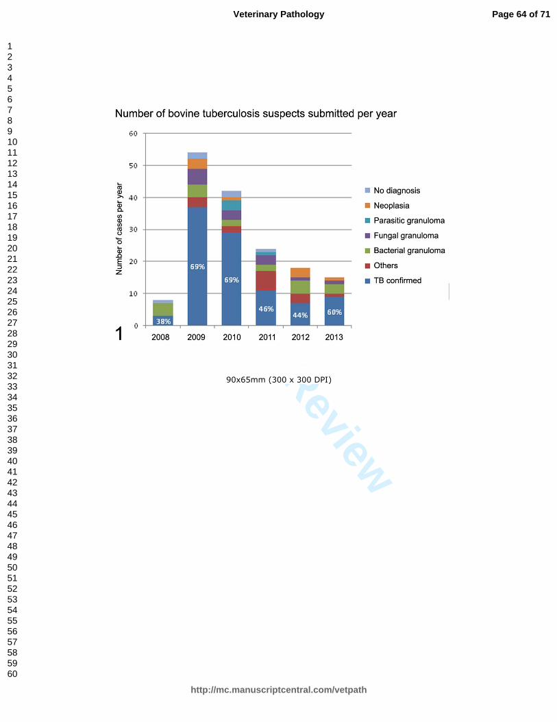
For Peer Review
90x65mm (300 x 300 DPI)
Page 64 of 71
http://mc.manuscriptcentral.com/vetpath
Veterinary Pathology
123456789101112131415161718192021222324252627282930313233343536373839404142434445464748495051525354555657585960

For Peer Review
180x127mm (300 x 300 DPI)
Page 65 of 71
http://mc.manuscriptcentral.com/vetpath
Veterinary Pathology
123456789101112131415161718192021222324252627282930313233343536373839404142434445464748495051525354555657585960

For Peer Review
180x127mm (300 x 300 DPI)
Page 66 of 71
http://mc.manuscriptcentral.com/vetpath
Veterinary Pathology
123456789101112131415161718192021222324252627282930313233343536373839404142434445464748495051525354555657585960
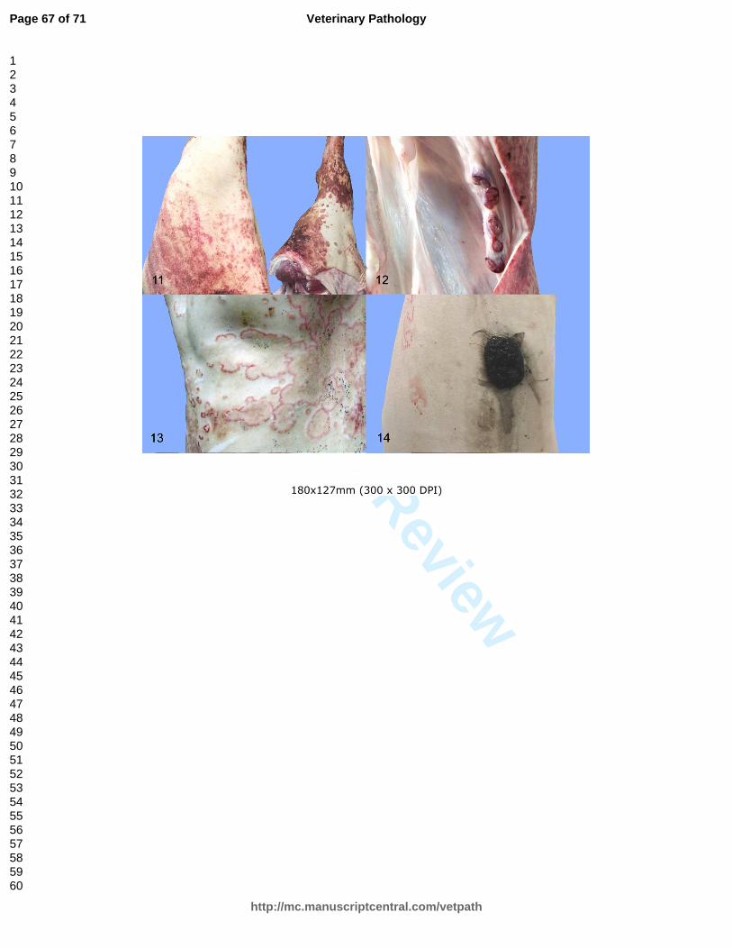
For Peer Review
180x127mm (300 x 300 DPI)
Page 67 of 71
http://mc.manuscriptcentral.com/vetpath
Veterinary Pathology
123456789101112131415161718192021222324252627282930313233343536373839404142434445464748495051525354555657585960

For Peer Review
180x127mm (300 x 300 DPI)
Page 68 of 71
http://mc.manuscriptcentral.com/vetpath
Veterinary Pathology
123456789101112131415161718192021222324252627282930313233343536373839404142434445464748495051525354555657585960

For Peer Review
90x52mm (300 x 300 DPI)
Page 69 of 71
http://mc.manuscriptcentral.com/vetpath
Veterinary Pathology
123456789101112131415161718192021222324252627282930313233343536373839404142434445464748495051525354555657585960

For Peer Review
180x191mm (300 x 300 DPI)
Page 70 of 71
http://mc.manuscriptcentral.com/vetpath
Veterinary Pathology
123456789101112131415161718192021222324252627282930313233343536373839404142434445464748495051525354555657585960

For Peer Review
180x191mm (300 x 300 DPI)
Page 71 of 71
http://mc.manuscriptcentral.com/vetpath
Veterinary Pathology
123456789101112131415161718192021222324252627282930313233343536373839404142434445464748495051525354555657585960
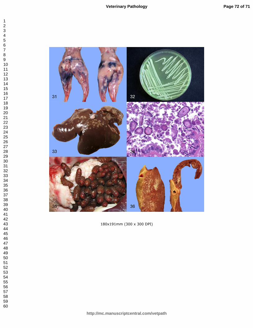
For Peer Review
180x191mm (300 x 300 DPI)
Page 72 of 71
http://mc.manuscriptcentral.com/vetpath
Veterinary Pathology
123456789101112131415161718192021222324252627282930313233343536373839404142434445464748495051525354555657585960