Embed Size (px)
Citation preview

11
Vibrational
Spectroscopy
(IR, Raman)Vibrational
spectroscopy
Vibrational
spectroscopy is an energy sensitive method. It is based on periodic changes
of dipolmoments
(IR)
or polarizabilities
(Raman)
caused by molecular vibrations of molecules or groups of atoms and the combined discrete energy transitions and changes of frequen-
cies
during absorption (IR)
or scattering (Raman)
of electromag- netic
radiation of wavelengths from 1 to 300 µm (selection rules).
One can get/detect:• the presence of known compounds (finger print)• the components of an unknown compound (functional groups)• and thus a likely structure of a compound• changes in the concentration of a species during a reaction• the properties of bonds (bond strength, force constants)• state and order parameters of phase transitions
22
Vibrational
Spectroscopy
(IR, Raman)Vibrational
spectroscopy
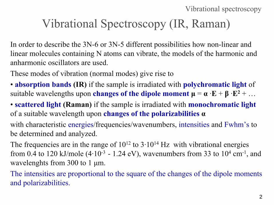
In order to describe the 3N-6 or 3N-5 different possibilities how non-linear and linear molecules containing N atoms can vibrate, the models of the harmonic and anharmonic
oscillators are used.
These modes of vibration (normal modes) give rise to•
absorption bands
(IR)
if the sample is irradiated with polychromatic light
of
suitable wavelengths upon changes of the dipole moment
μ
= α
·E + β
·E2
+ …•
scattered light
(Raman)
if the sample is irradiated with monochromatic light
of a suitable wavelength upon changes of the polarizabilities
αwith characteristic energies/frequencies/wavenumbers, intensities
and Fwhm’s
to
be determined and analyzed.The frequencies are in the range of 1012
to 3·1014
Hz with vibrational
energies from 0.4 to 120 kJ/mole (4·10-3 -
1.24 eV), wavenumbers
from 33 to 104
cm-1, and wavelenghts
from 300 to 1 μm.
The intensities are proportional to the square of the changes of
the dipole moments and polarizabilities.

33
Vibrational
Spectroscopy
Wavelengths and energies in vibrational
spectroscopy
Wavenumber
reciprocal of λ:
1/λ
(cm-1)
Wavelength
in nm
Vis, IR, and Raman
areas
drawnin a scale
of linear wavenumbers
and some
lasers
sources

44
Vibrational
Spectroscopy -
the main principleVibrational
Spectroscopy
H Clr0
Center of mass is not allowedoo shift during the vibration
Extension from r0
(equilibrium distance)Absorption of energy E
Relaxation to r0
Vibration
Together with molecular vibrations also molecular rotations are excitedas well since rotational energies are much smaller (~ 0.01·Evib.
)!
Hooke´s law
F = -
k xV = ∫-F dx
= ∫k x dx
V = ½
k x2
Spring with rate/spring constant k
k

55
Harmonic vibrational
levels
Warning: Molecular vibrations are essentially anharmonic!
Vibrational
Spectroscopy
Zero-point
vibration
Zero-opint
energy
E0
Potential curveV =
μν /~~ k
= Wavenumberk
= rate/force constant
μ
= reduced mass
ν~
21
21
mmmm+
=μ
F = -
k xV = ∫-F dx
= ∫k x dx
V = ½
k x2
Harmonic
Oszillator
F = -kx
= m·b
= m·d2x/dt2
→ ν0
= (1/2π)·(k/m)1/2
66
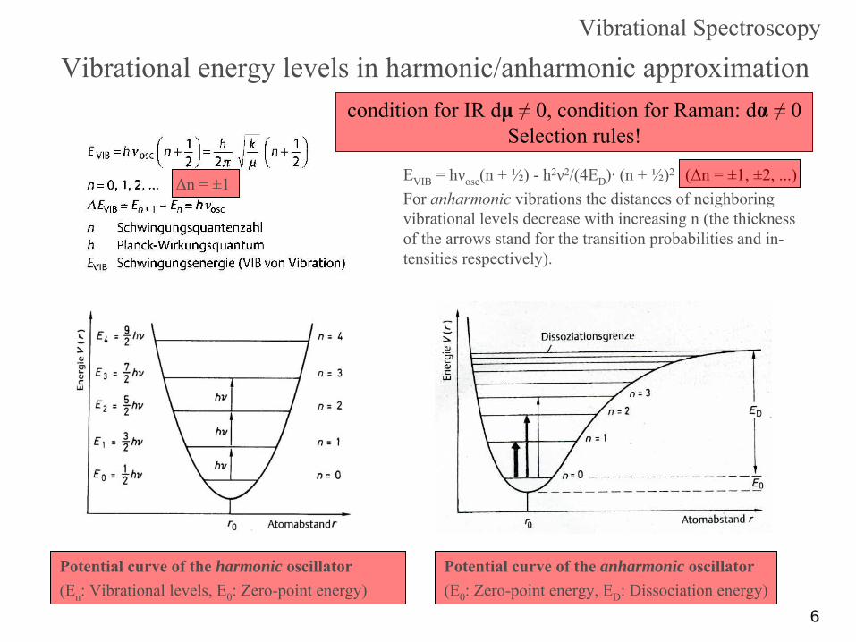
Vibrational
energy levels in harmonic/anharmonic
approximation
Potential curve of the harmonic oscillator(En
: Vibrational
levels, E0
: Zero-point energy)
Δn = ±1
Vibrational
Spectroscopy
Potential curve of the anharmonic oscillator(E0
: Zero-point energy, ED
: Dissociation energy)
EVIB
= hνosc
(n
+ ½) -
h2ν2/(4ED
)·
(n + ½)2 (Δn
= ±1, ±2, ...)For anharmonic
vibrations the distances of neighboring vibrational
levels decrease with increasing n
(the thickness of the arrows stand for the transition probabilities and in-
tensities
respectively).
condition for IR dμ
≠
0, condition for Raman: dα
≠
0 Selection rules!

77
Vibrational
states and frequencies
Vibrational
coupling
in zig-zag
chains
of different lengths
Excitation
of a vibrational
state
in the
electronic
ground
state
S0
by
a: infrared
absorption, b: Raman
scattering, c: inelastic
neutron
scattering, d: fluorescence.
Variation of frequencies
in case
of a free
molecule
(a),
static
(b) and dynamical
(c) coupling
in a crystal
lattice,
and dependence
on the
wave
vector
k
for
all unit
cells
Vibrational
Spectroscopy
88
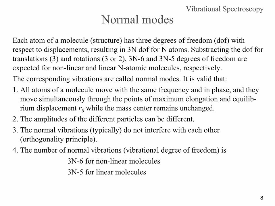
Vibrational
SpectroscopyNormal modes
Each atom of a molecule (structure) has three degrees of freedom
(dof) with respect to displacements, resulting in 3N dof
for N atoms. Substracting
the dof
for
translations (3) and rotations (3 or 2), 3N-6 and 3N-5 degrees of freedom are expected for non-linear and linear N-atomic molecules, respectively.The corresponding vibrations are called normal modes. It is valid that:1.
All atoms of a molecule move with the same frequency and in phase, and they
move simultaneously through the points of maximum elongation
and equilib- rium
displacement r0
while the mass center remains unchanged.2. The amplitudes of the different particles can be different.3.
The normal vibrations (typically) do not interfere with each other (orthogonality
principle).
4. The number of normal vibrations (vibrational
degree of freedom) is3N-6 for non-linear molecules3N-5 for linear molecules

99
Vibrational
SpectroscopyNormal modes
Every vibrational
mode
exhibits its own “pattern
(vector, matrix)”
for the atomic displacements (±Δx, ±Δy, ±Δy), leading to normal coordinates, but the vibrational
modes
are usually not known:
Assignment of the vibrational
modes via symmetry properties of the
molecules (point group, irreducible representation, character, character tables).
Symmetry of vibrations (symmetry species = Rassen, types of vibration) Symmetry species (Rassen)
of the modes are denoted after Mulliken:
A = symmetric, B = antisymmetric
with respect to Cn
; E, F, G, H = 2-, 3-, 4-, 5-fold degenerate with respect to Cn
;g = symmetric with respect to i (from German gerade);u = antisymmetric
with respect to i (from German ungerade);
Index subscripts of A or B: 1 = symmetric, 2 = antisymmetric
with respect to Cn
or Sn
(a mirror plane);Example: A2g
is a vibration that is symmetric with respect to Cn
and i (character = 1) and antisymmetric
with respect to Sn
or σ
(character = -1).

1010
C3v E 2C3 3σv
A1 1 1 1 z x2+y2, z2
A2 1 1 -1 Rz
E 2 -1 0 (x,y) (Rx
,Ry
) (x2-y2, xy) (xz, yz)
Point groupSymbol
Symmetry operations
Active vibrations in
Symmetry species
(Rassen)Group characters Combinations of the symbols x, y, z, Rx, Ry
and Rz, the first three of which represent the coordinates x, y and z, and the last three of which stand for rotations about these axes. These are related to transformation properties and basis representations of the group.
Character table for space group C3v
IR Raman
Normal modes
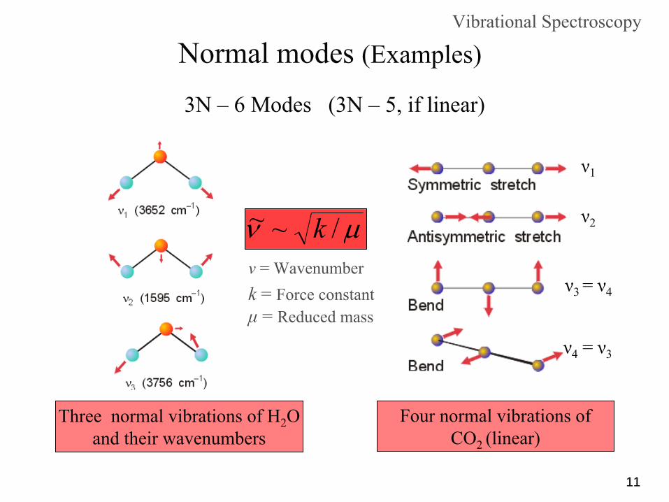
1111
Normal modes
(Examples)
Three normal vibrations of H2
O and their wavenumbers
Vibrational
Spectroscopy
3N –
6 Modes
(3N –
5, if
linear)
ν
= Wavenumberk
= Force constant
μ
= Reduced mass
μν /~~ k
Four normal vibrations of CO2 (linear)
ν1
ν2
ν3 = ν4
ν4
= ν3

1212
Auswahlregeln
Normal modes of vibration (IR-) activityDipole moment changes during the vibration!
O C O
O C O
O C O
O C O
HO
H
HO
H
HO
Hνsym: 1596 cm-1
νsym: 3652 cm-1
νasym: 3756 cm-1
666 cm-1 entartet
νasym: 2350 cm-1
νasym: 1340 cm-1
+ - +666 cm-1 entartet
IR-aktivIR-aktiv
IR-aktiv IR-inaktiv(Raman-aktiv)
IR-aktiv
Stretching vibrationChanges of bond
lengths
Bending vibration Changes of bond
anglesIR-aktiv
Vibrational
Spectroscopy

1313
S14
Normal modes
(Examples)

1414
Normal modes
(Examples)

1515
Normal modes
(Examples)

1616
Typical values for stretching and bending vibrations
“Molecule“ stretching bending
C -
H 2800 -
3000
N -
N 3300 -
3500
H2
O 3600 -
3000 1600
C = O 1700
C = C 1600
SO32- 970 (νs
)930 (νas
)620 (γ)470 (δ)
Schwingungsspektroskopie

1717
Sources for IR-
(and Raman-) radiation
Conventional lamps are not adequate, because near IR: ~ 700 to 1400 nm; mid-wavelength/far IR: > 1400 nm
IR-Spectroscopy
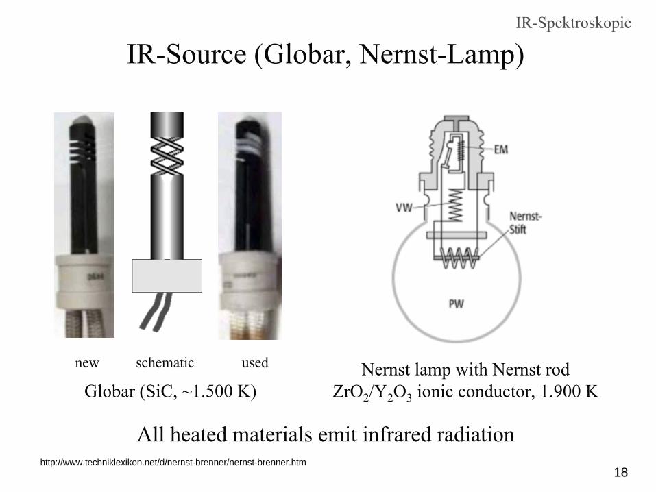
1818http://www.techniklexikon.net/d/nernst-brenner/nernst-brenner.htm
Nernst lamp with Nernst rod ZrO2
/Y2
O3
ionic conductor, 1.900 K
All heated materials emit infrared radiation
IR-Source (Globar, Nernst-Lamp)
new schematic used
Globar
(SiC, ~1.500 K)
IR-Spektroskopie

1919
Range
Source Monochromator
Detector
Far IR Nernst rod CsI-prism; grating
Bolometer
(ceramic rodMid IR
with heating coil)
LiF-prism; grating
Bolometer
Near IR
Light-bulb quartz-prism PbS-Cell; Se-Cell
Nernst rod:
cub. ZrO2
stabilized by rare earth elements (e.g.. Y3+)
Near
IR: ~ 700 bis 1400 nm; Mid-wavelenght/Far
IR: > 1400 nm
IR-sources, monochromators
and detectorsIR-Spektroskopie

2020
IR-Detectors
Main principle and a picture of a bolometer: A cooled metal foil (Pt, Au) absorbs IR radiation. The resulting
rise of the temperature is detected by a resistor-type thermometer.
IR-Spectroscopy

2121
IR -
Spectrometer
Double beam, optical grating
IR-Spectroscopy
Fourier-Transform
(FT)

2222
Fourier-transform spectroscopyIR-Spectroscopy
“Classical”
(grating, prism) IR spectroscopy has been replaced by the much faster FTIR spectroscopy. In the case of the “classical”
(i.e. non FT) infrared
spectroscopy the different wavelengths had to be measured successively. In the case of the FTIR technique the complete range of interest is measured at once.The fundamental instrument for FTIR is the Michelson interferometer that replaces the monochromator. The sample is irradiated by polychromatic light and a movable mirror produces a time dependent signal that is transformed by Fourier transformation into a frequency spectrum.

2323
Fouriertransform
(FT) spectrometry
Schematic representation of a Michelson interferometer (a) with interferogram
(b) and spectrum (c) obtained by Fourier transform.
a) Michelson InterferometerS radiation source, Sa sample chamber, D detector,A amplifier, M1 fixed, M2 movable mirror,BS beam splitter, x mirror deflection, L distance
b) InterferogramSignal recorded by the detector
c) SpectrumObtained by Fourier transform (FT)From the interferogram
IR-Spectroscopy

24243500 3000 2500 1300 1150 1000 850 700 400ν/cm-1
Sr(HPO OH)2 2
RT1226
1235
1225
12411164
1155
1170
1168
758
779
694
685
686
2881 2439 2433
2434244028993032
3019
2400
2432
2300
23033176
31752963
2783
3191
32143080
RT
TT
TT
Sr(SeO OH)2 2
Tran
smis
sion
νB(OH)
ν(PH)
δ(OH)
γ(OH)
Cs2
CrCl5
·4H2
O BaSO3
KMn(SeO2
OH)3
Examples from current research activities IR-Spectroscopy

2525
Raman spectroscopyRaman spectroscopy
Irradiation of a sample with monochromatic light of a suitable wave length may force oscillations of the electrons.A small portion (IRayleigh
/I0
~ 10-5) of the absorbed radiation energy is scattered with the same frequency as the incident light into all directions of space (elastic, Rayleigh scattering).An even smaller portion (IRaman(Stokes)
/I0
~ 10-8) of the irradiated energy will be transformed into molecular vibrations (with a corresponding change of the polarizability
tensor α) and thus leads to an absorption of vibrational
energy from
the scattered light. The scattered light therefore has a lower frequency than the irradiated light (inelastic, Raman scattering, Stokes).If the radiation interacts with a vibrational
excited molecule then the scattering
process may result in an emission of energy to the scattered light. Thus the scattered light has a larger frequency compared to the irradiated light (inelastic, Raman scattering, anti-Stokes, IRaman(anti-Stokes)
/I0
~ 10-11).Raman scattering requires a change of the polarizability
α. The intensity of the
scattered light is proporional
to the square of the change of the polarizability.

2626
Schematic representation of the energy levels Raman scattering, anti-Stokes; Rayleigh scattering; Raman scattering, Stokes
(ν0
= Frequency of the irradiated light, νM
= vibrational
frequency of the molecule)Resonance Raman bands with high intensity occur if instead of a forced oscillation an
electronically excited state is generated (absorption).
Raman spectroscopy
(IRayleigh
~ 10-5·I0
)
(IRaman(St)
~ 10-8·I0
)(IRa(anti-St)
~ 10-11·I0
)
Selection rules Δn
= ±1 (harmonic) Δn
= ±1, ±2, ±3 …
(anharmonic) Condition: dα
≠
0

2727
Molecular vibrations of PCl3
and comparable molecules
Vibrational
spectroscopy
PCl3
is a tetra-atomic molecule with C3v
symmetry
νasνs δasδs

2828
Raman spectrum of PCl3
(liquid) in the Stokes- and anti-Stokes
range exciting line 514,5 nm ≡
19436 cm-1
(Ar
Laser)
Raman spectroscopy
νs, as
δs
δas
δs
δas
νs, as

2929
The low intensities of the scattered radiation (IRa(St)
~ 10-8·I0
, IRa(anti-St)
~ 10-11·I0
) requires the use of lasers.
Raman spectroscopy
(IRay
~ 10-5·I0
)
(IRa(St)
~ 10-3·IRay
, IRa(anti-St)
~ 10-3·IRa(St)
)
(z.B. Ar-Laser)
Schematic representation for the observation of the Raman effect in 90o
arrangement
3030
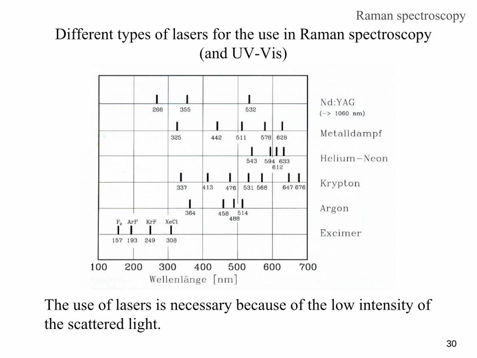
The use of lasers is necessary because of the low intensity of the scattered light.
Raman spectroscopyDifferent types of lasers for the use in Raman spectroscopy
(and UV-Vis)

3131
Raman and IR active vibrational
modes of CO2
IR-/Raman spectroscopy
Raman active are those vibrations, at which the polarizability
α
changes (different in the inversion points).
IR active are those vibrations, at which the dipole moment μ
changes (different in the inversion point). IR condition: dμ
≠
0
Raman condition: dα
≠
0

3232
Polarizability
changes (δα/δq)
of the fundamental modes (νas
, νs
, δ) and IR and Raman spectra of the CS2
molecule
IR-/Raman spectroscopy
Exclusion rule in IR and Raman spectra

3333
IR-/Raman spectroscopy
IR/Raman spectra and vibrational
modes of Hg2
I2
Exclusion rule in IR and Raman spectra
→ Exclusion rule
Vibrations symmetrical with respect to i
(g) are IR inactive but
Raman-ctiveVibrations non-symmetrical with respect to i (u) are IR active but Raman inactive
νs
(HgI) νas
(HgI) ν(HgHg) δs
(HgHgI) δas
(HgHgI) νT
.
A1g A2u A1g 2Eg 2Eu Eg
Ra. IR Ra. Ra IR Ra
Hg2
I2 (I4/mmm) D∞h
=> 3N-5=7Hg HgI I

3434
FIR and Raman spectra of Hg2
Br2
IR-/Raman spectroscopyTemperature effects and exclusion rule
Lattice vibration
ν(HgHg)
δ(HgHgBr)νas
(HgI)
3535
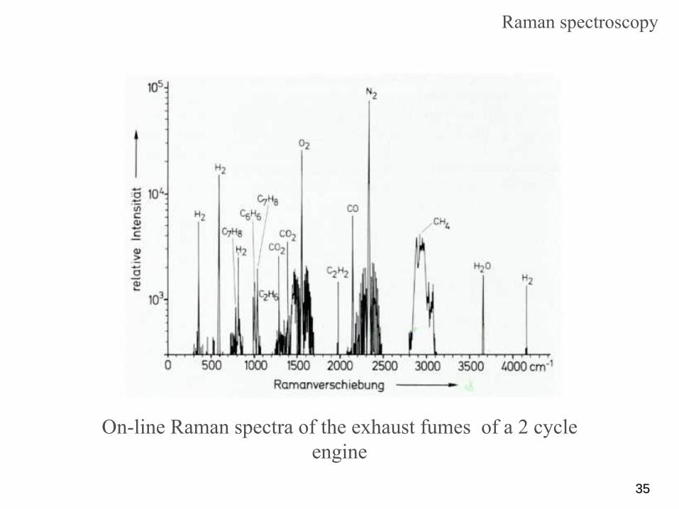
On-line Raman spectra of the exhaust fumes of a 2 cycle engine
Raman spectroscopy

3636
Schematic representation of the energy-levels and spectra of different spectroscopic transitions. The anharmonicity
of a vibration is exaggerated
and the distance S1
-S0
is strongly compressed. A = Absorption, I = Intensity
IR-/Raman spectroscopy

3737
Literatur
IR-/Ramanspektroskopie
Untersuchungsmethoden in der Chemie – Einführung in die moderneAnalytik von H. Naumer und W. Heller, Wiley-VCHOptische Spektroskopie von W. Schmidt, Wiley-VCHSchwingungsspektroskopie von J. Weidlein, U. Müller, K. Dehnike,Georg Thieme VerlagAnwendungen der Schwingungsspektroskopie in der AnorganischenChemie von H. Siebert, Springer VerlagInfrared and Raman Spectroscopy, Methods and Applications byB. Schrader, Wiley-VCHMolecular Vibrations by E. Bright Wilson Jr., J.C. Decius, P.C. Cross,McGraw-Hill