Embed Size (px)
Citation preview
Lobacz, M. A., Serra, F., Hammond, G., Oevermann, A. and Haley, A. C.
(2016) Imaging diagnosis: magnetic resonance imaging of diffuse
leptomeningeal oligodendrogliomatosis in a dog with "dural tail sign".
Veterinary Radiology and Ultrasound
There may be differences between this version and the published version.
You are advised to consult the publisher’s version if you wish to cite from
it.
Lobacz, M. A., Serra, F., Hammond, G., Oevermann, A. and Haley, A.
C. (2016) Imaging diagnosis: magnetic resonance imaging of diffuse
leptomeningeal oligodendrogliomatosis in a dog with "dural tail
sign". Veterinary Radiology and Ultrasound,(doi:10.1111/vru.12441)
This article may be used for non-commercial purposes in accordance with
Wiley Terms and Conditions for Self-Archiving.
http://eprints.gla.ac.uk/131451/
Deposited on: 20 June 2017
Enlighten – Research publications by members of the University of Glasgow
http://eprints.gla.ac.uk
1
Imaging Diagnosis – Magnetic Resonance Imaging Of Diffuse Leptomeningeal 1
Oligodendrogliomatosis In A Dog With “Dural Tail Sign” 2
3
Monika Anna Lobacz, Fabienne Serra, Gawain Hammond, Anna Oevermann, Allison C. 4
Haley. 5
6
Key words: leptomeningeal oligodendrogliomatosis, dural tail, dog 7
8
School of Veterinary Medicine, College of Medicine, Veterinary Medicine & Life 9
Sciences, University of Glasgow, 464 Bearsden Road, Glasgow G61 1QH, UK 10
Preliminary case report was presented as a Poster on 77th Diagnostic Imaging 11
International Congress in Milan organized by SCIVAC 23rd March 2013. 12
Presented the poster in the ACVP (American College of Veterinary Pathologists) 13
meeting in 2014 in Atlanta. 14
15
Abstract 16
A case of diffuse leptomeningeal oligodendrogliomatosis affecting the brain and spinal 17
cord of a dog is presented. A 7.5 year old, male neutered Staffordshire bull terrier 18
presented for evaluation of a chronic history of tetraparesis and seizures, with a 19
multifocal neuroanatomical localization was determined. Extra-axial intradural lesions 20
with an atypical presentation of a dural tail sign were seen on magnetic resonance 21
imaging (MRI). Histologically, the lesions were consistent with a leptomeningeal 22
oligodendrogliomatosis. To the authors’ knowledge a dural tail sign has not previously 23
2
been reported as an MRI characteristic of diffuse leptomeningeal 24
oligodendrogliomatosis in dogs. 25
26
Signalment, history and clinical findings: 27
A 7.5 year old, male neutered Staffordshire bull terrier presented for evaluation of 28
tetraparesis. Tail paresis was detected 1 year prior to presentation but further 29
investigation was not performed at the time. Nine months prior to presentation the 30
patient began having seizures, which were treated with anticonvulsant therapy 31
(Phenobarbital, 60mg PO BID). Six months prior to presentation the patient developed 32
an abnormal thoracic limb gait. Over the following six months the signs progressed to 33
dribbling urine, and occasional collapse in the pelvic limbs when walking. On 34
presentation the dog was quiet but alert and responsive with an ambulatory tetraparesis 35
and proprioceptive ataxia. Postural reactions were reduced in all four limbs but worse 36
on the right side. Spinal reflexes were reduced in some limbs, with an absent 37
withdrawal of the left thoracic limb and reduced flexion of the tarsus of the right pelvic 38
limb. Perineal reflex and tail tone were reduced. Decreased menace response and 39
positional ventral strabismus of the left eye was present. The neuroanatomic 40
localization was determined to be multifocal based on these findings. 41
42
Imaging, diagnosis and outcome: 43
Magnetic resonance (MR) images of the brain and the spinal cord were acquired using 44
a 1.5 Tesla magnet (Magnetom Essenza, Siemens, Camberley, United Kingdom) with 45
head and spine coils. The protocol included T2-weighted (T2w) and T1-weighted (T1w) 46
3
turbo spin echo sequences, T2 –weighted FLAIR (T2-FLAIR), T2 *-weighted gradient 47
recalled echo (GRE), and T1w images after manual intravenous administration of 48
gadoterate meglumine (0.1 mmol/kg of gadopentetate dimeglumine, Magnevist; Bayer 49
HealthCare Pharmaceuticals, United Kingdom). Post – contrast T1w images were 50
obtained immediately after injection in sagittal, transverse and dorsal planes. 51
Within the calvarium, an irregular, predominantly left sided, parasagittal lesion was 52
found extending along the skull base, around the pituitary gland and optic chiasm, and 53
into the brainstem. In comparison to gray matter, the lesion was homogeneously 54
hyperintense on T2w images, while on T2-FLAIR images it had mildly increased 55
intensity. On T1w images the lesion was mildly hypointense to normal gray matter, and 56
on post-contrast images it showed marked homogeneous enhancement. Post-contrast 57
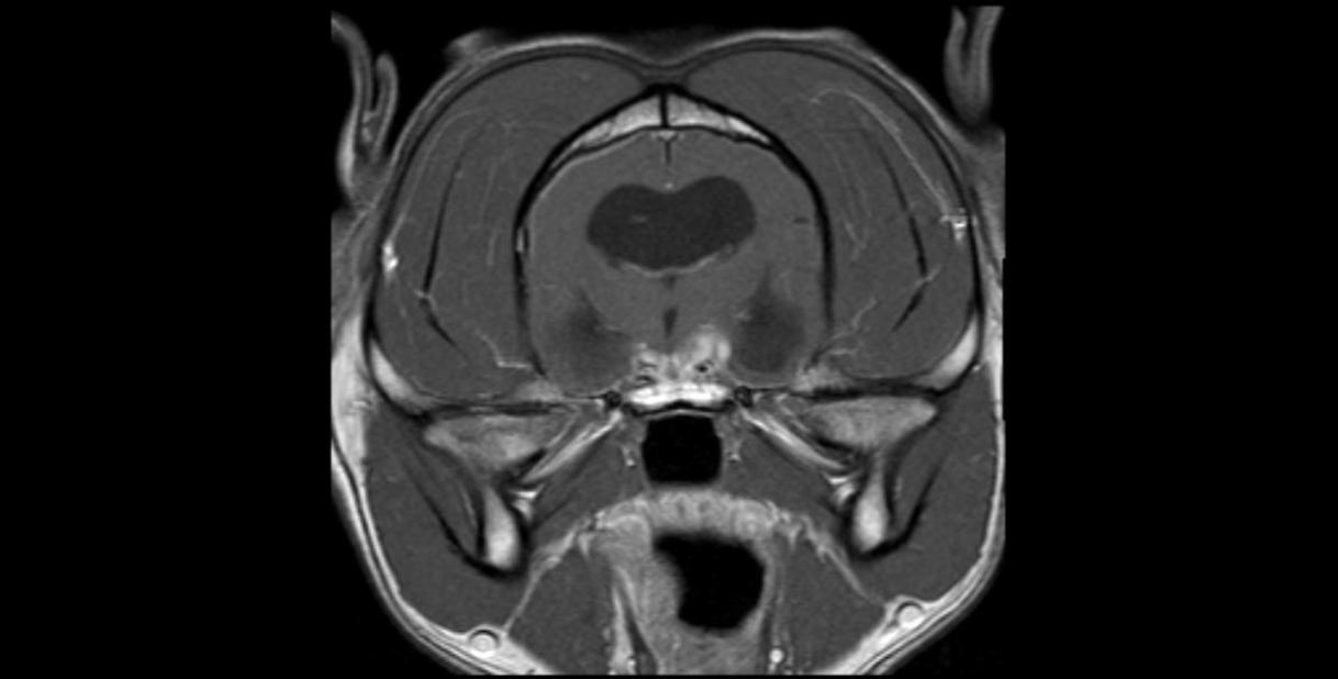
images revealed thickening of the adjacent meninges consistent with a “dural tail sign” 58
extending caudal and lateral to the lesion (Fig. 1A & Fig. 1B). No evidence of 59
susceptibility artifact on GRE images was detected. On T2w and GRE images in the 60
region of the cerebral arterial circle well-defined, macroscopically normal, intralesional 61
blood vessels could be appreciated. Extension of the lesion ventral to the rostral area 62
of the brainstem (to the level of the colliculi) was present bilaterally, although this finding 63
was more prominent on the left than on the right side at level of the foramen magnum. A 64
large, fluid-filled, irregular cystic structure was visible dorsorostral to the cerebellum, 65
causing caudal displacement of the lamina quadrigemina, marked compression and 66
slight herniation of the cerebellum through the foramen magnum. Severe dilation of the 67
ventricular system and of the olfactory recesses were also noted. There was a reduced 68
amount of cerebrospinal fluid in the sulci, compression of the third ventricle and 69
4
compression/distortion of the interthalamic adhesion. Mentioned above findings are 70
suggestive of increased intracranial pressure. 71
72
The intracranial lesion continued through the foramen magnum to merge with the lesion 73
within the cervical vertebral canal. At the level of the second cervical vertebra (C2) the 74
lesion had a thickened, solid appearance and then became cavitated cranially. This 75
lesion extended continuously along the entire vertebral canal as an irregular intradural 76
lesion ventral to the cord. At the level of L5-S1, the lesion could be seen invading nearly 77
the entire height of the spinal canal and extending caudally, mainly along the ventral 78
aspect of the vertebral canal. (Fig. 2). The spinal cord lesion was T2w hyperintense, 79
T1w isointense and showed marked, fairly homogeneous contrast enhancement with 80
similar intensity characteristics as the intracranial lesion. Throughout the length of the 81
spinal cord, but most marked in the cranial cervical spine there was marked dilation of 82
the central canal, consistent with syringohydromyelia. 83
84
The imaging diagnosis was an intradural - extramedullary diffuse infiltrative disease. 85
Taking in consideration duration of the clinical signs, differential diagnoses included 86
meningiomatosis, less likely lymphoma, carcinomatosis or histiocytic sarcoma. 87
Infectious diseases was considered less likely as a differential diagnosis because 88
patient did not have travel abroad history. The presence of an arachnoid diverticulum 89
rostral to the cerebellum, hydrocephalus, and syringohydromyelia were also consistent 90
with increased cerebrospinal fluid pressure. Due to the poor prognosis, the patient was 91
5
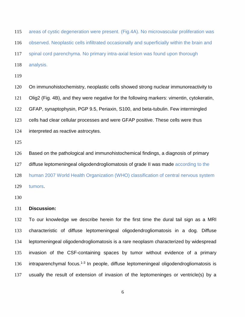
euthanized immediately after imaging. A cerebrospinal fluid (CSF) sample was not 92
obtained. 93
94
On gross examination, a grey, gelatinous, extra-axial lesion was diffusely expanding the 95
meningeal space along the entire spinal cord, causing severe compression. The 96
process extended to the ventro-lateral aspect of the brain. No primary solid mass lesion 97
was observed. Severe hydrocephalus was observed, together with secondary lesions 98
including syringomyelia, rupture of the septum pellucidum, and bilateral diverticula. The 99
latter extended into the striatal body beneath the internal capsule. No lesion was 100
present in any other organ system. 101
102
Formalin – fixed brain sections and histology through the same level as on Fig 1B with 103
“dural tail sign” are provided on Fig. 3A, 3B and 3C). On histological examination, the 104
leptomeninges of the brain and spinal cord were diffusely and markedly enlarged by 105
loosely arranged sheets of monomorphous neoplastic cells. These neoplastic cells had 106
predominantly round nuclei with a finely stippled chromatin and small nucleoli, and they 107
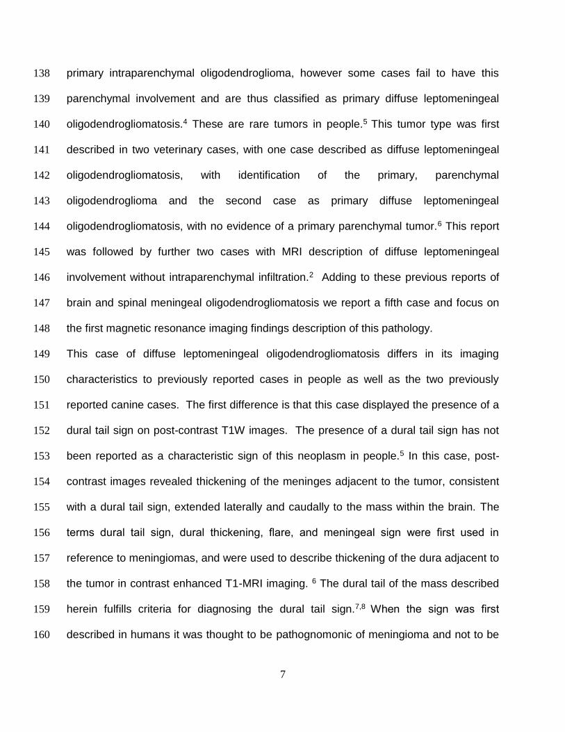
had either small to moderate eosinophilic eccentric cytoplasm or perinuclear optically 108
empty halos which were reminiscent of a honeycomb pattern. The cells were embedded 109
in pale amphophilic to basophilic matrix compartmentalized by thin cytoplasmic 110
processes. In some areas, the neoplastic cells had more hyperchromatic nuclei. In other 111
areas, the cell nuclei were more irregular and arranged in different patterns such as 112
packets, palisades and rows. Cellular atypia was moderate. Mitotic index was up to 5 113
mitotic figures per 10 high power fields in the denser areas of the tumor. Multifocal 114
6
areas of cystic degeneration were present. (Fig.4A). No microvascular proliferation was 115
observed. Neoplastic cells infiltrated occasionally and superficially within the brain and 116
spinal cord parenchyma. No primary intra-axial lesion was found upon thorough 117
analysis. 118
119
On immunohistochemistry, neoplastic cells showed strong nuclear immunoreactivity to 120
Olig2 (Fig. 4B), and they were negative for the following markers: vimentin, cytokeratin, 121
GFAP, synaptophysin, PGP 9.5, Periaxin, S100, and beta-tubulin. Few intermingled 122
cells had clear cellular processes and were GFAP positive. These cells were thus 123
interpreted as reactive astrocytes. 124
125
Based on the pathological and immunohistochemical findings, a diagnosis of primary 126
diffuse leptomeningeal oligodendrogliomatosis of grade II was made according to the 127
human 2007 World Health Organization (WHO) classification of central nervous system 128
tumors. 129
130
Discussion: 131
To our knowledge we describe herein for the first time the dural tail sign as a MRI 132
characteristic of diffuse leptomeningeal oligodendrogliomatosis in a dog. Diffuse 133
leptomeningeal oligodendrogliomatosis is a rare neoplasm characterized by widespread 134
invasion of the CSF-containing spaces by tumor without evidence of a primary 135
intraparenchymal focus.1-3 In people, diffuse leptomeningeal oligodendrogliomatosis is 136
usually the result of extension of invasion of the leptomeninges or ventricle(s) by a 137
7
primary intraparenchymal oligodendroglioma, however some cases fail to have this 138
parenchymal involvement and are thus classified as primary diffuse leptomeningeal 139
oligodendrogliomatosis.4 These are rare tumors in people.5 This tumor type was first 140
described in two veterinary cases, with one case described as diffuse leptomeningeal 141
oligodendrogliomatosis, with identification of the primary, parenchymal 142
oligodendroglioma and the second case as primary diffuse leptomeningeal 143
oligodendrogliomatosis, with no evidence of a primary parenchymal tumor.6 This report 144
was followed by further two cases with MRI description of diffuse leptomeningeal 145
involvement without intraparenchymal infiltration.2 Adding to these previous reports of 146
brain and spinal meningeal oligodendrogliomatosis we report a fifth case and focus on 147
the first magnetic resonance imaging findings description of this pathology. 148
This case of diffuse leptomeningeal oligodendrogliomatosis differs in its imaging 149
characteristics to previously reported cases in people as well as the two previously 150
reported canine cases. The first difference is that this case displayed the presence of a 151
dural tail sign on post-contrast T1W images. The presence of a dural tail sign has not 152
been reported as a characteristic sign of this neoplasm in people.5 In this case, post-153
contrast images revealed thickening of the meninges adjacent to the tumor, consistent 154
with a dural tail sign, extended laterally and caudally to the mass within the brain. The 155
terms dural tail sign, dural thickening, flare, and meningeal sign were first used in 156
reference to meningiomas, and were used to describe thickening of the dura adjacent to 157
the tumor in contrast enhanced T1-MRI imaging. 6 The dural tail of the mass described 158
herein fulfills criteria for diagnosing the dural tail sign.7,8 When the sign was first 159
described in humans it was thought to be pathognomonic of meningioma and not to be 160
8
seen in any other intracranial or extra-cranial tumors.9 However, it is now reported in a 161
growing number of human tumors, other than meningioma, as well as infectious, 162
autoimmune and vascular diseases. Tumors that have displayed a dural tail sign in 163
human medicine include glioblastoma, acoustic schwannoma, carcinoma, 164
hemangiopericytoma, pituitary adenoma, other sellar tumors.10-16 So far in veterinary 165
medicine masses such as meningioma, pituitary macroadenoma, histiocytic sarcoma, 166
chromophobe adenocarcinoma, lymphoma granular cell tumors and CNS blastomycosis 167
have been reported as demonstrating a dural tail sign.17-24 Many possible causes for the 168
dural tail sign have been hypothesized, such as expansion of the connective tissue and 169
hypervascularity, and different opinions exist regarding the diagnostic and prognostic 170
value of this imaging sign. Among these causes, expansion of connective tissue 171
secondary to tumoral invasion is important in determining the therapeutic plan, 172
particularly for planning surgical margins and radiation therapy planning. The area of 173
the dural tail in our case histologically was characterized by infiltration with the 174
neoplastic cells. 175
Another unique imaging characteristic of this case was the degree of contrast 176
enhancement the tumor displayed. A linear relationship has been established between 177
the degree of contrast enhancement and volume of peritumoral edema in human 178
gliomas.25, 26 This does not appear to be the case in our patient. The MR images of the 179
lesion we present here had a homogeneous appearance on all sequences with no 180
significant perilesional edema on FLAIR sequence, yet displayed marked, 181
homogeneous contrast enhancements on post-contrast T1W images. Histopathological 182
examination confirmed this lack of significant peritumoral edema. Presented herein was 183
9
a mass that had imaging characteristics of a diffuse extra-axial and intradural lesion with 184
marked, homogeneous contrast uptake. Similar MRI post contrast characteristics have 185
been described in glioblastomas in a dog and man. 27, 28 In the current veterinary 186
literature reported gliomas have variable contrast enhancement ranging from none to 187
variably isointense, nonuniform or ring-like enhancement, whereas in this case the 188
mass had marked and homogeneous contrast uptake.25, 26 Contrast enhancement is 189
reported to be more common in high-grade tumors (III or IV) than in low-grade tumors 190
(II), due to microvascular proliferation. 29 No microvascular proliferation was observed in 191
examined histopathological sections to explain this contrast uptake, and it remains to be 192
determined whether this is a characteristic of purely leptomeningeal 193
oligodendrogliomas. 2 However, it has been described that uptake of gadolinium by 194
extra-axial lesions is usually seen because they lack a blood-brain barrier 30 and 195
because of their tendency to develop congestion and interstitial edema. 31 196
Due to the extensive spread of the lesion along the vertebral column in this patient 197
surgical resection was not an option. Leptomeningeal oligodendrogliomatosis is 198
potentially treatable with chemotherapy and radiation. 32 The suitability of radiation 199
therapy was not explored as the owners elected euthanasia. 200
201
In conclusion, diffuse leptomeningeal oligodendrogliomatosis should be included on 202
differential lists for dogs with an extensive, extramedullary tumor, a dural tail sign, and 203
other MRI characteristics described in this case. 204
205
10
List of Author Contributions 206
Category 1 207
(a) Conception and Design 208
Author name (s) Monika Anna Lobacz, Fabienne Serra 209
(b) Acquisition of Data 210
Author name (s) Monika Anna Lobacz, Fabienne Serra, Allison Haley, Gawain 211
Hammond, Anna Oevermann 212
(c) Analysis and Interpretation of Data 213
Author name (s) Monika Anna Lobacz, Fabienne Serra, Allison Haley, Gawain 214
Hammond, Anna Oevermann 215
216
Category 2 217
(a) Drafting the Article 218
Author name (s) Monika Anna Lobacz, Fabienne Serra 219
(b) Revising Article for Intellectual Content 220
Author name (s) Monika Lobacz, Fabienne Serra, Anna Oevermann, Allison Haley, 221
Gawain Hammond 222
223
Category 3 224
(a) Final Approval of the Completed Article 225
Author name(s) Monika Lobacz, Fabienne Serra, Anna Oevermann, Allison Haley, 226
Gawain Hammond 227
228
11
229
Acknowledgments: We would like to thank the Pathology Department in Glasgow 230
Veterinary School for their technical support - with the native slides they sent us for the 231
extra immunos- staining. 232
233
Authorship statement 234
235
List of Author Contributions 236
Category 1 237 (a) Conception and Design 238 Author name (s) 239 (b) Acquisition of Data 240 Author name (s) 241 (c) Analysis and Interpretation of Data 242 Author name (s) 243
244
Category 2 245 (a) Drafting the Article 246 Author name (s) 247 (b) Revising Article for Intellectual Content 248 Author name (s) 249
250
Category 3 251 (a) Final Approval of the Completed Article 252 Author name(s) 253
254
255
References: 256
1. Beck DJK, Russell DS. Oligodendrogliomatosis of the cerebrospinal fluid pathway. 257
Brain 1942;65:352-372. 258
12
2. Canal S, Bernardini M, Pavone S, Mandara MT. Primary diffuse leptomeningeal 259
gliomatosis in 2 dogs. Can Vet J 2013;54:1075–1079 . 260
3. Kovi RC, Wunschmann A, Armien AG, Hall K, Carlson T, Shivers J, Oglesbee MJ. 261
Spinal meningeal oligodendrogliomatosis in two boxer dogs. Vet Pathol. 2013 262
Sep:50(5):761-4. 263
4. Mathews MS, Pare LS, Kuo JV, Kim RC. Primary Leptomeningeal 264
oligodendrogliomatosis. J Neuro-oncology. 2009;94(2):275-278. 265
5. Armao DM, Stone J, Castillo M, Mitchell KM, Bouldin TW, Suzuki K. Diffuse 266
leptomeningeal oligodendrogliomatosis: radiologic/pathologic correlation. Am J 267
Neuroradiol 2000; 21 (6):1222-1226. 268
6. Wilms G, Lammens M, Marchal G, Van Calenbergh F, Plets C, Van Fraeyenhoven L, 269
Baert AL. Thickening of dura surrounding meningiomas: MR features. J Comput Assist 270
Tomogr 1989;13:763–768. 271
7. Goldsher D, Litt AW, Pinto RS, Bannon KR, Kricheff II. Dural “tail” associated with 272
meningiomas on Gd-DTPA-enhanced MR images: characteristics, differential diagnostic 273
value, and possible implications for treatment. Radiology 1990;176:447–450. 274
8. Nägele T, Peterson D, Grodd W, Opitz H, Voigt K. The “dural tail” adjacent to 275
meningiomas studies by dynamic contrast – enhanced MRI: a comparison with 276
histopathology. Neuroradiol 1994:36:303-307. 277
9. Guermazi A, Lafitte F, Miaux Y, Adem C, Bonneville JF, Chiras J. The dural tail sign 278
beyond meningioma. Clin Radiol 2005;60(2):171–188. 279
10. Bourekas EC, Wildenhain P, Lewin JS, Tarr RW, Dastur KJ, Raji MR, Lanzieri CF. 280
The dural tail sign revisited. AJNR Am J Neuroradiol 1995;16(7):1514–1516. 281
13
11. Morioka T, Matsushima T, Ikezaki K, Nagata S, Ohta M, Hasuo K, Fukui M. Intra-282
cranial adenoid cystic carcinoma mimicking meningioma: report of two cases. 283
Neuroradiology 1993;35:462–465. 284
12. Chiechi MV, Smirniotopoulos JG, Mena H. Intra-cranial hemangiopericytomas: MR 285
and CT features. AJNR Am J Neuroradiol 1996;17:1365–1371. 286
13. Nakasu Y, Nakasu S, Ito R, Mitsuya K, Fujimoto O, Saito A. Tentorial enhancement 287
on MR images is a sign of cavernous sinus involvement in patients with sellar tumors. 288
AJNR Am J Neuroradiol 2001;22:1528–1533. 289
14. Celli P, Cervoni L, CantoreG. Dural tail in pituitary adenoma. J Neuroradiol 290
1997;24:68–69. 291
15. Paz-Fumagalli R, Daniels DL, Millen SJ, Meyer GA, Thieu TM. Dural ‘‘tail’’ 292
associated with an acoustic schwannoma in MR imaging with gadopentetate 293
dimeglumine. Am J Neuroradiol 1991;12:1206. 294
16. Atlas SW, Lavi E. Intra-axial brain tumors. In: Atlas SW, eds. Magnetic resonance 295
imaging of the brain and spine. Second edition. Philadelphia, Lippincott-Raven, 296
1996:315–422. 297
17. Tamura S, Tamura Y, Nakamoto Y, Ozawa T, Uchida K. MR imaging of histiocytic 298
sarcoma of the canine brain. Vet Radiol Ultrasound 2009;50:178–181. 299
18. Wisner ER, Dickinson PJ, Higgins R. Magnetic resonance imaging features of 300
canine intracranial neoplasia. Vet Radiol Ultrasound 2011;52:S52-S61. 301
19. Graham JP, Newell SM, Voges AK, Roberts GD, Harrison JM. The dural tail sign in 302
the diagnosis of meningiomas. Vet Radiol Ultrasound 1998;39:297-302. 303
14
20. Troxel TM, Vite CH, Massicotte C, McLear RC, Van Winkle TJ, Glass EN, Tiches D, 304
Dayrell-Hart B. Magnetic resonance imaging features of feline intracranial neoplasia: 305
Retrospective Analysis of 46 cats. J Vet Intern Med 2004;18:176-189. 306
21 Anwer CC, Vernau KM, Higgins RJ, Dickinson PJ, Sturges BK, LeCouteur RA, 307
Bentley RT, Wisner ER. Magnetic resonance imaging features of intracranial granular 308
cell tumors in six dogs. Vet Radiol Ultrasound 2013;54:271-277. 309
22. Lipitz L, Rylander H, Forrest LJ, Foy DS. Clinical and magnetic resonance imaging 310
features of central nervous system blastomycosis in 4 dogs. J Vet Intern Med 311
2010;24:1509-1514. 312
23. Holodny AI, Nusbaum AO, Festa S, et al. Correlation between the degree of 313
contrast enhancement and the volume of peritumoral edema in meningiomas and 314
malignant gliomas. Neuroradiology 1999;41:820-825. 315
24. Pronin IN, Holodny AI, Petraikin AV. MRI of high-grade glial tumors: correlation 316
between the degree of contrast enhancement and the volume of surrounding edema. 317
Neuroradiology 1997;39:348-350. 318
25. Stoica G, Levine J, Wolff J, Murphy K. Canine Astrocytic Tumors: A comparative 319
Review. Vet Pathology 2011;48(1):266-275. 320
26. Provenzale JM, Mukundan S, Dewhirst M. The role of blood-brain barrier 321
permeability in brain tumor imaging and therapeutics. Am J Roentgenol 2005;185:763-322
767. 323
27. Rothlisberger A, Lehmbecker A, Beineke A, Mischke R, Dziallas P, Meyer-324
Lindenberg A, Tipold A. Suspected primary glioblastoma multiforme in the canine spinal 325
cord. J Small Anim Pract 2012;53:604-607. 326
15
28. Kneissl S, Fuchs-Baumgartinger A, Probst A. Cross-sectional pathology amends 327
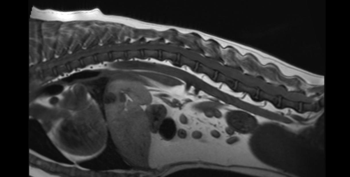
cross-sectional imaging. (abstract). Vet Radiol Ultrasound 2010;51:582. 328
29. Young BD, Levine JM, Porter BF, Chen-Allen AV, Rossmeisl JH, Platt SR, Kent M, 329
Fosgate GT, Schatzberg SJ. Magnetic resonance imaging features of intracranial 330
astrocytomas and oligodendrogliomas in dogs. Vet Radiol Ultrasound 2011;52:132-141. 331
30. Smirniotopoulos JG, Murphy FM, Rushing EJ, Rees JH, Schroeder JW. Patterns of 332
contrast enhancement in the brain and meninges. Radiographics 2007;7:525-551. 333
31. Rogers LR, Estes ML, Rosenbloom SA, Harrold L. Primary leptomeningeal 334
oligodendroglioma: case report. Neurosurgery 1995;36:166–169. 335
32. Leep Hunderfund AN, Zabad RK, Aksamit AJ, Morris JM, Meyer FB, Thorell WE, 336
Parisi JE, Giannini C. Diffuse anaplastic leptomeningeal oligodendrogliomatosis 337
mimicking neurosarcoidosis. Neurol Clin Pract. 2013 Jun;3(3):261-265. 338
339
Figure Legends: 340
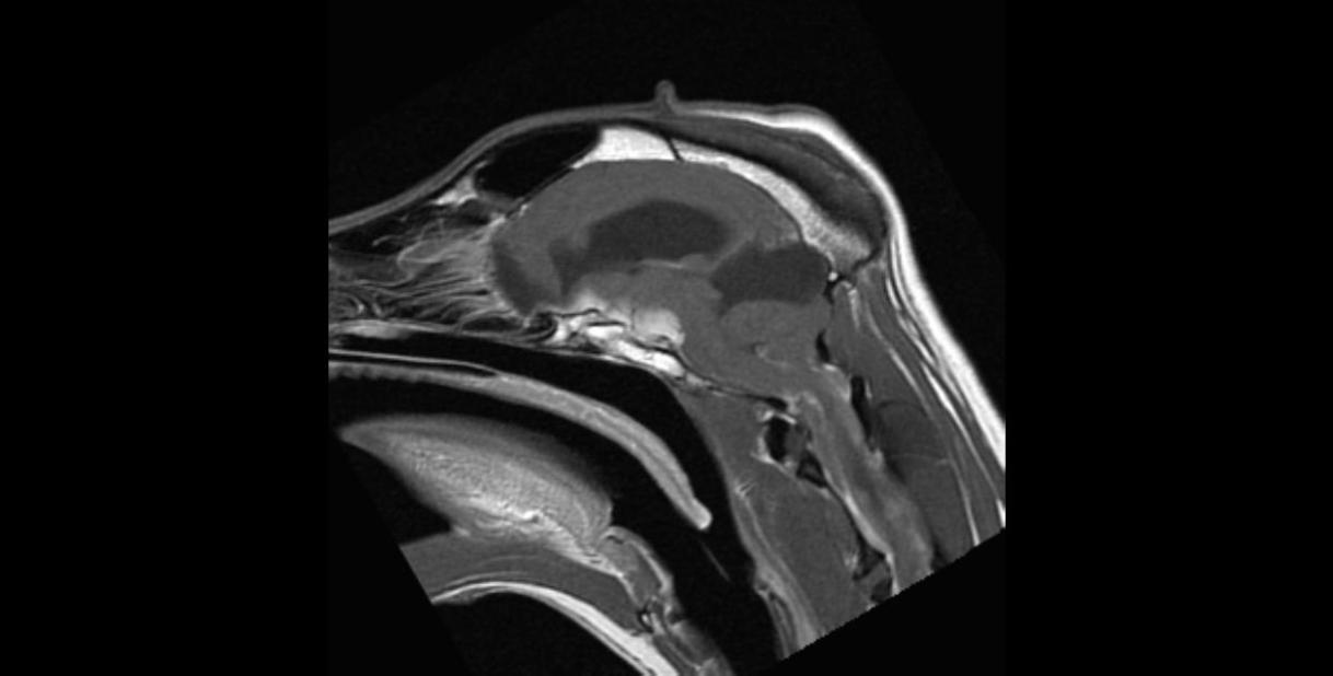
Fig. 1 A. Sagittal T1 – weighted postgadolinium. MR images revealed presence of 341
thickening of the adjacent meninges “dural tail” sign extending caudad (arrow) to the 342
lesion. Dilation of the central canal consistent with syringohydromyelia is seen (white 343
star). 344
Fig. 1 B. Transverse T1 - weighted postgadolinium. MR images revealed presence of 345
thickening of the adjacent meninges “dural tail” sign extending lateral (arrow heads) to 346
the lesion. Syrinx within the cervical spinal column also seen. 347
348
349
16
Fig. 2. Sagittal T1 – weighted postgadolinium images of the spine. MR images showed 350
the lesion extending along the entire vertebral canal as an irregular intradural lesion 351
ventral to the cord (arrows). 352
353
Fig. 3 A. Formalin-fixed brain section at the thalamic level showing the extra-axial mass 354
(see arrow). Severe secondary hydrocephalus is evident. 355
Fig. 3 B. The area of dural tail sign (arrow heads) was histologically characterized by 356
infiltration by the neoplastic cells. An extra-axial mass is indicated by the arrow. 357
Fig. 3 C. The larger magnification shows the presence of the neoplastic cells within the 358
subarachnoid space correlating with the dural tail sign on MRI. 359
360
Fig.4. Microphophotographs through the leptomeninges of the brain, correlating with 361
the MR images of Fig. 1A and Fig. 1B. (A) HE x20. Sheets, packets, palisades and 362
rows of neoplastic cells are embedded in an aboundant pale eosinophilic to amphophilic 363
matrix. (B) Oligo2 staining x 20. Neoplastic cells show strong nuclear reactivity for 364
Oligo2. 365
366