Embed Size (px)
Citation preview

Page 42 - VETcpd - Vol 2 - Issue 4
Laryngeal paralysis in dogs and cats
Alasdair Hotston Moore MA VetMB CertSAC CertVR CertSAS CertMedEd MRCVS
Alasdair Hotston Moore is Head of Referral Surgery at Bath Veterinary Referrals.He qualified from Cambridge in 1990. Following graduation, he joined Bristol Vet School, first as Intern in Small Animal Medicine and subsequently as CSTF Resident in Small Animal Soft Tissue Surgery. He was Lecturer and latterly Senior Clinical Fellow in Small Animal Soft Tissue Surgery at Langford from 1997until leaving in 2009. Alasdair accepts cases in all areas of Soft Tissue Surgery. He speaks on his subject area nationally and interna-tionally and has published widely in academic and professional journals. He holds post graduate qualifications in Veterinary Radiology, Small Animal Cardiology, Small Animal Surgery and Medical Education.Bath Veterinary Referrals Rosemary Lodge Veterinary Hospital Wellsway, Bath, Somerset BA2 5RLTel: 01225 832521Fax: 01225 835265Email: [email protected]
IncidenceLaryngeal paralysis (LP) is one of the two com-monest causes of upper airway obstruction in dogs, the other being brachycephalic airway obstruction (Millard and
Tobias 2009). In the author’s referral prac-tice, these present with similar frequency.
LP is much less common in cats than in dogs. In this species, in which upper airway obstruction is in any case rare, LP is a less dominant cause of laryngeal obstruc-tion than it is in dogs (causing 14 of 35 cases, Taylor et al. 2009).
Clinical signsDogs with LP generally have a chronic presentation, and most owners, when questioned, will report signs that have been present for months, although the final episode prompting veterinary attention may be acute. A chronic change in bark (dysphonia) is reported in most dogs, with the bark becoming muted or hoarse. In cats, change in voice is less often reported, although some owners mention that their pet becomes distressed when purring.
A chronic, somewhat characteristic, retching cough is reported in most dogs, sometimes described as a throat clearing cough. Coughing is not a common feature in cats.
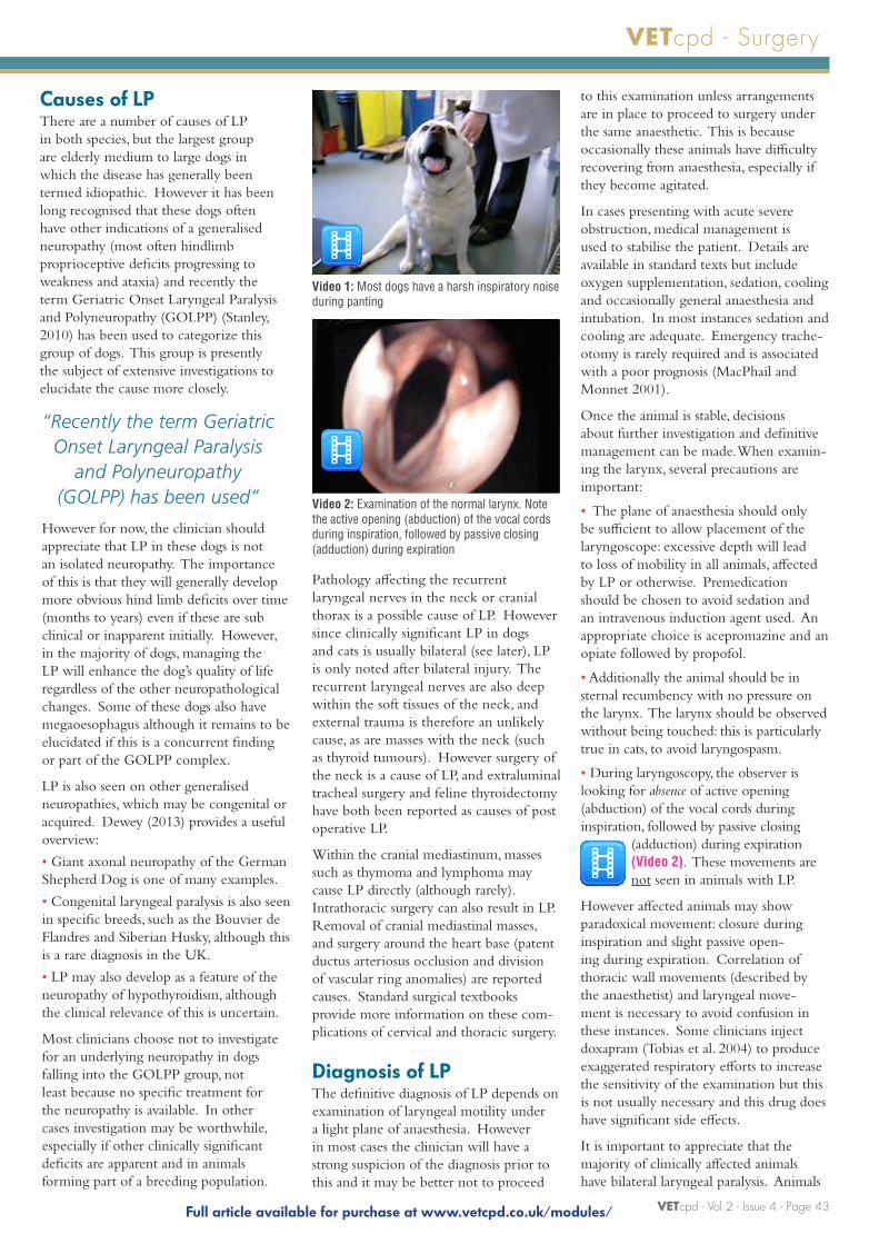
Most dogs also have a harsh inspiratory noise during pant-ing, stridor (Video 1), which
may not be apparent in the early stages and is less obvious when the dog is at rest. Stridor may not be appreciated by own-ers because it is of a gradual onset, and in dogs that are calm during clinic visits it is sometimes inapparent to inexperienced clinicians. Stridor is also less apparent in small dogs and cats, although it may be more obvious if the larynx is auscultated
VETcpd - Surgery
Laryngeal paralysis is one of the two commonest causes of upper airway obstruction in dogs, the other being brachycephalic airway obstruction. In the author’s referral practice, these present with similar frequency. This article will review the presenta-tion, causes and diagnosis and management of laryngeal paralysis in dogs. The disease is significantly rarer in cats but this species will be mentioned where relevant.
Key Words: canine, surgery, larynx, dyspnoea.
with a stethoscope. Exercise tolerance is generally reduced in affected animals, and in many cases is the immediate reason for presentation. However, animals with LP are generally old and exercise intolerance is often attributed to concurrent disease, such as osteoarthritis or cardiac insuffi-ciency, or owners simply assume that their pets are “slowing up” with age.
Cats are often not observed exercising as they are not taken for walks, therefore exercise intolerance is not noticed until the laryngeal obstructions is almost complete.
Obstruction due to LP is dynamic and worsened by increased respiratory effort: for this reason, signs of dyspnoea are worsened by excitement, heat or stress. In some dogs, this manifests as restlessness and panting especially in centrally heated houses.
Signalment The signalment is typical in the majority of dogs with LP. Since the commonest cause is a geriatric degenerative neu-ropathy (Millard and Tobias 2009 and see below), it is most often seen in elderly medium to large breed dogs. Typical breeds are Labrador and Golden Retriev-ers, Springer Spaniels and Border Collies, from 10 years of age and older. Males are slightly over represented. It is also seen in a variety of breeds of similar size (Flat Coated Retrievers, Greyhounds, Afghan Hounds, Saluki, Standard Poodles) and crossbreds. Giant breeds are also affected, such as the Newfoundland Retriever, but these are often somewhat younger at presentation (from five years). LP however is seen in a small number of dogs of all ages and types, sometimes associated with a specific neuropathy or other pathology, and often without obvious cause.
The signalment is less well defined in cats, although most are in middle or older age.
vetcpd.co.uk/urt
Video icons indicate video clips
For Soft Tissue Surgery Referrals in your area: www.vetindex.co.uk/STSMarket your referrals in VetIndex 2016! For further information call us on 01225 445561 or e-mail: [email protected]
Peer Reviewed
Full article available for purchase at www.vetcpd.co.uk/modules/ VETcpd - Vol 2 - Issue 4 - Page 43
Causes of LP There are a number of causes of LP in both species, but the largest group are elderly medium to large dogs in which the disease has generally been termed idiopathic. However it has been long recognised that these dogs often have other indications of a generalised neuropathy (most often hindlimb proprioceptive deficits progressing to weakness and ataxia) and recently the term Geriatric Onset Laryngeal Paralysis and Polyneuropathy (GOLPP) (Stanley, 2010) has been used to categorize this group of dogs. This group is presently the subject of extensive investigations to elucidate the cause more closely.
“Recently the term Geriatric Onset Laryngeal Paralysis
and Polyneuropathy (GOLPP) has been used”
However for now, the clinician should appreciate that LP in these dogs is not an isolated neuropathy. The importance of this is that they will generally develop more obvious hind limb deficits over time (months to years) even if these are sub clinical or inapparent initially. However, in the majority of dogs, managing the LP will enhance the dog’s quality of life regardless of the other neuropathological changes. Some of these dogs also have megaoesophagus although it remains to be elucidated if this is a concurrent finding or part of the GOLPP complex.
LP is also seen on other generalised neuropathies, which may be congenital or acquired. Dewey (2013) provides a useful overview:
• Giant axonal neuropathy of the German Shepherd Dog is one of many examples.
• Congenital laryngeal paralysis is also seen in specific breeds, such as the Bouvier de Flandres and Siberian Husky, although this is a rare diagnosis in the UK.
• LP may also develop as a feature of the neuropathy of hypothyroidism, although the clinical relevance of this is uncertain.
Most clinicians choose not to investigate for an underlying neuropathy in dogs falling into the GOLPP group, not least because no specific treatment for the neuropathy is available. In other cases investigation may be worthwhile, especially if other clinically significant deficits are apparent and in animals forming part of a breeding population.
Pathology affecting the recurrent laryngeal nerves in the neck or cranial thorax is a possible cause of LP. However since clinically significant LP in dogs and cats is usually bilateral (see later), LP is only noted after bilateral injury. The recurrent laryngeal nerves are also deep within the soft tissues of the neck, and external trauma is therefore an unlikely cause, as are masses with the neck (such as thyroid tumours). However surgery of the neck is a cause of LP, and extraluminal tracheal surgery and feline thyroidectomy have both been reported as causes of post operative LP.
Within the cranial mediastinum, masses such as thymoma and lymphoma may cause LP directly (although rarely). Intrathoracic surgery can also result in LP. Removal of cranial mediastinal masses, and surgery around the heart base (patent ductus arteriosus occlusion and division of vascular ring anomalies) are reported causes. Standard surgical textbooks provide more information on these com-plications of cervical and thoracic surgery.
Diagnosis of LP The definitive diagnosis of LP depends on examination of laryngeal motility under a light plane of anaesthesia. However in most cases the clinician will have a strong suspicion of the diagnosis prior to this and it may be better not to proceed
Video 1: Most dogs have a harsh inspiratory noise during panting
Video 2: Examination of the normal larynx. Note the active opening (abduction) of the vocal cords during inspiration, followed by passive closing (adduction) during expiration
VETcpd - Surgery
to this examination unless arrangements are in place to proceed to surgery under the same anaesthetic. This is because occasionally these animals have difficulty recovering from anaesthesia, especially if they become agitated.
In cases presenting with acute severe obstruction, medical management is used to stabilise the patient. Details are available in standard texts but include oxygen supplementation, sedation, cooling and occasionally general anaesthesia and intubation. In most instances sedation and cooling are adequate. Emergency trache-otomy is rarely required and is associated with a poor prognosis (MacPhail and Monnet 2001).
Once the animal is stable, decisions about further investigation and definitive management can be made. When examin-ing the larynx, several precautions are important:
• The plane of anaesthesia should only be sufficient to allow placement of the laryngoscope: excessive depth will lead to loss of mobility in all animals, affected by LP or otherwise. Premedication should be chosen to avoid sedation and an intravenous induction agent used. An appropriate choice is acepromazine and an opiate followed by propofol.
• Additionally the animal should be in sternal recumbency with no pressure on the larynx. The larynx should be observed without being touched: this is particularly true in cats, to avoid laryngospasm.
• During laryngoscopy, the observer is looking for absence of active opening (abduction) of the vocal cords during inspiration, followed by passive closing
(adduction) during expiration (Video 2). These movements are not seen in animals with LP.
However affected animals may show paradoxical movement: closure during inspiration and slight passive open-ing during expiration. Correlation of thoracic wall movements (described by the anaesthetist) and laryngeal move-ment is necessary to avoid confusion in these instances. Some clinicians inject doxapram (Tobias et al. 2004) to produce exaggerated respiratory efforts to increase the sensitivity of the examination but this is not usually necessary and this drug does have significant side effects.
It is important to appreciate that the majority of clinically affected animals have bilateral laryngeal paralysis. Animals