Embed Size (px)
Citation preview
VESTIBULAR SYSTEM
ANATOMY AND PHYSIOLOGY
Professor.Dr. M.K.Rajasekar MS., DLO.,

Life is hard for those who don’t have a VOR
During a walk I found too much motion in my visual
picture of the surroundings to permit recognition of fine
detail. I learned that I must stand still in order to read
the lettering on a sign
--J.C, 1952
M.D. with no
vestibular system
His vision was disturbed by head movements as small as
those induced by the beat of his heart while at rest.

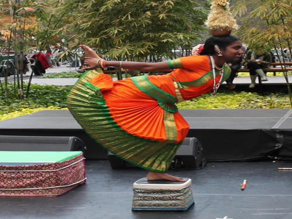
The vestibular system has important
sensory and motor functions
contributing to the perception of self
motion , head position and spatial
orientation relative to gravity
► Peripheral sensory apparatus
detects & relays information about head angular &
linear velocity to central processing system
orients the head with respect to gravity
► Central processing system
processes information in conjunction with other
sensory inputs for position and movement of head
in space
► Motor output system
generates compensatory eye movements and
compensatory body movements during head &
postural adjustments

Bony Part: SemiCircular Canals, Vestibule, Cochlea
SC ducts, Utricle, Saccule
Choclear membranes
Membranous Part
Buried deep in the temporal bone
the main peripheral component of
the vestibular system the elaborate
set of interconnected chambers, the
labyrinth in continuous with the
cochlea
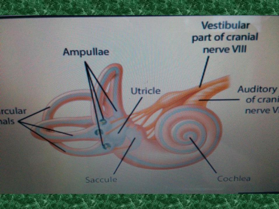
The labyrinth consists of two otolith organs
the utricle and the saccule and three
semicircular canals the vestibular haircells
like the cochlear hair cells produce minute
displacements into relevant receptor
potentials are located in the utricle and
saccule and three jug like swellings called the
ampulla located at the base of the
semicircular canals next to the utricle.

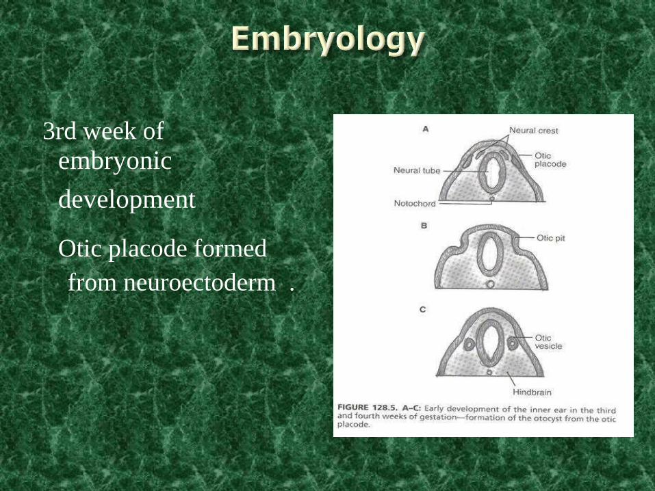
3rd week of
embryonic development
Otic placode formed
from neuroectoderm .

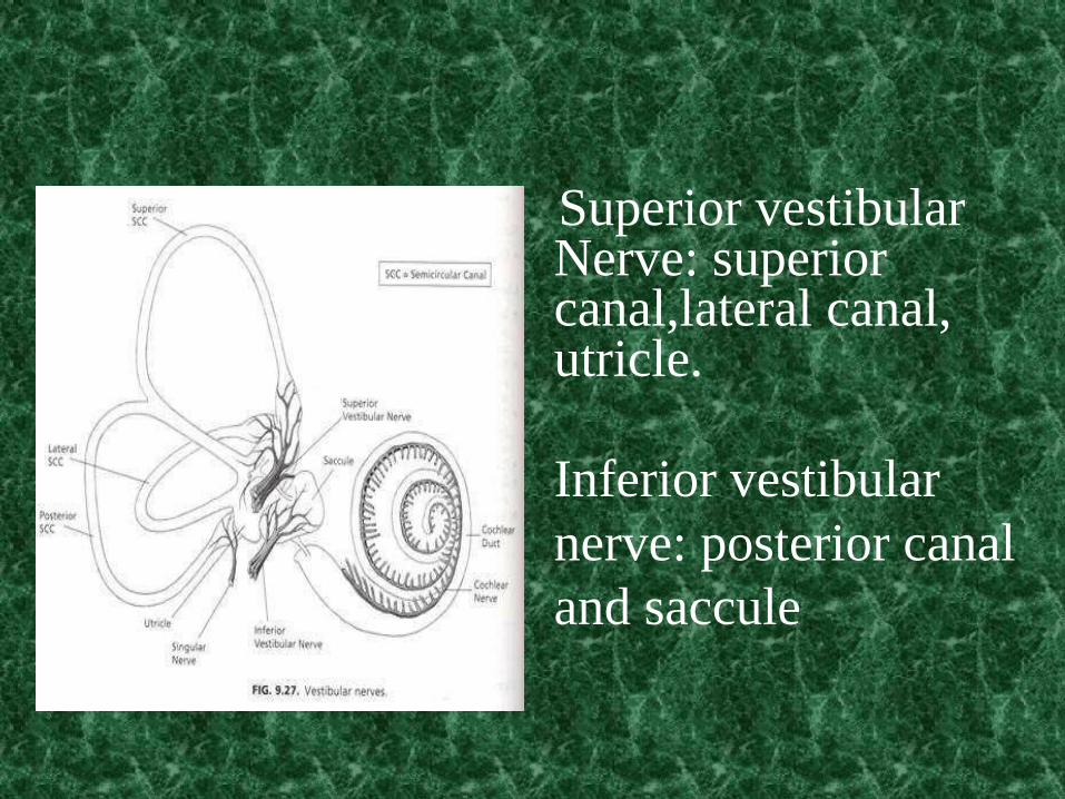
Superior vestibular Nerve: superior canal,lateral canal, utricle. Inferior vestibular
nerve: posterior canal
and saccule


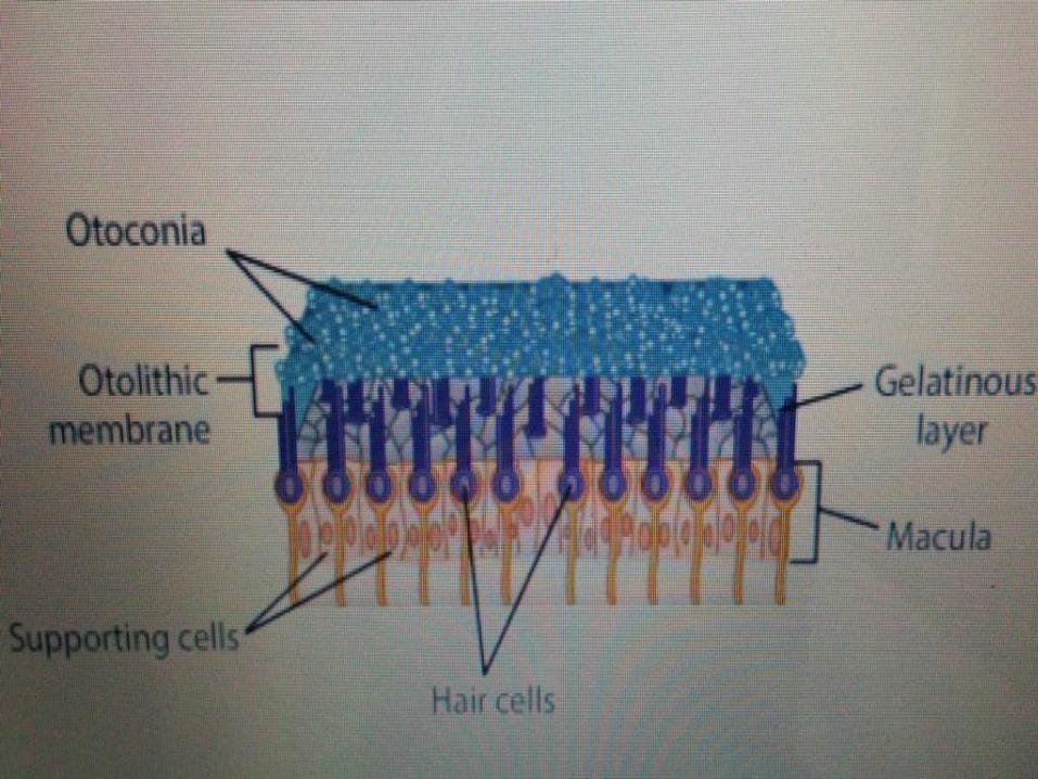
In the utricle and saccule the sensory epithelium
, the macula consists of hair cells and associated
supporting cells overlying the haircells and their
stereocilia is a gelatinous layer, above this layer
is a fibrous structure the otolith membrane in
which are embedded crystals of calcium
carbonate called otoconia the crystals give the
otolith organ the name(otolith in greek for ear
stones)
The otolith make the otoconial
membrane considerably heavier than
the structures and fluids surrounding
it thus when the head tilts gravity
causes the membrane to shift
relative to the sensory epithelium.
Semicircular canals sense angular Acceleration. Otolithic organs (utricle and
saccule)sense linear acceleration
Utricle in horizontal axis
Saccule in the vertical axis.

The resulting shearing motion between
the otolith membrane and the macula
displaces the hair bundle which are
embedded in the lower gelatinous
surface of the membrane this generates a
receptor potential in the hair cells , that
is dependent on the direction of the tilt.
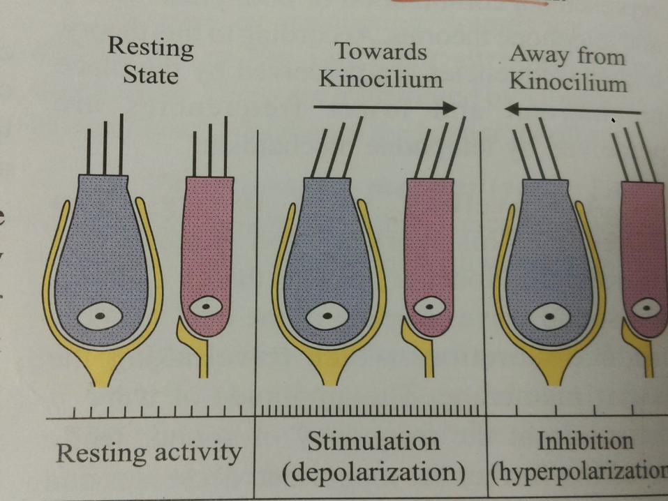
Movement of the stereocilia towards the
kinocilia causes the potassium channels to
open depolarizing the hair cells this results in
neurotransmitter release and excitation of
vestibular nerve fibers . Movement of the
stereocilia away from the kinocilia causes
closing of the potassium channels causing
hyperpolorisation of the cell thus reducing
vestibular nerve activity.

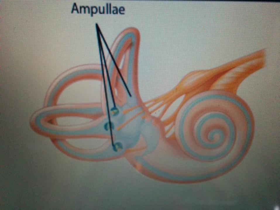
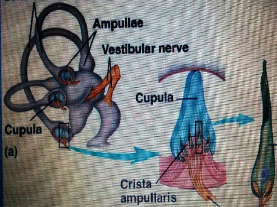
Each of the scc has a base expansion
called the ampulla which houses the
sensory epithelium or the crista that
contain the hair cells, the structure
of the canal suggest how they detect
the angular acceleration that arise
thro rotation of the head.

The hair bundles extend out of the crista
into the gelatinous mass,the cupula that
bridges the width of the gelatinous mass
forming a viscous barrier through which
endolymph cannot circulate. as a result
the compliant cupula is distorted by
movements of the endolymphatic fluid.
When the head turns in the direction of the
one of the plane of scc the inertia of
endolymph produces force along the cupula
distending it away from the direction of head
movement causing the displacement of hair
bundles within the crista in contrast the linear
acceleration of the head produces force equal
on both sides, so hair bundles are not
displaced.
The orientation of the hscc makes it
selectively sensitive to rotation in
the horizontal plane more
specifically the hair cells in the
canal on which side the head is
turning is depolarized while those
on the other side is hyperpolarized.