Embed Size (px)
Citation preview

331
Palestrica of the third millennium – Civilization and SportVol. 15, no. 4, October-December 2014, 331–335
CASE STUDIESSTUDII DE CAZ
Vertebral fracture - the first clinical sign of osteoporosis – Case reportFractura vertebrală, prima manifestare clinică a osteoporozei – Prezentare de caz
Viorela Ciortea, Rodica Ungur, Laszlo Irsay, Mădălina Nechita, Alina Popa, Ioan Onac, Ileana Monica Borda Rehabilitation Department, ”Iuliu Haţieganu” University of Medicine and Pharmacy, Cluj-Napoca, RomaniaClinical Rehabilitation Hospital, Cluj-Napoca, Romania
AbstractBackground. Osteoporosis is the most frequent metabolic bone disease characterized by: reduction of bone mass, altera-
tion of bone architecture, deterioration of bone quality, and increase of fracture risk. The frequency of osteoporotic vertebral fractures reported by the literature varies between 33-85%; of these, only 25-33% evidence a clinical manifestation when they occur. Regardless of the bone mineral density value, the presence of a vertebral fracture increases the risk of other vertebral fractures by 5 times, the risk of hip fractures by 1.8 times, and the risk of non-vertebral fractures by 1.6 times.
Aims. The purpose of this study was to assess a vertebral fracture as the first clinical sign of osteoporosis, and to distinguish predisposing factors for this disease complication.
Methods. The case report brings into discussion a patient in whom a vertebral fracture was the first clinical manifestation of osteoporosis.
Patient LP, aged 57 years, presented to our service in April 2013 for marked pain in the dorsolumbar spine, with onset 3 weeks before. Of the patient’s personal history and living conditions, we mention the onset of menopause at the age of 39 years, and smoking for the last 25 years, 20 cigarettes/day.
The objective examination of the patient at the time of presentation: BMI=23 kg/m², spontaneous pain at the percussion and mobilization of the dorsolumbar spine, without dural or neurological signs. Dorsolumbar spine X-ray, vertebral CT and the measurement of bone mineral density using the method of dual X-ray absorptiometry allowed us to make a positive diagnosis and to initiate adequate antiosteoporotic treatment. Any change in the shape, size, contour or structure of a vertebral body should be interpreted in a clinical context. The majority of osteoporotic fractures are located in the thoracic or thoracolumbar region; an osteoporotic fracture above T7 is unusual and the suspicion of malignancy should be eliminated.
Results. After 2 weeks of treatment (strontium ranelate 2 g/day, calcium 1000 mg/day and vitamin D3 1000 UI/day), we obtained an important improvement of symptomatology, and 4 weeks after the initiation of treatment, the patient resumed most of her daily activities.
Conclusions. An adequate oral treatment for osteoporosis, in combination with specific kinesitherapy, can reduce the pain and increase the mobility of the patient.
Key words: osteoporosis, vertebral fracture.
RezumatPremize. Osteoporoza este cea mai frecventă boală metabolică osoasă caracterizată prin: reducerea masei osoase, alterarea
arhitecturii osoase, deteriorarea calităţii osului şi creşterea riscului de fractură. Frecvenţa fracturilor vertebrale osteoporotice variază în literatură între 33-85%; dintre acestea doar 25-33% au manifestare clinică în momentul producerii. Indiferent de valoarea densităţii mineral osoase, prezenţa unei fracturi vertebrale creşte de 5 ori riscul de apariţie al altora la nivel vertebral, de 1,8 ori riscul de apariţie a fracturii de şold şi de 1,6 ori riscul de apariţie a unei fracturi non-vertebrale.
Obiective. Obiectivul acestui studiu este reprezentat de monitorizarea unei fracturi vertebrale, ca primă manifestare clinică a osteoporozei, şi de evidenţiere a factorilor de risc pentru această complicaţie a bolii.
Metode. Prezentarea de caz aduce în discuţie cazul unei paciente, în care fractura vertebrală a fost prima manifestare clinică a osteoporozei.
Copyright © 2010 by “Iuliu Haţieganu” University of Medicine and Pharmacy Publishing
Received: 2014, October 3; Accepted for publication: 2014, November 6; Address for correspondence: Rehabilitation Department, ”Iuliu Haţieganu” University of Medicine and Pharmacy Cluj-Napoca,
Clinical Rehabilitation Hospital, No. 46-50, Viilor St. 400437, Cluj-Napoca E-mail: [email protected], [email protected]; Corresponding author: Ileana Monica Borda

332
Viorela Ciortea et al.
IntroductionThe World Health Organization (WHO) defines
osteoporosis as a systemic disease of the skeleton, characterized by the reduction of bone mass and the deterioration of bone tissue microarchitecture, with a consecutive increase of bone fragility and fracture risk.
Bone mass reaches the highest level around the age of 26-30 years, and the normal bone mass loss rate is about 2% per year. In postmenopausal women, bone loss is accelerated, being approximately 4% per year, causing in this way the disappearance of 25-30% of the skeletal mass in 5-10 years. In the same time period, men lose approximately 12% of their bone mass. 60-80% of the bone mass is genetically determined, the rest of 20-40% being attributed to nutrition, physical exercise, medication, lifestyle (Jie et al., 2013).
WHO considers osteoporosis as one of the major diseases of the modern era, estimating that in the next 25 years, the number of osteoporosis cases will increase three times. Thus, osteoporosis and the fractures it causes are a major health problem for society and must be given priority attention (Briggs et al., 2007).
Vertebral fractures are found in approximately one third of women aged over 65 years, their incidence and prevalence being two times higher in women than in men (Khan et al., 2014).
The incidence of fractures that complicate osteoporosis increases with age. Thus, 97.2% of femoral neck fractures are found in subjects aged over 50 years; for these fractures, mortality in the first year is 12-24% (Păun, 1999).
The major clinical consequence of osteoporosis remains fracture, which occurs following a minor trauma or in its absence, which is why these fractures are termed insuffi-ciency fractures. The most common osteoporotic fracture sites are trabecular bone rich areas. The proportion of trabecular bone is different depending on the considered areas: lumbar vertebrae 75%, calcaneus 70%, proximal femur 50-70%, distal radius 25% (Pongchaiyakul et al., 2005).
The frequency of osteoporotic vertebral fractures reported by the literature varies between 33-85%; of these, only 25-33% evidence a clinical manifestation when they occur. Regardless of the bone mineral density (BMD)
value, the presence of a vertebral fracture increases the risk of other vertebral fractures by 5 times, the risk of hip fractures by 1.8 times, and the risk of non-vertebral fractures by 1.6 times (Lunt et al., 2002).
Radiologically, osteoporotic vertebral fractures induce changes in the shape, size, contour and structure of the vertebral body: concave vertebral endplate(s); lack of parallelism between the lines of the vertebral endplate, reduction of height compared to other vertebrae; reduction of anterior height compared to posterior height; a horizontal vertebral angle; a concave anterior margin of the vertebral body; opacities below the vertebral endplate given either by the compression of bone trabeculae or by the formation of callus (they are found particularly in central fractures in which the external margin remains intact); interrupted but not destroyed vertebral endplates (differential diagnosis with fractures secondary to metastases, primary tumors, myeloma); a step appearance; loss of vertical continuity with the adjacent vertebra (Mughal, 2002).
The majority of osteoporotic vertebral fractures are located in the thoracic or thoracolumbar region; any fracture situated above the thoracic vertebra 7 is unusual and should be suspected of malignancy. The most frequently affected vertebrae are D12 and L1, followed in decreasing order by the adjacent dorsal and lumbar vertebrae (Topini et al., 2014).
Vertebral compression is of several types: sometimes compression is predominantly anterior (Fig. 1), the vertebral body acquiring a trapezoid or cuneiform appearance. Cuneiform vertebrae situated in the dorsal region cause kyphosis in that segment of the spine. Sometimes compression is uniform, the vertebral body having a rectangular appearance from the side (collapsed vertebra). In the lumbar region, osteoporosis translates into a cupuliform deformation of vertebral endplates, which are depressed under the pressure transmitted by the discs, leading to a concave or biconcave appearance of the vertebra, depending on whether one or both endplates are affected (Fig. 1, Fig. 2, Fig. 3a, 3b, 3c, 3d) (Korkmaz et al., 2014).
Compression osteoporotic fracture of L2 with a height reduction of 55% in the anterior and middle region and 25% in the posterior region.
Pacienta, în vârstă de 57 ani s-a prezentat în serviciul nostru, în luna aprilie 2013 pentru o durere accentuată la nivelul coloanei dorso-lombare, cu debut de aproximativ 3 săptămâni. Dintre antecedentele personale şi condiţiile de viaţă ale pa-cientei, menţionăm instalarea menopauzei la 39 de ani, la o pacientă fumătoare de aproximativ 25 ani, 20 ţigări/zi.
Examenul obiectiv al pacientei în momentul prezentării: IMC=23Kg/m², durere spontană, la percuţia şi la mobilizarea coloanei vertebrale dorso-lombare, fără semne durale sau neurologice. Radiografia de coloană dorso-lombară, examinarea computer tomograf la nivel vertebral şi determinarea densităţii mineral osoase prin metoda absorţiometriei bifotonice cu raze X au stabilit diagnosticul pozitiv şi ne-au permis iniţierea unui tratament adecvat antiosteoporotic. Orice modificare de formă, dimensiune, contur şi structură ale unui corp vertebral, trebuie interpretată în context clinic. Majoritatea fracturilor osteoporo-tice sunt localizate în regiunea toracală sau toraco-lombară; o fractură osteoporotică deasupra T7 este neobişnuită şi trebuie eliminată suspiciunea de malignitate.
Rezultate. După 2 săptămâni de tratament (ranelat de stronţiu 2g/zi, calciu 1000 mg/zi şi vitamina D3 1000 UI/zi) am obţinut ameliorarea importantă a simptomatologiei, iar la 4 săptămâni după iniţierea tratamentului pacienta şi-a reluat majori-tatea activităţilor zilnice.
Concluzii. Tratamentul adecvat antiosteoporotic în combinaţie cu kinetoterapia specifică a ameliorat semnificativ durerea şi mobilitatea acestei paciente.
Cuvinte cheie: osteoporoză, fractură vertebrală.

333
Vertebral fracture - the first clinical sign of osteoporosis
Fig. 1 – Wedge osteoporotic fracture of T12 with a height reduc-tion of 30% in the anterior region and 20% in the middle region.
Fig. 2 – Compression osteoporotic fracture of L3 with a height reduction of about 40%.
Initially, the fracture appears as a small central depression that increases with time. At 6 months, around the collapsed area, an osteosclerotic line is found. At one year, the fracture is consolidated through a dense osteosclerosis line.
Dual-energy X-ray absorptiometry (DEXA) is the gold standard, with a performance equaled only by incomparably more expensive methods such as quantitative computed tomography (QCT). In the absence of DEXA, the diagnosis of osteoporosis according to WHO criteria cannot be established.
Vertebral compression falsely increases the measured bone mineral density; the greater the compression, the higher the bone mineral density, due to the reduction of the vertebral projection area (Fig. 4a, 4b) (Topini et al., 2014).
Fig. 4a – Lumbar spine X-ray (LL)
Fig. 4b – Lumbar spine - DEXA
Fig. 4 – Vertebral compression of L3. Falsely increased BMD at the level of this vertebra.
HypothesisWe assessed an osteoporotic vertebral fracture in
a 57-year-old female patient, without clinical signs of osteoporosis, as an example for clinical management.
Material and methodsThe study was performed in accordance with all current
deontological rules. The approval of the Ethics Committee of the ”Iuliu Haţieganu” University of Medicine and Pharmacy Cluj-Napoca and the patient informed consent were obtained.
Research protocolPeriod and place of the researchThis is the case of a patient, female, aged 57 years, who
presented to the Clinical Rehabilitation Hospital in April 2013, complaining of very intense pain in the dorsolumbar spine, with onset 3 weeks before, following a trunk movement.
Subjects and groupsOf the patient’s physiological and pathological personal
history and living conditions, we mention menarche at the age of 14 years, the onset of menopause at the age of 39 years, and smoking for the last 25 years, 20 cigarettes/day.
The general objective and locomotor system exami-nation at the time of presentation evidenced a normal
Fig. 3a – Dec 2011 Fig. 3b – May 2012 Fig. 3c – Dec 2012 Fig. 3d – CT Dec 2012 Fig. 3 – Compression fracture of L2 during evolution. Lateral lumbar X-ray.
334
Viorela Ciortea et al.
weight patient with a BMI = 25.5 kg/m² (weight 63 kg, height 1.57 m), spontaneous pain at the percussion and mobilization of the dorsolumbar spine, without dural or neurological signs. The patient had no associated disorders and was not under observation for any chronic disease.
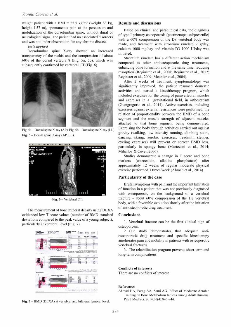
Tests appliedDorsolumbar spine X-ray showed an increased
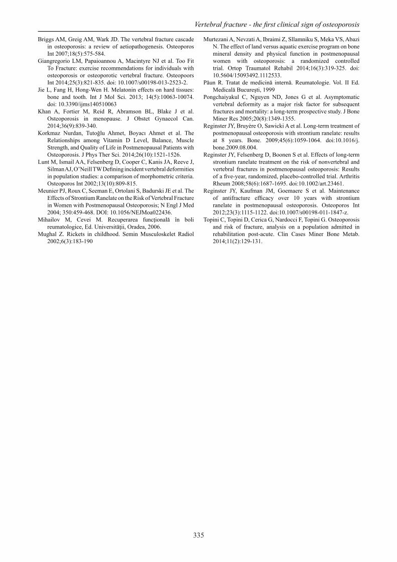
transparency of the rachis and the compression of about 60% of the dorsal vertebra 8 (Fig. 5a, 5b), which was subsequently confirmed by vertebral CT (Fig. 6).
Fig. 5a – Dorsal spine X-ray (AP) Fig. 5b – Dorsal spine X-ray (LL)Fig. 5 – Dorsal spine X-ray (AP, LL).
Fig. 6 – Vertebral CT.
The measurement of bone mineral density using DEXA evidenced low T score values (number of BMD standard deviations compared to the peak value of a young subject), particularly at vertebral level (Fig. 7).
Fig. 7 – BMD (DEXA) at vertebral and bilateral femoral level.
Results and discussionsBased on clinical and paraclinical data, the diagnosis
of type I primary osteoporosis (postmenopausal/presenile) with a 60% compression of the D8 vertebral body was made, and treatment with strontium ranelate 2 g/day, calcium 1000 mg/day and vitamin D3 1000 UI/day was initiated.
Strontium ranelate has a different action mechanism compared to other antiosteoporotic drug treatments, enhancing bone formation and at the same time, reducing resorption (Reginster et al., 2008; Reginster et al., 2012; Reginster et al., 2009; Meunier et al., 2004).
After 2 weeks of treatment, symptomatology was significantly improved, the patient resumed domestic activities and started a kinesitherapy program, which included exercises for the toning of paravertebral muscles and exercises in a gravitational field, in orthostatism (Giangregorio et al., 2014). Active exercises, including exercises against external resistances were performed, the relation of proportionality between the BMD of a bone segment and the muscle strength of adjacent muscles attached to that bone segment being demonstrated. Exercising the body through activities carried out against gravity (walking, low-intensity running, climbing stairs, dancing, skiing, aerobic exercises, treadmill, stepper, cycling exercises) will prevent or correct BMD loss, particularly in spongy bone (Murtezani et al., 2014; Mihailov & Cevei, 2006).
Studies demonstrate a change in T score and bone markers (osteocalcin, alkaline phosphatase) after approximately 12 weeks of regular moderate physical exercise performed 3 times/week (Ahmad et al., 2014).
Particularity of the caseBrutal symptoms with pain and the important limitation
of function in a patient that was not previously diagnosed with osteoporosis, on the background of a vertebral fracture - about 60% compression of the D8 vertebral body, with a favorable evolution shortly after the initiation of antiosteoporotic drug treatment.
Conclusions1. Vertebral fracture can be the first clinical sign of
osteoporosis.2. Our study demonstrates that adequate anti-
osteoporotic drug treatment and specific kinesiterapy ameliorates pain and mobility in patients with osteoporotic vertebral fractures.
3. The rehabilitation program prevents short-term and long-term complications.
Conflicts of interestsThere are no conflicts of interest.
ReferencesAhmad HA, Farag AA, Sami AG. Effect of Moderate Aerobic
Training on Bone Metabolism Indices among Adult Humans. Pak J Med Sci. 2014;30(4):840-844.
335
Vertebral fracture - the first clinical sign of osteoporosis
Briggs AM, Greig AM, Wark JD. The vertebral fracture cascade in osteoporosis: a review of aetiopathogenesis. Osteoporos Int 2007;18(5):575-584.
Giangregorio LM, Papaioannou A, Macintyre NJ et al. Too Fit To Fracture: exercise recommendations for individuals with osteoporosis or osteoporotic vertebral fracture. Osteopoors Int 2014;25(3):821-835. doi: 10.1007/s00198-013-2523-2.
Jie L, Fang H, Hong-Wen H. Melatonin effects on hard tissues: bone and tooth. Int J Mol Sci. 2013; 14(5):10063-10074. doi: 10.3390/ijms140510063
Khan A, Fortier M, Reid R, Abramson BL, Blake J et al. Osteoporosis in menopause. J Obstet Gynaecol Can. 2014;36(9):839-340.
Korkmaz Nurdan, Tutoğlu Ahmet, Boyacı Ahmet et al. The Relationships among Vitamin D Level, Balance, Muscle Strength, and Quality of Life in Postmenopausal Patients with Osteoporosis. J Phys Ther Sci. 2014;26(10):1521-1526.
Lunt M, Ismail AA, Felsenberg D, Cooper C, Kanis JA, Reeve J, Silman AJ, O’Neill TW Defining incident vertebral deformities in population studies: a comparison of morphometric criteria. Osteoporos Int 2002;13(10):809-815.
Meunier PJ, Roux C, Seeman E, Ortolani S, Badurski JE et al. The Effects of Strontium Ranelate on the Risk of Vertebral Fracture in Women with Postmenopausal Osteoporosis; N Engl J Med 2004; 350:459-468. DOI: 10.1056/NEJMoa022436.
Mihailov M, Cevei M. Recuperarea funcţională în boli reumatologice, Ed. Universităţii, Oradea, 2006.
Mughal Z. Rickets in childhood. Semin Musculoskelet Radiol 2002;6(3):183-190
Murtezani A, Nevzati A, Ibraimi Z, Sllamniku S, Meka VS, Abazi N. The effect of land versus aquatic exercise program on bone mineral density and physical function in postmenopausal women with osteoporosis: a randomized controlled trial. Ortop Traumatol Rehabil 2014;16(3):319-325. doi: 10.5604/15093492.1112533.
Păun R. Tratat de medicină internă. Reumatologie. Vol. II Ed. Medicală Bucureşti, 1999
Pongchaiyakul C, Nguyen ND, Jones G et al. Asymptomatic vertebral deformity as a major risk factor for subsequent fractures and mortality: a long-term prospective study. J Bone Miner Res 2005;20(8):1349-1355.
Reginster JY, Bruyère O, Sawicki A et al. Long-term treatment of postmenopausal osteoporosis with strontium ranelate: results at 8 years. Bone. 2009;45(6):1059-1064. doi:10.1016/j.bone.2009.08.004.
Reginster JY, Felsenberg D, Boonen S et al. Effects of long-term strontium ranelate treatment on the risk of nonvertebral and vertebral fractures in postmenopausal osteoporosis: Results of a five-year, randomized, placebo-controlled trial. Arthritis Rheum 2008;58(6):1687-1695. doi:10.1002/art.23461.
Reginster JY, Kaufman JM, Goemaere S et al. Maintenance of antifracture efficacy over 10 years with strontium ranelate in postmenopausal osteoporosis. Osteoporos Int 2012;23(3):1115-1122. doi:10.1007/s00198-011-1847-z.
Topini C, Topini D, Cerica G, Nardocci F, Topini G. Osteoporosis and risk of fracture, analysis on a population admitted in rehabilitation post-acute. Clin Cases Miner Bone Metab. 2014;11(2):129-131.


![1 16000939-01 Vertebral Compression Fracture Management Series ADDRESSING THE BURDEN OF VERTEBRAL COMPRESSION FRACTURES Presented by: [Name] [Title] [Institution]](https://img.dokumen.tips/doc/110x75/56649dbb5503460f94aad300/1-16000939-01-vertebral-compression-fracture-management-series-addressing-the.jpg)


