Embed Size (px)
Citation preview

CPG-1
Oxygen Therapy Verbal Check off outline NAME: ___________________________
Based on the CPG of the AARC DATE:______________________
Situation:
INDICATIONS:
� Documented hypoxemia � in adults, children, and infants older than 28 days, arterial oxygen tension (PaO2) of < 60 torr or arterial
oxygen saturation (SaO2) of < 90% in subjects breathing room air or with PaO2 and/or SaO2 below desirable range for specific clinical situation(1,2)
� in neonates, PaO2 < 50 torr and/or SaO2 < 88% or capillary oxygen tension (PcO2) < 40 torr(1,3,4) � An acute care situation in which hypoxemia is suspected(1,5,6-8)--substantiation of hypoxemia is required
within an appropriate period of time following initiation of therapy � Severe trauma(7,8) � Acute myocardial infarction(1,9) � Short-term therapy (eg, post-anesthesia recovery)(7,10)
CONTRAINDICATIONS:
� No specific contraindications to oxygen therapy exist when indications are judged to be present
PRECAUTIONS AND/OR POSSIBLE COMPLICATIONS:
� With PaO2 > or = 60 torr, ventilatory depression may occur in spontaneously breathing patients with elevated PaCO2.(8,11,12)
� With FIO2 > or = 0.5, absorption atelectasis, oxygen toxicity, and or depression of ciliary and/or leukocytic function may occur.(12,13)
� In newborns � In premature infants PaO2 of > 80 torr should be avoided because of the possibility of retinopathy of
prematurity.(2,14) � Increased PaO2 can contribute to closure or constriction of the ductus arteriosus-a possible concern in
infants with ductus-dependent heart lesions.(15) � Supplemental oxygen should be administered with caution to patients suffering from paraquat
poisoning(16) and to patients receiving bleomycin.(17) � During laser bronchoscopy, minimal levels of supplemental oxygen should be used to avoid intratracheal
ignition.(18) � Fire hazard is increased in the presence of increased oxygen concentrations. � Bacterial contamination associated with certain nebulization and humidifications systems is a possible
hazard.(19-21) Comments: ____________________________________________________________________ ______________________________________________________________________________ Passed _______ Failed ________ DATE: ___________ Instructor: ____________________

CPG-2
Pulse Oximetry Verbal Check off outline NAME: ___________________________
Based on the CPG of the AARC DATE:______________________
Situation:
INDICATIONS:
� The need to monitor the adequacy of arterial oxyhemoglobin saturation(1,4,6,9) � The need to quantitate the response of arterial oxyhemoglobin saturation to therapeutic intervention(4,9,10)
or to a diagnostic procedure (eg, bronchoscopy) � The need to comply with mandated regulations(11,12) or recommendations by authoritative groups(13,14)
CONTRAINDICATIONS:
� The presence of an ongoing need for measurement of pH, PaCO2, total hemoglobin, and abnormal hemoglobins may be a relative contraindication to pulse oximetry.
HAZARDS/COMPLICATIONS:
� Pulse oximetry is considered a safe procedure, but because of device limitations, false-negative results for hypoxemia(4) and/or false-positive results for normoxemia(15,16) or hyperoxemia(17,18) may lead to inappropriate treatment of the patient. In addition, tissue injury may occur at the measuring site as a result of probe misuse (eg, pressure sores from prolonged application or electrical shock and burns from the substitution of incompatible probes between instruments).
DEVICE LIMITATIONS/VALIDATION OF RESULTS:
� Factors, agents, or situations that may affect readings, limit precision, or limit the performance or application of a pulse oximeter include
� motion artifact(2,5,8,9,20) � abnormal hemoglobins (primarily carboxyhemoglobin [COHb] and met-hemoglobin
[metHb])(1,3,5,8,9,21) � intravascular dyes(1,3,8,9) � exposure of measuring probe to ambient light during measurement(2,3,8,9) � low perfusion states(1,3,4,8,9,21) � skin pigmentation(5,9,10,21) � nail polish or nail coverings with finger probe(9) � inability to detect saturations below 83%(22) with the same degree of accuracy and precision seen at higher
saturations(9,10,21,23,24)
Comments: ____________________________________________________________________ ______________________________________________________________________________
Passed _______ Failed ________ DATE: ___________ Instructor: ____________________

CPG-3
Bland Aerosol Verbal Check off outline NAME: ___________________________
Based on the CPG of the AARC DATE:______________________
Situation:
INDICATIONS:
� The presence of upper airway edema--cool bland aerosol(1,2) � Laryngotracheobronchitis (LTB)(1,2) � Subglottic edema(1,2) � Postextubation edema(1,2) � Postoperative management of the upper airway � The presence of a bypassed upper airway(3) � The need for sputum specimens(3,4)
CONTRAINDICATIONS:
� Bronchoconstriction(1,3,5,6) � History of airway hyperresponsiveness(1,2,5,6)
HAZARDS/COMPLICATIONS:
� Wheezing or bronchospasm(1,3,5,6) � Bronchoconstriction when artificial airway is employed(7-11) � Infection(12) � Overhydration(12) � Patient discomfort � Caregiver exposure to droplet nuclei of Mycobacterium tuberculosis or other airborne contagion produced
as a consequence of coughing, particularly during sputum induction
ASSESSMENT OF NEED Comments: ____________________________________________________________________ ______________________________________________________________________________
Passed _______ Failed ________ DATE: ___________ Instructor: ____________________

CPG-4
Aerosol delivery device Verbal Check off outline NAME: ___________________________
Based on the CPG of the AARC DATE:______________________
Situation:
INDICATIONS:
� The need to deliver--as an aerosol to the lower airways--a medication from one of the following drug classifications:
� Beta adrenergic agents Anticholinergic agents (antimuscarinics) Anti-inflammatory agents (eg, corticosteroids) Mediator-modifying compounds (eg, cromolyn sodium) Mucokinetics
CONTRAINDICATIONS:
� No contraindications exist to the administration of aerosols by inhalation. � Contraindications related to the substances being delivered may exist. Consult the package insert for
product-specific contraindications.
HAZARDS/COMPLICATIONS:
� Malfunction of device(3-5) and/or improper technique(6-12) may result in underdosing. � The potential exists for malfunction of device and/or improper technique (inappropriate patient use) to
result in overdosing. � Complications of specific pharmacologic agent may occur. � Cardiotoxic effects of Freon have been reported as an idiosyncratic response that may be a problem with
excessive use of MDI.(13-18) � Freon may affect the environment by its effect on the ozone layer.(19-21) � Repeated exposure to aerosols has been reported to produce asthmatic symptoms in some caregivers
Comments: ____________________________________________________________________ ______________________________________________________________________________
Passed _______ Failed ________ DATE: ___________ Instructor: ____________________

CPG-5
Delivery to Lung Parenchyma outline NAME: ___________________________
Verbal Check off based on the CPG of the AARC DATE:______________________
Situation:
INDICATIONS:
� The indication for selecting a suitable device is the need to deliver a topical medication (in aerosol form) that has its site of action in the lung parenchyma or is intended for systemic absorption. Such medications may possibly include antibiotics, antivirals, antifungals, surfactants, and enzymes.
CONTRAINDICATIONS:
� No contraindications exist to choosing an appropriate device for parenchymal deposition. � Contraindications related to the substances being delivered may exist. Consult the package insert for
product-specific contraindications to medication delivery.
HAZARDS/COMPLICATIONS:
� Malfunction of device and/or improper technique may result in underdosing or overdosing. � In mechanically ventilated patients, the nebulizer design and characteristics of the medication may
affect ventilator function (eg, filter obstruction, altered tidal volume, decreased trigger sensitivity) and medication deposition.(10,11)
� Complications related to specific pharmacologic agents can occur. � Aerosols may cause bronchospasm or irritation of the airway. � Exposure to medications(12-23) and patient-generated droplet nuclei may be hazardous to
clinicians.(24) � Exposure to medication should be limited to the patient for whom it has been ordered. Nebulized
medication that is released into the atmosphere from the nebulizer or exhaled by the patient becomes a form of "secondhand" exposure that may affect health-care providers and others in the vicinity of the treatment.
� The Centers for Disease Control and Prevention recommend addressing exposure control issues by (1) administrative policy, (2) engineering controls, and (3) personal protective equipment, in that order.(27,28)
� Administrative controls:Should include warning signs to apprise all who enter a treatment area of potential hazards of exposure. Accidental exposures should be documented and reported according to accepted standards.
Comments: ____________________________________________________________________ ______________________________________________________________________________
Passed _______ Failed ________ DATE: ___________ Instructor: ____________________

CPG-6
Delivery Aerosol to Upper Airway outline NAME: ___________________________
Verbal Check off Based on the CPG of the AARC DATE:______________________
Situation:
INDICATIONS:
� Upper airway inflammation (eg, to relieve inflammation due to laryngotracheobronchitis(2)) � Anesthesia (eg, to control pain and gagging during endoscopic procedures(3-7)) � Rhinitis (eg, to relieve inflammation and vascular congestion(8,9)) � Systemic disease (eg, to deliver peptides such as insulin(10))
CONTRAINDICATIONS:
� Known hypersensitivity to the medication being delivered
HAZARDS/COMPLICATIONS:
� Administration of medications for upper airway inflammation may result in � bronchospasm,(11,12) � rebound of symptoms, � systemic side effects. � Administration of medications for anesthesia(3-7) may result in � inhibition of gag reflex, � choking, � dehydration of epithelium, � allergic reactions, � excessive systemic effect, � bronchospasm, � Nosocomial infection from contaminated delivery device or medication (See AARC CPG on Selection of
Aerosol Delivery Device(13)).(14) � Administration of medications for rhinitis(15-21) may result in � nasal rebound (including rhinitis medicamentosa) after extended use of alpha adrenergic decongestants; � delayed effect (eg, effects of steroids are not immediate); � sensation of irritation and burning in the nose; � sneezing attacks (immediately following administration); � mucosal ulceration and bleeding; � postnasal drip. � Systemic disease--medication may cause nasal irritation or toxic effects.(9,10)
Comments: ____________________________________________________________________ ______________________________________________________________________________
Passed _______ Failed ________ DATE: ___________ Instructor:_________________

CPG-7
Incentive Spirometry Verbal Check off outline NAME: ___________________________
Based on the CPG of the AARC DATE:______________________
Situation:
INDICATIONS:
� Presence of conditions predisposing to the development of pulmonary atelectasis � upper-abdominal surgery(2,4,9-14) � thoracic surgery(9,10,13-15) � surgery in patients with chronic obstructive pulmonary disease (COPD)(7,13-15) � Presence of pulmonary atelectasis(16) � Presence of a restrictive lung defect associated with quadraplegia and/or dysfunctional
diaphragm.(6,8,14,17,18)
CONTRAINDICATIONS:
� Patient cannot be instructed or supervised to assure appropriate use of the device. � Patient cooperation is absent(2,16) or patient is unable to understand or demonstrate proper use of the
device.(16) � IS contraindicated in patients unable to deep breathe effectively (eg, with vital capacity [VC] less than about
10 mL/kg or inspiratory capacity [IC] less than about one third of predicted). � The presence of an open tracheal stoma is not a contraindication but requires adaptation of the spir
PRECAUTIONS AND/OR POSSIBLE COMPLICATIONS:
� Ineffective unless closely supervised or performed as ordered(6) � Inappropriate as sole treatment for major lung collapse or consolidation � Hyperventilation � Barotrauma (emphysematous lungs)(19) � Discomfort secondary to inadequate pain control(15,18) � Hypoxia secondary to interruption of prescribed oxygen therapy if face mask or shield is being used � Exacerbation of bronchospasm � Fatigue(20,21)
Comments: ____________________________________________________________________ ______________________________________________________________________________ Passed _______ Failed ________ DATE: ___________ Instructor: ____________________

CPG-8
Intermittent Positive Pressure Breathing NAME: ___________________________ Verbal Check off outline DATE:______________________
Based on the CPG of the AARC
Situation:
INDICATIONS:
� The need to improve lung expansion � The need for short-term ventilatory support for patients for are hypoventilated as an alternative to tracheal
intubation and continuous ventilatory support(12-2l) � The need to deliver aerosol medication (We are not addressing aerosol delivery for patients on long-term
mechanical ventilation)(4)
CONTRAINDICATIONS: Although no absolute contraindications to the use of IPPB therapy (except the oft-cited tension pneumothorax) have been reported, the patient with any of the following should be carefully evaluated before a decision is made to initiate IPPB therapy. � Intracranial pressure (ICP) > 15 mm Hg � Hemodynamic instability � Active hemoptysis � Nausea � Radiographic evidence of bleb � Singulation (hiccups)
HAZARDS/COMPLICATIONS:
� Barotrauma, pneumothorax(34) � Nosocomial infection(34) � Hemoptysis(4,35) � Hyperoxia when oxygen is the gas source(34) � Gastric distention(34) � Psychological dependence(34) � Impedance of venous return(34) � Hypoventilation � Air trapping, auto-PEEP, overdistended alveoli
Comments: ____________________________________________________________________ ______________________________________________________________________________
Passed _______ Failed ________ DATE: ___________ Instructor: ____________________
CPG-9
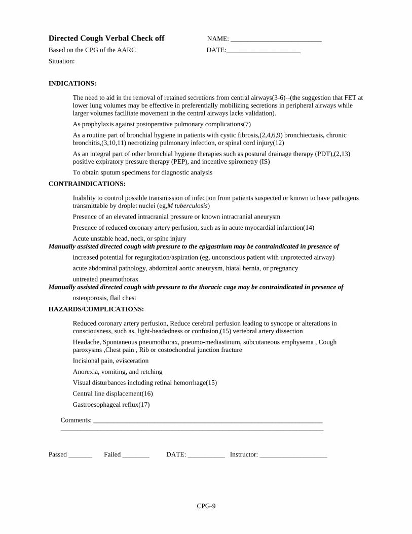
Directed Cough Verbal Check off NAME: ___________________________
Based on the CPG of the AARC DATE:______________________
Situation:
INDICATIONS:
� The need to aid in the removal of retained secretions from central airways(3-6)--(the suggestion that FET at lower lung volumes may be effective in preferentially mobilizing secretions in peripheral airways while larger volumes facilitate movement in the central airways lacks validation).
� As prophylaxis against postoperative pulmonary complications(7) � As a routine part of bronchial hygiene in patients with cystic fibrosis,(2,4,6,9) bronchiectasis, chronic
bronchitis,(3,10,11) necrotizing pulmonary infection, or spinal cord injury(12) � As an integral part of other bronchial hygiene therapies such as postural drainage therapy (PDT),(2,13)
positive expiratory pressure therapy (PEP), and incentive spirometry (IS) � To obtain sputum specimens for diagnostic analysis
CONTRAINDICATIONS:
� Inability to control possible transmission of infection from patients suspected or known to have pathogens transmittable by droplet nuclei (eg,M tuberculosis)
� Presence of an elevated intracranial pressure or known intracranial aneurysm � Presence of reduced coronary artery perfusion, such as in acute myocardial infarction(14) � Acute unstable head, neck, or spine injury Manually assisted directed cough with pressure to the epigastrium may be contraindicated in presence of � increased potential for regurgitation/aspiration (eg, unconscious patient with unprotected airway) � acute abdominal pathology, abdominal aortic aneurysm, hiatal hernia, or pregnancy � untreated pneumothorax Manually assisted directed cough with pressure to the thoracic cage may be contraindicated in presence of � osteoporosis, flail chest
HAZARDS/COMPLICATIONS:
� Reduced coronary artery perfusion, Reduce cerebral perfusion leading to syncope or alterations in consciousness, such as, light-headedness or confusion,(15) vertebral artery dissection
� Headache, Spontaneous pneumothorax, pneumo-mediastinum, subcutaneous emphysema , Cough paroxysms ,Chest pain , Rib or costochondral junction fracture
� Incisional pain, evisceration � Anorexia, vomiting, and retching � Visual disturbances including retinal hemorrhage(15) � Central line displacement(16) � Gastroesophageal reflux(17)
Comments: ____________________________________________________________________ ______________________________________________________________________________
Passed _______ Failed ________ DATE: ___________ Instructor: ____________________

CPG-10
Use of Positive Airway Pressure Adjuncts to Bronchial Hygiene Verbal Check off NAME: ___________________________
Based on the CPG of the AARC DATE:______________________
Situation:
INDICATIONS:
� To reduce air trapping in asthma and COPD(16,29-31) � To aid in mobilization of retained secretions (in cystic fibrosis and chronic bronchitis)(14,15,17-24,32,33) � To prevent or reverse atelectasis(6-13,34-36) � To optimize delivery of bronchodilators in patients receiving bronchial hygiene therapy(37,38)
CONTRAINDICATIONS: Although no absolute contraindications to the use of PEP, CPAP, or EPAP mask therapy have been reported,4,39 the following should be carefully evaluated before a decision is made to initiate PAP mask therapy. � Patients unable to tolerate the increased work of breathing (acute asthma, COPD) � Intracranial pressure (ICP) > 20 mm Hg, Hemodynamic instability(4) � Recent facial, oral, or skull surgery or trauma, Acute sinusitis, Epistaxis � Esophageal surgery, Active hemoptysis(39) � Nausea, Known or suspected tympanic membrane rupture or other middle ear pathology � Untreated pneumothorax
HAZARDS/COMPLICATIONS:
� Increased work of breathing(4) that may lead to hypoventilation and hypercarbia � Increased intracranial pressure � Cardiovascular compromise � myocardial ischemia � decreased venous return(4) � Air swallowing,(4) with increased likelihood of vomiting and aspiration � Claustrophobia(4) � Skin break down and discomfort from mask(4) � Pulmonary barotrauma(4)
Comments: ____________________________________________________________________ ______________________________________________________________________________
Passed _______ Failed ________ DATE: ___________ Instructor: ____________________

CPG-11
Postural Drainage Therapy Verbal Check off NAME: ___________________________
Based on the CPG of the AARC DATE:______________________
Situation:
INDICATIONS: Turning � inability or reluctance of patient to change body position. (eg, mechanical ventilation, neuromuscular
disease, drug-induced paralysis) � poor oxygenation associated with position(20,22,48-50) (eg, unilateral lung disease) � potential for or presence of atelectasis(24,26,30) � presence of artificial airway
Postural Drainage � evidence or suggestion of difficulty with secretion clearance � difficulty clearing secretions with expectorated sputum production greater than 25-30 mL/day
(adult)(3,7,9,11,12,27,38,40, 46,51-53) � evidence or suggestion of re-tained secretions in the presence of an artificial airway � presence of atelectasis caused by or suspected of being caused by mucus plugging(24,26,29,30,54) � diagnosis of diseases such as cystic fibrosis,(1,5,6,13-15,18,36,55) bronchiectasis,(4,5,14) or cavitating
lung disease � presence of foreign body in airway(56-58)
External Manipulation of the Thorax � sputum volume or consistency suggesting a need for additional manipulation (eg, percussion and/or
vibration) to assist movement of secretions by gravity, in a patient receiving postural drainag
CONTRAINDICATIONS: The decision to use postural drainage therapy requires assessment of potential benefits versus potential risks. Therapy should be provided for no longer than necessary to obtain the desired therapeutic results. Listed contraindications are relative unless marked as absolute (A). � All positions are contraindicated for, intracranial pressure (ICP) > 20 mm Hg, head and neck injury until
stabilized active hemorrhage with hemodynamic, instability recent spinal surgery (eg, laminectomy) or acute spinal injury acute spinal injury or active hemoptysis, empyema ,bronchopleural fistula, pulmonary edema associated with congestive heart failure, large pleural effusions, pulmonary embolism aged, confused, or anxious patients who do not tolerate position changes rib fracture, with or without flail chest surgical wound or healing tissue
� Trendelenburg position is contraindicated for intracranial pressure (ICP) > 20 mm Hg, patients in whom increased intracranial pressure is to be avoided (eg, neurosurgery, aneurysms, eye surgery) , uncontrolled hypertension, distended abdomen, esophageal surgery, recent gross hemoptysis related to recent lung carcinoma treated surgically or with radiation therapy,uncontrolled airway at risk for aspiration (tube feeding or recent meal)
� Reverse Trendelenburg is contraindicated in the presence of hypotension or asoactive medication External Manipulation of the Thorax, In addition to contraindications previously listed subcutaneous emphysema, recent epidural spinal infusion or spinal anesthesia recent skin grafts, or flaps, on the thorax, burns, open wounds, and skin infections of the thorax, recently placed transvenous pacemaker or subcutaneous pacemaker (particularly if mechanical devices are to be used)
CPG-12
Postural Drainage Therapy (continued) #2 HAZARDS/COMPLICATIONS:
� Hypoxemia Action To Be Taken/Possible Intervention: Administer higher oxygen concentrations during procedure if potential for or observed hypoxemia exists. If patient becomes hypoxemic during treatment, administer 100% oxygen, stop therapy immediately, return patient to original resting position, and consult physician. Ensure adequate ventilation. Hypoxemia during postural drainage may be avoided in unilateral lung disease by placing the involved lung up-permost with patient on his or her side.(20,22,48-50)
� Increased Intracranial Pressure Action To Be Taken/Possible Intervention: Stop therapy, return patient to original resting position, and consult physician.
� Acute Hypotension during Procedure Action To Be Taken/Possible Intervention: Stop therapy, return patient to original resting position, and consult physician.
� Pulmonary Hemorrhage Action To Be Taken/Possible Intervention: Stop therapy, return patient to original resting position, call physician immediately. Administer oxygen and maintain an airway until physician responds.
� Pain or Injury to Muscles, Ribs, or Spine Action To Be Taken/Possible Intervention: Stop therapy that appears directly associated with pain or problem, exercise care in moving patient, and consult physician.
� Vomiting and Aspiration Action To Be Taken/Possible Intervention: Stop therapy, clear airway and suction as needed, administer oxygen, maintain airway, return patient to previous resting position, and contact physician immediately.
� Bronchospasm Action To Be Taken/Possible Intervention: Stop therapy, return patient to previous resting position, administer or increase oxygen delivery while contacting physician. Administer physician-ordered bronchodilators.
� Dysrhythmias Action To Be Taken/Possible Intervention: Stop therapy, return patient to previous resting position, administer or increase oxygen delivery while contacting physician.
Comments: ____________________________________________________________________ ______________________________________________________________________________
Passed _______ Failed ________ DATE: ___________ Instructor: ____________________

CPG-13
Nasotracheal Suctioning Verbal Check off NAME: ___________________________
Based on the CPG of the AARC DATE:______________________
Situation:
INDICATIONS:
� The need to maintain a patent airway and remove secretions or foreign material from the trachea in the presence of
� inability to clear secretions;(6)
� audible evidence of secretions in the large/central airways that persist in spite of patient's best cough effort.(4,7,8-10)
CONTRAINDICATIONS:
� Occluded nasal passages � Nasal bleeding � Epiglottitis or croup (absolute) � Acute head, facial, or neck injury � Coagulopathy or bleeding disorder(2) � Laryngospasm(2) � Irritable airway � Upper respiratory tract infection
HAZARDS/COMPLICATIONS:
� Mechanical trauma, Laceration of nasal turbinates � Perforation of the pharynx, Nasal irritation/bleeding � Tracheitis, Mucosal hemorrhage(13) � Hypoxia/hypoxemia, Cardiac dysrhythmias/arrest � Bradycardia(1,19,22-24) � Increase in blood pressure(1,19,21) � Hypotension, Respiratory arrest(7) � Gagging/vomiting, Laryngospasm, Bronchoconstriction/bronchospasm � Nosocomial infection(15,16,23) � Atelectasis(5,14) � Misdirection of catheter((15,18) � Increased intracranial pressure (ICP)(21,26,27) � Intraventricular hemorrhage(21) � Exacerbation of cerebral edema
Comments: ____________________________________________________________________ ______________________________________________________________________________
Passed _______ Failed ________ DATE: ___________ Instructor: ____________________

CPG-14
Sampling for Arterial Blood Gas Analysis NAME: ___________________________
Verbal Check off based on the CPG of the AARC DATE:______________________
INDICATIONS:
The need to evaluate the adequacy of ventilatory (PacO2) acid-base (pH and PaCO2), and oxygenation (PaO2 and SaO2) status, and the oxygen-carrying capacity of blood (PaO2, HbO2, Hbtotal, and dyshemoglobins)
The need to quantitate the patient's response to therapeutic intervention(1,2,5) and/or diagnostic evaluation (eg, oxygen therapy, exercise testing)
The need to monitor severity and progression of a documented disease process.
CONTRAINDICATIONS: Contraindications are absolute unless specified otherwise.
Negative results of a modified Allen test (collateral circulation test) are indicative of inadequate blood supply to the hand' and suggest the need to select another extremity as the site for puncture.
Arterial puncture should not be pertormed through a lesion or through or distal to a surgical shunt (eg, as in a dialysis patient). If there is evidence of infection or peripheral vascular disease involving the selected limb, an alternate site should be selected.
Agreement is lacking regarding the puncture sites associated with a lesser likelihood of complications; however, because of the need for monitoring the femoral puncture site for an extended period, femoral punctures should not be performed outside the hospital.
A coagulopathy or medium-to-high-dose anticoagulation therapy (eg, heparin or coumadin, streptokinase, and tissue plasminogen activator but not necessarily aspirin) may be a relative contraindication for arterial puncture.
HAZARDS/COMPLICATIONS:
Hematoma, Arteriospasm Air or clotted-blood emboli, Anaphylaxis from local anesthestic Introduction of contagion at sampling site and consequent infection in patient; introduction of contagion to
sampler by inadvertent needle 'stick.’ Hemorrhage, Trauma to the vessel Arterial occlusion, Vasovagal response Pain
Comments: ____________________________________________________________________ ______________________________________________________________________________
Passed _______ Failed ________ DATE: ___________ Instructor: ____________________

CPG-15
ABG LIMITATIONS OF METHOD/VALIDATION OF RESULTS:
Artery may be inaccessible due to periarterial tissues (overlying muscle, connective tissue, or fat).
Pulse may not be palpable.
Arteriospasm may preclude collection despite successful introduction of needle into the artery.
Arterial blood specimens withdrawn from the body only reflect the physiologic condition at the moment of sampling (eg, pain from the puncture itself may lead to hyperventilation with consequent changes in values).
Specimens drawn at peak exercise best reflect response to exercise; however, specimens drawn within 15 seconds or less of termination of exercise may be acceptable (otherwise results do not reflect ventilatory status during dynamic activities and may yield false-negatives for hypoxemic events).(9-10)
Specimens from mechanically ventilated patients with minimal pulmonary pathology adequately reflect the effects of oxygen concentration change 10 minutes after the change.(7,8,11) In spontaneously breathing patients, at least 20-30 minutes should elapse following oxygen concentration change (patients with obstructive defects and increased residual volumes may require the full 30 minutes or longer).
Specimens held at room temperature must be analyzed within 10-15 minutes of drawing; iced samples should be analyzed within 1 hour.' The PaO2 of samples drawn from subjects with elevated white cell counts may decrease very rapidly. Immediate chilling is necessarySome dual-purpose electrolyte/blood gas analyzers stipulate immediate analysis without chilling because of possible elevations in potassium from chilling;(14) however, the accuracy of the blood gas results should not be affected by the chilling.
Validation of results:
Sample must be obtained anaerobically and anticoagulated, with immediate expulsion of air bubbles. Sample should be immediately chilled or analyzed within 10-15 minutes if left at room temperature.(15)
When a sample is obtained, date, time, patient's body temperature, position, activity level, respiratory rate, sample site, results of Allen test, inspired oxygen concentration(4,6-8) or supplemental oxygen flow, and mode of supported ventilation4 should be documented in the patient' s medical record with the results of blood gas analysis.
Appropriate sample size depends on (1) the anticoagulant used, (2) the requirements of the specific analyzers to be used, and (3) the presence of a need for other assays.
If liquid heparin (sodium or lithium, 1,000 units/mL of blood) is used, excess heparin (all except that filling the dead space of the syringe and needle) should be expelled and a blood sample of 2-4 mL be drawn (liquid heparin dilutes the specimen and changes PCO2 and PO2 in direct relationship to the heparin volume).(16)
If lyophilized heparin is used, the minimum volume drawn depends on the design of the analyzers and the need for other assays.
If other assays are required (eg, electrolyte determination), the choice of anticoagulant and the volume of the blood sample should be guided by the analyzer manufacturer's recommendations.

CPG-16
Capillary Blood Gas Sampling for Neonatal & Pediatric Patients NAME: ___________________________
Verbal Check off based on the CPG of the AARC DATE:______________________
INDICATIONS: Capillary blood gas sampling is indicated when
Arterial blood gas analysis is indicated but arterial access is not available. Noninvasive monitor readings are abnormal: transcutaneous values, end-tidal CO2, pulse oximetry. Assessment of initiation, administration, or change in therapeutic modalities (ie, mechanical ventilation) is
indicated. A change in patient status is detected by history or physical assessment. Monitoring the severity and progression of a documented disease process is desirable.
CONTRAINDICATIONS:
Capillary punctures should not be performed at or through the following sites posterior curvature of the heel, as the device may puncture the bone the heel of a patient who has begun walking and has callus development the fingers of neonates (to avoid nerve damage); previous puncture sites; inflamed, swollen, or edematous tissues; cyanotic or poorly perfused tissues; localized areas of infection peripheral arteries. on patients less than 24 hours old, due to poor peripheral perfusion; when there is need for direct analysis of oxygenation; when there is need for direct analysis of arterial blood; Relative contraindications include peripheral vasoconstriction; polycythemia (due to shorter clotting times); hypotension may be a relative contraindication.
HAZARDS/COMPLICATIONS:
Infection , Introduction of contagion at sampling site and consequent infection in patient, including calcaneus osteomyelitis and cellulitis
Inadvertent puncture or incision and consequent infection in sampler Burns, Hematoma, Bone calcification, Nerve damage Bruising, Scarring, Puncture of posterior medial aspect of heel may result in tibial artery laceration(11) Pain, Bleeding Inappropriate patient management may result from reliance on capillary PO2 values
Comments: ____________________________________________________________________ ______________________________________________________________________________
Passed _______ Failed ________ DATE: ___________ Instructor: ____________________

CPG-17
CBG LIMITATIONS OF METHOD/ VALIDATION OF RESULTS:
Limitations
Inadequate warming of the site prior to a puncture may result in capillary values that correlate poorly with arterial pH and PCO2 values.(19,20)
Undue squeezing of the puncture site may result in venous and lymphatic contamination of the sample.(14)
Variability in capillary PO2 values precludes using these samples for assessing oxygenation status.(1,2,3,18,20,21)
Validation of results
Sample must be anticoagulated and obtained anaerobically with capillary tube filled completely and air bubbles expelled immediately. Sample should be immediately chilled or analyzed within 10-15 minutes if left at room temperature.(22)
A respiratory assessment of the patient should be documented in the medical record at the time a capillary sample is performed (See 11.0 Monitoring).
An arterial sample may be analyzed to compare with the capillary pH and PCO2 values.

CPG-18
Removal of the Endotracheal Tube NAME: ___________________________
Verbal Check off based on the CPG of the AARC DATE:______________________
INDICATIONS/OBJECTIVES: When the airway control afforded by the endotracheal tube is deemed to be no longer necessary for the continued care of the patient, the tube should be removed. In general, the patient should be capable of maintaining a patent airway and adequate spontaneous ventilation and should not require high levels of positive airway pressure to maintain normal arterial blood oxygenation.
Patients in whom further medical care is considered (and explicitly declared) futile may have the endotracheal tube removed despite continuing indications for the artificial airway.
Acute artificial airway obstruction mandates immediate endotracheal tube removal if the obstruction cannot be cleared rapidly. Reintubation or other appropriate techniques for reestablishing the airway must be used to maintain effective gas exchange (ie, surgical airway management).
CONTRAINDICATIONS:
There are no absolute contraindications to extubation; however, some patients will require reintubation, positive pressure ventilation, continuous positive airway pressure, noninvasive ventilation, or high inspired oxygen fraction to maintain acceptable gas exchange after extubation. Airway protective reflexes are usually depressed immediately following and for some time after extubation and, therefore, measures to prevent aspiration should be considered.
HAZARDS/COMPLICATIONS:
Hypoxemia after extubation may result from but is not limited to failure to deliver adequate inspired oxygen fraction through the natural upper airway; acute upper airway obstruction; development of postobstruction pulmonary edema; bronchospasm; development of atelectasis, or lung collapse; pulmonary aspiration; hypoventilation Hypercapnia after extubation may be caused by but is not limited to: upper airway obstruction resulting from edema of the trachea, vocal cords, or larynx; respiratory muscle weakness; excessive work of breathing; bronchospasm. Death may occur when medical futility is the reason for removing the endotracheal tube.
LIMITATIONS OF METHODOLOGY/VALIDATION OF RESULTS Patients may need reintubation immediately or after some interval due to inappropriate extubation, progression of underlying disease, or development of a new disorder. A trial of extubation may be used in some marginal patients with the expectation that the need for reintubation is likely.
The need to reinsert an artificial airway following extubation is not necessarily an indication of poor practice. Inadequate airway maintenance and failure of reintubation may be an indication of poor practice.
The failure and complication rates of extubation can be used as quality monitors.
Comments: ____________________________________________________________________ ______________________________________________________________________________
Passed _______ Failed ________ DATE: ___________ Instructor: ____________________
CPG-19
Patient-Ventilator System Checks NAME: ___________________________
Verbal Check off based on the CPG of the AARC DATE:______________________
INDICATIONS: A patient-ventilator system check must be performed on a scheduled basis (which is institution- specific) for any patient requiring mechanical ventilation for life support. In addition, a check should be performed
prior to obtaining blood samples for analysis of blood gases and pH; prior to obtaining hemodynamic or bedside pulmonary function data; following any change in ventilator settings; as soon as possible following an acute deterioration of the patient's condition (this may or may not be
heralded by a violation of ventilator-alarm thresholds); any time that ventilator performance is questionable.(9)
CONTRAINDICATIONS: There are no absolute contraindications to performance of a patient-ventilator system check. If disruption of PEEP or FDO2 results in hypoxemia, bradycardia, or hypotension, portions of the check requiring disconnection of the patient from the ventilator may be contraindicated.(10,11)
HAZARDS/COMPLICATIONS:
Disconnecting the patient from the ventila-tor during a patient-ventilator system check may result in hypoventilation, hypoxemia, brady-cardia, and/or hypotension.(10,11)
Prior to disconnection, preoxygenation and hyperventilation may minimize these complications.(12-19) When disconnected from the patient, some ventilators generate a high flow through the patient circuit that
may aerosolize contaminated condensate, putting both the patient and clini-cian at risk for nosocomial infection.(20)
LIMITATIONS OF PROCEDURE/ VALIDATION OF RESULTS:
Measurements of volumes and inspired oxygen concentration are affected by the accuracy and reproducibility of the monitoring instruments.
Volume monitoring devices should be cali-brated at regular intervals. Volume monitoring accuracy should be ±10% of the measured volume.(21)
Oxygen analyzers should be calibrated at regular intervals. Oxygen analyzer accuracy should be ±3% of actual concentration.
Comments: ____________________________________________________________________ ______________________________________________________________________________
Passed _______ Failed ________ DATE: ___________ Instructor: ____________________

CPG-20
Management of Airway Emergencies NAME: ___________________________
Verbal Check off based on the CPG of the AARC DATE:______________________
INDICATIONS:
Conditions requiring management of the airway, in general, are impending or actual (1) airway compromise, (2) respiratory failure, and (3) need to protect the airway. Specific conditions include but are not limited to
Airway emergency prior to endotracheal intubation Obstruction of the artificial airway Apnea Acute traumatic coma(1) Penetrating neck trauma(2) Cardiopulmonary arrest and unstable dysrhythmias(3) Severe bronchospasm(4-8) Severe allergic reactions with cardiopulmonary compromise(9,10) Pulmonary edema(11,12) Sedative or narcotic drug effect(13) Foreign body airway obstruction(3) Choanal atresia in neonates(14) Aspiration Risk of aspiration Severe laryngospasm(15) Self-extubation(16,17)
Conditions requiring emergency tracheal intubation include, but are not limited to Persistent apnea Traumatic upper airway obstruction (partial or complete)(18-20) Accidental extubation of the patient unable to maintain adequate spontaneous ventilation(16,17) Obstructive angioedema (edema involving the deeper layers of the skin, subcutaneous tissue, and
mucosa)(21-23) Massive uncontrolled upper airway bleeding(2,24) Coma with potential for increased intracranial pressure(25) Infection-related upper airway obstruction (partial or complete) Epiglottitis in children or adults(26,27) Acute uvular edema(28) Tonsillopharyngitis or retropharyngeal abscess(29) Suppurative parotitis(30) Laryngeal and upper airway edema(31)
Neonatal- or pediatric-specific Perinatal asphyxia(32,33) Severe adenotonsillar hypertrophy(34,35) Severe laryngomalacia(36,37) Bacterial tracheitis(38-40)

CPG-21
Management of Airway Emergencies (continued 2 of 4) Neonatal epignathus(41,42) Obstruction from abnormal laryngeal closure due to arytenoid masses(43) Mediastinal tumors(44) Congenital diaphragmatic hernia(45) Presence of thick and/or particulate meconium in amniotic fluid(46-48) Absence of airway protective reflexes Cardiopulmonary arrest Massive hemoptysis(49) The patient in whom airway control is not possible by other methods may require surgical placement of an
airway (needle or surgical cricothyrotomy).(20,50,51) Conditions in which endotracheal intubation may not be possible and in which alternative techniques may
be used include but are not limited to restriction of endotracheal intubation by policy or statute;
Management of Airway Emergencies
difficult or failed intubation in the presence of risk factors associated with difficult tracheal intubations(52) such as
Short neck,(53) or bull neck(54) Protruding maxillary incisors(53) Receding mandible(53) Reduced mobility of the atlanto-occipital joint(55) Temporomandibular ankylosis(55) Congenital oropharyngeal wall stenosis(56) Anterior osteophytes of the cervical vertebrae, associated with diffuse idiopathic skeletal hyperostosis(57) Large substernal and/or cancerous goiters(58) Treacher-Collins syndrome(59) Morquio-Brailsford syndrome(60) Endolaryngeal tumors(61) when endotracheal intubation is not immediately possible
CONTRAINDICATIONS: Aggressive airway management (intubation or establishment of a surgical airway) may be contraindicated when the patient's desire not to be resuscitated has been clearly expressed and documented in the patient's medical record or other valid legal document.(62-64)
PRECAUTIONS/HAZARDS AND/OR COMPLICATIONS: The following represent possible hazards or complications related to the major facets of management of airway emergencies:
Translaryngeal intubation or cricothyrotomy is usually the route of choice. It may be necessary occasionally to use a surgical airway. Controversy exists as to whether intubation is hazardous in the presence of an unstable injury to the cervical spine. In one series the incidence of serious cervical spine injury in a severely injured population of blunt trauma patients was relatively low, and commonly used methods of precautionary airway management rarely led to neurologic deterioration.(65-67)
Failure to establish a patent airway(68-70)

CPG-22
Management of Airway Emergencies (continued 3 of 4) Failure to intubate the trachea(68,69) Failure to recognize intubation of esophagus(25,68,71-81) Upper airway trauma, laryngeal, and esophageal damage(82) Aspiration(70,74,82,83) Cervical spine trauma(67,84,85) Unrecognized bronchial intubation(25,68,72,82,86,87) Eye injury(70) Vocal cord paralysis(88) Problems with ETT tubes Cuff perforation(89) Cuff herniation(89) Pilot-tube-valve incompetence(90) Tube kinking during biting(70,89) Inadvertent extubation(17,25,68,72,86,91-93) Tube occlusion(17,72,82,89,93,94) Bronchospasm(68,70,74) Laryngospasm(72) Dental accidents(70) Dysrhythmias(94) Hypotension and bradycardia due to vagal stimulation(94) Hypertension and tachycardia(94,95) Inappropriate tube size(89,96-99) Bleeding Mouth ulceration(82)
Nasal-intubation specific Nasal damage including epistaxis Tube kinking in pharynx
Management of Airway Emergencies
Sinusitis(100-102) and otitis media Tongue ulceration Tracheal damage including tracheoesophageal fistula, tracheal innominate fistula, tracheal stenosis, and
tracheomalacia(103-107) Pneumonia(108) Laryngeal damage with consequent laryngeal stenosis,(82,101,107,109,110) laryngeal ulcer, granuloma,
polyps, synechia Surgical cricothyrotomy or tracheostomy specific(111,112)
Stomal stenosis(82,113) Innominate erosion(113) Needle cricothyrotomy specific(114-118) Bleeding at insertion site with hematoma formation Subcutaneous and mediastinal emphysema(117)

CPG-23
Management of Airway Emergencies (continued 4 of 4) Esophageal perforation Emergency ventilation Inadequate oxygen delivery(119-121) Hypo- or hyperventilation(122-124) Gastric insufflation and/or rupture(125,126) Barotrauma(127-129) Hypotension due to reduced venous return secondary to high mean intrathoracic pressure(130-132) Vomiting and aspiration(125) Prolonged interruption of ventilation for intubation(3,126) Failure to establish adequate functional residual capacity in the newborn(133-135) Movement of unstable cervical spine (more than by any commonly used method of endotracheal
intubation).(136) Failure to exhale due to upper airway obstruction during percutaneous transtracheal ventilation.(118,136)
LIMITATIONS OF PROCEDURE: Despite adequate management of airway emergencies, desired outcome may not be achieved because of the patient's underlying condition and progression of the process leading to the need for emergency airway management.
ASSESSMENT OF NEED: The need for management of airway emergencies is dictated by the patient's clinical condition. Careful observation, the implementation of basic airway management techniques, and laboratory and clinical data should help determine the need for more aggressive measures. Specific conditions requiring intervention include
Inability to adequately protect airway (eg, coma, lack of gag reflex, inability to cough) with or without other signs of respiratory distress.
Partially obstructed airway. Signs of a partially obstructed upper airway include ineffective patient efforts to ventilate, paradoxical respiration, stridor, use of accessory muscles, patient's pointing to neck, choking motions, cyanosis, and distress. Signs of lower airway obstruction may include the above and wheezing.
Complete airway obstruction. Respiratory efforts with no breath sounds or suggestion of air movement are indicative of complete obstruction.
Apnea. No respiratory efforts are seen. May be associated with cardiac arrest. Hypoxemia, hypercarbia, and/or acidemia seen on arterial blood gas analysis, oximetry or exhaled gas
analysis. Respiratory distress. Elevated respiratory rate, high or low ventilatory volumes, and signs of sympathetic
nervous system hyperactivity may be associated with respiratory distress. Comments: ____________________________________________________________________ ______________________________________________________________________________
Passed _______ Failed ________ DATE: ___________ Instructor: ____________________
CPG-24
Ventilator Circuit Changes NAME: ___________________________
Verbal Check off based on the CPG of the AARC DATE:______________________
INDICATIONS: The decision to change a ventilator circuit should be governed by
length of time the existing circuit has been in use;(1-11) type of circuit and humidification device in use;(1,3,6-20) circuit function (presence of a malfunctioning circuit or a circuit that leaks); appearance of ventilator circuit (circuits that are not clean in appearance should be replaced).
CONTRAINDICATIONS:
Presence of conditions in the patient's cardiopulmonary or neurologic status that might make tolerance of disconnection from mechanical ventilation hazardous to the patient Inability to safely and effectively ventilate or maintain patient during the ventilator circuit change Absence of a clean and functional circuit to use as a replacement
HAZARDS/COMPLICATIONS:
Patient's condition may predispose him or her to harm or injury during the changing process: hemodynamic instability, hypo- or hyperoxia with sequelae, hyped or hypocapnia, airway obstruction, artificial airway displacement, contamination of patient or staff from exposure to material in circuit.(8,21) Patient may not be safely maintained during disconnection from the ventilator: inappropriate or inadequate ventilation (f and/or VT), inappropriate or inadequate oxygenation (FI02 and/or PEEP), inappropriate increase in work of breathing, airway obstruction. Inability to assure that the replacement circuit has been satiety and effectively disinfected and that it is operationally sound(29,30) Transmission of pathogens to patient and to health care personnel Hazards of exposure to residual toxic disinfectants or associated disinfectant by products(30) Malfunctioning or suboptimally functioning ventilator or circuit Failure to assure proper ventilator function with patient reconnection (ie, correct settings, absence of leaks,
functioning alarms, proper valve placement(31)) Potential t'or patient-ventilator disconnection(31) Manipulation and disconnection of the ventilator tubing can cause contaminated ventilator condensate to
spill into the patient's airway, exposing the patient to further risk of infection,(8,21-24,26-28) Changing ventilator circuits more frequently than is necessary may increase the risk of nosocomial
pneumonia.(8,23) Failure to assure proper ventilator function prior to reinstituting mechanical ventilation may endanger
patient.(31)
LIMITATIONS OF PROCEDURE:
Evidence suggests that routine circuit changes (more frequently than every 48 hours) do not provide any advantage in controlling infections acquired by patients while being mechanically ventilated.(3-5,13,23,28,32)
Controversy exists concerning the frequency at which ventilator circuits should be changed to minimize patient infection.(1-11,13-20,23,24,26,32-36)

CPG-25
Ventilator Circuit Changes (continued 2 of 2) Controversy exists concerning the influence that circuit design and the presence of various circuit components
have on the safe interval of circuit change. The type of humidification device used may influence circuit change interval.(8,12-5-20,24,26,33-37) Use of heated vs non-heated circuits may influence circuit change interval.(6,7,9,10) Use of disposable vs reusable circuits may influence circuit change interval.(4,24,26,27) Use of aerosol generators (nebulizers) may influence circuit change interval.(2,21,29,31) Changing ventilator circuits too frequently may incur added patient or institutional expense and deliver no
additional infectious disease protection to the patient.(4,6,11-23)
ASSESSMENT OF NEED: Objective--limiting the transmission of infection
Reliance on published Centers for Disease Control and Prevention (CDC) recommendations(25-29) Reliance on institutional standards established by monitoring and surveillance and/or published research
Objective--to prevent malt'unction and to maintain optimal performance Competency of the circuit should be monitored for tubing leaks. In-line filters should be assessed for increased resistance.33-37 Equipment affected over time by water (eg, spirometers) should be monitored.
Objective--to maintain a circuit clean in appearance: Inspect appearance of circuit.
Comments: ____________________________________________________________________ ______________________________________________________________________________
Passed _______ Failed ________ DATE:___________ Instructor: _______________