Embed Size (px)
Citation preview

18
Ventilator-Induced Lung Injury: Mechanisms and Future Therapeutic Interventions
Maria A. Hegeman1,2, Marcus J. Schultz1,2, Adrianus J. van Vught3 and Cobi J. Heijnen4
1Laboratory of Experimental Intensive Care and Anesthesiology (LEICA), 2Department of Intensive Care Medicine, Academic Medical Center, Amsterdam,
3Department of Pediatric Intensive Care, 4Laboratory of Neuroimmunology and Developmental Origins of Disease (NIDOD),
University Medical Center Utrecht, Utrecht, The Netherlands
1. Introduction
Mechanical ventilation has become a standard technique to support the life of the critically ill patient in the intensive care unit (ICU) (Tobin, 2001). In general, mechanical ventilation is applied when the patient’s spontaneous ventilation is inadequate to maintain life. Especially patients who have developed acute respiratory failure require mechanical ventilation (Slutsky, 1993). Patients diagnosed with acute lung injury (ALI) suffer from severe pulmonary dysfunction which may persist for a long period of time. The extent and severity of ALI differs among patients, with the acute respiratory distress syndrome (ARDS) being the most severe manifestation of this lung disease (Schwarz, 2001). ALI and ARDS are characterized by the acute onset of diffuse neutrophilic alveolar infiltrates, protein-rich edema due to enhanced alveolar-capillary permeability and hypoxemic respiratory failure (ratio of arterial oxygen partial pressure to fractional inspired oxygen concentration, PaO2/FiO2 < 300 for ALI or < 200 for ARDS) (Ashbaugh et al, 1967; Petty & Ashbaugh, 1971). These pulmonary disorders may result from local injuries like pneumonia, gastric aspiration, near-drowning and lung contusion, but also from systemic events like severe sepsis, shock and blood transfusions (Hudson et al, 1995; Ware & Matthay, 2000). Although mechanical ventilation is a life-saving procedure, it has the potential to cause damage in healthy lung tissue or aggravate damage in diseased lung tissue (Dreyfuss & Saumon, 1998; Slutsky, 1999). The pulmonary complications secondary to mechanical ventilation are frequently referred to as ventilator-associated lung injury (VALI) in the clinical setting and ventilator-induced lung injury (VILI) in the experimental setting. As this chapter will primarily focus on experimental findings, the term VILI will be used.
2. Pathogenesis of ventilator-induced lung injury
VILI is characterized by enhanced alveolar-capillary permeability, accumulation of protein-rich lung edema, disturbed alveolar fibrin turnover, production of inflammatory mediators, and - ultimately - impaired gas exchange. Interestingly, these patterns of injury are very similar to those seen in ALI and ARDS (Tsuno et al, 1991).
www.intechopen.com

Front Lines of Thoracic Surgery
322
Over the years, various experimental models have been used to gather further insights into the mechanism(s) that may underlie the pathogenesis of VILI. These in vitro and in vivo studies support the clinical observations that mechanical ventilation evokes stretch trauma due to cyclic opening and collapse and/or overdistention of alveoli (Dreyfuss et al, 1985; Webb & Tierney, 1974). Ventilator-induced cyclic opening and closing of alveoli results in depletion of surfactant and renders the lung more prone to collapse (Dreyfuss et al, 1988; Mead et al, 1970). Extensive research demonstrated that surfactant depletion initiates loss of alveolar-capillary barrier function (Lachmann et al, 1987; Seeger et al, 1993; Verbrugge et al, 1997). In turn, serum proteins will leak from the circulation into pulmonary tissue. Protein accumulation in the lung will lead to surfactant inactivation, causing even more protein leakage and edema formation and eliciting a vicious circle of progressive lung injury (Ikegami et al, 1984; Lachmann et al, 1994). Besides surfactant dysfunction, other mechanisms have been proposed to be crucially involved in the development of VILI as well. It is reasonable to assume that these underlying mechanisms are interconnected.
2.1 Inflammatory response Leukocyte-endothelial interactions have been described to be important in the pathogenesis of severe inflammatory diseases related to VILI, like ALI/ARDS (Abraham, 2003; Moraes et al, 2003). In inflammation, pulmonary endothelial cells undergo a phenotypic shift (Orfanos et al, 2004). Activated endothelial cells secrete chemotactic cytokines (chemokines) which are essential for the recruitment of leukocytes to the site of inflammation (Luscinskas & Gimbrone, Jr., 1996). The main pulmonary chemoattractant is interleukin (IL)-8 or its rodent equivalents macrophage inflammatory protein (MIP)-2 and keratinocyte-derived chemokine (KC) (Donnelly et al, 1993). Besides release of chemokines and other inflammatory mediators, activated endothelial cells express various adhesion molecules on their cell surface thereby directing the multi-step cascade of leukocyte transmigration from blood vessels into affected tissue (Luscinskas & Gimbrone, Jr., 1996). The initial step of transmigration involves members from the selectin family (P- and E-selectin) which tether circulating leukocytes to vascular endothelium and facilitate rolling of leukocytes along the blood vessel wall (Carlos & Harlan, 1994). Subsequent leukocyte adhesion and extravasation are mediated by the immunoglobulin (Ig) superfamily comprising vascular cell adhesion molecule (VCAM)-1, intercellular adhesion molecule (ICAM)-1 and platelet-endothelial cell adhesion molecule (PECAM)-1. In ALI and ARDS patients, Gando et al. observed that soluble P-selectin, E-selectin, ICAM-1 and VCAM-1 levels were elevated within 24 hours after diagnosis (Gando et al, 2004). In addition, these authors showed a marked increase in these soluble adhesion molecules when subdividing patients into survivors and non-survivors. Together, these findings imply that adhesion molecules may have a prognostic value for development and clinical outcome of inflammatory lung diseases (Gando et al, 2004). Activated leukocytes release oxygen-based free radicals and proteolytic enzymes to restore physiological conditions of inflamed tissue. However, activated leukocytes may obstruct micro-capillaries in the lung due to an increased cellular size leading to ischemia and augmented oxygen-based free radical production. Accumulation of these inflammatory cells in the alveoli may therefore cause severe disruption of pulmonary epithelial-endothelial barriers leading to impaired gas exchange (Abraham, 2003; Lee & Downey, 2001). Kawano et al. were the first to describe that granulocyte depletion prior to injurious mechanical ventilation ameliorates pulmonary dysfunction in a rodent model of surfactant deficiency, stressing the importance of inflammatory mediators in the pathogenesis of VILI (Kawano et al, 1987). Later, Tremblay et al. revealed that alveolar stretch imposed by ex vivo mechanical ventilation caused IL-1┚, tumor necrosis factor (TNF)-┙, IL-6, MIP-2, interferon
www.intechopen.com

Ventilator-Induced Lung Injury: Mechanisms and Future Therapeutic Interventions
323
(IFN)-┛ and IL-10 expression and introduced the term “biotrauma” to describe the ventilator-induced secretion of inflammatory mediators (Tremblay et al, 1997; Tremblay & Slutsky, 1998). In line with this concept, it has been shown that alveolar macrophages secrete cytokines and chemokines upon in vitro applied mechanical stretch (Pugin et al, 1998). Although alveolar macrophages are considered to be the most important source of pulmonary cytokines and chemokines, other cell types like fibroblasts, leukocytes, epithelial and endothelial cells are also capable of producing inflammatory mediators (Kelley, 1990). In this respect, in vitro studies clearly showed that alveolar epithelial and capillary endothelial cells indeed release cytokines and chemokines upon mechanical stretch (Iwaki et al, 2009; Vlahakis et al, 1999). Ranieri et al. were the first to provide clinical evidence for the biotrauma-hypothesis (Ranieri et al, 1999). In a randomized controlled trial, these authors observed elevated concentrations of IL-1β, TNF-α, IL-6 and IL-1 receptor antagonist in the bronchoalveolar lavage fluid (BALf) of ALI/ARDS patients when excessive tidal volumes were used for 36 hours (Ranieri et al, 1999). Based on experimental and clinical observations, it has been hypothesized that the ventilator-induced inflammatory response in the lung may lead to pulmonary injury (Haitsma et al, 2003; Wilson et al, 2003; Wilson et al, 2005). Most experimental models of VILI, however, applied excessively high tidal volumes and/or inspiratory pressures compared to those applied in the clinical setting (Copland et al, 2004; Haitsma et al, 2003; Wilson et al, 2003). In an attempt to better reflect the human situation, we used clinically more relevant ventilator settings in our animal studies (Hegeman et al, 2010). In this way, we were able to prevent shock, metabolic acidosis and substantial damage to lung architecture commonly associated with high tidal volumes and/or inspiratory pressures (Wolthuis et al, 2009b). However, even in our relatively mild model of VILI, mechanical ventilation with either low (LVT) or high tidal volumes (HVT) caused increased cytokine, chemokine and adhesion molecule expression compared to non-ventilated control (NVC) animals, which was accompanied by marked granulocyte infiltration (Hegeman et al, 2011b). BALf neutrophil numbers and inflammatory mediator levels were even higher when comparing lungs of HVT-ventilated mice to lungs of LVT-ventilated mice (Hegeman et al, 2011b). Since lower PaO2/FiO2 ratios were only observed after HVT-ventilation, it seems plausible that ventilator-induced lung inflammation ultimately impairs gas exchange. In this respect, investigators suggested that enhanced pro-inflammation due to mechanical ventilation makes the patient more susceptible to a “second hit” (Bregeon et al, 2002). Mechanical ventilation itself may be the “second hit” when ventilating critically ill patients suffering from pulmonary injuries like ALI/ARDS or systemic events like sepsis (Headley et al, 1997; Meduri et al, 1995; Slutsky & Tremblay, 1998).
2.1.1 Local versus systemic inflammation An important clinical observation is that most ventilated critically ill patients do not succumb to acute respiratory failure but rather to progressive multiple organ failure (MOF) (Ferring & Vincent, 1997; Montgomery et al, 1985; Valta et al, 1999). A definition of MOF has been provided by John Marshall, who defined it as “the development of potentially reversible physiologic derangement involving two or more organ systems not involved in the disorder that resulted in ICU admission, and arising in the wake of a potentially life-threatening physiologic insult” (Marshall, 2001). In a clinical study, comprising 3147 patients from 198 ICUs in 24 European countries, Vincent et al. observed significant organ dysfunction in 71% of the ICU patients most of them being septic patients (Vincent et al, 2006). Moreover, they found a positive correlation between the number of organ systems failing and ICU mortality stressing the importance of MOF in the clinical outcome of critically ill patients (Vincent et al, 2006).
www.intechopen.com

Front Lines of Thoracic Surgery
324
Fig. 1. Mechanical ventilation induces endothelial activation and inflammation in pulmonary, hepatic and renal tissue.
www.intechopen.com

Ventilator-Induced Lung Injury: Mechanisms and Future Therapeutic Interventions
325
Reprinted with permission of Critical Care (Hegeman et al, 2009). To investigate whether alveolar stretch may cause endothelial activation and inflammation in the lung and distal organs, healthy mice were mechanically ventilated for 1, 2 or 4 hours with high pressures (i.e. high level of alveolar stretch). Non-ventilated mice served as a reference group. Spontaneous breathing animals were placed in an oxygen saturated box for 4 hours (FiO2 of 1.0, hyperoxia) to evaluate whether the high FiO2 associated with our ventilation strategy may contribute to changes in the immune response. A, E, I: In total lung, liver and kidney homogenates, respectively, mRNA expression of the adhesion molecule E-selectin was determined by real-time RT-PCR. B-C, F-G, J-K: In addition, mRNA expression of the pro-inflammatory cytokine interleukin (IL)-1┚ and the chemokine keratinocyte-derived chemokine (KC) was determined. Levels were normalized for expression of internal controls, i.e. the average value of ┚-actin and glyceraldehyde 3-phosphate dehydrogenase (GAPdH). D, H, L: In total lung, liver and kidney homogenates, respectively, myeloperoxidase (MPO) activity was determined as a measure of granulocyte infiltration. Levels were normalized for total protein concentration. Data are depicted relative to NVC and expressed as mean ± SEM for n = 4-8 animals. * p<0.05, ** p<0.01, *** p<0.001 (parameters were analyzed by one-way ANOVA with least significant difference (LSD) post-hoc test). NVC = non-ventilated controls; 1h, 2h, 4h = ventilated for either 1, 2 or 4 hours; O2 = hyperoxia for 4 hours; MV = mechanical ventilation.
There is convincing evidence that leukocyte-endothelial interactions are not only important in the pathogenesis of pulmonary disorders (Abraham, 2003; Moraes et al, 2003) but also in systemic events like sepsis and MOF (Whalen et al, 2000). It has been proposed that an elevation of adhesion molecule expression might contribute to tissue injury and ultimately to MOF by facilitating leukocyte activation and migration (Bone, 1991; Parrillo, 1993). In this regard, we examined whether alveolar stretch due to mechanical ventilation results in endothelial activation and inflammation in healthy mice, not only in the lung but also in organs distal to the lung (Hegeman et al, 2009). We observed that 4 hours of mechanical ventilation with high pressures, i.e. high level of alveolar stretch, induced de novo synthesis of various adhesion molecules in pulmonary but also in hepatic and renal tissue (figures 1a, e, i). In addition, increased cytokine and chemokine mRNA levels were found in the lung and distal organs after mechanical ventilation (figures 1b-c, f-g, j-k) accompanied by enhanced granulocyte infiltration (figures 1d, h, l). Our data imply that ventilator-induced endothelial activation in the lung, liver and kidney facilitates migration and adhesiveness of activated immune cells to inflamed tissue, which in turn may lead to tissue injury in these organs (Hegeman et al, 2009). Since we observed increased endothelial activation and inflammation after mechanical ventilation in the lung and distal organs, it seems plausible that mechanical ventilation plays a significant role in the development of both VILI and MOF (Hegeman et al, 2009). Supporting this hypothesis, earlier experimental research provided evidence that mechanical ventilation evokes detrimental effects in distal organs (Imai et al, 2003; Nin et al, 2006). Imai et al. demonstrated that mechanical ventilation with high tidal volumes induces epithelial cell apoptosis in the kidney and small intestine (Imai et al, 2003). Moreover, these authors showed that mechanical ventilation leads to elevated levels of serum markers such as creatinine indicative for renal failure (Imai et al, 2003). Ventilator-induced cardiovascular and hepatic injury has been described as well (Nin et al, 2006). It should be noted that these previous studies used animals with pre-existing lung injury whereas our study used healthy
www.intechopen.com

Front Lines of Thoracic Surgery
326
animals, suggesting that already existing inflammation is not a prerequisite for unveiling the negative effects of mechanical ventilation on distal organs. In line with this notion, a recent clinical trial showed that mechanical ventilation contributes to the development of acute kidney injury (AKI) in critically ill patients without ALI at onset of mechanical ventilation (Cortjens et al, 2011). Nonetheless, infectious events are known to aggravate ventilator-induced effects (Imai et al, 2003; O'Mahony et al, 2006) and possibly underlie the high incidence of MOF in critically ill patients ventilated with high tidal volumes and/or inspiratory pressures (Schultz, 2008). It remains to be determined which underlying mechanisms initiate the onset of endothelial activation and inflammation in distal organs during mechanical ventilation. Haitsma et al. and Tutor et al. demonstrated that ventilator-induced permeability of alveolar-capillary barriers provokes inflammatory mediator release into the blood circulation (Haitsma et al, 2000; Tutor et al, 1994). Although there is quite some evidence that inflammatory mediators will be released from inflamed pulmonary tissue into the systemic environment, a causal relationship between ventilator-induced spill-over and the pathogenesis of MOF has not been confirmed yet. Our experimental data indicate that ventilator-induced changes in de novo expression of adhesion molecules, cytokines and chemokines occur simultaneously in the lung, liver and kidney (Hegeman et al, 2009). Moreover, granulocyte recruitment to the liver and kidney was already observed after 1 hour of mechanical ventilation (Hegeman et al, 2009). In view of this spatiotemporal pattern of inflammatory mediator expression, we suggest that release or spill-over of cytokines and chemokines from the lung into the blood circulation is probably not the only cause of the enhanced pro-inflammatory environment in distal organs during mechanical ventilation. It cannot be excluded, however, that inflammatory mediators released into the circulation may contribute to de novo synthesis of adhesion molecules, cytokines and chemokines in distal organs, since cytokines like IL-1┚ and TNF-┙ are known to induce the production of a host of inflammatory mediators (Goodman et al, 2003). Since cytokines have in general only a very short half-life in plasma (± 20 minutes), it is more logical that locally produced mediators dictate the level of distal organ involvement. Local induction of cytokines by e.g. oxygen-based free radicals may be another important pro-inflammatory source in distal organs. Previously, it has been suggested that the physical stress of mechanical ventilation may trigger the sympathetic nervous system leading to an increase in catecholamine secretion (Plotz et al, 2004). Locally released catecholamines have been described to activate transcription factors like nuclear factor (NF)-κB in macrophages and promote production of IL-1β, TNF-α and IL-8 (Elenkov et al, 2000; Flierl et al, 2009; Spengler et al, 1990). These findings imply that stimulation of sympathetic nerve terminals in e.g. the liver and kidney may evoke a pro-inflammatory response in these peripheral organs (Elenkov et al, 2000; Straub et al, 2000). Subsequently, the presence of IL-1β, TNF-α and IL-8 may result in an acute phase response in the liver possibly via ┙–adrenergic activation (Elenkov et al, 2000; Flierl et al, 2009; Spengler et al, 1990). Based on these notions, we would like to propose that ventilator-induced activation of sympathetic nerve terminals in distal organs may contribute to the pro-inflammatory state of these organs. In turn, this pro-inflammatory state in distal organs may prime for the development of MOF in the critically ill patient (Imai et al, 2003; O'Mahony et al, 2006; Schultz, 2008). If so, modulation of adrenergic receptor function might be advantageous for the outcome of patients with MOF. Supporting this hypothesis, Miksa et al. showed in experimental models of sepsis that an ┙2A-adrenocepter antagonist reduced
www.intechopen.com

Ventilator-Induced Lung Injury: Mechanisms and Future Therapeutic Interventions
327
plasma levels of aspartate aminotransferase (AST), alanine aminotransferase (ALT) and creatinine, indicative for attenuated liver and kidney injury (Miksa et al, 2009). Moreover, these authors showed that inhibition of ┙2A-adrenocepter activation significantly improved survival in septic animals from 45 to 75% (Miksa et al, 2009). Another pathway of neuroimmune regulation is considered to be involved in the pathogenesis of VILI and MOF as well. In various inflammatory diseases, the efferent vagus nerve mediates anti-inflammatory action by downregulating pro-inflammatory immune responses (Kumar & Sharma, 2010). Recently, dos Santos et al. showed that disruption of the vagus nerve aggravates pulmonary wet-to-dry ratio, neutrophil infiltration, IL-6 production and apoptotic cell death, whereas stimulation of the vagus nerve was attenuating these parameters of VILI (dos Santos et al, 2011). These recent findings imply that the immunoregulatory function of the vagus nerve is of importance in VILI (dos Santos et al, 2011). Also in systemic diseases like sepsis, vagus-mediated neuroimmune signaling has been suggested to be important (Kumar & Sharma, 2010). In this respect, Pavlov et al. revealed that pharmacological activation of the vagus nerve improved survival of septic mice by inhibiting pro-inflammation (Pavlov et al, 2007). Taken together, stimulating the cholinergic anti-inflammatory pathway by pharmacological agents may represent potential therapeutic intervention strategies in patients diagnosed with VILI and/or MOF. Besides an enhanced systemic pro-inflammatory milieu, it has been hypothesized that suppression of the peripheral immune system may also be crucially involved in the pathogenesis of MOF (Pinsky et al, 1993; Syrbe et al, 1994). Initially, suppression of the peripheral immune function may provide a compensatory mechanism to restore homeostasis under physiological circumstances (Bone, 1996). Yet, the compensatory reaction may become maladaptive when existing for prolonged periods of time consequently leading to excessive suppression of peripheral lymphocytes and augmented susceptibility to infections. In this respect, Angele and Faist reported that many ventilated, critically ill patients suffer from unexplained immune suppression and thus have an associated increased risk for infections and MOF (Angele & Faist, 2002). Therefore, locally enhanced production of cytokines by e.g. endothelial cells, hepatic or renal cells in the distal organs - in combination with a hampering immune function - may be detrimental for the patient to survive. Previously, we demonstrated that mechanical ventilation suppresses lymphocyte function outside the lung (Plotz et al, 2002; Vreugdenhil et al, 2004). In children without lung pathology, the functional capacity of peripheral blood leukocytes to produce TNF-┙, IL-6 and IFN-┛ in vitro was significantly reduced after 2 hours of mechanical ventilation (Plotz et al, 2002). This was accompanied by a reduction in natural killer (NK) cell activity (Plotz et al, 2002). Also in healthy rats, we observed that 4 hours of mechanical ventilation with high pressures suppressed peripheral immune functioning, i.e. reduced NK cell activity, mitogen-induced splenocyte proliferation and cytokine production (Vreugdenhil et al, 2004). Supporting these earlier findings, 5 hours of HVT-ventilation resulted in diminished mitogen-induced splenocyte proliferation compared to NVC in our murine model of VILI (figure 2). Taken together, our data indicate that mechanical ventilation does not only induce a pro-inflammatory environment in organs distal to the lung, but also impairs the functioning of peripheral lymphocytes. Whether organ failure results from ongoing pro-inflammation of the environment (Pinsky et al, 1993) and/or persistent suppression of the immune function (Syrbe et al, 1994), these dysregulated immune responses may contribute to the development of MOF and subsequently cause increased morbidity and mortality in the critically ill patient (Rittirsch et al, 2008).
www.intechopen.com

Front Lines of Thoracic Surgery
328
Fig. 2. Mechanical ventilation with high tidal volumes induces suppression of splenocyte proliferation.
To investigate whether mechanical ventilation suppressed functioning of peripheral lymphocytes, healthy mice were mechanically ventilated for 5 hours with either low or high tidal volumes. Non-ventilated mice served as a reference group. In vitro mitogen-induced splenocyte proliferation was assessed as a measure of peripheral lymphocyte functioning. Splenocytes were stimulated in vitro with αCD3 (1µg/ml), a T cell specific antibody. After 48 hours 3H-thymidine was added and 16 hours later incorporation of 3H-thymidine was determined. Data are depicted relative to LVT and expressed as mean ± SEM for n = 10-16 animals. * p<0.05 (parameter was analyzed by one-way ANOVA with least significant difference (LSD) post-hoc test). NVC = non-ventilated controls; LVT, HVT = ventilated with either low or high tidal volumes.
2.2 Alveolar-capillary permeability The primary site of pulmonary gas exchange is the < 0.2 µm thin part of the alveolar-capillary membrane, consisting of alveolar epithelial and capillary endothelial cells (Burns et al, 2003; Weibel, 1984). Back in the 1980s, investigators observed that mechanical ventilation and the subsequent stretch of pulmonary tissue induces damage to epithelial-endothelial barriers thereby impairing gas exchange (Dreyfuss et al, 1985; Egan, 1982; Parker et al, 1984). In addition, Dreyfuss et al. demonstrated that enhanced microvascular permeability was responsible for the formation of pulmonary edema during mechanical ventilation with high tidal volumes (Dreyfuss et al, 1985). Besides apoptotic and/or necrotic cell death, loss of alveolar epithelial and capillary endothelial cell integrity is thought to play a crucial role in the ventilator-induced disruption of alveolar-capillary barriers (Pugin, 2003).
2.2.1 Cell death
Apoptosis is distinguished by cell shrinkage and nuclear fragmentation, while cell organelles and plasma membrane maintain their integrity for a prolonged period (Chang & Yang, 2000; Rossi & Gaidano, 2003). Apoptotic cell death is primarily regulated by cysteine aspartyl-specific proteases (caspases) and consists of two distinct routes: the intrinsic (mitochondria-mediated) and extrinsic (death receptor-mediated) pathway (Lavrik et al, 2005a). In response to oxidative stress, DNA damage and other types of severe intracellular stress, mitochondria undergo marked changes in membrane integrity and activate the intrinsic apoptotic pathway (Denecker et al, 2001; Festjens et al, 2004). Enhanced
www.intechopen.com

Ventilator-Induced Lung Injury: Mechanisms and Future Therapeutic Interventions
329
mitochondrial membrane permeability leads to release of pro-apoptotic proteins, like cytochrome c, from the mitochondria into the cytosol. Cytochrome c associates with apoptototic protease-activating factor (APAF)-1 and procaspase-9 to generate an apoptosome complex (Acehan et al, 2002; Jiang & Wang, 2000). In the apoptosome, procaspase-9 is cleaved to its mature form and activates the executioner caspase-3. Consequently, caspase-dependent DNAses are activated thereby causing chromatin condensation and DNA degradation (Chang & Yang, 2000). The extrinsic apoptotic pathway is initiated by triggering cell surface death receptors like the TNF receptor (TNFR)-1 and Fas (Lavrik et al, 2005b). Triggering of death receptors evokes formation of a death-inducing signaling complex (DISC) (Danial & Korsmeyer, 2004). The DISC brings procaspase-8 molecules in close proximity to each other resulting in their autoproteolytic activation (Chang et al, 2003). After autocleavage of procaspase-8, active caspase-8 is released into the cytosol and cleaves important executioner caspases (Chang et al, 2003). Caspase-8 is also involved in an indirect pathway linking the extrinsic and intrinsic routes of apoptotic cell death (Korsmeyer et al, 2000; Zamzami & Kroemer, 2003). In this pathway, activation of caspase-8 leads to the cleavage of Bid to truncated Bid (tBid) which induces permeability of the mitochondrial membrane. An important mediator of both the intrinsic and extrinsic apoptotic pathway is p53, a tumor suppressor molecule (Haupt et al, 2003). P53 is a transcription factor for several Bcl-2 family genes, including Bid and Bax, thereby promoting cytochrome c release from mitochondria and initiating the intrinsic apoptotic pathway (Haupt et al, 2003). In addition, p53 induces APAF-1 expression which is required for formation of the apoptosome complex (Kannan et al, 2001). P53 may also initiate the extrinsic apoptotic pathway through induction of Fas and activation of caspase-8 (Ding et al, 2000). Necrosis is characterized by irreversible plasma membrane damage, cytoplasmic swelling, organelle breakdown and, ultimately, cell rupture with leakage of cellular contents into the extracellular space (Fiers et al, 1999; Kroemer et al, 2005). Consequently, this type of cell death is associated with a marked inflammatory response. It has been proposed that necrosis might provide a backup suicide mechanism when caspase-dependent pathways of apoptosis cannot be properly activated (Fiers et al, 1999; Leist & Jäättelä, 2001) although this hypothesis may be too simplistic. Depletion of adenosine triphosphate (ATP) for instance may favor a switch from apoptosis to necrosis, in part because ATP is necessary for optimal activation of caspases (Leist et al, 1997). For many years, necrosis has been considered to be an uncontrolled process. However, recent evidence suggests that the course of necrotic cell death might be as tightly regulated as apoptotic cell death (Festjens et al, 2006; Golstein & Kroemer, 2007). It has to be kept in mind though, that necrotic and apoptotic traits might co-exist (Kroemer et al, 2009) and that the same cell death inducers may promote either necrosis or apoptosis depending on the specific environmental setting (Galluzzi et al, 2009). In this regard, previous research showed that triggering of TNFR-1 by TNF does not only lead to activation of the extrinsic pathway of apoptosis but also to programmed necrosis involving receptor-interacting protein (RIP) kinases (Galluzzi et al, 2009; Hitomi et al, 2008). Data from Cho et al., He et al. and Zhang et al. implicate RIP3 as a pivotal switch between TNF-induced apoptosis and necrosis (Cho et al, 2009; He et al, 2009; Zhang et al, 2009a). Based on this notion, the protein complex containing RIP1/RIP3 has been proposed to function as a “necrosome” (Declercq et al, 2009). In this setting, RIP3 promotes mitochondrial dysfunction, subsequent production of reactive oxygen species (ROS) and eventually necrotic cell death. Besides the production of ROS, necrotic cell
www.intechopen.com

Front Lines of Thoracic Surgery
330
death is associated with sustained elevation of cytosolic calcium levels (Harwood et al, 2005). Calcium overload leads to activation of calcium-dependent cysteine proteases, calpains. Active calpains promote release of lysosomal catabolic aspartyl proteases, cathepsins, which cleave essential cytoskeletal components like spectrin (also known as fodrin), microtubule subunits and microtubule-associated proteins thereby resulting in enhanced destabilization of the cell and ultimately in necrotic cell death (Artal-Sanz & Tavernarakis, 2005; Zhang et al, 2009b). Apart from necrosis, autophagy has been recognized as a distinct form of non-apoptotic cell death. Authophagy is the main cellular pathway for degradation of damaged cytoplasmic organelles or denatured proteins (Levine & Klionsky, 2004). In response to stress (e.g. hyperoxia, oxidative stress, pro-inflammation), autophagic vacuoles sequester damaged organelles or proteins. Subsequently, autophagic vacuoles fuse with lysosomes where the internalized contents will be degraded by lysosomal enzymes like cathepsins (Ryter & Choi, 2010). While basal activation of autophagy ascertains the physiological turnover of cytoplasmic organelles and proteins, dysregulation or excessive activation of autophagy may induce non-apoptotic cell death (Galluzzi et al, 2008; Maiuri et al, 2007). A potential cross-talk has been proposed between the mechanisms that regulate autophagy and those regulating apoptosis or necrosis (Galluzzi et al, 2008). Cell death has been considered to play a crucial role in the disruption of alveolar-capillary barriers (Pugin, 2003). Previously, in vitro studies demonstrated that mechanical stretch may activate apoptotic cell death (dos Santos et al, 2004; Edwards et al, 1999; Hammerschmidt et al, 2004). Moreover, several investigators reported enhanced apoptosis after 2 to 5 hours of mechanical ventilation with high tidal volumes (Chiang et al, 2011; Le et al, 2008; Li et al, 2007). Although the precise role of necrotic cell death has not been elucidated in VILI yet, it has been demonstrated in vitro that mechanical stretch as such induces necrosis (Hammerschmidt et al, 2004; Tschumperlin et al, 2000). Therefore, we investigated whether apoptotic and/or necrotic cell death pathways were activated in lungs of healthy mice exposed to either 5 hours of LVT or HVT-ventilation. Protein expression of cleaved caspase-8, -9 and -3 was determined in total lung homogenates as measures of caspase-dependent cell death. Protein expression of cleaved ┙-fodrin (145/150kDa fragments yielded by calcium-activated calpains) was determined as a measure of caspase-independent cell death. Our data demonstrate unequivocally that basal cleavage of caspase-8 was not enhanced by LVT or HVT-ventilation (figure 3a). Furthermore, cleaved caspase-9 and -3 were below detection level in all experimental groups implying that caspase-dependent pathways of apoptosis were not activated. Cleaved ┙-fodrin levels, however, were significantly increased in lungs of ventilated mice in comparison to NVC, which was independent of ventilation strategy (figure 3b). On the basis of these findings, it is tempting to speculate that 5 hours of mechanical ventilation induces cell death primarily via caspase-independent necrotic pathways. In contrast to our present data, Wolthuis et al. observed in a comparable model of VILI that the number of caspase 3-positive cells on lung sections was higher in HVT-ventilated mice than in control animals although this increase did not reach statistical significance (p=0.055) (Wolthuis et al, 2009a). Taken together, we propose that apoptosis is not the major executive mechanism of cell death in our experimental model of VILI. Nonetheless, we cannot exclude that caspase-dependent pathways of cell death are modestly activated which - together with necrotic activities induced in the target cell - will determine the level of cell death.
www.intechopen.com

Ventilator-Induced Lung Injury: Mechanisms and Future Therapeutic Interventions
331
Fig. 3. Mechanical ventilation activates caspase-independent pathways of cell death. To investigate whether mechanical ventilation activated caspase-dependent and/or caspase-independent pathways of cell death, healthy mice were mechanically ventilated for 5 hours with either low or high tidal volumes. Non-ventilated mice served as a reference group. A: In total lung homogenates, protein expression of cleaved caspase-8 was determined by Western blotting as a measure for activation of caspase-dependent cell death pathways. Cleaved caspase-9 and -3 were below detection level in all experimental groups. B: In addition, protein expression of cleaved ┙-fodrin (145/150kDa fragments yielded by calcium-activated calpains) was determined as a measure for activation of caspase-independent cell death pathways. Levels were normalized for total protein levels (Coomassie staining). Inset: representative Western blot depicting immunodetectable cleaved capase-8 and ┙-fodrin, respectively. Data are depicted relative to NVC and expressed as mean ± SEM for n = 5-8 animals. * p<0.05, *** p<0.001 (parameters were analyzed by one-way ANOVA with least significant difference (LSD) post-hoc test). NVC = non-ventilated controls; LVT, HVT = ventilated with either low or high tidal volumes.
2.2.2 Loss of vascular integrity
Under normal conditions, quiescent vascular endothelium establishes a tight barrier which controls the movement of plasma and/or leukocytes from the circulation into the underlying tissue (Cines et al, 1998). To achieve this barrier function, tight cell-cell contacts are formed by junctional transmembrane proteins such as vascular endothelial (VE)-cadherin (Dejana, 2004). Vascular leakage is an important feature of inflammatory disorders and is predominately caused by hyperpermeability of the endothelial barrier (Lentsch & Ward, 2000; Michel & Curry, 1999). One of the crucial systems regulating vascular cell integrity is the angiopoietin (Ang)-Tie2 system (Fiedler & Augustin, 2006). Ang-1 and Ang-2 are, respectively, an agonist and antagonist of the tyrosine kinase receptor Tie2 (Fiedler et al, 2003). Constitutive Ang-1 expression and Tie2 phosphorylation in adult vasculature implies that Ang-1−mediated Tie2 signaling is required to maintain endothelial cell integrity and quiescence (Fiedler &
www.intechopen.com

Front Lines of Thoracic Surgery
332
Augustin, 2006). Binding of Ang-1 leads to autophosphorylation of Tie2, subsequently activating several intracellular signaling pathways and maintaining endothelial cell integrity (Fiedler & Augustin, 2006). One of the signal transduction routes initiated upon Tie2 activation is the phosphatidylinositol 3 kinase (PI3K)–Akt pathway, which in general promotes cell survival (DeBusk et al, 2004; Kim et al, 2000; Papapetropoulos et al, 2000). In this regard, it has been shown that Ang-1−mediated Tie2 signaling may prevent endothelial cell apoptosis by activating Akt and upregulating survivin, an apoptosis inhibitor (Papapetropoulos et al, 2000). Daly et al. proposed a molecular mechanism through which Ang-1−mediated Akt activation may promote vascular stability (Daly et al, 2004). These authors demonstrated that Akt activation inactivates the forkhead transcription factor FKHR (FOXO1) which regulates genes associated with endothelial cell apoptosis (survivin) and vascular destabilization (Ang-2) (Daly et al, 2004). These findings imply that Ang-1−mediated Tie2 signaling prevents Ang-2 expression and subsequent loss of vascular integrity. Another signal transduction route that may be influenced upon Tie2 phosphorylation is the NF-κB pathway. Hughes et al. showed that Tie2 interacts with A20-binding inhibitor of NF-κB (ABIN)-2, a NF-κB regulatory protein (Hughes et al, 2003). As a consequence of ABIN-2 recruitment, Ang-1-mediated Tie2 signaling inhibits activation of the NF-κB pathway and results in an anti-inflammatory and quiescent status of the endothelial barrier (Hughes et al, 2003). Finally, Gavard et al. described that Ang-1−mediated Tie2 signaling protects against vascular endothelial growth factor (VEGF)-stimulated endothelial permeability by inhibiting VEGF-triggered endocytosis of VE-cadherin, a junctional transmembrane protein establishing tight cell-cell contacts (Gavard et al, 2008). Until now, an increase in Ang-2 levels has been recognized to be the modulating factor of the Ang-Tie2 system. In situations of endothelial activation, Ang-2 proteins are released from endothelial specific storage granules (Weibel-Palade bodies) and compete with Ang-1 for binding to the Tie2 receptor (Fiedler & Augustin, 2006). Accordingly, Ang-2 may exert antagonistic functions on Ang-1−mediated Tie2 signaling and alter vascular integrity, destabilize the endothelial barrier and prime the endothelium to attain responsiveness to pro-inflammatory mediators (Fiedler & Augustin, 2006). In line with this, van der Heijden et al. demonstrated that circulating Ang-2 and increased Ang-2/Ang-1 ratios are related to pulmonary permeability edema and severity of ALI/ARDS in ventilated patients with or without sepsis (van der Heijden et al, 2008). Furthermore, Gallagher et al. showed that high circulating Ang-2 levels in ALI/ARDS patients are associated with a poor outcome (Gallagher et al, 2008). Since 67% of these ALI/ARDS patients were mechanically ventilated, the authors proposed that ventilatory support alone does not cause enhanced Ang-2 levels (Gallagher et al, 2008). However, there is also conflicting evidence that Ang-2 may induce Tie2 activation in stressed endothelial cells (Daly et al, 2006). Both clinical and experimental studies recognized the importance of the Ang-Tie2 system in the pathogenesis of lung diseases related to VILI, like ALI and ARDS (van der Heijden et al, 2009). Karmpaliotis et al. demonstrated that vascular permeability and pulmonary edema were accompanied by reduced Ang-1 and enhanced VEGF levels in lungs of lipopolysaccharide (LPS)-challenged mice (Karmpaliotis et al, 2002). The same authors proposed that changes in the balance between Ang-1 (anti-leakage) and VEGF (pro-leakage) might contribute to the pathophysiology of ALI (Karmpaliotis et al, 2002). It may well be that a disturbed balance between Ang-1 and VEGF plays an indispensable role in VILI as well. Therefore, we examined whether 5 hours of either LVT or HVT-ventilation would influence the Ang-Tie2 system and VEGF expression in lungs of healthy
www.intechopen.com

Ventilator-Induced Lung Injury: Mechanisms and Future Therapeutic Interventions
333
mice (Hegeman et al, 2010). Particularly in lungs of HVT-ventilated mice, marked changes in the Ang-Tie2 system were observed. We demonstrated that Ang-1, Ang-2 and Tie2 mRNA expression was significantly reduced in HVT-ventilated mice in comparison with NVC (figures 4a-c). At this time point, Ang-1 and Ang-2 protein expression also tended to decrease (figures 4d-f). Moreover, we observed that HVT-ventilation caused increased expression of VEGF (figure 4g) which is in agreement with a prior report (Nin et al, 2008). Our findings strongly suggest that changes in the Ang-Tie2 system, together with elevated VEGF expression, are involved in the development of VILI (Hegeman et al, 2010).
Fig. 4. Mechanical ventilation affects the angiopoietin (Ang)-Tie2 system and vascular endothelial growth factor (VEGF) expression. Reprinted with permission of PLoS One (Hegeman et al, 2010). To investigate whether mechanical ventilation influences the Ang-Tie2 system, healthy mice were mechanically ventilated for 5 hours with high tidal volumes. Non-ventilated mice served as a reference group. A-C: In total lung homogenates, mRNA expression of Ang-1, Ang-2 and Tie2 was determined by real-time RT-PCR. Levels were normalized for expression of internal controls, i.e. the average value of ┚-actin and glyceraldehyde 3-phospate dehydrogenase (GAPdH). D-E: In total lung homogenates, protein expression of Ang-1 and Ang-2 was determined by Western blotting. Membranes were reprobed with antibody recognizing total Akt (Akt1/PKB┙) to control for equal loading. No group differences in total Akt were found. Inset: representative Western blot depicting immunodetectable Ang-1 and Ang-2, respectively. F-G: In total lung homogenates, protein expression of Tie2 and VEGF was determined by ELISA. Levels were normalized for total protein concentrations. Data are
www.intechopen.com

Front Lines of Thoracic Surgery
334
depicted relative to NVC and expressed as mean ± SEM for n = 6-12 animals. *** p<0.001 (parameters were analyzed by independent T-test). NVC = non-ventilated controls; HVT = ventilated with high tidal volumes.
In addition to the Ang-Tie2 system, Rho guanosine triphosphatases (GTPases) are recognized to be crucially involved in the mechanosensitive regulation of vascular permeability (Birukov, 2009; Spindler et al, 2010). Rho GTPases act as molecular switches that regulate a wide variety of signal transduction pathways involved in cell behavior (Schmidt & Hall, 2002; Wojciak-Stothard & Ridley, 2002). Rho GTPases cycle between an active guanosine triphosphate (GTP)-bound and an inactive guanosine diphosphate (GDP)-bound conformational state. Switching between the active and inactive form is tightly regulated. Guanine nucleotide exchange factors (GEFs), for example, promote the exchange from GDP to GTP leading to activation of Rho GTPases (Wojciak-Stothard & Ridley, 2002). Rac1 and Cdc42 are the main Rho GTPases needed for maintaining endothelial barrier function, whereas RhoA primarily has a destabilizing effect on endothelial barrier properties (Spindler et al, 2010). RhoA activation has been described to induce formation of stress fibers comprising actin and myosin (Ridley & Hall, 1992; Wojciak-Stothard et al, 1998). Consequently, actin-myosin contractility is suggested as the major mechanism of RhoA-mediated impairment of endothelial barrier properties (Spindler et al, 2010). RhoA signals through a downstream effector kinase, so-called Rho-kinase or Rho-associated coiled-col forming kinase (ROCK) (Liao et al, 2007). Activation of ROCK by RhoA inhibits myosin phosphatase acitivty subsequently leading to myosin light chain phosphorylation and stress fiber formation (Amano et al, 1997; Kimura et al, 1996).
3. Therapeutic interventions in critically ill patients with ventilator-induced lung injury
The recognition that conventional mechanical ventilation itself provokes detrimental effects in ventilated patients has led to the introduction of “lung-protective” ventilation strategies (Gillette & Hess, 2001). So far, mechanical ventilation with reduced tidal volumes is the only therapeutic approach effectively attenuating pulmonary injury and subsequent morbidity and mortality in critically ill ALI/ARDS patients (Amato et al, 1998; the ARDS network, 2000; Villar et al, 2006). In this regard, the ARDS Network revealed a 22% relative risk reduction in mortality rate when ventilating ALI/ARDS patients with “lung-protective” strategies, i.e. mechanical ventilation with tidal volumes of 6 ml/kg instead of with 12 ml/kg (the ARDS network, 2000). Notably, Determann et al. showed mechanical ventilation with lower tidal volumes also to attenuate development of lung injury in patients without ALI at the onset of mechanical ventilation (Determann et al, 2010). Recent research however, provided striking evidence that even “lung-protective” ventilator settings may result in the development of important aspects of VILI (Cobelens et al, 2009; Vaneker et al, 2007; Wolthuis et al, 2009b). We confirmed these prior reports in a murine model of VILI. We observed that not only conventional ventilation strategies with high tidal volumes but also clinically relevant ventilation strategies with low tidal volumes causes inflammation and vascular leakage in the lung (Hegeman et al, 2010; Hegeman et al, 2011b). Interestingly, a recent clinical trial revealed that mechanical ventilation with tidal volumes of 6ml/kg did not protect against the development or worsening of AKI in critically ill patients without ALI at onset of mechanical ventilation (Cortjens et al, 2011). Apart from preserving alveolar integrity by reducing tidal volumes, additional therapeutic intervention strategies that prevent the detrimental effects of mechanical stretch are therefore
www.intechopen.com

Ventilator-Induced Lung Injury: Mechanisms and Future Therapeutic Interventions
335
urgently needed. Based on the mechanisms that may underlie the pathogenesis of VILI (as mentioned above), therapies manipulating the course of inflammation and/or alveolar-capillary permeability might be advantageous for the clinical outcome of ventilated patients.
3.1 Inhibition of ventilator-induced lung inflammation: does it preclude lung injury?
It has been recognized that granulocytes and inflammatory mediators are important in the pathogenesis of VILI (Haitsma et al, 2003; Wilson et al, 2003; Wilson et al, 2005). Consequently, anti-inflammatory agents like glucocorticoids are suggested to attenuate or prevent the detrimental effects induced by mechanical ventilation (Brower et al, 2001; Luce, 2002). Glucocorticoids are a class of steroid hormones that exert their anti-inflammatory and immunosuppressive effects by binding to intracellular glucocorticoid receptors (GRs). After binding, the GR complex migrates from the cytosol to the nucleus where it regulates a wide range of gene activity, including inhibition of NF-κB and activator protein (AP)-1 driven expression of inflammatory genes (Barnes, 2006). Furthermore, glucocorticoids are known to suppress granulocyte recruitment and activation, preserve endothelial cell integrity and control vascular permeability (Ohta et al, 2001; Thompson, 2003). Although the efficacy of synthetic glucocorticoids to treat ALI/ARDS in critically ill patients is still under debate (Agarwal et al, 2007; Fernandes et al, 2005; Meduri et al, 2008), previous experimental research demonstrated that glucocorticoid treatment has the potential to attenuate ventilator-induced lung inflammation (Held et al, 2001; Ohta et al, 2001). Unfortunately, the anti-inflammatory effects of synthetic glucocorticoids cannot be separated from their metabolic effects. In particular when given at higher doses and for longer periods, systemic administration of glucocorticoids is associated with severe side effects like elevated blood glucose levels, deposition of body fat, suppressed systemic immunity and increased susceptibility to infections (Schacke et al, 2002). One way to reduce the unwanted side effects of glucocorticoid treatment is to selectively deliver these therapeutic agents into the diseased tissue (Asgeirsdottir et al, 2007). In this respect, liposomal formulations are valuable drug delivery systems as they can act as a depot from which the encapsulated drug will be slowly released to enable prolonged, local drug exposure at low concentrations (Storm & Crommelin, 1998). Moreover, liposomes preferably extravasate into tissues with increased capillary permeability (Storm & Crommelin, 1998) facilitating delivery at sites of inflammation and/or mechanical stretch. Previously, Ásgeirsdóttir et al. clearly showed that delivery of liposome-encapsulated dexamethasone inhibits pro-inflammatory gene expression without affecting blood glucose levels (Asgeirsdottir et al, 2007), one of the first clinically relevant side effects of free dexamethasone treatment (Feldman-Billard et al, 2006; Weinstein et al, 1995). Because local delivery of glucocorticoids by liposomal formulations could be of therapeutic importance in the context of VILI as well, we studied whether liposomes containing dexamethasone (Dex-liposomes) inhibited ventilator-induced lung inflammation (Hegeman et al, 2011a). We observed that administration of Dex-liposomes at initiation of ventilation diminished crucial inflammatory parameters of VILI such as IL-1┚, IL-6 and KC mRNA expression, especially in LVT-ventilated mice (Hegeman et al, 2011a). Yet, this formulation was not as effective as free dexamethasone in preventing granulocyte infiltration into pulmonary tissue (Hegeman et al, 2011a). As granulocytes are considered to be important in the development of VILI (Kawano et al, 1987), we hypothesized that phagocytosis of liposomes by activated granulocytes may be advantageous and should therefore be enhanced (Hegeman et al, 2011a). We proposed that IgG-modified Dex-liposomes (IgG-Dex-liposomes) may be more efficient in attenuating ventilator-induced lung inflammation than Dex-liposomes due to interaction with Fc┛-
www.intechopen.com

Front Lines of Thoracic Surgery
336
receptors (Fc┛Rs) on activated granulocytes and macrophages (McKenzie & Schreiber, 1998). Indeed, IgG-Dex-liposomes were pharmacologically more effective than Dex-liposomes particularly in preventing granulocyte infiltration induced by either LVT or HVT-ventilation (Hegeman et al, 2011a). Additionally, IgG-Dex-liposomes inhibited most parameters of ventilator-induced lung inflammation as efficient as free dexamethasone. Our experimental data imply that conjugation of IgG to Dex-liposomes significantly improves their efficacy in attenuating ventilator-induced lung inflammation (Hegeman et al, 2011a). Importantly, the use of liposomes may favor local release of dexamethasone thereby preventing unwanted systemic side effects (Asgeirsdottir et al, 2007). At present, it is thought that ventilator-induced lung inflammation may precede lung injury. Therefore, we hypothesized that reducing the inflammatory response by therapeutic intervention strategies would prevent vascular leakage and impaired gas exchange associated with mechanical ventilation. We observed, however, that the potent anti-inflammatory agent dexamethasone did not prevent occurrence of the more crude parameters of VILI like the elevation in BALf protein level, the increase in pulmonary wet-to-dry ratio and the reduction in PaO2/FiO2 ratio (Hegeman et al, 2011b). Our findings oppose previous studies describing protective effects of glucocorticoid therapy on lung injury (Nin et al, 2006; Ohta et al, 2001). In a rat model of VILI, dexamethasone was shown to restore pulmonary function after 75 minutes of mechanical ventilation as indicated by improved PaO2/FiO2 ratios (Nin et al, 2006). Moreover, Ohta et al. demonstrated that administration of methylprednisolone caused a marked leftward shifting of the pressure-volume (P-V) curve of rats ventilated for 40 minutes (Ohta et al, 2001). However, the deterioration of the P-V curve was still evident regardless of the significant reduction in granulocyte infiltration (Ohta et al, 2001). Ohta et al. explained these findings by the effects of mechanical stretch on lung tissue like stress failure of pulmonary capillaries, which may contribute to lung injury to a great extent (Ohta et al, 2001; West et al, 1991). Since mice were exposed to 5 hours of mechanical ventilation in our experimental model of VILI, it is tempting to speculate that the progressive lung injury induced by prolonged mechanical stretch may not be influenced by the anti-inflammatory action of dexamethasone (anymore). Similar to dexamethasone, administration of the vessel protective factor Ang-1 only inhibits inflammatory aspects of VILI (Hegeman et al, 2010). In a murine model of VILI, we observed that intravenous Ang-1 treatment at initiation of ventilation inhibited granulocyte infiltration and inflammatory mediator release, and completely abolished the increase in VEGF protein in lungs of HVT-ventilated mice (Hegeman et al, 2010). Although Ang-1 was preventing development of these important aspects of VILI, it did not influence the end points of VILI such as vascular leakage and impaired gas exchange (Hegeman et al, 2010). Our data are in apparent contrast with previously reported protective effects of Ang-1 on vascular leakage in endotoxin-challenged animals (Huang et al, 2008; McCarter et al, 2007; Mei et al, 2007; Witzenbichler et al, 2005). Using a similar dosing and administration procedure of Ang-1, David et al. showed that Ang-1 therapy also stabilized endothelial barrier function in an murine model of polymicrobiological abdominal sepsis as evidenced by attenuation of protein leakage from lung capillaries into the alveolar compartment (David et al, 2011). These findings underline that the pathways involved in sepsis- and ventilator-induced lung injury are different. An explanation for this discrepancy might be that the enhanced inflammation is not the primary inducer of vascular leakage and impaired gas exchange during HVT-ventilation as is the case in the
www.intechopen.com

Ventilator-Induced Lung Injury: Mechanisms and Future Therapeutic Interventions
337
induction of lung injury by sepsis. Indeed, it has been shown that ventilator-induced mechanical stretch itself may destabilize alveolar-capillary barrier function thereby resulting in increased vascular permeability and pulmonary edema (Dreyfuss et al, 1985; Egan, 1982; Parker et al, 1984). Ang-1 treatment will probably affect the capillary-endothelial but not the alveolar-epithelial barrier since the Tie2 receptor is mainly expressed on endothelial cells (Lemieux et al, 2005). Thus, the possibility remains that Ang-1 treatment is not capable of restoring lung injury induced by HVT-ventilation as it only modulates endothelial inflammation (Hegeman et al, 2010). The fact that Ang-1 prevents pulmonary vascular leakage in animals exposed to LPS, which induces a generalized inflammation primarily in the endothelial cells of the lung (Weppler & Issekutz, 2008), supports this hypothesis. In this respect, it is of interest that the TNF-α inhibitor Etanercept diminished inflammation and coagulation in the lungs of ventilated mice without affecting alveolar-capillary permeability and pulmonary edema (Wolthuis et al, 2009a), which is in line with our findings. Taken together, we would like to suggest that prevention of lung inflammation does not preclude a loss of pulmonary function in our experimental model of VILI, in contrast to what has been shown in models of endotoxin- or sepsis-induced ALI. Probably the mechanism of injury caused by mechanical ventilation is different from the injury caused by endotoxin-challenge. In this regard, our in vivo studies revealed that endotoxin-challenge provokes lung injury via both caspase-dependent apoptosis and caspase-independent necrosis (unpublished data by Kooijman et al.). Lung injury induced by either LVT or HVT-ventilation, however, is for the greater part caspase-independent and probably executed by pathways involving destabilization of the cytoskeleton via proteolytic activity of calpains (figure 3). Thus, the primary functional deficit induced by mechanical ventilation may be more geared at early cellular death due to alveolar (over)stretch which will most likely not be counteracted by anti-inflammatory intervention strategies.
3.2 Focus of future therapeutic interventions
We have clearly shown that anti-inflammatory intervention strategies do not prevent aspects of VILI driven by mechanosensitive alterations in barrier properties but will only regulate the pulmonary inflammation (Hegeman et al, 2010; Hegeman et al, 2011b). It may well be that it is difficult to diminish the effect of mechanical stretch on lung injury solely by anti-inflammatory therapy. So, we now propose that anti-inflammatory agents should not be applied to combat the mechanosensitive aspects of VILI. However, anti-inflammatory intervention strategies may well be considered when inflammation is the primary inducer of lung injury like in non-ventilated patients diagnosed with ALI or ARDS.
3.2.1 Inhibitors of RhoA and/or ROCK
Our findings imply that future therapeutic interventions in the ventilated, critically ill patient should aim at attacking the ventilator-induced impairment of alveolar-capillary barrier function. As Rho GTPases have been described to be important in the mechanosensitive regulation of endothelial barrier function (Birukov, 2009), inactivation of RhoA and/or ROCK may have a protective effect on endothelial barrier function in the context of VILI. In this respect, we investigated the effects of the selective ROCK inhibitor Y-27632 in a rat model of LPS-induced ALI (unpublished data by Kooijman et al.). ROCK inhibition significantly diminished endothelial permeability in LPS-challenged lungs, which was associated with decreased pulmonary inflammation and suppressed activation of caspase-dependent and
www.intechopen.com

Front Lines of Thoracic Surgery
338
caspase-independent cell death pathways. Our results are supported by previous studies in experimental models of ALI. Tasaka et al. showed that treatment with the ROCK inhibitor Y-27632 attenuated pulmonary edema formation and neutrophil infiltration induced by LPS (Tasaka et al, 2005). Also in an experimental model of oleic acid-induced ALI, ROCK inhibition resulted in diminished lung injury as demonstrated by improved lung histology and reduced myeloperoxidase (MPO) activity (Koksel et al, 2005). Together these data imply that ROCK inhibition might be a beneficial therapeutic strategy in preventing lung injury, at least in the absence of mechanosensitive aspects of mechanical ventilation. GEF inhibitors may be superior to direct pharmacological inhibitors of RhoA or ROCK (Birukova et al, 2010). Pharmacological targeting of GEF enables inhibition of excessive Rho signaling resulting from pathological conditions, while basal RhoA activity will remain unchanged. GEF factor H1 (GEF-H1) has been characterized as a RhoA-specific GEF (Ren et al, 1998). Consequently, (excessive) activation of GEF-H1 is associated with endothelial permeability. Evidence for the importance of GEF-H1 in endothelial barrier dysfunction has been provided by Birkova et al. (Birukova et al, 2006; Birukova et al, 2010). These authors showed that small interfering (si)RNA-based knockdown of GEF-H1 abolished Rho signaling, stress fiber formation and permeability in an in vitro model of cell stretch (Birukova et al, 2010). Depletion of GEF-H1 also showed to attenuate vascular leakage in a murine model of VILI (Birukova et al, 2010). In view of these findings, GEF-H1 might be an attractive target to prevent the detrimental effects induced by the pathological mechanosensitive aspects associated with mechanical ventilation.
3.2.2 Inhibitors of cell death
Pulmonary cell death is thought to be involved in the ventilator-induced disruption of alveolar-capillary barriers and thus in the impairment of alveolar-capillary barrier function (Pugin, 2003). In lungs of LVT and HVT-ventilated mice, we observed elevated protein levels of cleaved α-fodrin, but not of cleaved caspases, signifying that 5 hours of mechanical ventilation primarily activates the caspase-independent cell death pathway (figure 2). In view of this notion, it would be of interest to evaluate the effects of calpain inhibitors in our experimental model of VILI. Protective effects of calpain inhibitors have already been shown in experiment models of inflammation (Cuzzocrea et al, 2000; Cuzzocrea et al, 2002; Rose et al, 2006). Cuzzocrea et al. described that treatment with calpain inhibitors diminishes granulocyte infiltration, lipid peroxidation and the degree of lung injury caused by intrapleural injection of carrageenan, a polysaccharide extracted from seaweed (Cuzzocrea et al, 2000). In addition, the same research group demonstrated that calpain inhibitors may decrease the degree of lung inflammation and injury induced by zymosan, which leads to the formation of reactive oxygen species (Cuzzocrea et al, 2002). More importantly, these authors demonstrated that inhibition of calpain activity did not only attenuate inflammation and injury in the lung but also in organs like the liver and intestine (Cuzzocrea et al, 2002). As we observed that mechanical ventilation with high pressures induces marked inflammation in both the lung and distal organs (Hegeman et al, 2009), treatment with calpain inhibitors may be beneficial in ventilated patients suffering from respiratory failure and/or MOF. For many years, necrosis has been considered to be an uncontrolled process. However, recent evidence suggests that the course of necrotic cell death might be as tightly regulated as apoptotic cell death (Festjens et al, 2006; Golstein & Kroemer, 2007). It has to be kept in mind though, that necrotic and apoptotic traits may co-exist (Kroemer et al, 2009) and the
www.intechopen.com

Ventilator-Induced Lung Injury: Mechanisms and Future Therapeutic Interventions
339
same cell death inducers may promote either necrosis or apoptosis depending on the specific environmental setting (Galluzzi et al, 2009). Triggering of TNFR-1 by TNF-┙ does not only lead to activation of the extrinsic pathway of apoptosis but also to programmed necrosis involving RIP-kinases (Galluzzi et al, 2009; Hitomi et al, 2008). Necrostatin has been described as a small molecule inhibitor of programmed cell necrosis which prevents RIP1 kinase activation (Degterev et al, 2005). Importantly, previous research already showed that inhibition of programmed cell necrosis by necrostatin protects against the development of brain injury (Degterev et al, 2005; Northington et al, 2011; You et al, 2008). Northington et al. described that necrostatin exerts its protective effects by inhibition of the RIP1-RIP3 interaction, decreased oxidative injury and suppression of the pro-inflammatory response (Northington et al, 2011). The effect of necrostatin has not yet been evaluated in models of lung injury which are clearly associated with increased cell death, oxidative injury and pro-inflammation. Since mechanical ventilation in our experimental model of VILI primarily activated the caspase-independent route of cell death, treatment with necrostatin might be a preventive strategy for the devastating mechanosensitive effects of mechanical ventilation.
3.2.3 Mesenchymal stem cells
Adult stem cells have retained the ability to differentiate into a variety of cell lineages (McCulloch & Till, 2005). One of the most well characterized adult stem cells are the mesenchymal stem cells (MSCs) (Pittenger et al, 1999). MSCs can be isolated from various tissues such as bone marrow, placenta and adipose tissue. MSCs have been described to home to injured tissue beds, interact with injured cells and secrete multiple paracrine factors that regulate endothelial and epithelial permeability, decrease inflammation, enhance tissue repair and inhibit bacterial growth (Lee et al, 2011). In addition, MSC therapy may support endogenous stem cells residing in pulmonary tissue (Lee et al, 2011). In view of these notions, cell–based therapy with MSCs has been proposed as a potential intervention strategy for severe lung diseases. Previously, MSCs have been shown to restore function of damaged tissue in experimental models of lung disease. To our knowledge, the effecs of MSCs were primarily studied in rodent models of ALI. In this regard, intravenous MSC administration reduced lung inflammation and prolonged survival in a murine model of bleomycin-induced lung injury and fibrosis (Ortiz et al, 2003; Rojas et al, 2005). Similar effects of MSC treatment have been described in murine models of endotoxin–induced lung injury (Mei et al, 2007; Xu et al, 2007; Xu et al, 2008). Xu et al. observed that intravenous MSC administration prevents endotoxin-induced lung injury, edema formation and inflammation in mice (Xu et al, 2007). Moreover, genetic engineering of MSCs showed to improve the protective effects of MSCs even more (Mei et al, 2007; Xu et al, 2008). Mei et al. evaluated the integrity of the alveolar-capillary membrane barrier by measuring total protein, albumin and IgM concentration in BALf in a murine model of LPS-induced ALI (Mei et al, 2007). They showed that treatment with MSCs alone already partially reduced these indicators of lung injury in LPS-exposed mice. MSCs overexpressing the vessel protective factor Ang-1, however, restored total protein, albumin and IgM to levels not different from naive control animals (Mei et al, 2007). It should be noted though, that mechanical ventilation provokes a different spectrum of pulmonary injury compared to LPS-challenge as demonstrated by the difference in efficacy of Ang-1 treatment in our model of VILI (Hegeman et al, 2010). Nonetheless, it is tempting to speculate that autologous stem cell transplantation could become a beneficial approach to protect ventilated patients against detrimental effects induced by mechanical ventilation.
www.intechopen.com
Front Lines of Thoracic Surgery
340
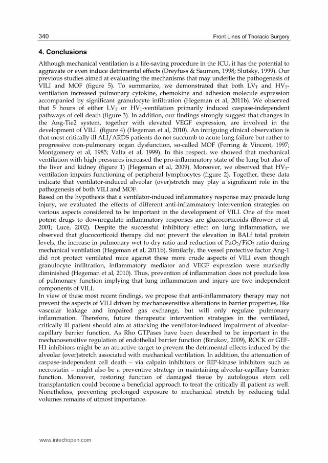
4. Conclusions
Although mechanical ventilation is a life-saving procedure in the ICU, it has the potential to aggravate or even induce detrimental effects (Dreyfuss & Saumon, 1998; Slutsky, 1999). Our previous studies aimed at evaluating the mechanisms that may underlie the pathogenesis of VILI and MOF (figure 5). To summarize, we demonstrated that both LVT and HVT-ventilation increased pulmonary cytokine, chemokine and adhesion molecule expression accompanied by significant granulocyte infiltration (Hegeman et al, 2011b). We observed that 5 hours of either LVT or HVT-ventilation primarily induced caspase-independent pathways of cell death (figure 3). In addition, our findings strongly suggest that changes in the Ang-Tie2 system, together with elevated VEGF expression, are involved in the development of VILI (figure 4) (Hegeman et al, 2010). An intriguing clinical observation is that most critically ill ALI/ARDS patients do not succumb to acute lung failure but rather to progressive non-pulmonary organ dysfunction, so-called MOF (Ferring & Vincent, 1997; Montgomery et al, 1985; Valta et al, 1999). In this respect, we showed that mechanical ventilation with high pressures increased the pro-inflammatory state of the lung but also of the liver and kidney (figure 1) (Hegeman et al, 2009). Moreover, we observed that HVT-ventilation impairs functioning of peripheral lymphocytes (figure 2). Together, these data indicate that ventilator-induced alveolar (over)stretch may play a significant role in the pathogenesis of both VILI and MOF. Based on the hypothesis that a ventilator-induced inflammatory response may precede lung injury, we evaluated the effects of different anti-inflammatory intervention strategies on various aspects considered to be important in the development of VILI. One of the most potent drugs to downregulate inflammatory responses are glucocorticoids (Brower et al, 2001; Luce, 2002). Despite the successful inhibitory effect on lung inflammation, we observed that glucocorticoid therapy did not prevent the elevation in BALf total protein levels, the increase in pulmonary wet-to-dry ratio and reduction of PaO2/FiO2 ratio during mechanical ventilation (Hegeman et al, 2011b). Similarly, the vessel protective factor Ang-1 did not protect ventilated mice against these more crude aspects of VILI even though granulocyte infiltration, inflammatory mediator and VEGF expression were markedly diminished (Hegeman et al, 2010). Thus, prevention of inflammation does not preclude loss of pulmonary function implying that lung inflammation and injury are two independent components of VILI. In view of these most recent findings, we propose that anti-inflammatory therapy may not prevent the aspects of VILI driven by mechanosensitive alterations in barrier properties, like vascular leakage and impaired gas exchange, but will only regulate pulmonary inflammation. Therefore, future therapeutic intervention strategies in the ventilated, critically ill patient should aim at attacking the ventilator-induced impairment of alveolar-capillary barrier function. As Rho GTPases have been described to be important in the mechanosensitive regulation of endothelial barrier function (Birukov, 2009), ROCK or GEF-H1 inhibitors might be an attractive target to prevent the detrimental effects induced by the alveolar (over)stretch associated with mechanical ventilation. In addition, the attenuation of caspase-independent cell death – via calpain inhibitors or RIP-kinase inhibitors such as necrostatin – might also be a preventive strategy in maintaining alveolar-capillary barrier function. Moreover, restoring function of damaged tissue by autologous stem cell transplantation could become a beneficial approach to treat the critically ill patient as well. Nonetheless, preventing prolonged exposure to mechanical stretch by reducing tidal volumes remains of utmost importance.
www.intechopen.com

Ventilator-Induced Lung Injury: Mechanisms and Future Therapeutic Interventions
341
Fig. 5. Possible mechanisms that may underlie the pathogenesis of ventilator-induced lung injury (VILI) and multiple-organ failure (MOF). Part of illustration adapted with permission of H.A.E. Vreugdenhil (Thesis “Mechanical ventilation and immune function” by H.A.E. Vreugdenhil, 2003). Although life-saving, mechanical ventilation may cause harm by itself. Our previous studies investigated the mechanisms that may underlie the pathogenesis of VILI and MOF in healthy mice. We showed that mechanical ventilation provokes endothelial activation (including changes in the angiopoietin (Ang)-Tie2 system), inflammation and cell death (primarily via activation of the caspase-independent route) in pulmonary tissue. Besides these local effects of mechanical ventilation, we also demonstrated enhanced endothelial activation and inflammation in hepatic and renal tissue (enhanced pro-inflammatory milieu of distal organs). In addition, we observed reduced mitogen-induced splenocyte proliferation after mechanical ventilation with high tidal volumes (suppressed peripheral lymphocyte functioning). Alv = alveolar epithelial cell; Mϕ = macrophage.
5. Acknowledgements
The studies were financially supported by the Catharijne Foundation (University Medical Center Utrecht, Utrecht, the Netherlands). For their contribution to abovementioned studies, the authors thank: A. Kavelaars, E. Kooijman, I. den Hartog, J. Zijlstra, K. Amarouchi, M.P. Hennus, N.J.G. Jansen & P.M. Cobelens (University Medical Center Utrecht, Utrecht, the Netherlands); G. Molema, H. Moorlag, H. Morselt, J.A.A.M. Kamps, M. van Meurs & R. Jongman (University Medical Center Groningen, Groningen, the Netherlands); B. Lachmann & P.A.C. Specht (Erasmus Medical Center, Rotterdam, the Netherlands).
www.intechopen.com

Front Lines of Thoracic Surgery
342
6. References
Abraham, E. (2003) Neutrophils and acute lung injury. Crit Care Med 31: S195-S199. Acehan, D., Jiang, X., Morgan, D. G. et al. (2002) Three-dimensional structure of the
apoptosome: implications for assembly, procaspase-9 binding, and activation. Mol Cell 9: 423-432.
Agarwal, R., Nath, A., Aggarwal, A. N. et al. (2007) Do glucocorticoids decrease mortality in acute respiratory distress syndrome? A meta-analysis. Respirology 12: 585-590.
Amano, M., Chihara, K., Kimura, K. et al. (1997) Formation of actin stress fibers and focal adhesions enhanced by Rho-kinase. Science 275: 1308-1311.
Amato, M.B., Barbas, C.S., Medeiros, D.M., et al. (1998) Effect of a protective-ventilation strategy on mortality in the acute respiratory distress syndrome. N Engl J Med 338: 347-354.
Angele, M. K. & Faist, E. (2002) Clinical review: immunodepression in the surgical patient and increased susceptibility to infection. Crit Care 6: 298-305.
Artal-Sanz, M. & Tavernarakis, N. (2005) Proteolytic mechanisms in necrotic cell death and neurodegeneration. FEBS Lett 579: 3287-3296.
Ásgeirsdóttir, S. A., Kamps, J. A., Bakker, H. I. et al. (2007) Site-specific inhibition of glomerulonephritis progression by targeted delivery of dexamethasone to glomerular endothelium. Mol Pharmacol 72: 121-131.
Ashbaugh, D. G., Bigelow, D. B., Petty, T. L. et al. (1967) Acute respiratory distress in adults. Lancet 2: 319-323.
Barnes, P. J. (2006) How corticosteroids control inflammation: Quintiles Prize Lecture 2005. Br J Pharmacol 148: 245-254.
Birukov, K. G. (2009) Small GTPases in mechanosensitive regulation of endothelial barrier. Microvasc Res 77: 46-52.
Birukova, A. A., Adyshev, D., Gorshkov, B. et al. (2006) GEF-H1 is involved in agonist-induced human pulmonary endothelial barrier dysfunction. Am J Physiol Lung Cell Mol Physiol 290: L540-L548.
Birukova, A. A., Fu, P., Xing, J. et al. (2010) Mechanotransduction by GEF-H1 as a novel mechanism of ventilator-induced vascular endothelial permeability. Am J Physiol Lung Cell Mol Physiol 298: L837-L848.
Bone, R. C. (1991) The pathogenesis of sepsis. Ann Intern Med 115: 457-469. Bone, R. C. (1996) Immunologic dissonance: a continuing evolution in our understanding of
the systemic inflammatory response syndrome (SIRS) and the multiple organ dysfunction syndrome (MODS). Ann Intern Med 125: 680-687.
Bregeon, F., Roch, A., Delpierre, S. et al. (2002) Conventional mechanical ventilation of healthy lungs induced pro-inflammatory cytokine gene transcription. Respir Physiol Neurobiol 132: 191-203.
Brower, R. G., Ware, L. B., Berthiaume, Y. et al. (2001) Treatment of ARDS. Chest 120: 1347-1367.
Burns, A. R., Smith, C. W. & Walker, D. C. (2003) Unique structural features that influence neutrophil emigration into the lung. Physiol Rev 83: 309-336.
Carlos, T. M. & Harlan, J. M. (1994) Leukocyte-endothelial adhesion molecules. Blood 84: 2068-2101.
Chang, D. W., Xing, Z., Capacio, V. L. et al. (2003) Interdimer processing mechanism of procaspase-8 activation. EMBO J 22: 4132-4142.
Chang, H. Y. & Yang, X. (2000) Proteases for cell suicide: functions and regulation of caspases. Microbiol Mol Biol Rev 64: 821-846.
www.intechopen.com

Ventilator-Induced Lung Injury: Mechanisms and Future Therapeutic Interventions
343
Chiang, C. H., Chuang, C. H., Liu, S. L. et al. (2011) Apocynin attenuates ventilator-induced lung injury in an isolated and perfused rat lung model. Intensive Care Med. 37:1360-1367.
Cho, Y. S., Challa, S., Moquin, D. et al. (2009) Phosphorylation-driven assembly of the RIP1-RIP3 complex regulates programmed necrosis and virus-induced inflammation. Cell 137: 1112-1123.
Cines, D. B., Pollak, E. S., Buck, C. A. et al. (1998) Endothelial cells in physiology and in the pathophysiology of vascular disorders. Blood 91: 3527-3561.
Cobelens, P. M., van Putte, B. P., Kavelaars, A. et al. (2009) Inflammatory Consequences of Lung Ischemia-Reperfusion Injury and Low-Pressure Ventilation. J Surg Res 153: 295-301.
Copland, I. B., Martinez, F., Kavanagh, B. P. et al. (2004) High tidal volume ventilation causes different inflammatory responses in newborn versus adult lung. Am J Respir Crit Care Med 169: 739-748.
Cortjens, B., Royakkers, A. A., Determann, R. M. et al. (2011) Lung-protective mechanical ventilation does not protect against acute kidney injury in patients without lung injury at onset of mechanical ventilation. J Crit Care.
Cuzzocrea, S., Chatterjee, P. K., Mazzon, E. et al. (2002) Effects of calpain inhibitor I on multiple organ failure induced by zymosan in the rat. Crit Care Med 30: 2284-2294.
Cuzzocrea, S., McDonald, M. C., Mazzon, E. et al. (2000) Calpain inhibitor I reduces the development of acute and chronic inflammation. Am J Pathol 157: 2065-2079.
Daly, C., Pasnikowski, E., Burova, E. et al. (2006) Angiopoietin-2 functions as an autocrine protective factor in stressed endothelial cells. Proc Natl Acad Sci U S A 103: 15491-15496.
Daly, C., Wong, V., Burova, E. et al. (2004) Angiopoietin-1 modulates endothelial cell function and gene expression via the transcription factor FKHR (FOXO1). Genes Dev 18: 1060-1071.
Danial, N. N. & Korsmeyer, S. J. (2004) Cell death: critical control points. Cell 116: 205-219. David, S., Park, J. K., van Meurs, M. et al. (2011) Acute administration of recombinant
Angiopoietin-1 ameliorates multiple-organ dysfunction syndrome and improves survival in murine sepsis. Cytokine 55: 251-259.
DeBusk, L. M., Hallahan, D. E. & Lin, P. C. (2004) Akt is a major angiogenic mediator downstream of the Ang1/Tie2 signaling pathway. Exp Cell Res 298: 167-177.
Declercq, W., Vanden Berghe, T. & Vandenabeele, P. (2009) RIP kinases at the crossroads of cell death and survival. Cell 138: 229-232.
Degterev, A., Huang, Z., Boyce, M. et al. (2005) Chemical inhibitor of nonapoptotic cell death with therapeutic potential for ischemic brain injury. Nat Chem Biol 1: 112-119.
Dejana, E. (2004) Endothelial cell-cell junctions: happy together. Nat Rev Mol Cell Biol 5: 261-270.
Denecker, G., Vercammen, D., Declercq, W. et al. (2001) Apoptotic and necrotic cell death induced by death domain receptors. Cell Mol Life Sci 58: 356-370.
Determann, R.M., Royakkers, A., Wolthuis, E.K., et al. (2010) Ventilation with lower tidal volumes as compared with conventional tidal volumes for patients without acute lung injury: a preventive randomized controlled trial. Crit Care 14: R1.
Ding, H. F., Lin, Y. L., McGill, G. et al. (2000) Essential role for caspase-8 in transcription-independent apoptosis triggered by p53. J Biol Chem 275: 38905-38911.
Donnelly, S. C., Strieter, R. M., Kunkel, S. L. et al. (1993) Interleukin-8 and development of adult respiratory distress syndrome in at-risk patient groups. Lancet 341: 643-647.
www.intechopen.com

Front Lines of Thoracic Surgery
344
dos Santos, C. C., Han, B., Andrade, C. F. et al. (2004) DNA microarray analysis of gene expression in alveolar epithelial cells in response to TNFalpha, LPS, and cyclic stretch. Physiol Genomics 19: 331-342.
dos Santos, C. C., Shan, Y., Akram, A. et al. (2011) Neuroimmune regulation of ventilator-induced lung injury. Am J Respir Crit Care Med 183: 471-482.
Dreyfuss, D., Basset, G., Soler, P. et al. (1985) Intermittent positive-pressure hyperventilation with high inflation pressures produces pulmonary microvascular injury in rats. Am Rev Respir Dis 132: 880-884.
Dreyfuss, D. & Saumon, G. (1998) From ventilator-induced lung injury to multiple organ dysfunction? Intensive Care Med 24: 102-104.
Dreyfuss, D., Soler, P., Basset, G. et al. (1988) High inflation pressure pulmonary edema. Respective effects of high airway pressure, high tidal volume, and positive end-expiratory pressure. Am Rev Respir Dis 137: 1159-1164.
Edwards, Y. S., Sutherland, L. M., Power, J. H. et al. (1999) Cyclic stretch induces both apoptosis and secretion in rat alveolar type II cells. FEBS Lett 448: 127-130.
Egan, E. A. (1982) Lung inflation, lung solute permeability, and alveolar edema. J Appl Physiol 53: 121-125.
Elenkov, I. J., Wilder, R. L., Chrousos, G. P. et al. (2000) The sympathetic nerve--an integrative interface between two supersystems: the brain and the immune system. Pharmacol Rev 52: 595-638.
Feldman-Billard, S., Du Pasquier-Fediaevsky, L. & Heron, E. (2006) Hyperglycemia after repeated periocular dexamethasone injections in patients with diabetes. Ophthalmology 113: 1720-1723.
Fernandes, A. B., Zin, W. A. & Rocco, P. R. (2005) Corticosteroids in acute respiratory distress syndrome. Braz J Med Biol Res 38: 147-159.
Ferring, M. & Vincent, J. L. (1997) Is outcome from ARDS related to the severity of respiratory failure? Eur Respir J 10: 1297-1300.
Festjens, N., van Gurp, M., van Loo, G. et al. (2004) Bcl-2 family members as sentinels of cellular integrity and role of mitochondrial intermembrane space proteins in apoptotic cell death. Acta Haematol 111: 7-27.
Festjens, N., Vanden Berghe, T. & Vandenabeele, P. (2006) Necrosis, a well-orchestrated form of cell demise: signalling cascades, important mediators and concomitant immune response. Biochim Biophys Acta 1757: 1371-1387.
Fiedler, U. & Augustin, H. G. (2006) Angiopoietins: a link between angiogenesis and inflammation. Trends Immunol 27: 552-558.
Fiedler, U., Krissl, T., Koidl, S. et al. (2003) Angiopoietin-1 and angiopoietin-2 share the same binding domains in the Tie-2 receptor involving the first Ig-like loop and the epidermal growth factor-like repeats. J Biol Chem 278: 1721-1727.
Fiers, W., Beyaert, R., Declercq, W. et al. (1999) More than one way to die: apoptosis, necrosis and reactive oxygen damage. Oncogene 18: 7719-7730.
Flierl, M. A., Rittirsch, D., Nadeau, B. A. et al. (2009) Upregulation of phagocyte-derived catecholamines augments the acute inflammatory response. PLoS ONE 4: e4414.
Gallagher, D. C., Parikh, S. M., Balonov, K. et al. (2008) Circulating angiopoietin 2 correlates with mortality in a surgical population with acute lung injury/adult respiratory distress syndrome. Shock 29: 656-661.
Galluzzi, L., Kepp, O. & Kroemer, G. (2009) RIP kinases initiate programmed necrosis. J Mol Cell Biol 1: 8-10.
Galluzzi, L., Vicencio, J. M., Kepp, O. et al. (2008) To die or not to die: that is the autophagic question. Curr Mol Med 8: 78-91.
www.intechopen.com

Ventilator-Induced Lung Injury: Mechanisms and Future Therapeutic Interventions
345
Gando, S., Kameue, T., Matsuda, N. et al. (2004) Systemic inflammation and disseminated intravascular coagulation in early stage of ALI and ARDS: role of neutrophil and endothelial activation. Inflammation 28: 237-244.
Gavard, J., Patel, V. & Gutkind, J. S. (2008) Angiopoietin-1 prevents VEGF-induced endothelial permeability by sequestering Src through mDia. Dev Cell 14: 25-36.
Gillette, M. A. & Hess, D. R. (2001) Ventilator-induced lung injury and the evolution of lung-protective strategies in acute respiratory distress syndrome. Respir Care 46: 130-148.
Golstein, P. & Kroemer, G. (2007) Cell death by necrosis: towards a molecular definition. Trends Biochem Sci 32: 37-43.
Goodman, R. B., Pugin, J., Lee, J. S. et al. (2003) Cytokine-mediated inflammation in acute lung injury. Cytokine Growth Factor Rev 14: 523-535.
Haitsma, J. J., Uhlig, S., Goggel, R. et al. (2000) Ventilator-induced lung injury leads to loss of alveolar and systemic compartmentalization of tumor necrosis factor-alpha. Intensive Care Med 26: 1515-1522.
Haitsma, J. J., Uhlig, S., Verbrugge, S. J. et al. (2003) Injurious ventilation strategies cause systemic release of IL-6 and MIP-2 in rats in vivo. Clin Physiol Funct Imaging 23: 349-353.
Hammerschmidt, S., Kuhn, H., Grasenack, T. et al. (2004) Apoptosis and necrosis induced by cyclic mechanical stretching in alveolar type II cells. Am J Respir Cell Mol Biol 30: 396-402.
Harwood, S. M., Yaqoob, M. M. & Allen, D. A. (2005) Caspase and calpain function in cell death: bridging the gap between apoptosis and necrosis. Ann Clin Biochem 42: 415-431.
Haupt, S., Berger, M., Goldberg, Z. et al. (2003) Apoptosis - the p53 network. J Cell Sci 116: 4077-4085.
He, S., Wang, L., Miao, L. et al. (2009) Receptor interacting protein kinase-3 determines cellular necrotic response to TNF-alpha. Cell 137: 1100-1111.
Headley, A. S., Tolley, E. & Meduri, G. U. (1997) Infections and the inflammatory response in acute respiratory distress syndrome. Chest 111: 1306-1321.
Hegeman, M. A., Cobelens, P. M., Kamps, J. A. et al. (2011a) Liposome-encapsulated dexamethasone attenuates ventilator-induced lung inflammation. Br J Pharmacol 163: 1048-1058.
Hegeman, M. A., Hennus, M. P., Cobelens, P. M. et al. (2011b) Inhibition of pulmonary inflammation by dexamethasone does not prevent ventilator-induced lung injury. Accepted as abstract for the 24th Annual Congress of the European Society of Intensive Care Medicine (ESICM), Berlin, Germany.
Hegeman, M. A., Hennus, M. P., Heijnen, C. J. et al. (2009) Ventilator-induced endothelial activation and inflammation in the lung and distal organs. Crit Care 13: R182.
Hegeman, M. A., Hennus, M. P., van Meurs, M. et al. (2010) Angiopoietin-1 treatment reduces inflammation but does not prevent ventilator-induced lung injury. PLoS ONE 5: e15653.
Held, H. D., Boettcher, S., Hamann, L. et al. (2001) Ventilation-induced chemokine and cytokine release is associated with activation of nuclear factor-kappaB and is blocked by steroids. Am J Respir Crit Care Med 163: 711-716.
Hitomi, J., Christofferson, D. E., Ng, A. et al. (2008) Identification of a molecular signaling network that regulates a cellular necrotic cell death pathway. Cell 135: 1311-1323.
Huang, Y. Q., Sauthoff, H., Herscovici, P. et al. (2008) Angiopoietin-1 increases survival and reduces the development of lung edema induced by endotoxin administration in a murine model of acute lung injury. Crit Care Med 36: 262-267.
www.intechopen.com

Front Lines of Thoracic Surgery
346
Hudson, L. D., Milberg, J. A., Anardi, D. et al. (1995) Clinical risks for development of the acute respiratory distress syndrome. Am J Respir Crit Care Med 151: 293-301.
Hughes, D. P., Marron, M. B. & Brindle, N. P. (2003) The antiinflammatory endothelial tyrosine kinase Tie2 interacts with a novel nuclear factor-kappaB inhibitor ABIN-2. Circ Res 92: 630-636.
Ikegami, M., Jobe, A., Jacobs, H. et al. (1984) A protein from airways of premature lambs that inhibits surfactant function. J Appl Physiol 57: 1134-1142.
Imai, Y., Parodo, J., Kajikawa, O. et al. (2003) Injurious mechanical ventilation and end-organ epithelial cell apoptosis and organ dysfunction in an experimental model of acute respiratory distress syndrome. JAMA 289: 2104-2112.
Iwaki, M., Ito, S., Morioka, M. et al. (2009) Mechanical stretch enhances IL-8 production in pulmonary microvascular endothelial cells. Biochem Biophys Res Commun 389: 531-536.
Jiang, X. & Wang, X. (2000) Cytochrome c promotes caspase-9 activation by inducing nucleotide binding to Apaf-1. J Biol Chem 275: 31199-31203.
Kannan, K., Kaminski, N., Rechavi, G. et al. (2001) DNA microarray analysis of genes involved in p53 mediated apoptosis: activation of Apaf-1. Oncogene 20: 3449-3455.
Karmpaliotis, D., Kosmidou, I., Ingenito, E. P. et al. (2002) Angiogenic growth factors in the pathophysiology of a murine model of acute lung injury. Am J Physiol Lung Cell Mol Physiol 283: L585-L595.
Kawano, T., Mori, S., Cybulsky, M. et al. (1987) Effect of granulocyte depletion in a ventilated surfactant-depleted lung. J Appl Physiol 62: 27-33.
Kelley, J. (1990) Cytokines of the lung. Am Rev Respir Dis 141: 765-788. Kim, I., Kim, H. G., So, J. N. et al. (2000) Angiopoietin-1 regulates endothelial cell survival
through the phosphatidylinositol 3'-Kinase/Akt signal transduction pathway. Circ Res 86: 24-29.
Kimura, K., Ito, M., Amano, M. et al. (1996) Regulation of myosin phosphatase by Rho and Rho-associated kinase (Rho-kinase). Science 273: 245-248.
Koksel, O., Yildirim, C., Tiftik, R. N. et al. (2005) Rho-kinase (ROCK-1 and ROCK-2) upregulation in oleic acid-induced lung injury and its restoration by Y-27632. Eur J Pharmacol 510: 135-142.
Korsmeyer, S. J., Wei, M. C., Saito, M. et al. (2000) Pro-apoptotic cascade activates BID, which oligomerizes BAK or BAX into pores that result in the release of cytochrome c. Cell Death Differ 7: 1166-1173.
Kroemer, G., El-Deiry, W. S., Golstein, P. et al. (2005) Classification of cell death: recommendations of the Nomenclature Committee on Cell Death. Cell Death Differ 12 Suppl 2: 1463-1467.
Kroemer, G., Galluzzi, L., Vandenabeele, P. et al. (2009) Classification of cell death: recommendations of the Nomenclature Committee on Cell Death 2009. Cell Death Differ 16: 3-11.
Kumar, V. & Sharma, A. (2010) Is neuroimmunomodulation a future therapeutic approach for sepsis? Int Immunopharmacol 10: 9-17.
Lachmann, B., Eijking, E. P., So, K. L. et al. (1994) In vivo evaluation of the inhibitory capacity of human plasma on exogenous surfactant function. Intensive Care Med 20: 6-11.
Lachmann, B., Hallman, M. & Bergmann, K. C. (1987) Respiratory failure following anti-lung serum: study on mechanisms associated with surfactant system damage. Exp Lung Res 12: 163-180.
www.intechopen.com

Ventilator-Induced Lung Injury: Mechanisms and Future Therapeutic Interventions
347
Lavrik, I. N., Golks, A. & Krammer, P. H. (2005a) Caspases: pharmacological manipulation of cell death. J Clin Invest 115: 2665-2672.
Lavrik, I. N., Golks, A. & Krammer, P. H. (2005b) Death receptor signaling. J Cell Sci 118: 265-267.
Le, A., Damico, R., Damarla, M. et al. (2008) Alveolar cell apoptosis is dependent on p38 MAP kinase-mediated activation of xanthine oxidoreductase in ventilator-induced lung injury. J Appl Physiol 105: 1282-1290.
Lee, J. W., Fang, X., Krasnodembskaya, A. et al. (2011) Concise review: mesenchymal stem cells for acute lung injury: role of paracrine soluble factors. Stem Cells 29: 913-919.
Lee, W. L. & Downey, G. P. (2001) Neutrophil activation and acute lung injury. Curr Opin Crit Care 7: 1-7.
Leist, M. & Jäättelä, M. (2001) Four deaths and a funeral: from caspases to alternative mechanisms. Nat Rev Mol Cell Biol 2: 589-598.
Leist, M., Single, B., Castoldi, A. F. et al. (1997) Intracellular adenosine triphosphate (ATP) concentration: a switch in the decision between apoptosis and necrosis. J Exp Med 185: 1481-1486.
Lemieux, C., Maliba, R., Favier, J. et al. (2005) Angiopoietins can directly activate endothelial cells and neutrophils to promote proinflammatory responses. Blood 105: 1523-1530.
Lentsch, A. B. & Ward, P. A. (2000) Regulation of inflammatory vascular damage. J Pathol 190: 343-348.
Levine, B. & Klionsky, D. J. (2004) Development by self-digestion: molecular mechanisms and biological functions of autophagy. Dev Cell 6: 463-477.
Li, L. F., Liao, S. K., Ko, Y. S. et al. (2007) Hyperoxia increases ventilator-induced lung injury via mitogen-activated protein kinases: a prospective, controlled animal experiment. Crit Care 11: R25.
Liao, J. K., Seto, M. & Noma, K. (2007) Rho kinase (ROCK) inhibitors. J Cardiovasc Pharmacol 50: 17-24.
Luce, J. M. (2002) Corticosteroids in ARDS. An evidence-based review. Crit Care Clin 18: 79-89, vii.
Luscinskas, F. W. & Gimbrone, M. A., Jr. (1996) Endothelial-dependent mechanisms in chronic inflammatory leukocyte recruitment. Annu Rev Med 47: 413-421.
Maiuri, M. C., Zalckvar, E., Kimchi, A. et al. (2007) Self-eating and self-killing: crosstalk between autophagy and apoptosis. Nat Rev Mol Cell Biol 8: 741-752.
Marshall J.C. (2001). The multiple organ dysfunction syndrome. In: Holzheimer RG, Mannick JA, eds. Surgical Treatment: Evidence-based and problem-oriented. Munich: Zuckschwerdt Verlag, 780-785.
McCarter, S. D., Mei, S. H., Lai, P. F. et al. (2007) Cell-based angiopoietin-1 gene therapy for acute lung injury. Am J Respir Crit Care Med 175: 1014-1026.
McCulloch, E. A. & Till, J. E. (2005) Perspectives on the properties of stem cells. Nat Med 11: 1026-1028.
McKenzie, S. E. & Schreiber, A. D. (1998) Fc gamma receptors in phagocytes. Curr Opin Hematol 5: 16-21.
Mead, J., Takishima, T. & Leith, D. (1970) Stress distribution in lungs: a model of pulmonary elasticity. J Appl Physiol 28: 596-608.
Meduri, G. U., Headley, S., Kohler, G. et al. (1995) Persistent elevation of inflammatory cytokines predicts a poor outcome in ARDS. Plasma IL-1 beta and IL-6 levels are consistent and efficient predictors of outcome over time. Chest 107: 1062-1073.
www.intechopen.com
Front Lines of Thoracic Surgery
348
Meduri, G. U., Marik, P. E., Chrousos, G. P. et al. (2008) Steroid treatment in ARDS: a critical appraisal of the ARDS network trial and the recent literature. Intensive Care Med 34: 61-69.
Mei, S. H., McCarter, S. D., Deng, Y. et al. (2007) Prevention of LPS-induced acute lung injury in mice by mesenchymal stem cells overexpressing angiopoietin 1. PLoS Med 4: e269.
Michel, C. C. & Curry, F. E. (1999) Microvascular permeability. Physiol Rev 79: 703-761. Miksa, M., Das, P., Zhou, M. et al. (2009) Pivotal role of the alpha(2A)-adrenoceptor in
producing inflammation and organ injury in a rat model of sepsis. PLoS ONE 4: e5504.
Montgomery, A. B., Stager, M. A., Carrico, C. J. et al. (1985) Causes of mortality in patients with the adult respiratory distress syndrome. Am Rev Respir Dis 132: 485-489.
Moraes, T. J., Chow, C. W. & Downey, G. P. (2003) Proteases and lung injury. Crit Care Med 31: S189-S194.
Nin, N., Lorente, J. A., de Paula, M. et al. (2008) Rats surviving injurious mechanical ventilation show reversible pulmonary, vascular and inflammatory changes. Intensive Care Med 34: 948-956.
Nin, N., Penuelas, O., de Paula, M. et al. (2006) Ventilation-induced lung injury in rats is associated with organ injury and systemic inflammation that is attenuated by dexamethasone. Crit Care Med 34: 1093-1098.
Northington, F. J., Chavez-Valdez, R., Graham, E. M. et al. (2011) Necrostatin decreases oxidative damage, inflammation, and injury after neonatal HI. J Cereb Blood Flow Metab 31: 178-189.
O'Mahony, D. S., Liles, W. C., Altemeier, W. A. et al. (2006) Mechanical ventilation interacts with endotoxemia to induce extrapulmonary organ dysfunction. Crit Care 10: R136.
Ohta, N., Shimaoka, M., Imanaka, H. et al. (2001) Glucocorticoid suppresses neutrophil activation in ventilator-induced lung injury. Crit Care Med 29: 1012-1016.
Orfanos, S. E., Mavrommati, I., Korovesi, I. et al. (2004) Pulmonary endothelium in acute lung injury: from basic science to the critically ill. Intensive Care Med 30: 1702-1714.
Ortiz, L. A., Gambelli, F., McBride, C. et al. (2003) Mesenchymal stem cell engraftment in lung is enhanced in response to bleomycin exposure and ameliorates its fibrotic effects. Proc Natl Acad Sci U S A 100: 8407-8411.
Papapetropoulos, A., Fulton, D., Mahboubi, K. et al. (2000) Angiopoietin-1 inhibits endothelial cell apoptosis via the Akt/survivin pathway. J Biol Chem 275: 9102-9105.
Parker, J. C., Townsley, M. I., Rippe, B. et al. (1984) Increased microvascular permeability in dog lungs due to high peak airway pressures. J Appl Physiol 57: 1809-1816.
Parrillo, J. E. (1993) Pathogenetic mechanisms of septic shock. N Engl J Med 328: 1471-1477. Pavlov, V. A., Ochani, M., Yang, L. H. et al. (2007) Selective alpha7-nicotinic acetylcholine
receptor agonist GTS-21 improves survival in murine endotoxemia and severe sepsis. Crit Care Med 35: 1139-1144.
Petty, T. L. & Ashbaugh, D. G. (1971) The adult respiratory distress syndrome. Clinical features, factors influencing prognosis and principles of management. Chest 60: 233-239.
Pinsky, M. R., Vincent, J. L., Deviere, J. et al. (1993) Serum cytokine levels in human septic shock. Relation to multiple-system organ failure and mortality. Chest 103: 565-575.
Pittenger, M. F., Mackay, A. M., Beck, S. C. et al. (1999) Multilineage potential of adult human mesenchymal stem cells. Science 284: 143-147.
www.intechopen.com

Ventilator-Induced Lung Injury: Mechanisms and Future Therapeutic Interventions
349
Plotz, F. B., Slutsky, A. S., van Vught, A. J. et al. (2004) Ventilator-induced lung injury and multiple system organ failure: a critical review of facts and hypotheses. Intensive Care Med 30: 1865-1872.
Plotz, F. B., Vreugdenhil, H. A., Slutsky, A. S. et al. (2002) Mechanical ventilation alters the immune response in children without lung pathology. Intensive Care Med 28: 486-492.
Pugin, J. (2003) Molecular mechanisms of lung cell activation induced by cyclic stretch. Crit Care Med 31: S200-S206.
Pugin, J., Dunn, I., Jolliet, P. et al. (1998) Activation of human macrophages by mechanical ventilation in vitro. Am J Physiol 275: L1040-L1050.
Ranieri, V. M., Suter, P. M., Tortorella, C. et al. (1999) Effect of mechanical ventilation on inflammatory mediators in patients with acute respiratory distress syndrome: a randomized controlled trial. JAMA 282: 54-61.
Ren, Y., Li, R., Zheng, Y. et al. (1998) Cloning and characterization of GEF-H1, a microtubule-associated guanine nucleotide exchange factor for Rac and Rho GTPases. J Biol Chem 273: 34954-34960.
Ridley, A. J. & Hall, A. (1992) The small GTP-binding protein rho regulates the assembly of focal adhesions and actin stress fibers in response to growth factors. Cell 70: 389-399.
Rittirsch, D., Flierl, M. A. & Ward, P. A. (2008) Harmful molecular mechanisms in sepsis. Nat Rev Immunol 8: 776-787.
Rojas, M., Xu, J., Woods, C. R. et al. (2005) Bone marrow-derived mesenchymal stem cells in repair of the injured lung. Am J Respir Cell Mol Biol 33: 145-152.
Rose, R., Banerjee, A. & Ramaiah, S. K. (2006) Calpain inhibition attenuates iNOS production and midzonal hepatic necrosis in a repeat dose model of endotoxemia in rats. Toxicol Pathol 34: 785-794.
Rossi, D. & Gaidano, G. (2003) Messengers of cell death: apoptotic signaling in health and disease. Haematologica 88: 212-218.
Ryter, S. W. & Choi, A. M. (2010) Autophagy in the lung. Proc Am Thorac Soc 7: 13-21. Schacke, H., Docke, W. D. & Asadullah, K. (2002) Mechanisms involved in the side effects of
glucocorticoids. Pharmacol Ther 96: 23-43. Schmidt, A. & Hall, A. (2002) Guanine nucleotide exchange factors for Rho GTPases: turning
on the switch. Genes Dev 16: 1587-1609. Schultz, M. J. (2008) Lung-protective mechanical ventilation with lower tidal volumes in
patients not suffering from acute lung injury: a review of clinical studies. Med Sci Monit 14: RA22-RA26.
Schwarz, M. A. (2001) Acute lung injury: cellular mechanisms and derangements. Paediatr Respir Rev 2: 3-9.
Seeger, W., Gunther, A., Walmrath, H. D. et al. (1993) Alveolar surfactant and adult respiratory distress syndrome. Pathogenetic role and therapeutic prospects. Clin Investig 71: 177-190.
Slutsky, A. S. (1993) Mechanical ventilation. American College of Chest Physicians' Consensus Conference. Chest 104: 1833-1859.
Slutsky, A. S. (1999) Lung injury caused by mechanical ventilation. Chest 116: 9S-15S. Slutsky, A. S. & Tremblay, L. N. (1998) Multiple system organ failure. Is mechanical
ventilation a contributing factor? Am J Respir Crit Care Med 157: 1721-1725. Spengler, R. N., Allen, R. M., Remick, D. G. et al. (1990) Stimulation of alpha-adrenergic
receptor augments the production of macrophage-derived tumor necrosis factor. J Immunol 145: 1430-1434.
www.intechopen.com

Front Lines of Thoracic Surgery
350
Spindler, V., Schlegel, N. & Waschke, J. (2010) Role of GTPases in control of microvascular permeability. Cardiovasc Res 87: 243-253.
Storm, G. & Crommelin, D. J. A. (1998) Liposomes: quo vadis? Pharm Sci Technol To 1: 19-31. Straub, R. H., Mayer, M., Kreutz, M. et al. (2000) Neurotransmitters of the sympathetic nerve
terminal are powerful chemoattractants for monocytes. J Leukoc Biol 67: 553-558. Syrbe, U., Meinecke, A., Platzer, C. et al. (1994). Improvement of monocyte function - a new
therapeutic approach? In: Reinhart K, Eyrich K, Sprung CL, eds. Sepsis: Current Perspectives in Pathophysiology and Therapy., Berlin: Springer-Verlag, 473-500.
Tasaka, S., Koh, H., Yamada, W. et al. (2005) Attenuation of endotoxin-induced acute lung injury by the Rho-associated kinase inhibitor, Y-27632. Am J Respir Cell Mol Biol 32: 504-510.
the ARDS network (2000) Ventilation with lower tidal volumes as compared with traditional tidal volumes for acute lung injury and the acute respiratory distress syndrome. The Acute Respiratory Distress Syndrome Network. N Engl J Med 342: 1301-1308.
Thompson, B. T. (2003) Glucocorticoids and acute lung injury. Crit Care Med 31: S253-S257. Tobin, M. J. (2001) Advances in mechanical ventilation. N Engl J Med 344: 1986-1996. Tremblay, L., Valenza, F., Ribeiro, S. P. et al. (1997) Injurious ventilatory strategies increase
cytokines and c-fos m-RNA expression in an isolated rat lung model. J Clin Invest 99: 944-952.
Tremblay, L. N. & Slutsky, A. S. (1998) Ventilator-induced injury: from barotrauma to biotrauma. Proc Assoc Am Physicians 110: 482-488.
Tschumperlin, D. J., Oswari, J. & Margulies, A. S. (2000) Deformation-induced injury of alveolar epithelial cells. Effect of frequency, duration, and amplitude. Am J Respir Crit Care Med 162: 357-362.
Tsuno, K., Miura, K., Takeya, M. et al. (1991) Histopathologic pulmonary changes from mechanical ventilation at high peak airway pressures. Am Rev Respir Dis 143: 1115-1120.
Tutor, J. D., Mason, C. M., Dobard, E. et al. (1994) Loss of compartmentalization of alveolar tumor necrosis factor after lung injury. Am J Respir Crit Care Med 149: 1107-1111.
Valta, P., Uusaro, A., Nunes, S. et al. (1999) Acute respiratory distress syndrome: frequency, clinical course, and costs of care. Crit Care Med 27: 2367-2374.
van der Heijden M., van Nieuw Amerongen, G. P., Koolwijk, P. et al. (2008) Angiopoietin-2, permeability oedema, occurrence and severity of ALI/ARDS in septic and non-septic critically ill patients. Thorax 63: 903-909.
van der Heijden M., van Nieuw Amerongen, G. P., Chedamni, S. et al. (2009) The angiopoietin-Tie2 system as a therapeutic target in sepsis and acute lung injury. Expert Opin Ther Targets 13: 39-53.
Vaneker, M., Halbertsma, F. J., van Egmond, J. et al. (2007) Mechanical ventilation in healthy mice induces reversible pulmonary and systemic cytokine elevation with preserved alveolar integrity: an in vivo model using clinical relevant ventilation settings. Anesthesiology 107: 419-426.
Verbrugge, S. J., Sorm, V. & Lachmann, B. (1997) Mechanisms of acute respiratory distress syndrome: role of surfactant changes and mechanical ventilation. J Physiol Pharmacol 48: 537-557.
Villar, J., Kacmarek, R.M., Perez-Mendez, L., et al. (2006) A high positive endexpiratory pressure, low tidal volume ventilatory strategy improves outcome in persistent acute respiratory distress syndrome: a randomized, controlled trial. Crit Care Med 34: 1311-1318.
www.intechopen.com

Ventilator-Induced Lung Injury: Mechanisms and Future Therapeutic Interventions
351
Vincent, J. L., Sakr, Y., Sprung, C. L. et al. (2006) Sepsis in European intensive care units: results of the SOAP study. Crit Care Med 34: 344-353.
Vlahakis, N. E., Schroeder, M. A., Limper, A. H. et al. (1999) Stretch induces cytokine release by alveolar epithelial cells in vitro. Am J Physiol 277: L167-L173.
Vreugdenhil, H. A., Heijnen, C. J., Plotz, F. B. et al. (2004) Mechanical ventilation of healthy rats suppresses peripheral immune function. Eur Respir J 23: 122-128.
Ware, L. B. & Matthay, M. A. (2000) The acute respiratory distress syndrome. N Engl J Med 342: 1334-1349.
Webb, H. H. & Tierney, D. F. (1974) Experimental pulmonary edema due to intermittent positive pressure ventilation with high inflation pressures. Protection by positive end-expiratory pressure. Am Rev Respir Dis 110: 556-565.
Weibel, E.R. (1984). The Pathway for Oxygen: Structure and Function in the Mammalian Respiratory System. In: Harvard Univ.Press. Cambridge, MA, 425.
Weinstein, S. P., Paquin, T., Pritsker, A. et al. (1995) Glucocorticoid-induced insulin resistance: dexamethasone inhibits the activation of glucose transport in rat skeletal muscle by both insulin- and non-insulin-related stimuli. Diabetes 44: 441-445.
Weppler, A. & Issekutz, A. C. (2008) Alveolar epithelium down-modulates endotoxin-but not tumor necrosis factor alpha-induced activation of endothelium and selectively inhibits neutrophil transendothelial migration. Exp Lung Res 34: 425-453.
West, J. B., Tsukimoto, K., Mathieu-Costello, O. et al. (1991) Stress failure in pulmonary capillaries. J Appl Physiol 70: 1731-1742.
Whalen, M. J., Doughty, L. A., Carlos, T. M. et al. (2000) Intercellular adhesion molecule-1 and vascular cell adhesion molecule-1 are increased in the plasma of children with sepsis-induced multiple organ failure. Crit Care Med 28: 2600-2607.
Wilson, M. R., Choudhury, S., Goddard, M. E. et al. (2003) High tidal volume upregulates intrapulmonary cytokines in an in vivo mouse model of ventilator-induced lung injury. J Appl Physiol 95: 1385-1393.
Wilson, M. R., Choudhury, S. & Takata, M. (2005) Pulmonary inflammation induced by high-stretch ventilation is mediated by tumor necrosis factor signaling in mice. Am J Physiol Lung Cell Mol Physiol 288: L599-L607.
Witzenbichler, B., Westermann, D., Knueppel, S. et al. (2005) Protective role of angiopoietin-1 in endotoxic shock. Circulation 111: 97-105.
Wojciak-Stothard, B., Entwistle, A., Garg, R. et al. (1998) Regulation of TNF-alpha-induced reorganization of the actin cytoskeleton and cell-cell junctions by Rho, Rac, and Cdc42 in human endothelial cells. J Cell Physiol 176: 150-165.
Wojciak-Stothard, B. & Ridley, A. J. (2002) Rho GTPases and the regulation of endothelial permeability. Vascul Pharmacol 39: 187-199.
Wolthuis, E. K., Vlaar, A. P., Choi, G. et al. (2009a) Recombinant human soluble tumor necrosis factor-alpha receptor fusion protein partly attenuates ventilator-induced lung injury. Shock 31: 262-266.
Wolthuis, E. K., Vlaar, A. P., Choi, G. et al. (2009b) Mechanical ventilation using non-injurious ventilation settings causes lung injury in the absence of pre-existing lung injury in healthy mice. Crit Care 13: R1.
Xu, J., Qu, J., Cao, L. et al. (2008) Mesenchymal stem cell-based angiopoietin-1 gene therapy for acute lung injury induced by lipopolysaccharide in mice. J Pathol 214: 472-481.
Xu, J., Woods, C. R., Mora, A. L. et al. (2007) Prevention of endotoxin-induced systemic response by bone marrow-derived mesenchymal stem cells in mice. Am J Physiol Lung Cell Mol Physiol 293: L131-L141.
www.intechopen.com

Front Lines of Thoracic Surgery
352
You, Z., Savitz, S. I., Yang, J. et al. (2008) Necrostatin-1 reduces histopathology and improves functional outcome after controlled cortical impact in mice. J Cereb Blood Flow Metab 28: 1564-1573.
Zamzami, N. & Kroemer, G. (2003) Apoptosis: mitochondrial membrane permeabilization--the (w)hole story? Curr Biol 13: R71-R73.
Zhang, D. W., Shao, J., Lin, J. et al. (2009a) RIP3, an energy metabolism regulator that switches TNF-induced cell death from apoptosis to necrosis. Science 325: 332-336.
Zhang, Z., Larner, S. F., Liu, M. C. et al. (2009b) Multiple alphaII-spectrin breakdown products distinguish calpain and caspase dominated necrotic and apoptotic cell death pathways. Apoptosis 14: 1289-1298.
www.intechopen.com

Front Lines of Thoracic SurgeryEdited by Dr. Stefano Nazari
ISBN 978-953-307-915-8Hard cover, 412 pagesPublisher InTechPublished online 03, February, 2012Published in print edition February, 2012
InTech EuropeUniversity Campus STeP Ri Slavka Krautzeka 83/A 51000 Rijeka, Croatia Phone: +385 (51) 770 447 Fax: +385 (51) 686 166www.intechopen.com
InTech ChinaUnit 405, Office Block, Hotel Equatorial Shanghai No.65, Yan An Road (West), Shanghai, 200040, China
Phone: +86-21-62489820 Fax: +86-21-62489821
Front Lines of Thoracic Surgery collects up-to-date contributions on some of the most debated topics intoday's clinical practice of cardiac, aortic, and general thoracic surgery,and anesthesia as viewed by authorspersonally involved in their evolution. The strong and genuine enthusiasm of the authors was clearlyperceptible in all their contributions and I'm sure that will further stimulate the reader to understand theirmessages. Moreover, the strict adhesion of the authors' original observations and findings to the evidencebase proves that facts are the best guarantee of scientific value. This is not a standard textbook where thewhole discipline is organically presented, but authors' contributions are simply listed in their pertainingsubclasses of Thoracic Surgery. I'm sure that this original and very promising editorial format which has andfree availability at its core further increases this book's value and it will be of interest to healthcareprofessionals and scientists dedicated to this field.
How to referenceIn order to correctly reference this scholarly work, feel free to copy and paste the following:
Maria A. Hegeman, Marcus J. Schultz, Adrianus J. van Vught and Cobi J. Heijnen (2012). Ventilator-InducedLung Injury: Mechanisms and Future Therapeutic Interventions, Front Lines of Thoracic Surgery, Dr. StefanoNazari (Ed.), ISBN: 978-953-307-915-8, InTech, Available from: http://www.intechopen.com/books/front-lines-of-thoracic-surgery/ventilator-induced-lung-injury-mechanisms-and-future-therapeutic-interventions