Embed Size (px)
Citation preview
Major veins of the human body
A. coronary circulation: coronary sinus B. pulmonary circulation: pulmonary veins
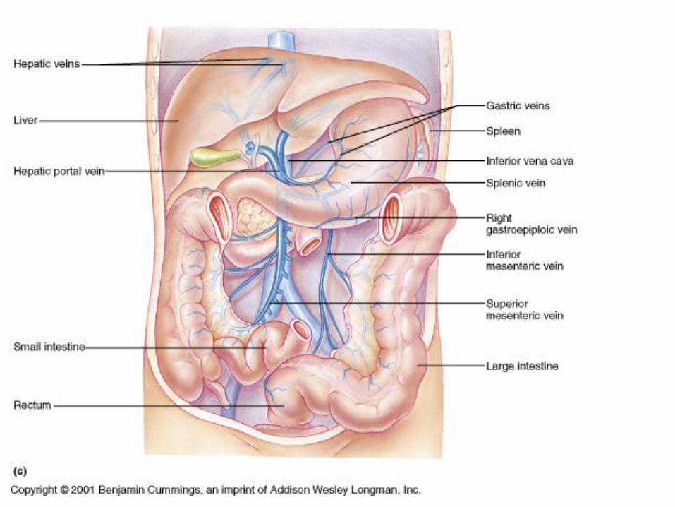
C. Systemic circulation D. hepatic portal circulation: hepatic portal vein drains 1. inferior mesenteric which drains
splenic vein 2. superior mesenteric vein
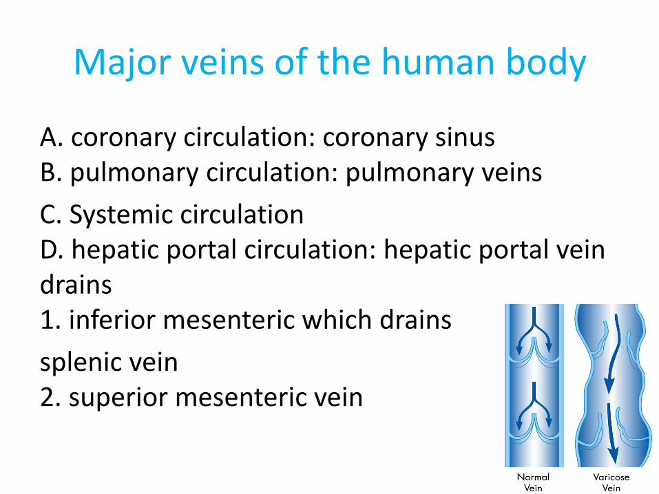
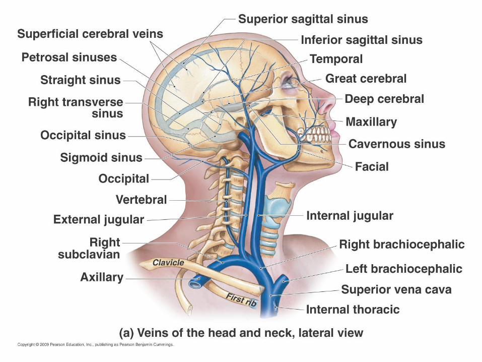
Cranial sinuses – Drain the venous
blood of the brain
Cavernous sinuses contains
Internal Carotid arteries and some cranial nerves run within them
• Dangerous if thrombosed
Venous sinuses come together as sigmoid sinus which becomes Internal Jugular vein
• Exits skull through jugular foramen
4
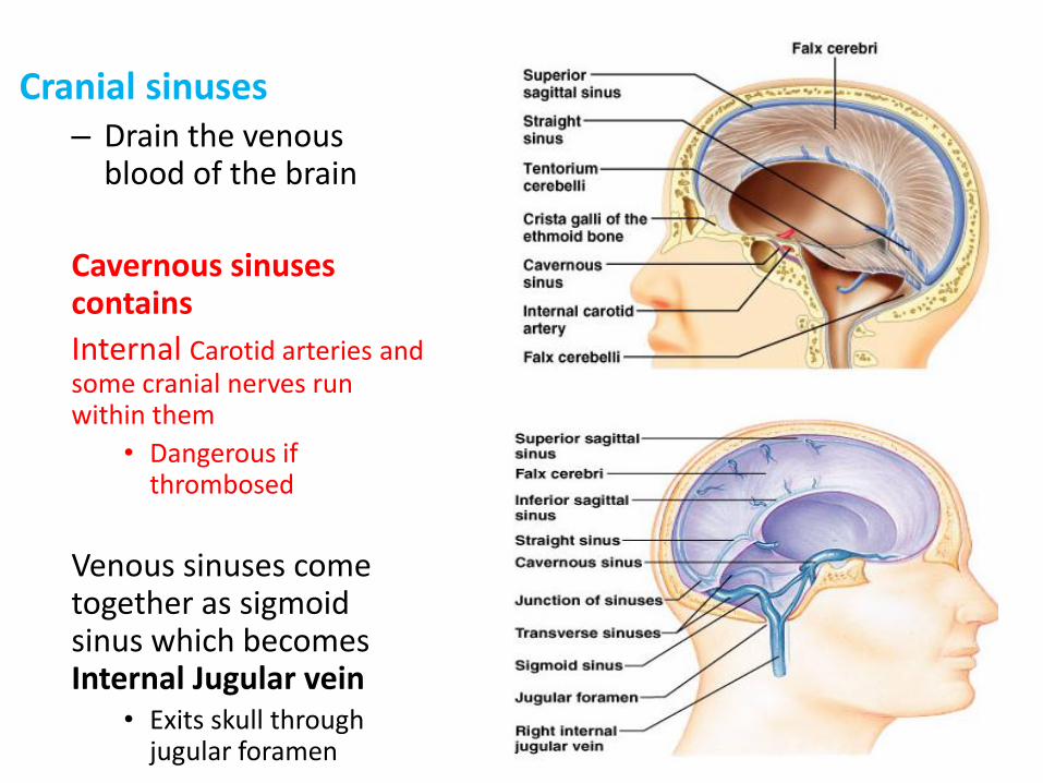
• Internal jugular veins Drain most of blood from brain
Run lateral to internal carotid then common carotid arteries in the carotid sheath. At base of neck joins subclavian v. to form brachiocephalic v.
• External jugular veins – drain some of scalp & face. It is formed by the junction of the posterior division of the retromandibular vein with the posterior
auricular vein. It ends in the subclavian vein
Superficial Veins of the Upper Limb
Dorsal venous arch
Cephalic vein
Basilic vein
Median vein of the forearm
Veins of the Upper Limb
The venous system of the upper limb
It can anatomically be divided into the superficial veins
and the deep veins.
Superficial Veins
The major superficial veins of the upper limb are
the cephalic and basilic veins. They are located within
the subcutaneous tissue of the upper limb.
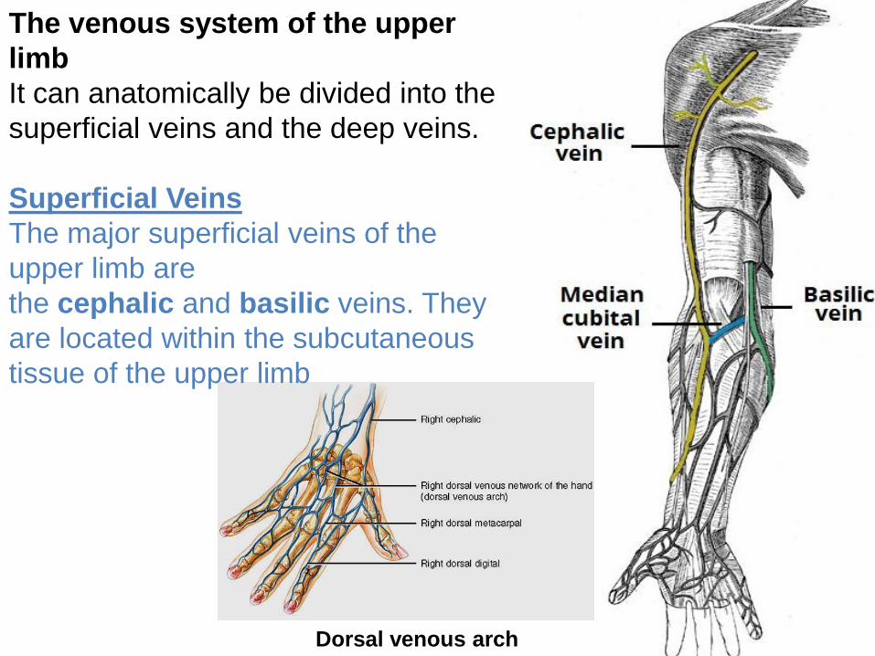
Dorsal venous arch
The venous system of the upper
limb
It can anatomically be divided into the
superficial veins and the deep veins.
Superficial Veins
The major superficial veins of the
upper limb are
the cephalic and basilic veins. They
are located within the subcutaneous
tissue of the upper limb
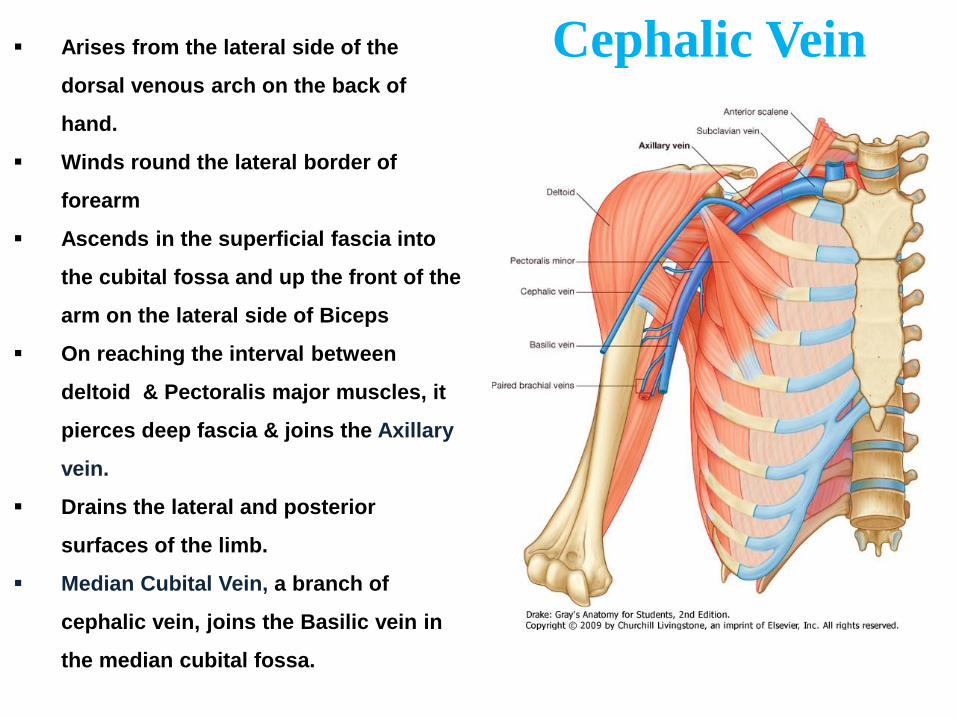
Cephalic Vein Arises from the lateral side of the
dorsal venous arch on the back of
hand.
Winds round the lateral border of
forearm
Ascends in the superficial fascia into
the cubital fossa and up the front of the
arm on the lateral side of Biceps
On reaching the interval between
deltoid & Pectoralis major muscles, it
pierces deep fascia & joins the Axillary
vein.
Drains the lateral and posterior
surfaces of the limb.
Median Cubital Vein, a branch of
cephalic vein, joins the Basilic vein in
the median cubital fossa.
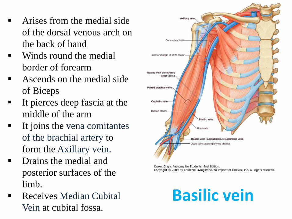
Arises from the medial side
of the dorsal venous arch on
the back of hand
Winds round the medial
border of forearm
Ascends on the medial side
of Biceps
It pierces deep fascia at the
middle of the arm
It joins the vena comitantes
of the brachial artery to
form the Axillary vein.
Drains the medial and
posterior surfaces of the
limb.
Receives Median Cubital
Vein at cubital fossa.
Basilic vein
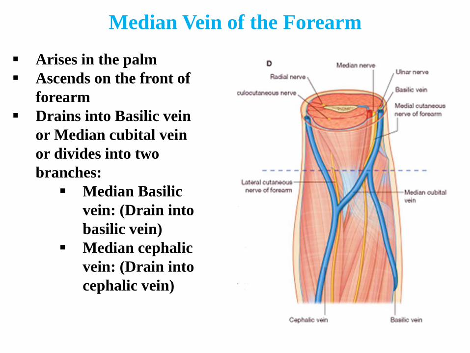
Median Vein of the Forearm
Arises in the palm
Ascends on the front of
forearm
Drains into Basilic vein
or Median cubital vein
or divides into two
branches:
Median Basilic
vein: (Drain into
basilic vein)
Median cephalic
vein: (Drain into
cephalic vein)
Deep Veins
The deep veins of the upper limb are situated
underneath the deep fascia.They are paired veins that
accompany and lie either side of an artery.
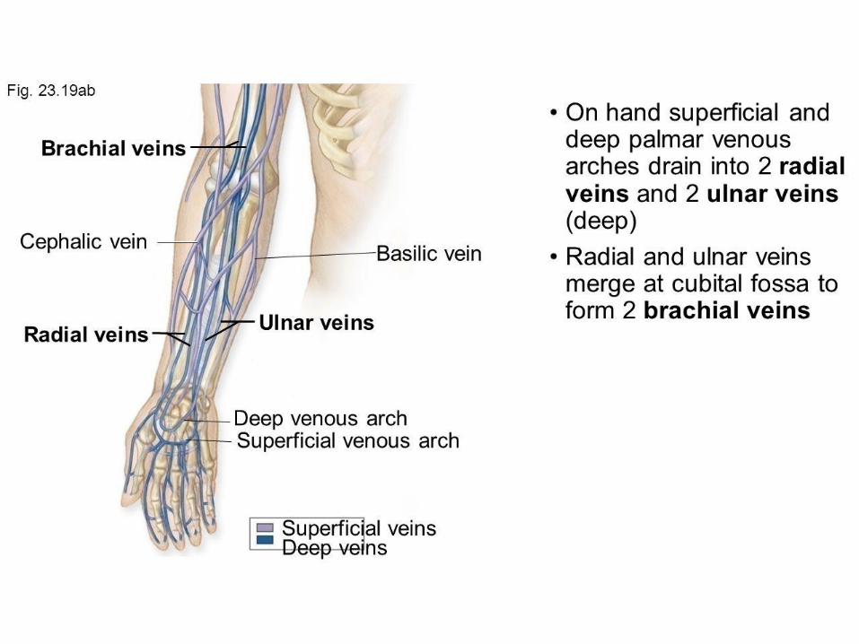
Superficial & deep palmar venous arches Lies in the
subcutaneous tissue proximal to
Metacarpophalangeal joints, they empty into the radial
& ulnar veins and drain the hand. These veins unite to
form the brachial veins, the latter are the larger in size,
and are situated either side of the brachial artery. The
pulsations of the brachial artery assists the venous
return. Veins that are structured in this way are known
as vena comitantes.
Perforating veins run between the deep and superficial
veins of the upper limb, connecting the two systems.
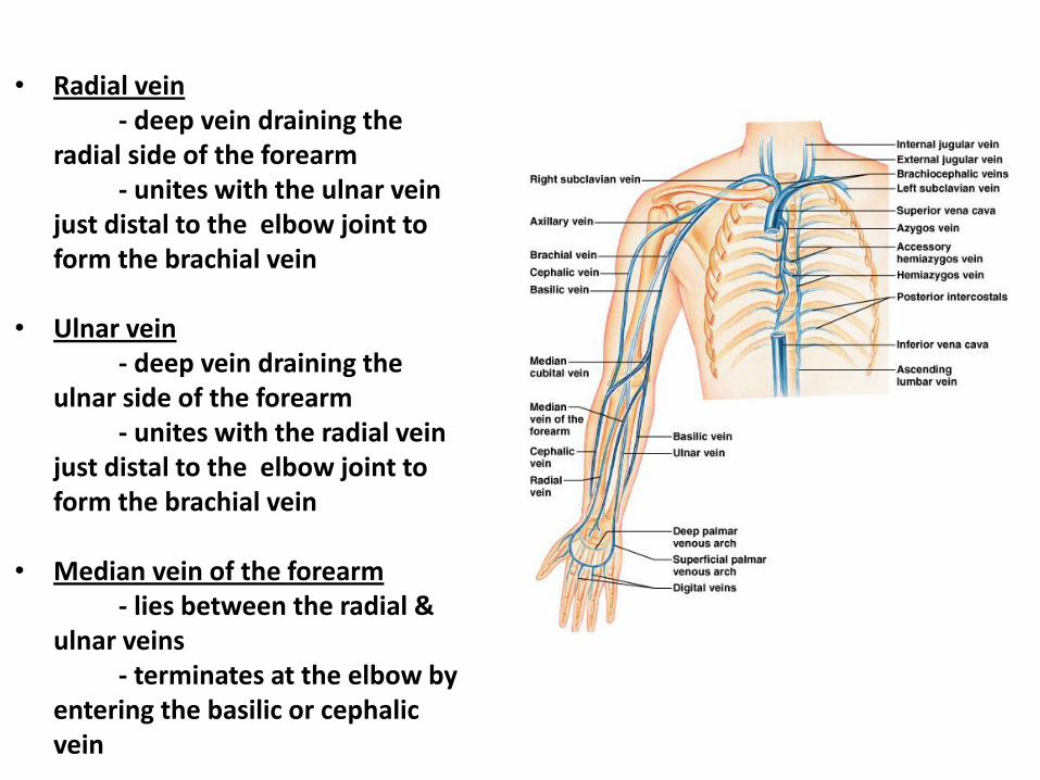
• Radial vein - deep vein draining the
radial side of the forearm - unites with the ulnar vein
just distal to the elbow joint to form the brachial vein
• Ulnar vein - deep vein draining the
ulnar side of the forearm - unites with the radial vein
just distal to the elbow joint to form the brachial vein
• Median vein of the forearm - lies between the radial &
ulnar veins - terminates at the elbow by
entering the basilic or cephalic vein
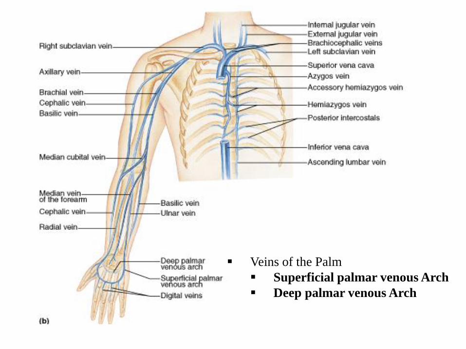
Veins of the Palm
Superficial palmar venous Arch
Deep palmar venous Arch
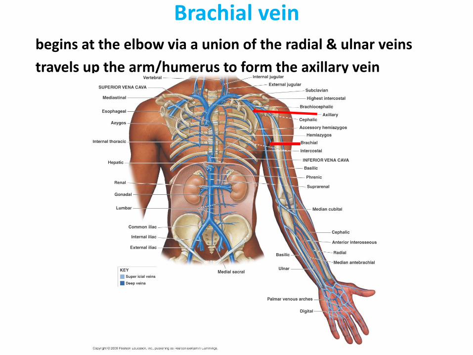
Brachial vein
begins at the elbow via a union of the radial & ulnar veins
travels up the arm/humerus to form the axillary vein
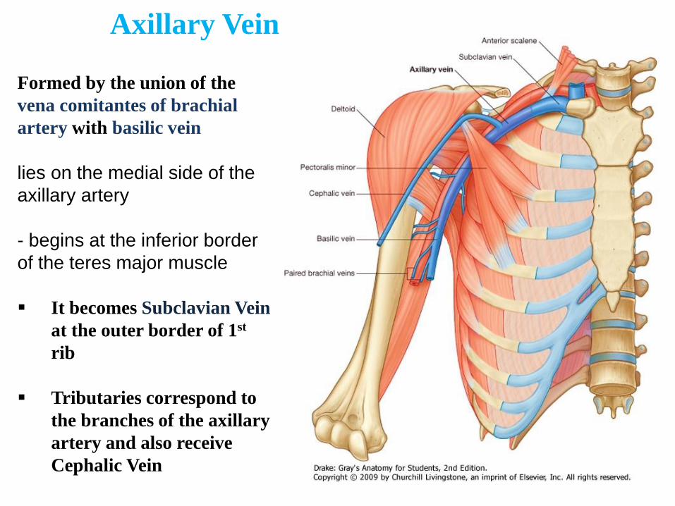
Axillary Vein
Formed by the union of the
vena comitantes of brachial
artery with basilic vein
lies on the medial side of the
axillary artery
- begins at the inferior border
of the teres major muscle
It becomes Subclavian Vein
at the outer border of 1st
rib
Tributaries correspond to
the branches of the axillary
artery and also receive
Cephalic Vein
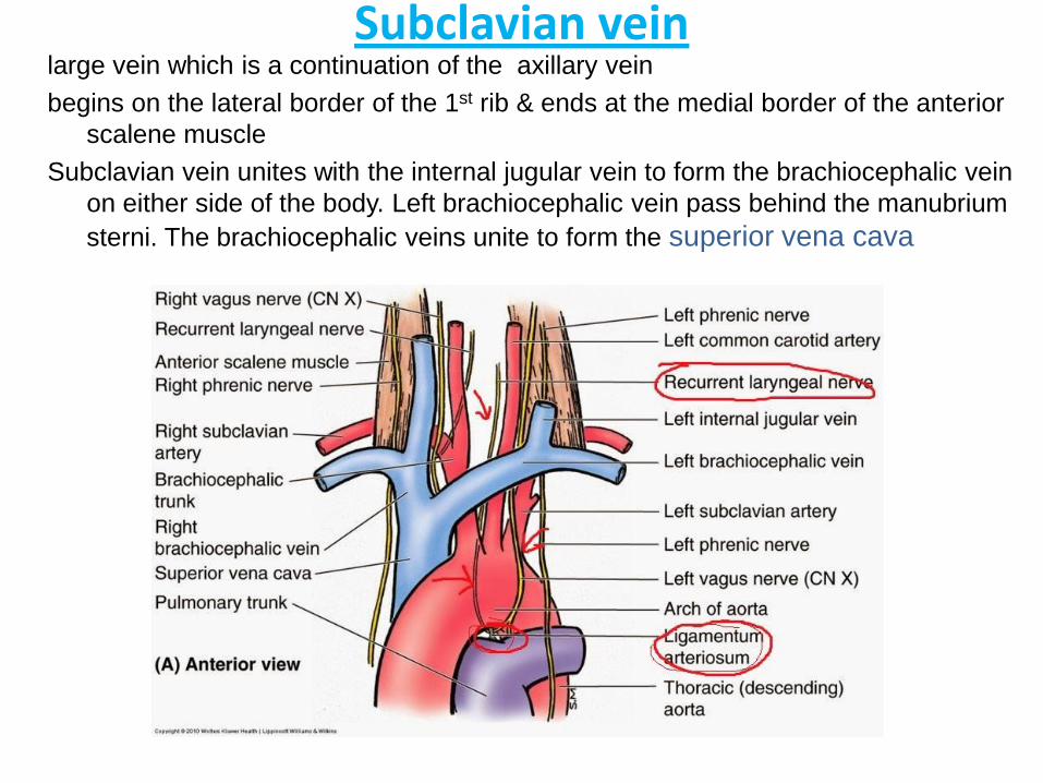
Subclavian vein
large vein which is a continuation of the axillary vein
begins on the lateral border of the 1st rib & ends at the medial border of the anterior
scalene muscle
Subclavian vein unites with the internal jugular vein to form the brachiocephalic vein
on either side of the body. Left brachiocephalic vein pass behind the manubrium
sterni. The brachiocephalic veins unite to form the superior vena cava
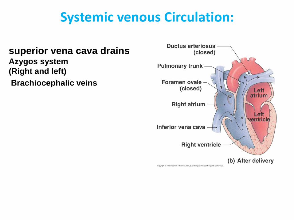
Systemic venous Circulation:
superior vena cava drains Azygos system
(Right and left)
Brachiocephalic veins
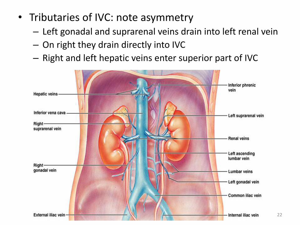
• 2. inferior vena cava drains a. hepatic veins b. phrenic veins
c. Right suprarenal vein d. paired renal veins e. right gonadal veins
f. paired common iliac veins
22
• Tributaries of IVC: note asymmetry – Left gonadal and suprarenal veins drain into left renal vein
– On right they drain directly into IVC
– Right and left hepatic veins enter superior part of IVC
23
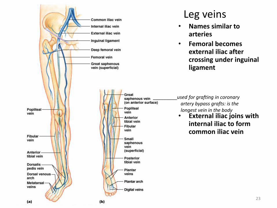
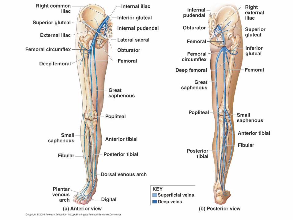
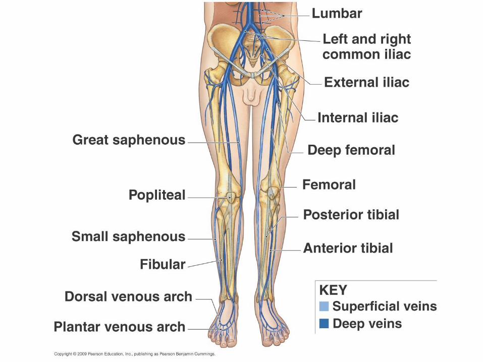
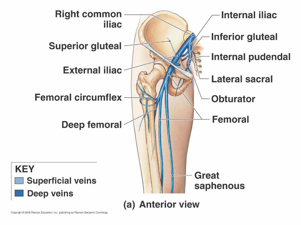
Leg veins • Names similar to
arteries
• Femoral becomes external iliac after crossing under inguinal ligament
• External iliac joins with internal iliac to form common iliac vein
_________used for grafting in coronary artery bypass grafts: is the longest vein in the body
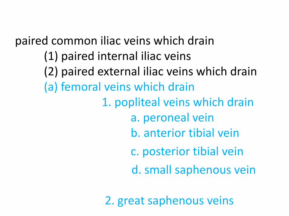
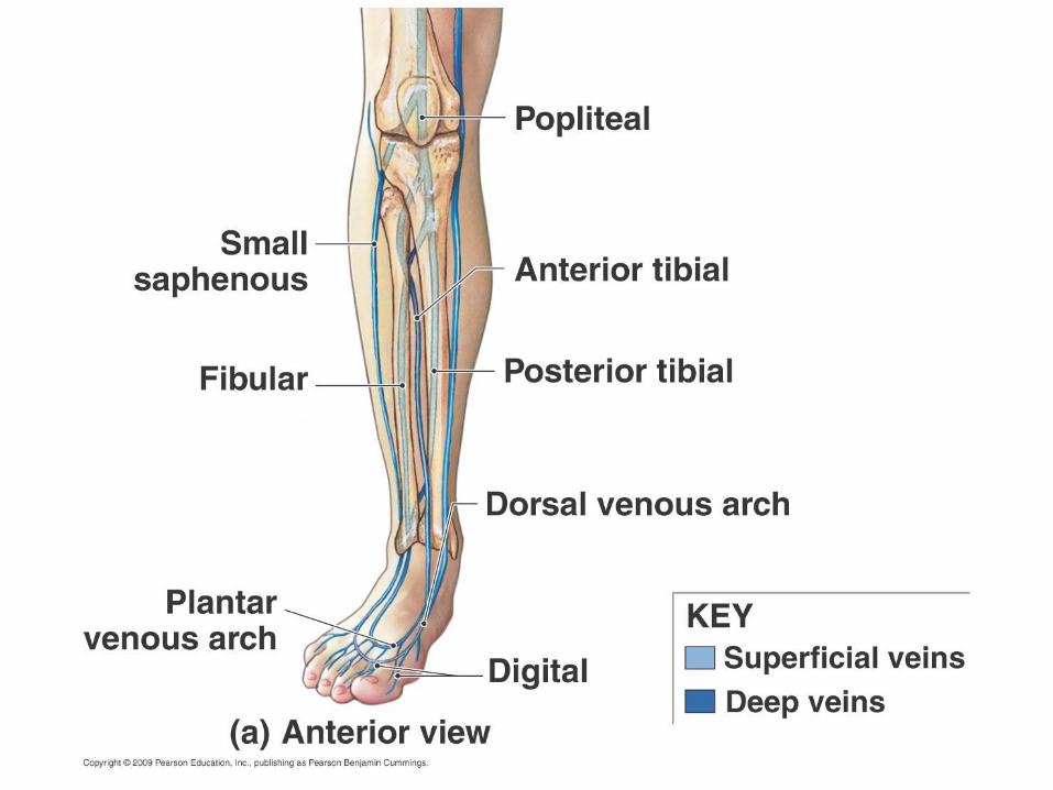
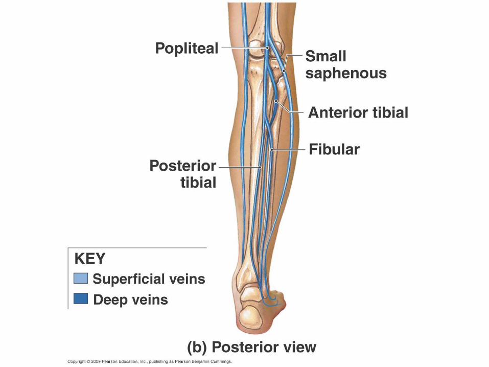
paired common iliac veins which drain (1) paired internal iliac veins (2) paired external iliac veins which drain (a) femoral veins which drain 1. popliteal veins which drain a. peroneal vein b. anterior tibial vein
c. posterior tibial vein
d. small saphenous vein 2. great saphenous veins
27
Systemic Veins
• 3 major vessels enter Right Atrium: – SVC (superior vena cava)
– IVC (inferior vena cava)
– Coronary sinus
• Many veins are very superficial (unlike arteries)
• Venous plexuses (networks of anastomoses and parallel veins) are very common
• Head and hepatic portal systems are unusual
Hepatic Portal System
• System of TWO capillary beds • Digested material is absorbed from the digestive
system by one capillary bed • Goes to the liver for processing through a second
capillary bed • Blood returns back from the liver to the venous
circulation (inferior vena cava) via the hepatic veins (2 – 4)
• Portal vein is formed from the – Superior mesenteric vein – Splenic vein – Inferior mesenteric vein
32
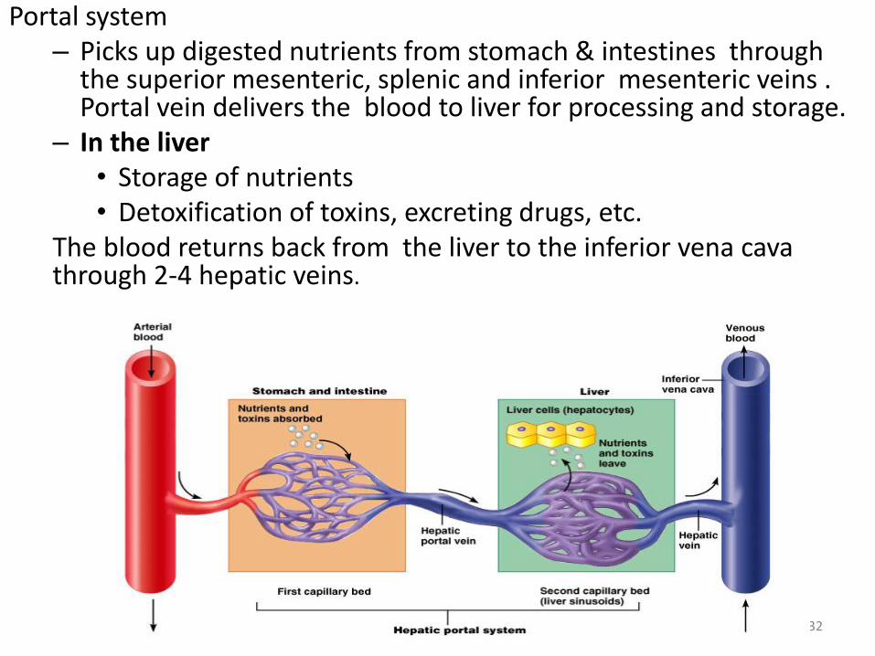
Portal system – Picks up digested nutrients from stomach & intestines through
the superior mesenteric, splenic and inferior mesenteric veins . Portal vein delivers the blood to liver for processing and storage.
– In the liver • Storage of nutrients • Detoxification of toxins, excreting drugs, etc.
The blood returns back from the liver to the inferior vena cava through 2-4 hepatic veins.
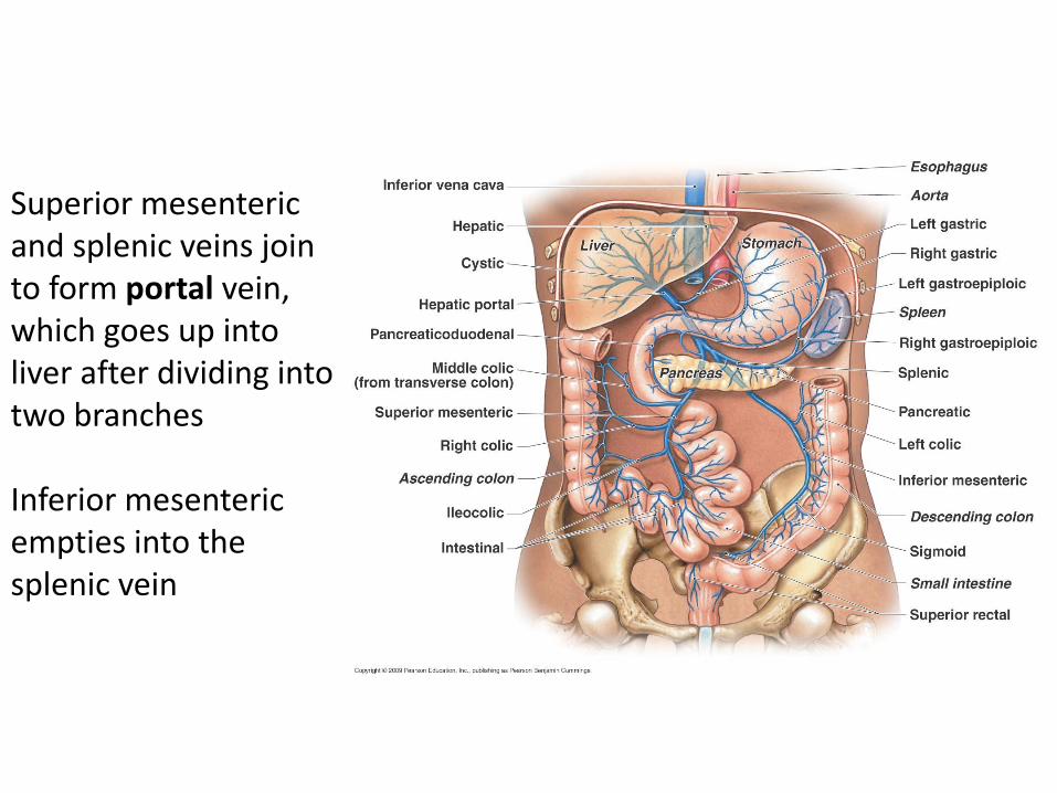
Superior mesenteric and splenic veins join to form portal vein, which goes up into liver after dividing into two branches Inferior mesenteric empties into the splenic vein
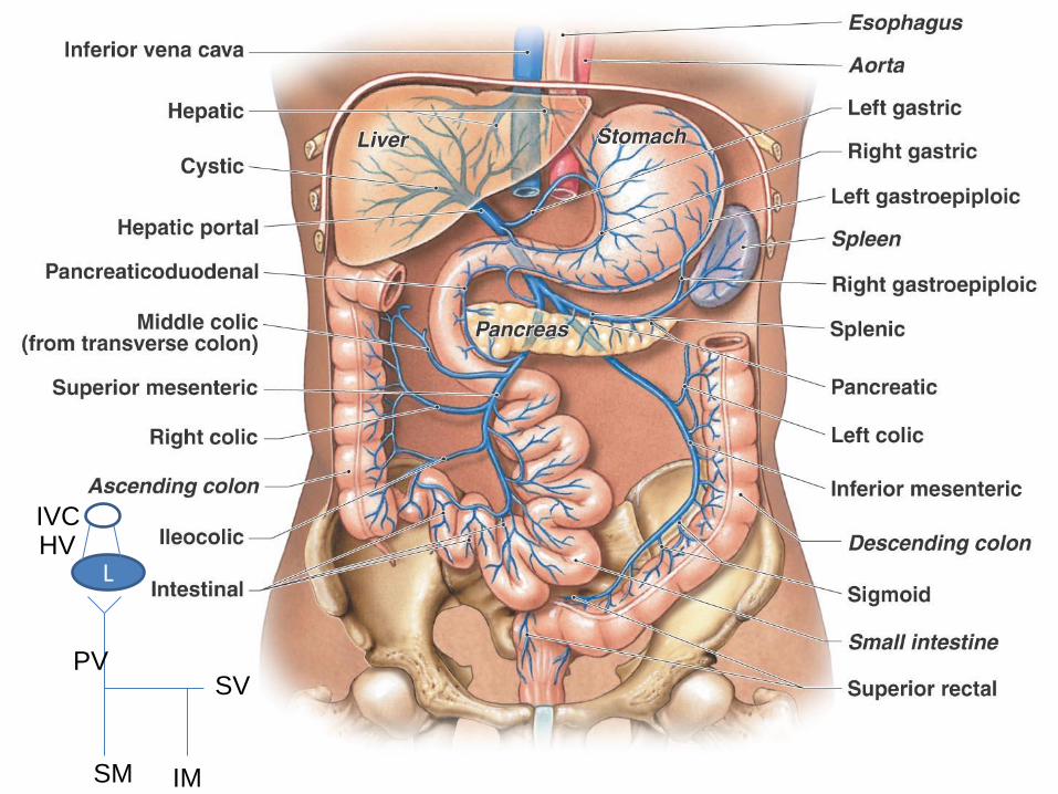
SV
IM SM
PV
HV L
IVC

Thank you