Embed Size (px)
Citation preview
VCT100Vacuum Cryo Transfer
VCT100:02_DM4000_6000M_dt_Brochure.qxd 23.04.2008 9:55 Uhr Seite 1
2
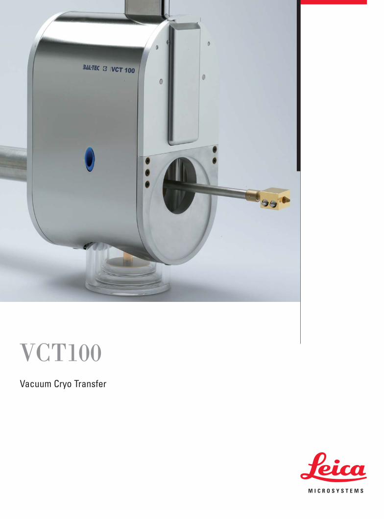
VCT100Versatile Specimen Transfer
� Key features include:
• Contamination free transfer
• Minimum load on analysis tool
• Resolution of analysis tool maintained
• Independent preparation and analysis
• Repeated preparation of same specimen
• Full availability of analysis tool during preparation
VCT100:02_DM4000_6000M_dt_Brochure.qxd 23.04.2008 9:55 Uhr Seite 2
3
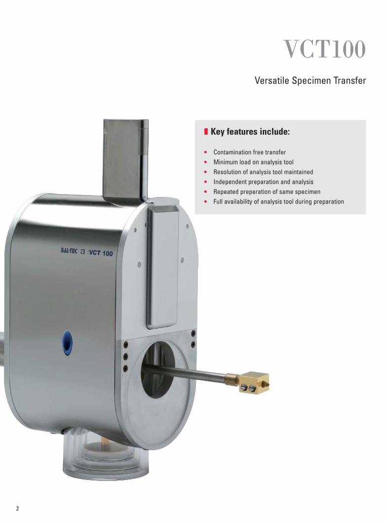
� Contamination free specimen transferbetween preparation and analysis
Transferring specimens to the chamber of an analysissystem is one of the most critical steps of most prepara-tion methods. It is essential to protect the samples fromcontamination. The VCT 100 concept was establishedparticularly to cross link preparation units with variousanalysis systems. Samples are transferred in a well de-fined environment, e.g. protective gas or high vacuumconditions. In addition the specimen can be kept at lowtemperature for cryo techniques. The sample holders usedfor specimen transfer are compatible to an integratedstage. Due to the modular stage concept, the stage canbe installed in any (ultra) high vacuum system or glovebox and used for both room temperature and cryo appli-cations. With the air lock, the transfer shuttle can bemounted to any SEM, FIB, AFM, SIMS or XPS chambers.The flexible system design enables adjustments to anycustomer specific application.
� Setup advantages• Contamination free transfer between preparation
and analysis unit
• Versatile transfer environment
• Minimal interference with the analysis systemfor highest possible resolution
• Independent sample preparation and analysis
• Efficient access to the analysis as well asto the preparation unit
• Preparation units can be linked to severalVCT100 adapted analysis units.
• Preparation and analysis can be performedat different locations
• Easy adjustment to configuration changes
X - Beam FESEM with VCT100
FESEM stage with installed VCT cryo stage
VCT100:02_DM4000_6000M_dt_Brochure.qxd 23.04.2008 9:55 Uhr Seite 3

4
� Cryo SEMLately Cryo Scanning Electron Microscopy (Cryo SEM)has been established as the surface analysis method ofchoice for wet (soft condensed and hydrated matter) andbeam sensitive specimens. Cryogenic sample preparationis quicker than conventional sample preparation techni-ques like chemical fixation with subsequent dehydrationand critical point drying. With the right experimental setupthe entire processing of the specimen from object ex-traction to immobilization (freezing), surface preparation,coating, and imaging can be done in less than one hour.A typical preparation procedure starts with cryo fixationof the specimen using techniques like:High Pressure Freezing with the HPM010 or HPM100, JetFreezing with the JFD030 or Plunge Freezing dependingon the nature of the specimen. The specimen is subse-quently transferred to one of the Leica preparation units:SCD500, MED020 or BAF060 using the VCT100 shuttle. Inthe preparation unit the specimen can be fractured, et-ched and coated. In the VCT100 shuttle the specimen isthen transferred, protected under high vacuum and by acold trap, onto the cryo stage in the SEM. For the actualSEM analysis the VCT100 shuttle is detached from theSEM.
� Cryo SEM advantages• Physical preparation
(no artefacts deriving from chemical treatment)
• No handling with toxic reagents
• Sole preparation technique for many applications
• Beam damage reduction due to low temperatureduring imaging
ESEM with VCT docking station and shuttle
X-ray system with VCT docking station on high vacuum loading chamber and VCT cryo dewaron ultra high vacuum chamber.
X-ray system with VCT cold finger and interface for cryo sample holder
VCT100:02_DM4000_6000M_dt_Brochure.qxd 23.04.2008 9:55 Uhr Seite 4
5
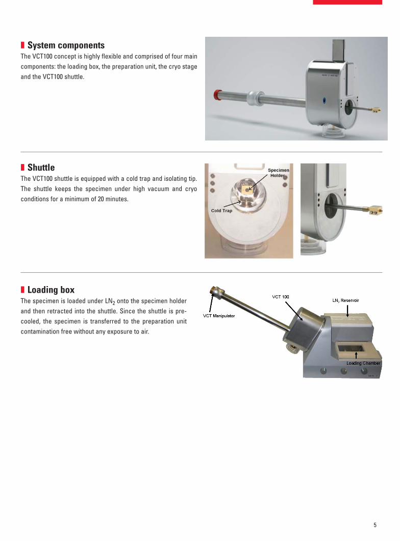
� System componentsThe VCT100 concept is highly flexible and comprised of four maincomponents: the loading box, the preparation unit, the cryo stageand the VCT100 shuttle.
� ShuttleThe VCT100 shuttle is equipped with a cold trap and isolating tip.The shuttle keeps the specimen under high vacuum and cryoconditions for a minimum of 20 minutes.
� Loading boxThe specimen is loaded under LN2 onto the specimen holderand then retracted into the shuttle. Since the shuttle is pre-cooled, the specimen is transferred to the preparation unitcontamination free without any exposure to air.
VCT100:02_DM4000_6000M_dt_Brochure.qxd 23.04.2008 9:55 Uhr Seite 5

6
� Preparation unitsThree preparation units are available for cryo SEM samplepreparation: the SCD500, the MED020 and the BAF060.
The SCD500 equipped with the freeze fracture/etchingunit allows mirror fracturing followed by sputter coatingfor subsequent cryo SEM analysis.
The MED020 configured as freeze fracturing/etching andcoating system is the main preparation device for cryoSEM. Cryo sputtering is the main configuration to producethin and uniform metal layers on the specimen surface.Using the optional E-gun module, films may be producedwith shadowing effects as well as replicas for TEM ana-lysis.
The BAF060 is a fully automatic freeze fracturing machinethat is designed for replica production for high resolutionTEM investigation. It is also the perfect tool to producedouble layer coated specimens for cryo SEM.
VCT100:02_DM4000_6000M_dt_Brochure.qxd 23.04.2008 9:55 Uhr Seite 6
7
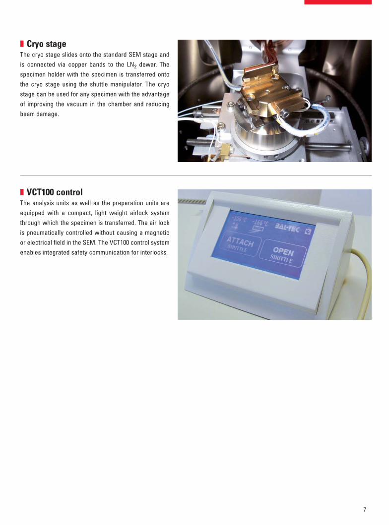
� Cryo stageThe cryo stage slides onto the standard SEM stage andis connected via copper bands to the LN2 dewar. Thespecimen holder with the specimen is transferred ontothe cryo stage using the shuttle manipulator. The cryostage can be used for any specimen with the advantageof improving the vacuum in the chamber and reducingbeam damage.
� VCT100 controlThe analysis units as well as the preparation units areequipped with a compact, light weight airlock systemthrough which the specimen is transferred. The air lockis pneumatically controlled without causing a magneticor electrical field in the SEM. The VCT100 control systemenables integrated safety communication for interlocks.
VCT100:02_DM4000_6000M_dt_Brochure.qxd 23.04.2008 9:55 Uhr Seite 7
8
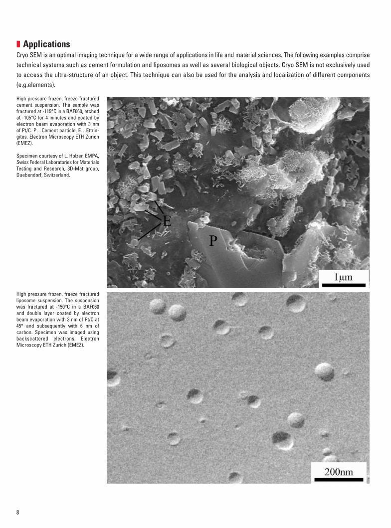
� ApplicationsCryo SEM is an optimal imaging technique for a wide range of applications in life and material sciences. The following examples comprisetechnical systems such as cement formulation and liposomes as well as several biological objects. Cryo SEM is not exclusively usedto access the ultra-structure of an object. This technique can also be used for the analysis and localization of different components(e.g.elements).
High pressure frozen, freeze fracturedcement suspension. The sample wasfractured at -115°C in a BAF060, etchedat -105°C for 4 minutes and coated byelectron beam evaporation with 3 nmof Pt/C. P…Cement particle, E…Ettrin-gites. Electron Microscopy ETH Zurich(EMEZ).
Specimen courtesy of L. Holzer, EMPA,Swiss Federal Laboratories for MaterialsTesting and Research, 3D-Mat group,Duebendorf, Switzerland.
High pressure frozen, freeze fracturedliposome suspension. The suspensionwas fractured at -150°C in a BAF060and double layer coated by electronbeam evaporation with 3 nm of Pt/C at45° and subsequently with 6 nm ofcarbon. Specimen was imaged usingbackscattered electrons. ElectronMicroscopy ETH Zurich (EMEZ).
VCT100:02_DM4000_6000M_dt_Brochure.qxd 23.04.2008 9:55 Uhr Seite 8

9
High pressure frozen, freeze fracturedmouse intestine biopsy. The samplewas fractured at -115°C in a BAF060,etched at -105°C for 5 minutes and co-ated by electron beam evaporationwith 3 nm of Pt/C. M…Mitochondria,N…Nucleus, NP…Nucleopores,G…Golgi, PM…Plasma membrane. El-ectron Microscopy ETH Zurich (EMEZ).
Specimen courtesy of René Fischer,Laboratory of Organic Chemistry, ETHZurich, Switzerland.
High pressure frozen, freeze fracturedsuspension of Vero cells. The samplewas fractured at -115°C in a BAF060and immediately coated by electronbeam evaporation with 3 nm of Pt/C.M…Mitochondria, N…Nucleus,NP…Nucleopores, G…Golgi, PM…Plasma membrane. Electron Microsco-py ETH Zurich (EMEZ).
Specimen courtesy of Peter Wild, Insti-tute of Veterinary Anatomy, Universityof Zurich, Switzerland.
VCT100:02_DM4000_6000M_dt_Brochure.qxd 23.04.2008 9:55 Uhr Seite 9
10
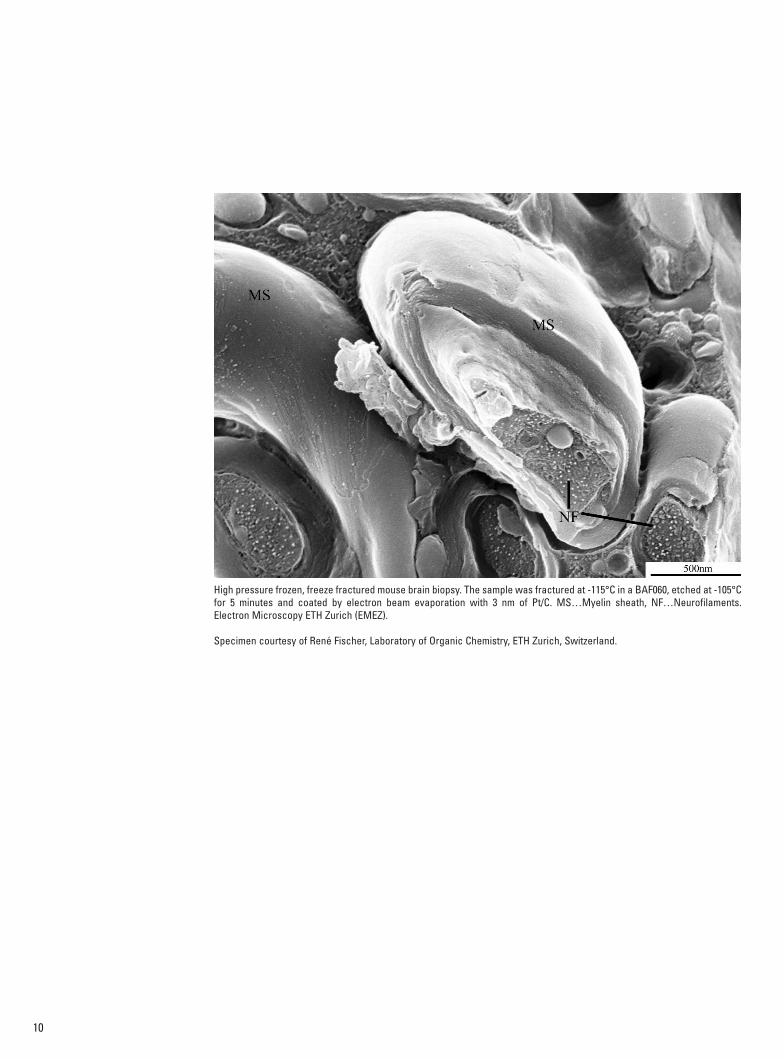
High pressure frozen, freeze fractured mouse brain biopsy. The sample was fractured at -115°C in a BAF060, etched at -105°Cfor 5 minutes and coated by electron beam evaporation with 3 nm of Pt/C. MS…Myelin sheath, NF…Neurofilaments.Electron Microscopy ETH Zurich (EMEZ).
Specimen courtesy of René Fischer, Laboratory of Organic Chemistry, ETH Zurich, Switzerland.
VCT100:02_DM4000_6000M_dt_Brochure.qxd 23.04.2008 9:55 Uhr Seite 10

11
High pressure frozen, freeze fractured suspension of yeast cells (bakers yeast). The sample was fractured at -120°C in a MED020, etched at -100°C for 1 minute andsputter coated with 2 nm of Pt. The high magnification image shows the protoplasmic fracture face of the plasma membrane with invaginations and hexagonal particlearrangements. Electron Microscopy ETH Zurich (EMEZ).
Wax surface on tulip leaf. A piece of tulip leaf was plunge frozen in liquid nitrogen, etched at -105°C for 5 minutes in a BAF060 and coated by electron beam evaporationwith 3 nm of Pt/C. Small wax tubes cover the whole surface of the leaf. Electron Microscopy ETH Zurich (EMEZ).
VCT100:02_DM4000_6000M_dt_Brochure.qxd 23.04.2008 9:55 Uhr Seite 11
12
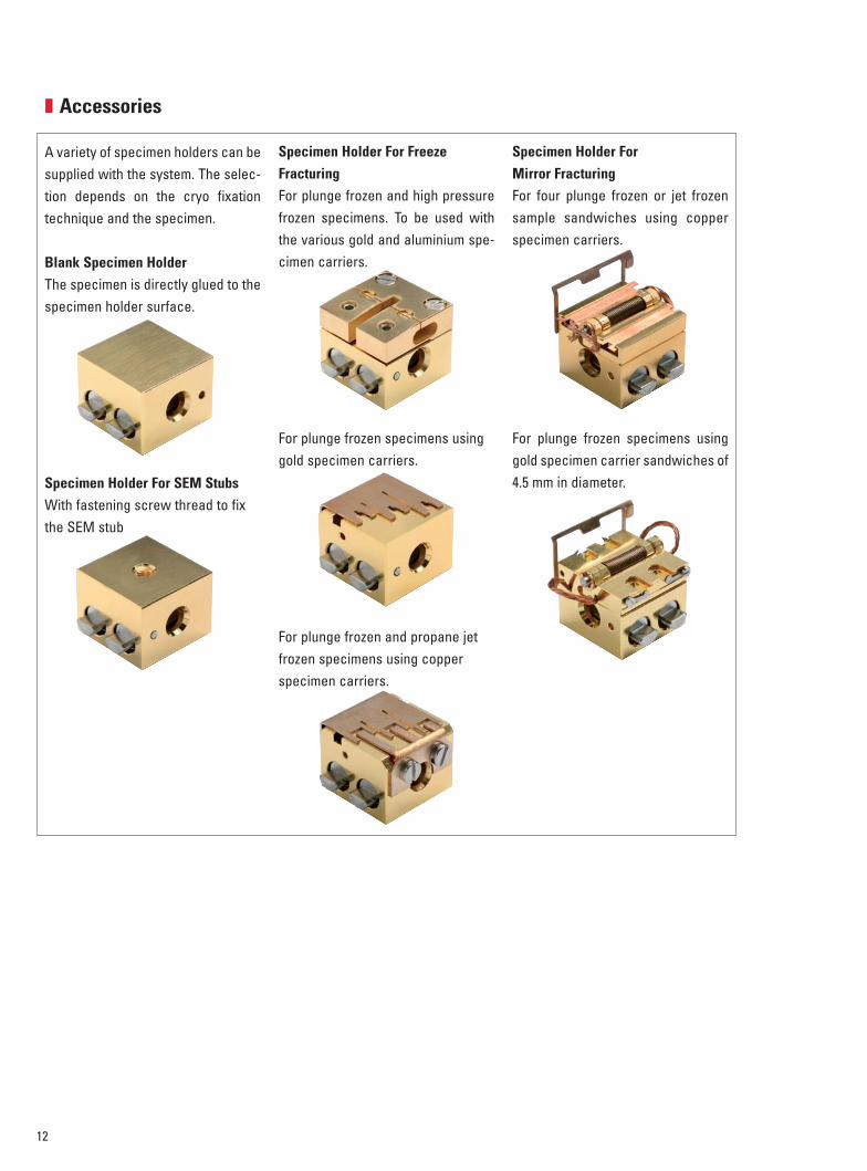
� Accessories
A variety of specimen holders can besupplied with the system. The selec-tion depends on the cryo fixationtechnique and the specimen.
Blank Specimen HolderThe specimen is directly glued to thespecimen holder surface.
Specimen Holder For SEM StubsWith fastening screw thread to fixthe SEM stub
Specimen Holder For FreezeFracturingFor plunge frozen and high pressurefrozen specimens. To be used withthe various gold and aluminium spe-cimen carriers.
For plunge frozen specimens usinggold specimen carriers.
For plunge frozen and propane jetfrozen specimens using copperspecimen carriers.
Specimen Holder ForMirror FracturingFor four plunge frozen or jet frozensample sandwiches using copperspecimen carriers.
For plunge frozen specimens usinggold specimen carrier sandwiches of4.5 mm in diameter.
VCT100:02_DM4000_6000M_dt_Brochure.qxd 23.04.2008 9:56 Uhr Seite 12
13
� Technical specifications
Loading Box
LN2 reservoir 2.5 l for ½ day operation
Dimensions
Metrics 410 mm (W) x 390 mm (D) x 280 mm (H)
Weight 11.5 kg
Shuttle
Temperature
Cold trap -170ºC for 20 min
Isolating tip Temperature of specimen stage maintained
(+0.5ºC per minute in high vacuum)
Vacuum
High vacuum 2 … 5 x 10⁻⁷ mbar for ~20 min
Dimensions
Length 580 mm
Manipulator transfer length 390 mm
Weight 3.6 kg
Airlock
Weight: 2 kg
SEM Cryo Stage
Cooling time < -140ºC within 45 min
Cooling Capacity ~3 h
Cold trap < -170ºC
Stage temperature: -140ºC … 60ºC
Specimen movement Free translation and ± 90º rotation
Drift < 2 nm/min
Preparation Units
SCD500
Vacuum
Pumping System Automatically controlled
Oil free
Membrane pump
Turbo molecular pump
Pumping time to < 1 x 10⁻⁶ mbar 20 min
Ultimate vacuum < 5 x 10⁻⁷ mbar
Cooling
Cryo stage Constant temperature control of the
specimen between -160Cº … +60ºC
Specimen protected by Meissner trap
Automatic bake out system
LN2 filling system Manual
LN2 capacity ½ day operation
Sample Preparation
Specimen handling Via shuttle
Fracturing / Etching Mirror fracturing
Simultaneous fracturing and sputter
coating possible
Etching by exact controlled specimen
temperature
Coating
Sputter Coating Planar Magnetron Sputtering
Sputter Control Permanent sputter coating control by quartz
crystal film thickness monitoring
(auto termination)
Sputter material Cr, W, Pt
Dimensions
Metrics 490 mm (W ) x 585 mm (D) x 553 mm (H)
Opened 792 mm (H)
Weight 87 kg
VCT100:02_DM4000_6000M_dt_Brochure.qxd 23.04.2008 9:56 Uhr Seite 13

� Technical specifications
MED020
VacuumPumping System Automatically controlled
Oil freeScroll pumpTurbo molecular pump
Pumping time < 1 x 10⁻⁶ mbar 20 minUltimate vacuum < 1 x 10⁻⁷ mbar
CoolingCryo stage Electronically controlled by
stepper motorTemperature control of thespecimen between -160ºC ... +60ºCSpecimen protected by MeissnertrapAutomatic bake out system
LN2 filling system ManualLN2 capacity ½ day operation
Sample PreparationSpecimen handling Via shuttleMicrotome Manually driven for fracturing
Automatic knife advance,precision ± 5 µm
Fracturing/Etching Etching by exact controlledspecimen temperature
CoatingSputter coating (standard) Planar Magnetron SputteringSputter control Permanent sputter coating
control by quartz crystal filmthickness monitor (auto termination)
Sputter material Cr, W, Pt
E-beam evaporation (optional)Evaporation sources Two individual digital program-
mable electron beam gunsAngle setting Shuttle manipulatorEvaporation techniques Unidirectional, bidirectionalEvaporation control Permanent evaporation control
by quartz crystal film thicknessmonitoring(auto termination)
Coating material Pt/C, Ta/W, Cr
DimensionsMetrics 850 mm (W) x 670 mm (D) x 615 mm (H)Weight 60 kg (without accessories)
BAF060
VacuumPumping system Automatically controlled
Oil freeStatus displayed by logic mimic diagramMembrane pumpMolecular drag turbo pump
Pumping time to < 1 x 10⁻⁶ mbar 20 minUltimate vacuum: < 5 x 10⁻⁸ mbar
CoolingCryo stage Rotary cryo stage electronically control-
led by stepper motor with variable speedConstant temperature control of thespecimen between -180ºC ... +60ºCSpecimen protected by Meissner trapAutomatic bake out system for the entirecooling system
LN2 filling system AutomaticLN2 capacity 60 l for one day operation
Sample PreparationSpecimen handling Microtome
Motor or manually drivenPrecision advance ± 1µm
Fracturing/Etching Simultaneous fracturing and evaporationpossibleEtching by exact controlled specimentemperature
CoatingElectron beam evaporationEvaporation source Two individual programmable electron
beam gunsAngle settings In-situ motorised angle adjustment
0º…..90ºEvaporation techniques Unidirectional, bidirectional, rotar
shadowing, Double Axis RotaryShadowing (DARS)
Evaporation control Permanent evaporation control byquartz crystal film thickness monitoring(auto termination)
Coating material Pt/C, Ta/W, Cr, W
DimensionsMetrics 1170 mm (W) x 885 mm (D) x 1590 mm (H)Weight 300 kg
VCT100:02_DM4000_6000M_dt_Brochure.qxd 23.04.2008 9:56 Uhr Seite 14
Leica Microsystems –the brand for outstanding products
Leica Microsystems’ mission is to be the world’s first-choice provider of innovative solutions to ourcustomers’ needs for vision, measurement and analysis of micro-structures.
Leica, the leading brand for microscopes and scientific instruments, developed from five brandnames, all with a long tradition: Wild, Leitz, Reichert, Jung and Cambridge Instruments. Yet Leicasymbolizes innovation as well as tradition.
Leica Microsystems – an international companywith a strong network of customer servicesAustralia: North Ryde Tel. +61 2 8870 3500 Fax +61 2 9878 1055
Austria: Vienna Tel. +43 1 486 80 50 0 Fax +43 1 486 80 50 30
Belgium: Groot Bijgaarden Tel. +32 2 790 98 50 Fax +32 2 790 98 68
Canada: Richmond Hill/Ontario Tel. +1 905 762 2000 Fax +1 905 762 8937
Denmark: Herlev Tel. +45 4454 0101 Fax +45 4454 0111
France: Rueil-Malmaison Tel. +33 1 47 32 85 85 Fax +33 1 47 32 85 86
Germany: Wetzlar Tel. +49 64 41 29 40 00 Fax +49 64 41 29 41 55
Italy: Milan Tel. +39 02 574 861 Fax +39 02 574 03392
Japan: Tokyo Tel. +81 3 5421 2800 Fax +81 3 5421 2896
Korea: Seoul Tel. +82 2 514 65 43 Fax +82 2 514 65 48
Netherlands: Rijswijk Tel. +31 70 4132 100 Fax +31 70 4132 109
People’s Rep. of China: Hong Kong Tel. +852 2564 6699 Fax +852 2564 4163
Portugal: Lisbon Tel. +351 21 388 9112 Fax +351 21 385 4668
Singapore Tel. +65 6779 7823 Fax +65 6773 0628
Spain: Barcelona Tel. +34 93 494 95 30 Fax +34 93 494 95 32
Sweden: Kista Tel. +46 8 625 45 45 Fax +46 8 625 45 10
Switzerland: Heerbrugg Tel. +41 71 726 34 34 Fax +41 71 726 34 44
United Kingdom: Milton Keynes Tel. +44 1908 246 246 Fax +44 1908 609 992
USA: Bannockburn/lllinois Tel. +1 847 405 0123 Fax +1 847 405 0164
and representatives of Leica Microsystemsin more than 100 countries.
Leica Microsystems operates internationally in four divi-sions, where we rank with the market leaders.
• Life Science Research DivisionLeica Microsystems’ Life Science Research Division sup-ports the imaging needs of the scientific community withadvanced innovation and technical expertise for the visu-alization, measurement and analysis of microstructures.Our strong focus on understanding scientific applicationsputs Leica Microsystems’ customers at the leading edgeof science.
• Industry DivisionThe Leica Microsystems Industry Division’s focus is tosupport customers’ pursuit of the highest quality end re-sult by providing the best and most innovative imagingsystems for their needs to see, measure and analyze themicrostructures in routine and research industrial appli-cations, in materials science and quality control, in foren-sic science investigations, and educational applications.
• Biosystems DivisionThe Biosystems Division of Leica Microsystems bringshistopathology labs and researchers the highest-quality,most comprehensive product range. From patient topathologist, the range includes the ideal product for eachhistology step and high-productivity workflow solutionsfor the entire lab. With complete histology systems fea-turing innovative automation and Novocastra™ reagents,the Biosystems Division creates better patient care throughrapid turnaround, diagnostic confidence and close cus-tomer collaboration.
• Surgical DivisionThe Leica Microsystems Surgical Division’s focus is topartner with and support micro-surgeons and their careof patients with the highest-quality, most innovative surgi-cal microscope technology today and into the future.
www.leica-microsystems.com
VCT100:02_DM4000_6000M_dt_Brochure.qxd 23.04.2008 9:56 Uhr Seite 15