Embed Size (px)
Citation preview

Vasoconstriction by ElectricalStimulation: New Approach to Control ofNon-Compressible HemorrhageYossi Mandel1,2, Richard Manivanh2, Roopa Dalal2, Phil Huie1,2, Jenny Wang1, Mark Brinton1
& Daniel Palanker1,2
1Hansen Experimental Physics Laboratory, Stanford University, Stanford, CA, 94305, USA, 2Department of Ophthalmology,Stanford University, Stanford, CA, 94305, USA.
Non-compressible hemorrhage is the most common preventable cause of death on battlefield and in civiliantraumatic injuries. We report the use of microsecond pulses of electric current to induce rapid constrictionin femoral and mesenteric arteries and veins in rats. Electrically-induced vasoconstriction could be inducedin seconds while blood vessels dilated back to their original size within minutes after stimulation. At highersettings, a blood clotting formed, leading to complete and permanent occlusion of the vessels. The latterregime dramatically decreased the bleeding rate in the injured femoral and mesenteric arteries, with acomplete hemorrhage arrest achieved within seconds. The average blood loss from the treated femoral arteryduring the first minute after injury was about 7 times less than that of a non-treated control. This newtreatment modality offers a promising approach to non-damaging control of bleeding during surgery, andto efficient hemorrhage arrest in trauma patients.
Trauma is the leading cause of death among US individuals younger than 44 years. Hemorrhagic shockaccounts for 30–40 percent of traumatic mortality1,2. Bleeding is also the most common preventable cause ofdeath on battlefield3. Applications of tourniquets to compressible hemorrhages4–8 caused a marked decrease
in limb exsanguinations3,4,9. As a result, according to the US army, hemorrhage not amenable to truncal tourni-quets (also called non-compressible hemorrhage) is now the leading cause of preventable death3. Part of the non-compressible hemorrhages occur due to bleeding into body cavities (such as the abdomen or chest), while othersare caused by wounds in the junction between the trunk and the limbs or neck. The latter ones, called junctionalhemorrhages, are recognized as a care gap, and those of the pelvic, buttock and groin area are of highestprevalence10. Though Combat GauzeTM is endorsed by the US Army for bleeding care in areas not amenableto a tourniquet, it is often ineffective in junctional hemorrhages such as groin, gluteal, axilla, shoulder andothers9,4,3,10. A novel mechanical compressing device, the Combat Ready Clamp, was recently introduced intothe US Army3,11, but has not yet been proven clinically. This device cannot be applied to wounds of the head, neck,abdomen and chest.
Effective prevention of blood loss in the pre-hospital arena offers the best opportunity to save soldiers withnon-compressible injuries12, therefore major efforts are undertaken to develop technologies for this unmet need.In early 70 s, it was demonstrated that thrombosis can be induced in a clamped blood vessel by minutes-longapplication of direct electric current13–15. However, associated thermal damage precluded the use of this techno-logy in clinical practice. Reduction in blood perfusion during electro-chemotherapy was also noted previously,and it was found to enhance the antitumor effect of the chemotherapy16,17. More recently, constriction of bloodvessels and thrombosis without thermal damage have been achieved with short (ms-ms) electric pulses18.However, these techniques have not been characterized in mammals, nor have they been evaluated for clinicaluse in various bleeding scenarios.
Recently we described significant decrease in blood loss from liver injury in rabbits treated by sub-millisecondelectrical pulses19. The current study evaluates the effect of microsecond pulses on blood vessels in two areas non-amenable to truncal tourniquets: the groin area (femoral) and the abdominal cavity (mesenteric). We dem-onstrate significant vasoconstriction and decrease in blood loss following injury of these blood vessels. Theseresults indicate a possibility of controlling non-compressible hemorrhage using non-thermal pulsed electricalstimulation.
OPEN
SUBJECT AREAS:PRE-CLINICAL STUDIES
PHYSIOLOGY
BIOPHYSICS
TRANSLATIONAL RESEARCH
Received1 March 2013
Accepted7 June 2013
Published4 July 2013
Correspondence andrequests for materials
should be addressed toY.M. (yossi.mandel@
gmail.com)
SCIENTIFIC REPORTS | 3 : 2111 | DOI: 10.1038/srep02111 1
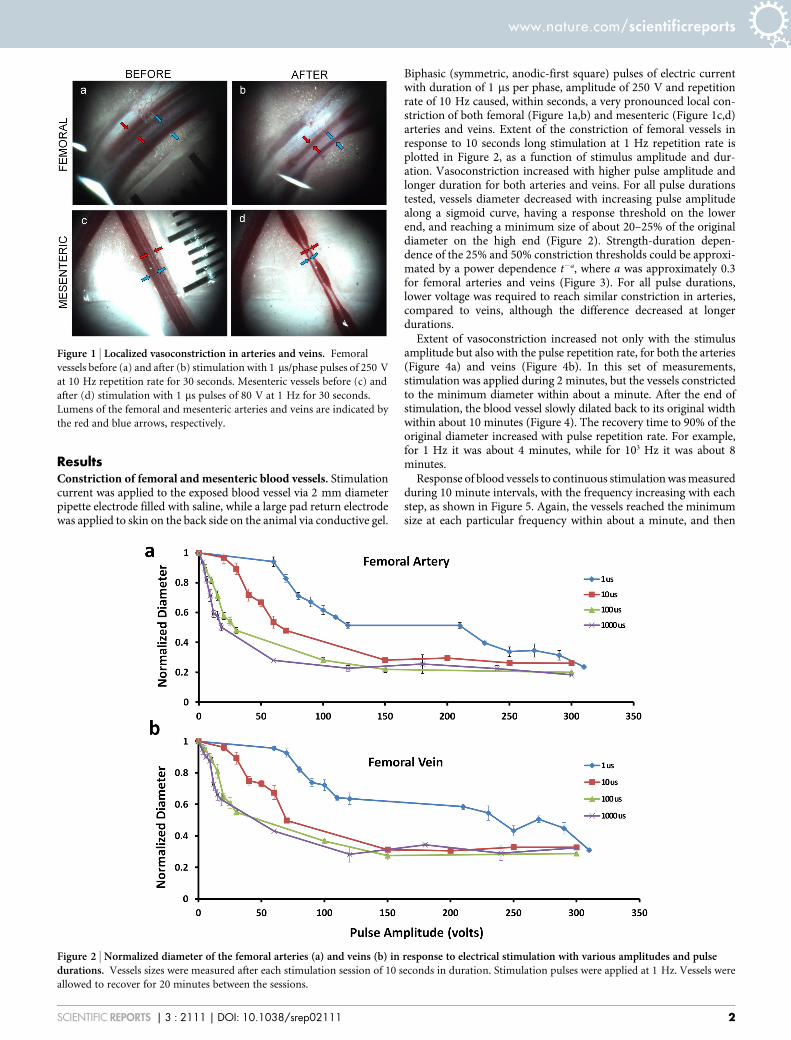
ResultsConstriction of femoral and mesenteric blood vessels. Stimulationcurrent was applied to the exposed blood vessel via 2 mm diameterpipette electrode filled with saline, while a large pad return electrodewas applied to skin on the back side on the animal via conductive gel.
Biphasic (symmetric, anodic-first square) pulses of electric currentwith duration of 1 ms per phase, amplitude of 250 V and repetitionrate of 10 Hz caused, within seconds, a very pronounced local con-striction of both femoral (Figure 1a,b) and mesenteric (Figure 1c,d)arteries and veins. Extent of the constriction of femoral vessels inresponse to 10 seconds long stimulation at 1 Hz repetition rate isplotted in Figure 2, as a function of stimulus amplitude and dur-ation. Vasoconstriction increased with higher pulse amplitude andlonger duration for both arteries and veins. For all pulse durationstested, vessels diameter decreased with increasing pulse amplitudealong a sigmoid curve, having a response threshold on the lowerend, and reaching a minimum size of about 20–25% of the originaldiameter on the high end (Figure 2). Strength-duration depen-dence of the 25% and 50% constriction thresholds could be approxi-mated by a power dependence t2a, where a was approximately 0.3for femoral arteries and veins (Figure 3). For all pulse durations,lower voltage was required to reach similar constriction in arteries,compared to veins, although the difference decreased at longerdurations.
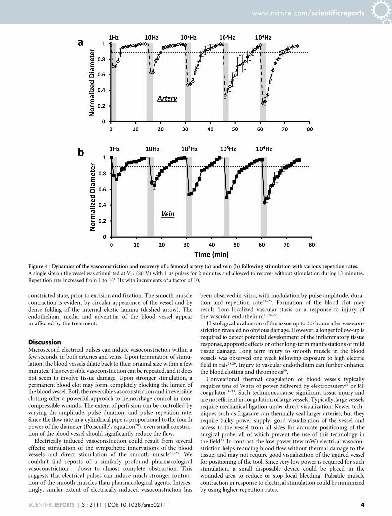
Extent of vasoconstriction increased not only with the stimulusamplitude but also with the pulse repetition rate, for both the arteries(Figure 4a) and veins (Figure 4b). In this set of measurements,stimulation was applied during 2 minutes, but the vessels constrictedto the minimum diameter within about a minute. After the end ofstimulation, the blood vessel slowly dilated back to its original widthwithin about 10 minutes (Figure 4). The recovery time to 90% of theoriginal diameter increased with pulse repetition rate. For example,for 1 Hz it was about 4 minutes, while for 103 Hz it was about 8minutes.
Response of blood vessels to continuous stimulation was measuredduring 10 minute intervals, with the frequency increasing with eachstep, as shown in Figure 5. Again, the vessels reached the minimumsize at each particular frequency within about a minute, and then
Figure 1 | Localized vasoconstriction in arteries and veins. Femoral
vessels before (a) and after (b) stimulation with 1 ms/phase pulses of 250 V
at 10 Hz repetition rate for 30 seconds. Mesenteric vessels before (c) and
after (d) stimulation with 1 ms pulses of 80 V at 1 Hz for 30 seconds.
Lumens of the femoral and mesenteric arteries and veins are indicated by
the red and blue arrows, respectively.
Figure 2 | Normalized diameter of the femoral arteries (a) and veins (b) in response to electrical stimulation with various amplitudes and pulsedurations. Vessels sizes were measured after each stimulation session of 10 seconds in duration. Stimulation pulses were applied at 1 Hz. Vessels were
allowed to recover for 20 minutes between the sessions.
www.nature.com/scientificreports
SCIENTIFIC REPORTS | 3 : 2111 | DOI: 10.1038/srep02111 2

remained at approximately steady state, with the extent of constric-tion dependent on the repetition rate. Vasoconstriction was strongerin arteries than in veins for each pulse frequency with both transient(Figure 4) and continuous (Figure 5) stimulation, and the differencewas more pronounced at higher repetition rates. Maximum responseto continuous stimulation (Figure 5) was smaller than to the tran-sient stimulation regime (Figure 4), especially at higher repetitionrates.
Mesenteric blood vessels (Figure 6) had similar kind of responseto that of the femoral arteries and veins. For the same pulse para-meters, the extent of vasoconstriction in mesenteric arteries washigher than in femoral arteries (Figure 2), and the differenceincreased with larger amplitudes. For example, with 1 ms pulses at200 V mesenteric arteries constricted by 76%, compared to 49%reduction in femoral arteries. Mesenteric veins constricted morethan the femoral veins at low amplitudes, while this ratio reversedat higher amplitudes.
Hemorrhage control during vascular injury. Complete cut of afemoral artery represents a model of traumatic injury leading toprofound loss of blood by the animal. Applying 100 ms pulses of150 V (corresponding to 75% constriction at 1 Hz repetition rate)at a repetition rate of 10 Hz for 30 seconds rapidly decreased thebleeding rate. In all 6 cases treated with this regime, a nearly completehemorrhage arrest has been achieved within a few seconds. Theaverage blood loss from the femoral artery measured during 30seconds of treatment and 30 seconds after that was about 7 timesless than that of a non-treated control (0.14 vs. 1.05 ml, p 5 0.001)(Figure 7). In all untreated animals, bleeding still continued after the1 minute-long blood collection, and the animal died within minutesif bleeding was not mechanically stopped at the end of the
measurements. When treated with pulse amplitude of 30 V at1 Hz (corresponding to 50% constriction threshold), there was nocomplete hemorrhage arrest, and therefore reduction in blood losswas less pronounced: (0.35 vs 1.05 ml, p 5 0.005), as shown inFigure 7). Strong decrease in blood loss was also observed in thesevered mesenteric arteries treated with 100 ms pulses of 40 V at1 Hz (corresponding to 75% constriction threshold), as shown inFigure 7.
Finite element computational modeling of the resistive heatingduring the maximum exposure (150 V, 100 ms/phase, 10 Hz, 30 s)demonstrated that the peak temperature rise at the end of the treat-ment was between 2.3uC with full blood vessel perfusion and 2.9uC,with no blood perfusion (see Supplementary Figure 1b). This estim-ate indicates that even the most intense regime of hemorrhage con-trol did not involve thermal damage to the treated vessels. Modelingof resistive heating for reversible constriction (80 V, 1 ms/phase,10 Hz, 2 min) showed a temperature rise of less than 0.02uC, indi-cating that the mechanism of vasoconstriction is not thermal.
Histological findings. Histological sections of the treated and non-treated femoral arteries are shown in Figure 8. Figure 8a shows across-section of a femoral artery following complete vessel dissectionand 30 seconds-long treatment with 100 ms/phase pulses of 150 V ata repetition rate of 10 Hz. For comparison, an untreated controltissue from the other leg is shown in Figure 8b. Treatment causedcomplete occlusion of the vessel and cessation of bleeding within afew seconds. Upon euthanasia and tissue fixation the vessels dilatedsomewhat, compared to the most constricted state because of smoothmuscle relaxation. In the middle of a circular lumen one can see anacute blood clot attached to the endothelium (solid arrow). Thewidth of the blood clot illustrates the size of the blood vessel in its
Figure 3 | Peak voltage required to induce 25% and 50% constriction (V25 and V50) with pulse durations of 1, 10, 100, and 1000 ms/phase (MEAN 1/2SE, n 5 6). Analytical fit demonstrated that V50 and V25 thresholds for both types of blood vessels scale with pulse duration as a power function of
approximately ,t20.3.
www.nature.com/scientificreports
SCIENTIFIC REPORTS | 3 : 2111 | DOI: 10.1038/srep02111 3
constricted state, prior to excision and fixation. The smooth musclecontraction is evident by circular appearance of the vessel and bydense folding of the internal elastic lamina (dashed arrow). Theendothelium, media and adventitia of the blood vessel appearunaffected by the treatment.
DiscussionMicrosecond electrical pulses can induce vasoconstriction within afew seconds, in both arteries and veins. Upon termination of stimu-lation, the blood vessels dilate back to their original size within a fewminutes. This reversible vasoconstriction can be repeated, and it doesnot seem to involve tissue damage. Upon stronger stimulation, apermanent blood clot may form, completely blocking the lumen ofthe blood vessel. Both the reversible vasoconstriction and irreversibleclotting offer a powerful approach to hemorrhage control in non-compressible wounds. The extent of perfusion can be controlled byvarying the amplitude, pulse duration, and pulse repetition rate.Since the flow rate in a cylindrical pipe is proportional to the fourthpower of the diameter (Poiseuille’s equation20), even small constric-tion of the blood vessel should significantly reduce the flow.
Electrically induced vasoconstriction could result from severaleffects: stimulation of the sympathetic innervations of the bloodvessels and direct stimulation of the smooth muscle21–25. Wecouldn’t find reports of a similarly profound pharmacologicalvasoconstriction - down to almost complete obstruction. Thissuggests that electrical pulses can induce much stronger contrac-tion of the smooth muscles than pharmacological agents. Interes-tingly, similar extent of electrically-induced vasoconstriction has
been observed in-vitro, with modulation by pulse amplitude, dura-tion and repetition rate23–25. Formation of the blood clot mayresult from localized vascular stasis or a response to injury ofthe vascular endothelium18,26,27.
Histological evaluation of the tissue up to 3.5 hours after vasocon-striction revealed no obvious damage. However, a longer follow-up isrequired to detect potential development of the inflammatory tissueresponse, apoptotic effects or other long-term manifestations of mildtissue damage. Long term injury to smooth muscle in the bloodvessels was observed one week following exposure to high electricfield in rats28,29. Injury to vascular endothelium can further enhancethe blood clotting and thrombosis30.
Conventional thermal coagulation of blood vessels typicallyrequires tens of Watts of power delivered by electrocautery31 or RFcoagulator31–33. Such techniques cause significant tissue injury andare not efficient in coagulation of large vessels. Typically, large vesselsrequire mechanical ligation under direct visualization. Newer tech-niques such as Ligasure can thermally seal larger arteries, but theyrequire bulky power supply, good visualization of the vessel andaccess to the vessel from all sides for accurate positioning of thesurgical probe, all of which prevent the use of this technology inthe field33. In contrast, the low-power (few mW) electrical vasocon-striction helps reducing blood flow without thermal damage to thetissue, and may not require good visualization of the injured vesselfor positioning of the tool. Since very low power is required for suchstimulation, a small disposable device could be placed in thewounded area to reduce or stop local bleeding. Pulsatile musclecontraction in response to electrical stimulation could be minimizedby using higher repetition rates.
Figure 4 | Dynamics of the vasoconstriction and recovery of a femoral artery (a) and vein (b) following stimulation with various repetition rates.A single site on the vessel was stimulated at V25 (80 V) with 1 ms pulses for 2 minutes and allowed to recover without stimulation during 13 minutes.
Repetition rate increased from 1 to 104 Hz with increments of a factor of 10.
www.nature.com/scientificreports
SCIENTIFIC REPORTS | 3 : 2111 | DOI: 10.1038/srep02111 4

In conclusion, electrical stimulation of vasculature by microse-cond pulses can be used to control blood perfusion and reducehemorrhage in non-compressible wounds. Temporary decrease inblood perfusion can be achieved in seconds using the reversible
vasoconstriction regime, with vessels dilating back to their originalsize within minutes after termination of stimulation. This modalitycould be used for non-damaging hemorrhage control in surgery andduring trauma care. Permanent blockage of bleeding is achievedupon vasoconstriction followed by initiation of clotting. For practicaluse in trauma care and for treatment of the battlefield injuries, aminiature device should be developed capable of delivering pulsedstimulation prior to arrival of the patient to the hospital. Due to lowenergy requirements a disposable battery-powered device can be justa few millimeters in width, so it can be inserted into the wound tostop local bleeding. Alternatively, a stimulator may remain outsidethe body, and electric current can be delivered to the area of interestvia percutaneous penetrating needle electrodes, similarly to tumorablation by electroporation [e.g.34].
MethodsAnimals. Wild-type Long Evans rats 55–67 days of age were used for all experiments(average weight 275 g). Animals were anesthetized with 75 mg/kg Ketamine HCl and5 mg/kg Xylazine, with additional maintenance half-dose applied every 45 minutes.Buprenorphine (0.01 mg/kg) and Hartman’s Lactated Ringer solution (114 mL/kg/24 hr 37uC) were applied sub-cutaneously before experiment for hydration and paincontrol. All animal care and experiments were carried out in accordance withguidelines for the humane care of animals and were approved by the StanfordAdministrative Panel on Laboratory Animal Care.
Electrical stimulation setup. After anesthesia, dorsal side of the animal was shavedand a conductive gel (Signa Gel, Parker Laboratories, Fairfield, NJ, USA) was applied.Animal was placed in supine position on a stainless steel return electrode and internalbody temperature was controlled at 37uC by a heating pad. Target vessel(s) weredissected from surrounding tissue and allowed to acclimate for 10 minutes beforestimulation. A constant drip of 37uC Hartman’s Lactated Ringer was kept on theexposed site throughout the experiment. Stimulation probe was fabricated from a1 mL plastic syringe with 2 mm diameter opening, filled with normal saline (0.9%NaCl). A platinum electrode inside the syringe and return pad electrode wereconnected to a customized biphasic pulse generator and to an oscilloscope
Figure 5 | Dynamics of vasoconstriction and recovery of a femoral artery (a) and vein (b) during continuous stimulation with various pulse repetitionrates. Blood vessels were stimulated at V25 (80 V) with 1 ms pulses for 10-minute periods at each repetition rate. (MEAN 1/2 SE, n 5 4).
Figure 6 | Constriction of mesenteric arteries (a) and veins (b) inresponse to stimulation with pulses of 1 and 100 ms/phase in duration.Vessels were stimulated at repetition rate of 1 Hz during 10 seconds, with
20 minutes recovery between sessions. (MEAN 1/2 SE, n 5 10).
www.nature.com/scientificreports
SCIENTIFIC REPORTS | 3 : 2111 | DOI: 10.1038/srep02111 5

(Tektronix, Beaverton, OR, USA). Electric pulses were composed of two squarephases of each polarity (positive followed by negative) separated by 10 ms. Pulse risingtime was recorded shorter than 100 ns.
Measurements of the constriction threshold. Femoral vessels were stimulated atthree sites beginning with the most distal one. Each site was stimulated for 10-secondsat 1 Hz repetition rate, and left untreated for 20 minutes for recovery. Anotherstimulation session was then applied to the same site. Mesenteric vessels werestimulated at the second bifurcation from the terminal arteries in intestines, and weretreated only once. Images of the blood vessels before and after stimulation were takenwith a digital camera (Sentech Inc., TC202USB-A), and the lumen diameters weremeasured using the ImageJ software.
Measurements of the frequency response. To assess frequency dependence of thevasoconstrictive response, femoral vessels were stimulated at the voltage levels
corresponding to 25-percent constriction threshold as determined previously for1 Hz. In one set of experiments the vessel was stimulated for 2-minutes at a certainfrequency, and then allowed to recover without stimulation for 13-minutes. This wasthen followed by stimulation at higher frequency, with the same recovery period(Figure 4). In another set of experiments a continuous stimulation was applied at eachfrequency for 10 minutes without recovery periods between them. Stimulation probewas removed from the treatment site for a few seconds every minute to capture animage of the tissue. In each step of these experiments repetition rate was increased by afactor of 10 from 1 Hz to 10 kHz, and then back to 1 Hz.
Measurement of the bleeding rate from the injured femoral artery. Femoral arterywas exposed and completely cut by surgical scissors. Animals in the treatment groupreceived 100 ms electric pulses at 150 V and pulse repetition rate of 100 Hz for 30seconds. In a control group electrode was similarly applied near the severed vessel,however no pulses were applied. In both groups hemorrhage volume was measured 1minute after the vascular injury by soaking a cotton wool near the vessel andweighting the absorbed blood, as previously reported19. Statistical significance of thedifferences between the two groups was evaluated using Student t-test.
Histology. After stimulation the femoral and mesenteric veins and arteries wereligated with 6-0 nylon sutures, and the treated area was dyed with India ink fordefinitive identification in pathology. Tissue was dissected and fixed in 10% bufferedformalin overnight at room temperature. Tissue was then dehydrated with a gradedseries of ethanols and fixed in paraffin, sectioned and stained with hematoxylin andeosin (H&E).
Computational modeling. The tissue temperature rise during electrical stimulationwas estimated using an axio-symmetric 2-dimensional finite element computationalmodel of the electric field and the resulting Joule heating (Comsol Multiphysics 4.1software, COMSOL inc., USA). The geometry is illustrated in supplemental Figure S1,and the constants are listed in Table S1. In short, the model contained five keycomponents: the muscle tissue, the blood vessel, the plastic pipette, the metalelectrode, and the saline in the pipette and in a thin layer above the tissue.
The height of the disk electrode inside the pipette (approximately 2 mm) wascalibrated with a saline-only model to match the impedance of 619 Ohms measuredin a saline-filled petri dish. The tissue resistivity of 300 Ohm-cm was set in the fullcomputational model to match the total impedance of 1.13 kOhm measured for theexperimental setup. After solving the electrostatic field (Poisson) equation whichdefines distribution of the current density in space, the time-averaged heat source(Joule heating rate per unit volume) was calculated and then added to the Pennes bio-heat equation35. To account for the blood flow in non-constricted vessel, a perfusionrate inside the blood vessel was set to 10.6 s21, corresponding to a flow rate of 1 mL/min through a 1 mm diameter blood vessel, where the heated length matches thepipette diameter. For treatment resulting in completely occluded vessels, this per-fusion was removed.
The room temperature and pipette saline temperature was assumed to be 20uC,while the saline covering exposed muscle tissue was 37uC. An equilibration time of100 s was used before calculating treatment-induced temperature rise to allow tissuetemperature to approach steady state after contact with cooler pipette (see details inSupplementary Material).
1. Center for Disease Control (CDC): Web-based Injury Statistics Query andReporting System (WISQARS). In: U.S. Department of Health and HumanServices, CDC, National Center for Injury Prevention and Control. (2002).
2. Kauvar, D. S., Lefering, R. & Wade, C. E. Impact of Hemorrhage on TraumaOutcome: An Overview of Epidemiology, Clinical Presentations, and TherapeuticConsiderations. J Trauma. 60(6 Suppl), S3–11 (2006).
3. Kragh, J. F. et al. New tourniquet device concepts for battlefield hemorrhagecontrol. US. Army Med. Dep. J. Apr–June, 38–48 (2011).
4. Kragh, J. F. et al. Battle casualty survival with emergency tourniquet use to stoplimb bleeding. J. Emerg. Med. 41(6), 590–7 (2011).
5. Kauvar, D. S., Lefering, R. & Wade, C. E. Impact of hemorrhage on traumaoutcome: an overview of epidemiology, clinical presentations, and therapeuticconsiderations. J. Trauma 60(6 Suppl), S3–11 (2006).
6. McManus, J. G., Eastridge, B. J., Wade, C. E. & Holcomb, J. B.. Hemorrhagecontrol research on today’s battlefield: lessons applied. J. Trauma 62(6 Suppl), S14(2007).
7. Kelly, J. F. et al. Injury severity and causes of death from Operation Iraqi Freedomand Operation Enduring Freedom: 2003–2004 versus 2006. J. Trauma64(2 Suppl), S21–6 (2008).
8. Scope, A., Farkash, U., Lynn, M., Abargel, A. & Eldad, A. Mortality epidemiologyin low-intensity warfare: Israel Defense Forces’ experience. Injury 32(1), 1–3(2001).
9. Kragh, J. F. et al. Practical use of emergency tourniquets to stop bleeding in majorlimb trauma. J. Trauma 64(2 Suppl), S38–49 (2008).
10. Ran, Y. et al. QuikClot Combat Gauze use for hemorrhage control in militarytrauma: January 2009 Israel Defense Force experience in the Gaza Strip--apreliminary report of 14 cases. Prehosp. Disaster Med. 25(6), 584–8 (2010).
11. US-Army Report. Tactical Combat Casualty Care Guidelines. (2011).12. Blackbourne, L. H. et al. Decreasing killed in action and died of wounds rates in
combat wounded. J. Trauma 69(Suppl 1), S1–4 (2010).
Figure 7 | Blood loss following a complete cut of femoral and mesentericarteries. After cutting, the femoral artery was stimulated for 30 seconds
with 100 ms pulses of 150 V at 10 Hz (white bar), or at 30 V and 1 Hz
(pink bar). Blood was collected during stimulation and for an additional 30
seconds after stimulation. Control vessels were exposed and severed in a
similar fashion, and the stimulation probe was placed above the vessel, but
no stimulation was applied. Mesenteric vessels were treated with 100 ms
pulses of 40 V (right white bar) at 1 Hz for 30 seconds, or not treated (red
bar). In both vessels types, treatment caused decrease or even complete
stoppage of bleeding after stimulation, while continuous bleeding was
observed in the untreated arteries. Statistical significance of the differences
between groups was evaluated using Student t-test: *p 5 0.001,**p 5
0.047 ***p 5 0.005,****p , 0.001.
Figure 8 | Histology of the treated (a) and untreated (b) femoral arteryfollowing complete vessel dissection. (a) Femoral artery treated with
100 ms pulses of 150 V at 10 Hz for 30 seconds. Treatment caused
complete occlusion of the vessel and termination of hemorrhage within a
few seconds. Constricted lumen is filled with acute blood clot attached to
the endothelium (solid arrow). Constriction is evident by round shape of
the vessel and folding in the internal elastic lamina (dashed arrow). (b)
Control femoral artery from the other side of the same animal was cut and
left bleeding for 60 seconds. Free flowing blood in the control artery
formed a detached clot during sample fixation.
www.nature.com/scientificreports
SCIENTIFIC REPORTS | 3 : 2111 | DOI: 10.1038/srep02111 6

13. Bourgain, R. H. & Six, F. A continuous registration method in experimentalarterial thrombosis in the rat. Thromb. Res 4(4), 599–607 (1974)
14. Guarini, S. A highly reproducible model of arterial thrombosis in rats.J. Pharmacol. Toxicol. Methods 35(2), 101–5 (1996).
15. Hladovec, J. The effect of some platelet aggregating and potential thrombosis-promoting substances on the development of experimental arterial thrombosis.Thromb. Diath. Haemorrh. 29(1), 196–200 (1973).
16. Gehl, A., Skovsgaard, T. & Mir, L. Vascular reactions to in vivo electroporation:characterization and consequences for drug and gene delivery. BiochimicaBiophysica Acta 1569, 51–58 (2002).
17. Sersa, G., Cemazar, M., Parkins, C. S. & Xchaplin, D. J. Tumour Blood FlowChanges Induced by Application of Electric Pulses. European Journal of Cancer35(4), 672–677 (1999).
18. Palanker, D., Vankov, A., Freyvert, Y. & Huie, P. Pulsed electrical stimulation forcontrol of vasculature: temporary vasoconstriction and permanent thrombosis.Bioelectromagnetics 29(2), 100–7 (2008).
19. Mandel, Y. et al. Hemorrhage Control of Liver Injury by Short Electrical Pulses.PLoS ONE 8(1), e49852, doi:10.1371/journal.pone.0049852 (2013).
20. Sutera, S. P. The history of Poiseuille’s law. Annu. Rev. Fluid Mech. 25, 1–19(1993).
21. Atkinson, J. et al. Noradrenaline inhibits vasoconstriction induced by electricalstimulation. Gen. Pharmacol. 18(3), 219–23 (1987).
22. Drummond, P. D. et al. Repeated cycles of electrical stimulation decreasevasoconstriction and axon-reflex vasodilation to noradrenaline in the humanforearm. Br. J. Clin. Pharmacol. 64(4), 421–7 (2007)
23. Ferrell, W. R. & Khoshbaten, A. Responses of blood vessels in the rabbit knee toelectrical stimulation of the joint capsule. J. Physiol. 423, 569–78 (1990).
24. Ferrell, W. R., Khoshbaten, A. & Angerson, W. J. Responses of bone and jointblood vessels in cats and rabbits to electrical stimulation of nerves supplying theknee. J. Physiol. 431, 677–87 (1990).
25. Khoshbaten, A. & Ferrell, W. R. Nerve-mediated responses of blood vessels in therabbit knee joint. J. Vasc. Res. 30(2), 102–7 (1993).
26. Darbousset, R., Thomas, G. M. et al. Tissue factor-positive neutrophils bindto injured endothelial wall and initiate thrombus formation. Blood 120(10),2133–43.
27. Bagot, C. N. & Arya, R. Virchow and his triad: a question of attribution. Br. J.Haematol. 143(2), 180–90 (2008).
28. Maor, E., Ivorra, A., Leor, J. & Rubinsky, B. The effect of irreversibleelectroporation on blood vessels. Technol. Cancer Res. Treat. 6(4), 307–12 (2007).
29. Maor, E., Ivorra, A., Mitchell, J. J. & Rubinsky, B. Vascular smooth muscle cellsablation with endovascular nonthermal irreversible electroporation. J. Vasc.Interv. Radiol. 21(11), 1708–15 (2010).
30. Geenen, I. L. et al. Coagulation on endothelial cells: the underexposed part ofVirchow’s Triad. Thromb. Haemost. 108(5), 863–71 (2012).
31. Massarweh, N. N., Cosgriff, N. & Slakey, D. P. Electrosurgery: history, principles,and current and future uses. J Am Coll Surg 202(3), 520–30 (2006).
32. Mulier, S. Complications of radiofrequency coagulation of liver tumours. BritishJournal of Surgery. 89(10), 1206–1222 (2002).
33. Alexiou, V. G., Tsitsias, T., Mavros, M. N., Robertson, G. S. & Pawlik, T. M.Technology-Assisted Versus Clamp-Crush Liver Resection: A Systematic Reviewand Meta-analysis. Surg Innov. (2012).
34. Cannon, R., Ellis, S., Hayes, D., Narayanan, G. & Martin, R. C. 2nd. Safety andearly efficacy of irreversible electroporation for hepatic tumors in proximity tovitalstructures. J Surg Oncol. doi: 10.1002/jso.23280 (2012).
35. Pennes, H. H. Analysis of tissue and arterial blood temperatures in the restinghuman forearm. J. Appl. Physiol. 1(2), 93–122 (1948).
AcknowledgmentsFunding was provided by the Government of Israel, Research Grant # 4440160018. Theauthors would like to thank LTC Elon Glassberg for a fruitful discussion.
Author contributionsConceived the idea and planned the research (YM, DP), performed experiments (YM, RM),thermal model (JW, MRB), histology (RD, PH), wrote the manuscript (YM, DP).
Additional informationSupplementary information accompanies this paper at http://www.nature.com/scientificreports
Competing financial interests: The authors declare no competing financial interests.
How to cite this article: Mandel, Y. et al. Vasoconstriction by Electrical Stimulation: NewApproach to Control of Non-Compressible Hemorrhage. Sci. Rep. 3, 2111; DOI:10.1038/srep02111 (2013).
This work is licensed under a Creative Commons Attribution-NonCommercial-NoDerivs 3.0 Unported license. To view a copy of this license,
visit http://creativecommons.org/licenses/by-nc-nd/3.0
www.nature.com/scientificreports
SCIENTIFIC REPORTS | 3 : 2111 | DOI: 10.1038/srep02111 7