Embed Size (px)
Citation preview
1
Vacuum Delivery Procedures
An Guide To Assisted Vaginal Delivery
This tutorial is designed to re-inforce previous training – and should, therefore, not be regared as sufficient guidance by itself
Lee Wright Midwifery Lecturer
Plymouth University and The Royal College of Midwives, United Kingdom

Assisted delivery may be classified as:
Outlet forceps or vacuum: – where the fetal skull is on the pelvic floor
Low forceps or vacuum: – where the fetal skull is at or below +2 station
Mid forceps or vacuum: – where the fetal head is engaged but above +2 station
2

Essential conditions for instrumental use include:
Vertex presentation
Complete dilatation of the cervix
Rupture of membranes
No known bony obstruction Being willing to stop the
procedure if it is not successful
3

It is important that the fetal head is engaged before attempting assisted
vaginal delivery
This means that:
The biparietal/largest diameter of the fetal head has passed through the pelvic inlet
The leading edge of the fetal skull is at or below the ischial spines
4

Instrumental delivery should be considered if the mother is demonstrating
any of the following conditions:
Maternal exhaustion Drug induced analgesia Soft tissue resistance
with failure to descend
Predisposing maternal illness
Haemorrhage Relative cephalopelvic
disproportion
Malposition Malpresentation
5

Vacuum delivery should be considered if the fetus is demonstrating either of the
following conditions:
Fetal compromise requiring immediate delivery in the second stage
Non-reassuring fetal heart rate
6
1 2

The instruments that you will need to have available are as follows:
Vacuum delivery:
Vacuum extractor with cup, vacuum pump and vacuum release
7

Procedure
There are 10 steps to be followed for vacuum delivery
In English, these are easily remembered as A-J
– This comes from the ALSO (Advanced Life Support in Obstetrics) organisation
8
10 Steps
Vacuum delivery – Step 1
Address the woman (explain the procedure and ask for consent)
Adequate anaesthesia
Abdominal palpatation
9
A Ask…
Ask for help

Vacuum delivery – Step 2
May need to be catheterised
10
B Bladder…
Bladder empty
Vacuum delivery – Step 3
Examine the woman
Cervix should be fully dilated
11
C Cervix…
Cervix fully dilated

Vacuum delivery – Step 4
– the anterior fontanelle is larger and forms a cross
– the posterior fontanelle is smaller and forms a Y
– assess for bending the ear
Remember moulding of the head makes assessment difficult
Think about dystocia (is the fetus going to fit through the pelvis?)
12
Determine the position of the fetal head
D Posterior fontanelle
Anterior fontanelle

Vacuum delivery – Step 5
Equipment and vacuum extractor need to be ready
13
Equipment
E
Vacuum delivery – Step 6
14
Apply the cup over the sagittal suture 3 cm in front of the posterior fontanelle
Flexion point: proper application of the cup results in flexion of the fetal head when traction is applied
Fontanelle
F Fontanelle…

Vacuum delivery – Step 6
15
CORRECT PLACEMENT
FLEXION POINT/FLEXING MEDIAN
POSTERIOR FONTANELLE
ANTERIOR FONTANELLE
This diagram shows correct positioning of the cup

Vacuum delivery – Step 7
16
Gentle traction at right angles to the plane of the cup
This must only be performed during contractions
Rotary force, or para median application will cause the cup to fall off
Gentle traction
G Gentle…

Vacuum delivery – Step 7
17
This diagram shows application of the cup and the procedure for vacuum extraction
Vacuum delivery – Step 8
18
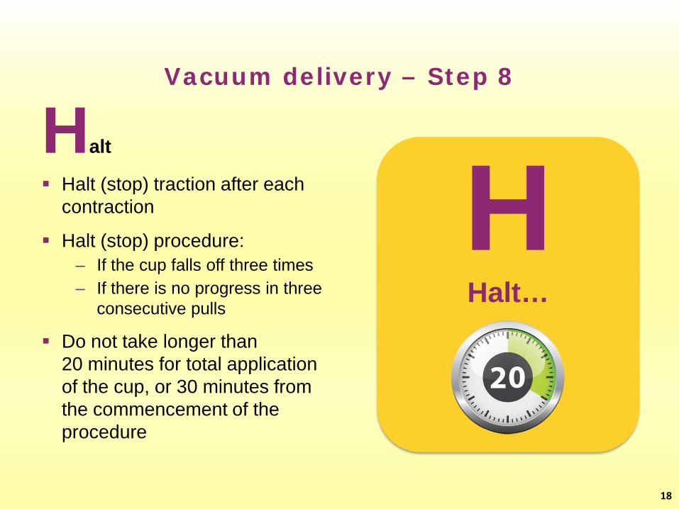
Halt (stop) traction after each contraction
Halt (stop) procedure: – If the cup falls off three times – If there is no progress in three
consecutive pulls
Do not take longer than 20 minutes for total application of the cup, or 30 minutes from the commencement of the procedure
H Halt…
Halt

Vacuum delivery – Step 9
19
Incision or episiotomy needs to be considered when the fetal head is being delivered
This is not always necessary for vacuum delivery although may be necessary for shoulder dystocia or difficult delivery
I Incision…
Incision
Vacuum delivery – Step 10
20
Release the vacuum when you are able to reach the baby’s jaw J
Jaw
Jaw
The disadvantages of vacuum delivery are that:
It may take longer than forceps
It needs the woman to co-operate
There needs to be minimal cephalopelvic disproportion i.e. the fetus should fit fairly easily through the mother’s pelvis
The cup needs to be placed properly
Traction is necessary to avoid losing vacuum
There may be a small increase in cephalhaematoma i.e. bruising under the baby’s scalp
21

Vacuum delivery should not be performed in the following cases:
Severe prematurity (less than 34 weeks)
The fetus is thought to have a blood-clotting problem
Breech, face, or brow presentation
Transverse lie
Incomplete cervical dilatation
The head is not engaged
Excessive traction is needed for delivery
22

The following care needs to be provided after vacuum delivery:
Mother: Examination of cervix and
vagina for injuries Baby: Examination of the new born
baby for birth trauma (injuries) and complications such as:
– scalp emphysema (fluid) – haematomas (bruising) – jaundice (yellowing of
the skin)
symptoms of internal bleeding
Record keeping is important
23

Vacuum delivery illustrated (1)
24
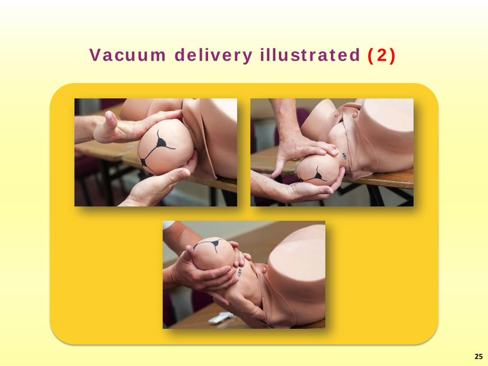
Vacuum delivery illustrated (2)
25


![Dystocia due to a Dichephalus Monster Fetus in Egyptian Buffalo: … Duplication of cranial part of the fetus is more common than of the caudal parts [5]. Polycephaly is a congenital](https://img.dokumen.tips/doc/110x75/601601e419609f28716fa2ce/dystocia-due-to-a-dichephalus-monster-fetus-in-egyptian-buffalo-duplication-of.jpg)