Embed Size (px)
Citation preview

CONTINUOUS ARTERIOVENOUS HEMODIAFIL1RATION


CONTINUOUS ARTERIOVENOUS HEMODIAFIL TRATION
continue anerioveneuze hemodiafiltratie
(met een samenvatting in het Nederlands)
PROEFSCHRIFT
Ter verkrijging van de graad van doctor aan de Erasmus Universiteit Rotterdam
op gezag van de Rector Magnificus Prof. Dr. C.J. Rijnvos en volgens het beslnit van het College van Dekanen.
De openbare verdediging zal plaatsvinden op woensdag 19 mei 1993 om 15.45 uur
door
MARGREET CORNELIA VOS
geboren te Bergen op Zoom

PROMOTIECOMMISSIE
Promotor: Prof.Dr. MA.D.H. Schalekamp
Overige !eden: Dr.Ir. W.A. van Duyl Prof.Dr. W. Weimar Dr. R. Krediet
Deskundige: Dr. H.H. Vincent
Things just happen in the right way, at the right time. At least they do when you let them, when you work with circumstances instead of saying, "'This isn't
supposed to be happening this way," and trying hard to make it happen some other way. [ ... ]
Later on, you can look back and say, "Oh, now I understand [ ... ]".Then you realize that even if you'd tried to make it all turn out perfectly, you couldn't have done better, and if you'd really tried, you would have made a mess of the whole
thing.
(Benjamin Hoff, The Tao of Pooh)
This study was financially supponed by the Dutch Kidney Foundation (Grants nr C88.821 and C89.0873), Hospal, MSD, Glaxo and Bayer. The printing of this thesis was financed by Roussel, which is gratefully acknowledged.
© 1993 M.C. Vos
Cover: D.J. Vos
Uitgeverij Eburon Postbus 2867 2611 CW Delft The Netherlands
No part of this book may be reproduced in any form, by print, photoprint, microfilm or any other means without written permission from the author.
ISBN 90-5166-324-2

Abbreviations
Chapter 1:
Chapter 2:
Chapter 3:
Chapter4:
Chapter 5:
Chapter 6:
Chapter 7:
Chapter 8:
Chapter 9:
Chapter 10:
CONTENTS
Introduction
Problems associated with intermittent hemodialysis in acute renal failure
The clinical practice of continuous arteriovenous hemodiafiltration
Determinants of blood flow and ultrafiltration: Theoretical predictions and laboratory and clinical observations
A mathematical model of solute transpon in continuous arteriovenous hemodiafiltration
Validation of the mathematical model of continuous arteriovenous hemodiafiltration: The assumptions of mixing cup
1
13
19
37
55
concentrations 69
Prediction of the solute mass transfer coefficient of diffusion and the solute clearance rate in continuous aneriovenous hemodiafiltration 83
Drug clearance by continuous arteriovenous hemodiafiltration:
Analysis of sieving coefficients and mass transfer coefficients of diffusion 93
Drug clearance by continuous aneriovenous hemodiafiltration: Prediction of drug clearance rate and recommended dose adaptations for seven antibiotics
Clearance of imipenem/cilastalin in acute renal failure patients
104
treated by continuous arteriovenous hemodiafiltration 121

Chapter 11: Acute renal failure in intensive care patients: A follow up of 236 patients treated by continuous arteriovenous hemo-diaiiltration or intennittent hemodialysis 128
Chapter 12: Summary 141
Chapter 13: Samenvattiog 146
List of publications 153
Nawoord 157
Curriculum vitae 159

ABBREVIATIONS
(X volume fraction of proteins [L/g]
(I) blood channel width [em]
l' viscosity [mmHg ·min]
AI integration constant []
A2 a constant []
c. solute concentration in dialysate [mmol/L]
Cr solute concentration in ultrafiltrate [mmol/L]
em solute concentration in membrane [mmol/L]
cp solute concentration in plasma [mmol/L]
c;.ro, plasma protein concentration [mmol/L]
COP colloid osmotic pressure [mmHg]
b blood channel half-height []
Cl clearance rate [ml/min]
Clcavh clearance rate by CA VH [ml/min]
Clcavhd clearance rate by CA VHD [ml/min]
~ plasma protein concentration [g/dl]
Cpa initial plasma concentration [mmol/L]
~ot-a plasma protein concentration at filter inlet [g/dl]
~. plasma concentration after a time span t [mmol/L]
S>rot-v plasma protein concentration at filter outlet [g/dl]
Cw solute concentration in plasma water [mmol/L]
f fractional volume distribution of solute in blood cells []
F free fraction of a drug []
h height of the fluid column in the UF-Iine [em]
Ht hematocrit []
·; at filter inlet []

J, solute flux [)llll/min l
Jv volume flux [)llll/min]
Kd mass transfer coefficient of diffusion [pm/min]
K.c mass transfer coefficient of diffusion of creatinine [)llll/min]
Kdp mass transfer coefficient of diffusion of phosphate [)lll1/min]
Kw mass transfer coefficient of diffusion of urea [)llll/min l
Km..x maximal Kd at infinite dialysate flow rate [pm/min]
L fiber length [em]
~ hydraulic membrane permeability [em/min • mmHg]
MAP mean arterial pressure [mmHg]
MI hydraulic permeability index of the membrane, or
filter membrane index [ml!h • mmHg]
MW molecular weight [Daltons]
Mw total solute mass in plasma water [mmol]
N number of fibers or blood chanoels of the filter []
n KJJv []
-. at filter outlet []
pb hydraulic pressure in blood compamnent [mmHg]
pd hydraulic pressure in dialysate compamnent [mmHg]
P;a hydraulic pressure in the artety [mmHg]
piv hydraulic pressure in the vein [mmHg]
P. hydraulic pressure in the arterial line [mmHg]
pv hydraulic pressure in the venous line [mmHg]
Qb blood flow rate [ml!min]
Q, ultrafiltration flow rate [ml/min]
Q. dialysate flow rate [ml!min]
Qw plasma water flow rate [ml/min]
Qpa artetial plasma flow rate [ml/min]

Qpred predilution flow rate [mllmin]
r radius of the fiber of the filter [pm]
R total resistance to diffusion [min/pm]
R,. resistances to flow of the arterial access [mmHg · min/ml]
Rb resistance to diffusion in blood compartment [min/pm]
Rd resistance to diffusion in dialysate compartment [min/pm]
Rr resistance to blood flow of the filter [mmHg . min/mll
Rm Rf corrected for blood viscosity (RJ)l) [lo'/ml]
R,. resistance to diffusion in membrane [min/pm]
R.. resistances to flow of venous access [mmHg . min/ml]
s sieving coefficient []
s membrane surface area [mz]
I time [hr]
TMP transmembrane pressure [mmHg]
T~ drug half life (hrs]
vz volume of distribution [L/kg]
w width (S!L) [m]


CHAPTER 1
INTRODUCTION
THE DEVELOPMENT OF BLOOD PURlFlCATION TECHNIQUES
History
The history of blood purification started with the judgement of different body fluids by color and consistency. Blood leuing, as popularized by Hippocrates (460-377 B.C.), was one of the most common therapeutic tools in antique medicine. HistOry tells us that too much blood leuing was weakening the Spartan army and may very well have contributed to its defeat by the Athenians. 1n the 17th and 18th century, blood purification was achieved by adequate purgation every week, application of a strong emetic once a month, and blood leuing twice a year, in spring and fall. 1n 1854, Graham demonstrated that a vegetable parchment, sealed with albumin, acted as a semipermeable membrane. When he placed a fluid, containing crystalloids and colloids, on one side of this membrane, he found that only cristalloids diffused
through the membrane. He named this phenomenon dialysis [1]. 1n 191 I, Haas in Giessen, Germany, who was confronted with numerous cases
of 'field nephritis', first tried to petform dialysis of the blood in uremic patients. In 1913, Abel reported the first successful dialysis on a live animal. 1n 1924, Haas
petformed a 15 minute dialysis in a uremic patient, by using a celloidin membrane. Two further developments were very important for the development of dialysis, i.e. the discovery of heparin in 1918 and the manufacturing of cellophane membranes in the 1930's, which were used as package material for sausages. Using this material,
Kolff, in 1943, treated his first patient in Kampen, the Netherlands, with his rorating drum dialyzer. Blood access was obrained by arteriotomies and venesections, which limited the number of dialysis treatments in one patient His 17th patient was the first that survived. This patient was the first human being whose life was saved by
dialysis [2]. Duling World War II, the hundreds of patients with renal failure from crush
injuries instigated further developments. Alwall, in Sweden, stressed the importance of fluid overload. He devised a 'dialyzer-ultrafilter' in order to petfonn ultrafiltration. 1n 1947, Malinow, in the USA, in an experimental study of dogs, produced ultrafiltrate from blood and replaced it with Ringers solution [3], thereby cleaning the blood. This was the first description of hemofiltration. 1n 1948, Kolff was invited to New Yorlc to continue his work in the United Srates in cooperation with Merrill. By
1

Imroduction
1952, Merrill reponed that about 45 rotating drum dialyzers were in use in 1he United States, mostly for the treatment of acute renal failure. A few years later, 1he Baxter company staned the commercial manufacturing of dialysis equipment. In 1960, Quinton and Scribner described a method for the chronic cannulation of blood vessels by the use of a teflon tubing by-pass of 1he anery and vein [1]. Both these developments made an enormous contribution to the spreading of the dialysis technique.
Mechanisms of solute transport and terminology
Nowadays, hemodialysis and hemofiltration are commonly used blood purification techniques. With both techniques, blood is led through a flat sheet or a capillary dialyzer. The dialyzer consists of a blood and an ultrafiltrate/dialysate companment,
separated by a membrane. The membrane is permeable to solutes of up to several thousand Daltons, dependlug on the membrane pore size, but not to proteins and blood cells. By virtue of the difference in hydrostatic pressure on both sides of the membrane, plasma water leaks through 1he membrane (ultrafiltration) (see Figure 1). If solutes have a molecular weight far below the 'cut-off' point of the membrane, they can pass the membrane together with the plasma water, so their concentration in the ultrafiltrate will be equal to that in plasma water. This transport is called convection. Therefore, for these solutes, the convective transport rate is determined by the rate of ultrafiltration alone. illtrafiltration does not affect the concentration of the solute in plasma water. Only if the ultrafiltration fluid is replaced with a clean substitution fluid, the solute concentration in the plasma water falls. This technique is called hemofiltration (see Figure 2) and is performed either intennittent or continuous. Another way to clean the blood is by ruoning a clean fluid through the dialysate comparttnent so as to cause diffusion of solutes from plasma water to 1he dialysate companment. This technique is called hemodialysis (see Figure 3). The diffusive transport rate is largely determined by the molecular size. Thus, the transport of small molecules is favored by hemodialysis whereas that of large molecules is favored by hemofiltration. With intennittent hemodialysis, it is now
common practice to use a high dialysate flow rate (500 ml/min) and it is possible to realize a fixed ultrafiltration flow rate, adjusted to the patient' overweight. Therefore, a relatively small convective transport occurs too, which can largely be neglected when compared to the large diffusive transport. The combination of hemodialysis and hemofiltration, in which both modalities function as major transport mechanisms, is called hemodiafiltration (see Figure 4), also performed either in the intermittent or continuous mode.
2

Chapter 1
Ul TRAFIL TRA TION
Figure 1. A schematic drawing of ultrafiltration. No clearance of solutes passing the membranes will occur as solute concentration in plasma water does not decrease. This method is used to witJuiraw an excess of body fluid.
substitution
HEMOFIL TRA TION
Figure 2. A schematic drawing of he11U)filtration. Solutes drawn here, have a 11U)lecular weight far below the cut-off point of the membrane. Therefore, solute concentration in the ultrafiltrate compartment will be the same as that in plasma water. Transpon of solutes is by comection. Substitution fluid is used to decrease the solute concentration in blood and to impede too much fluid withdrawal.
3

l ntroduction
0
0
HEMODIALYSIS 0 ° 0 0~·~-----111
Figure 3. A schematic drawing of hemodialysis. Dialysate flows countercurrently through the dialysate compartment, which causes a solute concentration difference over the membrane. Transpon is by diffusion.
0 0 0
0 0 0
0
0 0
HEMODIAFILTRATION
Figure 4. A schematic drawing of hemadiafiltration. Both by convection, by a high ultrafiltration flow rate, and by diffusion, by using dialysate, solutes are removed from biood. Both convection and diffusion contribute significantly to solute transport."
•
4
Figures 1-4 reprinted with permission from NTVG 1992, 136: 561-565 (see list of publications nr. 8)

Chapter I
Further refinement of the blood purification techniques
In the early years of dialysis, patients were treated for 8 to 12 hours, two to three
times a week. Understandably, the developments in those days were aimed at
improving the efficiency of the treatment so as to reduce treatment time. Accordingly, dialysis membranes became thinner and larger and blood and dialysate flow rates were increased. In 1964, however, the drawback of this development became clear when Peterson described acute encephalopathy during dialysis treatment. This was explained by the rapid fall in the concentration of solutes in plasma due to diffusion, leading to an osmotic 'disequilibrium' between plasma water and the brain tissue [4]. Dialysis induced hypotension was another frequent complication. In 1967, Henderson promoted hemofiltration [5]. With hemofiltration the efficiency of the removal of urea was lower but disequilibrium was seldom seen and blood pressure was more stable. Moreover, as Babb [6] pointed out, so called 'middle molecules', which were believed to contribute to the uremic syndrome, were removed more efficiently with this technique. This middle molecule hypothesis also led to the development of peritoneal dialysis techniques.
For hemofiltration, new membranes were developed, which allowed passage of water and solutes of up to several thousand Daltons at very low pressures. At the same time, hemodialysis was refined by controlled ultrafiltration, by the use of bicarbonate rather than acetate as a buffer substitute and by the use of higb or variable sodium concentrations in dialysate. By virtue of these developments, most patients with chronic renal failure can now adequately be treated by intermittent hemodialysis in two or three sessions of 410 5 hours a week. In case of acute renal failure, however, many problems remained to be solved.
Continuous arteriovenous hemodiaftltration
Acute renal failure often results from sepsis, a clinical syndrome that is accompanied also by massive edema and circulatory insufficiency, respiratory failure and sometimes neurological damage [7, 8]. Patients with acute renal failure are characterized by a higb catabolic rate and therefore need intensive dialysis treatment. The combination of low or unstable blood pressure and edema makes it qnite difficult to judge how much flnid should be withdrawn. The combination of ultrafilttarion and hemodialysis frequently leads to hypotension and neurological deterioration and may further jeopardize kidney function [9]. Furthermore, rapid
5

Introduction
correction of the acidosis together with the rise of inflammatory mediators that
results from the blood-membrane interaction may aggravate respiratory failure [10-
15]. Consequently, in many patients with acute renal failure, 'conventional'
hemodialysis is contraindicated. Therefore, this group of patients demands a differem
approach [16]. The prerequisiteS that must be met by the trealment can be summarized
as follows: l. It must be possible to stan trealment at any time;
2. It must be a very gradual treatment, so as to avoid sudden disturbances in the delicate equilibrium and fluid status of the patient;
3. It must be very efficient in order to treat the uremic syndrome.
In 1977 Kramer published a method to treat massive ove!h.ydration. He used a small
dialyzer and conneCted it to ca!h.eters in !h.e femoral artery and vein to obtain a
spontaneous blood flow driven by the blood pressure of the patiem [17]. No substitution fluid was given. This technique is now known as slow continuous
ultrafiltration (SCUF). Later, in order to comrol the rate of fluid loss, !h.e technique
was modified by the addition of fluid substitution [18]. In this way, the blood is also cleaned. This trealment is called continuous arteriDvenous hernofiltration (CA VH).
When small highly permeable dialyzers became generally available, an increasing
number of doctors relied on CA VH for the trealment of critically ill patients with acute
renal failure. In spite of very low blood pressures, the gradual fluid wi!h.drawal was
very well tolerated by these patients [19]. As far as the trealment of uremia was
concerned, the treatment proved to have its limitations. For !h.ese highly catabolic
patients, the rate of ultrafiltration was seldom enough to provide adequate clearance of uremic solutes. Indeed, it was common practice for patients on CA VH to have
intermittent machine dialysis treatment as well. Several adaptations of !h.e technique were suggested. Some advocated the application of a suction pump to the ultrafiltrate
compartment to increase !h.e ultrafiltration rate [20] whlle others relied on pumped
venovenous hemofiltration [21]. Kaplan demonstrated the enhancement of the
efficiency of CA VH by infusing the substitution fluid in the arterial line (predilution),
rather than in the venous line (postdilution) [22]. Ronco suggested additional dialysis
with the same dialyzer. He first used a dialysate flow rate of 20 L/hr for 2 hrs a day
[23] and reasoned that, at a high dialysate pressure, no ultrafiltration would take place.
The method was, of course, soon abandoned because of the considerable risk of 'backfiltration'. At the same time Geronemus suggested continuous hemodialysis rather
than hemofiltration. At a dialysate flow rate of 1-2 L/hr adequate clearance of urea was easily obtained. Unfortunately, with the dialyzer he used (a low hydraulic permeability
of the membrane) the ultrafiltration flow rate was very low [24]. Later he suggested
6

Chapter I
the use of highly penneable membranes [25]. In 1987, Vincent and van Geelen presented their first experience in the University Hospital in Rotterdam with continuous aneriovennus hemodiofiltration (CA VHD) [26, 27]. They stressed the importance of both convection and diffusion [27]. This remarkably simple method, which does not require complicated eqnipment or specially trained persontlel, proved both safe and
very effective. It was soon to become the first choice treatment of acute renal failure in the intensive care setting [27-30].
Clinical examples
The following case histories give a picture of the clinical setting in which CA VHD treatment is used and show the impact of CA VHD on the treatment of the patient.
Case 1: The patient is a 67 year old man, who had undergone an aneurysmectomy of the thoracic aona. The operative procedure had been technically difficult and had lasted for 8 hours. During the operation the blood pressure had been very low. In the immediate postoperative period, he reqnired ventilatory suppon and despite inotropic
treatment his blood pressure remained unstable. Urine output decreased to zero. On
the third postoperative day, the plasma level of urea was 56 mmoi/L, that of creatinine was 760 pmoi/L, and that of potassium was 6.2 mmoi/L. There was severe peripheral edema and the patient was judged to be oveihydrated. The mean arterial pressure was 60 mmHg. Treatment of !he acute renal failure was needed. Because of his circulatory instability !he patient was considered to be a poor candidate for intermittent hemodialysis and he was treated wiih CA VHD. Caiheters were imroduced into !he femoral attery and vein. Treatment was statted at a dialysate flow rate of 2 L/hr (1 L/hr is recommended in the standard protocol), because of hyperkalemia. On !he first day, a negative flnid balance of 1.5 liter was obtained.
Urea levels decreased to 38 mmoi/L, creatinine to 450 pmoi/L and potassium to 4.1 mmoi/L. Dialysate flow rate could be adjusted to 1 L/hr. The following days, urea
levels stabilized at 20 mmoi/L, creatinine at 210 pmoi/L and potassium levels were maintained at 4-5 mmoi/L. The mean anerial pressure increases to 70 mmHg and less vasopressor suppon was needed. After two weeks !he patient could be weaned from !he ventilator. Finally, on !he 16th day of treatment, !he filter was disconnected because renal function had recovered. The patient could then be mobilized. He was eventually dismissed from !he hospital after five weeks.
7

Introduction
Case 2: 1bis patient was a 59 year old mao, who was admitted to the intensive care unit,
because of respiratory failure and progressive loss of consciousness. A sepsis syndrome was diagnosed but the cause was unknown. Blood pressure was 100/50. Ventilator suppon was needed. The hemodynamic and respiratory conditions deteriorated rapidly and his condition was further complicated by acute oliguric renal failure. The patiem was treated with tobramycin and cefotaxime and large amoums of intravenous fluid were administered to maintain adequate blood pressure levels and the patients developed massive edema. At the second day, CA VliD treatment was staned. Catheters were introduced in the femoral vessels. Dialysate flow rate was 1 L/br. A net zero fluid balance was almed for. The dose of tobramycin was increased to accommodate for the clearance by the dialyzer. To eliminate a possible
source of infection, the femoral catheters were replaced after a week The condition of the patient did not improve. Because the anerial oxygen pressure remained low, possibly due to puimonary edema it was decided to withdraw as much as fluid as possible. A total of 21 liters were withdrawn over three days. The anerial oxygen pressure rose. CA VliD treatment was continued with an average fluid withdrawal of 1-2 liters per day for 25 days. Ovetall, the dialyzer was replaced 4 limes. Then the patient began to pass urioe, his renal function recovered. CA VliD treatment was stopped. Ventilatory suppon was still needed. Three days later, symptoms of sepsis reappeared. There were signs of pulmonary infection and Pseudomonas aeruginosa was isolated from blood cultures. Despite all possible medical suppon the infection could not be eradicated and on the 29th day of his iliness the patient died.
AIMS OF THE STUDY
As demonstrated by the above examples, in CA VliD blood access is usually obtained through femotal catheters, the blood flow rate is not routinely measured and the dialysate flow rate is arbitratily set to 1 to 2 L/br. In this way, the treatment proved genetally effective. However, sometimes problems were encountered. In some patients frequent clotting of dialyzers occurred, probably because blood flow rate was too low. Sometimes plasma urea levels did not come down as quickly as we had expected or levels of phosphate were found to become too low. There was vinually no insight in the determinants of transpon rates and the dialysate flow rate that is necessary. It was not known to what extent CA VliD treatment had influence on the disappearance rate of drugs. Therefore, in 1989 a study was begun of the
8

Chapter 1
detenninams of blood flow rate, ultrafiltration and solute transpon rate in CA VHD, so as to be able to optimize CA VHD treaonent. The aims of this srudy can be sUllllilalized as follows: 1. Analysis of the determinants of blood flow rate.
The resistance to flow rate of catheters and dialyzers was studied and the influence of blood viscosity was analyzed. A method was investigated for the determination of blood flow rate by using a probe outside the blood line.
2. Analysis of the determinants of ultrafiltration and convective rranspon rate. The transmembrane pressure difference and the hydraulic permeability of dialyzers were determined as well as the change in hydraulic permeability over time. Funhermore, the 'sieving coefficients' were determined for a number of clinically relevant solutes.
3. Analysis of the determinants of the diffusive transpon rate. A mathematical model of combined convection and diffusion was developed and used to determine the diffusive mass transfer coefficiem of a number of solutes as a function of solute characteristics and operational conditions.
4. Application of the new knowledge in mathematical models in order to predict blood flow, ultrafiltration and solute clearance rates.
5. Developmem of guidelines for the performance of CA VHD therapy with respect to equipment, operational conditions and adjustment of drug dosage and additional therapy.
6. Description of the outcome of patients with acute renal failure, treated with CAVHD.
9

Introduction
REFERENCES
1. Drukker WA. Haemodialysis, a historical review. In: Replacement of renal function by dialysis. Sec ed. 1986 Martinus Nijhoff Publishers Dordrecht. Ed. Drukker W A. Chapter 2, page 3-52.
2. Klinkmann H. Historical overview of renal failure therapy- A homage to Nlls Alwall. Contrib Nephrol 1990; 78: 1-23.
3. Ma!inow MR, Korzon W. An experimental method for obtaining an ultrafiltrate of the blood. J of Lab Clin Med 1947; 32: 461-471.
4. Peterson HdeC, Swanson AG. Acute encephalopathy occurring duriog hemodialysis. Archives io ioternal Medicioe 1964; 113:877-880.
5. Henderson LW, Besarab A, Michaels, Bluemle LW. Blood purification by ultrafiltration and fluid replacement (diafiltration). Trans Am Soc Artif Intern Organs 1967; 13: 216-226.
6. Babb AL, Popovich RP, Cristopher TG, Scribner BH. The genesis of the square meter-hour hypothesis. Trans Am Soc Artif Intern Organs 1971; 17: 81.
7. Steiohausen M, Parekh N. Principles of acute renal failure. Proceediogs of 9th ioternational congress of nephrology. Ed. Robioson RR. Springer Verlag New York 1984 page 702-710.
8. Cameron JS. Acute renal failure In the iotensive care uuittoday. Intensive Care Medicioe 1986; 12: 64-70.
9. Myers BD, Moran SM. Mechanism of disease. Hemodynamically mediated acute renal failure. New England Jountal of Medicioe 1986; 314: 97-105.
10. Shennan RA. The pathophysiologic basis for hemodialysis-related hypotension. Semioars io dialysis 1988; 1: 136-142.
11. Port FK, Joiotson WJ, Klass DW. Prevention of dialysis disequilibrium syndrome by using of high sodium concentration io the dialysate. Kidney International 1973; 3: 327-333.
12. Keshaviah P, Shapiro Fl., A critical examioation of dialysis-iodnced hypotension. Am J Kidney Dis 1982; 2: 58.
13. Henderson LW. Symptomatic hypotension duriog hemodialysis. Kidney International 1980; 17: 571-576.
14. Henrich WL, Woodard TD, B1acbley ID, Gomez-Sanchez C, Pettinger W, Cronin RE. Role of plasma osmolality io blood pressure stability after dialysis and ultrafiltration. Kidney International 1980; 18: 480-488
15. de Broe MA, Heynnan RM, De Backer WA, Ve~pooten GA. Venneire PA.
10
Pathogenesis of dialysis-ioduced hypoxentia: A short overview. Kidney lnt1988; 33 (supp 24): S 57-61.

Chapter 1
16. Lauer A, Saccagi A, Roncc C, Belledonne M, Glabman S, Bosch J. Continuous arteriovenous hemofiltration in the critically ill patient. Annals of internal medicine 1983; 99: 455-460.
17. Kramer P, Wigger W, Rieger J, Matthaei D, Scheler F. Arteriovenous haemofiltration: A new and simple method for treattnent of over-hydrated patients resistant to diuretics. Klin Wschr 1977; 55: 1121-1!22.
18. Kramer P, Seegers A, De Vivie D, Trautmann M, Scheler F. Therapeutic potential ofhemofiltration. Clinical Nephrology 1979; 11: 145-149.
19. Kaplan AA, Longnecker RE, Folkert VW. Continuous arteriovenous hemofiltration. A report of six months' experience. Annals of Internal Medicine 1984; 100: 358-367.
20. Kaplan AA, Longnecker RE, Folkert VW. Suction-assisted ccntinuous arteriovenous hemofiltration. Trans lun Soc Artif Intern Organs 1983; 29: 408-413.
21. Wendon J, Smithies M, Sheppard M, Bullen K, Tinker J, Bihati D. Continuous high volume veno-venous haemofiltration in acute renal failure. Intensive Care Med 1989; 15; 358-363.
22. Kaplan AA. Predilution versus postdilution for continuous arteriovenous hemofiltration. Trans Am Soc Artif Intern Organs 1985; 31: 28-32.
23. Roncc C, Brendolan A, Braganti L, Chiaramonte S, Fabris A, Ferriani M, Dell' AquilaR, Milan M, La Greca G. Arteriovenous hemodiafiltration associated with ccntinuous arteriovenous hemofiltration: A combined therapy for acute renal failure in the hypercatabolic patient. Blood Purification 1987: 5: 33-40.
24. Geronemus R, Schneider N. Ccntinuous arteriovenous hemodialysis: A new modality for treatment of acute renal failure. Trans Am Soc Arrif Intern Organs 1984; 30: 610-613.
25. Geronemus R, Schneider N. Further studies with continuous a,"teriovenous hemodialysis (CA VHD) in acute renal failure (ARF). Abstract Trans Am Soc Artif Intern Organs 1985; 14: 54.
26. Vincent HH, van Geelen JA. Continuous arteriovenous hemofiltration (CA VH)
and hemodiafiltration (CA VHD) in the critically ill. Experience with the AN-69 plate filter. lnt Symposium on Acute Renal Replacement Therapy, Boca Raton, Florida, March 1987 (Abstract).
27. van Geelen JA, Vincent HH, Schalekamp MADH. Continuous arteriovenous haemofiltration and haemodiafiltration in acute renal failure. Nephrol Dial transplant 1988; 2: 181-186.
11

Introduction
28. Raja R, Kramer M, Goldstein S, Caruana R, Lerner A. Comparison of
continuous aneriovenous hemofiltration and continuous aneriovenous dialysis in critically ill patients. Trans Am Soc Artif Intern Organs 1986; 32: 435-436.
29. Stevens PE, Riley B, Davies SP, Gower PE, Brown EA, Kox W. Continuous
arteriovenous haemodialysis in critically ill patients. The Lancet1988; 16: 150-
152.
30. Go1per T A. Continuous aneriovenous heme filtration in acute renal failure. Am
J Kidney Dis 1985; 6: 373-386.
12

CHAPrER2
PROBLEMS ASSOCIATED Willi INTERMI'ITENT HEMODIALYSIS IN ACUTE RENAL FAILURE
The term acute renal failure is used to indicate the total loss of kidney function within hours or days. This is usually caused by acute tubular necrosis, which is in fact a misnomer, resulting from ischemic or toxic damage to the tubular cells. It is encountered after a period of shock, often associated with sepsis, after aortic surgery and after rhabdomyolysis, administration oflarge doses of X-ray contrasl material or nephrotoxic antibiotics. Sometimes, acute renal failure is caused by immune complex mediated g!omerulonephrilis, resulting from an infection with Staphylococcus or Streptococcus species. Most patients in whom acute renal failure occurs are ctitically ill and their condition is frequently complicated by multiple organ failure with hemodynamic, respiratory or neurological instability. Acute renal failure is a potentially reversible condition. After acute tubular necrosis, if the patient's general condition improves, recovery of kidney function may be expected after a period of two to six weeks [I].
In acute renal failure patients, intermittent macltine hemodialysis is relatively contraindicated because of the clinical instability of the patient. In tltis chapter a shon overview is given of clinical problems encountered with intermittent hemodialysis, with emphasis on the possible consequences for critically ill patients.
Intermittent hemodialysis is a form of renal replacement therapy, that was funher developed for the treatment of patients with chronic renal failure. Patients are now dialyzed 2 or 3 times a week, for 3 to 6 hours per session. In these few hours,
clearance rates have to be very high to tide over the interdialytic time interval The high clearance rates are obtained by using a large membrane surface area, a blood flow rate of 200 lO 300 ml/min and a dialysate flow rate of 500 ml/min. During dialysis, flnid is withdrawn 10 an amount adjusted 10 the interdialytic weight-gain of the patient. Dialysis treattnent is supplemented by dietary treatment, which at least implies restriction of protein and flnid intake. Thus, every means is used to enable the removal of uremic solutes and water as fast as possible in the shonest possible time. However, there is another side of the coin.
Over-efficient dialysis is fraught with clinical complications. The most frequent complication is dialysis-induced hypotension [2]. It is known from clinical studies that rapid fluid removal during dialysis often leads to anerial hypotension. In the
13

Problems with IHD
early years of hemodialysis, this was atlributed to hypovolemia caused by a fluid removal rate that exceeds the rate of fluid mobilization from the interstitial space [3]. However, in 1978, Bergstrom et al showed that rapid fluid removal was better tolerated if performed without simultaneous dialysis [4]. Also, during simultaneous hemodialysis and hemofiltration, removal of water was easier to perform than during
hemodialysis alone [5, 6]. Furthermore, it was demonstrated that even with isovolemic hemodialysis a reduction in blood pressure could be observed [7]. This suggested that solute transpon during dialysis interferes with blood pressure control and therefore contributes to the side effects of the dialysis treatment. Although this phenomenon is still not fully explained, a number of explanations have been put forwatd. It has been stated that the rapid fall in plasma osmolality causes a fluid shift from the extracellular compartment into the intracellular compartment, the so called disequilibrium syndrome [4, 8, 9]. This internal fluid shift together with external
fluid removal by ultrafiltration results in a decreased intravascular volutne and hypotension [10]. Another possible explanation of hypoteusion during hemodialysis is an altered response of the autonomic nervous system [II, 12], either because of autonomic neuropathy or because of the fall in plasma osmolality. Schultze et al [13]
have demonstrated that wben using a dialysate containing a sodiutn concentration of 126 mmoi/L, mean anerial blood pressure dropped 30 mmHg and plasma levels of PGE,, PGF20 and plasma renin activity rose significantly in comparison with a dialysate containing 140 mmol/L. By using the latter dialysate, blood pressure drop was ouly 13 mmHg [13]. Also, the blood-membrane interaction leads to complement activation, which, probably through the production of arachidonic acid metabolites, causes vasodilatation [13, 14-16]. In general, the risk of dialysis-induced hypotension poses no severe restrictions to the treatment of patients with chronic renal failure. In hemodynamically unstable patients, however, the risk of hypotension with intennittent hemodialysis is often unacceptable. Even with a relatively slow dialysis, at a blood flow rate of 180 ml/min, recirculating dialysate and an ultrafiltration rate of ouly 200-300 ml/hr, hypotenSion frequently occurs in these patients [17].
Another side effect of hemodialysis is hypoxemia. This can be explained by several
causes. First, in the case of acetate-buffered dialysate, C02 loss into the dialysate and a decreased respiratory quotient that results from acetate metabolism will lead to both an insufficient respiratory drive andhypoventilarion [18, 19]. Second, hypoventilarion may also occur with bicarbonate-buffered dialysate, because of a rapid increase in
blood pH [18]. Third, membrane bio-incompatibility gives rise to the secretion of inflammatory mediators that may lead to pulmonary vasoconstriction [16, 20-22]. By using bicarbonate-containing dialysate and more biocompatible membranes (AN-69,
14

Chapter 2
polysulfone) rather than cuprophane, these ventilatory problems may largely be
prevented [20, 22]. Hemodialysis may cause neurological deterioration. Patiems may become
unconscious as a result of uremic encephalopathy itself. Also, the uremic state predisposes to the development of seizures [23]. Hemodialysis may, paradoxically,
impair the neurological condition as a result of sudden changes in pH and osmolality. Kennedy et al and La Greca et al have shown that during hemodialysis urea levels in cerebrospinal fluid decreased more slowly than plasma levels [24, 25]. Arieff et al have shown that in uremic dogs rapid dialysis causes brain edema and seizures. Slow dialysis, although resultiog in a similar reduction in urea concentration, was not associated with brain edema. The brain edema was caused by a fluid shift resultiog from an osmotic disequilibrium between plasma and cerebrospinal fluid. Interestiogly, it could be shown that the change in osmolality did not result from changes in urea concentration alone. The authors suggested that another, as yet undefined, osmotically active solute ('idiogeuic osmoles') is present in the brain, creatiog an
osmotic gradient between brain tissue and plasma [26]. In a clinical study Port et al demonstrated that the first dialysis treannent caused disturbances in the electroencephalogram in virtually all patients and subjective symptoms of disequilibrium in most of them. They compared two group of patients, one with a normal dialysate sodium concentration and one in which dialysate sodium concentration had been increased in order to prevent the fall in plasma osmolality. They found that by this intervention. neurological problems could be prevented [27]. Davenport et al showed that, in case of hepatic encephalopathy, cerebral perfusion pressure fell during intermittent hemofiltration treannent but not during contiouous atteriovenous hemofiltration (CA VH) [28]. These are indications that, in order to prevent neurological complications of dialysis, one should sim for slower solute transport rates. Therefore, in patients with neurological problems, contiouous treannent methods are preferable.
In conclusion, rapid removal of solutes and fluids in interntinent hemodialysis has some disadvantages. In general, these problems do not impede the treatment of
chronic renal failure patients. With acute renal failure patients, however, we are faced with hemodynamic. neurologic and/or respiratory instability, which is inherent to the cause of the renal failure. These problems call for a more restrained approach. i.e. by contiouous methods, such as contiouous arteriovenous hemofiltration (CA VH), continuous atteriovenous hemodiafiltration (CA VHD) or continuous peritoneal dialysis (CPD). With these methods, solute clearance rate is low, which prevent
osmotic disequilibrium and fluid removal occurs more gradually.
15

Problems with IHD
REFERENCES
1. Vincent HH, Vos MC The use of continuous aneriovenous hemodiafiltration in
multiple organ failure patients Applied Cardiopulmonary Pathophysiology 1991; 4: 109-116.
2. Rosa AA, Fryd DS, Kjellstrand CM. Dialysis symptoms and stabilization in long-term dialysis. Arch lnt Med 1980; 140: 804-807.
3. Maher JF, Schreiner GE. Hazards and complications of dialysis. The New England Journal of Medicine 1965; 12: 370-377.
4. Bergstrom J. Ultrafiltration without dialysis fit removal of fluid and solutes in
uremia. Clinical Nephrology 1978; 4: 156-164.
5. Leber HW, Wu:emann V, Goubeaud G, Rawer P, Schunerle G.
Hemodiafiltnuion: A new alternative to hemofiltnuion and conventional
hemodialysis. Artificial Organs 1987; 2: 150-153.
6. Leber HW, W12emann V, Goubeaud G, Rawer P, Schutterle G. Simultaneous
hemofiltration)hemodialysis an effective alternative to hemofiltration and conventional hemodialysis in the treatment of uremic patients. Clinical
Nephrology 1978; 9: 115-121. 7. Wehle 13, Asaba H, Castenfors J, Fiirst P, Gunnarsson 13, Shaldon S, Bergslrtjm
J. Hemodynamic changes during sequential ultrafiltration and dialysis. Kidney Imernational1979; 15: 4ll-418.
8. Henrich WI., Woodard TD, Blachley JD, Gomez-Sanchez C, Pettinger W,
Cronin RE. Role of plasma osmolality in blood pressure stability after dialysis
and ultrafiltration. Kidney International 1980; 18: 480-488.
9. Sherman RA. The pathophysiologic basis for hemodialysis-related hypotension.
Seminars in dialysis 1988; 1: 136-142. 10. van Stone, Bauer J, Carey J. The effect of dialysate sodium concentration on
body fluid distribution during hemodialysis. Trans Am Soc Artif Intern Organs
1980; 26: 383-386.
11. Baldamus CA, Ernst W, Koch KM. Sympathetic and hemodynamic response to volume removal during different forms of renal replacement therapy. Nephron
1982; 31: 324-332. 12. Maeda K, Fujita Y, Shinzato T, Morita H, Kobayakawa H, Takai I. Mechanism
of dialysis-induced hypotension. Trans Am Soc Artif Intern Organs 1989; 35:
245-247.
13. Schultze G, Maiga M, Neumayer HH, Wagner K, Keller F, Molzahn M, Nigam
S. Prostaglandin E,. promotes hypotension on low-sodium hemodialysis. Nephron
1984; 37: 250-256.
16

Chapter 2
14. Shaldon S, Deschodt G, Branger B, Granolleras C, Baldamus CA, Koch KM,
Lysaght MJ, Dinarello CA. Haemodialysis hypotension: The interleukin hypothesis restated. Proceedings EDT A-ERA 1985; 22: 229-243.
15. Basile C, Driiecke T. Dialysis membrane biocompatibility. Nephron 1989; 52:
113-118.
16. Hakim RM, Breilatt J, Lazarus M, Port F. Complement activation and hypersensitivity reactions to dialysis membranes. N Eng! J Med 1984; 311: 878-
882. 17. van Geelen JA, Woittiez AJJ, Schalekamp MADH. Bicarbonate versus acetate
hemodialysis in ventilated patients. Clin Nephrol !987; 28: 130-133.
18. De Brae MA, Heyrman RM, De Backer WA, Vetpeoten GA, Vermeire PA.
Pathogenesis of dialysis-induced hypoxemia: A short overview. Kidney Int!988;
33 (supp 24): S 57-61.
19. Blanchet F, Kaufer A, Cramer E. Benyahia A, Georges R, Mery JP, Amiel C. Relative conttibution of inttinsic lung dysfunction and hypoventilation to
hypoxemia duriug hemodialysis. Kidney lnt 1984; 26: 430-435. 20. Kolb G, Fischer W, Schoenemann H, Bathke K, Hoeffken H, Mueller T, Lange
H, Joseph K, Havemann K. Effects of cuprophan, hemophan, and polysulfone
membranes on the oxidative metabolism, degranulation reaction enzyme release
and pulmonary sequestration of granulocytes. Conttib Nephrol (Basel) 1989; 74:
10-21.
21. Henderson LW, Chenoweth D. Biocompatibility of artificial organs: an overview. Blood Purif 1987; 5: 100-111.
22. De Backer WA, Ve~pooten GA, Borgonjon DJ, Vermeire PJ, Lius RR, De Brae
ME. Hypoxemia duriug hemodialysis: effects of different membranes and
dialysate compositions. Kidney lnt 1983; 23: 738-743. 23. Tyler H. Neurologic disorders in renal failure. American Journal of medicine.
1968; 44: 734-748.
24. Kennedy AC, Linton AL, Eaton JC. Urea levels in cerebrospinal fluid after
haemodialysis. The Lancet !962; 24: 410-411.
25. La Greca G, Biasioli S, Boriu D, Brendolam A, Chiaramonte S, Fabris A,
Feriani M, Ronco C. Dialytic encephalopathy. Contrib. Nephrol. 1985; 45: 9-28.
26. Arieff AJ, Massry SG, Batrientos A, Kleeman CR Braiu water and electrolyte
metabolism in uremia: Effects of slow and rapid dialysis. Kidney International
1973; 4: 177-187.
17

Problems with IHD
27. Port FK, Johnson WJ, Klass DW. Prevention of dialysis disequilibtium
syndrome by use of high sodium concentration in the dialysate. Kidney International 1973; 3: 327-333.
28. Davenport A, Will EJ, Davison AM. Early changes in intracranial pressure duting haemofiltration treatment in patients with grade 4 hepatic encephalopathy
and acute oliguric renal failure. Nephrol Dial Transplant 1990; 5: 192-198.
18
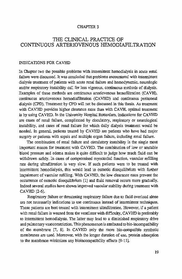
CHAPTER3
THE CLINICAL PRACTICE OF CONTINUOUS ARTERIOVENOUS HEMODIAFIL1RATION
INDICATIONS FOR CAVHD
In Chapter two !he possible problems with intermittent hemodialysis in acute renal failure were discussed. It was concluded that problems encountered with intermittent
dialysis treanuent of patients with acute renal failure and hemodynamic, neurologic
and/or respiratory instabiliry call for less vigorous, continuous methods of dialysis. Examples of these methods are continuous arteriovenous hemofiltration (CA VH),
continuous arteriovenous hemodiafiltration (CA VHD) and continuous peritoneal dialysis (CPD). Treattuent by CPD will not be discussed in this thesis. As treanuent
with CA VHD provides higher clearance rates !han with CA VH, optimal treatment
is by using CA VHD. In !he Universiry Hospital Rotterdam, indications for CA VHD
are cases of renal failure, complicated by circulatory, respiratory or neurological instability, and cases of renal failure for which daily dialysis treatment would be needed. In general, patients treated by CA VHD are patients who have had major
surgery or patients with sepsis and multiple organ failure, including renal failure.
The combination of renal failure and circulatory instability is !he singie most
important reason for treatment with CA VHD. The combination of low or unstable
blood pressure and edema makes it quite difficult to judge how much fluid can be withdrawn safely. In cases of compromised myocardial function, vascular refilling
rate during ultrafiltration is very slow. If such patients were to be treated with
intermittent hemodialysis, this would lead to ostnotic disequilibrium with further
impaittuent of vascular refilling. With CA VHD, !he low clearance rates prevent !he
occurrence of osmotic disequilibrium (I] and fluid removal occurs more gradually. Indeed several studies have shown improved vascular stability during treanuent with
CA VHD [2-6].
Respiratory failure or threatening respiratory failure due to fluid overload alone
are not necessarily indications to use continuous instead of intermittent techniques.
These patients are best treated with intermittent ultrafiltration. However, if a patient
with renal failure is weaned from !he ventilator with difficulty, CA VHD is preferably
to intermittent hemodialysis. The latter may lead to a diminished respiratory drive
and pulmonary vasoconstriction. This phenomenon is attributed to bio-incompatibility
of !he membrane [7, 8]. In CA VHD ouly !he more bio-compatible synthetic
membranes are used. Moreover, with !he longer duration of use, protein adsorption
to !he membrane minimizes any bioincompatibility effects [9-11].
19

Clinical practice
Neurological instability is another indication for CA VHD. Neurological disturbances during intermittent hemodialysis arise from an osmotic disequilibrium between blood and cerebrospinal fluid [12-14]. Again, with CA VHD, the lower solute clearance rates prevent osmotic disequilibrium. This has been borne out by the study of Davenport et al, who showed that in patients with hepatic encephalopathy cerebral perfusion pressure fell after intermittent hemofiltration but was preserved during treatment with CA VH [15].
The indications for continuous treannent mentioned thus far stem from a contraindication to intermittent treatment. Another reason to choose continuous rather than intermittent treatments is the need for daily dialysis treatment. Acute renal fallure patients, especially those with sepsis, are in a hypermetabolic state with fever, increased cardiac output and increased resting energy expenditure [16]. Therefore, there is an increased demand for both energy and proteins. It has been shown that
a positive cumulative caloric balance is associated with improved overall survival [16-18]. In order to achieve a positive caloric balance, patients often receive total parenteral nutrition, which may be complicated by vecy high rates of urea production and fluid overload. Until recently, this necessitated daily hemodialysis and
ultrafiltration. Today these problems are easily managed by CA VHD [19-22]. Indeed, Bartlett et al have demonstrated that patients with multiple organ fallure who were treated by CA VH showed a more positive energy balance and higher survival rates than patients, who were treated by intermittent methods [23]. Compared to intermittent hemodialysis, the rate of uremic solute removal is lower inCA VHD, but at usual dialysate flow rates the total removal per day is similar. Furthermore, intermittent therapies carry the risk of fluid overload in the period between successive treatments. This risk is avoided by CA VHD. It must be realized that with
both treatment modalities amino acids are removed to a certain extent [24-28] but the rate of removal is small when compared to the rate of admiuistration. Another
example of a situation, which used to be managed by daily intense dialysis treannent, is kidney transplantation in a patient with primacy hyperoxaluria. In these patients plasma oxalate levels should be kept below 20 pmol/L [29] until the graft functions well. This is virtually impossible with intermittent treatment. However, with CA VHD, at a dialysate flow rate of approximately 4-5 L/hr, a constant plasma oxalate clearance rate of 40-50 ml/min can be achieved, which suffices to control hyperoxalemia.
Logistic indications for treatment with CA VHD are especialiy justified if frequent dialysis treatment would interfere with other aspects of patient care and if the arterial blood access poses no particular problem. Frequent and prolonged
investigations or operations can proceed without disconnecting the filter.
20
Chapter 3
It has been hypothesized that continuous techniques can effectively remove inflammatory mediators associated with sepsis, the adult respiratory distress syndrome (ARDS) and multiple organ failure [30, 31]. This prompted Cosentino et
a! to study the effect of CA VH in patients with ARDS, who had no renal failure. In this study, a beneficial effect on the course of ARDS could not be demonstrated [32]. It is likely, however, that, for most peptides, endogenous clearance rates are much faster than their removal by CA VH or CA VHD. Therefore, current research still focuses on tbe precise pathophysiological mechanisms involved in the multiple organ failure syndrome. The trend is to look for ways to more selectively remove or
neutralize the key pathogenetic factors by affiulty columns or specific antibodies or drogs.
CONTRAINDICATION$ FOR CA VHD
If a patient has a vascular prothesis of the femoral artery located less than 20 em above the groin, the use of the femoral artery can be hazardous. Sometimes, due to extensive arteriosclerosis, canoulation of the femoral artery is impossible. In such cases, the axillary or brachial artety or, if blood pressure is high enough, a Scribner shunt may be used as an alternative vascular access. If none of these possibilities remains, then the use of pumped veno-venous hemodiafiltration should be considered. The main disadvantage of the pumped technique is that more specialized personnel are needed to watch this system. Moreover, if the pump is equipped with all the necessary alarms and safeguards, this implies that the pump can be interrupted which
may lead to clotting of the system.
In our view, CA VHD (or CVVHD) is contraindicated only if the extracmporeal circuit interferes with the mobilization of the patient. Thus, when a patient who has been treated with CA VHD, recovers, we tend to continue the treatment by
intermittent hemodialysis.
EQUIPMENT AND INSTRUCTIONS FOR USE
A list of the necessary equipment for CA VHD is given below: 1. Catheters for arterial and venous access (and the disposables used for their
introduction). 2. Small surface high-flux dialyzer.
3. Arterial/venous blood tubing set with at least an entrance pon in the arterial blood line for heparin administration and preferably an additional arterial and/or venous pon for administration of substitution fluid.
21

Clinical practice
4. illtrafiltration line. 5. Graduated ultrafiltration collection bag or urometer. 6. Heparin pump. 7. Substitution pump, which can deliver up to 1000 ml!hr and infusion line. 8. Dialysate pump, wllich can deliver up to approximately 5 L/hr, and
corresponding infusion line. 9. Weighing device. 10. Dialysate heater. 11. Heparin, substitution fluid and dialysate fluid. 12. Two liters of rinsing fluid: NaCl 0.9%, containing heparin 5000 U/L.
Priming and installation
The purpose of priming a hemofilter is to eliminate air and, with some hemofilters, glycerine, wllich is used to prevent dehydration of the membrane.
To prime the system, attach the blood lines and the ultrafiltrate line to the hemofilter. The ultrafiltrate outlet should be situated on the arterial site of the hemofilter to achieve a countercurrently dialysate flow. Place the hemofilter in a holder in the upright position, with the venous pon at the top. Connect the proximal end of the anerialline to the rinsing fluid bag and the distal end of the venous line to an empty sterile bag. Clatnp the ultrafiltrate line and run 1 to H~ liter of rinsing fluid through the blood lines and filter under gravity forces. Make sure that side pons have been rinsed as well and that all air has been eliminated. Then unclatnp the ultrafiltrate line and clamp the venous line. Run 'h liter of rinsing fluid through the system. In this way, forced ultrafiltration will occur and glycerine will be rinsed out of the pores of the membrane. Finally unclatnp the venous line and rinse the rest of the fluid through the system.
After priming, the pumps, heater, urimeter, heparin, substitution fluid and
dialysate fluid are connected to the system and the hemofilter, filled with rinsing fluid, is connected to the catheters. Figure 1 shows a schematic drawing of the
hemofilter and the requirements. It is customary to position the filter with the ultrafiltrate/dialysate outlet up (not shown in the Figure), in order to ensure that any
air in the dialysate companment leaves the companment immediately. The ultrafiltrate/dialysate container is positioned below the level of the filter in order to
create a negative pressure in the dialysate companment and thereby enhaoce the rate of ultrafiltration. There must he no direct contact between the sterile dialysate exit pan and the container in which ultrafiltrate and dialysate are collected. The filter is
22
Chapter 3
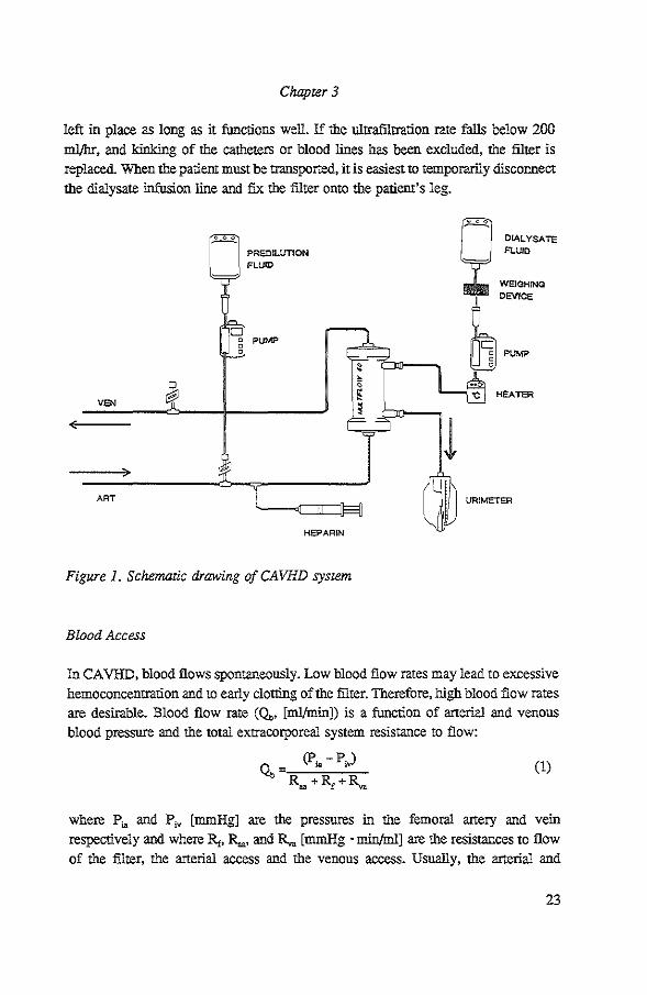
left in place as long as it functions well. If the ultrafiltration rate falls below 200 ml/hr, and kinking of the catheters or blood lines has been excluded, the filter is replaced. When the patient must be transported, it is easiest to temporarily disconnect the dialysate infusion line and fix the filter onto the patient's leg.
VEN
ART
PREDlLUTION FLUID
PUMP
HEPARIN
Figure 1. Schematic drawing of CA VHD system
Blood Access
l
0 0
DIALYSATE FLUID
WEIGHING
DEVICE
PUMP
HEATER
URIMETER
InCA VHD, blood flows spontaneously. Low blood flow rates may lead to excessive hemoconcentration and to early clorting of the filter. Therefore, high blood flow rates are desirable. Blood flow rate (Q •• [ml/min]) is a function of arterial and venous blood pressure and the total extracorporeal system resistance to flow:
(l)
where P~ and P,. [mmHg] are the pressures in the femoral artery and vein respectively and where R,. R,.. and R,. [mmHg • min/ml] are the resistances to flow of the filter, the arterial access and the venous access. Usually, the arterial and
23

Clinical practice
venous access constitute approximately one half of the total resistance to blood flow. For straight catheters and for capillary hemofilters the resistance to flow is determioed largely by the intemal radius of the lumen as given by Poiseuille's law:
(2)
Here L is the length, N is the number of the capillaries, r is the radius of the catheter or capillary, and Jl is the viscosity of the medium [mmHg ·min].
The best results have been obtained with special shon wide bore CA VHcatheters. Our experience is with L=ll.4 em catheters (M8CA VH4, Medcomp) with an internal diameter of 2.7 mm. With these catheters the average resistance to blood flow is 0.10 mmHg · min · ml"1 per catheter. Limited experience with 15 em 'Shaldon catheters' (MCY306PS, Medcomp) with an internal diameter of 2.3 mm (8F) indicates an average resistance to flow of 0.16 mmHg · min • mr'. With Scribner shunts, the average resistance to flow was 0.36 mmHg ·min · ml"1
, ie. 3 to 4 times higher than with CA VH-catheters (see Chapter 4). It should be noted that the contribution to the total resistance to flow of the tubings from the catheters to the hemofilter need not be considered. With 180 em of tubing with an internal diameter of 4.36 mm, it can be calculated that the resistance to flow exened by the tubing alone is 0.07 mmHg · min · ml"1• Shonening of the tubing will have a negligible influence on the overall resistance to flow of the extracorporeal circuit With CA VH-catheters, blood flow rates are typically in the range of 100 to 250 ml/min. Ideally, blood flow rate should be measured continuously. It is now technically feasible to measure blood flow rate by means of echo transit time measurement, using a flow probe around the blood line [33]. As an alternative, blood flow rate may be determined by injecting a 0.3 ml air bubble into the blood line and measuring the transit time of the air bubble over a known distance of the blood line [34, 35]. Calculation of blood flow from anerial and venous hematocrit and ultrafiltration rate is unreliable.
CA VH-catheters are best placed in the femoral anery and vein. These vessels are usually wide enough to accommodate the catheters and the introduction procedure is simple. If the femoral vessels cannot be used, one may consider using the axillary or brachial anery and the jugular or subclavian vein for access. Scribner shunts may be placed in the lower leg or in the forearm.
Reponed complications of anerial cannulation include dissection, thrombcsis, false aneurysm formation, fistula formation, infection, and superficial or retroperitoneal bleeding. Although this sounds alanning, Kramer et al have demonstrated that these complications were considerably reduced once specially
24
Chapter 3
designed short catheters were used exclusively [36]. In recent series, with the use of special CA VH-cathetets, the incidence of damage to the femoral artery was reported to be 2% [37-39]. Furthermore, in a retrospective analysis, Swann et al found no evidence for either sepsis or increased morbidity related to catheter infections [ 40].
The hemofilter
For CA VHD one needs a small surface highly permeable hemofilter with a low resistance to blood flow. Using a high surface hemofilter is not desirable. First, because it leads to an excessive ultrafiltrate production and thereby necessitates substitution infusion rates that are above the range of the intravenous infusion pumps that are generally available. Second, because a larger membrane surface area implies more extensive contact between blood and membrane material and, hence, a greater risk of clotting. High flux characteristics usually coincide with good biocompatibility [41]. For optimal function, a high diffusive permeability is also required. Protein adsorption to the membrane causes a decrease of membrane permeability over time.
In view of the long duration of use, obviously, protein adsorption is undesirable. The typical hemofilter used for CA VH or CA VHD has short capillaries with a diameter of between 200 and 280 pm and a membrane surface area of between 0.2 and 0.6 m2
•
Knowledge of the resistance to flow is important, as in CA VH(D) blood flow is spontaneous generated by the pressure difference between the arterial and veoous access. Especially because it is no common practice to measure blood flow rate continuously, one should aim for a hemofilrer with a low resistance to flow to preveot early clotting and therapy failure. lf one uses a blood pump, the resistance to flow is of minor concem Table 1 provides manufacturer data of several filters for CA VH(D) that are available. From these data, the resistance to flow for capillary hemofiltets is calculated by using Poiseuille's law (Eq. 2). The resistance to flow for plare filtets is calculated according to:
3·L·)l R, --;::-2-:· N~-m-:-'-;-· b''
(3)
where N is the number of blood channels, m is the blood channel width and b is the blood channel half-height [42]. The viscosity used to calculate resistance to flow as giveo in Table l, is calculated as described by Pallone et al [43] and by using a correction factor (see Chapter 4), with a hematocrit of 0.30, a protein concentration of 50 mg/L and a temperature of blood of 36°C. When the resistance to flow is calculated from the manufacturer data, most of these have a resistance to flow below
25

Clinical practice
0.30 mmHg · min/ml, which is suitable for CA VH(D). However, from in vivo as
well as laboratory studies it is known that, very often, the actual resistance to flow is higher than the value that was calculated from the official specifications [44,
Chapter 4]. Therefore, before a filter is used for CA VHD, one should best rely on
precise determination of the resistance to flow in a laboratory setting [45]. The
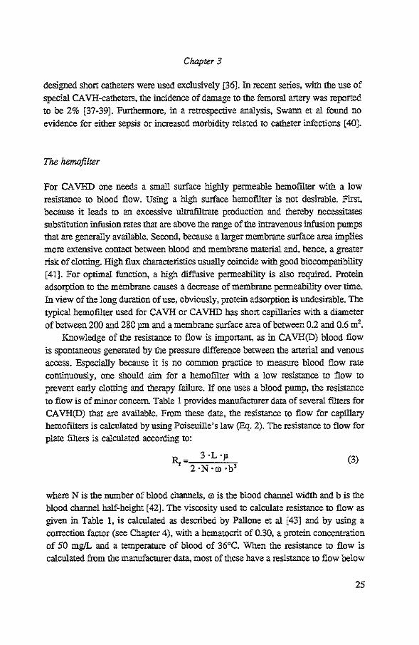
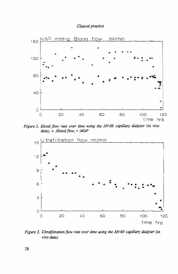
resistance to flow of a hemofilter has been found to be constant over time. Figure 2 shows a recording of mean arterial pressure and blood flow rate within the CA VHDcircuit over 5 days. Fluctuations in blood flow rate reflect those in mean arterial pressure (MAP, [mmHg]). Ouly after four days a precipitous drop in blood flow is seen. A few hours later the filter had become clotted completely.
The ultrafiltration rate (Q,, [ml/min]) is determined by the hydraulic permeability
of the membrane (4, [ml/mmHg ·min · m2]), the membrane surface area (S, [m2
])
and the transmembrane pressure (TMP, [mmHg]):
Q,=L,-S ·MP (4)
or
Q,=MI·TMP (5)
where MI [ml/min • mmHg] is the more commouly used hydraulic permeability
index of the membrane. A recording over time of ultrafiltration rate is shown in Figure 3. The data shown in Figures 2 and 3 were obtained from the same patient.
It is apparent from Figure 3 that, while mean pressure and blood flow rate remained
constant, ultrafiltration rate decreased over time. This indicates that MI and,
presumably, Lp decreased over time. This phenomenon has also been demonstrated
by others [46-48]. It has been shown that during continued use of a membrane, a
decline in the hydraulic permeability occurs as a result of protein adhesion [49, 50].
We have described the average rate of decline of MI with three different hemofilters
(Chapter 4). The data showed that the decline of MI was least apparent with a small
polysulfone capillary hemofilter. The influence of hemofilter geometry and
membrane material in this respect has not been studied in detail so far.
InCA VH, high-flux membranes are used in order to obtain adequate convective
clearance rates. In CA VHD, clearance occurs more by diffusion than by convection.
The rate of ultrafiltration must still be high enough to be able to withdraw several liters of fluid per day. When using a high flux membrane, a total membrane surface
area of 0.6 m2 is enough to obtain an ultrafiltration rate of 300 to 1000 ml/hr.
26

Chapter 3
Both hydraulic and diffusive permeability have been found to decrease over time (Chapter 4, 6). Thus, a very low hydraulic permeability is associated with a low diffusive permeability. Generally, if clearance rates are inadequate, they may be effectively increased by increasing dialysate flow rate (Chapter 6). However, when membrane permeability has become poor, as reflected by an ultrafiltration rate of less than 200 ml/hr, the filter is best replaced. Changing of the filter can nearly always be postponed so as to take place during working hours.
Table 1. Manufacturer data of CAVH(D) filters and calculated resistance to jltYW.
Fres.= Fresenius, AN-69=Acrylonitrile-69, PS=Polysulfone, PA=Polyamide, P AN=Polyacrylonitrile. Jl = 3.5 ·10"7 mmHg ·min. 1
: Channel height, 2 : Channel length,
' : Channel width, 4 :Number of blood channels, 5 : TMP = 55 mmHg,
6: Wet condition, 7
: Calculated from membrane surjae area.
filter membrane membrane fiber fiber number effective R, calc wall diameter length of fibers membrane
thickness pm em surface pm area. m2
Hospal plate AN-69 23 200+ 26.8" 15' 0.43 0.11' 0.26. 6.7' TMP'
Hospal AN-69 506 2406 15 6000 0.60 0.11 Mu!tiflow-60
AmiconD-20 PS 75 250 12.7 5000 0.40 0.09
AmiconD-30 PS 75 250 21.2 5000 0.70 0.15
GambroFH 22 PA 60 215 14.1 2100 0.16 0.44
Gambro FH 66 PA 60 215 17.2 6200 0.59 0.18
Fres. A-V 400 PS 35 220 25.5 4500 0.70 0.34
SorinHFT 02 PS 40 200 12.0 4600 0.24 0.23
Rcnaflo HF250 PS 40 280 14.0 28007 0.25 0.11
Rcnaflo HF500 PS 40 280 21.5 33507 0.50 0.15
Asahi APF-03 PAN 35 250 18.5 3400 0.30 0.20
Asahi APF-06 PAN 35 250 18.5 6400 0.60 0.11
27
Clinical practice
160 MAP mmHo. Blood flow ml/m1n
+ + + + + ++
+ 120 1- + + + + *• + + + ++
+ + + + +
+ + + ++ +
so • • • .•'14 ••• •• • <P • ••• 11 • 8 • • • • • • • "' + ... 40
+
+ 0
0 20 40 60 so 100 120 time hrs
Figure 2. Blood flow rate over time using the AN-69 capillary dialyzer (in vivo data).+ :Blood flow, • :MAP
15 Ultraf1ltrat1on flow ml/min
• 12 ..
9
6
3
0 0
• • • ••••
• • • • •
20 40 60
• • ••• • • .... • • • •• •
• • .. • so 100 120
time hrs
Figure 3. Ultrafiltration flow rate over time using the AN-69 capillary dialyzer (in
vivo data)
28

Chapter 3
Fluids and jlqw rates
In CA VHD, dialysate must be sterile in order to prevent back-transpon of products of pcssible contaminants, such as pyrogens, from dialysate to blood [51-53]. It is easiest to use a commercially available hemofiltration substitution fluid for dialysate. These fluids come in bags of up to 5 liters. In Table 2, several solutions are mentioned that can be used as dialysate fluid. Dialysate fluid for peritoneal dialysis is less suitable for use in CA VHD, mainly because of its low sodium content and high content of glucose. After a few days of treannent with CA VHD, at a dialysate flow rate of 1 L/hr, we frequently encountered hypckalemia, hypcphosphatemia and alkalosis. In our view therefore, the ideal dialysate solution should be compcsed so as to prevent electrolyte derangements during the stable phase of CA VHD treannent. At the stan of treatment, when serum potassium and phosphate are high and bicarbonate is low, the same dialysate fluid may srill be used albeit at a higher dialysate flow rate. We do not advocate the use of a range of different dialysate solutions, because it is impractical and because individual modifications or supplementations may still be needed. Therefore, it is recommended to measure serum levels of phosphate, potassium and sodium at least once a day. Until recently, all solutions contained either lactate or acetate as a buffer substitote, since the addition of bicarbonate would be precluded because of precipitation with calcium and magnesium. Presently, we use for dialysate a fluid to which bicarbonate is added just before use (Schiwa). When used within 24 hours, precipitation of calciumcarbonate is negligible. With bicarbonate containing fluids, the pcssible hannful effects of acetate and the inconvenience of spurious elevstion of blood lactate is eliminated. Furthermore, Jenkins et al have demonstrated that in children on CA VHD who have persistent metabolic acidosis, changing from lactate containing dialysate to bicarbonate containing dialysate considerably improves the metabolic acidosis [54].
Blood and dialysate run in oppcsite directions. This countercurrent flow increases the concentration gradient between blood and dialysate. In most cases a dialysate flow rate of 1 L/hr is enough to control the uremic state. The influence of dialysate flow rate on diffusion is extensively discussed in Chapter 6.
As substitution fluid one may use simple Ringer's lactate. In case of hyponatremia, the addition of 1 gr NaCl to 500 ml of Ringer's lactate so as to increase its sodium concentration to 180 mmol/L, will lead to gradually correction of the hypcnatremia. Alternatively, if the patient is no longer acidotic, one might consider the use of isotouic saline for substitution fluid. Substitution can be given either by infusing the fluid on the arterial line (predilution) or on the venous line of the filter (postdilution). Predilution has several advantages [55, 56]. By predilution
29

Clinical practice
the hematocrit and the plasma protein concentration are decreased, which leads to a decrease of the blood viscosity and an increase of blood flow rate. Also, at least theoretically, by lowering colloid osmotic pressure ultrafiltration rate will be increased. The flow rate of the substitution fluid must be adjusted hourly to the rate of ultrafiltration so as to achieve the desired fluid balance. It is recommended not to decrease the rate of ultrafiltration, as this would decrease clearance rates, especially of solutes with a high molecular weight.
Table 2. Composition of fluids which can be used for dialysate fluid in CA VHD (mmoUL).
Dialysa!e Na• K• ca'· Mg" cr Lact Ac· HCO; Glue
Fres. HF11 140 1 1.63 0.75 101 45.0 0 0 10.9
Fres. HF21 135 2 1.88 0.75 109 33.8 0 0 8.3
Fres. HF02 140 0 2.00 1.00 111 0 35 0 0
Scbiwa SH-44 HEP 140 2 1.75 0.50 112 2.9 0 31.4 5.6
Scbiwa SH-35 HEP 140 0 1.75 0.50 110 2.9 0 31.4 5.6
Scbiwa SH-36 HEP 140 4 1.75 0.50 114 2.9 0 39.7 5.6
Scbiwa SH-41 HEP 141 0 0 0.72 108 0 0 34.3 5.6
Hospal LO 140 0 1.75 0.75 100 45.0 0 0 0
Hospal L2 142 2 2.00 0.75 110 40.0 0 0 0
Hospal LG4D 140 4 2.00 0.75 110 40.0 0 0 6.1
Gambro 140 1 1.60 0.75 100 45.0 0 0 11.1 Hemofiltraso1 21
Gambro 140 0 1.60 0.75 100 45.0 0 0 11.1 Hemofiltrasol 22
Gambro 140 I 1.80 0.75 100 45.0 0 0 11.1 Hemofiltraso1 23
30

Chapter 3
Pumps and weighing devices
For safety reasons, we advise against the use of a pump in the blood line. If one were to use a pump in the blood line, this would at least necessitate the use of a bubble trap and monitoring of the pressures in the arterial blood line, intmediately
after the pump (in order to detect clotting of the filter) and preferably also in the venous blood line. One migbt also argue that the extracorporeal system ougbt to be supervised by a renal nurse.
For pumping dialysate fluid, a special pump that may deliver up to 3-5 L/br, is usefui but a simple screw clamp will also do. As dialysis fluid comes in big bags, it is necessary to use a weigbing device to check the amount of dialysate delivered evecy hour. The total amount of ultrafiltrate plus spent dialysate may be measured either by weigbing or by using a calibrated container.
When substitution fluid is given into the arterial line ('predilution'), an infusion pump must be used because of the bigb pressure. In view of the bigb ultrafiltration flow rate that occurs in the first few hours, this pump must be able to deliver up to 1000 ml/br.
Anticoagulation
Our practice is to stan with a heparin dose of 500 U/br, administered just before the hemofilter in the arterial line. No loading dose is given. The dose may be changed
to between 250 and 750 U/br as clinicaliy indicared. In case platelets counts are below 20·109/L we give no heparin at ali.
One ruigbt consider regional heparinization, i.e. heparin in the arterial line and protamine in the venous line [57]. Receotly, Mehta et al have published a scheme for citrate anticoaguiation in CA VHD [58, 59].
31

Clinical practice
REFERENCES
1. Twardowski ZJ, Nolph KD. Blood purification in acute renal failure. Annals of Internal Medicine 1984; 100: 447-449.
2. Kohen JA, Whitley KY, Kjellstrand CM Continuous arteriovenous hemofiltration: A comparison with hemodialysis in acute renal failure. Trans Am Soc Artif Intern Organs 1985; 31: 196-173.
3. Tam PYW, Huraib S, Mahan B, LeBlanc D, Lunski CA, Holtzer C, Doyle CE, Vas SI, Uldall PR. Slow continuous hemodialysis for the management of complicated acute renal failure in an intensive care unit Clinical Nephrology 1988; 30: 79-85.
4. Paganini EP. Suhoza K, Swann S, Golding L. Nakamoto S. Continuous renal
replacement therapy in patients with acute renal dysfunction undergoing
intraaortic balloon pump and/or left ventricular device support. Trans Am Soc Artif Intern Organs 1986; 32; 414-417.
5. Kaplan AA, Longnecker RE, Fol.ken VW. Continuous aneriovenous hemofiltration. A repon of six months' experience. Amtals of Internal Medicine 1984; 100: 358-367.
6. Paganini EP, O'Hara P, Nakamoto S. Slow continuous ultrafiltration in hemodialysis resistant oliguric acute renal failure patients. Trans Am Soc Artif Intern Organs 1984; 30: 173-177.
7. Hakim RM, Breilatt J, Lazarus M, Pon F. Complement activation and hypersensitivity reactions to dialysis membranes. N Eng! J Med 1984; 311: 878-882.
8. De BackerWA, Verpooten GA, BorgonjonDJ, Vermeire PJ, Lins RR, De Broe
ME. Hypoxemia during hemodialysis: effects of different membranes and dialysate compositions. Kidney Int 1983; 23: 738-743.
9. Kuwahara T, Marken M, Wauters JP. Protein adsorption on dialyzer membranes influences their biocompatibility properties. Contrib Nepbrol (Basel) 1989; 74: 53-57.
10. Kuwahara T, Marken M, Wauters JP. Modulations of leukopenia, thrombocytopenia and protein adsorption of 3 hemodialyzer membranes. A comparison of dialyzer reprocessing techniques. Kidney Int 1987; 32: 434-435.
II. Chenoweth DE, Cheung AK, Ward DM, Henderson L. Anaphylatoxin formation during hemodialysis: Comparison of new and re-used dialyzers. Kidney International 1983: 24: 770-774.
32

Chapter 3
12. Pon KF, Johnson WJ, Klass DW. Prevention of dialysis disequilibrium
syndrome by use of high sodium concentration in the dialysate. Kidney Int 1973;
3: 327-333.
13. Peterson HC, Swanson AG. Acute encephalopathy occurring during
hemodialysis. Archives of intemal medicine 1964; 113: 877-880.
14. La Greca G, Biasioli S, Borin D, Brendolam A, Chiaramonte S, Fabris A,
Feriani M, Ronco C. Dialytic encephalopathy. Contrib. Nepbrol 1985; 45: 9-28.
15. Davenpon A, Will EJ, Davison AM. Early changes in intracranial pressure
during haemofiltration treattnent in patients with grade 4 hepatic encephalopathy and acute oliguric renal failure. Nepbrol Dial Transplant 1990; 5: 192-198.
16. Shizgal HM, Martin MF. Caloric requirement of the critically ill septic patient.
Critical Care Medicine. 1988; 16: 312-317.
17. Mault JR, Bartlett RH, Dechert RE, Oartc SF, Swanz RD. Starvation: a major
contribution to mortality in acute renal failure? Trans Am Soc Artif Organs
1983; 29: 390-393.
18. Mault JR, Kresowik TF, Dechert RE, Amo1di DK, Swartz RD. Bartlett RH. Continuous arteriovenous hemofiltration: The answer to starvation in acute renal failure? Trans Am Soc Artif Organs 1984; 30: 203-205.
19. Raja R, Kramer M, Goldstein S, Caruana R. Lerner A. Comparison of
continuous arteriovenous hemofiltration and continuous aneriovenous dialysis in critically ill patients. Trans Am Soc Artif Intern Organs 1986; 32: 435-436.
20. Stevens PE, Riley B, Davies SP, Gower PE, Brown EA. Kox W. Continuous
arteriovenous haemodialysis in critically ill patients. The Lancet 1988; 16: 150-
152. 21. Maher ER. Han L, Levy D, Scobie JE, Baillod RA, Sweny P, Varghese Z,
Moorhead JF. Comparison of continuous aneriovenous haemofiltration and haemodialysis in acute renal failure. The Lancet 1988; 16: 129.
22. Keshaviah PR. Technical aspects of continuous and intennitrent therapies. Trans Am Soc Artif Intern Organs 1988; 34: 61-62.
23. Bartlett RH, Mault JR, Dechert RE, Palmer J, Swartz D, Port FK. Continuous
aneriovenous hemofiltration: Improved survival in surgical acute renal failure?
Surgery 1986; 100: 400-408.
24. Chanard J, Toupance 0, Gillery P, Lavaud S. Evaluation of protein loss during
hemofiltration. Kidney International 1988; 33 suppl24: 114-116. 25. Wolfson M, Jones MR. Kopp1e JD. Amino acid loss during hemodialysis with
infusion of amino acids and glucose. Kidney International 1982; 21: 500-506.
33

Clinical practice
26. Paganini EP, Haque J, Whitman G, Nakamoto S. Amino acid balance in patients
with oliguric acute renal failure undetgoing slow continuous ultrafiltration (SCUF). Trans Am Soc Artif Intern Organs 1982; 28: 615-620.
27. Davenpon A, Robens NB. Amino acid losses duting haemofiltration. Blood
Purif 1989; 7: 192-196.
28. Davenpott A, Robetts NB. Amino acid losses duting continuous higb-flux
hemofiltration in the critically ill patient. Critical Care Medicine. 1989; 17:
1010-1014. 29. Scheinman Jl. Therapy for primary hyperoxaluria Kidney International 1991;
40: 389-399. 30. Wolner E. Continuous aneriovenous hemofiltration. Applications other than for
renal failure. In: Paganini E, ed. Acute continuous renal replacement therapy,
Maninus Nijhoff Publishing. 1986: 283-292.
31. Coraim FJ Coraim HP, Ebennaon R, Stellwag FM. Acute respiratory failure
after cardiac failure: Clinical experience with the application of continuous
arteriovenous hemofiltration. Critical Care Medicine 1986; 14: 714-718.
32. Cosentino F, Paganini E, Lockrem J, Boswotth C, Stoller J, Wiedemaon H.
Continuous arteriovenous hemofiltration (CA VH) in the adult respiratory distress
syndrome (ARDS): a randomized contrOlled trial. Inrensivbehandlung 1990; 15:
103-104 (Abstract).
33. Button RG, Gorewit RC. llitrasonic flowmeter. Uses Wide-beam transit-rime
technique. Medical Electronics 1984; 15: 68-73. 34. Hoenich NA, Kerr FL. Dialysers. Replacement of renal function by dialysis. sec
edition. 1986. Ed. Drukker W. page 121-122.
35. Keshaviah PR, Shaldon S. Haemodialysis monitors and monitoring Replacement
of renal function by dialysis. sec edition. 1986. Ed. Drukker W. page 226.
36. Kramer P, Bohler J, Kehr A, Grone HJ, Schrader J, Matthaei D, Scheler F.
Intensive care potential of continuous atteriovenous hemofiltration. Trans Am Soc Artif Intern Organs 1982; 28: 28-32.
37. Vos MC, Vincent HH. Continue arterioveneuze hemodiafiltratie: een nieuwe
nierfunctie vervangende therapie. Nederlands Tijdschrift voor Geneeskunde
1992; 136: 561-565.
38. Olbricht CJ. Continuous arteriovenous hemofiltration. The control of azotemia
in acute renal failure. In: acute continuous renal replacement therapy. 1986, Maninus Nijhoff Publishing. Ed. E. Paganini.
39. Vmcent HH, Vos MC. The use of continuous arteriovenous hemodiafiltration in
multiple organ failure patients. Applied Cardiopulmonary Pathophysiology 1991; 4: 109-116.
34
Chapter 3
40. Swann S. Paganini E. The practical technical aspects of slow continuous
ultrafiltration (SCUF) and continuous atteriovenous hemofiltration (CA VH). Developments in Nephrology, Acute continuous renal replacement thernpy. Ed.
Paganini EP, Mattinus NiJboffPublishing, Boston 1986; 13:51-77.
41. Lysaght MJ. Hemodialysis membranes in transition. Contr. Nephrol. 1988: 61;
1-17.
42. Min-Shing Lih M. Transpon phenomena in medicine and biology. J Wiley & Sons, New Yolk 1975 (Ed.)
43. Pallone TL, Hyver S, Petersen J. The simulation of continuous atteriovenous
hemodialysis with a mathematical model. Kidney International 1989; 35: 125-133.
44. Olbricht CJ, Haubitz M, Habel U, Frei U, Koch K Continuous atteriovenous hemofiltration: In vivo functional characteristics and its dependence on vascular
access and filter design. Nephron 1990; 55: 49-57.
45. Vincem HH e.a on behalf of the Kwaliteits-kommissie DGN. Adviezen tav.
CA VH(D). DGN Nieuwsbrief 1992.
46. van Geelen JA, Vincent HH, Schalekamp MADH. Continuous atteriovenous
haemofiltration and haemodiafiltration in acute renal failure. Nephrol Dial Transplant 1988; 2: 181-186.
47. Sigler MH, Teehan BP. Solute trauspon in continuous hemodialysis: A new treatmem for acute renal failure. Kidney International. 1987; 32: 562-571.
48. Jenkins RD, Kuhn RJ, Funk JE. Permeability decay inCA VH hemofilters. Trans
Am Soc Artif Intern Organs 1988; 34: 590-593.
49. Colton CK Henderson LW, Ford CA, Lysaght MJ. Kinetics ofhemodiafiltration.
I. In vitro transpon characteristics of a hollow-fiber blood ultrafilter. Journal of
Laboratoty and Clinical Medicine 1975; 3: 355-371. 50. Bosch T, Schmidt B, Santleben W, Gurland HJ. Effect of protein adsorption on
diffusive aod convective trauspon through polysulphon membranes. Conrrib Nephrol 1985; 46: 14-22.
51. Golper TA, Leone M, Backtranspon of dialysate solutes during in vitro continuous aneriovenous hemodialysis. Blood Purif 1989; 7: 223-229.
52. Baurmeister U. Vienken J. Daum V. High-flux dialysis membranes: Endotoxin transfer by backfiltration can be a problem. Nephrol Dial Transplant 1989; 4
Suppl: 89-93.
53. Kolmos HJ. Hygienic problems in dialysis. Danish medical bulletin 1985; 32:
338-361.
54. Jenkins RD. Jackson E, Kuhn R. Funk J. Benefit of bicarbonate dialysis during
CA VHD. Trans Am Soc Artif Intern Organs 1990; 36: M465-466.
35

Clinical practice
55. Kaplan AA. The predilution mode for continuous arteriovenous hemofiltration. In: Developments in Nephrology, Acute continuous renal replacement therapy. Ed. Paganini EP, Marti:ous Nijhoff Publishing, Boston 1986 (Chapter 9. page 143-172).
56. Kaplan AA. Predilution versus postdilution for continuous arteriovenous hemofiltration. Trans Am Soc Artif Intern Organs 1985; 31: 28-32.
57. Kaplan AA, Petrillo R. Regional heparinization for continuous arterio-venous hemofiltration. Trans Am Soc Artif Intern Organs 1987; 33: 312-315.
58. Mehra RL, McDonald BR, Aguilar M, Ward DM Regional citrate anticoagulation for continuous arteriovenous hemodialysis in critically ill patients. Kidney International1990; 38: 976-981.
59. Mehta R, McDonald BR Regional citrate anticoagulation for continuous arteriovenous hemodiafiltration. Contrib Nephrol 1991; 93: 210-214.
36
CHAPTER4
DETERMINANTS OF BLOOD FLOW AND ULTRAFILTRATION: THEORETICAL PREDICTIONS AND
LABORATORY AND CLINICAL OBSERVATIONS•
INTRODUCTION
Continuous arteriovenous hemofiltration (CA VH) and hemodiafiltration (CA VHD) are based on spontaneous blood flow and spontaneous ultrafiltration [1]. Failure of the technique may occur due to an inadequate blood flow rate, leading to excessive hemoooncentration and clotting.
Theoretically, blood flow rate and ultrafiltration rate may be predicted on the
basis of filter geometry, membrane characteristics and blood visoosity. Pallone and
Petersen developed a malhemstical model by which they were able to predict filter perfonnanoe in a laboratory setting [2]. In their study the resistanoe to blood flow was in good agreement with model predictions. On the other hand, Olbricht et al in
a clinical study found that the resistanoe to blood flow was 3 times higher than expected [3]. The aim of the present study was to examine the feasibility of predicting filter perfonnanoe under clinical oonditions. In patients who were treated with CA VHD we perfonned serial measurements of blood flow rate, blood pressures and ultrafiltration rate, so as to estimate the filter resistance to blood flow and its hydraulic penneability. We also measured filter resistanoe to flow in a laboratory
setting. Clinical and laboratory data were then oompared with theoretical predictions.
METHODS
Clinical protocol
Twenty patients were treated with CA VHD for acute renal failure and sepsis and/or circulatory or neurological instability. A hemofiltration substitution solution (HF-!1,
Fresenius, Gennany) was used for dialysate. The CA VHD circuit was instailed as depicted in Figure 1 (for details, see Chapter 3). For vascular acoess we used special 11 em CA VH-catheters (Medcomp, 8 F) in the femoral vessels, or Scribner shunts. Three types ofhemofilters were used (all specifications according to manufacturers):
* Reprinted in revised fonn with permission from Nephrol Dial Transplant 1990,5: 1031-1037
37

Determinants of Q. and Q1
- the AN-69 plate fiher (15 compamnents, length 26.8 em, width 6.7 em, channel
height 200+0.26 • TMP pm, membrane surface 0.43 m2; SCU/CA VHD, Hospal,
France),
- the AN-69 capillary filter (6000 fibers, length 15 em, including 2 em within
petting, internal diameter of fibers 240 pm, and 220 pm within potting, membrane
surface 0.6 m2; Multiflow-60, Hospal, France)
- a pclysulfone capillary filter (4600 fibers, fiber length 85 em, internal diameter of
fibers 200 pm, membrane surface 024 m2; Spiraflo HFT02, Solin Biomedica, Italy).
d1alysat~
puO>p
Figure 1. CAVHD system
prcd!lvtion fluid
container
-T?N
Filters were prepared by rinsing the blood compamnent with 2 L of saline,
containing hepartn 5000 U/L, allowing filtration to take place (see Chapter 3 for rinsing procedure). Filters were placed in the upright position, with the ultrafiltrate
outlet up. Ultrafiltrate column height was about 60 em. Standard anticoagulation consisted of hepalin 500 U/br into the arterial line of the filter. No loading dose of
hepalin was given. No protamine was used. Tirree patients received no hepalin
38

Chapter4
because of excessive bleeding, thrombocytopenia or hepatic failure and coagulation disorders. Substitution fluid (Ringers lactate) was infused into the arterial line (predilution). The amount of substitution fluid was adjusted hourly to the net uitrafiltration (UF) rate. Reasons for filter replacement were clotting of the filter or a UF rate < 200 ml/br. Furthermore, filters were removed when dialysis was no longer necessary or when clinical improvement allowed mobilization of the patient, in which case patients were treated further with conventional intermittent hemodialysis. Thus, average filter 'survival' time was 3 days.
In a separate study we took blood for viscosity measurements from 10 patients, aged 38 to 66 years, who had been treated in the intensive care department for at least three days.
Laboratory protocol
The 0.6 m2 AN-69 capillary hemofilter was studied in the laboratory. Filters were prepared by rinsing the blood compartment with 2 I of saline, containing heparin 5000 U/L, allowing filtration to take place. After that, the dialysate compartment was closed. Blood tubing, with an inner diameter of 4.3 mm, containing side ports was used and adjuSted so we couid measure the pressure in the perfusion fluid 2 em before and after the hemofilter. An electromagnetic flow probe was inserted into the tubing so that mean flow rate could be measured continuously.
The experiments were performed at room temperature. Filters were in the horizontal position. Eight filters were used. They were all first perfused with saline, then two filters were perfused with a sucrose solution and three filters were perfused with pig blood. A Rhodia!• blood pump was used to generate pulsatile flow rates from 50 to 600 ml/min in case of saline perfusion and from 50 to 300 ml/min in case of blood or sucrose perfusion. The filter resistance to flow was thus detennined at different flow rates. Furthermore, with all three solutions we once changed the set up so as to create an open circuit with a container at a variable height, in which a constant flow through the filter was generated by gravity and the pump was used to return the fluid to the container.
We also measured the resistance to flow over 223 em of standard polyvinylc!J.!oride (PVC) blood tubing with an inner diameter of 4.36 mm, using saline as the perfusion fluid.
39

Determinants of Q, and Q1
Measurements
Electronic pressure transducers were connected to the anerial and the venous lines to measure the pre- and postfilter hydraulic pressures (P, and P, respectively). Both pressures were measured using the same pressure transducer. In the patients, we also measured intra-anerial and intra-venous (P~ and P,v) pressures by the same pressure transducers after clamping the line for l 0 seconds. The pressure midway in the ulttafiltrate-companment (P,, [mmHg]) was calculated from the height of the flnid column in the UF-line (h, [em]) according to:
h P=-
' 1.36 (1)
where the value 1.36 is needed for the conversion from cmH,.O to mmHg. Effective
transmembrane pressure (TMP) in the filter was calculated according to:
P.+P, TMP=-'- -P, -COP
2 (2)
where COP [mmHg] is the colloid osmotic pressure caused by the protein in the blood companmenL (Bemouilli effectS were thus neglected, see Discussion). Colloid osmotic pressure was calculated according to the equation of Landis and Pappenheimer:
COP= 2.1 ·C"', + 0.16 · C""2 + 0.009 • C,.,' (3)
where C"', is the mean plasma protein concentration (g/dl) in the filter. This equation had previously been found to fit experimental data closely over the range 0 to 25 %
of protein [4]. When we compared measured and calculated values, the difference was always less than 5 mmHg (n=l2). cpro, is calculated according to:
(4)
where cpro,~ and cpro,.v are the plasma protein concentrations at the filter inlet and outlet respectively; QP" [ml/lllin] is the plasma flow rate through the anerial catheter before addition of predllution flnid, which is calculated as:
40

Chapter4
Q,. = Q, ·(1 -Ht) (5)
where Ht is the blood hematocrit, Q""" is the predilution fluid infusion rate; Q, the ultrafiltration flow rate and Ht the hematocrit. In the patients, blood flow through the filter was determined in duplicate by measuring the time required for air bubble displacement over 13.0 ml of tubing. In the laboratory blood flow was measured with an electromagnetic flow probe (see laboratory protocol). Ultrafiltration rate was determined by timed collection of fluid from the ultrafiltrate line over five minutes after switching off the dialysate pump. Filter membrane index (MI, [ml/h • mmHg]) was defined as:
Q ·60 MI=-'
TMP (6)
Hydraulic membrane permeability of the membrane (L•, [em/min • mmHg]) was calcuiated from:
L =-MI-= Q, • S TMP·S
(7)
where S [ cm2] is the membrane surface area of the filter. The resistance of the filters to blood flow [R, mmHg · min/mll was calculated according to:
P. -P R,=-' -'
Q, (8)
and resistances of the arterial and venous access were likewise derived from the pressure dtop P"' • P1 and P, - P,v respectively. Resistances were normalized for blood viscosity according to:
(9)
where, for blood, viscosity (p., [mmHg ·min]) is calcuiated as described by Pallone et al [5], assuming a blood temperature of 37 'C in the patient for calcuiating the resistance of the arterial access and of 34 'C in the filter for calculating the resistance
of the filter and the venous access and taking the effect of ultrafiltration on hematocrit and protein concentration into account. In narrow tubes blood viscosity
may be decreased according to the Fabreus Lindqvist effect. With a diameter of 200
41

Determinants of Q, and Q1
to 240 Jllll, however, the decrease is less than 5 % and may therefore be neglected [6]. Values of viscosity for the salioe and sucrose solutions were taken from published tables [7]. Under the conditions of our study, with shear rates of 150 to 400 sec·', blood may be regarded as a Newtonian flnid [8]. Furthermore, within the fibers, blood flow is laminar. Therefore, R, may also be predicted according to Poiseuille's Law:
(10)
where L is the fiber length; N is the number of fibers and r is the internal radius of
the fibers of the hemofilter. For the flat plate filter, R., was predicted according to:
(11)
where N is the number of blood channels; oo is the blood channel width and b is the blood channel half-height [9].
Viscosity measurements were performed using an apparatus that measures the flow rate of blood at 37 oc through a glass capillary with a known resistance. The
internal diameter of the capillary is 1.05 mm and the velocity is approximately 30 mm/sec, resulting in shear rates similar to those within the fibers of the hemofilters.
Statistics
The clinical data are presented as means± I S.D. The time dependent decrease of hydraulic membrane permeability was analyzed by regression analysis. Previous experiments showed that a good fit was obtained by linear regression of the filter membrane index or hydraulic membrane permeability as a function of the logarithm of time (Ln t). This regression analysis was performed for each individual filter. An approximately equal number of data points were obtained before and after 12 h of use (for number of data, see Table 4). Slopes and extrapolated values at 12 h of use were compared by Wilcoxon's Rank test
Filter resistance, measured in the laboratoty, was plotted against flow rate and linear regression analyses were used to calculate the resistance at zero flow rate and to determine the slopes.
42

Chapter4
RESULTS
Blood path resistance and blood fi(JW rate
On starling CA VHD treatment, mean arterial pressure was below 70 mmHg in a third of the patients. The CA VH-catheters bad a resistance to blood flow of only 0.10 mmHg per ml/min, while for the Scribner shunt this value was fourfold bigber (Table 1). The average filter resistance to blood flow was between 0.24 and 0.35 mmHg per ml/min. Generally, there was no appreciable change in filter resistance with lime. ln a few cases, a sudden increase in the resistance to blood flow heralded clotting of the
filter. Using CA VH-catheters, overall blood path resistance during CA VHD treatment was approximately 0.45 to 055 mmHg per ml/min. This resulted in blood flow rates of 164 ±59 ml/min (range 63-284 ml/min).
The normalised resistance, R,, of the hemofilters we observed was two to three limes bigber thao the predicted value (Table 1). This was partly explsined by underestimation of blood viscosity. Blood viscosity proved to be 1.4 limes bigber than the value calculated according to Pallone et al [5] (Table 2).
Results of laboratory expeliments are given in Table 3 and Figures 2 and 3. For the PVC tube, the resistance to flow was in accordance with that predicted according to Poiseuille's law. For the hemofilter, both with saline and with the sucrose solution, the resistance was 40 % bigber than the calculated value. Similar values were obtained with either pulsatile or constant flows. The resistance rose with increasing flow rate. With blood, the resistance to flow was 160 % bigber thao the calculated value, and it did not significantly increase with flow rate.
43

Determinants of Q, and Q1
Table 1. Resistance to blood flow (clinical data). PS= polysulfone
R,- [mmHg • mill/mil Rrn [!o'!ml] n
CA VHD catheter art 0.!0 ± 0.04 3.8 ± 1.3 39
ven 0.!0 ± 0.04 3.7 ± 1.3 39
Scribner shunt art 0.36 ± 0.07 14.8 ± 3.! 6
ven 0.36 ± 0.!3 14.7 ± 5.4 6
AN-69 plate filter:
calculated 3.0 ± 0.2
observed 0.25 ± 0.06 8.8 ± 2.2 16
AN-69 cap. filter:
calculated 3.0
observed 0.24 ± 0.!1 9.0 ±4.0 17
PS cap. fllter:
calculated 4.7
observed 0.35 ± 0.14 !1.4 ± 2.3 17
44

Chapter4
Table 2. Whole Blood Viscosity Measurements. 1 See ref [5]
No. Ht c,... )l obslpred' 10"7 mmHg • min
1 21 55 2.84 1.33
2 31 49 3.55 1.49
3 25 55 2.66 1.17
4 21 53 3.23 1.54
5 26 36 3.06 1.53
6 33 45 3.69 1.55
7 30 75 3.93 1.41
8 34 54 3.96 1.54
9 26 54 3.05 1.33
10 24 56 3.11 1.38
mean± SD: 3.31 ± 0.43 1.43 ± 0.12
45

Determinants of Q. mui Q1
Table 3. Resistance w Blood F/(JW (Laboratory Data)
BLOOD TUBING
saline ]1=1.2·10"7
HEMOFJLTER
saline ]1=1.2·10"7
HEMOFJLTER
sucrose ]1=2.8•10"7
HEMOFJLTER
Pig blood Ht= 0.23-0.26 )1=2.8·!0·'
46
R., predicted IO'mr'
2.41
3.24
mean± SD:
3.24
mean± SD:
3.24
mean± SD:
R., measured IO'mr'
2.15
4.58
5.17
5.83
2.92
5.00
4.69
3.88
4.60
4.58 ± 0.82
5.43
3.98
4.71 ±0.72
7.93
8.11
8.93
8.33 ±0.44
Slope% per 100 ml/min
0.00
8.8
8.8
6.0
10.0
8.1
4.1
6.0
6.1
7.2 ± 1.8
5.1
4.9
5.0 ± 0.1
1.5
-0.4
0.5
0.5 ± 0.8

Chapter4
Resistance mmHg min I ml
0 , I + ++++
+ +++++ ++ + 0.081 + -+ +- +..,.. ++++ +++
_,...1-+ +++
0.06 r I
•·+ FILTER SALINE
I 0.04 ~
I I - -
0.02 r TUBING SALINE ---
oL------L-----~----------~------~----~ 0 100 200 300 400 500 600
Flow ml I min
Figu;re 2. Resistance to fWw of tubing and of AN-69 capillary filter, using saline.
(Eypredicted tubing= 0.03, R1predictedfilter = 0.04).
Resistance mmHg min I ml 0.3,-----------~--------------------------,
0·25 FILTER BLOOD
t++-l::r.ry_+-11- ++++++++ ++ ++ + + +
0.2
FILTER SUCROSE 0.15
0.1L-----~----~-------------L------L-----~
0 50 100 150 200 250 300 Flow ml I min
Figu;re 3. Resistance to fWw of AN-69 capillary filter, using sucrose and blood. (Ey predicted = 0.09).
47

Determinants of Q, and Q1
Table 4: Hydraulic membrane permeability. The average L,-values of2.77 and258 for the AN-69 filters were both lower than the value of 5.03 for the polysulfone filter (p<O.Ol) The slopes 0.72 and 0.69 for the capillary filters were both lower than the slope of 135 for the plate filter (p<0.05). IJ Value obtained by interpoltalon 21 Slope b in the formula L, = a - b In( time)
AN-69 capillary AN-69 plate PS capillary
L,' after 12 br slope' L,' after 12 br slope' L,' after 12 br slope•
2.69 0.39 2.81 0.67 6.47 0.55
3.66 !.55 0.56 1.62 5.94 !.30
2.57 0.60 5.70 1.79 4.11 0.05
2.14 0.34 2.34 0.84 6.27 0.70
2.90 0.73 2.26 1.05 4.50 1.27
2.60 0.98 2.87 2.01 6.53 0.53
2.81 0.87 2.02 1.53
2.97 1.26
3.35 1.05
2.85 1.10
3.23 1.08
0.92 1.03
1.71 2.53
average: 2.77 0.72 2.58 1.35 5.03 0.69
48

In vivo membrane lndel< ml/h~ pe,. mmHg
"
"
"
Chapter4
,. " ..
Figure 4. Decrease of filter membrane index with time.
AN-69 0. 4 m' AN-69 0.6 m' plate filter capi ll. fi Iter
Qb 250 ml/min
200
"0
"0
Qf 1-000 mJ!hr
800 y 600 ___::----.ao -------"h "h 200
60 80 100 120 60 80 1 00 120
MAP mmHg MAP mmHg
'" " TIME h<>urs
Polysul(one: 0. 2m1 cap ill. filter
/ ~ ~
60 80 "0 no MAP mmHg
Figure 5. Estimated average extracorporeal blt:iodflow and ultrafiltration rate as a junction of mean arterial pressure.
49

DeterminantS of Q, and Q1
Hydraulic membrane permeability
Hydraulic membrane penneability of the AN-69 membrane was less than that of the
polysulfone membrane (Table 4). This difference was largely made up for by the
difference in membrane surface area of the hemofilters. Hydraulic membrane
penneability decreased with time. The rate of the decrease was faster with the plate filter than with the capillary filters (see also Figures 4 and 5).
DISCUSSION
Blood flow
Pallone and Petersen have shown that, for capillary and flat plate hemofilters, the
resistance to flow may be predicted from the filter geometty [2, 5]. Their prediction
was based on Poiseuille's law. In their calculations it was assumed that blood
behaves as a Newtonian fluid and that flow through the filter is lamina!. In addition,
they used data from the literature which allow the calculation of blood viscosity from hematocrit, protein concentration and temperature. In our study we used the same
calculation methods. We found that the filter resistance to flow was two to three times higher than the predicted value. A similar finding has recently been reported
for a number of other hemofilters [3]. To explain this discrepancy we considered
clotting of fibers, underestimation of blood viscosity, turbulent flow and/or additional
pressure losses due to the sudden changes in the blood path diameter and deviations
of the internal diameter of the fibers.
Laboratory measurements showed that the resistance of a length of tubing was in accordance with the calculated value, which argues against gross methodological
mistakes. The resistance of the hemofilter, perfused with saline or a sucrose solution,
was 40 % higher than expected. Furthennore, it was to some extent flow dependent,
suggesting that turbulence does occur, most likely at the filter inlet. However, this
can hardly explain why the extrapolated resistance at zero flow rate was 40 % higher
than calculated. Also, when we applied Bemouilli 'slaw to estimate the pressure drop
that results from the sudden change in diameter at the filter inlet [7], we calculated
a pressure drop of far less than 1 mmHg. On the other hand, a 40 % increase in
resistance would be explained if the fiber diameter would be a mere 8 % lower than
the given value. When filters were perfused with blood, the viscosity-corrected filter resistance was higher than with saline or sucrose. With blood, a similar filter
resistance was found both in laboratory expetiments, where clotting was prevented
50

Chapter4
by a high dose of heparin, and in the clinic. This argues against a significant
contribution of clotting of fibers to filter resistance. Furthermore, in Chapter 3 it was
observed that filter resistance was constant over time. On the other hand, comparison
of actual blood viscosity with the calculated value confirmed our suspicion that we bad grossly underestimated blood viscosity. With blood, filter resistance no longer
seemed flow dependent, probably indicating that turbulence is diminished by the
higher viscosity of blood. If blood flow rate was prediCted on the basis of our
laboratoty data on filter resistance during saline perfusion and on the basis of a more accurate estimation of blood viscosity, flow rate would be overestimated by only
25%.
Ultrafiltration
The fall in ultrafiltration rate with time may be explained both by cbanges in the
hydraulic membrane penneability, presumably resulting from protein adsorption to
the membrane, and by a fall in the available membrane surface area, e.g. due to
inadvertent clotting of fibers. For reasons given above, under clinical conditions inadvertent clotting of fibers is unlikely to occur. Therefore, the decrease in hydraulic
membrane permeability with time is more likely to be due to ongoing protein
adsorption. It should be noted that according to Jenkins et al [10] the hemofilter
hydraulic membrane penneability may fall even after contact with water alone. To
our knowledge, however, this finding has not been reproduced by others. For the
AN-69 capillary filter we did not observe a fall in hydraulic membrane permeability between 1 and 48 hours of saline perfusion, but we do observe a fall in hydraulic
membrane penneability with plasma and blood (data not shown).
Clinical Implications
In critically ill patients, in whom CA VH(D) is indicated, blood pressure tends to be
low. A low resistance to flow of the extracorporeal circuit is therefore mandatoty. In
this respect CA VH catheters are clearly to be preferred to Scribner shunts. The use
of a Scribner shunt instead of catheters doubled the overall resistance of the
extracorporeal system. In our view Scribner shunts should be regarded as unsuitable
for CA VH(D).
The resistance to flow of the hemofilters studied was low enough to ensure an
adequate blood flow rate in most cases. By using laboratory data on filter resistance
51

and a more accurate estimation of blood viscosity, a reasonably accurate prediction
of blood flow rate under clinical conditions can be obtained. With the three filters Sllldied, ultrafiltration rate is always high enough for
correction of overb.ydration. On the other hand, due to the time-dependent fall in hydraulic membrane permeability, ultrafiltration rate will soon become too low for
adequate solute removal through convection alone. In case of CA VH this necessitates
frequent changes of the filter. In case of CA VHD the fall in filtration rate is less
important, as solute removal then occurs largely by diffusion.
52

Chapter4
ACKOWLEDGEMENTS
We are grateful to Dr. G.C.D. Kuiken from the Technical University, Delft, for his advice and to Dr. C. Slager from the department of Thoracic Technology of the Erasmus University, Ronerdam, for his help in perfonning viscosity measurements.
53
DeterminantS of Q. and Q1
REFERENCES
1. van Geelen JA, Vincent HH, Scbalekamp MADH. Continuous arterio-venous
haemofiltration and haemodiafiltration in acute renal failure. Nephi Dial Transpl 1988; 2: 181-186.
2. Pallone TL, Petersen J. Continuous arteriovenous hemofiltration: An in vitro simulation and mathematical model Kidney lnt 1988; 33: 685-698.
3. Olbrich! CJ, Haubitz M, Habel U, Frei U, Koch K-M. Continuous arteriovenous
hemofiltrarion: 1n vivo functional characteristics and its dependence on vascular
access and filter design. Nephron 1990; 55: 49-57.
4. Landis EM, Pappenheimer JR. Exchange of substances through the capillary
wall.ln: HandbookofPhysiology. Circulation, (volll Chapter29). Washington,
D.C., Am Physiol Soc, 1963, page 962-1034.
5. Pallone TL, Hyver S, Petersen J. The simulation of continuous arteriovenous
hemodialysis with a mathematical model. Kidney International 1989; 35: 125-
133. 6. Gaeh!gens P. Flow of blood through narrow capillaries: Rheological mechanisms
deterntining capillary hematocrit and apparent viscosity. Biorheology 1980; 17:
183-189.
7. Handbook ofbiochemistty. Selected data for molecular biology. Eds. So ben HA, Harte RA. The Chemical Rubber Co. Cleveland, Ohio, 1968.
8. MerrillEW, Pelletier GA. Viscosity of human blood: Transition from Newtonian
to non-Newtonian. J Appl Physiol 1967; 23: 178-182.
9. Min-Shing Lih M TtanSport phenomena in medicine and biology. J Wiley & Sons, New Yorlc 1975 (Ed.)
10. Jenkins RD, Kuhn RJ, Fuok JE. Penneability decay inCA VH hemofilters. Trans
Am Soc Artif Intern Organs 1988; 34: 590-593.
54

CHAPTERS
A MATHEMATICAL MODEL OF SOLUTE TRANSPORT IN CONTINUOUSARTEIDOVENOUSHEMOD~TRATION
~TRODUCTION
Continuous arteriovenous hemofiltration (CA VH) is characterized by the use of a
small surface highly permeable hemofilter, spontaneous blood flow and spontaneous
ultrafiltration. Solutes are removed solely by convection, i.e. plasma water is
removed together with all dissolved molecules that are able to pass through the
membrane pores. The driving force is the trans membrane pressure difference. Solute
clearance is limited by the amount of ultrafiltrate produced. It is techulcally feasible
to increase ultrafiltration rate by the use of a pump, either in the blood circult or in
the filtrate line, but the inherent hazards of such techulques necessitate the use of sophisticated safety measures and supervision by specially trained personnel. An easier way to increase solute clearance is by continuous arteriovenous
hemodiafiltration (CA VHD), a techulque in which CA VH is combined with
continuous slow dialysis [1, 2]. In this techulque, transpon occurs by combined convection and diffusion. Both processes have an influence on one another [3].
Ultrafiltration leads to an increased solute concentration in the dialysate side of the
dialyzer and thereby decreases diffusion. Dialysis lowers the solute concentration in
plasma water and thereby decreases the amount of solute that is removed by
ultrafiltration.
CA VHD is a new renal replacement techulque. Insight into the determinants of
solute clearance is needed to determine the optimal flow rates. Also, data on drug
clearance are needed. For some drugs dose adaptations may be necessary. For this, we needed a mathematical model that could be used to analyze solute tnmspon by
CA VHD. We must realize that CA VHD differs from conventional intermittent
hemodialysis and hemodiafiltration in several respects. First, solute transpon occurs
by simultaneous convection and diffusion. Second, dialysate flow rates (10-30 ml/min) are very low compared to blood flow rate (100-250 ml/min) and as a
consequence the change in dialysate solute concentration over the length of the filter cannot be taken to be linear, as in conventional models of dialysis [4]. The dialysate
flow rates that are used in CA VHD might be too low for optimal distribution of
dialysate over the dialysate compartment. Consequently, by low dialysate flow rates,
the resistance to diffusion exened by the dialysate compartment may be much higher
than in conventional hemodialysis. Third, CA VHD is a continuous techulque. With
prolonged use of the filter, deterioration of the membrane is likely not ouly to
55

Mathematical model
decrease the rate of ultrafiltration but also to impair diffusive transport. For the above reasons, existing models of hemodialysis are not likely to be useful for the analysis of solute transport. in CA VHD.
Previous models of solute transpon by hemodiolysis
In the model of hemodialysis, as published by Sargent and Gotch [4], a linear change in solute concentration over the length of the dialyzer is assumed. Convective transport. is neglected. Thus the rate of transport depends on the difference between the concentration in blood and that in dialysate and the mass transfer coefficient of diffusion (KJ. By using this model, the length-averaged K. may be calculated from the total solute transport. and the log-mean concentration difference over the membrane [4]. In CA VHD, the contribution of convective transport is relatively great. Also, because of the ratio of blood and dialysate flow rates, a curvilinear decrease of solute concentration over the length of the dialyzer is to be expected.
The mass transfer coefficient of diffusion is the inverse of the total resistance to diffusion, which consists of the resistance to diffusion of the blood compartment, that of the membrane and that of the dialysate compartment [5]. In conventional dialysis the greater pan of the resistance to diffusion is exened by the blood compattment and the membrane. According to Grimsrud and Babb [6], at a fast and turbulent dialysate flow, the resistance to diffusion in the dialysate compattment approaches zero. Accordingly, they developed a model of solute transpon in which the resistance to diffusion of the dialysate compattment was neglected. Also, in their model ultrafiltration rate was taken to be zero. In CA VHD, this approach is not valid because of the low dialysate flow rate and the relatively high rate of ultrafiltration.
Sigler and Teehan first described solute clearance by CA VHD in patients [7]. They did not discern between convective and diffusive clearance and the study does not provide any insight into the determinants of diffusive transport.
Pallone et al studied solute transport by CA VHD in a laboratory sening. They presented a model in which they defined the solute flux by convection and by diffusion. These fluxes were then integrated over the length of the dialyzer [8]. With their model, they were able to calculate the curvilinear change in solute concentration
along the dialyzer. Their model does not provide an analytical expression ofK.. They did not recognize the influence of differences in dialysate flow rate on K. (Chapter 6).
J affrin published a mathematical model of combined convection and diffusion, to be applied to intermittent hemodiafiltration [9]. He expressed K. as a function of
56

Chapter 5
the resistance of diffusion in blood, in the membrane and in dialysate. These values
are, however, not readily available. Furthermore, in order to derive K,, experiments had to be done at zero ultrafiltration rate. The model can therefore not be applied to data obtained from patients treated with CA VHD.
In view of the shortcomings of the above models, we developed a new model
of CA VHD. In the present chapter, we first define a model ofhemodiafiltration that can be easily implemented in any spread sheet program. Then, with some simplifications, the model equations are analytically solved, so as to provide an expression of K, as a function of blood, filtrate and dialysate flow rates and solute concentrations. By virme of this expression of K,, the model is well suited for the analysis of diffusive transport in CA VHD.
MathematicalrMdel of CA VHD
The model describes the flux of a solute from plasma water to dialysate by simultaneous convection and diffusion during countercurrent hemodiafiltration. The solute concentration at the dialysate inlet is taken to be zero.
Three important assumptions are made. Frrst, blood and dialysate flows are assumed to be homogeneously distributed over their respective compartments. Second, the membrane penneabilities for ultrafiltration and for diffusion are assumed to be constant over the entire membrane length. Third, within both the blood and dialysate compartments, the solute concentration varies in the axial direction but not in the radial direction, i.e. calculations are based on local "mixing cup" concentrations. In reality, the so called 'concentration polarisation' occurs on both sides of the membrane. Thus, in our model, diffusive solute transport results from a difference between mixed concentrations in plasma water and dialysate only. The influence of solute concentration polarization on the resistance to diffusion is thereby neglected. The validity of the latter assumption was experimentally studied (see next chapter).
Input variables of the model are:
blood flow rate at the inlet of the dialyzer (Q,), hematocrit (Ht),
plasma protein concentration (C""J, dialysate flow rate (Q.), ultrafiltration rate (Q,),
infusion rate of predilution fluid (Q,J, solute concentration in the arterial blood plasma (c;,),
57

MathemLltical model
total membrane surface area (S),
the length of the membrane (L) and the mass transfer coefficient of diffusion (K,).
Also the non-protein-bound fraction of the solute (F) and the sieving coefficient (s) are required. The sieving coefficient is the fraction of the solute mass that passes througb the membrane during ultrafiltration, defined as the solute concentration in ultrafiltrate divided by its concentration in plasma water. From these input values, we calculate the changes in solute concentration in blood and dialysate over the length of the dialyzer and, thereby, solute clearance.
Model equations
The flow rate of plasma water at the blood inlet (Qw;) is calculated by:
Qw; = Qb, ·(I- Ht) ·(1- a ·C,.,) + Qb, • Ht ·f + Q""" (1)
where f is the fractional solute distribution in red blood cells. For urea f is 0.8 [10]. For most other solutes f may be taken to be zero. C,., is the plasma protein concentration in giL and a, the volume fraction of proteins, is 0.00107 L/g [11]. The solute concentration in plasma water at the inlet of the dialyzer (C..J is calculated by:
c . - cp, . c~ - Q""") (2) ~ (I 0t ·C.,.,) Qwi
The plasma water flow rate at the blood outlet CQwol is calculated by:
~=~-~ ~
and the dialysate flow rate at the dialysate side outlet (Q.,J is:
Q., =Q, +Q, (4)
58

Chapter 5
The trans membrane pressure difference is the sum of the hydraulic pressure in blood and dialysate (P, and P, respectively) and the colloid osmotic pressure in blood (COP):
(5)
The value of COP may be estimated from the blood protein concentration by using the Landis-Pappenhelmer equation [12]. The hydraulic permeability coefficient of the membrane (Lp) is calculated from the rate of ultrafiltration, the length-averaged trans
membrane pressure difference (TMP) and the membrane surface area according to:
L - Q, ' TMP·S
(6)
The volume flux (J,=Q, I S) is a function of the trans membrane pressure difference (TMP) and the hydraulic permeability:
J =TMP·L • p (7)
Solute flux through the membrane (J.) occurs by convection and diffusion. The most rigorous expression of J, during combined convection and diffusion is:
H.·~ J ·s·[C -C ·e '] J = v w d
' ' (-1.·-,2 l -e ·
(8)
This equation is equivalent with the one derived by Levitt [13]. In case of high values of J, · s and low values of K,, the above equation reduces to:
J ·s for -'->>1
K, (9)
In case of low values of J, · s relative to the value of K,, eqoation 8 reduces to:
Cw+C, J =J ·s· +K ·(C -C)
$ v 2 d w d
J ·s for -'-<<1
K, (10)
In case of uremic solutes, protein-binding does not usually occur. With drugs, on the other hand, very often pan of the solute is bound to plasma proteins and ouly the free fraction (F) is avallable for convection and diffusion. To take the effect of protein binding into account, in Eq. 9 and 10 the value of C,. must be substituted by
59

A B c D E F G H
I x/L Qw Qd Mw Md Cw Cd Js
2 0.00 Qwi Qdo Qwi·Cwi Qdo.Cdo D2/B2 E2/C2 Jv·F2·P+Kd·(F2·P·G2)
3 0.01 Qwi-0.01·Qf Qdo-0.01-Qf D2-0.01•S•H2 E2·0.o! •S•H2 D3/B3 E3/C3 Jv•FJ•P+Kd{FJ•P-GJ)
4 0.02 Qwi·0.02-Qf Qdo-0.02-Qf D3 .0.01·S·H3 EJ-0,01 ·S·H3 D4/B4 E4/C4 Jv•F4·P+Kd{F4·P-G4) L__ -- ' -- --- ------ --· -·
101 0.99 Qwi-0.99-Qf Qdo-0.99·Qf DJ00.0.01·S·HJOO EJ00·0.01·S·HJOO DJOl/BJOJ ElOl/ClOJ Jv·FJOJ•F+Kd{FJOJ•F-GJOJ)
102 1.00 Qwo Qdi DJ01-0.01•S•HJOJ El01-0.01•S•HJOJ DJ02/BJ02 El02/Cl02 Jv·Fl02·F+Kd{FJ02·F-GJ02) ~ ------·-···"--
.__ - --- -
Figure I. Stepwise numerical integration in a spreadsheet program. Note that, an approximation procedure must be used to find the value of C.., at which the value of cell GJ02 approaches zero.

Clulpter 5
C, · F. This implies immediate re-equilibration between bound and free fractions as solute is removed from the blood. Thus, Eq. 9 becomes:
and Eq. 10 becomes:
J •S for _v_>>l
K,
J ·s for -v-<<1
K,
(ll)
(12)
For the calculation of total solute transport, Eq. 11 or Eq. 12 is integrated along the length of the dialyzer.
Implementatibn of the model in a spread sheet program
The above model is implemented in a spread sbeet program as follows (see Figure 1). The length of the dialyzer is divided into sections. At each section, d(x), Pb(x) is defined as a linear function ofP" and P .. ; TMP(x) is defined as a function ofPb(x), P, and COP. Since, in CA VHD, the ultrafiltration rate is small in relation to blood flow rate, one may use a single length-averaged value of the blood protein concentration and of COP. Also, at a low dialysate flow rate one may use a single value of P,. Jv(x) is then defined as a function of TMP(x). Q,.(x) and Q,(x) are
expressed as a function of Qw; and Jv(x) and of Q,. and Jv(x) respectively. The total
solute mass in plasma water, Mw(x), is defined as a function of MW,, which is the product of Qw; and Cw;. and J,(x). Likewise the total solute mass in dialysate is defined as a function of MW. and J,(x). Thus, to start the procedure, an estimated value of c,. is required (see below). C,.(x) and C.(x) are defined as a function of mass and flow rate. Fmally, at each section, J, is defined according to Eq. 11 or 12. An iterative approximation procedure is used to find the input value of c .. at which cdl becomes zero.
If c ... is known and K, must be determined, then an initial estimation of K, is required and the approximation procedure is followed to find the value of K, at which cdl becomes zero.
61

Mathematical model
Simplification of the model and analytical solutions
In the case that the free fraction, F, and the sieving coefficient, s, of the solute equal
one, as is true for urea, creatinine and phosphate, the transpon equation (Eq. 11) becomes:
J ·s for -v->>1 (13)
K•
If, for simplicity, we assume that Jv is constant along the length of the filter, it is possible to obtaio an analytical expression of C,.(x) and C.(x) as follows [14, 15]: From the condition of continuity over a differential length d(x) of the hemofilter it
follows that:
dQw(x) dQ.(x) --=---=-w·J
dx dx v
where w is the width, defined as S/L. Iotegtation of Eq. 14 yields:
Qw(x) =Q... -w ·Jv ·x
and
Q.(x) = W • Jv ·X - Q.,
Mass transfer of the solute io the blood compartment is given by:
d[~(x) ·Cw(x)] _ -w ·J (x)
dx '
and io the dialysate compartment by:
d[ Q.(x) • C.(x)] w ·J,(x)
dx
where the solute flux is:
62
(14)
(15)
(16)
(17)
(18)
(19)

Chapter 5
By combining Eqs. 14, 17 and 18 we get:
and
dC/x) K• · (Cw(x) - c.(x)) --~--=-W----~Q~w(~~~--
dC.(x) CJ. + K.) · (Cw(x) - C.(x)) --~-- = w -------.,Q.;<..,-(x") -----
(20)
(21)
On substracting Eq. 21 from Eq. 20 and eliminating d(x) by Eq. 14 we obtltin:
do C(x) n dQw(x) - (n + 1) dQ.(x) (22) oC(x) Qw(x) Q.(x)
where n = KJJ~ Integration of Eq. 22 along the length of the filter yields:
(C)x) - C.(x)) = [Qw(x)]" "[-Q.(x)r<',.'l ·( -Al) (23)
where A 1 is an integration constant The condition of conseiVation of solute, both
in blood and dialysate side, implies:
Q/x) ·Cw(x) + Q.(x) ·C.(x) = A2 (24)
where A2 is a constant Multiplying Eq. 23 by Qw(x) we find
Qw(x) ·(Cw(x) - C.(x)) = -AI [Qw(x)]"•' · [ -Q.(x)r<n·ll (25)
and multiplying Eq. 23 by Q.(x) we find:
Q.(x) · (Cw(x) - c.(x)) = Al [Qw(x)]" · [ -Q.(x)r" (26)
Substracting Eq. 25 from Eq. 24 and adding Eq. 26 to Eq. 24 we find:
c (x) = 1 A2 + A1 [Q (x)r' +n (x)J-<n•'l (27) d ~-Q., w .....
and
C (x) = 1 A2 + A1 [n (x)]" +" (xW" (28)
w ~Q., '<w .....
63

Mathe=tical model
From the boundary conditions at x = 0 we find:
AI= (C"- Cwi)(Q .. r' ·(Q..)-n (29)
and
A2=" ·C -" ·C "wi. wi "<do do (30)
Substitution of AI and A2 in Eqs. 27 and 28 and their further elaboration gives the concentration profile on the dialysate side:
and the concentration profile at the blood side by:
Because of the boundary condition C'" = 0, Eq. 31 gives us an expression for the ratio of the diffusive mass transfer coefficient to the volume flux:
1 (33)
When membrane total surface area, flow rates and solute concentrations are known, Eq. 33 may be used to calculate the value of n and of K,. By substituting the value of n in Eqs. 31 and 32 one may calculate the solute concentration profiles. Alternatively, for the case that both the sieving coefficient of the solute and the free fraction equal one. Convective solute flux may also be regarded as the volume flux times the solute concentration in the membrane (C,) approximated by (C, + C;J/2,
rather than that in plasma water (C,J. Now startiog from Eq. 12, by substituting Eq. 12 in Eqs. 17 and 18, we get another expression for the concentration in dialysate:
64

Chapter 5
(34)
and for the concentration in plasma water:
(35)
The ratio of the diffusive mass transfer ooefficient to the volume flux is obtained
from Eq. 34, using c'" = 0:
l/2 in[ (Qw;- Q,)Q"l -in[ Q., ·C,,- Qwi ·Cw;l K, ~ ·Q., Qw;(C,,- Cw;)
n=--=------~--~--~~~~~~--Jv in[ (Qw; Q,)Q.,]
~-~
(36)
K, values calculated by using Eq. 36 are slightly higher than those calculated by using Eq. 33. lf K, is known and C,, must be calculated, we use the formula derived by van
Geelen [16]:
(Q .j() \n•l- (() /() )n•l C = C . • Wl "<coo Xwo "<di
'' M (Qw;/Q.;)n _ (Q • .J~r' (37)
COMMENTS
Two models have been described, the numerical and the analytical model. The numerical model describes the process more clear. However, use of the numerical
model is more time-consuming. It may best be combined with the analytically derived expression of K, as an initial estimation to start the approximation procedure.
65

Mathematical model
The analytical model bas a few limitations. It is only applicable for S=l andF=l. This poses no problems when the model is used to study the transport of uremic toxins. Analysis of the consequences of the assumption that Jv is constant rather than
decreasing along the length of the dialyzer on the calcnlated value of K,, turnS out to be not significantly differem [17]. The analytical model presented here, cannot be used to calcnlate backfiltration from dialysate to blood. It is not applicable to proteinbound solutes, unless it is assumed that protein binding is irreversible, i.e. C. can be
substituted by C. ·F. A possible source of error in both the numerical and the analytical model is that
'unstirred layer effects', which wonld lead to an influence of the axial flow rates on the concentration gradient, are not taken into account.
In the next chapter, data are presented showing that, with low and variable dialysate flow rates, there is an appreciable effect of dialysate flow rate on the
calcnlated value of K,. A correction for this effect will be suggested in the following chapter.
66

Chapter 5
REFERENCES
1. van Geelen JA, Vincent HH, Schalekamp MADH. Continuous arteriovenous
haemofilttation and haemodiafilttation in acute renal failure. Nephrol Dial
Transpll988; 2: 181-186.
2. Geronemus R, Schneider N. Continuous arteriovenous hemodialysis: A new modality for treannent of acute renal failure. Trans Am Soc Artif Intern Organs
1984; 30: 610-613. 3. Husted FC, Nolph KD, Vitale FC, Maher JF. Detrimental effects of ulttafilttation
on diffusion in coils. J. Lab. Clin. Med. 1976; 3: 435-442. 4. Sargent JA, Gotch FA. Principles and biophysics of dialysis. In: Drukker W,
Parsons FM, Maher JF. Replacement of renal function by dialysis. Martinus
Nijhoff Medical Division Pub!, The Hague, 1978.
5. Colton CK, Lowrie EG. Hemodialysis,: Physical principles and technical
considerations, in: The Kidney, second edition 1981; 2: 2425-2489.
6. Grimsrud L, Babb AL Velocity and concentration profiles for laminar flow of
a newtonian flnid in a dialyzer. Chern Eng Progr Symp Ser no 66 1966; 62: 20-
31.
7. Sigler MH, Teeban BP. Solute transport in continuous hemodialysis: A new
treannent for acute renal failure. Kidney International 1987; 32: 562-571. 8. Pallone TL, Hyver S, Peterson J. The simulation of continuous arteriovenous
hemodialysis with a mathematical modeL Kidney lnt 1989; 35: 125-33. 9. Jaffrin MY, Ding L, Laurent JM. Simultaneous convective and diffusive mass
transfer in a hemodialyser. Trans of the ASME. 1990; 212: 212-219. 10. Colton CK, Sntith KA, Merrill EW, Reece JM. Diffusion of organic solutes in
stagnant plasma and red cell suspensions. Chern Eng Progr Symp Ser no 66, 1970; 99: 85-100.
11. Colton CK, Henderson L W, Ford CA. Lysaght MJ. Kinetics of hemodiafilttation
I. In vitro transport characteristics of a hollow-fiber blood ulttafilter. J Lab Clin
Invest 1975; 85: 355-371. 12. Landis EM, Pappenheimer JR. Exchange of substances through the capillary
wall, in: Handbook of Physiology. Circulation. Washington, Am Physiol Soc
1963, sect 2, vol11, chapt 29, p 962-1034.
13. Levitt DG. General continuum analysis of transport through pores. I. Proof of Onsager's reciprocity postulate for uniform pores. Biophys J 1975; 15: 533-51.
14. Akcahuseyin E, Vincent HH, van Ittersum FJ, van Duyl WA, Schalekamp
MADH. A mathematical model of continuous arterio-venous hemodiafilttation
(CA VHD). Computer Methods and Programs in Biomedicine 1990; 31 215-224.
67
Mathematical model
15. Vincent HH, van Ittersum FJ, Ak93huseyin E, Vos MC, van Duyl WA.
Schalekamp MADH. Solute transport in continuous arteriovenous hemodiafiltration: A new mathematical model applied to clinical data. Blood Purif 1990; 8: 149-159.
16. van Geelen JA. Hemodiafiltration, simultaneous application of hemodialysis and hemofiltration. Thesis, 1983. UniversityofLimburg,Maastricht, the Netherlands.
17. Ak~ahuseyin E, Vos MC, Vincent HH, van Duyl WA. A mathematical model on solute trauspon in continuous arteriovenous hemodiafiltration (CA VHD): The influence of taking into account the decrease in volume flux over the length of the dialyzer (in preparation).
68

CHAPTER6
VALIDATION OF TilE MATIIEMATICAL MODEL OF CONTINUOUS ARTERIOVENOUS HEMODIAFILTRATION: THE ASSUMPTION OF MIXING CUP CONCENTRATIONS
INTRODUCTION
In the previous chapter, a mathematical model of solute transpon in CA VHD was presemed. In this model a number of assumptions were made. The most imponant assumption was the so called 'mixing cup' concentrations within both the blood and dialysate companment. Assuming mixing cup concentrations is valid when companments are well stirred, a condition which could be approached if flows are turl:lu!ent, but at least in the blood companment, flow is laminar.
The true driving force for diffusion is the solute concentration gradiem over the membnme, which may be equated with the difference between the solute concentration in blood and dialysate close to the membnme. In both the numerical and analytical model, these concentrations are approached by taking the bulk or 'mixing cup' concentrations in blood and dialysate (Figure la). This assumption implies that possible radial concentration profiles within either of the respective companments are not taken into account. However, solute concentrations close to the membnme may be different from mixing cup concentrations, thereby inducing a radial concentration profile. It is evident that a concentration difference between the area close to the membnme and anywhere else in the blood or dialysate companment will be generated by solute diffusion from blood to dialysate (Figure lb) [6]. With assuming mixing cup concentrations, the concentration gradient over the membnme will be overestimated and K, may be underestimated. Aiso, the solute concentration in the blood companment can be higher near the membnme, as will be described below. Then, with assuming mixing cup concentrations, the concentration gradient over the membrane will be underestimated and K, values may be overestimated.
69

Validation of the model
cone. Cp Cm
blood comp. dialysate comp.
membr.
Figure I a. Illustration of mixing cup concentrations on both sides of the membranes. In both the blood and dialysate compartment, no gradient of concentration is regarded. Concentration difference over the membrane is calculated from concentrations in blood (C) and dialysate (C,J. Cm: concentration in the membrane.
cone. Cp
Cm
' .... _:~----
l.-,o-o-d _c_o_m_p __ _,--+--dl-al-y-sa_t_e _c_o_m-p. membr.
Figure 1 b. Illustration of a possible concentration profile in both the blood (C) and dialysate compartment (C,J due to diffusion from the blood to the dialysate compartment and due to concentration boundary layer formation. em: concentration in the membrane.
70

Chapter 6
Factors influencing the radial concentration profiles
A radial concentration gradient can be influenced by blood and dialysate flow rates. In the blood compartment, as blood flow in the fiber is laminar, the distribution of solutes over the fiber is not homogeneous (i.e. concentration boundary layer formation) [6]. Accordingly, the difference berween the concentration in the middle of the stream and that near the membrane may be expected to depend on the blood flow rate. Furthermore, with low blood flow rates, an inhomogeneous blood flow distribution might occur. Within the dialysate compartment, due to diffusive transport, the concentration near the membrane is higher than that more distant from the membrane. Furthermore, with low dialysate flow rates, a 'stagnant' fluid layer
surrounds the fiber, which is not mixed with fresh dialysate. Within this layer, solute concentration is higher than more distant from the membrane. Therefore, this layer adds to the resistance to diffusion. At higher dialysate flow rates, the stagnant layer will dimiuish and the solute concentration in dialysate near the membrane will
decrease until, at an infinitely high dialysate flow rate, no stagnant layers exist. Thus, the axial flow rates may be expected to have an influence on the radial
concentration gradient and thereby on the driving force for diffusion. As a result, when axial flow rates are changed, the calculated value of the mass transfer coefficient of diffusion (K,) may also be changed [1, 2].
The influence of ultrafiltration rate on the solute concentration gradient during CA VHD has not been previously studied. It is, however, conceivable that, in case of ultrafiltration, dragging leads to a higher blood solute concentration near the membrane [4, 7], which would enhance diffusive transport (Figure lc). On the other hand, ultrafiltration leads to an increased concentration of proteins near the
membrane. This 'protein boundary layer formation' (protein concentration polarization) may lead to diminished membrane permeability and may thereby dimiuish transport by diffusion and convection.
By the assumption of mixing cup concentrations the possible influences of axial flows and of ultrafiltration on diffusion gradients are neglected. We are, therefore,
faced with the question whether changes in blood- or dialysate flow rate and changes in ultrafiltration flow rate have an influence on the calculated value of K., based on the simplified model. In the present study we therefore measured solute clearance and calculated K• values at different blood, ultrafiltration and dialysate flow rates.
71

cone.
l
Validation of the model
Co
blood comp.
', Cd
-- -'---------: dialysate comp.
membr.
Figure 1 c. Illustration of a possible influence of ultrafiltration on the solute concentration gradient in the blood compartment. Solutes are dragged along with the ultrajiltrate and near the membrane a concentration build up will occur.
PROTOCOLS
Studies were perfonned during CA VHD in the Intensive Care Unit. Some additional
studies were perfonned in the laboratory. The clinical procedure has been described
in Chapter 4. The study protocols are described below.
I. Blood flow rate (clinical observations) This protocol was aimed at studying the effects of blood flow rate (Q,) on calculated I<. values. The protocol was perfonned if the spontaneous blood flow through the
filter was higher than 100 ml/mln, because blood flow rate could only be decreased
by clamping of the blood line. Pressures, spontaneous blood flow rate and
ultrafiltration flow rate (Q,) were measured two times, at the beginning and at the
end of the experiment At each experiment, by clamping the arterial or venous line,
blood flow was lowered 4 times to varying degrees and in random order. Dialysate
flow rate (Q,) was set to 3 L/hr. Ultrafiltration flow rate was measured at each blood
flow setting. Data were obtained with the AN-69, 0.6 m2 capillary dialyzer (Hospal,
Multiflow-60).
72
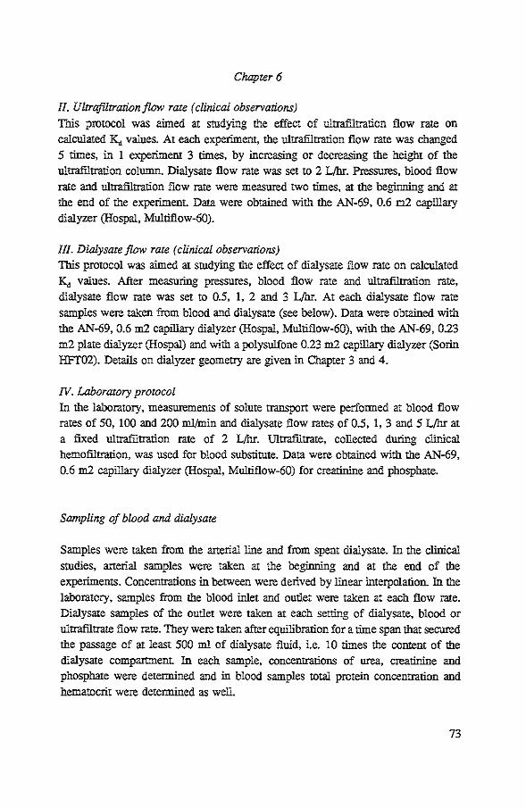
Chapter 6
II. Ultrafiltration/low rate (clinical observations)
This protocol was aimed at studying the effect of ultrafiltration flow rate on calculated K, values. At each experiment, the ultrafiltration flow rate was changed 5 rimes, in 1 experiment 3 rimes, by increasing or decreasing the height of the ultrafiltration column. Dialysate flow rate was set to 2 L/hr. Pressures, blood flow rate and ultrafiltration flow rate were measured two rimes, at the beginning and at the end of the experiment. Data were obtained with the AN-69, 0.6 m2 capillary dialyzer (Hospal, Multiflow-60).
!II. Dialysate flow rate (clinical observations)
This protocol was aimed at studying the effect of dialysate flow rate on calculated K, values. After measuring pressures, blood flow rate and ultrafiltration rate, dialysate flow rate was set to 0.5, 1, 2 and 3 L/hr. At each dialysate flow rate
samples were taken from blood and dialysate (see below). Data were obtained with the AN-69, 0.6 m2 capillary dialyzer (Hospal, Multiflow-60), with the AN-69, 023 m2 plate dialyzer (Hospal) and with a polysulfone 0.23 m2 capillary dialyzer (Solin HFT02). Details on dialyzer geometry are given in Chapter 3 and 4.
IV. Laboratory protocol
In the laboratory, measurements of solute transpOrt were performed at blood flow rates of 50, 100 and 200 ml/min and dialysate flow rates of 0.5, 1, 3 and 5 L/hr at a fixed ultrafiltration rate of 2 L/hr. Ultrafiltrate, collected during clinical hemofiltration, was used for blood substitute. Data were obtained with the AN-69, 0.6 m2 capillary dialyzer (Hospal, Multiflow-60) for creatinine and phosphate.
Sampling of blood and dialysate
Samples were taken from the arterial line and from spent dialysate. In the clinical studies, arterial samples were taken at the beginning and at the end of the experiments. Concentrations in between were derived by linear interpolation. In the laboratory, samples from the blood inlet and outlet were taken at each flow rate. Dialysate samples of the outlet were taken at each sening of dialysate, blood or
ultrafiltrate flow rate. They were taken after equilibration for a rime span that secured the passage of at least 500 m1 of dialysate fluid, i.e. 10 rimes the content of the dialysate compartment. In each sample, concentrations of urea, creatinine and phosphate were determined and in blood samples total protein concentration and
hematocrit were determined as well.
73

Validation of the model
TECHNIQUES OF MEASUREMENT
Arterial and venous line pressures were measured electronically and dialysate
pressure was calculated from the ultraiiltrate column height Blood fo;w rate was
measured in triplicate by air bubble displacement In the clinical studies where blood
flow rate was varied, it was measured both by air bubble displacement and by the echo transit time technique, using the Transonics• Clamp-on flowmeter [5]. Both
methods yielded similar values. In the laboratory studies blood flow rate was
measured by means of an electromagnetic flow probe. Dialysate foJw rate and
ultr<ifiltration rate were measured by electronic weighing and by timed collection.
CALCULATIONS
Hydraulic permeability index (MI, [ml · hr"1 • mmHg-1
]) was calculated as:
Q ·60 Ml=-'- (1)
TMP
where TMP is transmembrane pressure difference, calculated as described in Chapter
4. The filter resistance to bloodfltJW (R,, [mmHg ·min · ml"1]) was calculated as:
(2)
where P, and P, are the hydrostatic pressures at the inlet and outlet of the filter respectively. lf R, was higher than 0.40, clotring of the filter may be the case. For
this reason, l out of 6 experiments in protocol II and 5 out of 45 experiments in
protocol III were discarded.
The moss transfer coefficient of diffusion (K., [pm!min]) was calculated by using the
analytical expression given in Chapter 5:
In[ Q,, . c,, - Q"" • c"" l K, Q"" (C,, - C"")
n =- = --..,.,,.-...,_=--J. ln[ (Qwi- Q,)Q,,l
Qwi ·Q.
l (3)
74

Chaprer 6
The relationships between blood, filtrate and dialysate flow rates and K, were examined in two ways. First, we plotted K,. as a function of flow rate and performed linear regression analysis if applicable. Second, we used !he approach suggested by Wilson [6] (see below). The reciprocal of!(,., i.e. !he total resistance to diffusion (R, [min!pm]) is !he sum of !he resistances exerted by !he blood compartment (R,), by !he dialysate compartment (R,) and by !he membrane (R,J [8]:
(4)
According to Wilson, !he influence of dialysate flow rate on !he resistance to diffusion of the dialysate compartment may be examined by plotting the total resistance to diffusion as a function of Q, to the power -y, where !he value of y is empirically determined so as to yield a straight line. The y-intercept is taken to
represent R,+R, (see Figure 2). We likewise defined the total resistance to diffusion as follows:
(5)
where a · Q;' represents R,, b • Q,·Y represents R, and c represents R,. a, b, c, x and y are positive values, to be determined by linear regression analysis. Eq. 5 indicates that an increase of blood or dialysate flow rate results in a decrease of the resistance to diffusion, i.e. an increase of K, (see also ref [3]).
0.03 1/Kd m1n/um
Ml=:8.3
1/Kd"' 1/113 ... 0.14/0d . . . ,
0.02 .
... ·} _.---- Rd .•.
0.01 --· __ .-
0.00
} Rb•Rm
0.00 0.03 0.06 0.09 0.12 0.15
1/0d m1n/ml
Figure 2. Example of a Wilson plot and the regression junction obtained by linear regression analysis on data poims slwwn.
75

Validation of the model
RESULTS
I rifluence of blood flaw rate
In protocol I, initial blood flow rate ranged from 10510 195 ml/min. Filters had been used for 1 to 97 hours and MI ranged from 3 to 28 ml • hf1
• mmHg·'. During the experiment R, did not change. With two filters that had been used for less than two hours, MI decreased by one fifth of the initial value.
Figure 3 shows K. values at different blood flow rates. There was no apparent relationship. With linear regression analysis of K. as a function of Qb, 11 out of 13 slopes were not sigcificantly different from zero (2 SE) and, of the remaining two, one slope was positive and the other slope was negative. The average slope was 0.00 (see Table 1).
Table I. Effects of changes in blood and ultrafiltration flow rates on the mass
transfer coefficient of diffusion of creatinine (K.J and that of the reciprocal
of dialysate flaw rates on the resistance to diffusion of creatinine (R).
Results of regression analyses. n=number of experiments. MF-60: AN-69, 0.6 m2 capillary dialyzer (Multijlow-60, Hospal, France). HFT02: 0.23 m2 polysulfone capillary dialyzer (HFT-02, Sarin, Italy).
CJ: Corifidence Interval.
Flow range protocol ml/min
Dialyzer n Slope 95% CI )ll1l/ml of the mean
!(, VS Q,: 19-195 Clinic MF-60 13 0.00 -0.06- 0.06
50-200 Laboratory MF-60 3 0.00 -0.01 - 0.00
K, vs Or: 2-14 Clinic MF-60 5 0.06 -1.04- 1.16
RvsQ;': 8-50 Clinic MF-60 15 0.16 0.14- 0.18
8-85 Laboratory MF-60 4 0.19 0.17- 0.21
8-50 Clinic Plate 11 0.11 0.!0- 0.12
8-50 Clinic HFT02 14 0.11 O.o7- 0.15
76

Chapter 6
Influence of ultrafiltration rate
In protocol !I, initial ultrafiltration flow rate ranged from 6 to 11 ml/min. Filters had
been used for 12 to 18 hours. Values of the hydraulic permeability index ranged
from 11 to 14 nil • br'1 • mmHg·1•
Figure 4 shows K,. values at different ultrafiltration rates. Again, with linear
regression analysis, slopes were not significantly different from zero (2 SE). The average slope was 0.06 pm/ml (see Table 1).
Influence of dialysate flow rate
In protocol III, MI ranged from 3 to 21. As shown in Figure 5, K,. increased to a
maximum value with dialysate flow rates. Similar observations were made with the
AN-69 0.6 m2 capillaty dialyzer (n=l5 experiments), the 0.43 m2 plate dialyzer
(n=l!) and the po1ysulfone 023 m2 capillaty dialyzer (n=14).
For creatinine, the influence of dialysate flow rate was quantified according to
Eq. 5 (see CALCULATIONS). Slopes obtained from the regression analysis ofR as a function of Q,·' were all significantly different from zero (2 SE). The correlation
coefficients (r) of the regression lines ranged from 0.91 to 1.00.
Both with the AN-69 capillary and plate dialyzers, the intercept of the regression
line proved to be related to MI (Figure 6), whereas with the polysulfone capillary
dialyzer this relationship could not be determined. For the AN-69 capillary dialyzer,
we performed linear regression analysis on the intercept as a function of the
reciprocal of MI (r=0.76). As the intercept equals the minimal resistance to diffusion
obtained with infinite dialysate flow rates, the intercept was defined as the reciprocal
of I<._. The relationship between K""'= of creatinine and MI was given by:
MI K"""'=u·160 (forMI<12) K,._=160 (forMI::12) (6)
77

Validation of the m1Jdel
1 50 Kdc um/mm
120
90
60
30 -0 25 50 75 100 125 150 175 200
Figwe 3. Diffusive mass transfer coefficient of creatinine (K,J for the AN-69 capillary dialyzer as a junction of blood flow rate (Q,J. Each line represents one experiment.
125 Kdc um/m1n
100
75
50
25
0 ~----~----~----L-----~--~ 0 3 9 12 15
Of ml/m1n
Figwe 4. Diffusive mass transfer coefficient of creatinine (K,J for the AN-69 capillary dialyzer as a junction of ultrafiltration flow rate. Each line represents one experiment.
78
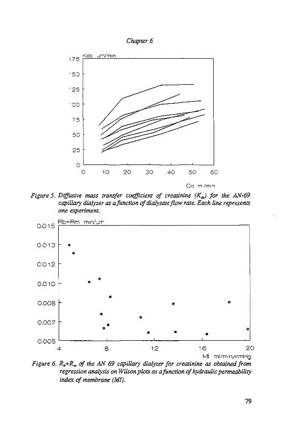
Chapter 6
175 Kdc um/m1n
150
125
100
75
50
25
0 0 10 20 30 40 50 60
Od ml/m1n
Figure 5. Diffusive mass transfer coefficient of creatinine (K.J for the AN-69 capillary dialyzer as a function of dialysate flow rate. Each line representS one experiment.
0 0 Rb+Rm m1n/um
. 15
0.013 •
• 0.012
0.010
0.008
o.oo? r-
0 •
•
• • •
• •
• • • • •
0.005 L------.1...------'-------'------_J
4 8 12 16 20 Ml ml/min.mmHg
Figure 6. R.+Rm of the AN 69 capillary dialyzer for creatinine as obtained from regression analysis on Wilson plots as a function of hydraulic permeability index of membrane (Ml).
79

Validation of the model
Results from laboratory experiments
Results from the laboratory experiments were in accordance with clinicaUindings. The average slopes obtained from linear regression analysis of K.. as a function of Q, and that obtained from the regression of R as a function of Q,"1 are given in
Table 1.
In all experiments, K.. values of urea and phosphate were closely related to the corresponding values of creatinine. The ratio of the K.. value of urea to that of creatinine averaged 1.32 (SEM=4, n=l09) and the ratio of K.. of phosphate to that of creatinine averaged 0.75 (SEM=3, n=l25).
DISCUSSION
With our model of combined convection and diffusion, we calcnlated the mass transfer coefficient of diffusion (K,) based on the assumption of mixing cup concentrations. In the present study, we examined whether the same value of K.. may be used to extrapolate to solute clearance at different settings. We found that blood flow rate and ultrafiltration flow rate have no measurable influence on K,.. Accordingly, one may indeed use the model to predict solute clearance at blood flow
and ultrafiltration rateS, different from those during the experiment in which K.. was determined. On the other hand, dialysate flow rates (Q,) does have an influence on K,.. Thus, when dialysate flow rate was increased from 0.5 to 3 L/hr, K.. values increased by approximately 100%. The effect of the dialysate flow rate on K. was analyzed as described by Wilson [6]. The 'Wilson plots' allowed us both to estimate R,+R.. and R• separately. The increase of K. with increasing Q, may be explained by the existence of an unstirred layer in the dialysate compartment, or, at low dialysate flow rates, the distribution of dialysate over the compartment is inadequate, resulting in a partial use of the membrane surface area. Therefore, the increase of K.. with increasing Q,, which may be ascribed to thinning of that unstirred layer in the dialysate compartment or an increase in the use of the membrane surface area, was quantified by a mathematical expression. Also, with the AN-69 dialyzers, K.. proved to be dependent on the hydraulic permeability index of the membrane (MI). Changes
inK.. with different values of MI are contributed to a vatiability of R,+R,.. As was shown in Chapter 4, MI decreases over rime, probably due to protein absOiption. Therefore, the vatiability of R, +R,. may be caused by changes in R,. due to protein
absorption on the membrane, whereas R, is not variable. This explanation is
80

Chapter 6
supported by the results of protocol L Also, the influence of MI on K, was quantified by a mathematical expression. By incorporating the mathematical relation ofK, with
both Q, and MI, one may allow for the influence of these parameters on K,. This maneuver should allow to extrapolate to solute clearance at different dialysate flow rates and at different values of MI. Thus, for the AN-69 capillary dialyzer the predicting formula of K, of creatinine becomes:
_l_ =R =-1-+ 0.15 ·Q;1
K, K"""" (7)
in which K"""" is given by Eq. 6. Since values of K, of other solutes showed a fixed relationship with the value of K, of creatinine obtained from the same experiment, this relationship can be used to estimate K, of other solutes as welL
81

Validation of the model
REFERENCES
1. Babb AL, Maurer CJ, Deloss LF, Popovich RP, McKee RE. The detennination of membrane permeabilities and solute diffusivities with application to hemodialysis. Chem Eng Progr Symposium Series no 84, 1968; 64: 59-68.
2. Kooyman J.M Comparative performance of artificial kidneys. Thesis, 1971. Teclmical University Delft, the Netherlands.
3. Sakai K. Teclmical determination of optimal dimensions of hollow fibre membranes for clinical dialysis. Nephrol Dial Transplant 1989; Suppl4: 73-77.
4. Popovich RP, Cristopher TG, Babb AL. The effects of membrane diffusion and ultrafiltration properties on hemodialyzer design and performance. Chem Eng Progr Symposium Series no 114, 67: 105-115.
5. Burton RG, Gorewit RC. illtrasonic flowmeter. Uses Wide-beam tranSit-time teclmique. Medical Electronics 1984; 15: 68-73.
6. Colton CK, Lowrie EG. Hemodialysis: Physical principles and teclmical considerations. In: The Kidney, Brenner, Rector. Saunders, New York 1981, 2nd ed., Vol. 2, page 2425-2489.
7. Jaffrin MY, Ding L, Laurent JM. Simultaneous convective and diffusive mass transfers in a hemodialyser. Journal of Biomechanical Engineering 1990; 112: 212-219.
8. Sigdell JE. Calculations of combined diffusive and convective mass transfer. Int J Anif Organs 1982; 5: 361-372.
82
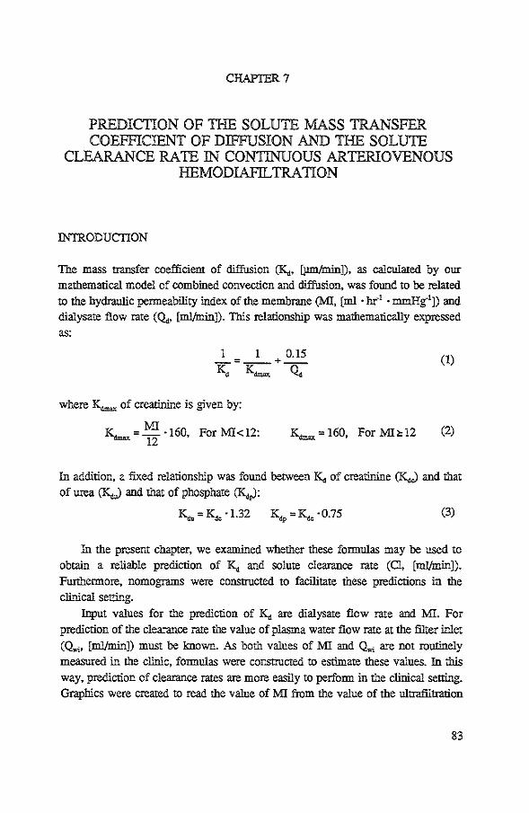
CHAPrER 7
PREDICTION OF THE SOLUTE MASS TRANSFER COEFFICIENT OF DIFFUSION AND THE SOLUTE
CLEARANCE RATE IN CONTINUOUS ARTERIOVENOUS HEMODIAFILTRATION
INTRODUCTION
The mass lrllllSfer coefficient of diffusion (K,, [pm!min]), as calculated by our mathematical model of combined convection and diffusion, was found to be related to the hydraulic permeability index of the membrane (MI, [ml • hr"1
• mmHg-1]) and dialysate flow rate (Q,, [ml/min]). This relationship was mathematically expressed as:
l l 0.15 -=--+--K, K"""' Q,
(1)
where ~ of creatinine is given by:
MI K"""' = lZ · 160, For MI < 12: K"""" = 160, For MI:: 12 (2)
1n addition, a fixed relationship was found between K. of creatinine (K,J and that of urea (K.,) and that of phosphate (K,,):
(3)
1n the present chapter, we examined whether these formulas may be used to obtain a reliable prediction of K, and solute clearance rate (Cl, [ml/min]). Furthermore, nomograms were constructed to facilitate these predictions in the clinical setting.
Input values for the prediction of K. are dialysate flow rate and MI. For prediction of the clearance rate the value of plasma water flow rate at the filter iulet (Qwi, [ml/min]) must be known. As both values of MI and Qwi are not routinely measured in the clinic, formulas were constructed to estimate these values. 1n this way, prediction of clearance rates are more easily to perform in the clinical setting. Graphics were created to read the value of MI from the value of the ultrafiltration
83

Prediction model
flow rate and the value of Qwi from the value of the mean arterial pressure of the patient. The influences of errors resulting from these estimations on the prediction of K,. and MI were calculated.
METIIODS
Data were obtained from several clinical experiments, i.e from the experiments described in Chapter 6 and from the measurements of clearance rates of drugs (see Chapter 8), in which creatinine, urea and phosphate were measured simultaneously. All data were obtained with the AN-69 capillary dialyzer. Hydraulic permeability index of the membrane, resistance to blood flow of the dialyzer (R, [mmHg ·min • m1·1]) and ultrafiltration flow rate (Q,, [ml!min]) were measured as described in Olapter 4. Dialysate flow rate was checked by an electronic weighing device. If the
resistance to flow was higher than 0.40, data were discarded. The mass transfer coefficient of diffusion was calculated using the analytical expression as described in O:tapter 5 (Eq. 33). K,. values of creatinine were predicted according to formulas 1 and 2 and that of urea and phosphate according to formulas 1, 2 and 3.
In accordance with our model, we defined solute clearance rate as:
" ·C Cl _ "'<do .::lo
c"' (4)
where Q,. and c,. are flow rate and solute concentration at the dialysate outlet side of the dialyzer respectively, and Cw; is the solute concentration in plasma water at the blood inlet side of the dialyzer. aearance rate was predicted by substituting the predicted value of K,. in the followiog formula derived by van Geelen [1]:
a- . (Qw/Q,.r 1- (Qw.fQ..r 1
- Q,. (~/Q,,)"- (Q..JQ,)"'l (5)
where n is K,. • S!Q, S is membrane surface area, Qwo the plasma water flow at the dialyzer outlet and Q.. and Q.. are dialysate flow rates at the dialyzer inlet and outlet respectively. For both K,. and a, lioear regression analysis was performed on the observed values as a function of the predicted values and 95% confidence intervals were detemtined.
84

Chapter 7
A nomogram was constructed to obtain the value of plasma water flow from the observed mean anerial pressure of the parient (MAP, [mmHg]) according to the following formula:
MAP-P. Qwi =
0_44
,. ·(1-Ht) ·(1-0.00107 ·C,.) +f·Ht (6)
where the intravenous pressure of the patient (P,., [mmHg]) was taken to be 10 mmHg, hematocrit (Ht) 0.30, total protein concentration in plasma Cc,.J 50 g/dl, the
fractional volume distribution of solutes in blood cells (!) zero and the total
resistance to flow of the catheterS and the dialyzer was taken to be 0.44, which equals mean observed values (see Chapter 4). Note that, if predilution is used, the value of the predilution flow rate has to be added up to the plasma water flow rate. For errors in the estimation of Qwi up to 50%, the resulting error in the value of the predicted clearance rate was calculated for Q,=l L/hr, MI=8 and Q,=8 ml/min. The value of Qwi and that of clearance rate at 0% error (startiug value) were 100 ml/ntin and 22 ml/min respectively.
Furthermore, a nomogram was constructed to obtain a value of MI from the observed values of Q, in ml/br and MAP according to the following formula, assuming equal values of the resistance to flow of both the catheters:
Q, Ml=~~~~-----MAP+P.
--..,..--'" + P, - COP 2
(7)
where the pressure in the dialysate compartment (P,, [mmHg]) was taken to be -37, which is obtained with an ultrafiltration column height of 50 em and the colloid osmotic pressure (COP, [mmHg]) was taken to be 20. Accordingly, for errors in the estimation of MI up to 50%, the resulting errors in the predicted value of K., for Q,=l and 3 L/hr were calculated by using Eq. I and 2 (startiug value of MI=8). Then, the impact was detennined of an underestimation and overestimation of K., up to 50% on the predicted clearance rate for Q,=8 ml/min and Q~=IOO ml/min. Herewith, startiug values of K., were predicted for Q,=O.S, 1 and 3 L/hr and MI=8.
85

170 ° 160
150
140
130
120
110
100
WI
90
80
70
60
50
[7
50
Prediction model
ml/m 1n
7 /
/ I v
/
I/ /
60 70 80 90 100 110 120
MAP mmHg
Figure I. Expected plasma water flow rate as a junction of mean arterial pressure (zero pre dilution flow rate and f=O ).
10 Cl dct-on er r o/c ore 1 I ro 0
8
6
4
2
v----0
-2
-4
-1 0 -50 -40 -30 -20 -10 0 10 20 30 40 50
Ow1 predlct10n error %
Figure 2. The error in terms of percentage in predicted clearance rates of creatinine as a junction of an error in terms of percentage in the estimation of Qw,·
86

Chapter 7
RESULTS
From Figure 1, one can read the expected value of Qw; by using the mean anerial pressure of the patient as input value. This Figure is based on values of the mean observed resistance of flow of the catheters and of the filter. Figure 2 shows the consequences of underestimation or overestimation of Qwi on clearance rate. In Figure 3, the value of MI can be read from ultrafiltration flow rates for MAP values of 50, 70, 90 and 110 mmHg. Figure 4 shows the effect of an error in terrns of percentage in MI on the prediction of K. for Q.,=1 and 3 L/br. The absolute error and the error in terrns of percentage in clearance rates effected by an error in the predicted value of K. are shown in Figures 5 and 6 respectively. Staning values forK. were 35, 55 and 81 pm/min at 0.,:0.5, 1 and 3 L/hr respectively. Figures 7 and 8 show the observed values of K, and Cl respectively of creatinine as a function of predicted values. The coefficient of determination (r) ofK. of creathline was 0.73 (n=251) and that of clearance rate was 0.81 (n=255). For urea, these values were 0.70 (=233) and 0.80 (n=249) and for phosphate 0.58 (n=230) and 0.71 (=240) respectively.
20 Ml ml/h mmHg
18
16
14
12
10
8
6
4
2
0
~ v----
/ v /// /~
v.........- / v ~ v
/ v
/
50/ v v v 70 ~0 ~ v v ~ v.......------v
200 300 400 500 600 700 800 900
Of ml/hr Figure 3. Expected values of MI as a junction of ultrafiltration flow rate for mean
arterial pressures of the patient of 50, 70,90 and 110 mmllg.
87

Prediction model
Kd rediction error %
/
0 3U>< v /
/ ....--;; ' """
20
0 / v
...--; :;::P
"' ' """ 0 /
/ // 00.3 m
-20
-30
/
-50 -50 -40 -30 -20 -10 0 10 20 30 40 50
Ml error %
Figure 4. The error in terms of percentage in predicted Kd as a junction of an error in terms of percentage in the estimation of Ml (Qd=l L!hr and 3 L!hr).
50 Cl . prediCted ml!min
Kd•81 0<:!•3 L/hr
30 ~ ~
K<I=SS 0d"'1 l/~r
4()
20 - Kd .. 35 O<:lzO.S L/hr
0 -50 --40 -30 -20 -10 0 10 20 30 40 50
Kd pred1Ct1on error %
Figure 5. Predicted clearance rates of creatinine as a function of an error in terms of percentage in the predicted value of K,.
88
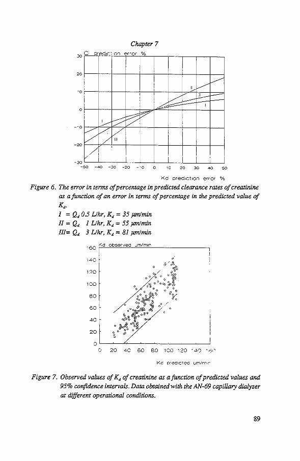
Chapter 7 I _Qred1Ct1on error %
20
~ Ill ~
~ ----3------/: I
0
f-::;: v f:7 ,_____._
/ VII ~ /
-w
-20
//; ' I I_L I I -30 -50 -40 -30 -20 -10 0 10 20 30 40 50
Kd pred1Ct1on error %
Figure 6. The error in terms of percentage in predicted clearance rates of creatinine
as a function of an error in terms of percentage in the predicted value of K •. I = Q. 0.5 Llhr, Ka = 35 pmlmin
II = Q. I Uhr, K• = 55 pm!min
Ill= Q. 3 Uhr, Ka = 81 pmlmin
1 60 Kd observed um/m1n
Kd ored1cted um/m•n
Figure 7. Observed values of K. of creatinine as a junction of predicted values and
95% confidence intervals. Data obtained with the AN-69 capillary dialyzer
at different operational conditions.
89

Prediction model
60 Clearance observed ml/m1n
0 0
50
40
30
20
oL---A---~--~----~--~--~
0 10 20 30 40 50 60
Clearance predicted ml/mrn
Figure 8. Observed values of creatinine clearo.nce rate as a function of predicted values and 95% confidence intervals. Data obtained with the AN-69 capillary dialyzer at different operational conditions.
DISCUSSION
In 1his chapter the applicability of the prediction fonnulas of K., of creatinine, as derived in Chapter 6, was examined. It was concluded that 73% of the variability of
K., of creatinine can be explained by variations in Q, and .MI. For prediction of K., values of urea and phosphate, the predicted value of the K., of creatinine was multiplied by the acocmpanying ratio. Comparing the predicted values of K., of urea
and phosphate with the observed values demonstrate that respectively 70% and 58% of the variability could be explained. Therefore, it can be concluded that the use of a fixed relationship between K., of creatinine and that of other solutes is justified (Eq. 3). The main purpose of predicting K.,, is to obtain an indication of the expected clearance rate at different operational conditions. As clearance in CA VHD takes place by combined convection and diffusion, prediction of values of clearance rates will always be more accurate than prediction of K., values. Therefore, explaining the variability in clearance rates was superior to that of K., values.
90

Chapter 7
The contribution of diffusive transport to the total transport of solutes increases with dialysate flow rate. Therefore, as was shown in Figure 6, the error of the predicted clearance rate generated by an error in the predicted K, increases with dialysate flow rate. However, as the error in K, was calculated in terms of percentage, the absolute change was higher at high dialysate flow rates with accordingly high K, values.
Since values of Qwi and :MI are not readily available, nomograms are constructed. An underestimation or overestimation of Qwi will induce a minimal error in the predicted clearance rate. Using an estimated value of :MI, the error in the value of the predicted clearance rate is minimized by using the observed value of Q,. In this way, an error in the estimation of :MI depends only on the estimation of the transmembrane pressure difference. (see Eq. 1, Chapter 6). The transmembrane pressure difference can be calculated from the MAP, and mean values of the P,,, the P,, and the COP. If the ultrafiltration column height is 50 em, the error in terms of percentage in :MI due to a deviation of P,, and COP from the values of 10 and 20 mmHg respectively, will not be higher than approximately 10%. Thus, when using the observed value of Q, and MAP, errors in the estimation of :MI would not be as high as is shown in Figure 4.
In conclusion, the prediction of clearance rates of different uremic solutes is
feasible and can be simplified to be used in the clinical setting. In the next chapter, the feasibility of prediction of the clearance rate of drugs is examined.
91

Prediction model
REFERENCE
1. van Geelen JA. Hemodiafiltration, simultaneous application of hemodialysis and
hemofiltration. Thesis, 1983. University ofLimburg, Maastricht, the Netherlands.
92

CHAPTER 8
DRUG CLEARANCE BY CONTINUOUS ARTERIOVENOUS HEMODIAFILTRATION: ANALYSIS OF
SIEVING COEFFICIENTS AND MASS TRANSFER COEFFICIENTS OF DIFFUSION
INTRODUCTION
With CA VHD, time-averaged clearance rates may be several fold higher than with either CA VH or conventional dialysis techniques. With the latter two techniques, it is known that some drugs are cleared to a significant extent [1-4]. Therefore, with CA VHD drug clearance rates may be high enough to warrant dosage adjustments. To determine dosage adjustments, insight into the determinants of transpon is needed.
Clearance rate by CA VHD of uremic soluteS can be reliable predicted by our mathematical model of combined convection and diffusion. This model can be further extended to transpon of drugs. lmponant determinants of transpon of drugs are the sieving coefficient and the mass transfer coefficient of diffusion (K,, [pm/min]). They have to be experimentally determined. When the values of these two parameters are known. our model may be used to calculate drug clearance rates under a variety of operational conditions. ln this study, clearaoce rateS of urea, creatinine, uric acid, phosphate and of the antibiotics cefuroxime, cefotaxime, ceftazidime, imipenem, ciprofl.oxacin, tobramycin and vancomycin are measured in patients who were treated with CA VHD. 1n patients receiving imipenem, concentrations of cilastatin were measured too. By using the numerical mathematical model of hemodiafiltrarion (see Chapter 5), the data were analyzed in terms of sieving coefficients and mass transfer coefficients of diffusion. The relationship between K, values of creatinine and K, values of drugs were determined.
METHODS
The clinical protocol is described in Chapter 4. Drugs were given as clinically indicated. lf an antibiotic was prescribed, the patient was eligible for this study.
93

Drug clearance
Study protocol
First, the infusion pumps for dialysate and substitution fluid were turned off. The dialyzer condition was established by measuring the resistance to blood flow (R,,
[mmHg • ruin/ml]) and the membrane hydraulic permeability index (MI, [ml/h • mmHg]). Samples of blood and ultrafiltrate were taken to determine the drug's apparent sieving coefficient (see Calculations) and the free fraction. Then, the substitution fluid infusion pump was turned on and samples of blood and ultrafiltrate were taken again. After that, the dialysate flow rate was set to 1 L/hr. Dialysate samples were taken to determine clearance rates and diffusion coefficients of creatinine, urea, uric acid, phosphate and of the drug. The same procedure was repeated at a dialysate flow rate of 3 L/hr. Samples were taken after equilibration for a time span that secured the passage of at least 500 ml of ultrafiltrate or dialysate
fluid, i.e. 10 times the content of the dialysate comparnnent Finally, the dialysate pump was turned off and Ml and R, were determined again. The study protocol implied blood sampling to a total volume of 35 ml, which is 20 ml in excess of the routine.
Measurements
Pressures in the arterial and venous blood line (P. and P., [mmHg]), pressure in the dialysate compartment (P., [mmHg]) and uitrafiltration rate (Q,, [ml/min]) were measured as described in Chapter 4. Dialysate flow rate at the inlet of the dialyzer (Q.u, [ml!min]) was checked using an electrouic weighing device. Blood flow (Q,,
[ml/min]) was determined either by measuring the time required for air bubble displacement over a certain length of tubing containing a volume of 13 ml or by measuring the transit time of an echo signal, using the TransouicsR HT101D flowmeter with a clamp-on probe around the blood line [5]. Both methods yielded similar values.
When the dialyzer condition was poor, i.e. when the resistance to blood flow was higher than 0.40 mmHg · min • ml"1 and/or ultrafiltration rate was less than 175 ml/min, the data were discarded.
94

Chapter 8
Calculations
The dialyzer resistance to flow and the hydraulic petmeability index of the membrane were calculated as described in Chapter 4. The sieving coefficient (s) was calculated as:
2 ·C, (1)
with C, the drug concentration in ultrafiltrate, and ~ and C,., the drug concentrations in plasma water at the blood inlet and outlet respectively (for
calculation of ~ and C,., see Chapter 5). Note that this is the clinical definition of the sieving coefficient, which is equivalent with the physical definition of 'apparent' sieving coefficient. The true sieving coefficient, i.e. the fraction of non protein bound
solute that passes through the membrane during ultrafiltration, is taken to be one. With high flux membranes, this assumption is valid for solutes of up to several thousand Daltons. The apparent sieving coefficient of a solute, s, as calculated according to Eq. 1, i.e. the product of the true sieving coefficient and the free fraction should then be equal to the free fraction.
Clearance (Cl, [ml!min]), defined as the rate of mass transfer divided by the total concentration in plasma at the blood inlet, was calculated as:
(2)
Where Q., and C,, are flow rate and solute concentration at the dialysate outlet of
the dialyzer respectively, and Cp; is the solute concentration in plasma at the blood inlet side of the dialyzer. From the observed flow rates and solute concentrations, K., was calculated using the numerical model as described in Chapter 5.
Drug assays
Blood samples were centrifugated and the plasma was stored at -80'C. Total (bound plus free) drug concentrations were measured in plasma and in filtrate 1 dialySate. Concentrations of tobramycin and vancomycin were detetmined by a fluorescence polatization immunoassay [ 6]. The day to day variability of the assay of tobramycin was 4% and that of vancomycin 7%. Concentrations of cefuroxime, cefotaxime, ceftazidime, imipenem, ciprofloxacin and cilastatin were detetmined by H.P J...C. [7-
95

Drug clearance
11] with slight modifications. The day to day vatiability of the H.P .L.C. assay was
6% for cefuroxime, 7% for cefotaxime, 7% for ceftazidime, 7% for imipenem and
5% for ciprofloxacin. To detennine drug protein binding, plasma samples were centrifuged through a
porous membrane of regenerated cellulose (Centri-free system, Amicon, U.S.A.) with
a molecular cut-off of 30, 000 Daltons. The free fraction was calculated as the ratio of the concentration in the filtrate to that in plasma water. Protein binding of
imipenem was not detennined, since plasma samples containing imipenem were immediately stabilized by buffering to pH 6.5, by which protein binding is reduced.
Protein binding of ciprofloxacin was not determined.
Table 1. Range of drug clearance rates during CAVH and CAVHD. 1
: Total of measurements ar dialysareflow of I Uhr and 3 Uhr.
DRUG n' Or Clcavh Cleavbd I L/hr
ml/hr ml/min ml/min
Cefuroxime 22 270-990 4-14 7-26
Cefotaxlme 13 380-990 5-!3 9-20
Ceftazidime 24 240-890 4-14 13-30
Jmipenem 23 180-720 4-12 8-34
Ciprofloxacin 4 410-490 7-8 18-23
Tobramycin 30 240-880 3-12 13-24
Vancomycin 6 330-740 4-8 8-20
Cilastatin 24 200-720 2-9 6-20
RESULTS
ClClJ.Vbd 3 L/hr
ml/min
10-42
17-25
17-38
17-49
24-39
17-32
11-20
7-24
The study protocol was performed 53 times in 32 patients with 19 dialyzers. With
9 protocols, the patient received two different antibiotics. Therefore, the total amount
of measurements of clearance of antibiotics was performed 62 times, each
measurement at two dialysate flow rates. Due to time the protocol needed to be
performed, sieving coefficients were not detennined at every protocol. Both for cefotaxime and ciprofloxacin, sieving coefficients were detennined two times
separately and at one of these occasions, clearance rate of cefotaxime was measured
only at a dialysate flow rate of 1 L/hr. For imipenem, at one protocol, the
96

Chapter 8
concentration of spent dialysate at a dialysate flow rate of 3 L/br was too low to detennine reliably and for tobramycin tbis occurred at 1 protocol for both dialysate flow rates. Blood flow tate was 125 (SD 32) ml/min and net ultrafiltration tate was 540 (SD 225) ml/br. At a dialysate flow tate of 1 L/hr, urea clearance tate was 24 (SD 4) ml/min and creatinine clearance was 21 (SD 5) ml/min. At a dialysate flow
tate of 3 L/hr these values were 42 (SD 9) and 32 (SD 7) ml/min respectively. At the time of measurements, dialyzers had been used for an average time of 50 hrs
(range 0.75-291 hrs). The hydraulic penneability index of the membrane and the resistance to flow have not changed during the expetiment.
Ranges of drug clearance rates are presented in Table 1. Sieving coefficients, measured free fractions and free fraction of nonnals are presented in Table 2. The value of the sieving coefficient itself was not related to either the hydraulic penneability index of the membrane (MI) or to the rate of ultrafiltration (see Figure 1). The sieving coefficient was not different from the corresponding free fraction, as detennined in the laboratory. The free fraction in our patients was, however, different from that in nonnal subjects (see Table 2).
The value of K, of creatinine was 71 ± 30 (range 20 to 155) )IIll/min. This variability was largely explained by differences in membrane penneability, as reflected by the hydraulic penneability index, and by differences in dialysate flow tate (see Chapter 7). K, values of a number of solutes, expressed as a percentage of the corresponding K, of creatinine, are given in Table 3. Seventeen K, values of drugs resulted in negative values or an error. K, values were related to the solute molecular weight (see Figure 2). Regression analysis of the logarithm of K, as a function of the logarithm of molecular weight (MW, [Daltons]) yielded the equation:
K, MW -OA2
r=<m) ,, (3)
For some of the drugs, however, K, values were appreciably higher than expected
according to the regression equation. Using ouly the mass transfer coefficient of antibiotics (n=7), regression analysis yielded an exponent of -0.44 (r=0.67).
97

Drug clearance
Table 2. Comparison of free fraction and sieving coefficient. The sieving coefficient during CA VH is not different from the free fraction as determined in the lohoratory. Values of the measwedfreefraction and the apparent sieving coefficient in!CU patients were averaged to obtain the value of F. which will be used to predict clearance rates (see Chapter 9).
DRUG Free fraction % sieving coeff. % F
normals [ref] patients n patients n
Cefuroxime 67 [18] 78±8 12 81 ± 10 21 0.80
CefotaXime 60 [19] 77±7 9 76± 12 14 0.76
· Ceftazidime 83 [20] 86±5 5 95± 5 12 0.92
Imipenem 80 [21] 100 ± 14 22 1.00
Ciprofloxacin 75 [22] 102±7 6 1.02
Tobramycin 100 [23] 79±5 9 83 ± 7 19 0.82
Vancomycin 45 [24] 56 1 68± 7 5 0.67
Cilastatin 20 [25] 68± 2 20 0.68
Siev. coeff. (actual/drug 1.3 ,----
average)
---------~
• • • <II
• • • '· • • • • • •
••• e e
0.7 I~
0.5 '----- -·-- . - _ _I~ • -- ___ __~__ __ ---'
0 5 10 15 20 25 30
Membrane Index (ml/hr.mmHg)
Figure 1. Relationship of the sieving coefficient qf drugs (s) with the hydraulic permeability index of the membrane (Ml).
98

Chapter 8
Table 3. Solute molecular weight (MW) and mass transfer coefficient of diffusion (K,J expressed as a percentage of the co"esponding Kd of creatinine (K,.J.
1 Estimated molecular weight in blood: At pH 7.40 with pKa 7 21, 39% is taken to be present as insoluble Ca(H,P04)2 and 62% as Ca(HP04). 2 In-vitro data supplied by D. Pouchoulin, Hospal C.O.T., Lyon, France.
SOLU1E M.W KJK.. SEM n %
Urea 60 132 4 109
Lactate 90 93 2 2
Creatinine 113 100
Uric acid 168 93 4 123
Phospha!e 174' 75 3 125
Vitamin B 12 1355 242 1 5
Imipenem 317 68 8 20
Ciprofloxacin 331 85 12 2
Cilastatin 384 56 5 21
Cefuroxime 424 72 7 21
Cefotaxime 455 37 7 11
Tobramycin 468 72 6 26
Ceftazidime 547 66 6 22
Vancomycin 1449 37 4 5
99

90
60
30
creatJntne
• o urate lactate
• phos.o
• cipro
Drug clearance
r=0.87
812 •
va11co •
0 L_ ______ L_ ______ L_ ______ L_ ______ L_ ____ ~
0 300 600 900 1200 1500
Malec we1ght Daltons
Figure 2. Relationship between Ka and solute molecular weight (MW). K, values were expressed as a fraction of the corresponding K, of creatinine (K.,). Numerical values are given in Table 3. Linear regression analysis of Ln(Kd I K.,) as a function of Ln(MW) yielded a correlation coefficient (r) of 0.87.
DISCUSSION
In the previous chapter, the feasibility of the prediction of the value of K, of creatinine based on dialysate fiow rate and the hydraulic permeability index of the membrane was demonstrated. To use this model for prediction of drugclearance, the ratio of K, of creatinine to that of the drug and the sieving coefficient of the drug
was determined. The present study yielded two imponant results. First, it was demonstrated that a drug's apparent sieving coefficient may be
equated to its non-protein bound fraction. The laner may be determined in the laboratory. Since, in patients, the non protein bound fraction may differ remarl<ably from values in normals, it would be worthwhile to have the free fraction routioely determined in a blood sample. The binding of acidic drugs may be decreased due to uremia, low levels of albuntio, high levels of bilirubin and free fatty acids or due to competition by other drugs [12-14]. The binding of basic drugs may be increased due
100

Chapter 8
to increased levels of a,-acid glycoproteins, one of the acute phase proteins [15]. In
general our data on sieving coefficients were roughly in accordance with data of several studies given by Bickley et a! [16] and Kroh et al [17].
Second, we found a relationship between I<aJK.o and solute molecular weight. With the AN-69 capillary dialyzer, drug clearance rates can now be predicted by the
model as follows. First, dialysate flow rate and the membrane hydraulic permeability
are determined for the estimation of K, of creatinine. Then K, of the drug is ealimated, either from published values of I<aJK.o (see Table 3) or from an estimation of K,IK,, based on the drug molecular weight. Then the free fraction, F, is either taken from published values or determined in the laboratoty. The values are used for input in the model, by which drug transpon may then be calculated for an average plastua water flow rate or ealimated from the mean arterial pressure of the patient. (the influence of differences in blood flow rate is small). The whole procedure will be demonstrated in Chapter 9, in which predicted values of clearance rate of drugs will be compared to observed values.
It should be noted that the findings in this study are restricted to the dialyzer we used, the AN-69 capillary dialyzer. In case of other membranes with a smaller pore size or electric charge might cause the drug sieving coefficient to be different than
its free fraction. Furthermore, it may be expected that the relationship between K, and molecular weight depends on the membrane resistance to diffusion and its contribution to the overall resistance to diffusion of the dialyzer. This methodology for the analysis of drug clearance by CA VHD, however, should be readily applicable to other dialyzers.
In conclusion, this study shows the feasibility of determining free fractions and K, values of drugs. With these parameters the clearance duriog CA VHD may be
predicted for different operational conditions. In the next chapter the reliability of such predictions will be examined and consequences for dosage adaptations will be considered.
101

Drug clearance
REFERENCES
1. Golper TA, Bennett WM. Drug removal by continuous arteriovenous haemofiltration. A review of the evidence in poisoned patients. Medical toxicology 1988; 3: 341-349.
2. Kroh UF, Dehne M, El Abed K, FeuBner KD, Hofmann W, Iennartz H. Drug dosage during continuous hemofiltration: Pharmacokinetics and practical implications. Contrib Neprol 1991; 93: 127-130.
3. S!ugg PH, Haug MT, Bosworth C, Paganini EP. Comparative vancomycin
kinetics in intensive care unit patients with acute renal failure: Intennittent hemodialysis versus continuous hemofiltration hemodialysis. Contrib Neprol 1991; 93: 140-142.
4. Bennett WM, Arronoff GR, Morrison G, Golper TA, Pulliam J, Wolfson M,
Singer L Drug describing in renal failure: Dosing guidelines for adults. Am J of Kidoey Disease 1983; 3: 155-193.
5. Burton RG, Gorewit RC. illtrasonic flowmeter. Uses Wide-beam transit-time technique. Medical Electronics 1984; 15: 68-73.
6. Jolley ME, Stroupe SD, Wang CJ. Fluorescence polarization immunoassay I. Monitoring aminoglycoside antibiotics in serum and plasma. Clin Cbem 1981; 27: 1190-1191.
7. Signs SA, File TM, Tan JS. High-pressure liquid chromatographic method for analysis of cephalosporins. Antimicrob Agents Chemother 1984; 26: 652-655.
8. Ayttan J. Assay of cefiazidime in biological fluids using high-pressure liquid chromatography. J Antimicrob Cbemother 1981; 8 (Suppl B): 227-231.
9. Gravallese DA, Musson DG, Pauliukonis LT, Bayne WF. Detettninations of imipenem in human plasma and urine by bigh-perfollltance liquid chromatography. Comparison with microbiological methodology and stability. J Chrom 1984; 310: 71-84.
10. Myers CM, Blumer JL. Detettnination of imipenem and cilastatin in serum by high pressure liquid chromatography. Antimicrobial Agents and Chemotherapy 1984; 26: 78-81.
11. Brogard JM, Jehl F, Momeil H, Adloff M, Blickle JF, Levy P. Comparison of high liquid chromatography and microbiological assay for the detettnination of
biliary elimination of ciprofloxacin in humans. Antimicrob Agents Chemother 1985; 28: 3!1-314.
12. Craig W A, Su B: Protein binding and the antimicrobial effects: Antibiotics in
laboratory medicine. Lorian V. editor, second edition. Williatns and Wilkins 1985, Chapter 13.
102

Chapter 8
13. Perucca E, Grimaldi R, Crema A: Interpretation of drug levels in acute and chronic disease states. Clinical Pharmacokinetics 10; 498-513 1985.
14. Bodemham A, Shelly MP. Park GR: The altered pharmacokinetics and pharmacodynamics of drugs commonly used in critically ill patients. Clinical Pharmacokinetics 14; 347-373 1988.
15. Piafsky KM: Disease-induced chaoges in the plasma binding of basic drugs. Clinical Pharmacokinetics 1980; 5: 246-262.
16. Bickley SK: Drug dosiog during continuous arteriovenous hemofiltration. Clinical Pharmacy 1988; 7: 198-206.
17. Kroh U, Hofmann W, Dehne M, El Abed K, Lennartz H: Dosisanpassung von pharmaka wWelld kontinnierlicher hamofiltration. Anaesthesist 1989; 38: 225-
232. 18. BundtzenRW, ToothakerRD,Nielson OS,MansenPO, WelliogPG, CraigWA:
Pharmacokinetics of cefuroxime io nonnal and impaired renal function: Comparison of high-pressure liquid chromatography and microbiological assays. Antimicrobial Agents and Chemotherapy 1981; 19: 443-449.
19. Fillastre JP, Leroy A, Humbert G. Godin M: Pharmacokinetics of cefotaxime io
subjectS with nonnal and impaired renal function. Journal of Antimicrobial Chemotherapy 1980; 6 suppl A: 103-111.
20. O'Callaghan CH, Acred P, Harped PB, Ryan DM, Kirby SM. Harding SM: GR 20263, a new broadspecttum cephalosporin with anti-pseudomonal activity.
Antimicrobial Agents and Chemotherapy 1980; 17: 876-883. 21. Gibson TP, Demettiades JL,. Bland JA: Imipenem/cilastatin: Pharmacokinetic
profile in renal iosufficiency. The American Journal of Medicioe 1985; 78 suppl 6a: 54-61.
22. Robberts DE, Williams JD: Ciprofloxacio io renal failure. Journal of Antimicrobial Chemotherapy 1989; 23: 820-823.
23. Gordon RC, Regamey C, Kirby WMM: Serum proteio bioding of the atninoglycoside antibiotics. Antimicrobial Agents and Chemotherapy 1972; 2: 214-216.
24. Moellering RC: Pharmacokinetics of vancomycio. Journal of Antimicrobial Chemotherapy 1984; 14 suppl D: 43-52.
25. Calandra GB, Brown KR, Grad LC, Akonkhai VI, Wang C, Aziz M. Review of experiences and tolerability io the first 2516 patients treated with imipenem/cilastatin. The American Journal ofMedicioe 1985; 78 suppl6a: 65-
70.
103

CHAPTER9
DRUG CLEARANCE BY CONTINUOUS ARTERIOVENOUS HEMODIAFILTRATION: PREDICTION OF DRUG
CLEARANCE RATE AND RECOMMENDED DOSE ADAPTATIONS FOR SEVEN ANTIBIOTICS
INTRODUCTION
In the previous chapter, clearance rates (Q, [ml!min]) hy CA VHD of different drugs were examined. Data were analyzed in tenns of sieving coefficients and mass transfer coefficients of diffusion (K., [pm/min]). The ratio of the mass transfer coefficient of
diffusion of drugs to that of creatinine was detennined. As was shown in Chapter 6,
clearance rates by CA VHD of uremic solutes other than creatinine could be predicted when the predicting formula of K.. of creatinine was extended by the ratio of K.. of the uremic solute to that of creatinine. In this chapter, the feasibility of predicting clearance rates of drugs, which have an apparent sieving coefficient lower than one, will be examined.
As patients treated by CA VHD are nearly always anuric, the removal of drugs which are normally excreted by the human kidney can occur predominamly by CA VHD. This is especially true if the protein binding of the drug is low, if the volume of distribution is small and if the non-renal clearance rate is low in comparison to the clearance rate by CA VHD. If a drug meets the demands as stated
above, treatment by CA VHD will warrant dosage adaptations. Most previous studies on clearance of drugs by continuous renal replacements techniques focused on CA VH [1-5]. Recently, some studies have been published on drug clearance by CA VHD [6-8]. However, little is known about the required dosage regimens of drugs during treatment by CA VHD.
In the present chapter, drug clearance rates by CA VHD at different operational conditions are predicted and compared to observed values. Dosage adaptations are calculated for anuric patients treated by CA VHD at dialysate flow rates of 1 L/hr and
3 L/hr.
104

Chapter 9
METI!:ODS
Prediction of K• and clearance rates of drugs
Clearance rates of cefuroxime, cefotaxime, ceftazidime, imipenem, tobramycin, vancomyCin, ciprofloxacin and cilastatin were measured during CA VH and during CA VHD at a dialysate flow rate of I and 3 L/llr. The srody protocol as well as the analysis of data has been described in Chapter 8.
The mass transfer coefficient of diffusion of drugs was predicted using the predicting formula of K., of creatinine as described in Chapter 6, multiplied by the ratio of K., of the drug to that of creatinine as given in Chapter 8. The mass transfer coefficient of diffusion was calculated by the numerical method as described in
Chapter 5. Predicted values of drug clearance were calculated by inco~porating the predicted
values of K., in the following formula:
cQ,JQ.,r'- <Q..,JQ,r' Cl = Q, · · F ·1.05
, <Q.JQ..!'- CQ.,tQ,.;r' (I)
where n = K., • Sf~. S [m2] is membrane surface area, Q~ [ml!min] the plasma
water flow at the dialyzer outlet, Q, and Q,, [ml/min] are dialysate flow rates at the dialyzer inlet and outlet respectively and F is the average of the apparent sieving coefficient and the free fraction of the drug concerned as given in Chapter 8, Table 2. To calculate plasma clearance rate from plasma water clearance rate, the latter is
multiplied by !.05, to account for the volume fraction of proteios. The whole procedure of calculating predicted values in the clinical setting is given in Figure I. Observed plasma clearance rates were calculated as:
(2)
Linear regression analysis was performed for both predicted K., values and clearance rateS by CA VHD as a function of the observed values aod 95% confidence intervals were calculated.
105

Prediction drugclearance and dosage
Example: Tobramycin ( K,/K,.,=0.72 F=0.82)
1 P.=68mmHg 11:= 20mmHg
2 1MP = 51 mmHg Q,= 500 ml/br
3 MI = !0 mlJlJr · mmHg
P, = 32 mmHg P, = -30 crnHzO --> 1MP = 51 mmHg
--> MI = 10 ml/br·mmHg
Q. = 2 L/hr --> !(,. = 83 )lill/mi:n
4 !(,. = 83 )lill/mi:n KJK., = 0.72 --> K, = 60 )lill/mi:n
5 Q,. = !17 ml!min. Q. = 33 ml/min Q, = 8 ml/min Q,...=5ml!min f = 0 S = 0.6 m' F = 0.82 Ht = 030 C,.,, = 50 mg!L K, = 60 jllll/min
(Eq. 2, Chp 4)
(Eq. 6, Chp 4/
(Eqs. 6, 7, Chp 6)
c¢ = 10 mmo!IL --> c" = 6 mmo!IL (Eq. 11, Chp 5) (fot procedure, see Figure !, Chp 5)
6 Q, = 41 ml/min cdo = 6 mmol/L c~ = 10 mmol/L --> Clearance- 22 ml/min
(Eq. 2, Chp 9)
Figure I. Algorithm for the prediction of drug clearance rate in CAVHD. For abbreviations see previous chapters or abbreviationlist. ·The value of Ml and Qw, can also be read from the nomograms (Chp 7).
Dosage adaptations
The drug half life (f.,Jl) during CA VHD treatment was calculated by using the formula:
TB=0.693·V, "' --,a=--"
(3)
where V, is the distribution volume of the drug [9]. Values of drug protein binding, of dislribulion volume, and of total and non-renal clearance rates were taken from
the literature (see Table l). If possible, all values, except the values of clearance rates in nonnals, were taken from studies, which obtained data from critically ill patients.
Recommendations regarding dose adjustments were based on changes in the drug
106

Chapter 9
clearance rates. For every drug, the average clearance rates by CA VHD at dialysate flow rates of I L/hr and 3 L/hr were used to calculate dose adaptations. A one compartment model of pbarmaccldnetics was used to calculate doses and dose intervals that led to the desired levels. Time related changes in drug ccncentrations were calculated according to:
Cl• t
C =C .~v;-,, pO (4)
where C,. is the initial plasma ccncentration, c;, is the plasma ccncentration after a time span t. Desired levels were deteiiDined by taking into account the drugs' mode of action and their toxicity [10-12]. Thus, for beta-lactams and imipenem, we aimed for plasma ccncentrations continuously above the minimal inhibitory concentration (MIC) of most ccmmon pathogens. For tobramycin the area under the curve (AUC) is probably the main deteiiDinant of efficacy [13]. Therefore, for this antibiotic, we aimed for high peak levels with trough levels low enough to reduce the risk of serious toxicity. For vancomycin the same strategy was followed. For ciprofloxacin, we aimed for high peak levels and trough levels not falling to low.
RESULTS
Figure 2 shows the correlation between the observed and predicted values of the K,. of all drug together (n=l20). The coefficiem of deteiiDination (r'-) was 0.42. Eight values were considered as outlayers.
Figure 3 shows predicted values of plasma clearance rate versus observed values of each drugs separately. In Figure 4, the correlation between the predicted clearance rates and the observed values are given for all drugs together. The coefficient of deteiiDination was 0.64, n= 146. Values of ranges of drug clearance rates by CA VH and by CA VHD at a dialysate flow rate of l and 3 L/hr are given in Chapter 8. Average values± SD of clearance rates at a dialysate flow rate of 1 L/hr and of 3 L/hr were for cefuroxime 15 ± 5 and 25 ± 9, for cefotaxime 14 ± 3 and 21 ± 3, for ceftazidime 18 ± 5 and 27 ± 6, for imipenem 18 ± 7 and 31 ± 9, for ciprofloxacin 21 ± 2 and 26 ± 6, for tobramycin 19 ± 3 and 26 ± 4, for vancomycin 15 ± 4 and 17 ± 4 and for cilastatin 12 ± 4 and 17 ± 5 ml/min respectively.
107

Prediction drugclearance and dosage
1 50 Kd observed um/min
125
100
75
50
25
0 0
oo
0 0
0
0
0 0 0 0 0
0 0 0 oo
0 0 0
0 0 og>o 8o q,08
0 0 0 O~Oo 0
0 0 § 0
€ 0 roo 0 0~
0 OtSiJ~ 0 0 0
o ~ooS 0 o Ooo
25 50
0
0 0 0 0
0 0 0
0 Oo 0'
0 0 0
"' @ 0
0 0 0
0 0
"' 0
75 100 125 150
Kd pred1cted um/min
Figure 2. Comparison of observed values of K, of eight different drugs calclilaJed from membrane surface area, flow rates and drug concentrations, with values predicted from dialysate flow rare, membrane Jrydraulic penneabiliry and the ratio KjK"". The lines show the 95% cor{idence intervals.
108

Chapter 9
50 Clearance observed ml/m1n
50 Clearance observed m11m1n
Cefurox1me Cefotax1me
40 n=22. r2=0.63 40 n= 13. r2=0.54
30 30 . . 20 20 :
. . : 10 .. 10 !
0 0 0 10 20 30 40 50 0 10 20 30 40 5:.·
Clearance pred1cted ml/m1n Clear once pred1cted m1/m1n
50 Clearance observed ml/m1n 50
Clearar1ce observed ml/m1n
CeftaZ1d1me lm1oenem
40 n=24, r2=0.44 40 n=23. r2=0.58
30 30
: 20 20
z
10 10
0 0 0 10 20 30 40 50 0 10 20 30 40 50
Clearance pred1cted m11m1n Clearance pred1Cted m1/m1n
Figwe 3. Clearance observed versus clearance predicted of each drug separaJely. Line of identity is given.
109

Prediction drugclearance and dosage
50 Clearance observed mllm1n
50 Clearance observed onl/m,n
' /
C•profloxac•n Tobramyc•n
40 n=4. r2=0.82 40 n=30. r2=0.71
30 30 .. ·.· .
20 20 . •• . .. . 10 10
0 0 0 10 20 30 40 50 0 10 20 30 40 ~;..:'
Clearance pred•cted ml/m•n Clearance pred•Cted ml/rn•r•
50 Clearance observed ml/m1n
50 C:earanCl-'_ ouserv(.:d ml/m1n
Vancomyc•n Cllastat•n
40 n=6, r2=0.75 40 n=24. r2=0.64
30 30
20 .. 20 •
• • 10 10 ,.
0 0 0 10 20 30 40 50 0 10 20 30 40 50
Clearance pred•cted ml/mrn Clearance prediCted ml/m11i
Figure 3.
110

Chapter 9
60 Clearance observed ml/min
50 0
0
0
40 0 0
0
0 OfjOo
0 0
0 0 0
30 0 0 0 o<Bo& 0 0
oo <:fJ 0o ~o o q,o &, o o
o 0 o 20 0 Clf};,§'<:l:> Oo0 0
<! 0 0~ 0 0 ~foo§o <S> e&o ~ 0
10 ~0~ 0 o o o0 '00
0 0 iO 20 30 40 50 60
Clearance predicted ml/m1n
Figure 4. Clearance observed versus clearance predicted of eight different drugs together. The lines s!ww the 95% confidence intervals.
111

Table 1. Volume of distribution, clearance rates and half lives in normals and In anurlc patients with and without treatment by CAVHD. 1 Data taken from literature. 2 Sum of non-renal clearance rate and average clearance by CAVHD. 3 Calculated by using Eq. 3 (body weight 70 kg).
Drug V,' [Likg] Clearance [ml/min]: Half life [hrl) References
nonnal1 anuric1 amrric an uric norrnal3 anuric3 an uric an uric CAVHD CAVHD CAVHD CAVHD I L/hr' 3 L/hr' I L/hr' 3 L!hr'
cefuroxime 0.23 115 5 20 30 1.7 38.7 9.3 6.2 [30]
cefotaxime 0.37 353 143 157 164 1.1 2.4 1.9 1.8 [31-35]
ceftazidime 0.18 115 8 26 35 1.7 25.3 5.6 4.2 [33, 34, 36-38]
imipenem 0.31 245 116 134 147 1.0 2.2 1.9 1.7 [4, 39-41]
ciprofloxacin 2.61 521 254 275 280 4.3 8.2 6.4 6.2 [42]
tobramycin 0.28 84 7 26 33 1.8 40.8 8.7 6.9 [43-45]
vancomycin 0.35 121 <5 20 22 3.7 90.2 14.2 12.9 [46-49]

Chapter 9
Mean drug clearance rates and corresponding plasma half lives are given in
Table 1. Differences between the values of clearance rate and half lives in the untreated anuric state and duting treatment by CA VHD, were most pronounced for cefuroxime, ceftazidime, tobramycin and vancomycin In the anuric state, adjustment of drug dosage was necessary for all antibiotics. During CA VHD treatment, further adjustment was required for cefuroxime, ceftazidime, robramycin and vancomycin Results of cilastatin will be discussed in Chapter 10. Guidelines for drug dosage in patients with untreated anuria and in patients treated by CA VHD are given in Table 2. Since the volume of distribution may effect drug half-life, the volume of distribution was varied in the model. An increasing or decreasing of V, by 1.5 times did not affect the results in such a way that calculated dose or dose intervals needed
to be adjusted.
Table 2: Guidelines for dosage adjustment in CAVHD and the aimed peak and trough levels. 1) Second and later administration: give haif the standard dose. 2) Give haif the standord dose. 3) Lean body mass 4) Adjust dosage according to peak and trough levels, measured respectively one hour cifter start of the infusion and shortly before a new dose.
Drug Standard Dose interval [his]: dose
nonnal anuric anuric anuric peak trough CAVHD CAVHD level level
1 L/hr 3 L/hr mg/L mg/L
cefuroxime 750mg 6 48 12 12
cefotaxim.e !OOOmg 6 8 8 8
ceftazicfune lOOOmg 8 48' 12' 12'
imipenem SOOmg 6 12 12 12
ciprofloxacin 200mg 12 122 12' 12'
tobramycin 3mgjklf 12' 48-72' 2448' 24' 8-10 <2
vancomycin SOOmg 6' 48-72' 2448' 2448' >25 < 10
113

Prediction drugclearance and dnsage
DISCUSSION
Prediction of clearance rates
In case of CA VH, the clearance rate is the product of the sieving coefficient and the rate of ultrafiltration. The latter is detennined by the trans membrane pressure difference, which depends on operational conditions, and on the hydraulic
petmeability of the dialyzer, which decreases over lime. Therefore, even for a given dialyzer, the clearance rate by CA VH is quite variable. It can, however, easily be estimated from the sieving coefficient and the rate of ultrafiltration.
Clearance by CA VHD, on the other hand, is detennined by convective and diffusive transpon. Diffusive transport is detennined by the concentration gradient between blood and dialysate and the mass transfer coefficient of diffusion. Moreover,
at high dialysate flow rates the mass transfer coefficient of diffusion is increased. A high ultrafiltration rate implies a high hydraulic permeability, which is associated with a high diffusive permeability, thus a high K, value. Furthermore, the value of K, corrected for operational conditions depends on the solute molecular weight Therefore, it can be concluded that a mathematical model is required to estimate drug clearance by CA VHD. The use of the prediction formulas of K, of creatinine, together with the ratio of K, of drugs to that of creatinine as introduced in Chapter 8, resulted in a fairly accurate estimation of the clearance rate of drugs.
Furthermore, one can use this prediction model to estimate the clearance rate of other drugs or of solutes used in case of self-poisoning. The molecular weight and protein binding are input parameters and, when an indication is needed for the contribution of CA VHD to the total clearance rate, the endogenous clearance rate and the volume of distribution has to be known.
Dose adjustments
Dose adjustments of antibiotics are usually necessary for anuric patients, especially when clearance in these patients is low compared to that in normal subjects. If these patients are treated by CA VHD, the dosage regimen may be adjusted again. One might detennine drug clearance by CA VHD in individual cases and detennine drug dosage accordingly. For clinical practice, however, it is more convenient to use standard dosage regimens for patients treated by CA VHD, based on average clearance rates by CA VHD. Moreover, if one use such dosage regimens, a possible delay in lime of establishing the desired plasma levels of the drug will be avoided.
114

Chapter 9
In this study, we calculated dosage regimens to be used for anuric patients treated by CA VHD. The averaged observed values of drug clearance rateS, used to calculate these dosage regimens, were approximately equal to the values of drug clearance rateS, predicted by taking an average value of ultrafiltration flow rate and membrane index. Therefore, it is concluded t.'Jat the calculated dose adaptations are based on average operational ccnditions. For each drug, the effect of the calculated dose adaptation on the plasma drug ccncentrations was ccnsidered when using input values of the drug clearance rate by CA VHD higher and lower as the averaged value. It was ccncluded that a ccnsiderable deviation in drug clearance rate by CA VHD from the value of the averaged clearance rate cculd take place before the reccmmended dose regimens were no longer appropriate. We used literature data on values of the volume of distribution measured in normal subjects. There have been several reportS in the literature on changes in distribution volume in intensive care patients [14-18]. Data are still to scarce, however, to determine whether and to what extent appreciable changes really occur.
When ccnstructing treatment regimens, the dose-efficacy relationship of antibiotics [10-12] and toxicity of certain antibiotics has to be regarded.
Killing of bacteria by beta-lactams is time dependent [12]. Furthermore, there is no ccncentration dependent killing above the MIC. These killing characteristics of beta-lactams would demand sustained concentrations above the MIC of the infectious organism. In animal models, ccntinuous serum ccncentrations above the MIC of the infecting organism have been shown to be more efficacious, ccmpared to transient peaks [11, 19]. Also, clinical outccme has been shown to be ccrrelated to the time above the MIC of the infecting organism [20]. Therefore, for these antibiotics, we aimed for plasma levels continuously above the MIC of most common pathogens.
In contrast to beta-lactams, aminoglyccsides do show concentration dependent killing activity on Gram-negative aerobes [10] and killing is relatively fast. This has
been shown in vitro [21] as well as in vivo studies [22]. High peak ccncentrations have been found to be a major determinant of clinical response [23]. Furthermore,
aminoglyccsides show a postantibiotic effect against staphylococci as well as against vatious Gram-negatives [24, 25]. Vogelman et al [13] found a reduction of efficacy
when the dosing interval was longer than the time period duting which the serum levels exceeded the MIC plus the duration of the postantibiotic effect. Several studies conclude that the AUC is the main determinant of efficacy [13, 26]. Apart from efficacy, toxicity is an Important issue. Nephrotoxicity, as well as oto- and vestibulotoxicity are major side-effects of aminoglyccsides. Aminoglyccsides are actively transponed into the proximal tubular cells, and this transpon is saturable [27, 28]. These findings have led to a decreased dosing frequency in the last few years
115

Prediction drugclearance and dosage
[29]. When reconstructing a dosage regimen of aminoglycosides during treatment by CA VHD, we considered these factors. Thus, we paid particular attention to trough levels low enough to minimize toxicity, and the dosing interval was increased rather than the dose was lowered.
The quinolones { ciprofloxacin) have a concentration dependent killing. Therefore, priority was given to high peak levels and a long dosing interval [12].
The most important finding of this study is that for cefuroxime, ceftazidime, tobramycin and vancomycin, the dosage regimen, used for anuric untreated patients, must be changed when the patient are treated by CA VHD. Failure to recognize this may easily lead to suboptimal drug levels and therapeutic failure. For the same reason, when there are alternative drugs of choice, clinicians may prefer drugs for which the influence of CA VHD on plasma levels can be neglected. Cefotaxime, imipenem and ciprofloxacin needed no dose adaptation during CA VHD. This was due to the relatively high non-renal clearance rates of these drugs, compared to that of CA VHD. It should be pointed out that the dosage regimens are theoretically derived from data obtained by a number of different studies. Although these dosage regimens are currently used in the clinic, the validity of these regimens needs confumation. In the future, studies have to be performed to determine plasma drug levels over time with the present dosage recommendations.
116

Chapter 9
REFERENCES
1. Bickley SK. Drug dosing during continuous arteriovenous hemofiltration.
Oinical Pharmacy 1988; 7: 198-206.
2. Go1per TA, Wedel SK, Kaplan AA, Saad AM, Donta ST, Paganini EP. Drug
removal during continuous arteriovenous hemofiltration: theory and clinical
obseiVations. The International Joumal of Artificial Organs 1985; 8: 307-312.
3. Keller E, Fecht H, Bohler J, Schollmeyer P. Single-dose kinetics of intipenem/cilastatin during continuous arteriovenous hemofiltration in intensive
care patients. Nephro1 Dial Transplant 1989; 4: 640-645. 4. Kroh U, Hofmann W, Dehne M, El Abed K, Lennartz H. Dosisanpassung von
pharmaka wiihrend kontinuierlicher Mmofiltration. Anaesthesistl989; 38: 225-
232. 5. Kroh UF, Dehne M, El Abed K, FeuJlner KD, Hofmann W, Lennartz H. Drug
dosage during continuous aneriovenous hemofiltration. Conttib Nephrol 1991;
93: 127-130. 6. Davies SP, Kox WJ, Brown EA. Clearance studies in patients with acute renal
failure treated by continuous arteriovenous haemodialysis. Contrib Nephro!l991;
93: 117-119.
7. Reetze-Bonorden P, Bohler J, Kohler C. Schollmeyer P, Keller E. Elimination
of vancomycin in patients on continuous arteriovenous hemodialysis. Conttib
Nephrol 1991; 93: 135-139. 8. Slugg PH, Haug MT, Bosworth C, Pagauini EP. Comparitive vancomycin
kinetics in intensive care uuit patients with acute renal failure: Intemrittent
hemodialysis versus continuous hemofiltration hemodialysis. Contrib Nephrol
1991; 93: 140-142. 9. Allen L, Kimura K, MacKichan J, Ritschell W A. Manual of symhols, equations
and defiuitions in pharmacokinetics. J C1in Pharmacal 1982; 22S: 1-23.
10. Vogehnan B, Craig WA. Kinetics of antimicrobial activity. Journal of Pediatrics
1986; 108: 835-840.
1 L Roosendaal R, Bakker-Woudenherg W, van den Berghe-van Raffe M, Vink-van
den Berg JC, Michel MF. Impact of the dosage schedule on the efficacy of
ceftazidime, gentamicin and ciprofloxacin in Klebsiella pneumoniae pneumonia
and septicemia in leucopenic rats. Eur J C1in Microbial Infect Dis 1989; 8: 878-
887. 12. Cars 0. Craig WA. General discussion on antibiotic dosing. Scand J Infect Dis
1991; 74S: 282-282.
117

Prediction drugclearance and dosage
13. Vogelman B, Gudmundsson S, Legett J, Tumidge J, Ebert S, Craig WA.
Correlation of antimicrobial pbannacokinetic parameters with therapeutic efficacy in an animal model The Journal of Infectious Disease 1988; 158: 831-845.
14. Perucca E, Grimaldi R, Crema A. Interpretation of drug levels in acute and chronic disease states. Clinical Pbannacokinetics 1985; 10: 498-513.
15. Bodembam A, Shelly MP, Paik GR. The altered pbannacokinetics and pbannacodynamics of drugs commonly used in critically ill patients. Clinical Phannacokinetics 1988; 14: 347-373.
16. Klotz U. Pathophysiological and disease-induced changes in drug distribution volume: Pbannacokinetic implications. Clinical Pbannacokinetics 1976; 1: 204-
218. 17. Gibaldi M. Drug distribution in renal failure. The American Journal of Medicine
1977; 62: 471-474. 18. van Dalen R, Vree TB. Pbannacokinetics of antibiotics in critically ill patients.
Intensive Care Medicine 1990; 16 S3: S235-S238. 19. Mordenti JJ, Quinliliam R, Nightingale CH. Combination antibiotic therapy,
comparison of constant infusion and intermittem bolus dosing in an experimemal animal model. J Anlimicrob Chemother 1985; ISS: 41-45.
20. Schentag JJ. Correlations of phannacokinetic parameters to efficacy of antibiotics: Relationship between serumconcentrations, MIC values, bacterial eradication in patients with gram-negative pneumonia. Scand J Infect Dis 1991; 74S: 218-234.
21. Auwera P van der, Klastersky J. Serum bactericidal activity and postantibiotic effect in serum of patients with urinary tract infection receiving high dose amikacin. Anlimicrob Agents Chemother. 1987; 31: 1061-8.
22. Flueckiger U, C Feller AU Gerber. Dose-response curves in a man-adapted model: A novel view of comparative in vivo assessment of antibiotics. Program and abstracts of the twenty-sixth interscience conference on antimicrobial agents and chemotherapy, New Orleans, LA. 1986. American Society for Microbiology, Washington, DC. Abstract 577, page 202.
23. Moore RD, Lietman PS, Smith CR. Clinical response to aminoglycoside therapy:importance of the ratio of peak concentration to minimal inhihitocy concentration. J Infectious Dis. 1987; 155: 93-9.
24. Vogelman BS, Craig W A. Postantibiotic effects. J Anlimicrob Chemother. 1985; 15 SA: 37-46.
118

Chapter 9
25. Craig WA, Gudmundsson S. The postantibiotic effect In: Lorian V. (ed)
Antibiotics in medicine, 2nd ed. Baltimore: Willaiams and Williams page 515-536.
26. Gerber AU, Wiprachler P, Stettlewr-Spichger U, Lebeh G. Constant infusion vs intermittent doses of gentamycin against Pseudomoas aeruginosa in vitro. J
Infectious Dis. 1982: 155: 93-9. 27. De Broe ME, Verbist L, Verpooten GA. Influence of dosage schedule on renal
cortical accumulation of amikacin and tobramycin in man. J Antimicrob Chemother. 1991; 27 SC: 41-47.
28. Verpooten GA, Giuliano RA, Verbist L, Eestermans G, De Broe ME. Once daily dosing decreases renal accumulation of gentamicin and netilmicin. Clin Pharmacology Therapeutics. 1989; 45: 22-7.
29. Tulkens PM. Efficacy and safety of aminoglycosides once-a-day: experimental and clinical data. Scand J Infect Dis 1991; 74S: 249-257.
30. Bundtzen RW, ToothakerRD, Nielson OS, Mansen PO, Welling PG, Craig WA. Pharmacokinetics of cefuroxime in normal and impaired renal function: Comparison of high-pressure liquid chromatography and microbiological assays. Antimicrobial Agents and Chemotherapy 1981; 19: 443-449.
31. Fu KP, Aswapokee P, Ho !, Matthijssen C, Neu H. Pharmacokinetics of cefotaxime. Antimicrobial Agents and Chemotherapy 1979; 16: 592-597.
32. Luthy R, Blaser J, Bonetti A, Simmen H, WiseR, Siegenthaler W. Comparative
multiple-dose pharmacokinetics of cefotaxime, moxalactam and ceftazidime. Antimicrobial Agents and Chemotherapy 1981; 20: 567-575.
33. Balant L, Dayer P, Auckenthaler R. Clinical pharmacokinetics of the third generation cephalosporins. Clinical Pharmacokinetics 1985; 10: 101-143.
34. de Klerk Sommers, Walters L, van Wyk M, Harding SM, Paton AM, Aynon J. Pharmacokinetics of ceftazidime in male and female volumeers. Antimicrobial Agents and Chemotherapy 1983; 23: 892-896.
35. Luthy R, M!!nch R, Blaser J, Bhend H, Giegenthaler W. Human pharmacology of cefotaxime (HR 756), a new cephalosporin. Antimicrobial Agents and Chemotherapy 1979; 16: 127-133.
36. Armstrong GC, Wise R, Brown RM. Hancox J. Comparison of ceftazidime and cefamando1e. Pharmacokinetics and blister fluid concentrations. Antimicrobial
Agents and Chemotherapy !981; 20: 356-358. 37. Mouton JW, Horrevorts AM, Mulder PGH, Prens EP, Michel MF.
Pharmacokinetics of ceftazidime in serum and suction blister fluid during continuous and intermittem infusions in healthy volunteers. Antimicrobial Agents and Chemotherapy 1990; 34: 2307-2311.
119

Prediction drugclearance and diJsage
38. Leroy A, Leguy F, Borsa F, Spencer GR, Fillastre JP, Humbert G.
Phannacokinetics of ceftazidime in normal and uremic subjectS. Amimicrobial
Agents and Chemotherapy 1984; 25: 638-642.
39. Gibson TP, Demetriades JL, Bland JA. Imipenem/cilastatin; Phannaookinetic profile in renal insufficiency. The American Journal of Medicine 1985: 78 suppl
6a: 54-61. 40. Wise R, Donovan MR, Lockley J, Drumm J, Andrews JM. The
phannacokinetics and tissue penetration of lmipenem. Journal of Amimicrobial Chemotherapy 1986; 18 supplE: 93-101.
41. Veipooten GA, Verbist L, Buntinx AP, Entwistle LA, Jones KH, de Broe ME.
The phannacokinetics of imipenem (thienamycin-formamidine) and the renal
dehydropeptidase inhibitor cilastatin sodium in normal subjectS and patients with
renal failure. British Journal ofPhannacology 1984; 18: 183-193.
42. Drusano GL, Weir M, Forrest A, Plaisance K, Emm T, Standiford HC. Phannacokinetics of intravenously administered ciprofloxacin in patients with
various degrees of renal function. Antimicrobial Agents and Chemotherapy 1987;
31: 860-864.
43. Adelman M, Evans E, Schentag JJ. Two-compartment comparison of gentamycin and tobramycin in normal volunteers. Antimicrobial Agents and Chemotherapy
1982; 22: 800-804. 44. Levy J, Smith AL, Koup JR, Williams-Warren J, Ramsey B. Disposition of
tobramycin in patientS with cystic fibrosis: A prospective comrolled study. The
Journal of Pediatrics 1984; 101: 117-124.
45. Zarowitch BJ, Anadan JV, Dtun!er F, Jayanshankar J, Levin N. Continuous
arteriovenous hemofiltration of aminoglycoside antibiotics in critically ill patients. J Clin Phannacol 1986; 26: 686-689.
46. Blouin RA, Bauer CA, Miller DD, Record KE, Griffen WO. Vancomycin
pharmacokinetics in nonnal and morbidly obese subjectS. Antimicrobial Agents and Chemotherapy 1982; 21: 575-580.
47. Brown N, Ho DHW,Fong KLL, Bogerd L,Maksymiuk A, BolivarR, Fainstein
V, Bodey GP. EffectS of hepatic function on vancomycin clinical phannaco1ogy.
Amimicrobial Agents and Chemotherapy 1983; 23: 603-609. 48. Blevins RD, Halstenson CE, Salem NG, Matzke GR. Pharmacokinetics of
vancomycin in patientS undergoing continuous ambulatory peritoneal dialysis. Antimicrobial Agents and Chemotherapy 1984; 25: 603-606.
49. Nielsen HE, Hansen HE, Korsager B, Skov PE. Renal excretion of vancomycin
in kidney disease. Acta Med. Scand. 1975; 197: 261-264.
120

CHAPI'ER 10
CLEARANCE OF IMIPENEM/CILASTATIN IN ACUTE RENAL FAILURE PATIENTS TREATED BY CONTINUOUS
ARTERIOVENOUS HEMODIAFILTRA TION•
INTRODUCTION
The antibiotic Tienam• (MSD!USA) is the combination of imipenem, a broad spectrum thienamycin antibiotic belonging to the antibiotic class of carbapenem, and of cilastatin, an inhibitor of renal dehydropeptidase-1, the enzyme responsible for the metabolism of imipenem in renal tubular cells. The addition of cilasratin serves both to prevent nephrotoxicity by imipenem and causes the intact drug to be excreted into the urine [1, 2]. Although imipenem and cilastatin differ with respect to protein binding, their volumes of distribution and routes of metabolism, their plasma half
lives are quite similar in normal subjects. lmipenem/cilastatin is often used in patients with acute renal failure due to sepsis. In case of renal failure, however, the plasma half life of imipenem is linle altered whereas that of cilastalin is vety much prolonged. It would seem questionable, therefore, whether the use of the fixed dose combination is justified in these patients. In Chapter 9, results of the rate of removal of imipenem and cilastatin with continuous arteriovenous hemodiafiltration were given. In this chapter, the consequences of using imipenem/cilastatin in a fixed dose combination are analyzed in patients with renal failure when untreated and when treated by continuous arteriovenous hemodiafiltration.
METHODS
The clinical and study protocols are described in Chapter 8. Analysis and calculations of these data were described in Chapters 8 and 9. Data of clearance rates of imipenem and cilastatin were compared and consequences of using these drugs in a fixed dose combination were considered. Concentrations in lime of imipenem and cilastalin were calculated for acute renal failure patients untreated and treated by CA VHD at a dialysate flow rate of 1 L/hr and 3 L/hr. Average values of drug distribution volume, clearance rates in normals and in anuric patients and plasma half
• ReprinTed in revised fonn from Intensive Care Med. 1992, 18: 282-285
121

Table 1: Pharmacokinetics of imipenem and ci/astatin. 1 Data taken from litera hire. 2 Sum of non-renal clearance rate and average clearance by CAVH and CAVHD. 3 Calculated by using Eq. 3, Chapter 9 (body weight 70 kg).
Drug V,' [L/kgl Clearance [mVmin]:
normal1 anuric1 an uric an uric an uric normal CAVH' CAVHD CAVHD
I L/hr' 3 L/hr''
imipenem 0.31 245 116 125 134 149 1.0
cilastatin 0.24 230 3 9 13 21 0.9
Calculated half life [hrs]3 References
an uric an uric an uric an uric CAVH CAVHD CAVHD
I L/hr 3L/hr
2.2 1.9 1.9 1.7 [3-6]
89 21.6 14.9 9.2 [3, 6-9]
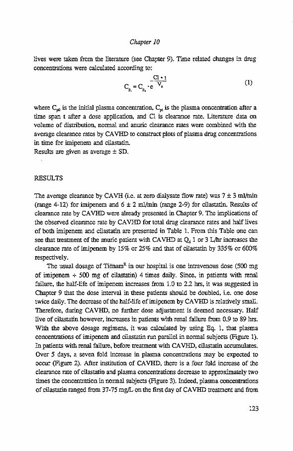
Chapter 10
lives were taken from the literarure (see Chapter 9). Time related changes in drug concentrations were calculated according to:
c =C .~v. P, Po
(I)
where Cp0 is the initial plasma concentration, C"' is the plasma concentration after a time span t after a dose application, and Cl is clearance rate. Literarure data on volume of distribution, normal and anuric clearance rates were combined with the average clearance rates by CA VHD to construct plots of plasma drug concentrations in time for imipenem and cilastatin. Results are given as average ± SD.
RESULTS
The average clearance by CA VH (i.e. at zero dialysate flow rate) was 7 ± 3 ml/min (range 4-12) for imipenem and 6 ± 2 ml/min (range 2-9) for cilastatin. Results of clearance rate by CA VHD were already presented in Chapter 9. The implications of the observed clearance rate by CA VHD for total drug clearance rates and half lives of both imipenem and cilastatin are presented in Table 1. From this Table one can see that treatmem of the anuric patient with CA VHD at Q. 1 or 3 L/hr increases the clearance rate of intipenem by 15% or 25% and that of cilastatin by 335% or 600% respectively.
The usual dosage of Tienam• in our hospital is one intravenous dose (500 mg of intipenem + 500 mg of cilastatin) 4 times daily. Since, in patients with renal failure, the half-life of imipenem increases from 1.0 to 22 hrs, it was suggested in Chapter 9 that the dose interval in these patients should be doubled, i.e. one dose twice daily. The decrease of the half-life ofintipenem by CA VHD is relatively small. Therefore, during CA VHD, no further dose adjustment is deemed necessary. Half live of cilastatin however, increases in patients with renal failure from 0.9 to 89 hrs. With the above dosage regimens, it was calculated by using Eq. 1, that plasma concentrations of imipenem and cilastatin run parallel in normal subjects (Figure 1). In patients with renal failure, before treatment with CA VHD, cilastatin accumulates. Over 5 days, a seven fold increase in plasma concentrations may be expected to occur (Figure 2). After institution of CA VHD, there is a four fold increase of the clearance rate of cilastatin and plasma concentrations decrease to approximately two times the concentration in normal subjectS (Figure 3). Indeed, plasma concentrations of cilastatin ranged from 37-75 mg/L on the first day of CA VHD treatment and from
123

lmipenem!cilastatin in CAVHD
13-49 mg/L thereafter. These concentrations depend on the duration of the treatment
with imipenem/cilastatin, the duration of the treatment by CA VHD and the time-span
between administering the drug and taking the blood sample.
200 Cone. m /1
175
150
125
100
75
50
0 2 3 4 5
Day~.;
Figure 1. Plasma concentration of imipenem and cilastatin in time in normal subjects.
200 Cone. m /1
175
150
Days
Figure 2. Plasma concentration ojimipenem and cilastatin in time in anuric patients without renal replacement therapy.
124

Chapter 10
200 Cone. m /1
175
Days
Figure 3. Plasma concentration ofimipenem and cilastatin in time in anuric patients in whom CAVHD treatment is started 5 days after initiating drug treatment. (With imipenem, plasma levels calculated for dialysate flow rates of 1 Llhr
and of 3 Ll hr are indistinguishable.)
DISCUSSION
For CA VH and CA VHD high flux membranes, such as AN-69, polysulfone and polyamide, are used. With these membranes solutes of up to several thousands Daltons, can freely pass thtough the membrane pores but proteins can not In Chapter 8, it was shown that, at least with AN-69, the apparent sieving coefficient of a thug
equals its non-protein boWld fraction. Accordingly, our data indicate that protein binding of imipenem (MW 317) was 0% and that of ci1astatin (MW 384) was 30%. These values, determined in intensive care patients, are considershly lower than those reponed for normal subjects, i.e. 20% for imipenem and 44% for cilastalin [3].
Dosage regimens of imipenem/cilastalin are based only on desired levels of the antibiotic imipenem. In the anuric state, even after dose adjustment, the use of the fixed dose combination will lead to an overdosage of cilastatin. Although no evident toxicity of cilastalin has been reponed so far [1, 3], the inevitable accumulation of this thug is to be regarded as undesirable. After hemofiltralion or dialysis treatment
125

lmipenem/cilastatin in CAVHD
is instituted, however, accumulation of cilastatin may be reversed, depending on the type of treattnent. This is caused by the contribution of such treattnent to the total
drug clearance, which is proportionally greater for cilastatin than for imipenem. With CA VHD, at a dialysate flow rate of ouly I L/hr, the clearance rate will be
twice as high as with CA VH due to the addition of diffusive transport. Serious accumulation of cilastatin is thereby avoided. In case of continuous vena-venous hemofiltration (CVVH), an ultrafiltration flow rate of 1200 ml/hr would be needed to achieve clearance rates similar to those obtained by CA VHD. In view of the above considerations, for patients with renal failure, who are treated with CA VHD, the use of the fixed dose combination of imipenem/cilastatin may be justified.
126

Chapter 10
REFERENCES
1. Birnbaum J, Kahan FM, Kropp H, Mcdonald JS: Carbapenems, a new class of
beta lactam antibiotics. The American Journal of Medicine 1985; 78 suppl 6a:
3-21.
2. Bennan SJ, Sugihari JG, Nakamura JM, Wong EGC, Musgrave JE, Wong LMF,
Siemsen AM. Multiple dose study of imipenem/cilastatin in patients with end
stage renal disease undergoing long tenn hemodialysis. The American Journal of Medicine 1985; 78 suppl 6a: 113-116.
3. Calandra GB, Brown KR, Grad LC, Akonkhai VI, Wang C, Aziz M Review of
experiences and tolerability in the first 2516 patients treated with imipenem/cilastarin. The American Journal of Medicine 1985; 78 suppl 6a: 65-
70. 4. Kroh U, Hofmann W, Dehne M, El Abed K, Lennartz H: Dosisanpassung von
pharmaka wahrend kontinuiedicher hamofiltration. Anaeslhesist 1989; 38: 225-
232. 5. Wise R, Donovan MR, Lockley J, Drumm J, Andrews JM: The
pharmacokinetics and tissue penetration of intipenem. Journal of Antimicrobial
Chemotherapy 1986; 18 supplE: 93-101.
6. Verpooten GA, Veroist L, Buntinx AP, Entwistle LA, Jones KH, de Broe ME:
The pharmacokinetics of intipenem (thienamycin-fonnamidine) and the renal
dehydropeptidase inhibitor cilastatin sodium in normal subjects and patients with renal failure. British Journal of Pharmacology 1984; 18: 183-193.
7. Drusano GL, Weir M, Forrest A, Plaisance K, Emm T, Standiford HC:
Pharmacokinetics of intravenously administered ciprof!oxacin in patients with
various degrees of renal function. Antimicrobial Agents and Chemotherapy 1987;
31: 860-864.
8. Keller E, Fecht H, Bohler J, Schollmeyer P: Single-dose kinetics of
imipenem/cilastatin during continuous aneriovenous hemofiltration in intensive
care patients. Nephrol Dial Transplant 1989; 4: 640-645.
9. Rogers ID, Meisinger MAP, Ferber F, Calandra GB, Demetriades JL, Bland JA:
Pharmacokinetics of intipenem and cilastatin in volunteers. Reviews of infectious
Diseases 1985; 7 suppl 3: 435-446.
127

CHAPTER 11
ACUTE RENAL FAll..URE IN INTENSIVE CARE PATIENTS: A FOLLOW UP OF 236 PATIENTS TREATED
BY CONTINUOUS ARTERIOVENOUS HEMODIAFTI...TRATION OR INTERMITTENT
HEMODIALYSIS
IN1RODUCTION
In the Second World War, monality of acute renal failure amongst woWlded servicemen was reponed to be 91% [1). Immediately upon the introduction of
hemodialysis in 1942 by Kolff, monality of post surgical tubular necrosis decreased to 50.60% [1). Monality rates of critically ill patieots, however, have remained very
high. Explanations for these high monality rates may be that nowadays patieots are older, more complicated surgery is performed and severely ill patients, who, in prior years, would not have survived, may develop reoal failure [2,3). Indeed, according to vatious studies, monality rates of critically ill patients with acute reoal failure range from 58% to 89% [4-9]. Differences between these figures are related to the need of reoal replacement therapy and 'co-morbidity'.
After the introduction of contiouous methods of dialysis, a number of studies were performed to examine whether this teclmique may improve survival Bartlett et al found a survival rare of 12% in patients treated by intermittent hemodialysis and a survival rate in 28% in patients tteated by contiouous dialysis but this was not a statistically significant difference. A positive caloric balance did significaruly improve the outcome. Therefore, he concluded that, as with contiouous methods a positive caloric balance was easier to obtain, this method was to be preferred [9, 10]. Weiss et al recorded a 45% survival rare of patients treated with CA VH [11). McDooald et al, described in their retrospective study a trend of decreasing monality from 85% to 72% in patients, who needed dialysis treatment, after introduction of CA VHD [12). Kierdorf described in his randomized retrospective study a significantly lower survival rate of 78% of patieots treated by contiouous venovenous hemofiltration (CVVH) compared to a survival rare of 93% of patieots treated by imermitteru hemodialysis [13). However, compatisons of outcome are always frustrated by the
heterogeneousness of the patient groups and the quality of medical care in general. Several studies describe the influence of clinical parameters on outcome. Corwin
et a1 found that the presence of sepsis and non-recovery of renal function were important predictors of death [8). The factors of old age, sepsis and gastrointestioal
128

Chapter 11
disease were associated with a poor prognosis in the study of Routh eta! [14]. Lobr et a! found fatal outcome to be related to a systolic blood pressure lower than 110 m.mHg at the stan of dialysis [15]. Furthermore, Lien et a! demonstrated that among 15 patients with hypotension for more than one week, who were treated by IHD, none survived [16]. In their stody, hypotension was defined as to exist when inotropic agents were necessary to maintain adequate blood pressure support.
In the University Hospital Dijkzigt, before 1984, acute renal failure used to be
treated by interminent methods. In case of severe hemodynamic shock, no dialysis treatment could be given at all. Continuous arteriovenous hemofiltration (CA VH) and hemodiafiltration (CA VHD) were first introduced in 1984 and 1986 respectively. With these techniques virtually all patients with acute renal failure could be treated.
In this chapter, the clinical outcome is presented of 236 intensive care patients with acute renal insufficiency who received dialysis treatment. A ctitical condition of the patient never precluded dialysis treatment, but did influence the choice of a specific dialysis technique, i.e. intennittent hemodialysis or CA VHD. As the choice of the dialysis technique depended on the condition of the patient, the effect of treatment modality on mortality cannot be determined. We did compare duration of renal insufficiency and clinical outcome for patients with renal failure of different
etiologies.
METHODS
Between 1985 and 1991, 236 patients with acute renal insufficiency received renal replacemem therapy. Acute renal insufficiency was diagnosed if the patiem had
remained anuric for more than 24 hours and/or if serum creatinine concemralion had
risen at least two-fold to more than 700 pmol/L. lf, at this stage, no spontaneous improvement was expected, renal replacement therapy was started. Eighty-nine patients were treated by intennittent hemodialysis (IHD) and 147 patients were treated by CA VHD. No patient with pre-existing chronic renal failure was included.
Patients were treated with CA VHD in case of hemodynamic instability and/or neurological instability. A threatening respiratory failure, not due to fluid overload, was also an indication for treatment by CA VHD. Most patients treated by CA VHD
required artificial ventilation as well as vasopressor support. CA VHD was performed as desctibed in Chapter 4. Intermittent hemodialysis was
performed with a AN-69 plate dialyzer (Hospal) with a dialysate flow rate of 500 ml/min, a blood flow rate of 100-150 ml/ntin and a sodium concentration in dialysate
fluid of 140-145 m.mol/L.
129

Outcome
PatieD!S were prospectively divided into four groups according to the clinical
setting in which acute renal failure occurred: -A,: patieD!S with untractable shock (systolic pressure <100 mmHg at start of
treannent).
-A1: patients who had undergone aortic surgery or cardiac valve surgery.
-A,.: patients who had been in shock, but whose blood pressure had now improved (including those who still had vasopressor suppon).
-A3: patient with renal failure due to some tubulotoxic injury.
Data on duration of renal failure and clinical outcome were collected. Renal function
was taken to have recovered on the day of the last dialysis session. Patients, whose
renal function had recovered, were followed for the time they were hospitalized, to
determine monailty (from any cause) in this period, defined as 'late hospital death'.
In patients treated by CA VHD, also the occurrence of sepsis and/or multi organ failure was recorded. Sepsis/MOF was diagnosed if the patient had a hemodynamic
shock with low systemic vascular resistance, together with an infection and fever and/or a thrombocytopenia or multi organ failure of undetermined origin. 1bree
patieD!S switched over to peritoneal dialysis, and from three patients, no data on follow up were available due to transfer to another hospital. Data of these patients
were excluded from analysis. Statistical analysis was performed by using the Chisquare test. Results are given as means ± standard deviation or, when appropriate,
medians and range.
RESULTS
From 1985 to 1991, an increasing percentage of patients was treated with CA VHD.
In 1991, 90% of all patients were treated by this method (Fignre 1). The age
distribution of all patients is shown in Fignre 2. Mean age was 58± 7. Mean age of
patients treated by CA VHD was not different from that of patients treated by IHD.
Monailty was not related to age (Fignre 2). Data on recovery from renal function and survival for each patient group are given in Table 1. From the patients treated by
CA VHD, 26% belonged to group A,, whereas this percentage was only 6% for the group of patients treated by IHD
The duration of renal insufficiency is shown in Fignre 3. The duration of
treannent for all patients was 8 days (range 1-71). For patients treated by IHD this
value was 7 days (range 1-62) and for patients treated by CA VHD this value was 10
days (range 1-71). Recovery of renal function occurred in 60% of all patients. Of
patients treated by IHD 69% recoveted and of the patients treated by CA VHD 53%.
130

Chapter 11
Table 2 shows p-values for recovery and survival for IHD, CA VHD and the 4
different patient groups. Patient groups A, and A,. showed significantly higher values of recovery and survival compared to those of patient group A,. Figure 4 and 5 show recovery and survival rate of each diagnostic group.
Overall patient survival rate was 44%. The survival rate of the group treated by IHD was 57% (n=86) and that of the group treated by CA VHD was 37% (n=l44). In patients who died, the period from starting dialysis treatment to death was 11 days (range 1-125). For patients treated by IHD this value was 10 days (range 1-96) and
for patients treated by CA VHD this value was 13 days (range l-125). Late hospital death occurred more frequently in the group of patients treated by CA VHD compared to that treated by IHD (p =0.09). Patient group A, showed the highest rate (37.5%) and patient group A, showed the lowest rate (20%) of late hospital death.
Table I. Outcome of patients treated by CA VHD or IHD. For definition of A0-A3
see METHODS.
CONTINUOUS ARTERIOVENOUS HEMOD!AFJLTRAT!ON
n Died during Recovered from renal Late hospital death dialysis treatment function
A, 37 23 14 6
A, 27 13 14 7
A, 70 27 43 10
A, 10 4 6 1
Total 144 67 n 24
!NTERMIITENT HEMODIALYSIS
n Died during Recovered from renal Late hospital death dialysis treannent function
A, 5 3 2 0
A, 34 15 19 3
A, 29 7 22 3
A, 18 2 16 4
Total 86 27 59 10
131

Outcome
Table 2: p-values calcuJated with the Chi-square test of differ em group of patiems. For abbreviations and definition of A0-A3 see METHODS.
RECOVERY SURVIVAL
GROUP p-va!ue p-value
lliD vs CA VHD total group 0.03 0.005
lliD vs CA VHD without sepsis/MOF 050 0.02
lliD vs CA VHD with sepsis/MOF 0.007 0.006
CA VHD; with sepsis/MOF vs without sepsis/MOF 0.11 0.95
A, vs A1 0.!6 020
A, vs A, 0.004 0.003
A, VS A, 0.002 0.004
A1 vs A, 0.20 0.10
Al VS A3 0.08 0.07
A, vs A, 0.07 058
132

Chapter ll
100 %
90 ~
---80 {?- ~
---••••••• < 70 .,>;,
---•·.
r~ ••••••
· .. 60 /,
~ •••••• A w-
50 -~~- '')~ >'·;,.: .. ·.··· f• ~ .. >·f-- ,;,~~ I 40 ># ')"' z!):::
••••••• ··.· .... -0/ #' ~; ·. %·.···· ···.· ... · r:;;;
I 30
"/ ...
"' 1 ..•...• ;-:-:' -:' ·. -fi~ -A;
~ ~ 20 ')' A:';, -~ · ..
.··.··.··
/4> .·• PB' ~: l •:2 •' ...... 4!- itt• ~ ... :;; 10 J y,f 1./ w~ 'f
;v/ . .. ¥1 .. 1'0 ;: I ;:{' #,, ' V;p.
0 1985 1986 1987 1988 1989 1990 1991
Figure 1. Percentage of use of/HD and CAVHDfrom 1985 to 1991. Dashed column: IHD Dotted column: CAVHD
90 ~ber of pat1ents
80
70
60
50
40
Year
Figure 2. Age distribution of patients with acute renal failure treated by IHD or CAVHD.
Dashed column: total numbers, Dotted column: number of patients who died during renal replacement treatment or thereafter. Percentages ofmonality of each age-group are given above each dotted column.
133

Outcome
100 Recover %
80
60
11-0. =86 II: CAVJ-0 W1l:t>out ~. =63 lit CAVK) Witt'\ Se-psos/MO!=. n=81
4 6
l 69%
II 62%
Ill 47%
8 10 12
T1me weeks
Figure 3. Recovery of relUll junction of patients treated by IHD (1), patients treated by CAVHD without sepsis and/or MOF (II) and patients treated by CAVHD with sepsis and/or MOF (Ill).
1 00 Recover %
A3 rr-:28. 79% 80
1>.2 n=99. 71%
A1 n=61. 54%
AO n=42. 38%
4 6 8 10 12 14
Time weeks
Figure 4. Recovery of relUlljunction of each diagnosis group. For definition of A0-
A3, see METHODS.
134
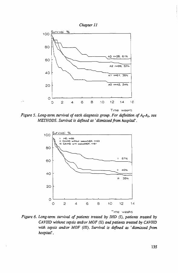
Chapter 11
100 Survtval %
80
A3 n=28. 61%
60
A2 n=99. 52%
40 A1 n=61. 38%
20 AO n=42, 24%
oL-~--~--~~--~--~--~--
o 2 4 6 8 10 12 14 16
Ttme weev.-s
Figure 5. Long-term survival of each diagnosis group. For definition of Ao-A3, see METHODS. Survival is defined as 'dismissed from hospital'.
100 Survival %
80
60
40
20
1: l!-0. =86 11: CAVI-0 wottlQut seosoS/MOI=. "~3 Ill: CAVI-0 w•th sep:;os/MOI=, =81
I 57%
II 40%
Ill 35%
oL---~~--~--~--~--~~
0 2 4 6 8 10 12 14
Ttme weeks
Figure 6. Long-term survival of patients treated by IHD (1), patients treated by CAVHD without sepsis and! or MOF (II) and patients treated by CAVHD with sepsis and/or MOF (III). Survival is defined as 'dismissed from hospital'.
135

Outcome
DISCUSSION
It is generally agreed that in patients with acute renal failure mortality is caused by the underlying disease. This is borne out by a study by Mentzer et al, showing that a mortality rate of only 6% in 236 patients with post-operative acute tubnlar necrosis of a transplanted kidney [17]. Although mortality is a function of the underlying disease, the choice of renal replacement therapy may pethaps influence the outcome. In several Studies continuous dialysis tecbniques are recommended for the treannent of critically ill patients. However, no randomized or severity matched prospective study has been realized yet, to evaluate the superiority of either the interminent or
the continuous mode of dialysis. Also this study cannot answer this question. Therefore, the first three p-values
in Table 2 can only be used to value the bias in the choice of a specific dialysis
technique. Indeed, from Table 2 and Figure 3 and 5, it is clear that patients treated by IHD have a significantly higher recovery and survival rate than patients treated by CA VHD. This difference is likely to be due to the large group of patients with sepsis/MOF, treated by CA VHD. However, a statistically difference between recovery or survival rateS of patients treated by CA VHD with or without sepsis/MOF conld not be detennined. OtheiS found that presence of sepsis was related to survival
[8, 14, 18], but Lien et a! demonstrated that !he presence of sepsis was not significantly different between survivors and non-survivoiS [16]. That we found no difference in outcome of our patients with or without sepsis/MOF may be explained by the critical illness of all patients treated by CA VHD, which accounts for high mortality rates, even in absence of sepsis or MOE This is confirmed by the statistically higher survival rate of patients treated by IHD compared to patients
without sepsis/MOF, who are treated by CA VHD. Also, as 'late hospital death' occurred more frequently in patients treated by CA VHD, it can be concluded that these patients were more severely ill than patients treated by IHD. Fur!hennore, patients from group Ao were ptedominantly (88%) treated by CA VHD, indicating that CA VHD was used primarily for patients with poor condition. This is in accordance with the approach of our department to treat these patients preferably by CAVHD.
In some studies it is reponed that mortality increases with age [7, 14, 19], but otheiS have not [20, 21]. Our resnlts did not show any relation of monality rate with age.
This retrospective study does not answer the question whether the introduction of CA VHD has improved the prognosis of severely patients with acute renal failure. Evidence suggesting that prognosis in severely ill patients can be improved by
136

Chapter 11
CA VHD, can be deduced from the observation that our patients with hemodynamic shock (A,), who were predominantly treated by CA VHD, showed a survival rate of 24%. Lien et a! [16] described no survivors in patients with hypotension for more than one week, who were treated by IHD. In conclusion, from 230 intensive care
patients with acute renal failure, a recovery rate of renal function of 60% and a patient survival rate of 44% was obtained. Compating these values with literature data, it can be concluded that these results are quite satisfactory.
137

Outcome
REFERENCES
1. Cameron JS. Acute renal failure in the intensive care today. Intensive Care Medicine 1986; 12: 64-70.
2. Stott RB, Cameron JS, Ogg CS, Bewick M. Why the persistently high monality in acute renal failure? Lancet 1972; 8: 75-78.
3. Butkus DE. Persistent high monality in acute renal failure. Are we asking the right questions? Archives of Internal Medicine 1983: 143: 209-212.
4. Kumon K, Tanaka K, Hirata T, Naito Y, Fujita T. Organ failutes due to
low cardiac output syntlrome following open heart smgery. Japanese Circulation Joutnal 1986; 50: 329-335.
5. Gillespie DJ, Marsh HMM, Divettie MB, Meadows JA. Clinical outcome of respiratory failure in patients requiring prolonged (>24 hours)
mechanical ventilation. Chest 1990; 3: 364-369. 6. Kratnan s. Khan F, Patel S, Seriff N. Renal failure in the respiratory
intensive care uoit. Critical Care Medicine 1979; 7: 263-266. 7. Spiegel DM, Ullian ME, Zerbe GO, Berl T. Detenninants of SutVival and
recovery in acute renal failure patients dialyzed in intensive-care uoits. Am J Nephrol 1991; 11: 44-47.
8. Corwin HL, Teplick RS, Schreiber MJ, Fang LST, Bonventre N, Coggins CH. Prediction of outcome in acute renal failure Am J Nephrol 1987; 7: 8-12.
9. Bartlett RH, Mault JR, Dechen RE, Palmer J, Swanz RD, Pon FK.
Contiouous arteriovenous hemofiltration: Improved SutVival in surgical acute renal failure? Surgery 1986; 100: 400-408.
10. Bartlett RH, Bosch J, Geronemus R, Paganini E, Ronco C, Swanz R. Contiouous arteriovenous hemofiltration for acute renal failure. Trans Am Soc Attif Intern Organs 1988; 34: 67-77.
11. Weiss L, Danielson BG, Wlkstr~m B, Hedstrand U, Wahlberg J. Contiouous arteriovenous hemofiltration in the treatment of 100 critically ill patients with acute renal failure: Repon on clinical outcome and nutritional aspects. Clinical Nephrology 1989; 4: 184-189.
12. McDonald BR, Mehta RL. Decreased monality in patients with acute renal failute undergoing contiouous arteriovenous hemodialysis Contr Nephro1 1991; 93: 51-56.
13. Kierdorf H. Contiouous versus intermittent treatment: Clinical results in acute renal failure. Contr Nephrol 1991; 93: 1-12.
138

Chapter 11
14. Routh GS, Briggs JD, Mone JG, Ledingham MeA. Survival from acute renal failure with and without multiple organ failu.11,. Postgraduate Medical Journal 1980; 56: 244-247.
15. Lohr JW, McFarlane MJ, Grantham JJ. A clinical index to predict survival in acute renal failure patients requiring dialysis. Am J Kidney Diseases 1988; 11: 254-259.
16. Lien J, Chan V. Risk factors influencing survival in acute renal failure treated by hemodialysis. Arch Intern Med 1985; 145: 2067-2069.
17. Mentzer SJ, Fryd DS, Kjellstrand CM. Why do patients with postsurgical acute tubular necrosis die? Arch. Sur. 1985; 120: 907-910.
18. Gornick CC, Kjellstrand CM Acute renal failure complicating aortic aneurysm surgery. Nephron 1983; 35: 145-157.
19. Knauss WA, Draper EA, Douglas MS, Wagner P, Zimmerman JE. Prognosis in acute organ-system failure. Ann Surg 1985;202: 685-693.
20. Rasmussen HH, Pitt EA, Ibels LS, McNeil DR Prediction of outcome in acute renal failure by discriminant analysis of clinical variables. Arch Intern Med 1985; 145: 2015-2018.
21. Abreo K, Moorthy V, Osborne M. Changing patterns and outcome of acute renal failure requiring hemodialysis. Arch Intern Med 1986; 146: 1338-1341.
139


CHAPTER 12
SUMMARY
Continuous aneriovenous hemodiafiltration (CA VHD) is a dialysis modality used to treat intensive care patients. 1n this thesis, the clinical use, the impact of illter design
aod membrane aod the mechaoism of solute transpon of continuous aneriovenous hemodiafiltration (CA VHD) are described.
A mathematical model of solute trangpon by combined convection and diffusion was
developed aod applied to clinical aod laboratory data. From these data, a prediction model of clearance rates of uremic solutes and drugs was derived. Funhermore, the
impact of clearance of drugs by CA VHD on dose adjusnnents was determined. Finally, the outcome of patients with acute renal insufficiency, treated by intermittent
hemodialysis or CA VHD was described.
Chapter 1:
Chapter I gives a shon history of hemodialysis. Different mechanism of solute
transpon aod terminology are discussed. Continuous aneriovenous hemoilltration (CA VH) and continuous aneriovenous
hemodiafiltration (CA VHD) were imroduced in the Dijkzigt University hospital in
1984 aod 1985 respectively. Continuous hemodialysis is performed 24 hours a day,
like the human kidney, intermittent hemodialysis is performed just a few hours a
day. CA VHD is characterized by the use of a small surface membrane with a high
hydraulic permeability, a spontaneous blood flow obtained by the pressure difference
between an anery and a vein, a spontaoeous ultrailltrate flow and a low dialysate
flow rate of l-3 L/hr.
Chapter 2:
1n this chapter, a shon overview is given of clinical problems encountered with
intermittent macltine-hemodialysis, with emphasis on the possible consequences for critically ill patients.
The most frequent complication is dialysis-induced hypotension. Also, with
neurologically unstable patients, intermittent hemodialysis may impair the
neurological condition as a result of sudden changes in pH and osmolality.
141

Summary
Chapter 3:
In this chapter, the indications for CA VHD are described. The most imponant indication for treatment by CA VHD is the combination of renal failure and circulatory instability. Neurological instability or the need for high clearance rates in severe catabolic patientS is another indication for treatment by CA VHD. Also, after kidney transplantation i11 a patient with primary hyperoxaluria,
plasma oxalate levels can be kept low by using CA VHD. CA VHD is contraindicated if tile extracorporeal circuit interferes witil the mobilisation of the patient In tilis chapter, tile necessities are described and guidelines for application for tile
techoique are given.
Chapter4:
For capillary hemofilters, tile resistance to flow may be predicted from tile filter geometry by using Poiseuille's law, assuming laminar flow. Blood viscosity can be calculated from hematocrit, protein concentration and temperature. However, observed filter resistance to flow was two to tilree times higher tilan tile predicted
value. Comparison of actual blood viscosity witil tile calculated value showed tilat
blood viscosity was grossly underestimated. Herewitil, pan of tile discrepancy between measured and predicted values could be explained. Further discrepancies between tilese values could be explained if tile fiber diameter would be a mere 8 % lower tilan tile value given by tile manufacturer.
Chapter 5:
Existillg models of hemodialysis are not likely to be useful for the analysis of solute transpon in CA VHD, as solute transpcn inCA VHD differs from tilat in ime!I!littent hemodialysis, The most impcnant differences are:
l. Witil CA VHD, solute transpcn occurs by simultaneous convecrion and diffusion. 2. Witil CA VHD, dialysate flow rates (10-30 ml/min) are very low compared to
blood flow rate (100-250 ml/min).
142
Also, dialysate flow rates might be too low for optimal distribution of dialysate over tile dialysate compartment

Chapter 12
3. CA VHD is a continuous technique. With prolonged use of the filter, deterioration of the membrane is likely not only to decrease the rate of nltrafiltration but also to impair diffusive mmspon.
Therefore, a new mathematical model of combined convection and diffusion in CA VHD was developed. Three important assumptions were made: 1. A homogeneously distribution of blood and dialysate over their respective
compartmems. 2. A constant membrane permeability for nltrafiltration and for diffusion over the
eutire membrane length. 3. No variations of solute concentration in the radial direction were considered, i.e.
calcnlations are based on local "mixing cup" concentrations.
Using this model, a numerical as well as an analytical solution of the mass transfer coefficient of diffusion (K,) conld be given.
Chapter 6:
In this chapter, the assumption of mixing cup concentrations in the model was validated. This assumption is valid when compartments are well stirred, a condition which can be approached if flows are turbulent. A radial concentration gradient can be influenced by axial flow rates. Blood flow rate had no measurable influence on the calcnlated value ofK.. Dialysate flow rates (Q,), however, did have an influence on K.· The calcnlated value of K. increased with increasing dialysate flow rates. This may be explained by the existence of an unstirred layer in the dialysate compartment, or, at low dialysate flow rates, the distribution of dialysate over the compartment is inadequate, resulting in a partial use of the membrane surface area. Also, K. proved to be dependent on the hydraulic permeability index of the membrane (MI). The relationship of K. with Q. and MI was quantified by a mathematical expression.
Chapter 7:
In this chapter the applicability of the prediction formulas of the mass transfer coefficient of diffusion (Kd) of creatinine, as derived in chapter 6, was eXatnined. It was concluded that 73% of the variability of Kd and 81% of the variability of the clearance rate of creatinine can be explained by variations in Qd and MI. The value
143

Summary
of plasma water flow rate at the filter inlet (Q,.i) is needed to predict clearance rate.
This value is, like the value of MI. not readily available in the clinical setting. Therefore, nomograms were constructed to read esthnated values of Qwi from the mean arterial pressure (MAP) and esthnated values of MI from the MAP and the ultrafiltration flow rate.
Chapter 8:
In this chapter, we applied the model to the transport of drugs. Sieving coefficients of the drugs and the relation of K, and molecular weight of the drugs were examined for cefuroxime, cefotaxime, ceftazidime, imipenem, cilastatin, ciprofloxacin, tobramycin and vancomycin. Two important results were obtained. First, it was demonstrated that the sieving coefficient of the drug may be equated to its non-protein bound fraction, which may be detennined in the laboratory. Second, we found a fixed relationship berween the ratio of K, of drugs to that of creatinine and the molecular weight of the drug.
Chapter 9:
In this chapter, the feasibility of predicting clearance rates of drugs and consequences for dose adaptations were considered. The use of the prediction formula of Kd of creatinine, together with the ratio of Kd of drugs to that of creatinine resulted in a fairly accurate esthnation of the clearance rate of drugs. This esthnation is accurate enough to be used in the clinic.
For each drug, dose adaptations were calculated based on average operational conditions. For anuric patients treated by CA VHD, dose adaptations were needed for cefuroxime, ceftazidime, tobramycin and vancomycin. Failure to recognize this may easily lead to suboptimal drug levels and therapeutic failure. For cefotaxime, imipenem and ciprofloxacin this was not needed. Therefore, the dosage regimens nf cefotaxim, imipenem and ciprofloxacin used for anuric patients, can also be used for anuric patients treated by CA VHD.
144

Chapter 12
Chapter 10:
TienamR is the combination of imipenem and cilastatin. When using this combination in patientS with renal failure, one should take imo account the more prolonged half life of cilastalin in comparison with that of imipenem. In the anuric state, even after dose adjusnnent, which is based on desired plasma levels of im.ipenem, the use of the fixed dose combination will lead to an overdosage of cilastatin. However, after institution of CA VHD, clearance rate of cilastalin increases four fold and serious accumulation of cilastalin is thereby avoided.
Chapter 11:
We studied the clinical outcome of 236 critically ill patients with acute renal insufficiency, who received dialysis treatment either by intermittent hemodialysis or
CA VHD. In general, mortality rates of these patients are very high. In our patients, recovery rate of the kidney was 60% and survival rate of the patiem was 44%. Survival rate depended on the clinical setting in which acute renal failure was acquired, or the clinical condition of the patient at start of the dialysis treatment Death after recovery of renal function occurred more frequently in the group of patients treated by CA VHD. This suggests, that patients treated by CA VHD were more severely ill than patients, who could be treated by intermittent techniques. Patients with umractable shock at start of dialysis treatment were predominantly treated by CA VHD and showed a survival rate of 24%. Before the intrOduction of CA VHD, dialysis could hardly be performed in such patients. Therefore, from this
observation it may be deduced that prognosis of these severely patients was improved by the intrOduction of CA VHD.
145

CHAPTER 13
SAMENV ATIING
Continue anerioveneuze hemodiafiltratie (CA VHD) is een dialyse teclmiek die gebruilct wordt voor de behandeling van patiemen met nierinsufficientie die verpleegd worden op een intensive care afdeling. In dit proefschrift is het klinisch gebruik, de invloed van de kunstnier op het slagen van de behandeling en het mechanisme van massatransport bij CA VHD beschreven.
Een wiskundig model van massatranspon bij het bestaan van een gelijktijdig convectief en diffusief transport werd onlwikkeld en meetresultaten verkregen in de kliniek en hetlaboratorium werden hiennee geanalyseerd. Gebaseerd op deze analyse kon een model onlwikkeld worden ten behoeve van de voorspelling van de klaring van uremische stoffen en geneesaniddelen. Tevens werd de noodzaak. tot aanpassen
van de dosis van diverse geneesmiddelen als gevolg van de klaring door CA VHD bestudeerd. Ten slotre zijn het herstel van nierfunctie en monaliteit beschreven van een groep patienten met acute nierinsufficientie, die behandeld werden met intennitterende hemodialyse of CA VHD.
Hoojdstuk 1:
In dit hoofdstuk wordt in het kart de historie van de hemodialyse beschreven. Oak worden de verschillende mechanismen van massatransport en de bijbehOrende tenninologie behandeld.
Continue anerioveneuze hemofiltratie (CA VH) en continue anerioveneuze hemodiafiltratie (CA VHD) werden respectievelijk in 1984 en 1985 gelntroduceerd
in het Academisch ziekenhuis Dijkzigt te Rotterdam. Continue dialyse wordt, overeenkomstig de menselijke nier, 24 uur per dag uitgevoerd; intennitterende
dialyse daarentegen wordt enkele uren per dag uitgevoerd. CA VHD wordt gekarakteriseerd door het gebruik van een membraan met een klein oppervlak en een hoge hydraulische permeabiliteit, een spontaan op gang gebrachte bloed stroom middels het dtukverschil tussen een anerie en een vene, een spontane ultrafiltratie stroom en een !age dialysaat stroomsnelheid van 1-3 L/uur.
146

Chapter 13
Hoofdstuk 2:
In dit hoofdstuk wordt een kart overzicht gegeven van klinische problemen zoals die kunnen ontstaan tijdens behandeling met intennitterende hemodialyse. Hierbij is de nadruk gelegd op de mogelijke consequenties voor patienten die verpleegd worden op een intenSive care afdeling.
De meest voorkomende complicatie tijdens dialyse is hypotensie. Ook kan, door te plotselinge veranderingen in de pH en de osmolariteit van het bloed, de neurologische toestand van een neurologisch reeds instabiele patiem verslechteren.
H oofdstuk 3:
In dit hoofdstuk komen de indicaties voor de behandeling met CA VHD aan bod. De belangrijkste indicatie voor behandeling met CA VHD, is de combinatie van nierfalen
en circulatoire instabiliteit. Overige indicaties zijn een neurologische instabiliteit of de behoefte aan hoge klaringssnelheden bij ernstig katabole patienten. Ook kan met behulp van behandeling met CA VHD de plasma spiegel van oxalaat van een patient met primaire hyperoxalurie voldoende laag gehouden worden na een
niertransplantatie. CA VHD is gecontraindiceerd als het extracorporele circuit interfereert met de mobilisatie van de patient. Verder worden in dit hoofdstuk de benodigdbeden en een leidraad voor het gebruik voor CA VHD beschreven.
Hoofdstuk 4:
Als men aanneemt dat er sprake is van een laminaire bloedstroom, kan de stromingsweerstand van capillaire hemofilters voorspeld worden met behulp van de wet van Poiseuille. De viscositeit van bloed kan berekend worden uit de waarde van de hematocriet, de eiwit concentratie en de temperaruur. Echter, de waarde van de
geobserveerde strcmingsweerstand van het filter was twee tot drie keer zo hoog dan de door ons voorspelde waarde. Vergelijking van de waarde van de gemeten
viscositeit van bleed met de berekende waarde liet echter zien dat de gebruikte berekening van de viscositeit in een te !age waarde resulteerde. Een dee! van de discrepantie tussen de geobserveerde en berekende stromingsweerstand kon hiennee verklaard worden. De overblijvende discrepantie russen de berekende en gemeten waarde van de sttomingsweerstand kon worden verklaard door de i.'l.terne diameter
147

Samenvatting
van de capillairen van bet filter 8% lager aan te nemen dan door de fabrikant wordt
opgegeveo.
Hoofdstuk 5:
Reeds bestaande modellen van bet massarransport tijdens hemodialyse kunnen niet
gebnrikt worden voor de analyse van het massatnmsport tijdens CA VHD. Redenen biervoor zijn dat het tnmsport tijdens CA VHD verscbilt met bet transport tijdens hemodialyse. De belangrijkste verschillen zijn:
1. Massatransport bij CA VHD vindt plaats middels gelijktijdige convectie en
diffusie. 2. Stroomsnelheden van dialysaat (10-30 ml/min) bij CA VHD zijn erg laag
vergeleken met StroOmsnelbeden van bloed (100-250 ml/min).
Ook zou de Stroomsnelheid van bet dialysaat te laag kunnen zijn om een
optimale distribu1ie van het dialysaat in bet dialysaat-compardment te
bewerlcstelligen.
3. CA VHD is een con1ioue techniek. Door bet langdurig gebruik van eenzelfde membraan, ligt het niet alleen voor de band dat de ultrafiltratie stroomsnelheid afneemt in de 1ijd, maar ook dat het cliffusieve tnmsport zou kunnen afnemen in
de tijd.
V anwege deze verschillen werd een nieuw wiskundig model, toepasbaar op de
situatie bij CA VHD, van gecombineerde convectie en cliffusie ontwikkeld. Hierbij zijn drie belangri jke aannamen gedaan:
1. De distributie van bloed en dialysaat in de respectieve compardmenten is bomogeen.
2. De permeabiliteit voor ultrafiltratie en diffusie is over de gebele lengte van de
membraan constant.
3. Variaties in concentraties in de radiale ricbting werden niet in bescbouwing
genomen, bierdoor werden de berekeningen dus gebaseerd op lokale "mixing
cup" concentraties.
Met dit model kon een numerieke en een analytische afieiding van de massatransport
coefficiem voor diffusie (K,) gegeven worden.
148

Chapter 13
Hoofdstuk 6:
In dit hoofdstuk werd de aanname 'de aanwezigheid van "mixing cup" concenttaties' gevalideerd. Deze aanname is valide, als de inhoud van de compartimenten homogeen zijn, een conditie die benaderd kan worden als de stromen twbulent zijn.
Een radiale concentratie gradient kan beiilvloed worden door axiale stroomsnelheden. De stroomsnelheid van bloed had geen meetbare invloed op de waarde van de K,. De stroomsnelheid van dialysaat (Q,) had echter wei een invloed op de waarde van de K,. Dit kan verldaard worden door het bestaan van een grenslaag in het dialysaatcompartiment, of, bij lage dialysaat stroomsnelheden, door een inadequate distributie van het dialysaat over het gehele compartiment wat resulteen in het
gebruik van slechts een gedeelte van het membraanoppervlak. Oak bleek de K, afhankelijk te zijn van de hydtaulische permeabiliteit index van de membraan (MI). De vastgestelde relatie tussen de K, met de Q. en MI werd uitgedrukt in een aantal formoles.
Hoojdstuk 7:
In dit hoofdstuk werd de bruikbaarlJ.eid van de formules, zoals afgeleid in Hoofdstuk 6, getoetst voor het voorspellen van de massatransport coefficient voor diffusie van
creatinine. Het bleek dat voor creatinine 73% van de variabiliteit in de K, en 81% van de variabiliteit in de klaring verldaard kon worden door variaties in de Q. en MI. Om de klaringssnelheid te voorspellen moet de waarde van de stroomsnelheid van plasma water (Q..,) bekend zijn. Deze waarde is, evenals de waarde van de MI, in de
klinische situatie niet direct voorhanden. Daarom werden nomogrammen geconstrueerd, waarin waarden van Qwi afgelezen kunnen worden met gebruik van de waarde van de gentiddelde arteriele druk en waarden van MI afgelezen kunnen worden met gebrnik van de waarde van de gentiddelde arterie1e druk en de stroomsnelheid van ultrafiltratie.
Hoofdstuk 8:
In dit hoofdstuk werd het transportmodel toegepast op transport van geneesntiddelen, i.e. antibiotica De "sieving" coefficienten van de geneesmiddelen en de relatie tussen de K, en het molekuul gewicht werden onderzocht voor cefuroxim, cefotaxim, ceftazidim, intipenem, cilastatine, ciprofloxacine, tobramycine en vancomycine.
149

Samenvatting
Hiermee werden twee belangrijke resultaten verkregen. Ten eerste, de "sieving" coefficient mocht gelijk gesteld worden aan de vrije fractie van het antibioticum, welke laatste gemeten kan worden in het laboratorium. Ten tweede, er bleek een vaste relatie te bestaan russen de ratio van de :£<. van de antibiotica t.o.v de K, van creatinine met het molekuul gewicht van het antibiotica
H oojdstuk 9:
In dit hoofdstuk werd de mogeli jkheid onderzocht of de klaringssnelheid van antibiotica door CA VHD voldoende betrouwbaar te voorspellen was. Het gebruik van
de voorspellingsformule van de K. van creatinine tezamen met de ratio van de :£<. van het antibioticum t.o.v. de Kd van creatinine, leverde een zodanige voorspelling
van de klaringssnelheid van deze antibiotica op, dat deze klinisch geed bruikbaar is. Tevens werd onderzocht of deze klaring consequenties heeft, zodanig dat het
doseringsschema van een patient die met CA VHD behandeld word!, veranderd moet worden. Doseringsaanpassingen, gebaseerd op gemiddelde condities van de kunsmier, werden berekend voor elk gemeten antibiotica. Het bleek, dat voor patienten met anurie die behandeld werden met CA VHD, een doseringsaanpassing nodig was bij het gebruik van cefuroxim, ceftazidim, tobramycine en vancomycine. Als aanpassing van de dosering tijdens behandeling met CA VHD wordt nagelaten, kunnen te !age
antibiolicaspiegels met daarbij falen van de antibiotische lherapie zeer wei mogelijk zijn. Bij het gebruik van cefotaXim, hrlipenem of ciprofloxine werd deze aanpassing rliet noodzakelijk geacht. Bij gebruik van deze laatst genoemde geneesmiddelen kan voor een anurische patient die behandeld wordt met CA VHD, hetzelfde doseringsschema gebruikt worden als dat voor anurische patienten, die rliet gedialyseerd worden.
Hoofdstuk 10:
Het geneesmiddel TienamR is een combinatie van 500 mg imipenem en 500 mg ciJastatine. Als men dit geneesmiddel gebnlikt bij palienten met anurie, moet men rekerling houden met het feit dat de halfwaardetijd van ciJastatine in vergelijking met imipenem sterker verlengd is. Als de patient anurisch is, wordt een aangepaste dosering gebruikt, berekend op gewenste spiegels van hrlipenem. Dit leidt tot een overdosis van cilastatine. Echter, als deze patient behandeld wordt met CA VHD, zal
!50

Chapter 13
de klaringssnelheid van cilastatine vietvoudig toenemen en daardoor wordt emstige accumulatie van cilastatine venneden.
Hoofdstuk 11:
In dit hoofdstuk is het herstel van de nierfunctie en de monaliteit bestudeerd van 236 patienten met acuut nierfalen die behandeld werden op een intensive care afdeling. Deze patienten werden ofbehande1d met intennitterende dialyse of met CA VHD. In bet algemeen is de monaliteit van deze patienten erg hoog. Bij de besclrreven groep patienten herstelde de nierfunotie in 60%. Het overlevingspercentage was 44%. Het percentage van overleving was gecorreleerd met de onderliggende ziekte of met de klinische toestand van de patient bij aanvang van de dialyse behandeling. De groep patienten die met CA VHD werden behandeld hadden een hogere monaliteit na herste1 van de nierfunotie. Dit impliceen dat patienten die een behandeling ntiddels CA VHD kregen, zieker waren dan patienten die een behandeling ntiddels intermitterende hemodialyse konden ondergaan. Van de patienten, die bij de aanvang van de dialysebehandeling in een onbehande1bare hemodynantische shock verkeerden, overleefden 24%. Deze patienten werden voornamelijk gedialyseerd ntiddels CA VHD. In het verleden, voor de introduotie van CA VHD, konden deze patienten niet of nauwelijks een dialyse behal1deling verdragen. Hieruit zou men voorzichtig
de conclusie kunnen trekken dat het gebruik van CA VHD, voor deze emstig zieke patienten een verbetering van hun overlevingskansen heeft gegeven.
151


LIST OF PUBLICATIONS
1. Vincent HH, van Ittersum FJ, Ak¢mseyin E, van Duyl WA, Vos MC, Schalekamp MADH. Solute transpon in continuous arteriovenous hemodiafiltration. A new mathematical model applied to clinical data. Blood Purification 1990; 8: 149-159.
2. Vincent HH, Ak9ahuseyin E, Vos MC. van Ittersum FJ, van Duyl WA, Schalekamp MADH. Detemtinants of blood flow and ultrafiltration in continuous arteriovenous haemodiafiltration: Theoretical predictions and laboratory and clinical observations. Nephrology Dialysis Transplantation 1990; 5: 1031-1037.
3. Vincent HH, Vos MC.
The use of continuous aneriovenous hemodiafiltration in multiple organ failure patients.
Applied Cardiopulmonary Pathophysiology 1991; 4: 109-116.
4. Ak9ahuseyin E, Vincent HH, Vas MC, van Duyl WA. Continue arterioveneuze hemodiafiltratie (CA VHD). Klinische Fysica 1991; 2: 87-92.
5. Vmcent HH, Ak9ahuseyin E, Vos MC, van Duyl WA, Schalekamp MADH. Continuous arteriovenous hemodiafiltration: Filter design and blood flow rate. Contributions to Nephrology 1991; 93: 196-198.
6. Vincent HH, Vos MC, van Duyl WA. Solute transpon in hemodiafiltration: A new mathematical model to analyse dialyser performance. Contributions to Nephrology 1991; 93: 199-201.
7. Vos MC, Vincent HH.
Continuous arteriovenous hemodiafiltralion: Predicting the clearance of drugs. Contributions to Nephrology 1991; 93: 143-145.
153

8. Vos MC, Vincent HH. Continue anerioveneuze haemodiafiltratie. Een nieuwe nierfunctie veiVangende techniek. Nederlands Tijdschrift voor Geneeskunde 1992; 136: 561-565.
9. Vincent HH, Vos MC, Ak9'iliuseyin E, van Duyl WA, Schalekamp MADH. Blood flow, ultrafiltration and solute tranSpOrt rate in continuous aneriovenous haemodiafiltration: The AN -69 plate haemofilter. Nephrology Dialysis Transplantation 1992; 7: 29-34.
10. Vos MC, Vincent HH, Uzennan EPF. Qearance of Imipenem/Cilastatin in acute renal failure patients treated by continuous hemodiafiltration (CA VHD). Intensive Care Medicine 1992; 18: 282-285.
11. Golper TA, Gleason JR, Vincent HH, Vos MC. Drug removal during high efficiency and high flux hemodialysis. In: Hemodialysis: High efficiency treatment. Ed. Juan P Bosch. Churchill Livingstone. New York 1993, page 175-208.
12. Vincent HH, Vos MC, Ak9"huseyin E, Goessens WHF, van Duyl WA, Schalekamp MADH.
Drug clearance by continuous aneriovenous hemodiafiltration (CA VHD). Analysis of sieving coefficient and mass transfer coefficient of diffusion. Blood Purification 1993: in press.
13. Vincent HH, Roggekamp MC, Vos MC. Qearance of desferrioxamine and aluminoxamine by dialysis using the 1.1 m2
cellulose-rriacetate dialyser. (Submitted).
14. Vos MC, Vincent HH, Uzennan EPF, Stamkott A, Vogel M, Mouton JW.
154
Drug clearance by continuous hemodiafiltration (CA VHD). Results with the AN-69 capillary hemofilter and recommended dose adjustments. (Submitted).

15. Vos MC, Vincent HH. Acute renal failure in intensive care patients: A follow up of 236 patients treated by continuous aneriovenous hemodiafiltration or intermittent hemodialysis. (Submitted).
16. Ak9ahuseyin E, Vos MC, Vincent HH, van Duyl WA. Sensitivity analysis of a mathematical model on continuous aneriovenous hemodiafiltration (CA VHD). (In preparation).
17. Ak9ahuseyin E, Vos MC, Vincent HH, van Duyl WA. A mathematical model on solute transport in continuous aneriovenous hemodiafiltration (CA VHD): The influence of taking into account the decrease in volume flux over the length of the dialyzer. (In preparation).
All published material as mentioned above, has been used in this thesis with pennission of the original source.
155


NAWOORD
Dit proefschrift is het resultaat van een samenwerlcingsproject tussen de afdelingen Interne Geneeskunde I en Biomedische Natuwkunde en Technologie van de Erasmus Universiteit Rotterdam. Velen hebhen hieraan hun medewerlcing verleend. Zonder
iemand tekort te willen doen, wil ik een aantal personen op deze plaats apart vermelden. Prof. Dr. M.A.D.H. Schalekamp, bedankt voor uw directe hereidwilligheid om tnijn promotor te zijn. Uw kritische commentaar op het manuscript is voor mij van grote waarde geweest Prof. Dr. W.A Weimar en Dr. R T. Krediet wil ik bedanken voor het kritisch lezen van het manuscript en hun waardevolle commentaar. Een hardwerkende en vooral gezellige onderzoeksgroep was de basis waarop dit proefschrift kon ontstaan.
Dr. H.H. Vincent, tnijn directe hegeleider, vettegenwoordigde hierin de klinische discipline, al was fysica hem niet vreemd en was de initiator van dit onderzoek. Beste Jeroen, je bent een "co-promotor'' van onschatbare waarde geweest. Je altijd goede humeur hoezeer ik ook 'een slabak van woorden' wist te creeren en je talent om alles altijd weer netjes op een rijtie te zetten heeft tnij enorm aangemoedigd. Jouw vermogen om een onderzoek op te zetten en de resultaten helder te interpreteren en formuleren, zal voor mij een voorbeeld blijven. De tomeloze energie die je op (en over) wist te brengen, heeft er mede toe geleid dat aan het eind van
onze 216-jarige samenwerking dit proefschrift voor een groot dee! voltooid kon zijn. Na deze periode wist ik je per post. flop, fax of telefoon lastig te vallen, waardoor
nog vele grote en kleine vragen tnijnerzijds beantwoord werden, waarvoortnijn dank. Ir. W.A. van Duyl vettegenwoordigde de hegeleiding van het fysische onderdeel
van dit onderzoek. Beste Wun, met weemoed denk ik terug aan de vrijdagen van 11 tot 1 en de altijd openstaande deur om fysische vraagsrukken door te nemen. Je kritische stem in de voortgang en analyse van het onderzoek, je aandacht en medeleven ook voor niet-wetenschappelijke problemen zijn onmisbaar geweest Ik wist je in je drukste lijd te overspoelen met telkens weer nieuwe hoofdstokken. maar toch vond je iedere keer weer de energie om er vee! aandacht aan te hesteden, waarvoor tnijn dank.
Ir. E. Ak9alJuseyin was ons rekenwonder. Beste Emin, heerlijk waren die discussies met jou, of het nu fysica, poliliek, trouwen, Turldje of Nederland hetrof.
Jouw vermogen om wiskundige afleidingen te creeeren is van groot belang geweest Zonder jouw "K," was dit proefschrift er dan ook niet geweest. Met veel geduld beb je me de essenlies van het transpOrt duidelijk gemaakt, waarvoor tnijn dank.
157
Hans de Bakker, bedankt voor je onmisbare technische steun lijdens de metingen en hulp bij het oplossen van vele problemen. De stagiaires Andre de Mol, Marco Poot en William Allheer dank ik voor hun aktieve en enlhousiaste uitvoetingen van de stromingsweerstand en flow metingen.
Alle medewerkers van de afdeliog Dialyse dank ik voor hun enlhousiasme en medeleven en koffie. Hun bewaarzucht voor alles wat anders in de prullenbak was verdwenen, heeft vee! in-vitro proeven mogelijk gemaakt. Fons, jij bent een speciale. De puinhoop en bet waterballet die ik op jouw kamer mocht veroorzaken, zullen me
nog lang heugen! De Intensive Care afdeliogen van het Dijkzigt ziekenhuis ben ik zeer erkentelijk
voor het geduld en de medewerking tijdens mijn regelmatig urenlange aanwezigheid. De afdeliog Mediscbe Microbiologie, in bet bijzonder Johan Mouton, Ed
Hzermans en Marius Vogel, wil ik bedanken voor de preuige samenwelking. Johan, ondanks mijn haast, mijn gebrek aan lijd en dat ik betliever gisteren dan vandaag
af wilde hebben, beb je gelukldg de moed nooit opgegeven. Een ieder verder, die ik benaderd beb voor meedenken, luisteren en
beantwoorden ofuitvoeren van alle mogelijke en onmogelijke vragen, wil ik op deze plaats hiervoor bedanken.
De paranimfen Jolien Tuyl en Gerda Verpoorte ben ik nog vee! verscbuldigd voor de hulp bij het regelen van alles wat er bij een promotie komt kijken. Bedankt!
Mijn huidige werkgever, de Maatscbap Artsen-microbioloog Brabant, wil ik op deze plaats bedanken voor de gelegenheid die zij mij bebben geboden om dit werk te voltooien.
Hans, allebei in het "promotie-schuitje" en toch hebben we bet gezellig weten te houden! A an mijn ouders, die mi j geslimuleerd hebben om te studeren en vooral verder te gaan dan dat alleen, wil ik dit proefschrift opdragen.
158

CURRICULUM VITAE
The author was born on the 17th of February 1961 in Bergen op ZOom, the Netherlands. She obtained the diploma 'Atheneum W at the 'Chrlstelijk Lyceum' in
Gouda. In 1981 she started the medical study at the Erasmus University Medical School in Rotterdam, where she graduated 'cum laude' in September 1988. From September 1988 !ill September 1989, she worked as a medical officer in the field of social security at the 'Gemeenschappelijk Administratie Kantoor (GAK) '. From September 1989 !ill March 1992, she was a research fellow, supervised by Dr. H.H. Vincent, nephrologiSt. Internal Medicine 1, University Hospital Dijkzigt (head of the department Prof.Dr. M.A.D.H. Schalekamp). This thesis was prepared at the depamnent of Internal Medicine 1 and at the department of Biomedical Fysics and Technology of the Erasmus University Rotterdam (Dr. W.A. van Duyl). In March 1992, she commenced a specialist training in Medical Microbiology at the
St. Elizabeth Hospital in T!lburg (Dr. M.F. Peeters).
159