Embed Size (px)
Citation preview

1
http://www.sci.cat
V Congrés Societat Catalana d’Immunologia
Programa Final
Barcelona, 17 i 18 de novembre 2011
“M e c a n i s m e s I m m u n i t a r i s M e c a n i s m e s I m m u n i t a r i s
B à s i c s d e l ’ A l · l è r g i a : B à s i c s d e l ’ A l · l è r g i a :
N oves pe rspec t i vesN oves pe rspec t i ves”

2 http://www.sci.cat
Comité organitzador -Junta SCI-:
President: Manel Juan Secretària: Mercè Martí Tresorera: Silvia Vidal Vicepresident: Jorge Lloberas Vocal 1: Francesc Borràs Vocal 2: Daniel Benítez Vocal 3: Margarita Bofill Vocal 4: Aura Muntasell
Col·laboradors
Secretaria Tècnica Congrés: Eva Palacios.
Col·laboradors Col·laboradors d’OR:d’OR:
Col·laboradors de Bronze:Col·laboradors de Bronze:
Col·laborador Col·laborador de Plata:de Plata:
Patrocinador dels premis a les millors comunicacions oral i pòster.
Patrocinador directe de la conferència del Dr. Sampson

3
http://www.sci.cat
Dijous, 17 de novembre
14:40
INAUGURACIÓ DEL CONGRÉS Dr. Manel Juan / Dr. Antonio Valero Presidents SCI i SCAIC. Hospital Clínic / CDB - IDIBAPS - UB, Barcelona.
Inauguració del Congrés
14:45-16:15
TAULA DE DEBAT: Modera i presenta: Dr. Moisés Labrador Servei d’Al·lergia. Hospital de la Vall d’Hebron. Barcelona. Dra. Mª Luisa Sanz Servicio de Alergia, Clinica Universitaria de Navarra. Pamplona. " TAB i al·lèrgia a fàrmacs “ Dra. Mª Carmen Vennera Unitat d'Al·lèrgia. Servei de Pneumologia. Hospital Clínic, Barcelona. "Tractaments anti-IgE "
Taula Rodona:
““Del diagnòstic Del diagnòstic al tractament”al tractament”
16:15 – 17:05
CComunicacions Orals I:omunicacions Orals I: IMMUNOPATOLOGIA I Modera: Dra. Eva Martínez-Cáceres
Llistat de comunicacions i ordre Pàgina 4
17:05- 17:30
Pausa café – VISITA general dels pòsters
17:30 – 18:20
Comunicacions Orals II:Comunicacions Orals II: IMMUNITAT INNATA Modera: Dr. Pablo Engel
Llistat de comunicacions i ordre Pàgina 4
18:20 – 19:15
Dr. Josep Mª Antó CREAL (Centre de Recerca en Epidemiologia Ambiental) , Barcelona. "L’epidèmia de les malalties "L’epidèmia de les malalties al·lèrgiques: un punt de vista al·lèrgiques: un punt de vista epidemiològic”. epidemiològic”. Modera: Dr. Jorge Lloberas. Universitat de Barcelona.
Conferència I
19:15 Final de la sessió
4 http://www.sci.cat
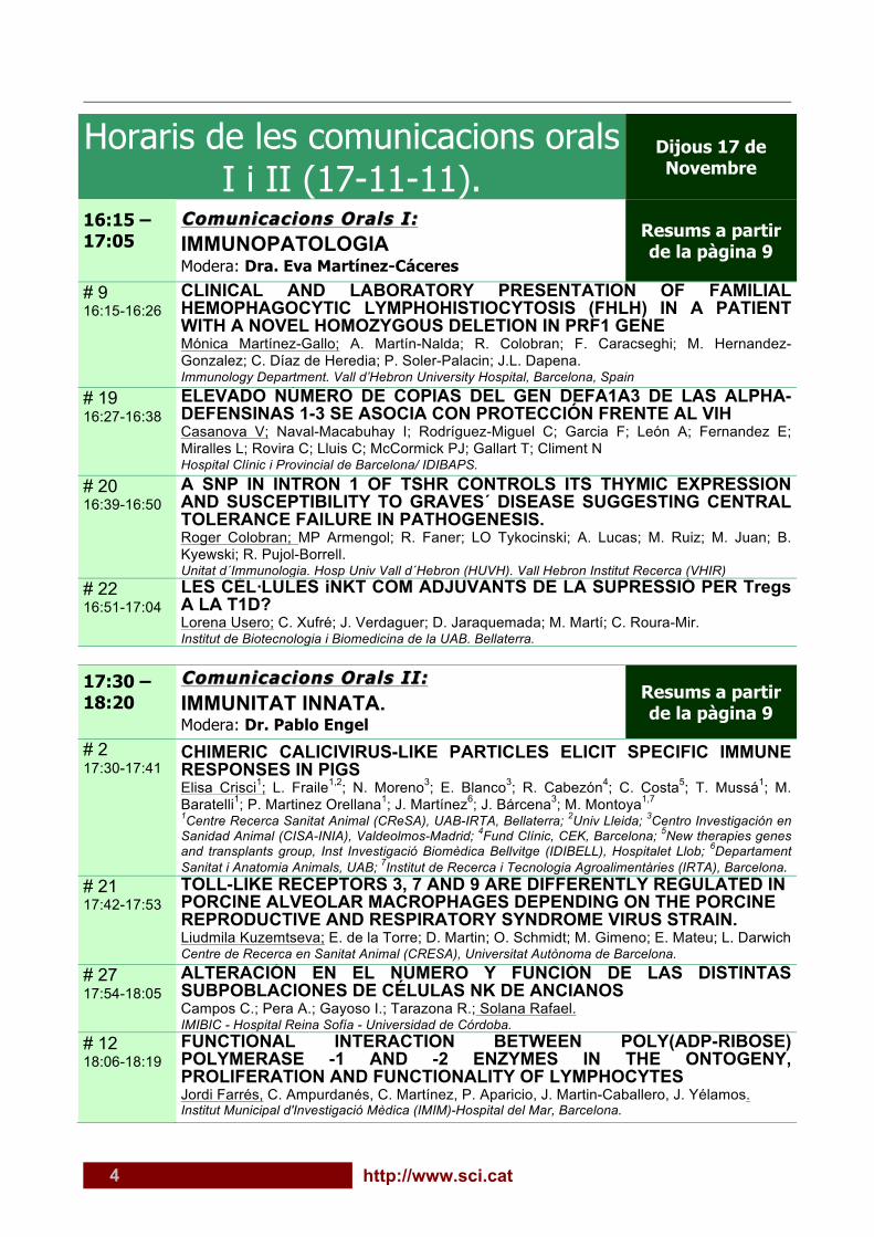
Horaris de les comunicacions orals I i II (17-11-11).
Dijous 17 de Novembre
16:15 – 17:05
Comunicacions Orals I:Comunicacions Orals I: IMMUNOPATOLOGIA Modera: Dra. Eva Martínez-Cáceres
Resums a partir de la pàgina 9
# 9 16:15-16:26
CLINICAL AND LABORATORY PRESENTATION OF FAMILIAL HEMOPHAGOCYTIC LYMPHOHISTIOCYTOSIS (FHLH) IN A PATIENT WITH A NOVEL HOMOZYGOUS DELETION IN PRF1 GENE Mónica Martínez-Gallo; A. Martín-Nalda; R. Colobran; F. Caracseghi; M. Hernandez-Gonzalez; C. Díaz de Heredia; P. Soler-Palacin; J.L. Dapena. Immunology Department. Vall d’Hebron University Hospital, Barcelona, Spain
# 19 16:27-16:38
ELEVADO NÚMERO DE COPIAS DEL GEN DEFA1A3 DE LAS ALPHA-DEFENSINAS 1-3 SE ASOCIA CON PROTECCIÓN FRENTE AL VIH Casanova V; Naval-Macabuhay I; Rodríguez-Miguel C; Garcia F; León A; Fernandez E; Miralles L; Rovira C; Lluis C; McCormick PJ; Gallart T; Climent N Hospital Clínic i Provincial de Barcelona/ IDIBAPS.
# 20 16:39-16:50
A SNP IN INTRON 1 OF TSHR CONTROLS ITS THYMIC EXPRESSION AND SUSCEPTIBILITY TO GRAVES´ DISEASE SUGGESTING CENTRAL TOLERANCE FAILURE IN PATHOGENESIS. Roger Colobran; MP Armengol; R. Faner; LO Tykocinski; A. Lucas; M. Ruiz; M. Juan; B. Kyewski; R. Pujol-Borrell. Unitat d´Immunologia. Hosp Univ Vall d´Hebron (HUVH). Vall Hebron Institut Recerca (VHIR)
# 22 16:51-17:04
LES CÈL·LULES iNKT COM ADJUVANTS DE LA SUPRESSIÓ PER Tregs A LA T1D? Lorena Usero; C. Xufré; J. Verdaguer; D. Jaraquemada; M. Martí; C. Roura-Mir. Institut de Biotecnologia i Biomedicina de la UAB. Bellaterra.
17:30 – 18:20
Comunicacions Orals II:Comunicacions Orals II: IMMUNITAT INNATA. Modera: Dr. Pablo Engel
Resums a partir de la pàgina 9
# 2 17:30-17:41
CHIMERIC CALICIVIRUS-LIKE PARTICLES ELICIT SPECIFIC IMMUNE RESPONSES IN PIGS Elisa Crisci1; L. Fraile1,2; N. Moreno3; E. Blanco3; R. Cabezón4; C. Costa5; T. Mussá1; M. Baratelli1; P. Martinez Orellana1; J. Martínez6; J. Bárcena3; M. Montoya1,7 1Centre Recerca Sanitat Animal (CReSA), UAB-IRTA, Bellaterra; 2Univ Lleida; 3Centro Investigación en Sanidad Animal (CISA-INIA), Valdeolmos-Madrid; 4Fund Clínic, CEK, Barcelona; 5New therapies genes and transplants group, Inst Investigació Biomèdica Bellvitge (IDIBELL), Hospitalet Llob; 6Departament Sanitat i Anatomia Animals, UAB; 7Institut de Recerca i Tecnologia Agroalimentàries (IRTA), Barcelona.
# 21 17:42-17:53
TOLL-LIKE RECEPTORS 3, 7 AND 9 ARE DIFFERENTLY REGULATED IN PORCINE ALVEOLAR MACROPHAGES DEPENDING ON THE PORCINE REPRODUCTIVE AND RESPIRATORY SYNDROME VIRUS STRAIN. Liudmila Kuzemtseva; E. de la Torre; D. Martin; O. Schmidt; M. Gimeno; E. Mateu; L. Darwich Centre de Recerca en Sanitat Animal (CRESA), Universitat Autònoma de Barcelona.
# 27 17:54-18:05
ALTERACIÓN EN EL NÚMERO Y FUNCIÓN DE LAS DISTINTAS SUBPOBLACIONES DE CÉLULAS NK DE ANCIANOS Campos C.; Pera A.; Gayoso I.; Tarazona R.; Solana Rafael. IMIBIC - Hospital Reina Sofía - Universidad de Córdoba.
# 12 18:06-18:19
FUNCTIONAL INTERACTION BETWEEN POLY(ADP-RIBOSE) POLYMERASE -1 AND -2 ENZYMES IN THE ONTOGENY, PROLIFERATION AND FUNCTIONALITY OF LYMPHOCYTES Jordi Farrés, C. Ampurdanés, C. Martínez, P. Aparicio, J. Martin-Caballero, J. Yélamos. Institut Municipal d'Investigació Mèdica (IMIM)-Hospital del Mar, Barcelona.
5
http://www.sci.cat
Divendres, 18 de novembre
08:30 - 09:00
Inscripció i Recollida de Documentació
09:00 - 10:00
Assemblea General Ordinària Assemblea General Ordinària SOCIETAT CATALANA d’IMMUNOLOGIA (08.45h – Primera convocatòria) MODIFICACIÓ D’ESTATUTS
Us hi esperem a tots: els socis i noUs hi esperem a tots: els socis i no -- socis!!socis!!
Assemblea Socis SCI
10:00 – 11:00
Dr. Manel Jordana Department of Pathology and Molecular Medicine, Centre for Gene Therapeutics, McMaster University, Hamilton, Ontario, Canadà.
" Asthma and food allergy: immune processes in two different mechanisms ".
Modera: Dra. Odette Viñas. Hospital Clínic.
Conferència II
11:00 - 11:30
Pausa café – VISITA 1 dels pòsters Posters N. 6, 13, 16 i 17 (Dra D Benítez i Dra M Martí)
11:30 – 12:30
Comunicacions Orals III:Comunicacions Orals III: AL·LÈRGIA -I- Modera: Dr. Antonio Valero i Dr. Manel Juan
Llistat de comunicacions i ordre Pàgina 7
12:30 – 13:30
Dra. Barbara Bohle. Institute of Pathophysiology, Center for Physiology, Pathophysiology and Immunology, Medical University of Vienna, Vienna, Austria. "Dissecting allergen-specific CD4+ T cell responses”. Modera: Dr. M. Juan. Hospital Clínic
Conferència III
13:30 – 13:35
“Congrés Virtual” SCI (www.congressci.com) Presenta: Manel Juan
Comentari
13:35 – 15:00
Dinar – VISITA 2ª de pòsters (Visita de 14:00 a 14:30) Pòsters N. 8, 26, 28 i 29 (Dra E. Martínez i Dra. MJ Amengual)
6 http://www.sci.cat
15:00 – 16:00
Dra. Barbara Ballmer-Weber. Allergy Unit, Department of Dermatology, University Hospital Zurich, Zurich, Switzerland.
”Component resolved diagnosis (single allergens) in the evaluation of food allergy”
Modera: Dr. J. Bartra. Hospital Clínic
Conferència IV
16:00 – 16:30
Comunicacions Orals IV:Comunicacions Orals IV: AL·LÈRGIA I DIVERSOS Modera: Dra. Maria José Amengual
Llistat de comunicacions i ordre Pàgina 7
16:30 – 17:00
Pausa café – VISITA 3 dels pòsters Valoració general (Dra M Bofill i Dr J Lloberas)
17:00 – 18:00
Comunicacions Orals V:Comunicacions Orals V: CÈL·LULES DENDRÍTIQUES I MACRÒFAGS Modera: Dr. Daniel Benítez
Llistat de comunicacions i ordre Pàgina 8
18:00 – 19:00
Dr. Hugh A . Sampson. Director Jaffe Food Allergy Institute. Mount Sinai New York, USA. " Immunotherapeutic Treatment of Food Allergy "
(Phadia ha patrocinat la totalitat de la despesa d’aquest ponent)
Modera: Dra. Mariona Pascal. Hospital Clínic.
Conferència V
19:00 – 19:15
Deliberacions: PREMI A LA MILLOR COMUNICACIÓ ORAL I POSTER (Patrocinat per Miltenyi)
19:15
Tancament del congrés i comiat.
CLAUSURA
7
http://www.sci.cat
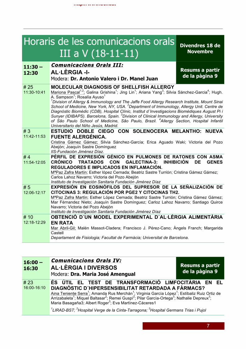
Horaris de les comunicacions orals III a V (18-11-11)
Divendres 18 de Novembre
11:30 – 12:30
Comunicacions Orals III:Comunicacions Orals III: AL·LÈRGIA -I- Modera: Dr. Antonio Valero i Dr. Manel Juan
Resums a partir de la pàgina 9
# 25 11:30-10:41
MOLECULAR DIAGNOSIS OF SHELLFISH ALLERGY Mariona Pascal1,2; Galina Grishina1; Jing Lin1; Ariana Yang3; Silvia Sánchez-Garcia4; Hugh. A. Sampson1; Rosalía Ayuso1 1Division of Allergy & Immunology and The Jaffe Food Allergy Research Institute, Mount Sinai School of Medicine, New York, NY, USA. 2Department of Immunology, Allergy Unit. Centre de Diagnòstic Biomèdic (CDB), Hospital Clínic, Institut d´Investigacions Biomèdiques August Pi i Sunyer (IDIBAPS). Barcelona, Spain. 3Division of Clinical Immunology and Allergy, University of São Paulo School of Medicine, São Paulo, Brazil. 4Allergy Section, Hospital Infantil Universitario del Niño Jesús, Madrid.
# 3 11:42-11:53
ESTUDIO DOBLE CIEGO CON SOLENOCERA MELANTHO: NUEVA FUENTE ALERGÉNICA. Cristina Gámez Gámez; Silvia Sánchez-García; Erica Aguado Waki; Victoria del Pozo Abejón; Joaquín Sastre Domínguez IIS-Fundación Jiménez Díaz.
# 4 11:54-12:05
PÉRFIL DE EXPRESIÓN GÉNICO EN PULMONES DE RATONES CON ASMA CRÓNICO TRATADOS CON GALECTINA-3: INHIBICIÓN DE GENES REGULADORES E IMPLICADOS EN INFLAMACIÓN. MªPaz Zafra Martín; Esther lópez Cernada; Beatriz Sastre Turrión; Cristina Gámez Gámez; Carlos Lahoz Navarro; Victoria del Pozo Abejón Instituto de Investigación Sanitaria Fundación Jiménez Díaz
# 5 12:06-12:17
EXPRESIÓN EN EOSINÓFILOS DEL SUPRESOR DE LA SEÑALIZACIÓN DE CITOCINAS 3: REGULACIÓN POR PGE2 Y CITOCINAS TH2. MªPaz Zafra Martín; Esther López Cernada; Beatriz Sastre Turrión; Cristina Gámez Gámez; Mar Férnandez Nieto; Joaquín Sastre Domínguez; Carloz Lahoz Navarro; Santiago Quirce Navarro; Victoria del Pozo Abejón Instituto de Investigación Sanitaria Fundación Jiménez Díaz
# 10 12:18-12:29
OBTENCIÓ D´UN MODEL EXPERIMENTAL D´AL·LÈRGIA ALIMENTÀRIA EN RATA Mar Abril-Gil; Malén Massot-Cladera; Francisco J. Pérez-Cano; Àngels Franch; Margarida Castell Departament de Fisiologia; Facultat de Farmàcia; Universitat de Barcelona.
16:00 – 16:30
Comunicacions Orals IV:Comunicacions Orals IV: AL·LÈRGIA I DIVERSOS Modera: Dra. Maria José Amengual
Resums a partir de la pàgina 9
# 23 16:00-16:10
ÉS ÚTIL EL TEST DE TRANSFORMACIÓ LIMFOCITÀRIA EN EL DIAGNÒSTIC D´HIPERSENSIBILITAT RETARDADA A FÀRMACS? Aina Teniente Serra1; Amanda Rus Merchán1; Virginia García López1; Estíbaliz Ruiz Ortiz de Arrizabaleta1; Miquel Baltasar2; Remei Guspí2; Pilar García-Ortega3; Nathalie Depreux3; Maria Basagaña3; Albert Roger3; Eva Martínez-Cáceres1 1LIRAD-BST; 2Hospital Verge de la Cinta-Tarragona; 3Hospital Germans Trias i Pujol
8 http://www.sci.cat
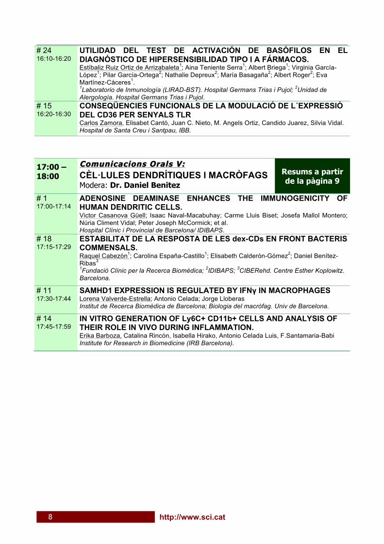
# 24 16:10-16:20
UTILIDAD DEL TEST DE ACTIVACIÓN DE BASÓFILOS EN EL DIAGNÓSTICO DE HIPERSENSIBILIDAD TIPO I A FÁRMACOS. Estíbaliz Ruiz Ortiz de Arrizabaleta1; Aina Teniente Serra1; Albert Briega1; Virginia García-López1; Pilar García-Ortega2; Nathalie Depreux2; María Basagaña2; Albert Roger2; Eva Martínez-Cáceres1. 1Laboratorio de Inmunología (LIRAD-BST). Hospital Germans Trias i Pujol; 2Unidad de Alergología. Hospital Germans Trias i Pujol.
# 15 16:20-16:30
CONSEQÜENCIES FUNCIONALS DE LA MODULACIÓ DE L´EXPRESSIÓ DEL CD36 PER SENYALS TLR Carlos Zamora, Elisabet Cantó, Juan C. Nieto, M. Angels Ortiz, Candido Juarez, Silvia Vidal. Hospital de Santa Creu i Santpau, IBB.
17:00 – 18:00
Comunicacions OraComunicacions Orals V:ls V: CÈL·LULES DENDRÍTIQUES I MACRÒFAGS Modera: Dr. Daniel Benitez
Resums a partir de la pàgina 9
# 1 17:00-17:14
ADENOSINE DEAMINASE ENHANCES THE IMMUNOGENICITY OF HUMAN DENDRITIC CELLS. Victor Casanova Güell; Isaac Naval-Macabuhay; Carme Lluis Biset; Josefa Mallol Montero; Núria Climent Vidal; Peter Joseph McCormick; et al. Hospital Clínic i Provincial de Barcelona/ IDIBAPS.
# 18 17:15-17:29
ESTABILITAT DE LA RESPOSTA DE LES dex-CDs EN FRONT BACTERIS COMMENSALS. Raquel Cabezón1; Carolina España-Castillo1; Elisabeth Calderón-Gómez2; Daniel Benítez-Ribas3 1Fundació Clínic per la Recerca Biomèdica; 2IDIBAPS; 3CIBERehd. Centre Esther Koplowitz. Barcelona.
# 11 17:30-17:44
SAMHD1 EXPRESSION IS REGULATED BY IFNγ IN MACROPHAGES Lorena Valverde-Estrella; Antonio Celada; Jorge Lloberas Institut de Recerca Biomèdica de Barcelona; Biologia del macròfag. Univ de Barcelona.
# 14 17:45-17:59
IN VITRO GENERATION OF Ly6C+ CD11b+ CELLS AND ANALYSIS OF THEIR ROLE IN VIVO DURING INFLAMMATION. Erika Barboza, Catalina Rincón, Isabella Hirako, Antonio Celada Luis, F.Santamaria-Babi Institute for Research in Biomedicine (IRB Barcelona).
9
http://www.sci.cat
Resums Orals Sessió IV divendres tarda
1. ADENOSINE DEAMINASE ENHANCES THE IMMUNOGENICITY OF HUMAN DENDRITIC CELLS. Victor Casanova Güell; Isaac Naval-Macabuhay; Carme Lluis Biset; Josefa Mallol Montero; Núria Climent Vidal; Peter Joseph McCormick; et al. Hospital Clínic i Provincial / IDIBAPS. Barcelona
Introduction: Adenosine deaminase (ADA) is an immune modulator, which by bridging A2B adenosine receptors on dendritic cells (DC) and CD26 on T cells, enhances T cell activation, proliferation and cytokine secretion contributing to an increased effector, memory and Treg differentiation (1,2). ADA is also reported to increase immune responses to inactivated-HIV loaded DC (3). Although the role of ADA on Tcells has been extensively studied, its effect on human DC remains largely unexplored, despite the pivotal role of these cells in the initiation of immune responses. Therefore, the role of ADA on human DC has been here addressed. Methods: Human peripheral blood mononuclear cells (PBMCs) of healthy and HIVinfected donors were obtained using the Ficoll method. Monocytes were differentiated to iDCs in 5 days and then incubated in absence or presence of ADA or a cytokine-PGE2 cocktail for 48h. DCs phenotype, viability and allogeneic potential were assessed by flow cytometry. In addition, up to 25 mediators were measured in cell supernatants using the Luminex technique. Results: Incubation of iDCs with ADA resulted in an increased expression of DC maturation markers CD83, CD80, CD40 and CD86, both in healthy and in HIV-infected individuals. CCR7 expression was also up-regulated while no changes were observed in HLA-DR and HLA-ABC molecules. Inhibition of ADA enzymatic activity diminished but not blocked the up-regulation of CD83 and CD80, suggesting both enzymatic and extra-enzymatic activities contribute to the observed effects. Th-1/pro-inflammatory IL-12, IL-6, TNF-alpha cytokines and IL-8, MIP1-alpha/beta and RANTES chemokines were also increased in the presence of ADA. Increased proliferation of allogeneic T CD4+ and T CD8+ was observed when co-cultured with ADA-treated DCs, revealing an improved immunogenicity. Conclusions: ADA enhances the maturation of DC rendering them more immunogenic, a desired situation in therapeutic approaches to chronic infections such as HIV.
10 http://www.sci.cat
Resums Orals Sessió II dijous tarda
2. CHIMERIC CALICIVIRUS-LIKE PARTICLES ELICIT SPECIFIC IMMUNE RESPONSES IN PIGS.
Elisa Crisci1; Lorenzo Fraile1,2; Noelia Moreno3; Esther Blanco3; Raquel Cabezón4; Cristina Costa5; Tufaria Mussá1; Massimiliano Baratelli1; Pamela Martinez Orellana1; Jorge Martínez6; Juan Bárcena3; Maria Montoya1,7* 1Centre de Recerca en Sanitat Animal (CReSA), UAB-IRTA, Campus de la Universitat Autònoma de Barcelona, 08193 Bellaterra, Spain; 2Universitat de Lleida, Lleida, Spain; 3Centro de Investigación en Sanidad Animal (CISA-INIA), Valdeolmos, 28130 Madrid, Spain; 4Fundació Clínic per la Recerca Biomèdica, Centre Esther Koplowitz, Barcelona, Spain; 5New therapies of genes and transplants group, Institut d’Investigació Biomèdica de Bellvitge (IDIBELL), L’Hospitalet de Llobregat, 08908 Barcelona, Spain; 6Departament de Sanitat i Anatomia Animals, Universitat Autònoma de Barcelona, Spain; 7Institut de Recerca i Tecnologia Agroalimentàries (IRTA), Barcelona, Spain Virus-like particles (VLPs) have received considerable attention due to their potential application in veterinary vaccines and, in particular, VLPs from rabbit haemorrhagic disease virus (RHDV) have successfully shown to be good platforms for inducing immune responses against an inserted foreign epitope in mice. The aim of this study was to assess the immunogenicity of chimeric RHDV-VLPs as vaccine vectors in pigs. For this purpose, we generate chimeric VLPs containing a well-known T epitope of 3A protein of foot-and-mouth disease virus (FMDV). Firstly, in vitro immunogenicity of RHDV-VLPs in immature porcine bone marrow-derived (poBMDC) was shown. Secondly, fifty conventional 6-7 weeks old pigs were inoculated twice in two week interval with chimeric RHDV-VLPs at different doses (20, 60, 180 µg/pig) using intranasal or intramuscular inoculation. Moreover, one of the groups was inoculated intramuscularly with adjuvant MontanideTM ISA 206 (SEPPIC). After two immunizations, specific IgG and IgA antibodies against RHDV-VLPs were induced; humoral responses were higher in the adjuvanted group compared with other groups. Interestingly, higher anti RHDV-VLP IgA responses were observed in groups inoculated intramuscularly, despite the parenteral administration of the VLPs. Two weeks after the last immunization, specific IFN-γ-secreting cells against 3A epitope and against RHDV-VLPs were detected in PBMCs by ELISPOT. The adjuvanted group exhibited the highest IFN-γ-secreting cell numbers as well as the highest lymphoproliferative FMDV-specific and RHDV-VLP-specific T cells responses when compared with other groups. Lesion score of injection site was also higher in group inoculated with adjuvant than in the other groups. Also, RHDV-VLPs were able to stimulate immature human monocyte derived DCs in vitro, paving the way for further uses of RHDV-VLPs as vaccine vectors in other systems. This is the first immunological report on the potential use of chimeric RHDV-VLPs as antigen carrier in pigs.

11
http://www.sci.cat
Resums Orals Sessió III divendres matí
3. ESTUDIO DOBLE CIEGO CON SOLENOCERA MELANTHO: NUEVA FUENTE ALERGÉNICA. Cristina Gámez Gámez; Silvia Sánchez-García; Erica Aguado Waki; Victoria del Pozo Abejón; Joaquín Sastre Domínguez. Instituto de Investigación Sanitaria Fundación Jiménez Díaz. Madrid. Introducción: La gamba es uno de los principales agentes responsables de la alergia alimentaria, presentando alta reactividad cruzada con otros crustáceos, moluscos y ácaros. Dentro de los alérgenos de la gamba, la tropomiosina tiene un papel relevante. Material y Métodos: Se reclutaron 45 pacientes en la CM con síntomas alérgicos. SPTs y cuantificación de IgE específica a gamba (Solenocera melantho), tropomiosina de gamba (Pen a 1) y Dermatophagoides pteronyssinus por CAP fueron realizados. Para confirmar el diagnóstico de alergia a gamba se hizo provocación oral doble ciego controlada con placebo (PODCP) con extracto de gamba. Mediante inmunodetección se estudió el patrón de reconocimiento de estos pacientes a gamba, nPen m 1 y rDer p 10 y se hicieron estudios de reactividad cruzada. Por último se analizaron diferentes bandas del extracto de gamba mediante espectrometría de masas. Resultados: Los pacientes se dividen en 3 grupos: POP (provocación oral positiva), PON (negativa) y control. La totalidad de los pacientes POP presenta SPT positivo frente al 61% y 11% de los pacientes PON y control (p<0.01, p<0.005), respectivamente. Los pacientes POP tienen el nivel de IgE específica para gamba, Pen a 1 y D. pteronyssinus más alto (8, 14 y 5 veces mayor que los pacientes PON (p<0.005, p<0.05, p<0.05)). Se observa un patrón diferencial de reconocimiento IgE frente a gamba, nPen m 1 y rDer p 10 en los tres grupos. Entre los pacientes PON, solo 1/3 poseen IgE capaz de unirse a la tropomiosina del extracto de gamba, nPen m 1 y rDer p 10, frente al 100% de los pacientes POP (p<0.005). Estos datos sugieren una posible reactividad cruzada entre la gamba y el acaro debida a la tropomiosina tanto en el grupo POP y PON. Los ensayos de inhibición demuestran esta reactividad cruzada en la población estudiada. Se caracteriza una nueva fuente alergénica, la gamba Solenocera melantho, identificándose mediante espectrometría de masas la tropomiosina de esta especie, al igual que 2 nuevos alérgenos, una α-actinina (~ 98kDa) y una fructosa-bifosfato aldolasa 1 (~ 46kDa). Conclusiones: Se ha demostrado la reactividad cruzada entre gamba y acaro en un estudio doble ciego controlado con placebo en pacientes con provocación oral a gamba positiva y negativa, siendo la tropomiosina el sensibilizante primario. Se ha caracterizado Solenocera melantho como una nueva fuente alergénica, identificándose su tropomiosina y una fructosa-bifosfato aldolasa 1 como alérgenos principales.
12 http://www.sci.cat
Resums Orals Sessió III divendres matí
4. PÉRFIL DE EXPRESIÓN GÉNICO EN PULMONES DE RATONES CON ASMA CRÓNICO TRATADOS CON GALECTINA-3: INHIBICIÓN DE GENES REGULADORES E IMPLICADOS EN INFLAMACIÓN. MªPaz Zafra Martín; Esther lópez Cernada; Beatriz Sastre Turrión; Cristina Gámez Gámez; Carlos Lahoz Navarro; Victoria del Pozo Abejón. Instituto de Investigación Sanitaria Fundación Jiménez Díaz. Madrid.
El asma es una enfermedad caracterizada por un predominio de células Th2 e inflamación eosinofílica. Las proteínas supresoras de la señalización de citocinas (SOCS) actúan como reguladores negativos de la señalización de citocinas. En particular, SOCS1 y SOCS3 desempeñan un importante papel en la respuesta inmune, controlando el balance entre las células Th1 y Th2. En un estudio anterior de nuestro grupo, se demostró que en ratones con asma crónico, la terapia génica con un plásmido que codifica para la Galectina-3 (Gal-3) provocaba una mejoría en la inflamación alérgica de tipo Th2. Mediante el uso de la tecnología que nos ofrecen los microarrays, en este estudio se evaluaron los cambios producidos tras el tratamiento con terapia génica de Gal-3 en un modelo murino de inflamación crónica de las vías aéreas. Los resultados fueron confirmados por RT-PCR a tiempo real, Western blot y análisis inmunohistoquímico. Se identificaron un grupo de genes implicados en diferentes vías cuya expresión se encuentra coordinada, aumentándose ó disminuyéndose, en los ratones tratados con Gal-3. Se estableció una correlación entre el tratamiento con Gal-3 y la inhibición de la expresión de SOCS1 y SOCS3 en los pulmones de los animales tratados. En conjunto, estos resultados sugieren una buena aproximación terapeútica en enfermedades alérgicas a través de la regulación negativa de SOCS1 y SOCS3 tras el tratamiento con Gal-3.
13
http://www.sci.cat
Resums Orals Sessió III divendres matí
5. EXPRESIÓN EN EOSINÓFILOS DEL SUPRESOR DE LA SEÑALIZACIÓN DE CITOCINAS 3: REGULACIÓN POR PGE2 Y CITOCINAS TH2. MªPaz Zafra Martín; Esther López Cernada; Beatriz Sastre Turrión; Cristina Gámez Gámez; Mar Férnandez Nieto; Joaquín Sastre Domínguez; Carloz Lahoz Navarro; Santiago Quirce Navarro; Victoria del Pozo Abejón Instituto de Investigación Sanitaria Fundación Jiménez Díaz
El asma y la bronquitis eosinofílica no asmática (BENA) son dos enfermedades respiratorias caracterizadas por un predominio de células Th2 e inflamación eosinofílica. Las proteínas supresoras de la señalización de citocinas (SOCS) juegan un papel importante en las respuestas alérgicas Th2-mediadas, a través del control del balance entre células Th1 y Th2, particularmente, SOCS3 y SOCS5.
El objetivo de este estudio fue el de analizar la expresión de SOCS en eosinófilos purificados de sangre periférica de pacientes con asma, BENA y controles sanos.
La expresión de SOCS en eosinófilos quedó demostrada por RT-PCR a tiempo real, Western blot, microscopía confocal y análisis inmunohistoquímico. Los eosinófilos y las células TCD4+ de los pacientes tienen una mayor expresión de SOCS3 que los de los controles sanos. Además, hemos demostrado que la prostaglandina E2 (PGE2) y las citocinas Th2 son capaces de regular positivamente la producción de SOCS3 en
eosinófilos resultando en una atenuación de la degranulación.
En conclusión, se ha demostrado la capacidad de los eosinófilos de transcribir y traducir la proteína SOCS3 y pueden contribuir a la regulación del balance Th1/Th2 a través de la producción de SOCS3.
14 http://www.sci.cat
Resums Pòster Visita I divendres matí
6. SELECTIVE LOSS OF CHEMOKINE RECEPTOR EXPRESSION ON LEUKOCYTES AFTER CELL ISOLATION. Juan C. Nieto; Elisabet Canto; Carlos Zamora; Mª Angels Ortiz; Candido Juarez; Silvia Vidal.
Institut de Recerca Hospital de la Santa Creu i Sant Pau. Barcelona.
Chemokine receptors are distinctively exposed on cells to characterize their migration pattern. However, little is known about factors that may regulate their expression. To determine the optimal conditions for an accurate analysis of chemokine receptors, we compared the expression of CCR2, CCR4, CCR5, CCR6, CXCR3 and CXCR4 on different leukocyte subsets using whole blood (WB) plus erythrocyte lysis and density gradient isolation (Ficoll). Most WB monocytes were CCR2+ (93.5±2.9%) whereas 32.8±6.0% of monocytes from Ficoll-PBMC expressed CCR2 (p<0.001). Significant reductions of CCR6 and CXCR3 on monocytes were also observed after Ficoll isolation (WB: 46.4±7.5% and 57.1±5.5%; Ficoll: 29.5±2.2% and 5.4±4.3% respectively) (p<0.01). Although comparable percentages of WB and Ficoll-PBMC monocytes expressed CCR4, CCR5 and CXCR4, Ficoll isolation significantly reduced the levels of CXCR4 (WB: MFI 5±0.4 and Ficoll: MFI 3.3±0.1) (p<0.05). Similarly to monocytes, CCR2, CXCR3 and CXCR4 were also reduced on lymphocytes. In addition, Ficoll isolation significantly reduced the percentage of CCR4 positive lymphocytes (WB: 90.2±4.5% and Ficoll: 55±4.1%) (p<0.01). The loss of expression of chemokine receptors after isolation of monocytes was not dependent on either the anticoagulant or the density gradient method. It was irreversible and could not be restored by LPS activation or in vitro macrophage differentiation. Experiments tagged with anti-CCR2 antibodies prior to density gradient isolation demonstrated that Ficoll internalized chemokine receptors. The method for cell isolation may alter not only the expression of certain chemokine receptors but also the respective functional migration assay. The final choice to analyze their expression should therefore depend on the receptor to be measured.
15
http://www.sci.cat
La comunicación n. 7 ha estat retirada pels autors.
Resums Pòster Visita II divendres migdia
8. NEUTROPENIA NEONATAL NO ALOINMUNE EN DOS GEMELOS UNIVITELINOS CON DEFICIENCIA DE CD16B. Isaac Francos Quijorna; Laura Martínez-Martínez; Nuria Pardo; Núria Nogués; Marc Orta Mascaró; Óscar de la Calle-Martín. Immunologia - Hospital Sant Pau; Pediatría - Hospital Sant Pau; BST. Barcelona.
Introducción: La causa más común de Neutropenia Neonatal es la Neutropenia Neonatal Aloinmune (NNA) debida a aloanticuerpos generados por incompatibilidades alélicas entre las moléculas de superficie de los neutrófilos, siendo la más frecuente CD16b (HNA-1). Las moléculas CD16 son receptores de baja afinidad de la región Fc de la IgG (FcγRIII) que están codificadas por 2 genes homólogos, que en la especie humana están muy próximos dentro del cromosoma 1; FCGRIIIA (CD16a) que se expresa en células NK y monocitos, y FCGRIIIB (CD16b), que lo hace exclusivamente en neutrófilos. Caso clínico: Presentamos dos varones, gemelos univitelinos, hijos de padres no consanguíneos, que desde el nacimiento presentaban una Neutropenia Neonatal moderada inusunualmente mantenida, pero sin sintomatología infecciosa. Resultados y Discusión: Ante la sospecha de una NNA por diferencias en CD16b, analizamos la expresión de esta proteína. El análisis de las moléculas CD16 en los pacientes indicó una ausencia total en los neutrófilos (CD16b), pero una expresión correcta en las células NK y monocitos (CD16a). Los padres de los pacientes presentaban expresión de CD16a y CD16b, pero mientras en el padre era del subtipo NA2, en la madre era del NA1. En ambos progenitores se podía apreciar una disminución de la expresión de CD16b en los neutrófilos. Asimismo, se descartó la presencia de anticuerpos anti-CD16b en los pacientes y en la madre. La ausencia de estos anticuerpos, junto con el mantenimiento de la neutropenia y la existencia de CD16b en la madre nos llevó a descartar la NNA y pensar en una alteración genética como posible causa. El análisis genético de los gemelos reveló una deleción complete del gen FCGRIIIB. Por lo que a FCGRIIIA respecta, detectamos un alelo nativo y otro con una sustitución a nivel del intrón 1, lo que implicaba que los niños eran heterocigotos para CD16a. En los padres encontramos tanto las secuencias para CD16a (la madre era portadora del polimorfismo del intrón 1 hallado en los pacientes) como para CD16b. Conclusiones: Los dos hermanos aquí descritos presentan una Neutropenia Neonatal no Aloinmune que se asocia a una deleción completa del gen FCGRIIIB/CD16b. El estudio del gen FCGRIIIA, que se encuentra ligado a FCGRIIIB, nos permitió concluir que los hermanos eran heterocigotos compuestos para la deleción de CD16b puesto que el haplotipo de CD16 con la delección no era el mismo en el alelo paterno que en el materno. Sería relevante relacionar la deficiencia completa de CD16b en otros posibles pacientes con neutropenias neonatales moderadas.
16 http://www.sci.cat
Resums Orals Sessió I dijous tarda
9. CLINICAL AND LABORATORY PRESENTATION OF FAMILIAL HEMOPHAGOCYTIC LYMPHOHISTIOCYTOSIS (FHLH) IN A PATIENT WITH A NOVEL HOMOZYGOUS DELETION IN PRF1 GENE
M. Martínez-Gallo; A. Martín-Nalda; R. Colobran; F. Caracseghi; M. Hernández-González; C. Díaz de Heredia; P. Soler-Palacin; J.L. Dapena.
Immunology Department. Vall d’Hebron University Hospital, Barcelona, Spain
Introduction: Familial haemophagocytic lymphohistiocytosis (FHLH) is rare congenital disease caused by a dysfunction of cytotoxic T and NK cells. FHLH results in multiorgan dysfunction and haemophagocytosis within the reticuloendothelial system characterized by pancytopenia and organomegaly. The pathologic findings in FHLH are believed to arise from prolonged tissue exposure to abnormally elevated levels of circulating proinflammatory cytokines presumably originating from activated histiocytes and T cells. Mutations of PRF1 gene account for 60% of FHLH and are associated with reduced or absent expression of perforin. Case report: We report the case of a 5-weeks-old baby girl admitted with febrile urinary tract infection, pancytopenia, hypofibrinogenemia, hyperferritinemia, splenomegaly and progressive liver failure. The patient died a few days after from uncontrolled multiorganic failure, with severe lactic acidosis. The patient was the fourth child of a consanguineous family of Moroccan origin. Previously, two baby boys from the same family had died, the first one from unknown causes when he was 1 month old, the second one died shortly after birth from multiorgan failure, with pancytopenia, hypofibrinogenemia and metabolic acidosis of unknown origin. Laboratory findings showed haemophagocytosis in bone marrow, defective cytotoxic natural killer (NK) cell activity and absent perforin expression. Sequencing analysis of PRF1 showed a novel deletion (g.4483delG) in homozygosity resulting in a premature stop codon p.Gly477del X479 . Other metabolic disorders were properly ruled out. Conclusions: We report a novel deletion (g.4483delG) in perforin gene, which abolishes the protein expression and is associated with a severe early-onset familial haemophagocytic lymphohistiocytosis presentation and severe lactic acidosis. Rapid diagnostic tools are needed to provide early treatment and avoid the unfortunate end of this case.

17
http://www.sci.cat
Resums Orals Sessió III divendres matí
10. OBTENCIÓ D´UN MODEL EXPERIMENTAL D´AL·LÈRGIA ALIMENTÀRIA EN RATA
Mar Abril-Gil; Malén Massot-Cladera; Francisco J. Pérez-Cano; Àngels Franch; Margarida Castell. Departament de Fisiologia; Facultat de Farmàcia; Universitat de Barcelona.
L’al·lèrgia a aliments constitueix una de les formes més greus d’al·lèrgia que pot arribar a causar la mort del pacient per xoc anafilàctic. El disposar de models animals adequats pot ajudar al disseny d’intervencions terapèutiques i a l’avaluació de protocols d’inducció de tolerància. L’objectiu d’aquest treball ha estat l’obtenció d’un model experimental d’al·lèrgia en rata, induït mitjançant sensibilització amb una proteïna de la dieta, que presenti les característiques pròpies de l’al·lèrgia alimentària. S’ha disposat de rates de la soca Lewis i de la soca Brown Norway d’edats compreses entre les 3 i les 8 setmanes (des del deslletament fins a l’edat adulta). S’han aplicat diferents pautes de sensibilització oral amb ovoalbúmina (OVA): administració diària i p.o. d’OVA sense adjuvant, administració p.o. d’OVA juntament amb toxina colèrica per augmentar la permeabilitat intestinal, administració diària i p.o. d’OVA després d’una immunització amb OVA per via i.p. amb alum com a adjuvant i inclusió o no de toxina de Bordetella pertussis (toxina pertussis). S’ha determinat la concentració sèrica d’anticossos anti-OVA d’isotip IgE i dels isotips IgG1, IgG2a i IgG2b. En alguns casos també s’ha avaluat la resposta intestinal enfront d’OVA i la reacció davant d’una provocació oral posterior. Els resultats mostren que, tal com s’esperava, la soca Brown Norway és més sensible a la sensibilització oral que la soca Lewis. L’administració d’OVA p.o. sense adjuvant comporta la formació d’anticossos específics només en una baixa proporció d’animals (27%) i aquests anticossos no pertanyen a la classe IgE. De forma similar, l’administració d’OVA juntament amb toxina colèrica tot i que provoca la formació d’anticossos específics dels isotips IgG1, IgG2a i IgG2b en més animals, aquesta resposta no s’acompanya de producció d’IgE específica. Per últim, la incorporació de toxina pertussis a la immunització per via i.p. en rates Brown Norway estimula la formació d’IgE anti-OVA, el que indueix a xoc anafilàctic després d’una provocació oral. En les condicions experimentals assajades, es pot concloure que l’obtenció d’un model d’al·lèrgia a una proteïna de la dieta requereix la immunització prèvia conjuntament amb toxina de B. pertussis.

18 http://www.sci.cat
Resums Orals Sessió V divendres tarda
11. SAMHD1 EXPRESSION IS REGULATED BY IFNγ IN MACROPHAGES Lorena Valverde-Estrella; Antonio Celada; Jorge Lloberas Institut de Recerca Biomèdica de Barcelona; Biologia del macròfag. Univ de Barcelona.
Aicardi-Goutières syndrome (AGS) is a genetically determined encephalopathy and is associated with a perturbation of type-I interferon metabolism. Recent molecular advances have revealed that AGS can be caused by biallelic mutations in at least 5 genes encoding TREX1, the 3 subunits of RNaseH2 and SAMHD1. Evidences show that the nucleases defective in AGS are involved in removing endogenously produced nucleic acid species, and that a failure of this removal results in activation of the immune system. (Crow et al. 2006 and 2009) SAMHD1 (SAM domain HD domain-containing protein 1) is a protein that is encoded by the samhd1 gene in humans. This gene (located on chromosome 20 in humans and chromosome 2 in mice) might play a role in the regulation of the innate immune response, even is still unknown. TREX1 is a homodimeric protein which cleaves ssDNA in 3’->5’ direction. RNaseH2 is a heterotrimeric protein which removes RNA from DNA-RNA hybrids. Loss of one of these proteins causes Aicardi-Goutières syndrome. The objective of this work is to study rnaseh2 and samhd1 cellular and tissue expression and its role in macrophages during activation. Moreover, we want to characterize samhd1 gene during the pro-inflammatory response and further study samhd1 promoter. samhd1 is also induced by pro-inflammatory stimuli whereas rnaseh2 (neither of three subunits) is not well induced. Furthermore, we have observed that using macrophages from STAT1-/- mouse, the induction of samhd1 by IFNγ; is abolished, indicating that induction of samhd1 by IFNγ is through STAT1 pathway. With this work we can conclude that samhd1 as well as trex1 are exonucleases that mutations that are associated with AGS and are induced by pro-inflammatory stimuli whereas rnaseh2 is not induced by pro-inflammatory either anti-inflammatory cytokines.

19
http://www.sci.cat
Resums Orals Sessió II dijous tarda
12. FUNCTIONAL INTERACTION BETWEEN POLY(ADP-RIBOSE) POLYMERASE -1 AND -2 ENZYMES IN THE ONTOGENY, PROLIFERATION AND FUNCTIONALITY OF LYMPHOCYTES. Jordi Farrés, Coral Ampurdanés, Carlos Martínez, Pedro Aparicio, Juan Martin-Caballero, José Yélamos. Institut Municipal d'Investigació Mèdica (IMIM) - Hospital del Mar. Barcelona.
Poly(ADP-ribose) polymerase -1 and -2 (Parp-1 and -2) belong to a family of enzymes that catalyse poly(ADP-ribosyl)ation of proteins. Parp-1 and -2 double knock-out mice are lethal at an embryonic stage (E8.5). Both enzymes are crucial to ensure genetic stability. To study the effects of the double deficiency of Parp-1 and -2 in B cells a conditional knock-out of Parp-2 through the Cre-loxP system was generated, controlled under CD19 promoter. We found that these mice present a decrease in cellularity of both its spleen and bone marrow due to a decrease in the number of B cells, this decrease is not present in either of the single knock-outs (Parp-1 -/- or Parp-2 -/-). Moreover, we found that these B cells had a defect in proliferation when stimulated through a canonical pathway. These results suggest an overlapping and redundant function of both enzymes during the generation of B cells, as well as a crucial role for their development and function.
20 http://www.sci.cat
Resums Pòster Visita I divendres matí
13. GM-CSF EXERTS A STRONGER PROTECTION OF MACROPHAGES UPON DNA DAMAGE INDUCED BY ETOPOSIDE. Catrin Youssif; Carlos Sebastian; Monica Comalada; Antonio Celada; Jorge Lloberas. Institut de Recerca Biomèdica de Barcelona; Biologia del macròfag. Univ de Barcelona.
Macrophages perform critical functions during the immune response. Macrophages proliferate in response to specific growth factors, including Macrophage Colony-Stimulating Factor (M-CSF), Granulocyte-Macrophage Colony-Stimulating Factor (GM-CSF) and Interleukin-3 (IL-3). Like all eukaryotic cells, macrophages can be exposed to different DNA damaging agents, which might alter the crucial functions of these phagocytes. GM-CSF protects bone marrow cells from the apoptosis induced by DNA damaging agents. DNA damage was induced using Etoposide (50µM), a Topoisomerase II inhibitor that causes double-strand breaks. As a cellular model, we used primary cultures of bone marrow derived macrophages and we compared their response to DNA damage in the presence of GM-CSF and M-CSF. Performing viability assays by crystal violet technique, we observed less susceptibility to apoptosis in macrophages growing in the presence of GM-CSF compared to M-CSF. Moreover, GM-CSF led to a faster recovery of the DNA damage induced by Etoposide than M-CSF, measured by the phosphorylation of H2AX. Comparing the DNA damage checkpoint proteins, we further observed that under basal conditions, the basal expression of 53BP1 (p53 binding protein 1, a protein proposed to function as a transcriptional coactivator of the p53 tumor suppressor) was higher in M-CSF growing macrophages, as well as NBS1 expression and phosphorylation. In accordance, we also observed a lower phosphorylation and faster recovery of p53, when damage was induced in macrophages growing in the presence of GM-CSF compared to M-CSF. Taken together, these data indicate, that GM-CSF-growing macrophages show a lower activation of DNA damage checkpoint proteins and a faster recovery of the DNA damage induced compared to M-CSF-growing macrophages, leading to the observed protective effect of GM-CSF on macrophages.
21
http://www.sci.cat
Resums Orals Sessió V divendres tarda
14. IN VITRO GENERATION OF Ly6C+ CD11b+ CELLS AND ANALYSIS OF THEIR ROLE IN VIVO DURING INFLAMMATION. Erika Barboza, Catalina Rincón, Isabella Hirako, Antonio Celada Luis, F.Santamaria-Babi Institute for Research in Biomedicine (IRB Barcelona). Macrophage Biology Group, Univ de Barcelona. Introduction and objective Circulating monocytes provide defense against infections and also contribute to autoimmune diseases. Two types of blood monocytes were recently identified in mice. CD11b+ CCR2low Ly6C- CX3CR1high phenotype, migrate to uninjured tissues and differentiate into resident macrophages and dendritic cells (DCs). In contrast, a distinct inflamed monocyte subset with a CD11b+ CCR2highLy6ChighCX3CR1low phenotype infiltrates infected tissue and contributes to the development of inflammation. The aim of this study is to demonstrate the migration and inflammatory capacity of Ly-6C+CD11b+ cells generated in vitro, in two in vivo models of inflammation. Methodology We have optimized two different animal models to induce local inflammation in non-sensitizied, immunocompetent Balb/c mice. In the first model, 1-fluoro-2,4-dinitrobenzene (DNFB) was applied topically on the right ear to create skin-homing conditions (DNFB dermatitis model) . In this skin model, net ear swelling was calculated by weight subtraction between right and left ear after 24h and 48h of cells injection. In the second model, an injection of Notexin was applied into the anterior tibialis (AT) on the right leg to induce myoinjuy (Notexin model). Finally 1x10⁶ and 3x10⁶ Ly6C+CD11b+ cells generated in vitro previously were injected i.v in both animal models. Gene expression was measured by a quantitative real time PCR, cell migration was seen trough IVIS images, flow cytometry studies and histology assays in both models. Results DNFB dermatitis model: Net ear swelling depended on the number of cells injected in animals treated with DNFB. DNFB and Ly6C+CD11b+ cells injected mice presented an increased weight in relation to DNFB no-treated mice. IVIS experiments with Ly6C+CD11b+ cells demonstrated the presence of fluorescence only in the inflamed ear, even 3 days after i.v. injection. Notexin model: muscle injury depended on the number of cells injected in animals treated with Notexin. Notexin-treated and Ly6C+CD11b+ cells injected mice showed muscle injury. IVIS experiments with Ly6C+CD11b+ cells demonstrated the presence of fluorescence only in the inflamed muscle, even 3 days after i.v. injection. Histology clearly indicated the presence of injected cells in the inflammed tissues. Conclusion Our data demonstrated that DNFB dermatitis and Notexin animal models are good tools to study the molecular mechanisms involved in the migration of circulating Ly-6C+CD11b+ cells in inflammation.

22 http://www.sci.cat
Resums Orals Sessió IV divendres tarda
15. CONSEQÜENCIES FUNCIONALS DE LA MODULACIÓ DE L´EXPRESSIÓ DEL CD36 PER SENYALS TLR. Carlos Zamora, Elisabet Cantó, Juan C. Nieto, M. Angels Ortiz, Cándido Juárez, Silvia Vidal. Hospital de Santa Creu i Santpau, IBB. Barcelona. Introducció: Els receptor Toll-like (TLR) i els receptors scavenger són expressats en la superfície dels monòcits per participar en funcions rellevants del sistema immune innat. Està establert que els TLR poden discriminar estructures normalment absents en els hostes sans, mentre que el CD36 és un receptor scavenger de classe B que pot coordinar la resposta contra els agonistes TLR2:TLR6, a més de reconèixer i intervenir en l’eliminació de cèl·lules apoptòtiques. Objectiu: Volem investigar com les senyals TLR2 (Pam3CSK4 i FSL1) i TLR4 (LPS) regulen l’expressió de CD36, quin és el seu destí i quines són les consequències funcionals pels monòcits. Mètodes: Les cèl·lules mononuclears perifèriques (PBMC) de donants sans van ser cultivades amb Pam3CSK4 (TLR2:TLR1), FSL1 (TLR2:TLR6) i LPS (TLR4). Després de 24 i 48h, l’expressió de CD36 va ser analitzada en les diferents poblacions de monòcits per citometria de flux i la producció de citocines va ser analitzada per ELISA. La localització del CD36 es va analitzar per microscopia de confocal. La capacitat fagocítica dels monòcits estimulats amb lligands de TLR va ser analitzada per citometria de flux després d’un co-cultiu de 4h amb neutròfils apoptòtics. Resultats: Després de l’estímul amb lligands de TLR, observem una regulació a la baixa del CD36 en les diferents poblacions de monòcits, sent el LPS el lligand amb més efecte. L’anàlisi de la cinètica mostraque el pic de màxima regulació a la baixa és a les 48h. Degut a que la unió dels lligands de TLR indueixen la producció i secreció de citocines, es va analitzar si aquestes eren responsables de la regulació observada. El TNF-α; va regular l’expressió de CD36 en les poblacions de monòcits, no observant-ne efecte amb IL-10 i IL-6. Bloquejant específicament el TNF-α; es va recuperar els nivells del CD36 en els monòcits estimulats amb lligands de TLR2, però no de TLR4. Mitjançant microscòpia de confocal es va observar que després de l’estimulació amb TLR4, el CD36 es localitzava a l’interior dels monòcits. També es va observar una disminució significativa en l’eliminació de neutròfils apoptòtics dels monòcits cultivats amb LPS. Conclusions: Els TLR poden modular molècules en la superfície dels monòcits que modifiquen la seva funcionalitat.
23
http://www.sci.cat
Resums Pòster Visita I divendres matí
16. PERIVASCULAR ADIPOCYTES AND SIGNALING THROUGH TOLL-LIKE RECEPTORS: ROLE IN THE PATHOPHYSIOLOGY OF ATHEROSCLEROSIS.
Consol Benaiges; Xavier Garcia-Moll; Eder Fredy Mateus; Christian Muñoz; Elisabeth Cantó; Elena Pérez; Rubén Leta; Esther Moga; Silvia Vidal and Cándido Juárez.
Hospital de la Santa Creu i Sant Pau, IBB. Barcelona.
INTRODUCTION Perivascular adipose tissue has emerged as a critical regulator of vascular function implicated in the pathophysiology of atherosclerosis. Virtually all arteries are surrounded by a significant amount of perivascular adipose tissue. This tissue is a very active endocrine and paracrine source of inflammatory cytokines and adipokines. Moreover, adipocytes express toll-like receptors (TLRs) to be able to respond to lipids and other self and nonself molecules activating proinflammatory pathways. We analyze the TLR/JAK-STAT/SOCS transduction pathways in adipose tissue from patients with atherosclerosis to unravel the mechanisms implicated in the pathophysiology of atherosclerosis. MATERIALS AND METHODS Perivascular and subcutaneous adipose tissue was obtained from atherosclerosis and control patients. Adipose tissue was cultured in the presence of TLR ligands to analyze the activation of TLR/JAK-STAT pathways. Expression and activation levels of STATs were measured by western blot and immunohistochemistry analysis using specific anti STATs antibodies. The secretion of adipokines (Adiponectin, Resistin and Leptin) and cytokines (IL-6, IL-10 and TNF) in supernatants were quantified by ELISA. RESULTS Western blot and immunohistochemistry analysis of STAT proteins upon stimulation of adipose tissue by TLR ligands showed an increased expression of STAT-1, STAT-3, Phospo-STAT1, Phospo-STAT3 and Phospo-STAT5. Cytokine secretion increased in those supernatants that were stimulated with TLR ligands, especially IL-6 secretion. Adipokine secretion did not show significant variations after stimulation. CONCLUSIONS We identified alterations in the TLR/JAK-STAT signaling pathways in adipose tissue that would help to explain the deleterious effects of lipids and other TLR ligands in the pathophysiology of atherosclerosis. The results will be very useful to understand the role played by adipose tissue in atherosclerosis and to design therapeutic approaches to control this inflammatory process.

24 http://www.sci.cat
Resums Pòster Visita I divendres matí
17. IN VITRO CELLULAR SENESCENCE AS A TOOL TO STUDY THE EFFECTS OF AGING IN MACROPHAGES Rodríguez-Ruiz J, Youssif C, Comalada M, Celada A and Lloberas J. Macrophage Biology Group, Institute for Research in Biomedicine (IRB Barcelona), Barcelona, Spain. Aims: Macrophages play a key role in the immune response destroying pathogens directly or releasing mediators which can activate other cells. However, macrophages from aged mice present defects in their functional activities due to the aging process that alter the immune response. Cellular senescence is characterized by a permanent cell cycle arrest and is produced after continuous replication of a cell. The accumulation of senescent cells seems to be involved in and is responsible for the induction of aging. In the present study we have considered whether the functions of long-lasting cultures of macrophages from young mice resembles to the functions found in senescent cells responsible for the aging patterns previously described. Methods and results: We used bone-marrow derived macrophages from 6-8 weeks old Balb/c mice cultured in vitro for 7 (normal culture) and 21 (senescent culture) days. Macrophages from 21 days cultures were positive for two different senescence markers: telomere shortening and high β-galactosidase activity (determined by a specific pH-dependent staining). Macrophage senescence was correlated with a reduced proliferation (measured by 3H-thymidine incorporation) in response to specific growth factors (M-CSF and GM-CSF) due to an increase of cells in the G1 phase of the cell cycle (measured by flow cytometry) and in the p21/p27 expression. In addition, an increased LPS-induced pro-inflammatory cytokine expression (TNF-α, IL-1 β) and a reduced IA/IE expression induced by IFN-g were observed in senescent macrophages. Finally, an increase in ROS production (measured by flow cytometry) and a reduction of STAT-5 phosphorylation induced by GM-CSF was also observed in senescent macrophages, which correlate with that observed in macrophages from aged mice as we previously demonstrated. Gene and protein expression analysis were evaluated by quantitative-PCR and western blot or flow cytometry, respectively. Conclusion: Our results demonstrate that cellular senescence induced in long-lasting macrophage cultures may be a good tool to evaluate the altered macrophage functions during the aging process.
25
http://www.sci.cat
Resums Orals Sessió V divendres tarda
18. ESTABILITAT DE LA RESPOSTA DE LES dex-CDs EN FRONT BACTERIS COMMENSALS. Raquel Cabezón1; Carolina España-Castillo1; Elisabeth Calderón-Gómez2; Daniel Benítez-Ribas3 1Fundació Clínic per la Recerca Biomèdica; 2IDIBAPS; 3CIBERehd. Centre Esther Koplowitz. Barcelona. La incubació de les cèl·lules dendrítiques (CDs) amb agents immunosupressors, com la dexametasona (dex), fa que aquestes adquireixin propietats tolerogèniques (dex-CDs). Les dex-CDs generen una resposta immunològica atenuada després de ser activades amb agonistes de TLRs, com per exemple LPS, o altres estímuls. Aquestes propietats tolerogèniques fan d’aquestes cèl·lules unes potencials candidates a ser utilitzades pel tractament de malalties. Malgrat s’han demostrat les propietats tolerogèniques a estímuls concrets, no s’ha estudiat el comportament de les dex-CDs en front bacteris. Els microorganismes sencers, a diferència dels estímuls individuals, contenen múltiples PAMPs i estimulen a les CDs per diferents vies, el que representa una situació més fisiològica i que comporta una major càrrega d’estímuls. L’avaluació de l’estabilitat del perfil tolerogènic d’aquestes dex-DCs quan interaccionen amb microorganismes representa un aspecte important per la seva utilització terapèutica. Per tal d’estudiar l’efecte de la dexametasona sobre les CDs en resposta als microorganismes, vam generar CDs en presència de dex, i aquestes es van incubar amb bacteris comensals gram + i gram - inactivats per shock tèrmic (Enterococcus faecalis i Eschericha coli, respectivament). Per analitzar el tipus de resposta generada, vam analitzar l’expressió de molècules co-estimuladores i marcadors maduratius en les CDs, la secreció de citocines en els sobrenedants, i la resposta al·logènica de les CDs després d’haver estat co-cultivades amb les dues soques bacterianes. Els resultats que hem obtingut corroboren el que ja haviem observat anteriorment activant les dex-CDs amb estímuls individuals com LPS o CD40L. Els bacteris, especialment E. Coli, indueixen una resposta madurativa potent en les CDs augmentant l’expressió de molècules co-estimuladores en la superfície cel·lular. Per contra, la presència de dex durant el procès de diferenciació té un efecte inhibitori en la maduració de les CDs en resposta als bacteris, com reflecteix el fenotip. A més a més, hi ha una inhibició en la secreció de citocines proinflamatòries (IL-12, TNF-a), i un augment en la secreció de IL-10. A nivell funcional, les dex-CDs estímulades amb els bacteris presenten una capacitat immunogènica disminuida. En conclusió, el tractament de les CDs amb dex condiciona la resposta d’aquestes cèl·lules en front bacteris gram+ i gram-. Les propietats tolerogèniques que aporta aquest agent immunosupressor no reverteixen quan les CDs reconeixen microorganismes que podrien activar a les CDs per múltiples vies.
26 http://www.sci.cat
Resums Orals Sessió I dijous tarda
19. ELEVADO NÚMERO DE COPIAS DEL GEN DEFA1A3 DE LAS ALPHA-DEFENSINAS 1-3 SE ASOCIA CON PROTECCIÓN FRENTE AL VIH
Casanova V; Naval-Macabuhay I; Rodríguez-Miguel C; Garcia F; León A; Fernandez E; Miralles L; Rovira C; Lluis C; McCormick PJ; Gallart T; Climent N. Hospital Clínic i Provincial de Barcelona/ IDIBAPS.
INTRODUCCIÓN: La resistencia natural a la infección por VIH es rara y varia considerablemente entre los diferentes individuos. De esta forma, encontramos individuos expuestos al virus pero que no se infectan (EAR- Expuestos de Alto Riesgo). Entre los factores de resistencia del huésped descritos se encuentran, entre otros, una mayor secreción de defensinas por parte de las células dendríticas (DCs). Las α-defensinas 1-3 son péptidos efectores de la inmunidad innata con actividad antimicrobiana de amplio espectro incluyendo virus como el VIH. Son producidas por neutrófilos y DCs, entre otros tipos celulares. El gen que las codifica (DEFA1A3) presenta variaciones en el número de copias (CNV). Así pues, postulamos que la secreción aumentada de α-defensinas 1-3 en DCs, relacionada directamente con el CNV, podría ser un mecanismo clave para la inducción de una respuesta protectora contra el VIH. MÉTODOS: Análisis mediante ELISA de las α-defensinas 1-3 en sobrenadantes de células dendríticas derivadas de monocitos (CD-DMs) a día 5, en individuos EARs y en individuos sanos. Análisis del CNV del gen DEFA1A3 en los grupos de individuos EARs (n = 29) y sanos (n = 57). Los resultados fueron analizados mediante dúplex qRT-PCR (quantitative real-time PCR). Ensayo de determinación de la infectividad por VIH en individuos sanos con CNV elevados (CNV ≥ 9) versus individuos con CNV bajos (CNV ≤ 4). Para ello, se infectaron a bajas dosis (MOI=1) CD-DMs propias de cada individuo y se co-cultivaron ex vivo con PBMCs autólogas deplecionadas de monocitos. RESULTADOS Y DISCUSIÓN: Los resultados del análisis por ELISA de las α-defensinas 1-3 en sobrenadantes de CD-DMs en individuos EARs y sanos, indicaron una mayor secreción, estadísticamente significativa (p = 0.0260), de α -defensinas 1-3 en los individuos EARs (1328.7 pg/mL) versus el grupo de individuos sanos (445 pg/mL). Estos resultados sugieren que una mayor secreción de α -defensinas 1-3 podría ser indicativa de una mayor protección frente a la infección por VIH. De forma estadísticamente significativa el grupo de individuos EARs presentan un mayor porcentaje 87,9 % de CNV ≥ 6 respecto los individuos sanos que presentan un 66,6 % (p= 0.02). Así pues, un mayor número de copias del gen DEFA1A3 en individuos EARs sería un mecanismo innato por el que los individuos EARs estarían promoviendo una mayor secreción de α -defensinas 1-3 que preservarían de la infección por el VIH. Con el objetivo de analizar in vitro este mecanismo, se realizó un ensayo en el que se comparó la infectividad por VIH en individuos sanos con CNV elevados (CNV ≥ 9) versus individuos con CNV bajos (CNV ≤ 4). Los resultados de estos experimentos mostraron una mayor protección (5 veces más) a la infección en trans por el VIH en los individuos con CNV elevados. Todo ello nos permite concluir que la característica genética de CNV elevado para el gen DEFA1A3 confiere protección frente a la infección por el VIH.
27
http://www.sci.cat
Resums Orals Sessió I dijous tarda
20. A SNP IN INTRON 1 OF TSHR CONTROLS ITS THYMIC EXPRESSION AND SUSCEPTIBILITY TO GRAVES´ DISEASE SUGGESTING CENTRAL TOLERANCE FAILURE IN PATHOGENESIS.
Roger Colobran; Maria del Pilar Armengol; Rosa Faner; Lars-Oliver Tykocinski; Anna Lucas; Marta Ruiz; Manel Juan; Bruno Kyewski; Ricardo Pujol-Borrell.
Unitat d´Immunologia. Hospital Universitari Vall d´Hebron (HUVH). Vall d´Hebron Institut de Recerca (VHIR) Graves’ disease (GD) is the paradigm of an anti-receptor autoimmune disease with agonistic auto-antibodies against the thyrotropin receptor (TSHR) being the underlying pathogenic mechanism. TSHR belongs to the category of tissue-restricted antigens (TRAs), which are expressed by medullary thymic epithelial cells (mTECs) and thereby induce central T cell tolerance. In order to understand the association between TSHR gene polymorphisms and GD we tested the hypothesis that TSHR gene variants affect susceptibility to GD by influencing levels of TSHR transcription in the thymus. The results indicate that thymic glands from normal children homozygous for the rs179247 predisposing allele of TSHR had significantly fewer TSHR mRNA transcripts than carriers of the protective allele. In addition, in heterozygous, the TSHR predisposing allele was expressed at a lower level than the protective one as demonstrated by Allele Specific Transcript Quantification. The effect of TSHR SNP rs179247 was thymus-specific and not observed in thyroid glands. An unexpected new finding was the expression of TSHR in thymocytes at RNA and protein level, suggesting a role of TSHR in thymocyte development. These results constitute first evidence for the involvement of central tolerance in the loss of tolerance to TSHR in GD and underscore the concept that variable expression levels of major target autoantigens in the thymus influence the predisposition to autoimmunity presumably by changing the threshold of tolerance.
28 http://www.sci.cat
Resums Orals Sessió II dijous tarda
21. TOLL-LIKE RECEPTORS 3, 7 AND 9 ARE DIFFERENTLY REGULATED IN PORCINE ALVEOLAR MACROPHAGES DEPENDING ON THE PORCINE REPRODUCTIVE AND RESPIRATORY SYNDROME VIRUS STRAIN.
Liudmila Kuzemtseva; Eugenia de la Torre; David Martin; Oscar Schmidt; Mariona Gimeno; Enric Mateu; Laila Darwich
Centre de Recerca en Sanitat Animal (CRESA), Universitat Autònoma de Barcelona.
Porcine reproductive and respiratory syndrome virus (PRRSV) is a positive-single-stranded RNA virus of the Arteriviridae family. PRRSV causes significant losses to the swine industry worldwide. Infection with PRRSV predisposes pigs to infection by bacterial and other viral pathogens. PRRSV has a tropism for cells of a phagocytic lineage, especially porcine alveolar macrophages (PAMs). Toll-like receptor (TLR) ligands, basically TLR3, TLR7, and TLR9, are involved in innate immune responses by triggering the production of antiviral cytokines such as type-I IFN. Our objective was to determine whether different PRRSV strains regulate the expression of these TLRs in PAMs. Cultures of PAMs obtained from 4 week-old healthy pigs were infected with two field PRRSV isolates (IL-10+/TNF-α+ inducer and IL-10-/TNF-α- non inducer strains) and an attenuated vaccine at m.o.i=0.1 and 1.0. Cells were harvested at different time-points post-infection (PI), and analyzed for the expression of TLRs and viral antigens by flow cytometry (FC) and real-time PCR (TaqMan®) respectively. Using FC, TLR3 increased in infected PAMs after 24-36h PI, being this increase more evident in the case of the IL-10+/TNF-α+ isolate. TLR9 and TLR7 were also induced by the IL-10+/TNF-α+ strain at 48h PI. The IL-10+/TNF-α+ isolate replicated at lower titers than the IL-10-/TNF-α- (106.0 versus 107.3 TCID50/ml) and apoptosis was observed in a lower proportion of cells after 48 h of incubation. Also, clear-cut differences were more evident at m.o.i=1. In conclusion, different PRRSV isolates can affect the TLRs expression in a different way and, consequently the development of innate immunity could be affected.

29
http://www.sci.cat
Resums Orals Sessió I dijous tarda
22. LES CÈL·LULES iNKT COM ADJUVANTS DE LA SUPRESSIÓ PER Tregs A LA T1D?
Lorena Usero; Cristina Xufré; Joan Verdaguer; Dolores Jaraquemada; Mercè Martí; Carme Roura-Mir. Institut de Biotecnologia i Biomedicina de la UAB. Bellaterra. La Diabetis Tipus 1 (T1D) és una malaltia autoimmunitària òrgan-específica caracteritzada per la presència de cèl·lules T autorreactives que provoquen la destrucció de les cèl·lules beta del pancreas productores de insulina. Les cèl·lules T amb funció immunoreguladora com les cèl·lules Treg amb un fenotip CD4+CD25++Foxp3+ i les cèl·lules NKT invariants, Vα24Jα18CD3, són importants en el control de l’autorreactivitat. Totes dues poblacions tenen frequències i funcions alterades en determinades malalties autoimmunitàries humanes com a la T1D. Estudis previs indicaven que aquestes dues subpoblacions cel·lulars tenien una distribució diferencial al pàncrees d’un individu amb Diabetes Tipus 1 (T1D) al debut. Les cèl·lules iNKT es concentraven majoritàriament fora dels illots pancreàtics i les Tregs eren reclutades preferentment al seu interior. Aquesta distribució es va confirmar per immunofluorescència en pàncrees de ratolins NOD però només a l’estadi prediabètic (9 setmanes) perquè a ratolins al debut de la malaltia (14 setmanes) o diabètics (20 setmanes) s’incrementava la proporció de cèl·lules iNKT a l’interior dels illots. La colocalització d’ambdós tipus cel.lulars suggeria que podien interaccionar. Partint de la hipòtesi que ambdues subpoblacions de cèl·lules T reguladores podrien actuar conjuntament en el control del procés autoimmunitari, ens vam proposar analitzar la seva interacció funcional a pàncrees de pacients amb T1D. Per això es van seleccionar cèl·lules iNKT i Tregs a partir de PBMCs d’individus sans. Les NKTs es van seleccionar segons l’expressió de CD3 i CD56 i es van expandir amb APCs CD1d+ pulsades amb αGalactosilCeramida (αGalCer) obtenint poblacions de cèl·lules iNKTs (Vα24Jα18+ CD3+) d’un 99,6% de puresa. Les cèl·lules Tregs es van seleccionar per la seva expressió de CD4 i nivells alts de CD25 i es van expandir in vitro utilitzant el Mètode d’Expansió Ràpida (REM). Amb aquest mètode es va obtenir fins a un 99% de cèl·lules Treg (CD25++CD3+CD4+Foxp3+). Amb aquestes cèl·lules expandides es va analitzar primer la capacitat supressora de les cèl·lules Treg en assajos de proliferació on es mesurava la incorporació de H3-Timidina desprès de 5 dies de cultiu usant diferents ràtios de cèl·lules Treg:Teff. Un cop comprovada la funció supressora de les Tregs, es va analitzar la capacitat de les iNKTs de modificar l’efecte supressor de les Tregs afegint un número creixent de iNKTs al mateix cultiu. Els resultats obtinguts en aquests assajos indiquen que les cèl·lules iNKT i Treg poden ser expandides in vitro mantenint el seu fenotip i la seva funcionalitat. La capacitat supressora de les cèl·lules Tregs (36% de supressió) sobre la proliferació de les cèl·lules T efectores es veu incrementada fins a un 60% desprès de la addició de cèl·lules iNKT a l’assaig. Això indica, que les cèl·lules iNKT tenen un efecte adjuvant de la supressió de la proliferació de cèl·lules T efectores mediat per les cèl·lules Treg. Aquest fet junt amb les dades de colocalització d’ambdues poblacions cel·lulars (Tregs i iNKTs) al pàncrees humà i de ratolí NOD suggereixen que aquesta interacció funcional pot tenir lloc al teixit diana de la resposta a la T1D. Estem investigant el mecanisme pel qual les cèl·lules NKT milloren la capacitat supressora de les Tregs, ja sigui per contacte cel·lular o per la secreció de factors solubles.
30 http://www.sci.cat
Resums Orals Sessió IV divendres tarda
23. ÉS ÚTIL EL TEST DE TRANSFORMACIÓ LIMFOCITÀRIA EN EL DIAGNÒSTIC D´HIPERSENSIBILITAT RETARDADA A FÀRMACS?
Aina Teniente Serra1; Amanda Rus Merchán1; Virginia García López1; Estíbaliz Ruiz Ortiz de Arrizabaleta1; Miquel Baltasar2; Remei Guspí2; Pilar García-Ortega3; Nathalie Depreux3; Maria Basagaña3; Albert Roger3; Eva Martínez-Cáceres1 1LIRAD-BST; 2Hospital Verge de la Cinta-Tarragona; 3Hospital Germans Trias i Pujol. Badalona. Introducció: El paper dels limfòcits T en les reaccions d’hipersensibilitat retardada (tipus IV) a fàrmacs està ben establert. El test de transformació limfocitària (TTL) permet mesurar in vitro la proliferació dels limfòcits T en presència de l’antigen específic. Objectiu: Valorar la utilitat clínica dels TTL realitzats en el nostre laboratori des del 2007 fins a juliol del 2011. Materials i mètodes: Des de l’any 2007 s’han realitzat 75 TTL en pacients derivats dels serveis d’al·lergologia de l’Hospital Germans Trias i Pujol i de l’Hospital Verge Cinta de Tortosa que havien patit reaccions d’hipersensibilitat retardada a fàrmacs. Les cèl·lules mononucleades obtingudes mitjançant gradient de densitat a partir de sang perifèrica van ser cultivades 6 dies a diferents concentracions del fàrmac, optimitzades prèviament realitzant test de toxicitat. En 42 casos la proliferació cel·lular es va mesurar mitjançant citometria de flux amb tinció amb el colorant vital CFSE, mentre que en els 33 TTL restants es va mesurar per incorporació de timidina tritiada. Resultats: Del global de 75 TTL, van resultar positius 19 (25%). Dels realitzats amb CFSE van resultar positius 9 (21%) i dels 33 valorats per incorporació de timidina tritiada van ser positius 10 (30%). Els fàrmacs més sol·licitats per a realitzar l’estudi han sigut els contrastos iodats (24, dels quals 7 van ser positius) seguit d’antibiòtics betalactàmics (9, 2 positius), alopurinol (6, 1 positiu), metamizol (5, 2 positius) i antiepilèptics (5, cap positiu). Cal destacar que en 10 dels pacients en què el TTL va resultar positiu, se’ls va realitzar tests epicutanis del fàrmac a estudi en el servei d’al·lergologia. D’aquests, en 7 casos (70%) les proves epicutànies havien resultat negatives. Conclusions: Tot i que el TTL presenta una baixa sensibilitat, pot ser d’utilitat en el diagnòstic de reaccions d’hipersensibilitat retardada a fàrmacs, especialment en aquells pacients en què les proves epicutànies han resultat negatives.

31
http://www.sci.cat
Resums Orals Sessió IV divendres tarda
24. UTILIDAD DEL TEST DE ACTIVACIÓN DE BASÓFILOS EN EL DIAGNÓSTICO DE HIPERSENSIBILIDAD TIPO I A FÁRMACOS. Estíbaliz Ruiz Ortiz de Arrizabaleta1; Aina Teniente Serra1; Albert Briega1; Virginia García-López1; Pilar García-Ortega2; Nathalie Depreux2; María Basagaña2; Albert Roger2; Eva Martínez-Cáceres1. 1Laboratorio de Inmunología (LIRAD-BST). Hospital Germans Trias i Pujol; 2Unidad de Alergología. Hospital Germans Trias i Pujol. Badalona. INTRODUCCIÓN: Debido a las características de las reacciones de hipersensibilidad a fármacos, las pruebas in vitro son una herramienta útil para su diagnóstico. En los casos de hipersensibilidad tipo I, no siempre existe la posibilidad de cuantificar la IgE específica y cuando es posible, ésta presenta una baja sensibilidad, por ello es interesante incluir además el test de activación de basófilos (TAB) en el diagnóstico in vitro de estas reacciones. OBJETIVO: Evaluar la utilidad del test de activación de basófilos en el diagnóstico de alergia a fármacos. MATERIAL Y MÉTODOS: Estudio retrospectivo realizado en el Hospital Germans Trias i Pujol durante el periodo 2006 – septiembre 2011. Se revisaron las historias clínicas de 59 pacientes que habían acudido al servicio de Alergología con sospecha de reacción de hipersensibilidad tipo I a diferentes fármacos. A todos ellos se les realizó el TAB (BD Biosciences). En 35 pacientes se determinó además la IgE específica (ImmunoCAP, Phadia). Las pruebas cutáneas se efectuaron sólo a pacientes con reacciones menos graves (n=31). RESULTADOS: De los 59 pacientes incluidos en el estudio, 52 fueron finalmente diagnosticados de hipersensibilidad tipo I a fármacos. La clínica que presentaron los pacientes fue de urticaria (8 casos), anafilaxia (31 casos), angioedema (8 casos) y síndrome de Kounis (1 caso) y los fármacos implicados fueron AINES (n=25), antibióticos (n=22), contrastes yodados (n=2), metabisulfito (n=1), progesterona (n=1) y clorfeniramina (n=1). La sensibilidad y especificidad del TAB en nuestra cohorte de pacientes fue de un 36% y un 100% respectivamente, con un valor predictivo positivo de un 100% y negativo de un 15%. Cabe destacar que en 18 pacientes con reacción frente a metamizol, 13 (72%) obtuvieron un resultado positivo en el TAB y en 10 de estos se realizó adicionalmente el estudio de IgE específica a metamizol que resultó negativo. CONCLUSIONES: A pesar de la baja sensibilidad del TAB, su elevada especificidad hace que un resultado positivo en el diagnóstico de pacientes con hipersensibilidad tipo I a fármacos evite la realización de pruebas cutáneas y de provocación, hecho especialmente importante en las reacciones de hipersensibilidad a metamizol que son más frecuentes y más graves.
32 http://www.sci.cat
Resums Orals Sessió III divendres matí
25. MOLECULAR DIAGNOSIS OF SHELLFISH ALLERGY Mariona Pascal1,2; Galina Grishina1; Jing Lin1; Ariana Yang3; Silvia Sánchez-Garcia4; Hugh. A. Sampson1; Rosalía Ayuso1. 1Division of Allergy & Immunology and The Jaffe Food Allergy Research Institute, Mount Sinai School of Medicine, New York, USA. 2Dep Immunology, Allergy Unit. Centre de Diagnòstic Biomèdic (CDB), Hospital Clínic, Institut d´Investigacions Biomèdiques August Pi i Sunyer (IDIBAPS). Barcelona. 3Division of Clinical Immunology and Allergy, University of São Paulo School of Medicine, São Paulo, Brazil. 4Allergy Section, Hospital Infantil Universitario del Niño Jesús, Madrid. Background: Accurate diagnosis of food allergy and appropriate treatment options depend on the verification of clinically relevant allergen specific IgE antibodies (sIgE), as well as on the identification of the responsible allergenic molecule(s). Progress in biochemistry and molecular biology has allowed identification, cloning, and recombinant production of allergenic proteins, as well as the synthesis of IgE epitope-emulating peptides of a number of allergens. Component-Resolved Diagnosis (CRD) consists of the use of defined allergen molecules (“components”, either purified natural or recombinant) to determine the individual patient’s humoral reactivity profile (mainly IgE) in order to identify the real trigger of the allergic reaction. Moreover, IgE epitope mapping with synthetic sequential peptides may provide an additional tool for allergy diagnosis and prognosis. Shellfish allergy is known to be a common disorder, frequently persisting into adulthood. Tropomyosin is considered to be responsible for most of the allergenic activity of shrimp, but other allergens have been identified and characterized in crustaceans. Diagnosis of shellfish allergy is aided by clinical history, skin prick testing, and sera specific IgE; however, and basically due to the high cross-reactivity observed, DBPCFCs are still the most reliable method to confirm the clinical relevance and to identify putative species. Aim: To define shellfish allergy profiles of allergic sensitization resulting in different clinical patterns and to describe the potential role of each allergen in cross-reactivity. Methods: Eighty-six subjects were recruited (inclusion criterion: positive shrimp-SPT and/or shrimp-sIgE) and classified whether histories of immediate allergic reactions following shrimp ingestion were reported. DBPCFC was performed to all patients (except those with anaphylaxis within the previous 6 months). Two group controls were included: subjects allergic to house-dust mite and/or cockroach with no sensitization to shrimp and no reported histories of immediate allergic reactions after ingestion, and non-atopic individuals. Ten recombinant allergenic proteins of Crustacea were purified and tested for IgE reactivity by dot blotting. IgE and IgG4 reactivity to synthetic peptides of 5 shrimp allergens was also evaluated. Results: Challenge positive subjects recognize more recombinant proteins than challenge negative. IgE recognition of SCP-alpha and/or beta and TM seems to differentiate challenge outcome. IgE binding to SCP-alpha, SCP-beta and TM was statistically significantly more frequent than in all subjects with challenge negative. No major differences between children and adults IgE recognition within each group. Dust mite and/or cockroach allergic subjects not sensitized to shrimp only recognize arginine kinase and hemocyanin. Shrimp allergic subjects show further IgE and IgG4 diversity of binding than tolerant subjects. Also for them, more peptides were observed to bind both IgE and IgG4. Several differences in binding sites for IgE and IgG4 have been identified when comparing allergic and tolerant subjects using TileMap analysis (fold change >2). Conclusions: Component resolved diagnostics with recombinant proteins, especially TM, SCP beta and alpha, is useful to differentiate clinical reactivity among subjects sensitized to shrimp. AK and He IgE antibodies, especially in the absence of sensitization to other proteins such as TM, SCPs or MLC, could be markers of shrimp tolerance. Both are allergens in other arthropods and IgE reactivity is probably due to cross-reactivity issues with Crustacea. Further IgE and IgG4 diversity of peptide recognition, as well as, binding to certain sites of shrimp allergens is also useful to differentiate clinical reactivity using microarray analysis. The incorporation of further shrimp-allergens in routine diagnosis and the use of peptide-based microarrays would improve shrimp allergy clinical characterization and minimize the need of oral challenge.
33
http://www.sci.cat
Resums Pòster Visita II divendres migdia
26. MONOCLONAL EXPANSIONS OF TCR IN A DIABETIC PANCREAS. Eva Codina-Busqueta; Carlos Martínez-Torró; Manuela Costa; Cristina Xufré; Lorena Usero; Eloi Parladé; Eduard Vico; Dolores Jaraquemada; Carme Roura-Mir and Mercè Martí Institut de Biotecnologia i Biomedicina. Universitat Autònoma de Barcelona. T1D is an autoimmune disease characterized by the selective destruction of insulin-producing cells located within the Langerhans islets. Pathology is associated with CD4+ and CD8+ T cells infiltration around the pancreatic islets. The study of the TCR repertoire is an indirect measure of the T cell diversity that is taking part of the effector and regulatory immune response inside the islets. Several investigators have described the TRBV family repertoire of the NOD mouse intrapancreatic infiltrates from but not in humans, where the limited availability of affected-pancreatic tissue has been a handicap to performe such studies. We had the opportunity to study the TRBV repertoire of intrapancreatic T cells of a diabetic pancreas at onset (case 1) that was characterized by Somoza et al (J. Immunol 1994; 153: 1360) (Codina-Busqueta et al. J. Immunology 2011; 186: 3787). Methods. TCR analysis was performed by multiplex RT-PCR analysis. PCR products were directly analyzed by spectratyping. Some of the products were further cloned and sequenced. Results. Five monoclonal expansions were identified and their CDR3 sequenced: Vb1 (CASSVSTTDTQYF), Vb7 (CASSQVAGAGTGELFF), Vb11 (CASSDPGTQETQYF), Vb17 (CATSPLGMNNEQFF) and Vb22 (CASSEAQQGYSGELFF). All these clones were also expanded in the total digested tissue sample except for the Vb11 clone that was only found at low frequency, not showing a pattern of monoclonal expansion. When further analyzing the Vb11 TRBV family, using total digest, spleen and PBMCs from the same patient, we found that the distribution of Vb11 peaks did not show a normal pattern, because only a few dominant peaks with similar area were visible, corresponding to CDR3 sizes of 10, 11, 12, 14 and 16aa, respectively. Of 34 sequenced clones, only six different sequences were identified, all at a frequency range of 9-26%, without any evidence of one dominant sequence. This was not the case for any other TRBV family, where a normal distribution of CDR3 sizes was always associated with several sequences for each CDR3 length. All Vb11 peaks present in the pancreas sample but not detectable in the islets, were also detected in the spleen of the same patient, but the monoclonal 12aa CDR3 clone identified in the islets has so far not been sequenced from any of the spleen samples analyzed. The T cells isolated from the total digest were also expanded and oligoclonal lines were generated. A large number of Vb11+ positive cell lines could be isolated, many of which shared one of the sequences found in the pancreas, but not the expanded clone from the islets. This sequence was not found in the patient’s spleen. These data suggest that in this particular case of T1D, the Vb11 family of TRBV can be relevant for the disease, showing an unusual pattern of CDR3 size distribution, with several dominant expansions, some of which appear to be directly involved in islet recognition. The mechanisms involved and the functional relevance of these cells are under study.
34 http://www.sci.cat
Resums Orals Sessió II dijous tarda
27. ALTERACIÓN EN EL NÚMERO Y FUNCIÓN DE LAS DISTINTAS SUBPOBLACIONES DE CÉLULAS NK DE ANCIANOS. Campos C.; Pera A.; Gayoso I.; Tarazona R.; Solana R. IMIBIC - Hospital Reina Sofía - Universidad de Córdoba.
El proceso de inmunosenescencia afecta tanto a la respuesta inmune innata como adquirida, y
estudios previos, incluyen a las células NK dentro de las poblaciones afectadas.
OBJETIVO: Estudiar in vitro el efecto de la edad en el fenotipo y función de las subpoblaciones de
células NK, cuantificando el contenido basal de granzimas A y B.
MATERIAL Y METODOS: El análisis fenotípico y cuantificación de las distintas subpoblaciones de
células NK se realizó por citometría de flujo. El análisis de la citotoxicidad se realizó mediante
marcaje intracellular de PBMCs para citometría de flujo.
RESULTADOS: Encontramos un descenso significativo en las células CD56brightCD16low/null de
ancianos, así como un aumento significativo en las células NK totales y CD56nullCD16bright.
También observamos un aumento significativo en el contenido de las granzimas A y B,
especialmente en las células CD56brightCD16null de ancianos.
CONCLUSIÓN: Los resultados obtenidos indican que las células NK se encuentran entre las
poblaciones linfocitarias afectadas por el proceso de “inmunosenescencia” en fenotipo y función.
Estos cambios afectan de diferente forma a las distintas subpoblaciones de células NK. La
subpoblación CD56bright es la más afectada en este proceso, adquiriendo una mayor capacidad
citotóxica con la edad.

35
http://www.sci.cat
Resums Pòster Visita II divendres migdia
28. EVOLUCIÓN DE POBLACIONES LINFOCITARIAS Y SU RELACIÓN CON EL DESARROLLO DE LA ENFERMEDAD VASCULAR DEL INJERTO (EVI) EN EL TRASPLANTE CARDÍACO. IDENTIFICACIÓN DE POBLACIONES PRONÓSTICO. Cristina Roldán1, Sonia Mirabet2, Cristina Cecilia1, Vicenç Brossa2, Yolanda Álvaro1, Elisabeth Moltó1, Laura López2, Eulalia Roig2, Carmen Gelpí1. 1Servei d’Immunologia; 2Servei de Cardiologia. Hospital de Sant Pau, IIB. Barcelona.
La enfermedad vascular del injerto (EVI) se caracteriza por la disfunción endotelial de las arterias coronarias del corazón trasplantado. Se consolida como rechazo crónico y es una de las causas limitantes de la supervivencia a largo plazo del paciente, lo que determina la importancia de un diagnóstico precoz. La respuesta inmunológica del receptor es una de las causas principales del desarrollo de EVI. La determinación clínica actual implica el uso de técnicas invasivas y económicamente costosas y ello conduce a la búsqueda de otros métodos. El objetivo de este estudio es la identificación de poblaciones linfocitarias en sangre periférica que correlacionen con el progreso clínico del paciente en relación al desarrollo de EVI y el establecimiento de la subpoblación CD4+CD45RO+CD25+/low CD127hi como uno de los marcadores pronóstico. Se realizó el estudio prospectivo de 32 pacientes trasplantados en el Hospital de Sant Pau entre 2007 y 2010. Asimismo se estudiaron 6 pacientes de larga evolución con y sin EVI establecida. El grupo control sano lo conformaron 12 voluntarios del banco de sangre. La obtención de muestras coincidió con las visitas de control clínico de los pacientes. Se estudiaron las poblaciones linfocitarias CD3+, CD4+, CD8+, CD19+, CD56+, CD3+/IFNγ+ (Th1), CD4+CD25hiFoxP3+ (Treg), CD4+CD45RO+CD25+/low CD127hi (Tact), CD4+CD45RO+CD25hi CD127-(Treg CD127-). El estudio se completó con biopsias endomiocárdicas periódicas y coronariografías anuales. Los resultados del análisis mediante citometría de flujo mostraron que el porcentaje de linfocitos Th1 aumentaba significativamente durante el seguimiento en los pacientes con peor evolución clínica, en tanto que la evolución de linfocitos Tact no difería entre pacientes diagnosticados o no de EVI. La población Treg mostraba una respuesta variable. El estudio poblacional mediante índices determinó que la ratio Th1/Treg aumentaba durante el seguimiento (P=0.02). El índice Tact/ TregCD127- aumentaba también en los casos de peor evolución (P=0.007), adelantándose en la mayoría de los pacientes a la respuesta del índice Th1/Treg. Concluimos que: La evolución individual de cada población linfocitaria no permite diferenciar tendencias en el progreso de los pacientes. Sin embargo, el establecimiento del estudio de la respuesta inmune al trasplante mediante índices poblacionales, resulta determinante para la diferenciación significativa de grupos de respuesta post-trasplante y la clasificación de los pacientes en ellos. Los índices entre poblaciones efectoras y reguladoras se confirman como posibles marcadores orientativos de desarrollo de EVI. El estudio de otras poblaciones linfocitarias junto al aumento del tamaño muestral y del tiempo de seguimiento del estudio ampliarán el conocimiento del desarrollo de EVI.

36 http://www.sci.cat
Resums Pòster Visita II divendres migdia
29. DETECCIÓ DE CANVIS EN LA FLUÏDESA DE MEMBRANA DE LIMFÒCITS D´OVELLA DEGUTS A L´ENVELLIMENT*. Marta Giralt1,2,3; Paz Martínez1; Antoni Morros2,3. 1Immunologia Aplicada, Institut de Biotecnologia i Biomedicina, (UAB); 2Unitat de Biofísica, Departament de Bioquímica i de Biologia Molecular, Facultat de Medicina, (UAB); 3Centre d’Estudis en Biofísica, (UAB). Barcelona.
Introducció: L’alteració del sistema immunitari que es produeix amb l’edat, referida com a immunosenescència, s’associa a un increment de la susceptibilitat a infeccions, càncers i malalties autoimmunes. Les causes de la immunosenescència no estan clares encara, però s’han descrit múltiples alteracions en la capacitat proliferativa, vies de senyalització i funcionalitat de cèl•lules del sistema immune. Degut al paper important que desenvolupen en el sistema immunitari els limfòcits, les seves alteracions funcionals podrien ser clau en la contribució a la immunosenescència. La fluïdesa de la membrana plasmàtica d’una cèl·lula influeix en la funcionalitat dels seus receptors I proteïnes de membrana. La rigidificació de la membrana cel·lular de limfòcits, o d’altres cèl·lules del sistema immunitari, podria influir en el curs d’un procés inflamatori i en els seus efectes. En l’envelliment, la fluïdesa de la membrana de limfòcits podria estar afectada, entre d’altres factors, per la peroxidació lipídica, generada per una presència excessiva d’agents oxidants. Objectiu de l’estudi: Realitzar un estudi preliminar sobre els canvis que es produeixen en la fluïdesa de la membrana de limfòcits degut a l’envelliment. Material i Mètodes: Com a model experimental es va escollir Ovis aries. L’ovella permetia l’obtenció freqüent i abundant de mostra, factor necessari per a posar a punt la tècnica de mesura de fluïdesa al nostre laboratori. Es van considerar vells els individus de més de 10 anys i joves els de menys de 2. En ambdós casos es van escollir individus que eren considerats sans pel personal de manteniment de la granja i que eren mantinguts en el cicle de producció de xais. Es van obtenir limfòcits d’ovelles joves i velles amb l’objectiu d’estudiar la fluïdesa de la seva membrane plasmàtica. La puresa dels limfòcits va ser superior al 95%, analitzada per citometria de flux. La fluïdesa de membrana es va analitzar utilitzant la tècnica d’anisotropia fluorescència, que permet valorar la mobilitat d’una sonda fluorescent incorporada a la membrana. La sonda utilitzada va ser el 1,6-difenil-1,3,5-hexatriè (DPH). El DPH és una molècula fortament lipofílica, així que, en una mostra de limfòcits la seva mobilitat depèn de la fluïdesa de la membrana plasmàtica on s’integra. La mobilitat de la sonda s’expressa mitjançant el paràmetre anisotropia (r), que depèn inversament de la fluïdesa de membrana. A més de determinar la fluïdesa de membrana dels limfòcits a temperatura fisiològica, també es va analitzar la seva variació, o comportament termotròpic, entre 3 i 84ºC. Aquesta estratègia, posada a punt al nostre laboratori, suposa un estudi més complet i acurat, que permet apreciar diferencies atribuïbles a canvis de composició lipídica de les membranes. Resultats: Es va estudiar la fluïdesa de membrana de limfòcits de 3 individus vells i 4 de joves. Els valors de fluïdesa van resultar significativament diferents entre individus vells i joves a temperatura fisiològica i a la majoria de les temperatures mostrejades (P ≤ 0,005, t-Student). El comportament termotròpic de les membranes de limfòcits també va resultar significativament diferent entre limfòcits d’individus vells i joves. Conclusions: Els resultats d’aquest estudi preliminar indiquen que la fluïdesa de membrana és menor en limfòcits d’ovelles velles que en els d’ovelles joves i podrien atribuir-se a canvis en la composició lipídica de les membranes. (*)Treball de Recerca presentat per Marta Giralt en Màster Interuniversitari d’Immunologia UB-UAB 2010-11.

37
http://www.sci.cat
SCI IV Congrés 2010 18 i 19 de novembre de 2010
IV Congrés de la Societat Catalana d'Immunologia: " Limfòcits T: en el centre de tota la resposta immunitària”.
Lloc: Auditori Acadèmia. c/ Major de Can Caralleu 1-7, Barcelona: A.- Sessió Inaugural – Taula de debat: Introducció / Moderació: “La plasticitat dels limfòcits T CD4+: quelcom més enllà dels Treg o els Th17 ?” Dr. Francesc Borràs. LIRAD – BST. FIGTiP. Departament Immunologia. UAB.
1. ”Tregs i ImmunopatologiaTregs i Immunopatologia”. Dra. Mercè Martí. Unitat d’Immunologia. Departament de Biologia Cel·lular, Fisiologia i Immunologia UAB, Barcelona. 2. - ”Th17 i ImmunopatologiaTh17 i Immunopatologia". Dra. Eva Tolosa. Institute for Neuroimmunology and Clinical Multiple Sclerosis Research, University Medical Centre. Hamburg-Eppendorf, Hamburg (Alemanya)..
3.- "Propagation of the tr iggering wave within ol igomeric TcR complexePropagation of the tr iggering wave within ol igomeric TcR complexe". Dr. Balbino Alarcón Martí. Centre Biologia Molecular Severo Ochoa, CSIC, Madrid. 4. "TT--cells in Tumour Immunologycells in Tumour Immunology”. Dra. Danila Valmori. Institut National de la Santé et de la Recherche Médicale, CLCC René Gauducheau, Boulevard Jacques Monod, Saint Herblain, France. 5.- "Tracking and dissecting allergenTracking and dissecting allergen --specific CD4+ T cell responses with MHC specific CD4+ T cell responses with MHC class II tetramersclass II tetramers". Dr. William W. Kwok. Benaroya Research Institute. Virgina Mason Center. Seattle, USA. 6.- "Molecular basis of T cell immunodeficiencies and treatment by gene Molecular basis of T cell immunodeficiencies and treatment by gene therapytherapy". Dr. Bobby Gaspar. Molecular Immunology Unit. UCL Institute of Child Health London, UK. 7.- "How the TCR measures antigen affinity: the basis of tolerHow the TCR measures antigen affinity: the basis of toler ance and ance and autoimmunityautoimmunity" Dr. Ed Palmer. Experimental Transplantation Immunology, Department of Biomedicine, University Hospital-Basel, Basel, Switzerland. B- Presentació de comunicacions lliures dels treballs portats a terme pels diferents grups d’Immunologia de Catalunya. C- Entrega de 1 premi a la millor comunicació oral i 1 premi al millor pòster durant el IV Congrés consistent en (PREMIS DE LA SOCIETAT CATALANA D’IMMUNOLOGIA) 500 i 250 €: Guanyador premi millor comunicació oral: Irene Puga per la comunicación “NEUTROPHILS AND B CELL ACTIVATION”. Guanyador premi millor pòster: José C. Nieto per la comunicació “LOS LIGANDOS DE RECEPTORES TOLL-LIKE REGULAN EL PATRÓN MIGRATORIO DE LOS LEUCOCITOS EN SANGRE PERIFÉRICA”. Beques congrés: - 18 inscripcions per al III Congrés de la SCI (18-19/11/2010).
38 http://www.sci.cat
SCI Activitats 2011 Programa de Formació continuada: Dijous, 3 de febrer de 2011: Conferència: "Nuevas estrategias terapéuticas en la diabetis mellitus tipus I”. Dr. Miguel Barajas. Clinica Universitaria de Navarra, Pamplona. Dijous, 3 de març de 2011: Conferència: “Extracellular vesicles, a new biological sources for biomarker discovery” Dr. Juan Manuel Falcón Pérez. Lab. Neuro-Immuno-Gastroenterologia, Servei Aparell Digestiu, Hosp. Vall d´Hebron + “CD4 and CD8 T cell immune activation during chronic HIV infection: Roles of Homeostasis, HIV, Type-I IFN and IL-7” Dra. Marta Catálfamo. Laboratory Immunoregulation; National Institute Allergy Infectious Diseases; NIH, Bethesda, USA. Dijous, 7 d'abril de 2011: Conferència: “CD4 T-cell hyperactivation in HIV infection and HAART treatment”. Dr. Julià Blanco. (IrsiCAIXA.Germans Trias i Pujol. Badalona) Dijous, 28 d'abril de 2011: Dia de la Immunologia-Jornada Conjunta SCB-SCI
- Conferència: “Inmunoterapia experimental de la enfermedad celíaca” Dr. Francisco León, Àrea d’Immunologia i Medicina Translacional (Centocor R&D - Chesterbrook, USA) - Taula Rodona: “Recordant César Milstein, molt més que la metodologia dels hibridomes” Dr. Ramon Vilella (Hospital Clínic), Dra. Margarita Bofill (IRSI-Caixa) i Dra. África González (Univ de Vigo). - Conferència: "Immunoteràpia per al melanoma" Dr. Antoni Ribas, Universitat de Califòrnia. Àrea Immunologia Tumoral, UCLA Jonsson Comprehensive Cancer Cent - Los Angeles, (USA) Dijous, 5 de maig de 2011 Conferència: "Immunomodulation by antiviral monoclonal antibodies : a novel approach for inducing endogenous protective immunity in infected hosts". Dra. Mireia Pelegrín. Institut de Génétique Moléculaire de Montpellier.
Dimecres, 2 de juny de 2011 Conferència: "Cloud computing, una nova forma de treballar, ... també en entorns sanitaris, docents i de recerca”. Dr. Albert Arnó. OnMedic. Barcelona + “Cadenas ligeras libres en suero (sFLC) en discrasias de las células plasmáticas: State of the art” conferencia del Dr. Nuno Carvalho patrocinada por Binding Site. Dijous, 7 de juliol de 2011 Conferència: “Diseccionando el papel de TACI en células B humanas”. Dra. Mónica Martínez-Gallo. Hospital Vall d’Hebron. Barcelona + “Inmunodeficiencias y respuesta vacunal” conferencia de Dra. Silvia Largacha patrocinada por Binding Site. Dijous, 6 de Octubre de 2011 Conferència: “Notch e IL-7R en el desarrollo de los linfocitos T y la generación de leucèmia”. Dr. Maria Toribio; Centro de Biología Molecular Severo Ochoa; Madrid. Dijous, 3 de Novembre de 2011 Conferència: “Antigen presenting cell dysregulation as a mechanism underlying immune evasion by helminth parasites”. Dr. Roshanak T. Semnani. Helminth Immunology Section, Laboratory of Parasitic Diseases, National Institute of Allergy and Infectious Diseases, National Institutes of Health, Bethesda (USA). Premi a la millor comunicació oral i al millor pòster en el V Congrés SCI: Per segon any consecutiu i gràcies al patrocini de Miltenyi Biotec , aquest any s’otorgarà també el premi a la millor comunicació oral (500 €) i al millor pòster (250 €) del present congrés. El comité cientific i la Junta de la SCI seleccionaran entre les comunicacions presentades en cada categoria aquella que destaca tant pels seus valors cientícs com per aspectes relacionats amb la pròpia presentació. En les deliberacions estarà present amb veu però sense vot i un membre de l’empresa Miltenyi. Les resolucions es faran públiques a la fi del congrés.
39
http://www.sci.cat
Nous socis SCI
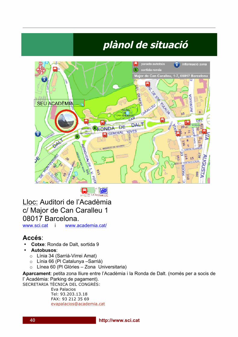
40 http://www.sci.cat
plànol de situació
Lloc: Auditori de l’Acadèmia c/ Major de Can Caralleu 1 08017 Barcelona. www.sci.cat i www.academia.cat/ Accés: • Cotxe: Ronda de Dalt, sortida 9 • Autobusos: o Línia 34 (Sarrià-Virrei Amat) o Línia 66 (Pl Catalunya –Sarrià) o Línea 60 (Pl Glòries – Zona Universitaria)
Aparcament: petita zona lliure entre l’Acadèmia i la Ronda de Dalt. (només per a socis de l’ Acadèmia: Parking de pagament). SECRETARIA TÈCNICA DEL CONGRÉS: Eva Palacios Tel: 93.203.13.18 FAX: 93 212 35 69 [email protected]
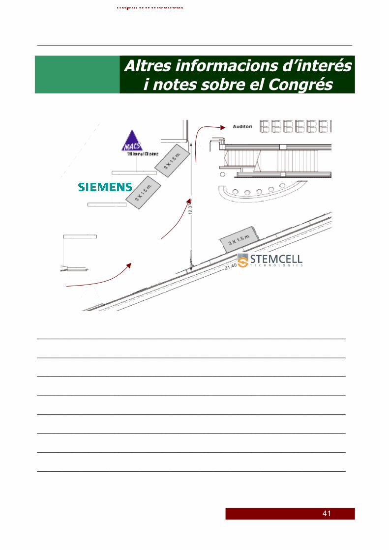
41
http://www.sci.cat
Altres informacions d’interés i notes sobre el Congrés
________________________________________________________________________
________________________________________________________________________
________________________________________________________________________
________________________________________________________________________
________________________________________________________________________
________________________________________________________________________
________________________________________________________________________
________________________________________________________________________
42 http://www.sci.cat
V Congrés Societat Catalana d’Immunologia
Barcelona, 17 i 18 de novembre 2011
Aquest congrés ha estat possible també gràcies a la col·laboració de la SCAIC (Societat Catalana d’Al·lèrgia i Immunologia Clínica)
i la SEI (Sociedad Española de Inmunología)
Sociedad Española de Inmunología
El contingut d’aquest congrés el podreu consultar en pocs mesos a la nostra web www.congressci.com
amb el format de congrés virtual!!!! Entreu-hi!!!