Embed Size (px)
Citation preview

UvA-DARE is a service provided by the library of the University of Amsterdam (http://dare.uva.nl)
UvA-DARE (Digital Academic Repository)
Methodology and implications of lymphatic mapping and sentinel lymphadenectomy
Tanis, P.J.
Link to publication
Citation for published version (APA):Tanis, P. J. (2002). Methodology and implications of lymphatic mapping and sentinel lymphadenectomy.
General rightsIt is not permitted to download or to forward/distribute the text or part of it without the consent of the author(s) and/or copyright holder(s),other than for strictly personal, individual use, unless the work is under an open content license (like Creative Commons).
Disclaimer/Complaints regulationsIf you believe that digital publication of certain material infringes any of your rights or (privacy) interests, please let the Library know, statingyour reasons. In case of a legitimate complaint, the Library will make the material inaccessible and/or remove it from the website. Please Askthe Library: https://uba.uva.nl/en/contact, or a letter to: Library of the University of Amsterdam, Secretariat, Singel 425, 1012 WP Amsterdam,The Netherlands. You will be contacted as soon as possible.
Download date: 13 Aug 2019

CHAPTERR THIRTEEN
Feasibilityy of Sentinel Node Lymphoscintigraphyy in Stage I
Testicularr Cancer
P.J.P.J. Tunis, S. Horenblas, R.A. Valdés Olmos, CA. Hoefnagel, O.E. Nieweg DepartmentsDepartments of Surgery, Urology and Nuclear Medicine, The Netherlands
CancerCancer Institute / Antoni van Leeuwenhoek Hospital, Amsterdam, thethe Netherlands
Eurr J Nucl Med, in press

126 6 Chapterr XIII
Thee management of the regional lymph nodes in stage I testicular cancer is stilll surrounded by controversy. As a result of the low incidence of occult lymphh node metastases, primary treatment for non-seminomatous germ celll tumours (NSGCT) in the form of retroperitoneal lymphadenectomy or
chemotherapyy results in overtreatment with associated morbidity in 70% to 75% of thee patients. Overtreatment in seminomatous germ cell tumours (SGCT) with routinee adjuvant radiotherapy is even higher, because occult metastases are present inn less than 20% of the patients. On the other hand, a surveillance policy results in detectionn of lymph node metastases at a later stage and requires an intensive, frequentt follow up with costly examinations. Until now, methods for selection of patientss who need adjuvant therapy are not satisfactory. Theree is a need for techniques to predict which patients are most likely to have occultt retroperitoneal disease. Positron emission tomography (PET) has been used in testicularr cancer for the evaluation of postoperative beds to find residual tumour, butt was found to be unreliable for staging.1 The technique of lymphatic mapping withh sentinel node biopsy can potentially be the solution for the selection problem. Beforee the sentinel node era, lymphoscintigraphy using intratesticular injection of colloidall gold (198Au) has been described in literature.2-3 The purpose of the present studyy was to determine the feasibility of lymphoscintigraphy using technetium-99m (99mTc)) nanocolloid for identification of the sentinel node in testicular cancer.
Patientss and methods
Fivee patients with clinically stage I testicular cancer underwent lymphoscintigraphy. Thee mean age of the patients was 36 years (range 21 to 52 years). The primary tumourr was situated on the right side in one patient and on the left side in four patients.. Except for the first patient, local anaesthesia was obtained with a funicular blockk using lidocain 2%, performed by the urologist in the outpatient clinic. In the nuclearr medicine department, a single dose of 99mTc-nanocolloid (Amersham Cygne, Eindhoven,, the Netherlands) in a mean volume of 0.22 ml (range 0.15 to 0.30) was injectedd with a fine type of needle into the funiculus in the first patient and into the testicularr parenchyma in the following four patients. The mean dose was 99 MBq withh a range from 52 to 135 MBq. Shortlyy after injection, anterior and lateral dynamic twenty-second images were obtainedd with a dual-head gamma camera (ADAC Vertex, Milpitas, CA, USA) duringg ten minutes to visualise the lymphatic flow and to determine the sequence of hott spots that appear. Static images with a cobalt-57 source behind the patient were obtainedd during five minutes immediately following the dynamic study. Late static imagess were obtained two to 24 hours after injection. Inn the last two patients, lymphoscintigraphy was followed by intraoperative lymphaticc mapping via a transperitoneal laparoscopic approach on the same day. Thee sentinel node was identified with the aid of intratesticular injected patent blue dyee in a volume of 1.0 ml and an endoscopic gamma ray detection probe (Europrobe,, Euromedical Instruments, Le Chesnay, France). This study protocol was approvedd by the Medical Ethical Committee and informed consent was obtained.
Testicularr lymphoscintigraphy 127 7
Results s
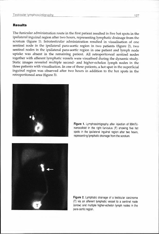
Thee funicular administration route in the first patient resulted in five hot spots in the ipsilaterall inguinal region after two hours, representing lymphatic drainage from the scrotumm (figure 1). Intratesticular administration resulted in visualisation of one sentinell node in the ipsilateral para-aortic region in two patients (figure 2), two sentinell nodes in the ipsilateral para-aortic region in one patient and lymph node uptakee was absent in the remaining patient. All retroperitoneal sentinel nodes togetherr with afferent lymphatic vessels were visualised during the dynamic study. Staticc images revealed multiple second- and higher-echelon lymph nodes in the threee patients with visualisation. In one of these patients, a hot spot in the superficial inguinall region was observed after two hours in addition to the hot spots in the retroperitoneall area (figure 3).
Figuree 1. Lymphoscintigraphy after injection of 99mTc-nanocolloidd in the right funiculus (F) showing five hot spotss in the ipsilateral inguinal region after two hours, representingg lymphatic drainage from the scrotum.
Figuree 2. Lymphatic drainage of a testicular carcinoma (T)) via an afferent lymphatic vessel to a sentinel node (arrow)) and multiple higher-echelon lymph nodes in the para-aorticc region.

128 8 Chapterr XIII
Lapararoscopicc exploration revealed two radioactive sentinel nodes without blue discolorationn adjacent to the aorta near the origin of the renal vessels in one patient. Explorationn in the patient without visualisation on lymphoscintigraphy did not resultt in identification of a radioactive or blue-stained sentinel node, but two lymph nodess without tracer-uptake were harvested. Al l excised lymph nodes were free of tumour. .
Discussion n
Thiss study shows the feasibility of lymphoscintigraphy to identify sentinel lymph nodess in testicular cancer patients. The method is easy to perform and is well-tolerated.. No side effects were observed. Lymphoscintigraphy can potentially be of clinicall value in combination with an intraoperative technique of lymphatic mapping too identify occult lymph node metastases in clinically node-negative testicular cancer.. This approach enables better selection of patients who may benefit from adjuvantt therapy after orchiectomy and avoids unnecessary morbidity. In our institution,, the current adjuvant treatment for stage I testicular cancer consists of radiotherapyy to the para-aortic and ipsilateral pelvic nodes in SGCT and chemotherapyy in the form of BEP (bleomycin, etoposide and cisplatin) with or withoutt salvage surgery in case of relapse during surveillance in NSGCT. The main disadvantagee of radiotherapy to this region is the two- to three-fold increased risk of secondaryy solid tumours. Chemotherapy regimens for NSGCT are associated with long-termm toxicity such as decrease in glomerular filtration, Raynaud's phenomenon, impairedd spermatogenesis, ototoxicity and neurotoxicity, besides the acute toxicity.4

Testicularr lymphoscintigraphy 129
Inn other institutions, routine retroperitoneal lymphadenectomy is still applied in NSGCTT patients. The use of nerve-sparing and unilateral lymphadenectomy has reducedd morbidity, but ejaculatory dysfunction, for example, occurs in about 10%.5
Somee studies have tried to solve the problem of patient selection by identifying factorss associated with occult lymph node metastases, such as tumour size, tumour invasionn of testicular lymphatics and veins, and tumour marker half-life.6'7 However, patientt selection based on these factors wil l result in high false-negative and false-positivee rates. Detectionn of occult lymph node metastases in a direct way has been shown to be moree promising. Following the work of Cabanas and Morton who were the pioneers inn exploring the sentinel node concept, the technique of lymphatic mapping has gainedd widespread popularity in the treatment of several malignancies.8 The hypothesiss of sequential dissemination was also explored in testicular cancer. In 1987,, Weissbach and Boedefeld stated that "a more limited approach strictly for the purposee of pathological staging, which aims at the prevention of long-term damage withoutt compromising diagnostic accuracy, must be based on the knowledge of the pathwayss of lymphatic dissemination and, particularly, on the first site of nodal involvement".99 The primary sites of lymphatic drainage have been defined by lookingg for the sites of a solitary metastasis in retroperitoneal dissections.9 The variabilityy in anatomical patterns as shown in these studies requires the use of the techniquee of lymphatic mapping to identify the lymph nodes on a direct drainage pathwayy from the primary tumour. Inn analogy to funicular lymphangiography, we started with funicular injection of the radiocolloidd for lymphoscintigraphy, because this was assumed to be the least painfull administration route. But at the level of the funiculus, only a few collecting lymphaticc vessels are present and it is hard to inject the radiocolloid exactly into thesee vessels unless these are surgically canulated. Consequently, the radiocolloid wil ll be absorbed by lymphatic capillaries of the scrotum with subsequent drainage to superficiall inguinal nodes (figure 1). Therefore, we changed the administration route afterr the first patient. Even when intratesticular injection is performed, inguinal drainagee may occur because of spill in the needle tract through the scrotum as we observedd in one patient (figure 3). The rapid lymphatic drainage form the testicle necessitatess dynamic gamma camera acquisition to enable differentiation between first-- and second-echelon lymph nodes in the retroperitoneum. Late images seem to bee indicated to exclude unexpected drainage patterns. Followingg the lymphoscintigraphy, we performed a laparoscopic retroperitoneal sentinell node biopsy in the same session as orchiectomy in two patients. The laparoscopicc approach requires longer operative time than an open procedure, but it iss a minimal invasive procedure with low rates of intraoperative and postoperative complicationss and short convalescence to normal life. In none of the two explored patientss blue lymphatics or blue nodes were encountered, probably due to inadequatee timing of injection. Further experience is needed to determine the proper usee and value of blue dye, especially in patients without scintigraphic visualisation. Whatt might be the consequences for adjuvant treatment in the future if the described sentinell node procedure turns out to be a technically successful and reliable

130 0 Chapterr XIII
technique?? Sentinel node positive patients can be treated with an adjuvant short-coursee chemotherapy or radiotherapy in case of NSGCT or SGCT respectively. Investigatorss who used two courses of BEP to treat clinical stage I NSGCT show that moree than 95% of the patients with occult lymph node metastases can be cured with shortt course chemotherapy.10 This results in reduction of chemotherapy and dose relatedd morbidity, because otherwise NSGCT patients with lymph node metastases duringg surveillance would have been treated with four courses of chemotherapy in a laterr phase. The approximate 80% reduction in the routine application of adjuvant radiotherapyy for SGCT obviously leads to a substantial reduction of morbidity as well. .
Conclusion n
Preliminaryy data shows that lymphoscintigraphy for sentinel node identification is feasiblee in stage I testicular cancer using intratesticular radiocolloid administration. Inn combination with laparoscopic sentinel node biopsy, identification of occult lymphh node metastases may prevent under- or overtreatment in the future.
References s
1.. Albers P, Bender H, Yilmaz H, et al. Positron emission tomography in the clinical staging of patientss with stage I and II testicular germ cell tumors. Urology 1999;53:808-11.
2.. Karparov M. Vesceral radioisotopic lymphography of the testicle. Neoplasma 1975;22:91 -8. 3.. Spaventi S, Bosnar M, Agbaba M, Paic G. Selective scintigraphic lymphography of testis. Radiol
Diagnn (Berl) 1974;15:111-4. 4.. Stuart NS, Woodroffe CM, Grundy R, Cullen MH. Long-term toxicity of chemotherapy for
testicularr cancer - the cost of cure. Br J Cancer 1990;61:479-84. 5.. Donohue JP, Foster RS. Retroperitoneal lymphadenectomy in staging and treatment. The
developmentt of nerve-sparing techniques. Urol Clin North Am 1998;25:461-8. 6.. Heidenreich A, Sesterhenn IA, Mostofi FK, Moul JW. Prognostic risk factors that identify patients
withh clinical stage I nonseminomatous germ cell tumors at low risk and high risk for metastasis. Cancerr 1998;83:1002-11.
7.. Warde P, Gospodarowicz MK, Banerjee D, et al. Prognostic factors for relapse in stage I testicularr seminoma treated with surveillance. J Urol 1997;157:1705-9,
8.. Tanis PJ, Nieweg OE, Valdés Olmos RA, Rutgers EJT, Kroon BBR. History of sentinel node and validationn of the technique. Breast Cancer Res 2001 ;3:109-12.
9.. Weissbach L, Boedefeld EA. Localization of solitary and multiple metastases in stage II nonseminomatouss testis tumor as basis for a modified staging lymph node dissection in stage I. JJ Urol 1987;138:77-82.
10.. Cullen MH, Stenning SP, Parkinson MC, et al. Short-course adjuvant chemotherapy in high-risk stagee I nonseminomatous germ cell tumors of the testis: a Medical Research Council report. J Clinn Oncol 1996;14:1106-13.