Embed Size (px)
Citation preview
UvA-DARE is a service provided by the library of the University of Amsterdam (http://dare.uva.nl)
UvA-DARE (Digital Academic Repository)
Early risk stratification in patients with chest painBholasingh, R.
Link to publication
Citation for published version (APA):Bholasingh, R. (2004). Early risk stratification in patients with chest pain.
General rightsIt is not permitted to download or to forward/distribute the text or part of it without the consent of the author(s) and/or copyright holder(s),other than for strictly personal, individual use, unless the work is under an open content license (like Creative Commons).
Disclaimer/Complaints regulationsIf you believe that digital publication of certain material infringes any of your rights or (privacy) interests, please let the Library know, statingyour reasons. In case of a legitimate complaint, the Library will make the material inaccessible and/or remove it from the website. Please Askthe Library: http://uba.uva.nl/en/contact, or a letter to: Library of the University of Amsterdam, Secretariat, Singel 425, 1012 WP Amsterdam,The Netherlands. You will be contacted as soon as possible.
Download date: 08 Apr 2019
Chapterr 6
Arnoo H.M. Moons Petruss M. van der Zee
Radhaa Bholasingh Auguestee Sturk
C.. Erik Hack Joostt CM. Meijers
Ottoo Kamp Jann H. Cornel
Ronn J.G. Peters Robbertt J. de Winter
Thee value of hemostatic markerss in the triage of patientss with chest pain presentingg with a normal or non-diagnostic c electrocardiogram m
Submitted Submitted
ValueValue of hemostatic markers
ABSTRACT T
Background d
Ass thrombus formation after coronary plaque disruption plays a major rote in patients with an
acutee coronary syndrome, we evaluated whether hemostatic markers could provide an
importantt diagnostic tool to detect acute coronary syndrome in patients with chest pain
presentingg to the emergency department with an initial non-diagnostic electrocardiogram.
Methods s
Wee measured plasma levels of coagulation and fibrinolytic markers on admission in
patientss with chest pain presenting with a normal or non-diagnostic electrocardiogram to
thee emergency department within 6 hours after symptoms onset. Patients were divided in
cardiacc troponin T negative (<0.06 ug/L, n=33) or positive (>0.06 ug/L, n=65) acute
coronaryy syndrome cases and control subjects (n=62) without a history of acute coronary
syndromee and who presented with non-cardiac chest pain.
Results s
Theree was no difference in the levels of prothrombin fragment Fi+2, thrombin-
antithrombinn (TAT) complexes, soluble tissue factor (TF), and tissue factor pathway
inhibitorr (TFPI) activity between troponin T negative or positive acute coronary syndrome
casess and controls. Elevated levels of plasminogen activator inhibitor (PAI) were
observedd in troponin T negative and positive acute coronary syndrome cases versus
controll subjects (72.3 and 57.8 vs 44.2 ng/ml, respectively), which was statistically
significantt for troponin T negative acute coronary syndrome cases compared to controls
(p=0.014).. The positive and negative predictive values to detect acute coronary syndrome
casess by these markers varied between 68% and 75% and between 40% and 43%,
respectively. .
Conclusions s
Thee measurement of Fi+2, TAT complexes, soluble TF, TFPI activity, and PAI in plasma
didd not contribute to the triage of patients presenting with chest pain and a normal or non-
diagnosticc electrocardiogram.
78 8

ChapterChapter 6
INTRODUCTIO N N
Off the patients presenting with chest pain at the emergency department, the early
diagnosiss of acute coronary syndromes is a major challenge for physicians. In addition to
medicall history and physical examination, the electrocardiogram is critical for the initial
evaluationn of these patients. However, in 50%-80% of the cases, the electrocardiogram is
normall or non-diagnostic at presentation 137), which makes the early differentiation from
non-cardiacc causes of chest pain difficult. Currently, biochemical cardiac markers have
becomee an important diagnostic tool for acute coronary syndrome. Moreover, elevations
inn myocardial injury markers, such as cardiac Troponin T and I, are associated with early
andd late recurrent adverse cardiac events(l31;138-I40). Serial assessment of cardiac
markers,, including troponins, creatine kinase-MB isoenzyme and myoglobin* results in a
highh sensitivity to detect myocardial damage(29;41;44), but the absence of evidence of
myocardiall damage does not exclude acute coronary syndrome(47). Therefore, novel early
markerss of acute coronary syndrome are needed.
Thee main cause of acute coronary syndrome is atherosclerotic plaque disruption with
superimposedd arterial thrombus fbrmation(l4l-143), and tissue factor (TF) induced
thrombinn generation plays a pivotal role in this process(144). Significantly higher levels
Off circulating TF(145-148), that contains prpcoagulant activity 149), free and total tissue
factorr pathway inhibitor-antigen (TFPI, the physiologic inhibitor of TF)(147;148;150),
andd TFPI-activity(150;151), were found in patients with acute coronary syndrome
comparedd to patients with stable angina and healthy control subjects. Conflicting results,
however,, have been reported about thrombin generation markers as prothrombin fragment
FI+22 and mrombin-antithrombin (TAT) complexes, which showed to be significantly
increased(147;152;I53),, while others observed comparable or even reduced levels(l54-
156)) in patients with unstable angina or myocardial infarction compared with stable
anginaa patients or healthy individuals. In all of these studies, patients with acute coronary
syndromee presented with characteristic changes on the electrocardiogram, whereas the
diagnosticc value of hemostatic markers may be useful in the triage of patients presenting
withh a non-diagnostic electrocardiogram.
Thee purpose of this study was to evaluate the diagnostic value of coagulation markers (Le.
markerss of thrombin generation (Fi+2 and TAT complexes), soluble TF, and TFPI
activity),, and a fibrinolytic marker (plasminogen activator inhibitor (PAI)) for the early
79 9
ValueValue of hemostatic markers
identificationn of acute coronary syndrome in patients presenting to the emergency
departmentt with chest pain and a normal or non-diagnostic electrocardiogram.
METHOD S S
Patients s
Studyy patients were selected from a consecutive, observational cohort of patients who
weree admitted to the emergency department of one large teaching hospital and two
academicc hospitals. All patients suffered from typical chest pain suggestive of myocardial
ischemiaa that started within 6 hours of presentation and without electrocardiographic
signs,, at presentation, that are typical of acute myocardial infarction(I57) or
ischemia(5;I8).. Exclusion criteria were severe skeletal muscle damage or trauma, cardiac
resuscitation,, known or suspected thromboembolic disease, current use of oral
anticoagulation,, and infectious disease or signs of inflammation. AH patiënte were
observedd at the emergency department for <24 hours before discharge or hospital
admission.. The study population of the present study consisted of cases with an acute
coronaryy syndrome and control subjects. The cases were divided into two groups: group 1,
whichh included cases with recurrent chest pain, a negative troponin T (defined as a peak
troponinn T < 0.06 ug/L in two serial measurements), and dynamic ST-segment or T-wave
changess on the electrocardiogram during observation; and group 2, which included cases
withh recurrent chest pain and a positive troponin T (defined as a peak troponin T > 0.06
ug/LL in two serial measurements). The control subjects were patients without a history of
cardiovascularr disease, who were considered to have chest pain of non-cardiac origin, and
whoo were event free during subsequent 6 months of follow-up.
Thee study complies with the Declaration of Helsinki. The study protocol was approved by
thee institutional review boards, and all patients gave informed consent.
Bloodd sampling and assays
Onn admission to the emergency department, venous blood samples were taken from each
subject,, and collected in citrate containing tubes for the measurements of hemostatic
markers,, and in another tube without additional substrates for the measurement of
troponinn T. At 12 hours after the onset of symptoms, an additional blood sample was
collectedd from all study patients for the second measurement of troponin T. All blood
80 0
ChapterChapter 6
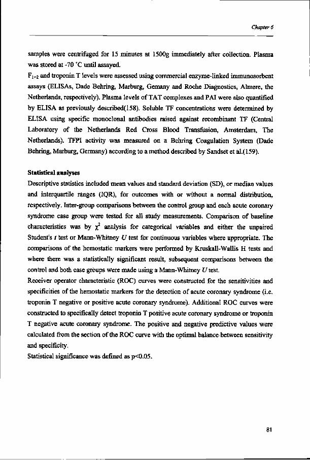
sampless were centrifuged for 15 minutes at 1500g immediately after collection. Plasma
wass stored at -70 °C until assayed.
F1+22 and troponin T levels were assessed using commercial enzyme-linked immunosorbent
assayss (ELISAs, Dade Bearing, Marburg, Gemany and Roche Diagnostics, Almere, the
Netherlands,, respectively). Plasma levels of TAT complexes and PAI were also quantified
byy ELISA as previously described(158). Soluble TF concentrations were determined by
ELISAA using specific monoclonal antibodies raised against recombinant TF (Central
Laboratoryy of the Netherlands Red Cross Blood Transfusion, Amsterdam, The
Netherlands).. TFPI activity was measured on a Behring Coagulation System (Dade
Behring,, Marburg, Germany) according to a method described by Sandset et al.(159).
Statisticall analyses
Descriptivee statistics included mean values and standard deviation (SD), or median values
andd interquartile ranges (IQR), for outcomes with or without a normal distribution,
respectively.. Inter-group comparisons between the control group and each acute coronary
syndromee case group were tested for all study measurements. Comparison of baseline
characteristicss was by x2 analysis for categorical variables and either the unpaired
Student'ss f test or Mann-Whitney U test for continuous variables where appropriate. The
comparisonss of the hemostatic markers were performed by Kruskall-Wallis H tests and
wheree there was a statistically significant result, subsequent comparisons between the
controll and both case groups were made using a Mann-Whitney U test.
Receiverr operator characteristic (ROC) curves were constructed for the sensitivities and
specificitiess of the hemostatic markers for the detection of acute coronary syndrome (i.e.
troponinn T negative or positive acute coronary syndrome). Additional ROC curves were
constructedd to specifically detect troponin T positive acute coronary syndrome or troponin
TT negative acute coronary syndrome. The positive and negative predictive values were
calculatedd from the section of the ROC curve with the optimal balance between sensitivity
andd specificity.
Statisticall significance was defined as p<0.05.
81 1
ValueValue of hemostatic markers
RESULTS S
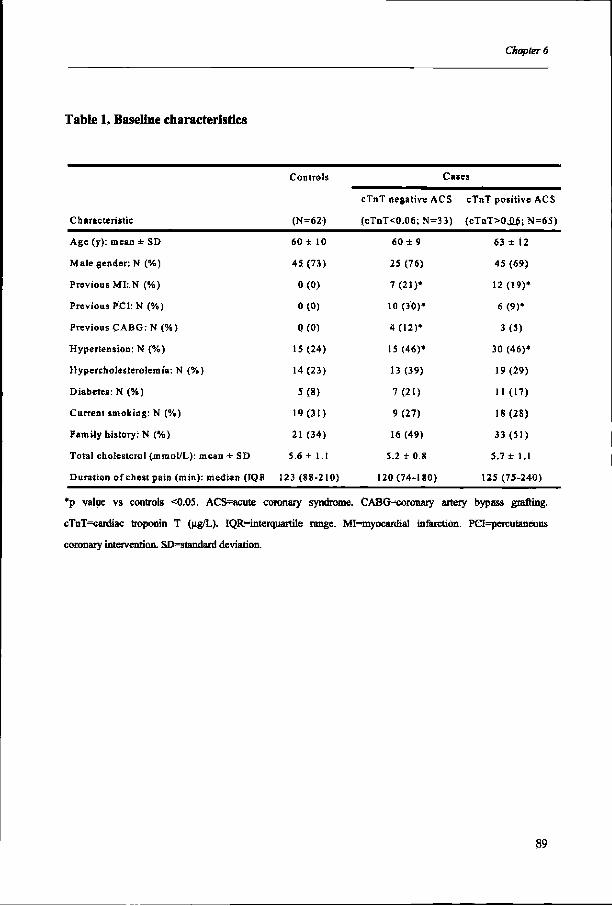
Baselinee characteristics
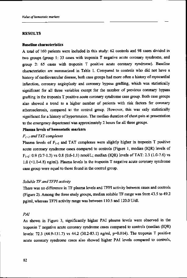
AA total of 160 patients were included in this study: 62 controls and 98 cases divided in
twoo groups (group 1: 33 cases with troponin T negative acute coronary syndrome, and
groupp 2: 65 cases with troponin T positive acute coronary syndrome). Baseline
characteristicss are summarized in Table 1. Compared to controls who did not have a
historyy of cardiovascular disease, both case groups had more often a history of myocardial
infarction,, coronary angioplasty and coronary bypass grafting, which was statistically
significantt for all three variables except for the number of previous coronary bypass
graftingg in the troponin T positive acute coronary syndrome case group. Both case groups
alsoo showed a trend to a higher number of patients with risk factors for coronary
atherosclerosis,, compared to the control group. However, mis was only statistically
significantt for a history of hypertension, The median duration of chest pain at presentation
too the emergency department was approximately 2 hours for all three groups.
Plasmaa levels of hemostatic markers
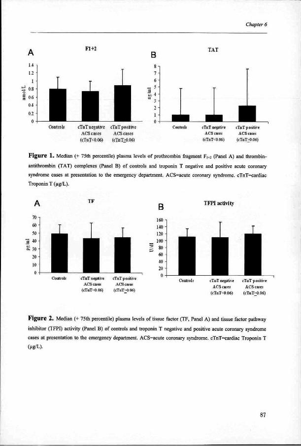
Fi+2Fi+2 and TAT complexes
Plasmaa levels of FH2 and TAT complexes were slightly higher in troponin T positive
acutee coronary syndrome cases compared to controls (Figure 1, median (IQR) levels of
Fi+2:: 0.9 (0.7-1.3) vs 0.8 (0.6-1.1) nmoI/L; median (IQR) levels of TAT: 2.3 (1.0-7.6) vs
1.00 (<1.0-4.8) ng/ml). Plasma levels in the troponin T negative acute coronary syndrome
casee group were equal to those found in the control group.
SolubleSoluble TF and TFPI activity
Theree was no difference in TF plasma levels and TFPÏ activity between cases and controls
(Figuree 2). Among the three study groups, median soluble TF range was from 43.5 to 49.2
pg/ml,, whereas TFPI activity range was between 110.5 and 120.0 U/dl.
PAI PAI Ass shown in Figure 3, significantly higher PAI plasma levels were observed in the
troponinn T negative acute coronary syndrome cases compared to controls (median (IQR)
levels:: 72.3 (44.9-131.7) vs 44.2 (30.2-83.1) ng/ml, p=0.014). The troponin T positive
acutee coronary syndrome cases also showed higher PAI levels compared to controls,
82 2
ChapterChapter 6
however,, it did not reach statistical significance (median (IQR) levels: 57.8 (35.5-107.2)
vss 44.2 (30.2-83.1) ng/ml, p=0.1).
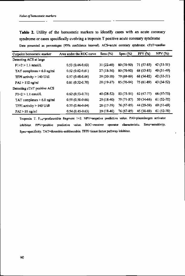
Diagnosticc utilit y of hemostatic markers
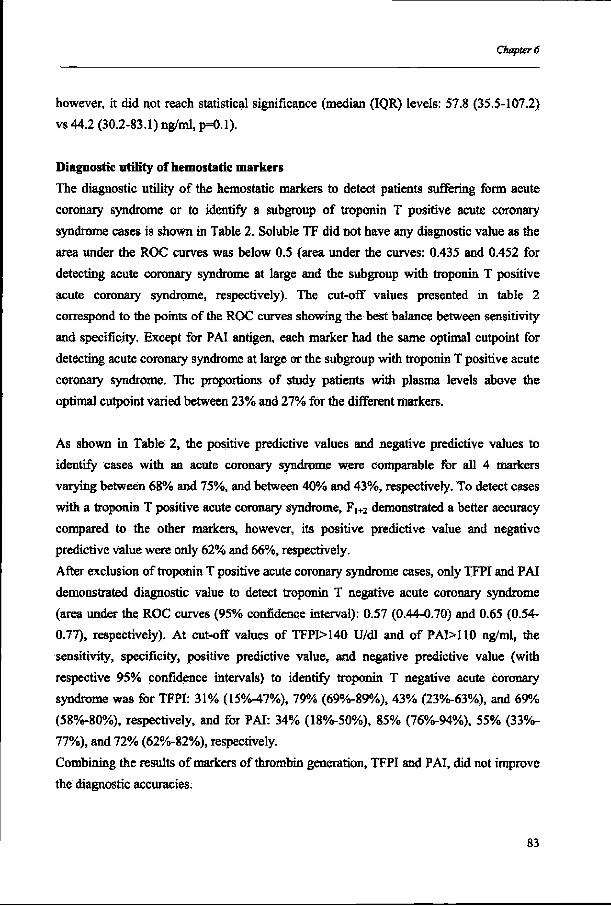
Thee diagnostic utility of the hemostatic markers to detect patients suffering form acute
coronaryy syndrome or to identify a subgroup of troponin T positive acute coronary
syndromee cases is shown in Table 2. Soluble TF did not have any diagnostic value as the
areaa under the ROC curves was below 0.5 (area under the curves: 0.435 and 0.452 for
detectingg acute coronary syndrome at large and the subgroup with troponin T positive
acutee coronary syndrome, respectively). The cut-off values presented in table 2
correspondd to the points of the ROC curves showing the best balance between sensitivity
andd specificity. Except for PAI antigen, each marker had the same optimal cutpoint for
detectingg acute coronary syndrome at large or the subgroup with troponin T positive acute
coronaryy syndrome. The proportions of study patients with plasma levels above the
optimall cutpoint varied between 23% and 27% for the different markers.
Ass shown in Table 2, the positive predictive values and negative predictive values to
identifyy cases with an acute coronary syndrome were comparable tor all 4 markers
varyingg between 68% and 75%, and between 40% and 43%, respectively. To detect cases
withh a troponin T positive acute coronary syndrome, Fi+2 demonstrated a better accuracy
comparedd to the other markers, however, its positive predictive value and negative
predictivee value were only 62% and 66%, respectively.
Afterr exclusion of troponin T positive acute coronary syndrome cases, only TFPI and PAI
demonstratedd diagnostic value to detect troponin T negative acute coronary syndrome
(areaa under the ROC curves (95% confidence interval): 0.57 (0.44-0.70) and 0.65 (0.54-
0.77),, respectively). At cut-off values of TFPI>140 U/dl and of PAI>110 ng/ml, the
sensitivity,, specificity, positive predictive value, and negative predictive value (with
respectivee 95% confidence intervals) to identify troponin T negative acute coronary
syndromee was for TFPI: 31% (15%^7%), 79% (69%-89%), 43% (23%-63%), and 69%
(58%-80%),, respectively, and for PAI: 34% (18%-50%), 85% (76%-94%), 55% (33%-
77%),, and 72% (62%-82%), respectively.
Combiningg the results of markers of thrombin generation, TFPI and PAI, did not improve
thee diagnostic accuracies.
83 3

ValueValue of hemostatic markers
DISCUSSION N
Inn the present study, we measured coagulation markers (thrombin generation markers
(Fi+22 and TAT complexes), soluble TF, and TFPI activity) and a fibrinolytic marker (PAI)
inn patients admitted to the emergency department with chest pain and an initially normal
orr non-diagnostic electrocardiogram, within 6 hours after symptom onset. Except for PAI
antigen,, there were no significant differences in the plasma levels of these markers
betweenn cases with a troponin T negative or positive acute coronary syndrome and control
subjects. .
AA large study of Pope et al. showed that 5.4% of patients with a non-diagnostic
electrocardiogramm who were discharged from the emergency department were ultimately
foundd to have an acute coronary syndrome(3). Serial measurement of specific markers of
myocardiall necrosis such as creatine kinase-MB isoenzymes and troponins are used as a
diagnosticc tool. However, an acute coronary syndrome can not be excluded when these
markerss are not increased(47). Although TF mediated thrombus formation after coronary
plaquee disruption plays a significant role in the initiation of acute coronary
syndrofne(144),, systemic measurement of hemostatic markers did not demonstrate
additionall diagnostic value to the early triage of our study patients with chest pain.
Ourr results do not confirm earlier studies showing significantly increased plasma
concentrationss of TF, TFPI, F1+2 and TAT-complexes on admission, in patients with
unstablee angina or acute myocardial infarction compared to patiënte with stable
angina(145-147;152;153).. However, in these studies, patients with acute coronary
syndromee were included if the electrocardiogram demonstrated characteristic changes of
thee ST-segments or T-waves and were likely at higher risk. It has been demonstrated that
amongg patients with unstable angina, those with reversible ST-segment changes have
significantlyy higher levels of thrombin activity, as measured by fibrinopeptide A levels,
comparedd to those without these changes, and ST changes correlated significantly with the
presencee of angiographic thrombus(160;161). However, this could not be confirmed by
Beckerr et aL, who reported no differences in thrombin activity and thrombin generation
betweenn patients with and without electrocardiogram changes(162). In addition, others
failedd to show significant differences in plasma levels of F]+2 and TAT complexes
betweenn patients with acute myocardial infarction, unstable angina and stable angina,
despitee the characteristic electrocardiogram changes presented by patients with an acute
84 4
ChapterChapter 6
coronaryy syndrome(154;155). Thus, symptomatic myocardial ischemia may not primarily
bee related to acute thrombin generation, but is possibly the result of a combination with
platelett aggregation, transient increased vasomotor tone and transient increased
myocardiall oxygen demand(163). Alternatively, local thrombin generation in the coronary
arteriess mayy be insufficiently reflected by systemic plasma levels.
Wee observed elevated plasma levels of PAI antigen in both acute coronary syndrome case
groupss compared to controls, but this was only significant in cases with a troponin T
negativee acute coronary syndrome. An important source of PAI during atherosclerotic
thrombuss formation may be the secretion by endothelial cells and activated platelets(164).
However,, increased PAI plasma levels are also associated with common risk factors for
coronaryy artery disease such as hypertension and particularly diabetes(164). These two
factorss were more prevalent in both case groups, in particular in the troponin T negative
acutee coronary syndrome case group, and this contributed to the enhanced PAI levels in
thesee groups compared to controls.
Patientss suffering from troponin T positive acute coronary syndrome previously
demonstratedd a significantly increased coagulation activation compared to patients with
troponinn T negative unstable angina(165;166). However, we could not confirm this in our
studyy population. We observed no differences in plasma levels of the hemostatic markers
betweenn cases with troponin T positive and negative acute coronary syndrome. Despite
evidencee of myocardial necrosis, the extent of coagulation activation may be limited in
casess with a troponin T positive acute coronary syndrome, and whose electrocardiogram
doess not show characteristic changes of myocardial ischemia. This was reflected by the
poorr diagnostic performance of the hemostatic markers to identify these cases.
Prothrombinn fragment Fi+2 showed the best diagnostic utility, but the positive predictive
valuee and negative predictive value were only 62% and 66%, respectively. Since these
patientss can be identified after serial measurement of cardiac serum markers, it remains a
difficultt task to detect patients suffering from an acute coronary syndrome, without
evidencee of myocardial damage and presenting with a non-diagnostic electrocardiogram.
Inn the present study, after excluding the troponin T positive acute coronary syndrome
casess from the analyses, only TFPI and PAI demonstrated diagnostic value to detect
troponinn T negative acute coronary syndrome cases, but with low positive predictive
valuess of 43% and 55%, respectively. Based on these markers, as much as 66%-69% of
thee patients with troponin T negative acute coronary syndrome would still not have been
85 5
ValueValue of hemostatic markers
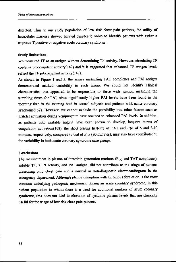
detected.. Thus in our study population of low risk chest pain patients, the utility of
hemostaticc markers showed limited diagnostic value to identify patients with either a
troponinn T positive or negative acute coronary syndrome.
Studyy limitation s
Wee measured TF as an antigen without determining TF activity. However, circulating TF
containss procoagulant activity 149) and it is suggested that enhanced TF antigen levels
reflectt the TF procoagulant activity(147).
Ass shown in Figure 1 and 3, the assays measuring TAT complexes and PAI antigen
demonstratedd marked variability in each group. We could not identify clinical
characteristicss that appeared to be responsible to these wide ranges, including the
samplingg times for PAI, since significantly higher PAI levels have been found in the
morningg than in the evening both in control subjects and patients with acute coronary
syndrome<167).. However, we cannot exclude the possibility mat other factors such as
platelett activation during venipuncture have resulted in enhanced PAI levels. In addition,
ass patients with unstable angina have been shown to develop frequent bursts of
coagulationn activation(168), the short plasma half-life of TAT and PAI of 5 and ÏM0
minutes,, respectively, compared to that of Fi+2 (90 minutes), may also have contributed to
thee variability in both acute coronary syndrome case groups.
Conclusions s
Thee measurement in plasma of thrombin generation markers (Fj+2 and TAT complexes),
solublee TF, TFPI activity, and PAI antigen, did not contribute to the triage of patients
presentingg with chest pain and a normal or non-diagnostic electrocardiogram in the
emergencyy department. Although plaque disruption with thrombus formation is the most
commonn underlying pathogenic mechanism during an acute coronary syndrome, in this
patientt population in whom there is a need for additional markers of acute coronary
syndrome,, this does not lead to elevation of systemic plasma levels that are clinically
usefull for the triage of low risk chest pain patients.
86 6
ChapterChapter 6
A A 1.44 -
122 -
11 -
tt 0.8-
11 0 . 6-0.4--
0.2 2 | |
T T
IE E Controlss cTuT negative
ACSS cases (cTnT<0.06) )
I I I cTnTT positive
ACSS cases (cTuT>0.06) )
B B «1 1 7--
6 6 __ 5 -
§4--2--
1 1
HHHControlss cTuT negative
ACSS cases (cTnT<0.06) )
I I I cTuTposithe e
ACSS cases (cTuT^O.06) )
Figur ee 1. Median (+ 75th percentile) plasma levels of prothrombin fragment F 1*2 (Panel A) and thrombin-
antithrombinn (TAT) complexes (Panel B) of controls and troponin T negative and positive acute coronary
syndromesyndrome cases at presentation to die emergency department. ACS=acute coronary syndrome. cTnT=cardiac
Troponinn T (ug/L).
A A TF F
Controlss cTnT negative cTnT positive ACSS «b-es ACS cases
(cTnT-=0.06)) (cTnT>0.06)
B B TFPII activity
Controlss cTnTneaathe cTnT positive ACSS cases ACS cases
(cTnT-0.06)) (cTnT>0.06)
Figur ee 2. Median (+ 75th percentile) plasma levels of tissue factor (TF, Panel A) and tissue factor pathway
inhibitorr (TFPI) activity (Panel B) of controls and troponin T negative and positive acute coronary syndrome
casess at presentation to the emergency department. ACS=acute coronary syndrome. cTnT=cardiac Troponin T
(ug/L). .
87 7

ValueValue of hemostatic markers
PAI I p=0.014 4
HEN N 1200 -
1000 -
11 80 -
PP 60
400 -
200 -
0 --Controls s cTnTT negative
ACSS cases
(cTnTO.06) )
cTnTT positive ACSS cases
(cTnT>0.06) )
Figuree 3 Mediann (+ 75th percentile) plasma levels of plasminogen activator inhibitor (PAI) of controls and troponin T
negativee and positive acute coronary syndrome cases at presentation to the emergency department. ACS=acute
coronaryy syndrome. cTnT=cardiac Troponin T (ug/L).
ChapterChapter 6
Tablee 1, Baseline characteristics
Characteristic c
Agee (y): meao SD
Malee gender: N (%)
Previouss MI : N (%)
Previouss PCI: N (%)
Previouss CABG:N (%)
Hypertension:: N {%)
Hypercholesterolemia:: N (%)
Diabetes:: N (%)
Currentt smoking: N (%)
Familyy history: N (%)
Totall cholesterol (mmoI/L): mean - SD
Duration,, of chest pain (mm): mediann (IQK
Controls s
(N=62) )
60++ 10
455 (73)
0(0) )
0(0) )
0(0) )
15(24) )
144 (23)
5(8) )
19(31) )
211 (34)
5.66 1.1
123(88-210) )
Cases s
cTnTT negative ACS
(cTnT<0.06;N= =
600 9
255 (76)
7(21)* *
100 (30)*
4(12)* *
15(46)* *
133 (39)
7(21) )
9(27) )
166 (49)
5.22 0.8
120(74-180) )
33) )
cTnTT positive ACS
{cToT>0J^;N=65) )
633 12
455 (69)
12(19)* *
6(9)* *
3(5) )
300 (46)*
19(29) )
UU (17)
18(28) )
333 (51)
5.77 1.1
1255 (75-240)
*pp value vs controls <0.05. ACS~aeute coronary syndrome. CABCHcoronary artery bypass grafting.
cTnT=cardiacc troponin T (ug/L). IQR=interquartilé range. MI=myocardia! infarction. PCI=percutaneous
coronaryy intervention. Substandard deviation.
89 9
ValueValue of hemostatic markers
Tablee 2. Utility of the hemostatic markers to identify cases with an acute coronary
syndromee or cases specifically evolving a troponin T positive acute coronary syndrome
Dataa presented as percentages (95% confidence interval). ACS=acute coronary syndrome. cTnT=eardiac
Cutpointt hemostatic marker Area under the ROC curve Sens(%) Spec(%) PPV(%) NPV(%)
Detectingg ACS at large
Fl+2>> I.] nmol/L 0.53 (0.44-0.63)
TATT complexes > 6.0 ng/ml 0.52 (0.42-0.61)
TFPII activity > 140 U/dl 0.57 (0.48-0.66)
PAII > 110 ng/ml 0.61 (0.52-0.70)
Detectingg cTnT positive ACS
Fl+2>> 1.1 nmol/L 0.62(0.53-0.71)
TATT complexes > 6.0 ng/ml 0.59 (0.50-0.68)
TFPII activity > 140 U/dl 0.55 (0.464).64)
PAII > 95 ng/ml 0.54 (0.45-0.63)
31(22^0)) 80(70-90) 71(57-85) 42(33-51)
277 (18-36) 80 (70-90) 68 (53-83) 40 (31-49)
299 (20-38) 79 (69-89) 68 (54-82) 42 (33-51)
28(19-37)) 85(76-94) 75(61-89) 43(34-52)
400 (28-52) 83 (75-91) 62 (47-77) 66 (57-75)
29(18-40)) 79(71-87) 50(34-66) 61(52-70)
288 (17-39) 76 (67-85) 44 (29-59) 60 (51-69)
299 (18-40) 76 (67-85) 45 (30-60) 61 (52-70)
Troponinn T. F1+2=prothrombin fragment 1+2. NPV=negative predictive value. PAI=plasminogen activator
inhibitor.. PPV=positive predictive value. ROC=receiver operator characteristic. Sens=sensitivity.
Spec=specificity.. TAT=uu»mbin-antiüirambm. TFPI=tissue factor pathway inhibitor.
90 0


![Diagnosti[1].. documento final popayán 2009](https://img.dokumen.tips/doc/110x75/55752f27d8b42a56388b557f/diagnosti1-documento-final-popayan-2009.jpg)