Embed Size (px)
Citation preview

UvA-DARE is a service provided by the library of the University of Amsterdam (http://dare.uva.nl)
UvA-DARE (Digital Academic Repository)
Deciduous molar hypomineralisation, its nature and nurture
Elfrink, M.E.C.
Link to publication
Citation for published version (APA):Elfrink, M. E. C. (2012). Deciduous molar hypomineralisation, its nature and nurture.
General rightsIt is not permitted to download or to forward/distribute the text or part of it without the consent of the author(s) and/or copyright holder(s),other than for strictly personal, individual use, unless the work is under an open content license (like Creative Commons).
Disclaimer/Complaints regulationsIf you believe that digital publication of certain material infringes any of your rights or (privacy) interests, please let the Library know, statingyour reasons. In case of a legitimate complaint, the Library will make the material inaccessible and/or remove it from the website. Please Askthe Library: https://uba.uva.nl/en/contact, or a letter to: Library of the University of Amsterdam, Secretariat, Singel 425, 1012 WP Amsterdam,The Netherlands. You will be contacted as soon as possible.
Download date: 20 Mar 2020
7
Deciduous Molar Hypomineralisation and Molar
incisor Hypomineralisation
Based on:
Deciduous Molar Hypomineralization and Molar incisor Hypomineralization
MEC ElfrinkJM ten CateVWV JaddoeA HofmanHA MollJSJ Veerkamp
J Dent Res 2012, Feb 27 [Epub ahead of print]
R1R2R3R4R5R6R7R8R9
R10R11R12R13R14R15R16R17R18R19R20R21R22R23R24R25R26R27R28R29R30R31R32R33R34R35R36R37R38R39
Chapter 7
110
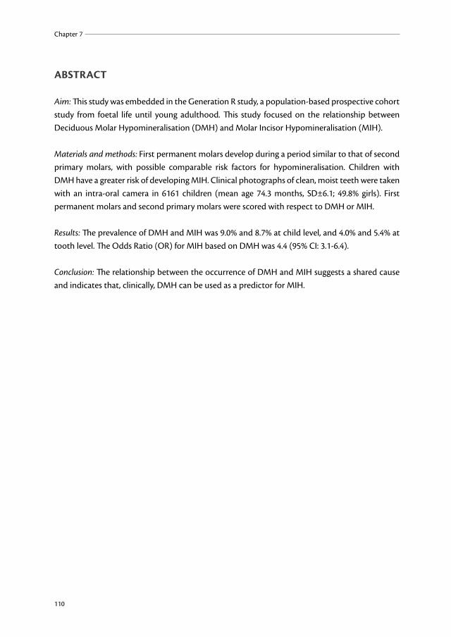
ABstrACt
Aim: This study was embedded in the Generation R study, a population-based prospective cohort study from foetal life until young adulthood. This study focused on the relationship between Deciduous Molar Hypomineralisation (DMH) and Molar Incisor Hypomineralisation (MIH).
Materials and methods: First permanent molars develop during a period similar to that of second primary molars, with possible comparable risk factors for hypomineralisation. Children with DMH have a greater risk of developing MIH. Clinical photographs of clean, moist teeth were taken with an intra-oral camera in 6161 children (mean age 74.3 months, SD±6.1; 49.8% girls). First permanent molars and second primary molars were scored with respect to DMH or MIH.
Results: The prevalence of DMH and MIH was 9.0% and 8.7% at child level, and 4.0% and 5.4% at tooth level. The Odds Ratio (OR) for MIH based on DMH was 4.4 (95% CI: 3.1-6.4).
Conclusion: The relationship between the occurrence of DMH and MIH suggests a shared cause and indicates that, clinically, DMH can be used as a predictor for MIH.
R1R2R3R4R5R6R7R8R9R10R11R12R13R14R15R16R17R18R19R20R21R22R23R24R25R26R27R28R29R30R31R32R33R34R35R36R37R38R39
Deciduous Molar Hypomineralisation and Molar Incisor Hypomineralisation
111
7
introDuCtion
Developmental defects of dental enamel are common in both deciduous and permanent dentitions and are classified into hypomineralisation and hypoplasia (1, 2). Enamel hypoplasia is a quantitative defect of the enamel, resulting from a disturbance to the ameloblasts during matrix formation (1, 3, 4). Enamel hypomineralisation is a qualitative defect of the enamel because of a disturbance during initial calcification and/or during maturation (1, 3). Hypomineralised parts of teeth are weaker and the enamel may chip off easily, resulting in posteruptive loss of enamel. It can be difficult to distinguish between hypoplasia and posteruptive enamel loss (4). In the primary dentition, the second primary molar generally presents with more caries than the first primary molar (5-7). A recent study showed that hypomineralisation was an important risk factor for caries in the primary dentition (8). Also, in the permanent dentition, rapid caries progression was observed in hypomineralised molars (9, 10). Hypomineralised permanent molars are frequently combined with hypomineralised incisors. Molar Incisor Hypomineralisation (MIH) is defined as hypomineralisation of systemic origin of 1 to 4 permanent first molars combined with affected incisors (11). MIH-like defects are also seen on second primary molars and permanent cuspids (3). These MIH-like defects in the primary molars are now described as Deciduous Molar Hypomineralisation (DMH) (8, 12). In the Netherlands, the prevalence of DMH has been reported at 4.9% at child level (13) while the prevalence of MIH was higher (6-14%) (9, 14). The severity of MIH as well as DMH varies between patients, but also within a patient. Opacities are considered the mildest form of MIH and DMH, and atypical extractions as the most severe manifestation (9). MIH can cause serious pain due to posteruptive enamel loss, rapid caries progression, and pain during restorative treatment (9, 10). Children with MIH need more dental treatments and - probably as a consequence - are generally more fearful than their peers (10). Therefore, it is important to diagnose MIH as early as possible to reduce the vulnerability of the MIH-affected molars by focusing on their restorative and preventive needs. The development of the second primary molars starts at around the same time as the development of the first permanent molars and permanent incisors, but the maturation of the permanent teeth occurs more slowly (see Table 7.1) (15, 16). If a risk factor occurs during this overlapping period, hypomineralisation might occur in the primary as well as in the permanent dentition (17). Therefore, DMH might be used as a predictor for MIH. The parallel development of the second primary molar and the first permanent molar, both developmentally and with respect to their location in the jaw, might be indicative of a common cause for the hypomineralisation process. Our aim was therefore to study the association between DMH in the second primary molars and MIH in the first permanent molars.

R1R2R3R4R5R6R7R8R9
R10R11R12R13R14R15R16R17R18R19R20R21R22R23R24R25R26R27R28R29R30R31R32R33R34R35R36R37R38R39
Chapter 7
112
table 7.1. Starting age of development of the incisors and first molars of the permanent dentition and the second molars of the primary dentition (16).
Tooth 1st permanent incisor
2nd permanent incisor
1st permanent molar
2nd primary molar
Upper jawStart calcification 3 months 11 months 32 weeks in utero 19 weeks in uteroCrown completed 4.5 years 5.5 years 4.3 years 11 monthsEruption 7.3 years 8.3 years 6.25 years 29 months
Lower jawStart calcification 3 months 3 months 32 weeks in utero 18 weeks in uteroCrown completed 3.5 years 4 years 3.8 years 10 monthsEruption 6.3 years 7.5 years 6 years 27 months
MAteriAls AnD MetHoDs
Participants. This study was embedded in the Generation R study, a population-based prospective cohort study from foetal life until young adulthood. The Generation R study was designed to identify early environmental and genetic determinants of growth, development, and health and has previously been described in detail (18, 19). Briefly, the cohort included 9778 mothers and their children living in Rotterdam, the Netherlands. Enrolment of mothers occurred in their early pregnancy (gestational age <18 wks). All children were born between April 2002 and January 2006 and formed a prenatally enrolled birth-cohort. Of all eligible children in the study area, 61% were enrolled in the study at birth (18). The Medical Ethics Committee of the Erasmus Medical Centre, Rotterdam, approved the study. Written informed consent was obtained from all participants.For the postnatal phase of the study, 7893 children were available. About half of the mothers (51.0%) and the children were of Dutch origin (54.8%) (19). At the age of 5 to 6 yrs, the children were invited for a check-up visit at the Erasmus Medical Centre. From March 2008 until November 2011, 6487 children (77 twins) visited the Erasmus Medical Centre. As part of this visit, intra-oral photographs of the teeth were made, which was successfully done in 6161 children (95.0%). In cases where a few teeth could not be scored, only the teeth visible on the photographs were used in the analysis. A flowchart of the participants is shown in Figure 7.1.

R1R2R3R4R5R6R7R8R9R10R11R12R13R14R15R16R17R18R19R20R21R22R23R24R25R26R27R28R29R30R31R32R33R34R35R36R37R38R39
Deciduous Molar Hypomineralisation and Molar Incisor Hypomineralisation
113
7
Assessed for eligibility
Pregnancies (n=9778)
Children (n=9897)
Excluded (n=152)
intra-uterine death (n=78)
abortion (n=29)
various reasons (n=45)
Enrolment
Analysis
Live births (n=9745)
Availability
Excluded (n=1852)
no consent (n=38)
various reasons e.g. moved from study area
(n=1814)
Postnatal participants
(n=7893)
Excluded (n=1406)
failed participation (n=1406)
Eligible for analysis (n=6487)
Excluded (n=326)
no or only one photograph was made
(n=326)
Analysed
(n=6161)
figure 7.1. Flowchart of the participants.
Dental examination. After teeth were brushed, photographs of clean and moist teeth were taken by trained nurses and dental students (excess saliva was removed with a cotton roll). It took 1-2 min to take approximately 10 photographs of all teeth of the child. For this purpose, an intra-oral camera was used [Poscam USB intra-oral autofocus camera (Digital Leader PointNix), 640 x 480 pixels]. An example of such a photograph is shown in Figure 7.2.

R1R2R3R4R5R6R7R8R9
R10R11R12R13R14R15R16R17R18R19R20R21R22R23R24R25R26R27R28R29R30R31R32R33R34R35R36R37R38R39
Chapter 7
114
figure 7.2. Tooth 26 (1st permanent molar upper left) showing MIH (yellow opacity) and tooth 65 (2nd
primary molar upper left) showing DMH (yellow opacity and post-eruptive enamel loss).
The minimal scene illumination was f 1.4 and 30 lx. In an earlier study, the validity of this camera for visualizing DMH was shown to be high (sensitivity 72.3% and specificity 92.8%). The reliability was good for inter-observer agreement (kappa 0.62) and excellent for intra-observer agreement (kappa 0.95) (12).DMH and MIH were scored on the intra-oral photographs according to the EAPD criteria (3, 12):Mild:
• Opacity: A defect that changes the translucency of the enamel, variable in degree. The defective enamel is of normal thickness with a smooth surface and can be white, yellow or brown in colour. The demarcated opacity is not caused by caries, ingestion of excess fluoride during tooth development or amelogenesis imperfecta etc.
Severe:• Posteruptive enamel loss: A defect that indicates surface enamel loss after eruption of the
tooth, e.g., hypomineralisation related attrition. Enamel loss due to erosion was excluded, and/or

R1R2R3R4R5R6R7R8R9R10R11R12R13R14R15R16R17R18R19R20R21R22R23R24R25R26R27R28R29R30R31R32R33R34R35R36R37R38R39
Deciduous Molar Hypomineralisation and Molar Incisor Hypomineralisation
115
7
• Atypical caries: The size and form of the caries lesion do not match the present caries distribution in the child’s mouth, and/or
• Atypical restoration: The size and form of the restoration do not match the present caries distribution in the child’s mouth, and/or
• Atypical extraction: Absence of a molar that does not fit in the dental development and caries pattern of the child.
A first permanent molar or a second primary molar was diagnosed as having MIH or DMH when at least one of these criteria or a combination was found. If in a child one or more DMH molars were scored as severe, the child was scored as having a severe form of DMH.On the photographs, the number of shed teeth was also measured. The following criteria were used for the eruption of the permanent teeth:
• Not erupted / not shed: gingiva distally from the 2nd primary molar was shown on the photograph, but nothing from the 1st permanent molar can be seen. Permanent incisor not visible yet.
• Partly erupted / primary tooth missing due to shedding: 1st permanent molar was partly covered by the gingiva. Permanent incisor is not visible yet, but primary incisor was lost, most likely due to shedding.
• Erupted: permanent molar was not covered by the gingiva anymore, permanent incisor is (partly) visible.
• If the tooth, or the place were the tooth has to be, was not shown on the photographs, the tooth was scored as ‘not able to be judged’
The photographs were shown on a computer in full-screen mode, and were scored by one calibrated dentist (ME). To test the inter-observer agreement in this study, another calibrated dentist (JV) independently scored the data for 648 children. The Cohen’s kappa score in this study for DMH was 0.60 and for MIH 0.69. In the event of disagreement, the photographs were studied again and a joint consensus decision made. A separate group of 649 children was scored again by the first dentist (ME), at least 6 wks after the first scoring. The intra-observer agreement reached the following Cohen’s kappa scores: 0.82 (DMH) and 0.85 (MIH).Statistics. Statistical analyses were made with SPSS version 18.0 (SPSS Inc., Chicago, IL, USA). To test if children with DMH also had MIH more often, the Odds Ratio (OR) was computed (logistic regression analysis).
results
From the 6487 participating children, in 95.0% a good series of photographs was made, in 2.9% only one photograph was made, and in 2.1% no photographs were made. In this study, on a child level the data from 6161 children were used (mean age 74.3 months, SD±6.1; 49.8% girls). On tooth level, the data from 5561 children could be used for DMH diagnosis and from 2327 children

R1R2R3R4R5R6R7R8R9
R10R11R12R13R14R15R16R17R18R19R20R21R22R23R24R25R26R27R28R29R30R31R32R33R34R35R36R37R38R39
Chapter 7
116
for MIH diagnosis, mostly due to limitations in the judging of individual teeth. The prevalence of DMH was 9.0% (n=499) and of MIH was 8.7% (n=203) at child level. Of all eligible second primary molars (n=23722), DMH was present in 4.0% (n=955) of the teeth, and of all eligible first permanent molars (n=6545), MIH was present in 5.4% (n=355). Most children with DMH had a severe form of DMH (302 out of 499). Of the children with DMH, 76.6% (n=382) had opacities, 31.9% (n=159) post-eruptive enamel loss, 14.6% (n=73) atypical caries, 19.4% (n=97) atypical fillings, and 11.2% (n=56) atypical extractions. Often children had only one molar affected; the mean number of affected molars per child was 1.9 for DMH and 1.7 for MIH. These results are described in more detail in Table 7.2.
table 7.2: Distribution of DMH and MIH at child level and tooth level.Distribution DMH
n (%)MiHn (%)
Children 499/5561 (9.0%) 203/2327 (8.7%)Molars 955/23722 (4.0%) 355/6545 (5.4%)Number of affected molars per child per dentition DMH
n=499MiH
n=203One molar 216 (43.3%) 105 (51.7%)Two molars 157 (31.5%) 59 (29.1%)Three molars 79 (15.8%) 24 (11.8%)Four molars 47 (9.4%) 15 (7.4%)Mean number of affected molars (sd) 1.9 (1.0) 1.7 (0.9)Severity of hypomineralisation DMH
n=499MiH
Mild 197 (39.5%) Not scoredSevere 302 (60.5%)Affected molars per quadrant DMH
n=955MiH
n=355Maxillary right 245 (4.1%) 96 (5.0%)Maxillary left 221 (3.7%) 87 (4.6%)Mandibular left 265 (4.5%) 92 (6.5%)Mandibular right 224 (3.8%) 80 (6.0%)
Children with DMH in more than one molar had a higher Odds Ratio (OR) of developing MIH molars when compared with children with only one molar affected (Table 7.3). The OR seemed to increase when more molars were affected with DMH, and for children with three DMH molars, the OR was lower (table 7.3). Of the children with DMH, 49 (26.5%) children were also diagnosed as having MIH. Children with DMH had an OR of 4.4 (95% CI: 3.1-6.4) for MIH compared with children without DMH. Analysis on tooth level did not show statistically significant differences.Children with mild DMH (n=197) had an OR of 5.3 (95%CI: 2.9-9.4), and children with severe DMH had an OR of 4.0 (95% CI: 2.6-6.3).

R1R2R3R4R5R6R7R8R9R10R11R12R13R14R15R16R17R18R19R20R21R22R23R24R25R26R27R28R29R30R31R32R33R34R35R36R37R38R39
Deciduous Molar Hypomineralisation and Molar Incisor Hypomineralisation
117
7
tabl
e 7.
3: D
istrib
utio
n of
DM
H in
chi
ldre
n w
ith a
nd w
ithou
t MIH
.W
ithou
t sel
ectio
nO
ne o
r mor
e 1st
per
man
ent m
olar
s eru
pted
an
d al
l mol
ars a
ble
to b
e ju
dged
All
four
1st p
erm
anen
t mol
ars e
rupt
ed a
nd a
ll m
olar
s abl
e to
be
judg
edCh
ildre
n w
itho
ut M
iH
(n=
2124
)
Child
ren
wit
h M
iH
(n=
203)
or
(95%
Ci)
Child
ren
wit
hout
MiH
(n
=17
48)
Child
ren
wit
h M
iH
(n=
138)
or
(95%
Ci)
Child
ren
wit
hout
MiH
(n
=79
9)
Child
ren
wit
h M
iH
(n=
57)
or
(95%
Ci)
No
DM
H m
olar
s17
4215
4Re
fere
nce
1639
106
Refe
renc
e75
849
Refe
renc
eO
ne o
r mor
e m
olar
s w
ith D
MH
136
494.
4 (3
.1-6
.4)
109
324.
2 (2
.7-6
.6)
418
2.8
(1.2
-6.2
)
One
mol
ar w
ith D
MH
59
183.
9 (2
.3-6
.8)
4511
3.8
(1.9
-7.5
)13
44.
8 (1
.5-1
5.1)
Two
mol
ars w
ith D
MH
3816
5.4
(3.0
-10.
0)32
115.
3 (2
.6-1
0.8)
153
3.1
(0.9
-11.
0) n
sTh
ree
mol
ars w
ith D
MH
227
4.1
(1.7
-9.8
)18
43.
4 (1
.1-1
0.3)
71
2.2
(0.3
-18.
3) n
sFo
ur m
olar
s with
DM
H17
86.
1 (2
.6-1
4.3)
146
6.6
(2.5
-17.
6)6
00
(0-0
) ns
ns: n
ot si
gnifi
cant
R1R2R3R4R5R6R7R8R9
R10R11R12R13R14R15R16R17R18R19R20R21R22R23R24R25R26R27R28R29R30R31R32R33R34R35R36R37R38R39
Chapter 7
118
DisCussion
The prevalence of DMH found in this group was higher than the prevalence previously reported for 5-year-olds in the Netherlands (13). The prevalence of MIH was in line with earlier studies performed in the Netherlands and abroad (9, 14, 20). This paper is the first to present data on the relationship between DMH and MIH, showing that patients with DMH have an OR of 4.4 for developing MIH. There is a tendency that ORs increase when the number of DMH-affected molars goes up. Caution must be taken, since the number of children with three or four DMH-affected molars are rather small, resulting in considerably large confidence intervals.Most DMH-affected molars were scored as ‘severe’, meaning that they not only had opacities but also showed posteruptive enamel loss, atypical restoration, or atypical caries, or had been extracted. The age of the children probably influenced the severity.At the age of 5 or 6, the second primary molars have been in function for 3-4 yrs, thus increasing the likelihood of enamel breakdown or caries. The difference in OR found between the children with mild and those with severe DMH is interesting. This difference can possibly be explained by the onset and period of influence of possible aetiological factors. When the onset is early, the second primary molar is most actively formed (see Table 7.1). When the onset of the causative factor is later, the maturation in the second primary molar is already at a later stage, and the influence is less. In the first permanent molar, in contrast, the maturation process is more active and more influenced by the aetiological factor(s). For the results to be appreciated, some limitations need to be discussed. The percentages of mothers from different ethnicities and lower socio-economic status were lower among the participants than expected from the population figures in Rotterdam (19). The trend toward a more affluent and healthy population might influence the generalisability of the results. The inter- and intra-observer agreements were adequate for DMH, but good agreement was found for the inter-observer agreement for MIH. The Cohen’s kappa scores for DMH were similar to those found in the previous study for inter-observer agreement, but were somewhat lower for the intra-observer agreement (12). After the photographs were discussed with no initial disagreement, agreement was reached in all cases. Most discussions arose concerning partly erupted first permanent molars.In some of these young children, it was difficult to take the photographs. Unsuccessful pictures were generally seen in cases where the children were not able to breathe nasally, e.g., from common colds, thus creating moisture on the lens of the camera. Due to the limited numbers of missing photographs, the results are considered representative. In the present study, DMH was scored on intra-oral photographs, while in previous studies, DMH was scored clinically.The difference in prevalence of DMH between this study and a previous study in the Netherlands (12) should not be attributed to the scoring method, because the validity and reliability of scoring DMH on intra-oral photographs were good.

R1R2R3R4R5R6R7R8R9R10R11R12R13R14R15R16R17R18R19R20R21R22R23R24R25R26R27R28R29R30R31R32R33R34R35R36R37R38R39
Deciduous Molar Hypomineralisation and Molar Incisor Hypomineralisation
119
7
The second primary molar and first permanent molar have a shared period of development and mineralisation (see Table 7.1), and an observed relationship between DMH and MIH has already been hypothesized (3). The development of the second primary molar and the first permanent molar start at the same time, but the maturation phase of the permanent molar is considerably longer (15). If a risk factor occurred during this overlapping period, hypomineralisation might occur in the primary as well as in the permanent dentition (17).A genetic predisposition for hypomineralisation or a chronic or frequent recurrent disease within a particular time span, instead of only one risk factor, might affect, first, the second primary molars and, later, the first permanent molars.Because the second primary molars erupt four years earlier in life than first permanent molars, DMH might clinically be used as an indicator for MIH. Whether this will lead to clinical consequences such as more tooth destruction or pain in deciduous teeth is subject to further research. If MIH can be diagnosed as early as possible, a greater effect of preventive measures (e.g., fluoride applications and Casein PhosphoPeptides and Amorphous Calcium Phosphate (CPP-ACP) (21) can be expected.The relationship between DMH and MIH found in this study is an additional tool in the study of possible aetiological factors such as exposure to dioxins from breastfeeding, antibiotic use, perinatal problems, infectious diseases, etc. (9, 22-25), because they might lead to both DMH and MIH. MIH-affected molars are typically in need of restoration soon after eruption, and they may cause pain (9, 10). Therefore, in clinical practice, extra attention needs to be paid to children with DMH in the period when their permanent molars and incisors are erupting, given their increased risk of having MIH. The use of DMH as a predictor for MIH could help with this important early diagnosis.This study shows an association between the prevalence of DMH and MIH in 5- to 6-year-old children. This relationship suggests a shared cause and indicates that, especially, mild DMH can clinically be used as an indicator for MIH.
AcknowledgementsThe Generation R study was conducted by the Erasmus MC in close collaboration with the Erasmus University Rotterdam, School of Law and Faculty of Social Sciences, the Municipal Health Service Rotterdam area, Rotterdam, the Rotterdam Homecare Foundation, Rotterdam, and the Stichting Trombosedienst & Artsenlaboratorium Rijnmond (STAR), Rotterdam. We gratefully acknowledge the contribution of general practitioners, hospitals, midwives, and pharmacies in Rotterdam. The first phase of the Generation R study was made possible by financial support from the Erasmus MC, Rotterdam, Erasmus University Rotterdam and the Netherlands Organization for Health Research and Development (ZonMw). The present study was supported by an additional and unrestricted grant from GABA, Therwil, Switzerland. The authors declare no potential conflicts of interest with respect to the authorship and/or publication of this article.

R1R2R3R4R5R6R7R8R9
R10R11R12R13R14R15R16R17R18R19R20R21R22R23R24R25R26R27R28R29R30R31R32R33R34R35R36R37R38R39
Chapter 7
120
literAture
1. Jalevik B, Noren JG. Enamel hypomineralization of permanent first molars: a morphological study and survey of possible aetiological factors. Int J Paediatr Dent 2000;10(4):278-89.
2. William V, Messer LB, Burrow MF. Molar incisor hypomineralization: review and recommendations for clinical management. Pediatr Dent 2006;28(3):224-32.
3. Weerheijm KL, Duggal M, Mejare I, Papagiannoulis L, Koch G, Martens LC, Hallonsten AL. Judgement criteria for molar incisor hypomineralisation (MIH) in epidemiologic studies: a summary of the European meeting on MIH held in Athens, 2003. Eur J Paediatr Dent 2003;4(3):110-3.
4. Fearne J, Anderson P, Davis GR. 3D X-ray microscopic study of the extent of variations in enamel density in first permanent molars with idiopathic enamel hypomineralisation. Br Dent J 2004;196(10):634-8; discussion 25.
5. Holt RD. The pattern of caries in a group of 5-year-old children and in the same cohort at 9 years of age. Community Dent Health 1995;12(2):93-9.
6. Gizani S, Vinckier F, Declerck D. Caries pattern and oral health habits in 2- to 6-year-old children exhibiting differing levels of caries. Clin Oral Investig 1999;3(1):35-40.
7. Elfrink ME, Veerkamp JS, Kalsbeek H. Caries pattern in primary molars in Dutch 5-year-old children. Eur Arch Paediatr Dent 2006;7(4):236-40.
8. Elfrink ME, Schuller AA, Veerkamp JS, Moll HA, ten Cate JM. Factors increasing the caries risk of second primary molars in 5-year-old Dutch children. Int J Paediatr Dent 2010;20(1):151-7.
9. Weerheijm KL, Groen HJ, Beentjes VE, Poorterman JH. Prevalence of cheese molars in eleven-year-old Dutch children. ASDC J Dent Child 2001;68(4):259-62, 29.
10. Jalevik B, Klingberg GA. Dental treatment, dental fear and behaviour management problems in children with severe enamel hypomineralization of their permanent first molars. Int J Paediatr Dent 2002;12(1):24-32.
11. Weerheijm KL. Molar incisor hypomineralisation (MIH). Eur J Paediatr Dent 2003;4(3):114-20.
12. Elfrink ME, Veerkamp JS, Aartman IH, Moll HA, ten Cate JM. Validity of scoring caries and primary molar hypomineralisation (DMH) on intraoral photographs. Eur Arch Paediatr Dent 2009;10(S1): 5-10.
13. Elfrink ME, Schuller AA, Weerheijm KL, Veerkamp JS. Hypomineralized second primary molars: prevalence data in Dutch 5-year-olds. Caries Res 2008;42(4):282-5.
14. Jasulaityte L, Weerheijm KL, Veerkamp JS. Prevalence of molar-incisor-hypomineralisation among children participating in the Dutch National Epidemiological Survey (2003). Eur Arch Paediatr Dent 2008;9(4):218-23.
15. Butler PM. Comparison of the development of the second deciduous molar and first permanent molar in man. Arch Oral Biol 1967;12(11):1245-60.
16. Profitt W, Fields H. Contemporary Orthodontics. 3rd ed. St Louis: Mosby; 2000.
17. Aine L, Backstrom MC, Maki R, Kuusela AL, Koivisto AM, Ikonen RS, Maki M. Enamel defects in primary and permanent teeth of children born prematurely. J Oral Pathol Med 2000;29(8):403-9.
18. Jaddoe VW, Mackenbach JP, Moll HA, Steegers EA, Tiemeier H, Verhulst FC, Witteman JC, Hofman A. The Generation R Study: Design and cohort profile. Eur J Epidemiol 2006;21(6):475-84.
R1R2R3R4R5R6R7R8R9R10R11R12R13R14R15R16R17R18R19R20R21R22R23R24R25R26R27R28R29R30R31R32R33R34R35R36R37R38R39
Deciduous Molar Hypomineralisation and Molar Incisor Hypomineralisation
121
7
19. Jaddoe VW, van Duijn CM, van der Heijden AJ, Mackenbach JP, Moll HA, Steegers EA, Tiemeier H, Uitterlinden AG, Verhulst FC, Hofman A. The Generation R Study: design and cohort update 2010. Eur J Epidemiol 2010;25(11):823-41.
20. Jalevik B. Prevalence and Diagnosis of Molar-Incisor- Hypomineralisation (MIH): A systematic review. Eur Arch Paediatr Dent 2010;11(2):59-64.
21. Lygidakis NA, Wong F, Jalevik B, Vierrou AM, Alaluusua S, Espelid I. Best Clinical Practice Guidance for clinicians dealing with children presenting with Molar-Incisor-Hypomineralisation (MIH): An EAPD Policy Document. Eur Arch Paediatr Dent 2010;11(2):75-81.
22. Alaluusua S, Lukinmaa PL, Koskimies M, Pirinen S, Holtta P, Kallio M, Holttinen T, Salmenpera L. Developmental dental defects associated with long breast feeding. Eur J Oral Sci 1996;104(5-6): 493-7.
23. Alaluusua S, Lukinmaa PL, Torppa J, Tuomisto J, Vartiainen T. Developing teeth as biomarker of dioxin exposure. Lancet 1999;353(9148):206.
24. Whatling R, Fearne JM. Molar incisor hypomineralization: a study of aetiological factors in a group of UK children. Int J Paediatr Dent 2008;18(3):155-62.
25. Laisi S, Ess A, Sahlberg C, Arvio P, Lukinmaa PL, Alaluusua S. Amoxicillin may cause molar incisor hypomineralization. J Dent Res 2009;88(2):132-6.