Embed Size (px)
Citation preview

FEBS Open Bio 2 (2012) 6–11
journal homepage: www.elsevier .com/locate / febsopenbio
Utrophin ABD binds to F-actin in an open conformation
Mike J.F. Broderick a,b,1, Andrey Bobkov c,1, Steve J. Winder a,⇑a Department of Biomedical Science, University of Sheffield, Firth Court, Western Bank, Sheffield S10 2TN, UKb IBLS, University of Glasgow, Glasgow G12 8QQ, UKc Sanford-Burnham Institute for Medical Research, 10901 North Torrey Pines Road, La Jolla, CA 92037, USA
a r t i c l e i n f o
Open access under CC BY-NC-ND license.
Article history:Received 16 January 2012Accepted 18 January 2012
Keywords:a-ActininActin binding domainCalponin homology domainDifferential scanning calorimetryDystrophinSpectrin
2211-5463 � 2012 Federation of European Biochemicdoi:10.1016/j.fob.2012.01.001
Abbreviations: ABD, actin binding domain; CH, caldichroism; DSC, differential scanning calorimetry; EM,filamentous actin; NTCB, 2-nitro-5-thiocyanobenzododecyl sulphate poly-acrylamide electrophoresis;UTR261, utrophin residues 1-261⇑ Corresponding author. Fax: +44 114 222 2787.
E-mail address: [email protected] (S.J. Wind1 These authors made equivalent contributions.
a b s t r a c t
Structural analyses of actin binding regions comprising tandem calponin homology domains aloneand when bound to F-actin have revealed a number of different conformations with calponinhomology domains in ‘open’ and ‘closed’ positions. In an attempt to resolve these issues we haveexamined the properties of the utrophin actin binding domain in open and closed conformationsin order to verify the conformation when bound to F-actin. Locking the actin binding domain ina closed conformation using engineered cysteine residues in each calponin homology domainreduced the affinity for F-actin without affecting the stoichiometry furthermore differential scan-ning calorimetry experiments revealed a reduction in melting temperature on binding to actin.The data suggest the amino-terminal utrophin actin binding domain is in an open conformationin solution and when bound to F-actin.
� 2012 Federation of European Biochemical Societies. Published by Elsevier B.V.
1. Introduction
Calponin homology (CH) domains are found primarily, but notexclusively, in proteins that interact with the F-actin cytoskeleton.In most cases a functional actin binding domain (ABD) comprisestwo structurally equivalent but functionally distinct CH domains[1]. Tandem-CH domain ABDs are found in a large number ofF-actin binding proteins with roles as structural linkers includingthe spectrin family of proteins, the spectroplakin family and otherF-actin bundling or cross-linking proteins such as filamin andfimbrin. High-resolution atomic structures have been determinedfor members of each of these groups [2–4]. Furthermore, in an at-tempt to understand the structural and functional determinantsfor actin binding, several of these proteins have also been analysedin complex with F-actin by electron microscopy (EM); however,analysis of the utrophin ABD by this route has resulted in a numberof conflicting models, reviewed in [5]. Part of the controversy mayhave arisen because in the original crystal structure of the utrophinABD [6], despite being a monomer in solution the ABD crystallised
al Societies. Published by Elsevier
ponin homology; CD, circularelectron microscopy; F-actin,ic acid; SDS-PAGE, sodium
Tm, melting temperature;
er).
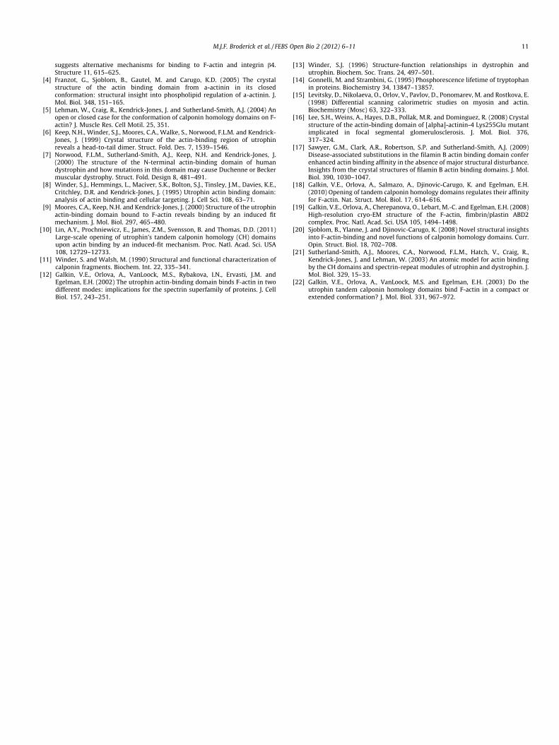
as a dimer. Moreover, because of the orientation of the individualchains within the dimer it was suggested that a three-dimensionaldomain swap may have occurred (Fig. 1). The crystal structures ofutrophin and dystrophin ABD reveal dimers in an extended confor-mation with CH domain 1 (CH1) of one crystallographic dimerinteracting with CH domain 2 (CH2) of the other crystallographicdimer (Fig. 1A and B, red and blue structures) [6,7]. The a-actininABD on the other hand, which shares considerable sequence andstructural homology [4,8], crystallised as a monomer with CH1and CH2 from the same molecule in close apposition and in anorientation similar to CH1 and CH2 from opposite dimers of theutrophin or dystrophin structure (Fig. 1A and B, green structure).
Nonetheless the idea that the utrophin ABD could exist in anopen extended and closed compact form was attractive, and toan extent supported by some of the available data [9]. One of themain arguments against the open conformation stemmed fromavailable crystal structures. With the exception of utrophin andits close homologue dystrophin, all other tandem CH domains thathad been crystallised, did so in a closed conformation. Because ofthe apparent domain swap in the utrophin and dystrophin crystals,possibly induced by the crystallisation at low pH, it has beenargued that the extended conformation is an artefact of crystallisa-tion and does not reflect a true state in solution [5], whereas morerecent studies do suggest an open conformation [10]. We havetherefore examined the binding properties of utrophin with F-actinusing native utrophin that is allowed to adopt any conformation(open or closed) and in a closed conformation by introducing
B.V. Open access under CC BY-NC-ND license.

Fig. 1. The utrophin ABD structure. (A and B) Ribbon diagrams of two differentviews of the UTR261 crystal structure 1QAG comprising a dimer with twomolecules shown in red and blue. These are overlayed with the structure of a-actinin 1WKU to demonstrate the apparent two-dimensional domain swap.Annotation to CH1 and CH2 refer to the corresponding CH domains of theUTR261 structure and the a-actinin fit. (C and D) Images of utrophin monomers inthe open conformation and closed conformation derived from the structure in (A)and (B). The position of threonine 36 and serine 241 are shown in yellow spacefill.
M.J.F. Broderick et al. / FEBS Open Bio 2 (2012) 6–11 7
cysteine residues into each CH domain to form an inter-CH domaindisulphide that locks the two CH domains together. The utrophinABD is easily expressed in bacteria, highly soluble (up to mMconcentrations) and is stable. Furthermore, unlike dystrophin anda-actinin the primary sequence contains no cysteine residuesmaking it an ideal model to introduce cysteine residues to addressfunctional changes.
2. Methods
2.1. Purification and characterisation of UTR261 cysteine mutants
The double cysteine mutant construct was generated byQuikChange mutagenesis of UTR261, first the T36C mutation wasgenerated to give UTR261T36C, and then the S242C mutation wasintroduced into UTR261T36C to generate UTR261T36C/S242C.UTR261T36C and UTR261T36C/S242C are expressed in soluble formin Escherichia coli BL21(DE3) and purified under the sameconditions as wildtype UTR261 [8]. Cleavage of UTR261 by 2-nitro-5-thiocyanobenzoic acid (NTCB) was carried out as describedpreviously [11]. Oxidation or reduction of UTR261T36C/S241C wasachieved overnight in 20 mM Tris (pH 8.0) in the presence of 4 mMo-phenanthroline and 1 mM CuSO4 or 1 mM Tris(2-carboxyethyl)phosphine hydrochloride, respectively, proteins were thendialysed back into 20 mM Tris for functional studies.
2.2. High speed co-sedimentation actin binding assays
Rabbit skeletal muscle actin was purified as described previ-ously [8]. High speed co-sedimentation of 5 lM F-actin in the pres-ence of increasing concentrations of UTR261, reduced UTR261T36C/
S241C and oxidised UTR261T36C/S241C were carried out as previouslydescribed [8].
2.3. Fluorescence spectroscopy and differential scanning calorimetry
Tryptophan fluorescence spectroscopy was measured using aShimadzu RF-5301PC spectrofluorophotometer. Protein sampleswere excited at 296 nm and fluorescence emission data wererecorded between 300 and 450 nm. Differential scanning calorim-etry (DSC) experiments were carried out in a N-DSC II differentialscanning calorimeter from Calorimetry Sciences Corp. (Provo,UT), at scanning rate of 1 K/min under 3.0 atm of pressure. DSCsamples contained 10 lM UTR261 (wildtype or mutants) usingbuffer conditions identical to those described previously [3].UTR261T36C and UTR261T36C/S241C samples under reducing condi-tions were kept with 1.0 mM DTT at all times and diluted 10-foldwith DTT-free buffer immediately before loading into calorimeter.Where stated 10 lM F-actin or 20 lM F-actin + 20 lM phalloidinwere also added.
3. Results and discussion
Based on the previous studies of de Pereda and colleagues on theplectin ABD [3], and using a notional closed conformation of theutrophin ABD derived from the crystallographic dimer (Fig. 1Cand D), we identified threonine 36 in CH1 and serine 242 in CH2that would be close together in a predicted closed conformation.UTR261 T32 was mutated to cysteine, and then using thisUTR261T36C as template, the second site was mutated to giveUTR261T36C/S242C. DNA sequencing of the mutated construct con-firmed the presence of both cysteine substitutions, which was fur-ther demonstrated by chemical cleavage at the cysteines withNTCB. As can be seen from Fig. 2B, compared to UTR261 which con-tains no cysteines, the UTR261T36C/S242C protein was susceptible tocleavage by NTCB. Furthermore, chemical oxidation of UTR261T36C/
S242C revealed a mobility shift on non-reducing SDS-PAGE consis-tent with the formation of the intra-chain disulphide, with no evi-dence of inter-chain disulphide formation leading to dimerisation(Fig. 2C). The latter was also confirmed by analytical gel filtration,with the oxidised protein eluting as a monodisperse peak with acalculated mass of 28kDa (data not shown).
Analysis of the F-actin binding properties of wild type andcysteine mutants of UTR261, in either reduced or oxidised formas shown in Fig. 3. UTR261 bound to F-actin with similar stoichi-ometry (Bmax; 1:1) and dissociation constant as reported previ-ously (19.2 ± 2.2 lM; [12,13]); however, introduction of the twocysteine residues did have an effect on the dissociation constantbut without affecting the stoichiometry. Threonine 36 is withinthe conserved KTFT motif, also termed ‘ABS1’ in earlier mappingstudies of actin binding regions within the amino-terminal actinbinding domains of dystrophin and utrophin [8]. Whether thisregion is in direct contact with F-actin or is simply required forstructural integrity of CH1 remains equivocal. CD spectra ofUTR261 and cysteine mutants showed no significant changes inoverall secondary structure (data not shown); however, therewas a reduction in tryptophan fluorescence on introduction ofT36C and S242C but there was little difference between reducedand oxidised UTR261T36C/S242C (Fig. 4). The reduction in affinityfor F-actin could be due to an effect of T36C on ABS1 or this struc-turally conserved region, and the drop in tryptophan fluorescence

Fig. 2. Purification and characterisation of UTR261 cysteine mutants. (A) SDS-PAGEof stages in the purification of UTR261T36C. Lane 1, pre-induction; 2, post-induction;3, soluble fraction; 4, post ion exchange pool; 5, purified UTR261T36C following sizeexclusion chromatography. (B) Chemical cleavage at cysteine residues. In theabsence of NTCB (�) UTR261 and UTR261T36C/S241C run as single bands, whilst in thepresence (+) of NTCB there is no cleavage of UTR261 due too the lack of cysteines,but UTR261T36C/S241C is cleaved into two prominent bands presumed based onrelative mass to correspond to the cleavage at C242 (upper band, arrowed) and C36(lower band). (C) Non-reducing SDS-PAGE of reduced (red) and incompletelyoxidised (ox) UTR261T36C/S241C which clearly demonstrates a small size shift onformation of the disulphide, marked by an arrow and S–S. Position of molecularmass standards are indicated in kDa.
Fig. 3. Binding of UTR261 to F-actin. High speed co-sedimentation of 5 lM F-actinin the presence of increasing concentrations of UTR261 (A), reduced UTR261T36C/
S241C (B) and oxidised UTR261T36C/S241C (C) were carried out as previously described[8]. Data presented are the mean of three independent experiments (mean ± SEM)with binding parameters shown within each graph.
8 M.J.F. Broderick et al. / FEBS Open Bio 2 (2012) 6–11
may result from cysteine quenching [14] of the nearby W40, andW128 which is close in the structure. Furthermore, as determinedby DSC, the Tm for all reduced proteins in solution is within 3 �C(Table 1), suggesting that there are not large scale structuralchanges. The double cysteine mutants were also slightly redshifted compared to UTR261. The oxidised form of the double cys-teine mutant, however bound to F-actin with an even lower affinitythan the reduced form (74.8 ± 19 lM reduced, 123 ± 14 lM oxi-dised) suggesting that either the open form of the ABD bound toactin better, or that a greater degree of flexibility was requiredfor the interaction with F-actin which was inhibited by lockingthe two CH domains closed.
In order to test further the conformation of UTR261 whenbound to F-actin we carried out differential scanning calorimetryon UTR261 and cysteine mutants, either alone or in the presenceof F-actin (Table 1, Fig. 5). UTR261 denatured in DSC experimentsas a single peak with Tm = 53.3 �C (Table 1, Fig. 5A). In studies con-ducted under otherwise identical conditions the Tm of UTR261 wasmuch lower than the Tm of the plectin ABD either in solution orwhen in complexed with actin: 63.9 �C vs 59.1 �C, [3]. The plectinABD DSC data were interpreted by Garcia-Alvarez et al., to suggestthat uncomplexed plectin ABD was in a closed state and plectinABD in complex with F-actin was in an open state [3]. The Tm ofUTR261 in complex with F-actin increased (rather than decreased
as in the case of plectin) but only slightly to 55.5 �C. Theseobservations, along with those of the recent studies performedusing spin labelling [10] suggest that UTR261 adopts an open con-formation in solution. To verify this we have used UTR261T36C/S242C
with two cysteines introduced at T36 and S242 positions, whichbased on the prediction from Fig. 2, the formation of disulphidebond should lock UTR261 in the closed state. In DSC experimentsoxidised UTR261T36C/S242C denatured at much higher temperaturethan UTR261 (Table 1, Fig. 5D). The Tm = 68.1 �C was as high as thatof the analogous plectin ABD T74C/S277C mutant in the oxidised,i.e. closed state [3]. This similarity suggests that we have also suc-ceeded in locking UTR261T36C/S242C in the closed conformation.
To verify that the structural effect associated with increased Tm
of oxidised UTR261T36C/S242C is due to S–S cross-linking and notcysteine mutation per se, we ran DSC on reduced UTR261T36C/
S242C and reduced UTR261T36C (with a single cysteine at T36). Themelting profile and Tm of UTR261T36C were very close to those ofUTR261 (Table 1, Fig. 5B) suggesting no significant effect of theT36C mutation on the conformation of UTR261 in solution. Themelting profile of reduced UTR261T36C/S242C had two peaks (Table1, Fig. 5C), which indicates the presence of two UTR261 populationswith different conformations in this sample. The greater fraction ofUTR261T36C/S242C under reduced conditions melted withTm = 56.3 �C, close to that of UTR261. It is logical to assume that thispopulation is in the open state in solution. The smaller fraction ofreduced UTR261T36C/S242C melted with Tm = 68.6 �C, very close tothat of oxidised UTR261T36C/S242C. This population likely exists inthe closed state. Since no cross-linking was detected on theSDS-PAGE for reduced UTR261T36C/S242C, this result indicate thatintroduction of two cysteines in the CH1–CH2 interface shifts the

Fig. 4. Tryptophan fluorescence of UTR261 and cysteine mutants. Tryptophan fluorescence of 30 lM samples of each of UTR261 (red), UTR261T36C, (green) reducedUTR261T36C/S241C, (dark blue) and oxidised UTR261T36C/S241C (light blue). The introduction of cysteines slightly reduced the fluorescence emission, and furthermore thepresence of two cysteines caused a slight red-shift of the spectrum whether the UTR261T36C/S241C was reduced or oxidised.
Table 1Denaturation temperatures for DSC scans shown in Fig. 5.
Proteins Tm1 (�C) Tm2 (�C) Tm3 (�C)
UTR261 WT 53.3 – –UTR261 1C 52.6 – –UTR261 2C reduced 56.3 68.6 –UTR261 2C oxidised – 68.1 –F-actin – – 69.1UTR261 WT + F-actin 55.5 – 69.8UTR261 1C + F-actin 55.0 – 68.7UTR261 2C reduced + F-actin – Shoulder at �67 69.7UTR261 2C oxidised + F-actin – Shoulder at �67 69.6F-actin-phalloidin – – 80.0UTR261 WT + F-actin-phalloidin 56.2 – 79.8UTR261 1C + F-actin-phalloidin 56.5 Shoulder at �69 80.6UTR261 2C reduced + F-actin-
phalloidin– 68.5 80.8
UTR261 2C oxidised + F-actin-phalloidin
– 68.2 79.4
The absolute errors in Tm values did not exceed 0.2 �C. WT = wildtype sequence,UTR261 1C = UTR261T36C and UTR261 2C = UTR261T36C/S242C.
M.J.F. Broderick et al. / FEBS Open Bio 2 (2012) 6–11 9
equilibrium towards the closed state. Overall, DSC analysis ofUTR261, UTR261T36C and UTR261T36C/S242C preparations demon-strated that similar to plectin ABD, utrophin ABD can adopt twoconformations, closed and open. However, in contrast to plectinABD which exists in a predominantly closed state in solution,unmodified utrophin ABD in solution is likely to be in a predomi-nantly open state.
In the presence of F-actin the Tm of UTR261 increased to 55.5 �C(Table 1, Fig 5A). While this increase reflects UTR261 binding toF-actin, the relatively small amplitude of the effect (2.2 �C) indi-cates that there are no major changes in the conformation ofUTR261 and thus, it is likely that it binds F-actin in the same stateas in solution ie presumed to be open. Interestingly, unmodifiedplectin ABD also bound F-actin in the open state [3]. The behaviourof UTR261T36C in the presence of F-actin (Fig. 5D) was very similarto that of UTR261. In the DSC profile of F-actin complexed with oxi-dised UTR261T36C/S242C (Fig. 5D), the main peak with Tm = 69.7 �C(associated with melting of F-actin) has a shoulder at �67 �C. Thisshoulder likely represents melting of UTR261T36C/S242C. To resolvethe peaks of F-actin and oxidised UTR261T36C/S242C we haverepeated this experiment in the presence of phalloidin. As reportedbefore [15] and seen in Fig. 5, phalloidin increases the meltingtemperature of F-actin by �10 �C as a result of its strong stabilizing
effect on the inter-subunit contacts in the actin filaments. In thesample with phalloidin we have also doubled the amount ofF-actin to check if the actin effect on UTR261 conformation is sat-urated. It can be seen clearly that melting profiles of oxidisedUTR261T36C/S242C in the presence and absence of phalloidin-F-actinare very similar (Table 1, Fig. 5D). Thus, oxidised UTR261T36C/S242C
binds phalloidin-F-actin in the closed state. To verify thatphalloidin does not alter the interaction of UTR261 with F-actinwe also performed DSC on WT UTR261 in the presence ofphalloidin-F-actin. Results showed that the effects of F-actin andphalloidin-F-actin on the conformation of WT UTR261 are similar(Table 1, Fig. 5A).
The melting profile of the reduced UTR261T36C/S242C in the com-plex with F-actin (Fig. 5C) was very similar to that of oxidisedUTR261T36C/S242C (Fig. 5D). Again, to resolve reduced UTR261T36C/
S242C and F-actin peaks we repeated the experiment with phalloi-din. As one can see, in both scans reduced UTR261T36C/S242C meltsas a single peak with Tm �68 �C (Table 1, Fig. 5C). Thus, the vastmajority of the reduced UTR261T36C/S242C molecules adopt theclosed conformation on F-actin, while in the absence of F-actinmore molecules (�60%) are in the open state (Fig. 5D). Theseresults indicate that for UTR261T36C/S242C mutant F-actin favoursthe closed state. Whilst the vast majority of WT UTR261 bindsF-actin in the open state, we cannot exclude that a small fractionmay be in the closed state on F-actin.
In other tandem-CH domain ABD structures such as those fora-actinin or filamin, mutations in the inter-CH domain interfaceaffect actin binding [16,17]. In all cases the mutations in theCH1:CH2 interface region do not alter the gross structural confor-mation, in that both a-actinin 4 and filamin B crystal structuresadopt a compact structure whether or not the mutations are pres-ent. However the presence of the mutations does increase theaffinity of both the a-actinin and filamin ABDs for F-actin[16,17]. The a-actinin mutants appeared to retain their compactshape as determined by analytical ultracentrifugation [16],whereas the filamin B mutants are also associated with a reductionin the melting temperatures for this ABD. This would argue at thevery least in favour of inter-CH domain rearrangement on bindingto F-actin, or even the possibility of the proteins adopting an openconformation as shown previously [18]. By contrast the cysteinemutants in utrophin increased the melting temperature and re-duced actin binding suggesting that a loss of CH1–CH2 flexibilityreduced their affinity for F-actin. The highest resolution cryo-EM

Fig. 5. Differential scanning calorimetry. DSC traces of UTR261 and cysteine mutants alone and in the presence of 10 lM F-actin or 20 lM F-actin + 20 lM phalloidin. UTR261(A), UTR261T36C (B), reduced UTR261T36C/S241C (C), and oxidised UTR261T36C/S241C (D) scans are shown in red in all traces, F-actin alone in green dashed lines, F-actin with thecorresponding UTR261 protein in solid green line. F-actin stabilised with phalloidin alone in blue dashed lines, F-actin/phalloidin with the corresponding UTR261 protein insolid blue line.
10 M.J.F. Broderick et al. / FEBS Open Bio 2 (2012) 6–11
reconstructions of F-actin and F-actin with a tandem-CH domainABD – that of fimbrin, however, demonstrate unequivocally thatthe two CH domains remain in a closed conformation with very lit-tle rearrangement required to match the crystal structure [19] andreviewed in [20]. However the situation regarding the utrophinABD is less clear.
A number of cryo-EM reconstructions of UTR261 with F-actinusing different methods of analysis have arrived at different con-clusions. The earliest models had been derived from helical recon-structions had proposed that utrophin bound to F-actin in an openconformation, but that there was an induced fit onto actin requir-ing some rearrangement of the orientation of the CH domainsrelative to their position in the crystal structure [9]. However usinga different method of analysis – iterative helical real space recon-struction, the Egelman group arrived at an alternative model[12]. In this model, although again the utrophin ABD was fittedin an open conformation, it was able to interact with F-actin intwo different states depending on whether one or both CH do-mains were in contact with actin. Furthermore, in the Egelmanstudy, questions were raised over the validity of using helical aver-aging techniques to derive a reconstruction from heterogeneouslydecorated actin filaments, and also as to the polarity of the fila-ment used in the reconstructions [12]. However a further recon-struction comprising the utrophin ABD and the first spectrinrepeat bound to F-actin, arrived at a third model – that of a closedconformation for the utrophin (and dystrophin) ABD on F-actin[21]. A further reassessment of all the evidence by the Egelmanlab provided convincing arguments for utrophin binding to actinin different modes but in an open conformation, see [22] and dis-cussions therein. The actin binding and DSC data presented hereindicate that utrophin ABD binds to actin in an open conformationand add further compelling weight to the open conformation
hypothesis. More recently, and despite evidence from solutionstudies and crystal structures of a closed conformation fora-actinin CH domains [4,22], [16] a cryo-EM reconstruction ofa-actinin bound to F-actin predicted an open conformation [18].The use of cysteine mutagenesis has also been employed in a re-cent electron paramagnetic resonance study by Lin and colleagues[10] to examine the opening and closing of the utrophin CH do-mains in solution and on binding to actin. Interestingly, in solutionthey identified a conformation almost identical to that of a singleutrophin ABD as seen in the crystal structure (as in Fig. 1C) butin apparent equilibrium with an equally abundant species with amore closed conformation. However on binding to actin, there isonly one population evident and this has an even more open con-formation [10]. Thus the authors also conclude that utrophin bindsto actin in an open conformation, but via an induced fit mecha-nism, ironically a conclusion also reached from the earliest EMreconstructions a decade earlier [9].
Acknowledgements
This work was funded by an MRC studentship to M.J.F.B. andMRC Career Establishment Grant G0000104 to S.J.W. We gratefullyacknowledge the assistance of Dr. Rosie Staniforth with CD andtryptophan fluorescence experiments.
References
[1] Gimona, M., Djinovic-Carugo, K., Kranewitter, W. and Winder, S. (2002)Functional plasticity of CH domains. FEBS Lett. 513, 98–106.
[2] Winder, S.J. (2003) Structural insights into actin-binding, branching andbundling proteins. Curr. Opin. Cell Biol. 15, 14–22.
[3] Garcia-Alvarez, B., Bobkov, A., Sonnenberg, A. and Pereda, J.M.D. (2003)Structural and functional analysis of the actin binding domain of plectin
M.J.F. Broderick et al. / FEBS Open Bio 2 (2012) 6–11 11
suggests alternative mechanisms for binding to F-actin and integrin b4.Structure 11, 615–625.
[4] Franzot, G., Sjoblom, B., Gautel, M. and Carugo, K.D. (2005) The crystalstructure of the actin binding domain from a-actinin in its closedconformation: structural insight into phospholipid regulation of a-actinin. J.Mol. Biol. 348, 151–165.
[5] Lehman, W., Craig, R., Kendrick-Jones, J. and Sutherland-Smith, A.J. (2004) Anopen or closed case for the conformation of calponin homology domains on F-actin? J. Muscle Res. Cell Motil. 25, 351.
[6] Keep, N.H., Winder, S.J., Moores, C.A., Walke, S., Norwood, F.L.M. and Kendrick-Jones, J. (1999) Crystal structure of the actin-binding region of utrophinreveals a head-to-tail dimer. Struct. Fold. Des. 7, 1539–1546.
[7] Norwood, F.L.M., Sutherland-Smith, A.J., Keep, N.H. and Kendrick-Jones, J.(2000) The structure of the N-terminal actin-binding domain of humandystrophin and how mutations in this domain may cause Duchenne or Beckermuscular dystrophy. Struct. Fold. Design 8, 481–491.
[8] Winder, S.J., Hemmings, L., Maciver, S.K., Bolton, S.J., Tinsley, J.M., Davies, K.E.,Critchley, D.R. and Kendrick-Jones, J. (1995) Utrophin actin binding domain:analysis of actin binding and cellular targeting. J. Cell Sci. 108, 63–71.
[9] Moores, C.A., Keep, N.H. and Kendrick-Jones, J. (2000) Structure of the utrophinactin-binding domain bound to F-actin reveals binding by an induced fitmechanism. J. Mol. Biol. 297, 465–480.
[10] Lin, A.Y., Prochniewicz, E., James, Z.M., Svensson, B. and Thomas, D.D. (2011)Large-scale opening of utrophin’s tandem calponin homology (CH) domainsupon actin binding by an induced-fit mechanism. Proc. Natl. Acad. Sci. USA108, 12729–12733.
[11] Winder, S. and Walsh, M. (1990) Structural and functional characterization ofcalponin fragments. Biochem. Int. 22, 335–341.
[12] Galkin, V.E., Orlova, A., VanLoock, M.S., Rybakova, I.N., Ervasti, J.M. andEgelman, E.H. (2002) The utrophin actin-binding domain binds F-actin in twodifferent modes: implications for the spectrin superfamily of proteins. J. CellBiol. 157, 243–251.
[13] Winder, S.J. (1996) Structure-function relationships in dystrophin andutrophin. Biochem. Soc. Trans. 24, 497–501.
[14] Gonnelli, M. and Strambini, G. (1995) Phosphorescence lifetime of tryptophanin proteins. Biochemistry 34, 13847–13857.
[15] Levitsky, D., Nikolaeva, O., Orlov, V., Pavlov, D., Ponomarev, M. and Rostkova, E.(1998) Differential scanning calorimetric studies on myosin and actin.Biochemistry (Mosc) 63, 322–333.
[16] Lee, S.H., Weins, A., Hayes, D.B., Pollak, M.R. and Dominguez, R. (2008) Crystalstructure of the actin-binding domain of [alpha]-actinin-4 Lys255Glu mutantimplicated in focal segmental glomerulosclerosis. J. Mol. Biol. 376,317–324.
[17] Sawyer, G.M., Clark, A.R., Robertson, S.P. and Sutherland-Smith, A.J. (2009)Disease-associated substitutions in the filamin B actin binding domain conferenhanced actin binding affinity in the absence of major structural disturbance.Insights from the crystal structures of filamin B actin binding domains. J. Mol.Biol. 390, 1030–1047.
[18] Galkin, V.E., Orlova, A., Salmazo, A., Djinovic-Carugo, K. and Egelman, E.H.(2010) Opening of tandem calponin homology domains regulates their affinityfor F-actin. Nat. Struct. Mol. Biol. 17, 614–616.
[19] Galkin, V.E., Orlova, A., Cherepanova, O., Lebart, M.-C. and Egelman, E.H. (2008)High-resolution cryo-EM structure of the F-actin, fimbrin/plastin ABD2complex. Proc. Natl. Acad. Sci. USA 105, 1494–1498.
[20] Sjoblom, B., Ylanne, J. and Djinovic-Carugo, K. (2008) Novel structural insightsinto F-actin-binding and novel functions of calponin homology domains. Curr.Opin. Struct. Biol. 18, 702–708.
[21] Sutherland-Smith, A.J., Moores, C.A., Norwood, F.L.M., Hatch, V., Craig, R.,Kendrick-Jones, J. and Lehman, W. (2003) An atomic model for actin bindingby the CH domains and spectrin-repeat modules of utrophin and dystrophin. J.Mol. Biol. 329, 15–33.
[22] Galkin, V.E., Orlova, A., VanLoock, M.S. and Egelman, E.H. (2003) Do theutrophin tandem calponin homology domains bind F-actin in a compact orextended conformation? J. Mol. Biol. 331, 967–972.


![CYTOSKELETON NEWS - fnkprddata.blob.core.windows.net · Dynamic remodeling of the actin cytoskeleton [i.e., rapid cycling between filamentous actin (F-actin) and monomer actin (G-actin)]](https://img.dokumen.tips/doc/110x75/609edd2b88630103265d18ee/cytoskeleton-news-dynamic-remodeling-of-the-actin-cytoskeleton-ie-rapid-cycling.jpg)