Embed Size (px)
Citation preview

Vol. 18, No. 5JOURNAL OF CLINICAL MICROBIOLOGY, Nov. 1983, p. 1167-11730095-1137/83/111167-07$02.00/0Copyright © 1983, American Society for Microbiology
Utility of Egg Yolk Medium for Cultivation of Mycoplasmapneumoniae
TSUGUO SASAKI,* MIHARU SHINTANI, AND KOJO KIHARA
Department of General Biologics Control, National Institute of Health, Shinagawa-ku, Tokyo 141, Japan
Received 4 April 1983/Accepted 8 August 1983
Chicken egg yolk broth medium containing 10% (vol/vol) phosphate-bufferedsaline extract of egg yolk instead of horse serum showed better growth-promotingactivity for Mycoplasma pneumoniae than did Chanock medium and was effectivein promoting the growth of small numbers of M. pneumoniae. Moreover, thephosphate-buffered saline extract of egg yolk proved superior to horse serum inthe following respects: (i) it was consistent in growth-promoting activity for M.pneumoniae among different batches; (ii) it could be preserved at 4°C for at least 2years; (iii) it is inexpensive and easy to obtain; and (iv) it contains large amountsof lipoproteins, cholesterol, and phospholipids.
Mycoplasmas require cholesterol for growth.Among the many mammalian sera, horse serum(HS) is widely used as an excellent cholesterolsource in the cultivation of mycoplasmas, butthe growth-promoting ability of HS for myco-plasmas often differs considerably among differ-ent lots of serum. For diagnosis of Mycoplasmapneumoniae infection, the quality of HS has adirect effect on the isolation of M. pneumoniae,and thus it is important to obtain HS of goodquality. In addition, when large quantities ofmycoplasmal antigen are to be prepared, the useof HS becomes costly.Many investigators have tried to find other
materials to replace HS in the growth mediumfor mycoplasmas (8, 10, 11, 24, 26). Among themany substitutes tried, chicken egg yolks havebeen found by Hayatsu (8), Jensen et al. (10),and Tamura et al. (24) to be one of the mostsuitable. They have used egg yolks extractedwith organic solvents or with distilled water forcultivation of mycoplasmas.
Considering the instability of lipoproteins inegg yolks, which are the natural cholesterolsource for the growth of mycoplasmas in vitro(16), it may be undesirable to denature thelipoproteins with organic solvents. Although na-tive lipoproteins are not always necessary tosustain the growth of mycoplasmas (4, 25), it isdifficult to dissolve the lipid components sepa-rated from the lipoproteins with organic solventsin the medium, and these components are not aseffective in promoting the growth of mycoplas-mas as is HS (8, 10). Although the growth-promoting ability of the water extract of eggyolks is comparable to that of HS (8, 24), theextract is unstable and increases in turbidityduring long periods of storage.
In the present study, we investigated theefficacy of the supernatant of a chicken egg yolksuspension in phosphate-buffered saline (re-ferred to as EY) for cultivation of M. pneumo-niae by comparing EY-containing medium withthe standard medium reported by Chanock et al.(2), which contains HS. We found that EY wasstable over a long period and that its growth-promoting ability was superior to that of HS.
MATERIALS AND METHODS
Mycoplasmas. The FH and Mac strains of M. pneu-moniae have been maintained for many years in ourlaboratory. Organisms grown in seed culture mediumwere stored at -20°C until use.
Preparation of EY. Fresh unfertilized chicken eggs(White Leghorn) were disinfected in 0.1% (wt/vol)Hibitane digluconate solutions (Sumitomo ChemicalIndustries, Ltd., Osaka) for 10 min and air dried. Theegg yolks were suspended in sterile phosphate-buff-ered saline without Ca2' and Mg2+ [PBS(-)] at aconcentration of 20% (wt/vol). After being stirredthoroughly, the suspension was centrifuged at 15,000x g for 40 min. Penicillin G was added to the superna-tant fluid to a concentration of 500 IU/ml. This super-natant fluid is referred to as EY. After passing tests forabsence of mycoplasmas and for growth-promotingactivity for M. pneumoniae, EY was stored at 4°C inglass vessels with rubber stoppers.
Yeast extract. Yeast extract was prepared by heatinga 25% (wtlvol) aqueous suspension of Nitten DryYeast (Nippon Beet Sugar Manufacturing Co., Ltd.,Tokyo) in a boiling water bath for 30 min; it was storedat -20°C until use (14).
Media. Basal broth medium consisted of 2.1%(wt/vol) PPLO broth (lot 671893, Difco Laboratories,Detroit, Mich.), 1% (wt/vol) glucose, and 0.002%(wt/vol) phenol red. Basal agar medium consisted of2.1% (wt/vol) PPLO broth and 1.2% (wt/vol) Wakospecial agar B (Wako Pure Chemical Industries, Ltd.,Osaka).
1167
on July 6, 2018 by guesthttp://jcm
.asm.org/
Dow
nloaded from

1168 SASAKI, SHINTANI, AND KIHARA
The seed culture medium consisted of 87% (vol/vol)basal broth medium and 13% (vol/vol) serum fractionfor mycoplasma (Kitasato Institute, Tokyo). The agarmedium for colony counting consisted of 77% (vol/vol)basal agar medium, 13% (vol/vol) serum fraction formycoplasma, and 10% (vol/vol) yeast extract.The broth media to be tested for their growth-
promoting activity for M. pneumoniae were as fol-lows: HS medium consisted of 80% (vol/vol) basalbroth medium and 20% (vol/vol) HS (lot 29211115;Flow Laboratories, Rockville, Md.). Chanock mediumconsisted of 70% (vol/vol) basal broth medium, 20%(vol/vol) HS, and 10% (vol/vol) yeast extract (2). EYmedium consisted of90% (vol/vol) basal broth mediumand 10% (vol/vol) EY. EYY medium consisted of 80%(vol/vol) basal broth medium, 10% (vol/vol) EY, and10% (vol/vol) yeast extract. A single lot of eachingredient was used for all the test media.
Separation of lipoprotein fractions in EY. The meth-od used was essentially that of Havel et al. (7).Lipoproteins in EY were separated sequentially intofour fractions of increasing density (d): fraction 1 (Fr.1), d was <1.019; Fr. 2, d was 1.019 to 1.063; Fr.3, dwas 1.063 to 1.12; and Fr. 4, d was 1.12 to 1.21. Thedensity of the background salt solution was adjusted to1.019 by the addition of solid NaCl. The EY solutionwas ultracentrifuged at 34,500 rpm for 20 h in aBeckman SW 41 Ti rotor and cellulose nitrate tubes(14 by 89 mm) without caps. The top layer (Fr.1) wasreemulsified in PBS(-), the solvent density of theinfranatant was adjusted to 1.063 with NaCl, andultracentrifugation was repeated. The top layer (Fr. 2)was reemulsified in PBS (-), and Fr.3 was obtained inthe same manner. Fr.4 was obtained by the addition ofsolid NaBr in place of NaCl. All fractions were dia-lyzed for 24 h against 1,000 volumes of PBS(-)containing penicillin G at 500,000 IU/liter. After dialy-sis, each fraction was sterilized by filtration through a0.45-,um filter (Millipore Corp., Bedford, Mass.).
Assessment of mycoplasmal growth on agar media.The surfaces of agar plates to be tested for theirgrowth-promoting activity were spotted with 10 0.01-ml portions of each of serial dilutions of a mycoplas-mal suspension. In 10 spots in which an appropriatedilution was inoculated, the number of colonies wasdetermined and expressed as the log CFU per milliliterof the original suspension.The diameters of 12 colonies randomly selected in
one spot which contained about 30 to 70 colonies weremicroscopically measured and expressed as the divi-sion of the micrometer. One division of the microme-ter equaled 35 p.m.
Assessment of mycoplasmal growth in the test brothmedia. After inoculation of 1 ml of an appropriatedilution of seed culture into 200 ml of each of the testbroth media, the media were dispensed in 15-ml quan-tities into test tubes with cotton plugs and incubated at37°C. Every day or two, one tube was taken from eachgroup of the test media, four 0.01-ml portions of serialdilutions of the culture in the tube were inoculatedonto the agar plates for colony counting, and the pH ofthe medium in the tube was sometimes measured. Theconcentration of the viable organisms in the brothculture was expressed as the log CFU per milliliter.
Chemical analysis. Protein and total lipids weredetermined by the method of Gornall et al. (6) and themethod of Pande et al. (15), respectively. Total choles-
J. CLIN. MICROBIOL.
terol and free cholesterol were determined by enzy-matic methods with the commercial kits CholesterolC-Test (Wako) and Free Cholesterol C-Test (Wako),respectively. Phospholipids were determined by anenzymatic method with the commercial kit Phospho-lipids B-Test (Wako).
RESULTS
Transparency of EY medium. Many granulesand lipid lumps were contained in the egg yolk-PBS(-) suspensions before centrifugation.When such a suspension was added to the basalagar medium, it was impossible to distinguishthe mycoplasmal colonies from the lipid lumpsdispersed in the agar medium. However, theselipids and granules were easily removed fromthe suspension by centrifugation.The supernatant fluid obtained at a centrifugal
force of 5,000 x g for 40 min contained very fewlipid lumps, and no lipid lumps were found in thesupernatant fluid obtained at 7,500 x g or high-er. Moreover, with increases in centrifugalforce, clearer supernatant fluids were obtained.Accordingly, the conditions for centrifugationwere fixed at 15,000 x g for 40 min. EY agar andEY broth media supplemented with 10% (vol/vol) EY were sufficiently transparent.
Preservation of EY. Attempts to preserve EYby freezing or lyophilization were completelyunsuccessful, owing to the denaturation of lipo-proteins. When EY was stored frozen for a longtime, insoluble debris formed as a result ofdissociation of lipoproteins, and it was difficultto distinguish colonies from this insoluble debrisin EY agar medium. However, EY stored at 4°Cfor 2 years showed good growth-promoting ac-tivity, equal to that of freshly prepared EY, andwas free from precipitates (data not shown).Growth of M. pneumoniae on agar media.
Various amounts of EY or HS were added to thebasal agar medium with or without 10% (vol/vol)yeast extract. After inoculation with the M.pneumoniae strains FH and Mac, the plateswere incubated aerobically for 12 days, and thenumber and diameters of colonies were deter-mined (Fig. 1). There were no distinct differences in the log CFU on the plates supplementedwith 5% (vol/vol) or more EY or HS. The colonydiameters for strain FH were markedly in-creased by the addition of yeast extract to themedium containing HS, but the colony diame-ters for neither strain were increased by theaddition of yeast extract to the medium contain-ing EY.Growth of M. pneumoniae in broth media. The
growth curves of M. pneumoniae in each of thetest broth media are presented in Fig. 2. A closecorrelation was observed between the changesin pH of each medium and the CFU in the samemedium. The M. pneumoniae strains showed
on July 6, 2018 by guesthttp://jcm
.asm.org/
Dow
nloaded from

GROWTH OF M. PNEUMONIAE IN EGG YOLK MEDIUM
D L
z
0
E E
cz U- 4-x
0 ol
FH
2.5 s lo 20 lo 20
Mac
10 20
2.5 5 10 20 10 20 2.55 10 20 10 20
Concentration of HS or EY (%.IVN)FIG. 1. Growth ofM. pneumoniae strain FH and Mac on agar media containing various concentrations of HS
or EY. Basal agar medium was supplemented with HS (O), HS and 10% (vol/vol) yeast extract (R), EY (a), andEY and 10% (vol/vol) yeast extract (El). The number of colonies formed in a spot on agar plates is expressed asthe log CFU recovered per milliliter of a mycoplasmal suspension inoculated on the agar plates, and each columnon the upper graphs represents a mean of the values enumerated in 10 spots. Each column on the lower graphsrepresents the mean diameter of 12 colonies in a spot. Bars indicate 95% confidence intervals of the means.
the most rapid growth in EY medium. In Chan-ock medium and HS medium, the CFU on thefirst day after inoculation decreased sharplybelow the number of CFU in the inoculum.Consequently, the time until maximum growthin these media was longer than that in EYmedium. In EYY medium, strain FH showedgood growth, whereas the growth of strain Macwas poor.
Relationship between inoculum size and thegrowth ofM. pneumonuze. Growth of the FH andMac strains in the four test broth media wasdetermined by the pH changes in the medium(Fig. 3). At each inoculum size, both strainsshowed a more rapid growth in EY medium thanin any other medium, as shown by an earlier pHchange. EY medium was especially effective inpromoting the growth of small numbers of M.pneumoniae organisms. In addition, EY mediumalways showed higher growth-promoting activi-ty for both strains than did EYY medium.When the chanock medium and HS medium
were inoculated with concentrations of orga-nisms of about 40 to 60 CFU/ml or lower, nochanges in pH could be observed during the 2-week incubation period, but in EY medium,
both FH and Mac strains grew even when inocu-lated at 4 and 6 CFU/ml, respectively.
Protein and lipid content of EY and HS. Table1 shows the protein, phospholipid, and choles-terol content of EY and HS. EY was character-ized by high quantities of free cholesterol andphospholipids, which were about eight timeshigher than those in HS. In contrast, theamounts of protein and esterified cholesterol inEY were about one-fourth and one-half of thosein HS, respectively.
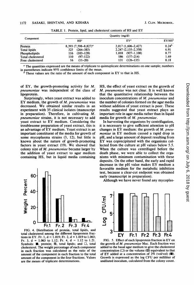
Effect of each lipoproteins fraction ofEY on thegrowth of M. pneumoniae. Figure 4 shows thedistribution of protein, total lipids, and totalcholesterol among the four lipoprotein fractionsin EY. Low-density lipoprotein fractions, Fr. 1and Fr. 2, constituted most of the lipoproteinspresent in EY.To investigate whether there are differences in
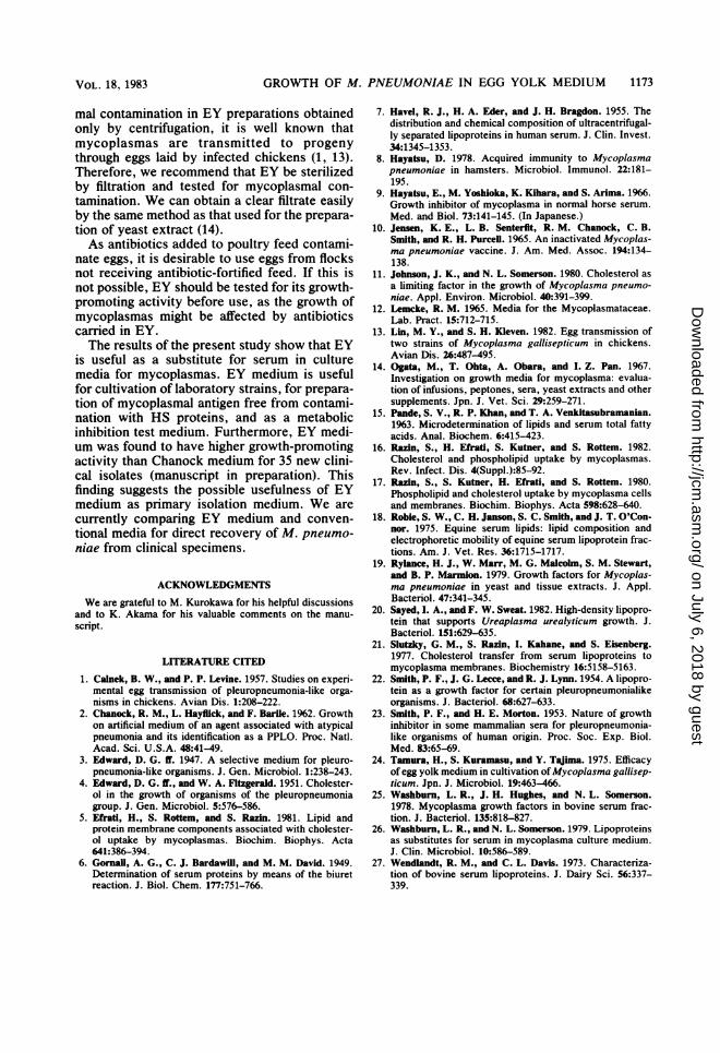
the growth-promoting activity for M. pneumo-niae strain Mac among the lipoprotein fractions,each fraction was added to the basal agar medi-um to give the cholesterol concentration or thevolume equivalent to that of EY added at aconcentration of 2% (vol/vol), which was foundby a preliminary experiment to be the minimum
VOL. 18, 1983 1169
on July 6, 2018 by guesthttp://jcm
.asm.org/
Dow
nloaded from

1170 SASAKI, SHINTANI, AND KIHARA
8
Q0.
I
I
J
FH Mac
a I,__ I --
7 9 110 1 3Days of Incubation
I I
5 7 9 ll
FIG. 2. Growth curves of M. pneumoniae strains and pH changes in the four test broth media. The meanvalue of log CFU per milliliter measured in four spots is represented by each point, and the 95% confidenceinterval of the mean is indicated by each bar. Symbols: 0, HS medium; 0, Chanock medium; A, EY medium; A,EYY medium.
concentration required to yield good growth ofM. pneumoniae (data not shown).When cholesterol was supplied at that mini-
mal amount or more to the basal medium by theEY lipoprotein fractions, there were no signifi-cant differences in the growth ofM. pneumoniaeon the agar media containing the different frac-tions (Fig. 5).
DISCUSSIONM. pneumoniae showed better growth in EY
broth medium than in Chanock broth medium.EY medium was especially effective in promot-ing the growth of small numbers of M. pneumo-niae. In addition, the growth-promoting activityof EY for M. pneumoniae was consistent among
different EY batches (data not shown). One ofthe reasons for the good growth-promoting ac-tivity of EY medium for M. pneumoniae may bethe absence of toxic substances in EY. General-ly, M. pneumoniae is sensitive to HS, and itsgrowth is often markedly affected by HS. Asshown in Fig. 2, in Chanock medium, the CFUafter incubation for 1 day decreased from that ofthe inoculum, which may be due to the presenceof toxic substances in HS (9, 23). In our experi-ence, the degree of inhibitory effect ofHS on thegrowth of M. pneumoniae differed considerablyamong different lots of HS, and the inhibitoryeffect was decreased to a great extent by heatingthe serum at 55°C for 30 min. Therefore, thetoxic substance in HS may be proteinaceous.
J. CLIN. MICROBIOL.
on July 6, 2018 by guesthttp://jcm
.asm.org/
Dow
nloaded from

GROWTH OF M. PNEUMONIAE IN EGG YOLK MEDIUM 1171
Mac
6x10²
6 CFU/ml0 2 4 6 8 10 12 14 0 2 4 6 8 10 12 14
Days of I ncubat ionFIG. 3. The relationship between the inoculum concentration and the growth of M. pneumoniae strains in the
four test broth media. Inoculum concentrations are indicated on each graph. Symbols: 0, HS medium; *,Chanock medium; A, EY medium; A, EYY medium.
Another possible explanation for the goodgrowth-promoting activity ofEY medium for M.pneumoniae is the high content of lipoproteinsrich in cholesterol and phospholipids. It is wellknown that serum lipoproteins are the naturalcholesterol source for animal mycoplasmas invitro and in vivo and that the growing cells ofmycoplasmal species take up significant quanti-ties of exogenous cholesterol and phospholipidsinto their plasma membrane from the medium (5,16, 17).HS is much richer in esterified cholesterol
than in free cholesterol; in contrast, EY con-tained much more of the latter, about eight timesthat of HS. Razin and co-workers (16, 17, 21)reported that mycoplasmal organisms incorpo-
rate more free cholesterol than esterified choles-terol into their plasma membranes during growthin a serum-containing medium. Therefore, a highcontent of phospholipids and free cholesterol inEY may be favorable for the growth of M.pneumoniae.Most of the lipoproteins in mammalian sera
belong to the high-density lipoprotein class (18,26, 27), whereas those in EY belong to the low-density lipoprotein class. Smith et al. (22) re-ported that alpha-1 lipoprotein (high-densityclass) in bovine serum is responsible for thegrowth of Mycoplasma spp., and Sayed et al.(20) also reported that alpha lipoprotein inequine serum had growth-promoting activity forUreaplasma urealyticum. However, in the case
FH8765
CL
3C0
7-
6-
7-
6-5l
7^
6-5
VOL. 18, 1983
A
--A
6 AnowI =a-dw
on July 6, 2018 by guesthttp://jcm
.asm.org/
Dow
nloaded from
1172 SASAKI, SHINTANI, AND KIHARA
TABLE 1. Protein, lipid, and cholesterol contents of HS and EYQuantity (mg/dl)
Component HS'a EYa EY/HSb
Protein 8,393 (7,598-8,827)a 2,017 (1,606-2,427) 0.24bTotal lipids 325 (264-385) 2,247 (2,135-2,358) 6.91Phospholipids 116 (103-128) 1,018 (927-1,108) 8.77Total cholesterol 110 (97-122) 186 (157-214) 1.69Free cholesterol 16 (11-20) 131 (126-135) 8.18
a The quantities expressed are the means of triplicate to quintuplicate determinations on one sample; numbersin parentheses indicate 95% confidence limits of the mean.
b These values are the ratio of the amount of each component in EY to that in HS.
of EY, the growth-promoting activity for M.pneumoniae was independent of the class oflipoprotein.
Surprisingly, when yeast extract was added toEY medium, the growth of M. pneumoniae wasdecreased. We obtained similar results in anexperiment with 35 clinical isolates (manuscriptin preparation). Therefore, in cultivating M.pneumoniae strains, it is not necessary to addyeast extract to EY medium. Considering thetroublesome preparation of yeast extract, this isan advantage of EY medium. Yeast extract is animportant constituent of the media for growth ofsome mycoplasma strains (3, 12), but little isknown about the nature of growth-promotingfactors in yeast extract (19). We showed thatcolony size of M. pneumoniae became larger bythe addition of yeast extract to agar mediumcontaining HS, but in liquid media containing
60c
0L-
i 203~
0Fr.1 Fr. 2 Fr.3 Fr.4
FIG. 4. Distribution of protein, total lipids, andtotal cholesterol among the different lipoprotein frac-tions in EY. Fr. 1, d < 1.019; Fr. 2, d = 1.019 to 1.063;Fr. 3, d = 1.063 to 1.12; Fr. 4, d = 1.12 to 1.21.Symbols: U, protein; 13, total lipids; and O, totalcholesterol. The weight percentage of each componentin each fraction was calculated as the ratio of theamount of the component in each fraction to the totalamount of the component in the four fractions. Valuesare the means of triplicate determinations.
HS, the effect of yeast extract on the growth ofM. pneumoniae was not clear. It is well knownthat the quantitative relationship between theinoculum concentrations of M. pneumoniae andthe number of colonies formed on the agar mediawithout addition of yeast extract is poor. Theseresults suggested that yeast extract plays animportant role in agar media rather than in liquidmedia for growth of M. pneumoniae.
In harvesting the organisms by centrifugation,it is necessary to give sufficient attention to pHchanges in EY medium: the growth of M. pneu-moniae in EY medium caused a rapid drop inpH, and a large amount of deposit resulting fromEY was coprecipitated with the organisms col-lected from the culture at pH values below 5.5.When the culture was centrifuged before thedeath phase, we were able to collect the orga-nisms with minimum contamination with thesedeposits. On the other hand, the early and rapiddecrease in the pH value makes EY medium afavorable medium for the metabolic inhibitiontest, because a clear-cut endpoint was obtainedearly (manuscript in preparation).Although we have never found any mycoplas-
-'7*E
EV Fril Fr.2 Fr.3 Fr.4FIG. 5. Effect of each lipoprotein fraction in EY on
the growth of M. pneumoniae Mac. Each fraction wasadded to the basal agar medium to give the cholesterolconcentration (LO) or the volume (E2l) equivalent to thatof EY added at a concentration of 2% (vol/vol) (U).Growth is expressed as the log CFU per milliliter ofundiluted inoculum, calculated from the colony count.
J. CLIN. MICROBIOL.
on July 6, 2018 by guesthttp://jcm
.asm.org/
Dow
nloaded from
GROWTH OF M. PNEUMONIAE IN EGG YOLK MEDIUM 1173
mal contamination in EY preparations obtainedonly by centrifugation, it is well known thatmycoplasmas are transmitted to progenythrough eggs laid by infected chickens (1, 13).Therefore, we recommend that EY be sterilizedby filtration and tested for mycoplasmal con-tamination. We can obtain a clear filtrate easilyby the same method as that used for the prepara-tion of yeast extract (14).As antibiotics added to poultry feed contami-
nate eggs, it is desirable to use eggs from flocksnot receiving antibiotic-fortified feed. If this isnot possible, EY should be tested for its growth-promoting activity before use, as the growth ofmycoplasmas might be affected by antibioticscarried in EY.The results of the present study show that EY
is useful as a substitute for serum in culturemedia for mycoplasmas. EY medium is usefulfor cultivation of laboratory strains, for prepara-tion of mycoplasmal antigen free from contami-nation with HS proteins, and as a metabolicinhibition test medium. Furthermore, EY medi-um was found to have higher growth-promotingactivity than Chanock medium for 35 new clini-cal isolates (manuscript in preparation). Thisfinding suggests the possible usefulness of EYmedium as primary isolation medium. We arecurrently comparing EY medium and conven-tional media for direct recovery of M. pneumo-niae from clinical specimens.
ACKNOWLEDGMENTS
We are grateful to M. Kurokawa for his helpful discussionsand to K. Akama for his valuable comments on the manu-
script.
LITERATURE CITED
1. Calnek, B. W., and P. P. Levine. 1957. Studies on experi-mental egg transmission of pleuropneumonia-like orga-nisms in chickens. Avian Dis. 1:208-222.
2. Chanock, R. M., L. Hayflick, and F. Barile. 1962. Growthon artificial medium of an agent associated with atypicalpneumonia and its identification as a PPLO. Proc. Natl.Acad. Sci. U.S.A. 48:41-49.
3. Edward, D. G. ff. 1947. A selective medium for pleuro-pneumonia-like organisms. J. Gen. Microbiol. 1:238-243.
4. Edward, D. G. ff., and W. A. Fitzgerald. 1951. Cholester-ol in the growth of organisms of the pleuropneumoniagroup. J. Gen. Microbiol. 5:576-586.
5. Efrati, H., S. Rottem, and S. Razin. 1981. Lipid andprotein membrane components associated with cholester-ol uptake by mycoplasmas. Biochim. Biophys. Acta641:386-394.
6. Gornall, A. G., C. J. Bardawill, and M. M. David. 1949.Determination of serum proteins by means of the biuretreaction. J. Biol. Chem. 177:751-766.
7. Havel, R. J., H. A. Eder, and J. H. Bragdon. 1955. Thedistribution and chemical composition of ultracentrifugal-ly separated lipoproteins in human serum. J. Clin. Invest.34:1345-1353.
8. Hayatsu, D. 1978. Acquired immunity to Mycoplasmapneumoniae in hamsters. Microbiol. Immunol. 22:181-195.
9. Hayatsu, E., M. Yoshioka, K. Kihara, and S. Arima. 1966.Growth inhibitor of mycoplasma in normal horse serum.Med. and Biol. 73:141-145. (In Japanese.)
10. Jensen, K. E., L. B. Senterfit, R. M. Chanock, C. B.Smith, and R. H. Purcell. 1965. An inactivated Mycoplas-ma pneumoniae vaccine. J. Am. Med. Assoc. 194:134-138.
11. Johnson, J. K., and N. L. Somerson. 1980. Cholesterol asa limiting factor in the growth of Mycoplasma pneumo-niae. Appl. Environ. Microbiol. 40:391-399.
12. Lemcke, R. M. 1965. Media for the Mycoplasmataceae.Lab. Pract. 15:712-715.
13. Lin, M. Y., and S. H. Kleven. 1982. Egg transmission oftwo strains of Mycoplasma gallisepticum in chickens.Avian Dis. 26:487-495.
14. Ogata, M., T. Ohta, A. Obara, and I. Z. Pan. 1967.Investigation on growth media for mycoplasma: evalua-tion of infusions, peptones, sera, yeast extracts and othersupplements. Jpn. J. Vet. Sci. 29:259-271.
15. Pande, S. V., R. P. Khan, and T. A. Venkitasubramanian.1963. Microdetermination of lipids and serum total fattyacids. Anal. Biochem. 6:415-423.
16. Razin, S., H. Efrati, S. Kutner, and S. Rottem. 1982.Cholesterol and phospholipid uptake by mycoplasmas.Rev. Infect. Dis. 4(Suppl.):85-92.
17. Razin, S., S. Kutner, H. Efrati, and S. Rottem. 1980.Phospholipid and cholesterol uptake by mycoplasma cellsand membranes. Biochim. Biophys. Acta 598:628-640.
18. Roble, S. W., C. H. Janson, S. C. Smith, and J. T. O'Con-nor. 1975. Equine serum lipids: lipid composition andelectrophoretic mobility of equine serum lipoprotein frac-tions. Am. J. Vet. Res. 36:1715-1717.
19. Rylance, H. J., W. Marr, M. G. Malcoln, S. M. Stewart,and B. P. Marmion. 1979. Growth factors for Mycoplas-ma pneumoniae in yeast and tissue extracts. J. Appl.Bacteriol. 47:341-345.
20. Sayed, I. A., and F. W. Sweat. 1982. High-density lipopro-tein that supports Ureaplasma urealyticum growth. J.Bacteriol. 151:629-635.
21. Slutzky, G. M., S. Razin, I. Kahane, and S. Eisenberg.1977. Cholesterol transfer from serum lipoproteins tomycoplasma membranes. Biochemistry 16:5158-5163.
22. Smith, P. F., J. G. Lecce, and R. J. Lynn. 1954. A lipopro-tein as a growth factor for certain pleuropneumonialikeorganisms. J. Bacteriol. 68:627-633.
23. Smith, P. F., and H. E. Morton. 1953. Nature of growthinhibitor in some mammalian sera for pleuropneumonia-like organisms of human origin. Proc. Soc. Exp. Biol.Med. 83:65-69.
24. Tamura, H., S. Kuramasu, and Y. Tajima. 1975. Efficacyof egg yolk medium in cultivation ofMycoplasma gallisep-ticum. Jpn. J. Microbiol. 19:463-466.
25. Washburn, L. R., J. H. Hughes, and N. L. Somerson.1978. Mycoplasma growth factors in bovine serum frac-tion. J. Bacteriol. 135:818-827.
26. Washburn, L. R., and N. L. Somerson. 1979. Lipoproteinsas substitutes for serum in mycoplasma culture medium.J. Clin. Microbiol. 10:586-589.
27. Wendlandt, R. M., and C. L. Davis. 1973. Characteriza-tion of bovine serum lipoproteins. J. Dairy Sci. 56:337-339.
VOL. 18, 1983
on July 6, 2018 by guesthttp://jcm
.asm.org/
Dow
nloaded from


