Embed Size (px)
Citation preview

Neurology: Clinical Practice Publish Ahead of PrintDOI: 10.1212/CPJ.0000000000001036
Utility of MRI Enhancement Pattern in Myelopathies with Longitudinally-extensive T2-lesions
Rafid Mustafa, MD,1 Theodore J. Passe, MD,2 Alfonso S. Lopez-Chiriboga, MD,3 Brian G.
Weinshenker, MD,1 Karl N. Krecke, MD,2 Nicholas L. Zalewski, MD,1 Felix E. Diehn, MD,2 Elia Sechi, MD,1 Jay Mandrekar, PhD,4 Timothy J. Kaufmann, MD,2 Padraig P. Morris, MD,2
Sean J. Pittock, MD,1,5 Michel Toledano, MD,1 Giuseppe Lanzino, MD,6 Allen J. Aksamit, MD,1 Neeraj Kumar, MD,1 Claudia F. Lucchinetti, MD,1 and Eoin P. Flanagan, MB, BCh.1,5
This is an open access article distributed under the terms of the Creative Commons Attribution-
NonCommercial-NoDerivatives License 4.0 (CC BY-NC-ND), which permits downloading and
sharing the work provided it is properly cited. The work cannot be changed in any way or used
commercially without permission from the journal.
Neurology® Clinical Practice Published Ahead of Print articles have been peer reviewed and
accepted for publication. This manuscript will be published in its final form after copyediting,
page composition, and review of proofs. Errors that could affect the content may be corrected
during these processes.
Copyright © 2021 The Author(s). Published by Wolters Kluwer Health, Inc. on behalf of the American Academy of Neurology.

Departments of Neurology,1 Radiology,2 Biostatistics,4 Laboratory Medicine and Pathology,5 and Neurologic Surgery,6 Mayo Clinic College of Medicine & Science, Rochester, MN, USA.
Department of Neurology, Mayo Clinic Florida, Jacksonville, FL, USA.3
Submission type: Article Title character count: 91 Abstract word count: 250 Total text word count: 2,857 Number of tables: 5 Number of figures: 2 References: 29 Corresponding Author: Eoin P. Flanagan [email protected]
Statistical Analysis was conducted by Jay Mandrekar, PhD, Department of Biostatistics, Mayo Clinic.
Search Terms: (1) MRI; (2) spinal cord; (3) transverse myelitis.
Study funding: No targeted funding reported.
Disclosures:
Dr. Mustafa reports no disclosures.
Dr. Passe reports no disclosures.
Dr. Lopez-Chiriboga reports no disclosures.
Dr. Weinshenker receives royalties from RSR Ltd, Oxford University, Hospices Civil de Lyon, and MVZ Labor PD Dr. Volkmann und Kollegen GbR for a patent of NMO-IgG as a diagnostic test for neuromyelitis optica spectrum disorders, served on adjudication committee for clinical trials in neuromyelitis optica spectrum disorders being conducted by MedImmune and Alexion, and consulted for Chugai/Roche/Genentech and Mitsubishi-Tanabe regarding a clinical trial for neuromyelitis optica spectrum disorders.
Dr. Krecke reports no disclosures.
Dr. Zalewski reports no disclosures.
Dr. Diehn reports no disclosures.
Copyright © 2021 The Author(s). Published by Wolters Kluwer Health, Inc. on behalf of the American Academy of Neurology.

Dr. Sechi reports no disclosures.
Dr. Mandrekar reports no disclosures relevant to this study.
Dr. Kaufmann reports no disclosures relevant to this study.
Dr. Morris reports no disclosures.
Dr. Pittock reports grants, personal fees and non-financial support from Alexion Pharmaceuticals, Inc.; grants from Grifols, Autoimmune Encephalitis Alliance; grants, personal fees, non-financial support and other from MedImmune, Inc.; Dr. Pittock has a patent # 9,891,219 (Application#12-573942) “Methods for Treating Neuromyelitis Optica (NMO) by Administration of Eculizumab to an individual that is Aquaporin-4 (AQP4)-IgG Autoantibody positive”. Dr Pittock also has patents pending for the following IgGs as biomarkers of autoimmune neurological disorders (septin-5, Kelch-like protein 11, GFAP, PDE10A and MAP1B.
Dr. Toledano reports no disclosures.
Dr. Lanzino is a consultant for Superior Medical Editing and Nested Knowledge.
Dr. Aksamit reports no disclosures.
Dr. Kumar reports no disclosures.
Dr. Lucchinetti reports no disclosures relevant to this study.
Dr. Flanagan is a site principal investigator in a randomized placebo-controlled clinical trial of
Inebilizumab (A CD19 inhibitor) in neuromyelitis optica spectrum disorders funded by
MedImmune/Viela Bio. He receives no personal compensation and just receives reimbursement
for the research activities related to the trial.
Copyright © 2021 The Author(s). Published by Wolters Kluwer Health, Inc. on behalf of the American Academy of Neurology.

ABSTRACT
Objective
To determine if MRI gadolinium enhancement patterns in myelopathies with longitudinally-
extensive T2-lesions can be reliably distinguished and assist in diagnosis.
Methods
We retrospectively identified 74 Mayo Clinic patients (1/1/1996–12/31/2019) fulfilling the
following criteria: 1) Clinical myelopathy; 2) MRI spine available; 3) Longitudinally-extensive
T2-hyperintensity (≥3 vertebral segments); and 4) Characteristic gadolinium enhancement
pattern associated with a specific myelopathy etiology. Thirty-nine cases with alternative
myelopathy etiologies, without previously described enhancement patterns, were included as
controls. Two independent readers, educated on enhancement patterns, reviewed T2-weighted
and post-gadolinium T1-weighted images and selected the diagnosis based on this knowledge.
These were compared to the true diagnoses and agreement was measured with Kappa coefficient.
Results
Among all cases and controls (n=113), there was excellent agreement for diagnosis using post-
gadolinium images (Kappa, 0.76) but poor agreement with T2-weighted characteristics alone
(Kappa, 0.25). A correct diagnosis was more likely when assessing post-gadolinium image
characteristics than with T2-weighted images alone (Rater 1: 100/113 [88%] vs 61/113 [54%]
correct, p<0.0001; Rater 2: 95/113 [84%] vs 68/113 [60%] correct, p<0.0001). Of the 74 with
characteristic enhancement patterns, 55 (74%) were assigned an alternative incorrect or non-
specific diagnosis when originally evaluated in clinical practice, 12 (16%) received
immunotherapy for non-inflammatory myelopathies, and 2 (3%) underwent unnecessary spinal
cord biopsy.
Copyright © 2021 The Author(s). Published by Wolters Kluwer Health, Inc. on behalf of the American Academy of Neurology.

Conclusions
Misdiagnosis of myelopathies is common. The gadolinium enhancement patterns characteristic
of specific diagnoses can be identified with excellent agreement between raters educated on this
topic. This study highlights the potential diagnostic utility of enhancement patterns in
myelopathies with longitudinally-extensive T2-lesions.
Copyright © 2021 The Author(s). Published by Wolters Kluwer Health, Inc. on behalf of the American Academy of Neurology.

Introduction Rapidly identifying the etiology of myelopathy is crucial as severe neurologic deficits may ensue
when diagnosis and disease-specific treatment are delayed. Prior studies have shown that
myelopathy diagnosis can be challenging, and in practice the term idiopathic transverse myelitis
(ITM) is often proposed as a diagnosis inappropriately.1 In one study of patients referred with a
presumptive diagnosis of ITM, only 54 percent had a confirmed inflammatory etiology.2
Previously, we reported that 70 percent of patients referred with a diagnosis of ITM were
diagnosed with an alternative, more specific myelopathy diagnosis. Presumptive diagnosis of
ITM may lead to premature termination of diagnostic evaluations and inappropriate treatment
decisions that drastically affect patient outcomes.1
The pattern of gadolinium enhancement accompanying longitudinally-extensive T2-
hyperintensities can be a critical clue to diagnosis and guide further investigations to confirm the
diagnosis. Several gadolinium enhancement patterns have been proposed to strongly suggest a
specific etiology including: linear craniocaudal strip of enhancement in spinal cord infarction
(SCI);3 tract-specific enhancement in paraneoplastic myelopathy;4 “pancakelike” transverse band
of enhancement in spondylotic myelopathy;5 dorsal subpial enhancement6, 7 and axial “trident”
sign8 in sarcoid myelopathy; ring/partial ring enhancement in neuromyelitis optica spectrum
disorders (NMOSD);9 “missing-piece” sign in spinal dural arteriovenous fistula (DAVF);10 and
“rim” and/or “flame” signs in intramedullary spinal cord metastases.11 Whether these can be
reliably identified or distinguished has not yet been established. In this study, we tested whether
such specific gadolinium enhancement patterns could be reliably distinguished and assist in
determining the diagnosis of myelopathy beyond the T2-weighted images alone.
Methods
Copyright © 2021 The Author(s). Published by Wolters Kluwer Health, Inc. on behalf of the American Academy of Neurology.

Standard protocol approvals, registrations, and patient consents
All patients in our study consented to the use of their medical records for research purposes. The
study was approved by the institutional review board of Mayo Clinic, Rochester, Minnesota
(IRB 19-004180).
Patients
Patients for this study were retrospectively identified from prior Mayo Clinic published studies
analyzing these gadolinium enhancement patterns including spinal cord infarct (n=8),3
paraneoplastic myelopathy (n=3),4 spondylotic myelopathy (n=8),5 sarcoid myelopathy (n=14),6,
8 aquaporin-4-IgG (AQP4-IgG) positive NMOSD (n=7),9 DAVF (n=3),10 intramedullary
metastasis (n=10),11 and from our database of non-traumatic myelopathy patients (n=21) for a
total of 74 patients over a time range of 1/1/1996 to 12/31/2019. For our gadolinium
enhancement pattern group we included these 74 patients fulfilling the following criteria: 1)
Clinical myelopathy; 2) MRI spine with and without gadolinium from time of clinical
presentation available for reanalysis; 3) Longitudinally-extensive T2-hyperintensity in the spinal
cord defined as a parenchymal T2-hyperintensity extending 3 or more vertebral segments on
sagittal sequences; and 4) A classic gadolinium enhancement pattern associated with a particular
myelopathy etiology as described in prior publications (Table 1). Images were reviewed by R.M.
and E.P.F. and consensus was reached if a characteristic enhancement pattern was present and
those deemed to have moderate or poor quality imaging that prevented a characteristic
enhancement pattern being visible were excluded. Also, patients not fulfilling the definitions for
each diagnosis below were excluded. Hyperintense T1 signal post-gadolinium in all cases was
true enhancement (i.e., not inherent pre-contrast T1 hyperintensity as can be seen with blood
products for example). The Mayo Clinic electronic medical records of all patients, including
detailed history and examination findings, laboratory values, and imaging findings were
Copyright © 2021 The Author(s). Published by Wolters Kluwer Health, Inc. on behalf of the American Academy of Neurology.

independently reviewed by two investigators for study inclusion (R.M., E.P.F.) and R.M.
abstracted clinical data. The initial diagnoses assigned to patients in clinical practice after
imaging was available were recorded as were final clinical diagnoses by criteria described below.
In cases where the initial diagnosis differed from the final diagnosis, additional details regarding
medical treatments prescribed to patients and/or surgical procedures performed were also
recorded.
Clinical diagnosis definitions in patients with enhancement patterns
The final clinical diagnosis was defined for each disorder based on published criteria or based on
the description from prior studies as follows: (1) SCI by its published diagnostic criteria;3 (2)
paraneoplastic myelopathy by clinical diagnosis and as an additional requirement an antibody
strongly associated with cancer (>80% positive predictive value) or a concurrent cancer in
accordance with its definition from a prior study;4 (3) spondylotic myelopathy with enhancement
by both clinical diagnosis and surgical decompression to have been performed as previously
described;5 (4) probable or definite neurosarcoidosis (pathologically confirmed systemically or
of the nervous system) by the consensus diagnostic criteria;12 (5) Aquaporin-4-IgG seropositive
NMOSD by the international consensus diagnostic criteria;13 (6) DAVF by identification of a
fistula on spinal cord angiography;14 and (7) intramedullary metastases by the clinical diagnosis
and/or pathologic confirmation within the cord as previously described.11 Each of the included
74 patients with characteristic enhancement patterns may not necessarily have completed all of
the comprehensive diagnostic workup discussed above, but did at minimum undergo the
appropriate diagnostic testing (e.g., spinal cord angiography for DAVF) for confirmation of the
final clinical diagnosis assigned.
Diagnoses in controls
Copyright © 2021 The Author(s). Published by Wolters Kluwer Health, Inc. on behalf of the American Academy of Neurology.

We selected 39 patients with other confirmed myelopathy diagnoses and a sagittal T2
longitudinally-extensive hyperintense lesion (≥3 vertebral segments) with accompanying
gadolinium enhancement from our myelopathy database of patients utilized in prior studies.5, 15,
16 The alternative confirmed diagnoses included: primary spinal cord tumor, 30 (ependymoma,
13; primary intramedullary spinal cord lymphoma, 7; hemangioblastoma, 5; astrocytoma, 2;
ganglioglioma, 1; primitive neuroectodermal tumor, 1; glioma not otherwise specified, 1); MOG
(myelin oligodendrocyte glycoprotein)-IgG-associated myelitis, 8; and spinal cavernous
malformation, 1.
Study design and radiology review
Two readers, a board certified neurologist (A.S.L.C.) and neuroradiologist (T.J.P.) not previously
involved in establishing any of the described enhancement patterns, were educated on the
described gadolinium enhancement patterns summarized in Table 1 and shown an example of
each similar to those outlined in Figures 1 and 2; the training set did not include cases used in the
study set. Representative MRIs were reviewed directly on Quick Query Radiographs and
Photographs Electronic Analysis and Display Station (QREADS) or Visage Picture Archiving
and Communication System (PACS) neuroimaging programs. Sagittal T2-weighted, axial T2-
weighted, post-gadolinium sagittal T1-weighted, and post-gadolinium axial T1-weighted
imaging sequences were collected for blinded assessment; both studies from Mayo Clinic and
from other institutions were included in the assessments.
Blinded to clinical details including diagnosis and imaging sequences other than MRI of the
spine, both readers initially reviewed the T2-weighted sequences for each case and
independently selected the most likely diagnosis from those available in Table 1 based on T2-
abnormalities (e.g., adjacent spinal cord flow-voids in DAVF). Subsequently, they independently
Copyright © 2021 The Author(s). Published by Wolters Kluwer Health, Inc. on behalf of the American Academy of Neurology.

reviewed the post-gadolinium T1-weighted sequences and selected the most likely diagnosis
from the same list. If a characteristic pattern suggestive of a specific myelopathy diagnosis could
not be identified, the readers were instructed to select “other” as the most likely diagnosis. The
selected diagnoses were compared with the established diagnoses.
Statistical analysis
Summary statistics were reported as percentage correct. Inter-reader agreement was assessed
using Cohen’s Kappa (κ) test, and McNemar’s test was used to compare the diagnostic accuracy
(percentage correct) of each reader with the availability of T2-weighted imaging sequences alone
versus with the additional availability of post-gadolinium T1-weighted imaging sequences (JMP
software 14.0; SAS Institute, Cary, NC). Terminology suggested by Fleiss17 was used to
associate descriptive terms to numerical values of the Kappa statistic. We defined Kappa (κ)
values of: <0.4 as poor agreement; 0.4-0.75 as fair to good agreement; and >0.75 as excellent
agreement.
Data Availability
Anonymized data used for this study are available from the corresponding authors on reasonable
request.
Results
The demographics and distribution of the 74 patients with confirmed gadolinium enhancement
patterns are summarized in Table 2. Among all cases and controls (n=113), there was excellent
agreement 94/113 (83%) between raters using post-gadolinium images (Kappa, 0.76) but poor
agreement 71/113 (63%) utilizing the T2-weighted pattern alone (Kappa, 0.25). When
considering T2 images alone, both reviewers made incorrect diagnoses 24% (27/113) of the time,
Copyright © 2021 The Author(s). Published by Wolters Kluwer Health, Inc. on behalf of the American Academy of Neurology.

whereas when considering post-gadolinium images, this only occurred 5% (6/113) of the time
(p=<0.001). Representative neuroimaging examples highlighting the utility of the gadolinium
enhancement pattern for each myelopathy diagnosis, including two examples of control cases
(MOG-IgG associated myelitis, ependymoma), are illustrated in Figures 1 and 2.
Each rater assigned the correct diagnosis based on the gadolinium enhancement pattern more
frequently than based on T2-image characteristics alone (Rater 1, 100/113 [88%] vs 61/113
[54%] correct, p<0.0001; Rater 2, 95/113 [84%] vs 68/113 [60%] correct, p<0.0001). Accuracy
of diagnosis by gadolinium enhancement pattern and T2-weighted images for each myelopathy
diagnosis by each of the two readers is summarized in Table 3. Of note, sarcoid myelopathy and
spondylotic myelopathy were identified with very high accuracy using post-gadolinium images
by each of the two raters (Rater 1, 22/23 [96%] accurate for sarcoid myelopathy, 13/13 [100%]
accurate for spondylotic myelopathy; Rater 2, 20/23 [87%] accurate for sarcoid myelopathy,
12/13 [92%] accurate for spondylotic myelopathy).
Among the 13 cases incorrectly identified by Rater 1 using post-gadolinium images, aquaporin-
4-IgG seropositive NMOSD was the least reliably identified in this study with 5 cases
misidentified as “other.” Similarly with Rater 2, of the 18 cases incorrectly identified using post-
gadolinium images, NMOSD again was the least reliably identified with 5 cases misidentified as
“other” and 1 misidentified as intramedullary metastasis. Table 4 summarizes all of the
incorrectly assigned diagnoses by each of the two readers after reviewing post-gadolinium
images.
Of the 74 patients with characteristic gadolinium enhancement patterns, 55 (74%) were assigned
an alternate, incorrect, or non-specific diagnosis when originally evaluated in clinical practice
Copyright © 2021 The Author(s). Published by Wolters Kluwer Health, Inc. on behalf of the American Academy of Neurology.

prior to having their final diagnosis confirmed (Table 5) despite the availability of post-
gadolinium sequences at that time. Twelve patients (16%) had received immunotherapy for non-
inflammatory myelopathies including: IV corticosteroids (11), plasma exchange (3), and IV
immune-globulin (1). Two patients (3%) with spondylotic myelopathy underwent spinal cord
biopsy revealing nonspecific inflammatory changes.
Discussion
This study showed that gadolinium enhancement patterns can be identified with excellent
agreement between two raters, increasing the diagnostic accuracy beyond the corresponding T2-
weighted imaging sequences alone. While our study is limited by the highly selected group of
patients we included, our findings suggest that improved knowledge on gadolinium enhancement
patterns may help reduce misdiagnosis of myelopathies with longitudinally extensive T2-lesions.
This study highlights the importance of obtaining post-gadolinium images in patients with
myelopathy accompanied by a longitudinally-extensive T2-hyperintense lesion. Following
education, these characteristic gadolinium enhancement patterns associated with specific
myelopathies were readily and reliably identified by a neurologist and neuroradiologist. This is
important because 74% of patients in this study were misdiagnosed initially, and in our
experience the gadolinium enhancement pattern is particularly useful in guiding the clinician
towards the correct diagnosis. The risks of misdiagnosis in myelopathy are not trivial. For
example, treating a spinal cord infarct with IV immune-globulin, which is prothrombotic, or with
plasma exchange, which can result in hypotension, could extend the infarct.3 Furthermore,
treatment with corticosteroids in patients with DAVF has the potential for irreversible
neurological impairment from increased venous hypertension.18 Spondylotic myelopathy with
enhancement is misdiagnosed in up to 70% of cases as neoplastic, subjecting patients to risk of
Copyright © 2021 The Author(s). Published by Wolters Kluwer Health, Inc. on behalf of the American Academy of Neurology.

morbidity from potential spinal cord biopsy, or inflammatory myelopathies, placing patients at
risk of long term immunosuppression; furthermore, delay in surgical decompression risks
accumulation of irreversible disability.5 Recognition of tract specific gadolinium enhancement in
paraneoplastic myelopathy facilitates decisions to search for an otherwise occult malignancy
responsible for the neurologic syndrome that often precedes cancer detection; paraneoplastic
antibodies also assist but are not always detected.4 Recognition of the enhancement pattern may
direct investigations to a specific diagnosis rather than a wider range of investigations and may
thereby be a cost reduction strategy. Accurate diagnosis may reduce the prescription of costly
ineffective medications where they are not appropriate (e.g., rituximab for spinal cord infarct).
Evaluation of neuroimaging alone is insufficient for diagnosis, though integrating neuroimaging
with clinical and laboratory features is particularly powerful. The time to nadir is particularly
important in myelopathy evaluation, and the presence of a hyperacute myelopathy along with the
linear craniocaudal strip/owl eye enhancement pattern can help increase the confidence for a
spinal cord infarct. Progression of the myelopathy beyond 21 days argues against ITM as the
cause and should raise suspicion for an alternative cause. In this situation identification of one of
the enhancement patterns can be very useful in suggesting the diagnosis. Laboratory studies such
as serum antibody biomarkers (e.g., AQP4-IgG and MOG-IgG) and cerebrospinal fluid analysis
provide powerful clues in evaluation of longitudinally-extensive T2-hyperintense cord lesions
and should be used in conjunction with the gadolinium enhancement pattern. However,
sometimes the enhancement pattern can be more specific than the serology.19
In this study we assessed multiple enhancement patterns occurring at varying frequencies. Some
patterns were more reliably detected than others. The dorsal subpial (Figure 1D.c) and axial
“trident” (Figure 1E.d) enhancement patterns of sarcoid myelopathy and “pancakelike”
Copyright © 2021 The Author(s). Published by Wolters Kluwer Health, Inc. on behalf of the American Academy of Neurology.

transverse band of enhancement on sagittal sequences (Figure 1C.c) with sparing of axial gray
matter (Figure 1C.d) of spondylotic myelopathy are particularly high yield patterns for clinicians
to recognize. These were very reliably identified in this study, distinguished from other
myelopathy etiologies very well in prior studies,5, 6 and are frequently encountered in our clinics.
Ring/partial ring enhancement in NMOSD was less reliably detected and was noted in some
controls (e.g., neoplasms) suggesting overlap may occur.
Our study design combining small numbers of multiple myelopathic diagnoses with hallmark
enhancement patterns precluded a reliable analysis of the specificity of each enhancement
pattern. However, sensitivity and specificity of several of these patterns have been reported in
other studies. The “rim and flame” sign each individually had a specificity of 97% and when
both were present concurrently the specificity was 100% versus primary spinal cord tumors.11
The “pancakelike” transverse-band pattern of enhancement in spondylotic myelopathy was not
found among 136 patients with alternative myelopathies.5 Ring/partial ring enhancement was
found in AQP4-IgG positive NMOSD myelitis and MS myelitis at similar frequency but not
found among 66 alternative myelopathy diagnoses.9 Linear dorsal subpial enhancement was
significantly more common in sarcoid myelopathy than in AQP4-IgG positive NMOSD
myelitis.6 The “missing-piece” sign in spinal DAVF was not found among 144 patients with
alternative myelopathies.10 The tract-specific enhancement of paraneoplastic myelopathy,
“trident” enhancement pattern of sarcoid myelopathy, and linear craniocaudal strip of
enhancement in spinal cord infarct have been reported or illustrated independently in other
publications from other investigators, although the frequency among control myelopathies has
not been as well studied.20-22 While the T2-patterns were overall less useful in determining the
cause (Figures 1 and 2), they remain useful for some etiologies (Table 3), such as: DAVF where
the presence of flow voids dorsal to the cord accompanying a thoracic longitudinally-extensive
Copyright © 2021 The Author(s). Published by Wolters Kluwer Health, Inc. on behalf of the American Academy of Neurology.

T2-signal abnormality is very suggestive (Figure 2C.a), SCI in which owl/snake eye T2-
appearance can assist (diffusion weighted imaging is also useful in this scenario), and
spondylotic myelopathy where the degree of narrowing and spondylotic changes on T2-weighted
sequences can be useful. However, MRI patterns detectable on T2-weighted and post-gadolinium
T1-weighted sequences are not always present simultaneously and patients with specific
gadolinium enhancement patterns often do not have accompanying specific abnormalities on T2-
weighted images. The enhancement patterns should be considered a helpful guide towards
diagnosis rather than pathognomonic and should not replace clinical judgement.
Our study focused on longitudinally extensive T2-hyperintense lesions with enhancement as this
is when the pattern is most useful, although some of the described patterns may still be useful
when accompanied by lesions spanning less than 3 vertebral segments. Also, imaging timing
could not be standardized in this retrospective study and can impact the presence or absence of a
T2-lesion and its length.3, 23-26 Other gadolinium enhancement patterns (e.g., ventral subpial
enhancement in a braid-like pattern in sarcoid myelopathy)21, 27 or unenhanced T2-patterns (e.g.,
bright spotty lesions with AQP4-IgG seropositive NMOSD, cap sign in ependymoma)28, 29 have
also been associated with myelopathies, but assessment of such cases was not undertaken in this
study.
This study has a number of limitations. The retrospective design is a limitation but prospective
studies of all myelopathies would be difficult given that some of these diagnoses are rare and
were collected over several decades. We selected cases based on their myelopathy enhancement
pattern and thus it is not surprising that the post-gadolinium sequences proved more useful than
the T2-images. It should not be assumed that post-gadolinium images are universally more
useful, but rather, when present, the specific patterns we report may significantly improve the
Copyright © 2021 The Author(s). Published by Wolters Kluwer Health, Inc. on behalf of the American Academy of Neurology.
diagnostic accuracy beyond T2-weighted images alone. Raters assessed T2-weighted images first
and therefore had more information available when reviewing T1-weighted post-gadolinium
images, but this closely resembles the order these are assessed in clinical practice. Our cases
were highly selected, with a focus on those with high quality imaging; it is uncertain whether the
results would be equivalent were cases with lower quality imaging included. Nonetheless, 74%
of patients in this study were initially assigned alternative non-specific or incorrect diagnoses
despite high quality images, suggesting recognition of these gadolinium enhancement patterns is
suboptimal.
In summary, this study suggests that certain well described gadolinium enhancement patterns
that assist with diagnosis of myelopathies associated with longitudinally extensive T2-lesions
can be reliably identified by two blinded raters educated on the topic. Prospective studies are
needed as are further studies of the sensitivity and specificity of the patterns we described.
Clinicians should learn to recognize gadolinium enhancement patterns in myelopathies with
longitudinally extensive T2-hyperintense lesions which will focus investigations, decrease cost,
and reduce morbidity by allowing earlier diagnosis and disease-specific treatment.
Copyright © 2021 The Author(s). Published by Wolters Kluwer Health, Inc. on behalf of the American Academy of Neurology.

Appendix 1: Authors
Name Location Contribution
Rafid Mustafa, MD Mayo Clinic,
Rochester, MN
Designed and conceptualized study;
analyzed and interpreted the data; drafted
the manuscript for intellectual content.
Theodore J. Passe, MD Mayo Clinic,
Rochester, MN
Major role in the acquisition of data;
revised the manuscript for intellectual
content.
Alfonso S. Lopez-
Chiriboga, MD
Mayo Clinic,
Jacksonville, FL
Major role in the acquisition of data;
revised the manuscript for intellectual
content.
Brian G. Weinshenker, MD Mayo Clinic,
Rochester, MN
Interpreted the data; revised the
manuscript for intellectual content.
Karl N. Krecke, MD Mayo Clinic,
Rochester, MN
Interpreted the data; revised the
manuscript for intellectual content.
Nicholas L. Zalewski, MD Mayo Clinic,
Rochester, MN
Interpreted the data; revised the
manuscript for intellectual content.
Felix E. Diehn, MD Mayo Clinic,
Rochester, MN
Designed and conceptualized study;
interpreted the data; revised the
manuscript for intellectual content.
Elia Sechi, MD Mayo Clinic,
Rochester, MN
Interpreted the data; revised the
manuscript for intellectual content.
Jay Mandrekar, PhD Mayo Clinic,
Rochester, MN
Analyzed and interpreted the data;
revised the manuscript for intellectual
content.
Copyright © 2021 The Author(s). Published by Wolters Kluwer Health, Inc. on behalf of the American Academy of Neurology.

Timothy J. Kaufmann, MD Mayo Clinic,
Rochester, MN
Interpreted the data; revised the
manuscript for intellectual content.
Padraig P. Morris, MD Mayo Clinic,
Rochester, MN
Interpreted the data; revised the
manuscript for intellectual content.
Sean J. Pittock, MD Mayo Clinic,
Rochester, MN
Interpreted the data; revised the
manuscript for intellectual content.
Michel Toledano, MD Mayo Clinic,
Rochester, MN
Interpreted the data; revised the
manuscript for intellectual content.
Giuseppe Lanzino, MD Mayo Clinic,
Rochester, MN
Interpreted the data; revised the
manuscript for intellectual content.
Allen J. Aksamit, MD Mayo Clinic,
Rochester, MN
Interpreted the data; revised the
manuscript for intellectual content.
Neeraj Kumar, MD Mayo Clinic,
Rochester, MN
Interpreted the data; revised the
manuscript for intellectual content.
Claudia F. Lucchinetti, MD Mayo Clinic,
Rochester, MN
Interpreted the data; revised the
manuscript for intellectual content.
Eoin P. Flanagan, MB, BCh Mayo Clinic,
Rochester, MN
Designed and conceptualized study;
analyzed and interpreted the data; revised
the manuscript for intellectual content;
study supervision.
References
1. Zalewski NL, Flanagan EP, Keegan BM. Evaluation of idiopathic transverse myelitis
revealing specific myelopathy diagnoses. Neurology 2018;90:e96-e102.
Copyright © 2021 The Author(s). Published by Wolters Kluwer Health, Inc. on behalf of the American Academy of Neurology.

2. Barreras P, Fitzgerald KC, Mealy MA, et al. Clinical biomarkers differentiate myelitis
from vascular and other causes of myelopathy. Neurology 2018;90:e12-e21.
3. Zalewski NL, Rabinstein AA, Krecke KN, et al. Characteristics of Spontaneous Spinal
Cord Infarction and Proposed Diagnostic Criteria. JAMA Neurol 2019;76:56-63.
4. Flanagan EP, McKeon A, Lennon VA, et al. Paraneoplastic isolated myelopathy: clinical
course and neuroimaging clues. Neurology 2011;76:2089-2095.
5. Flanagan EP, Krecke KN, Marsh RW, Giannini C, Keegan BM, Weinshenker BG.
Specific pattern of gadolinium enhancement in spondylotic myelopathy. Ann Neurol
2014;76:54-65.
6. Flanagan EP, Kaufmann TJ, Krecke KN, et al. Discriminating long myelitis of
neuromyelitis optica from sarcoidosis. Ann Neurol 2016;79:437-447.
7. Nesbit GM, Miller GM, Baker HL, Jr., Ebersold MJ, Scheithauer BW. Spinal cord
sarcoidosis: a new finding at MR imaging with Gd-DTPA enhancement. Radiology
1989;173:839-843.
8. Zalewski NL, Krecke KN, Weinshenker BG, et al. Central canal enhancement and the
trident sign in spinal cord sarcoidosis. Neurology 2016;87:743-744.
9. Zalewski NL, Morris PP, Weinshenker BG, et al. Ring-enhancing spinal cord lesions in
neuromyelitis optica spectrum disorders. J Neurol Neurosurg Psychiatry 2017;88:218-
225.
10. Zalewski NL, Rabinstein AA, Brinjikji W, et al. Unique Gadolinium Enhancement
Pattern in Spinal Dural Arteriovenous Fistulas. JAMA Neurol 2018;75:1542-1545.
11. Rykken JB, Diehn FE, Hunt CH, et al. Rim and flame signs: postgadolinium MRI
findings specific for non-CNS intramedullary spinal cord metastases. AJNR Am J
Neuroradiol 2013;34:908-915.
Copyright © 2021 The Author(s). Published by Wolters Kluwer Health, Inc. on behalf of the American Academy of Neurology.

12. Stern BJ, Royal W, 3rd, Gelfand JM, et al. Definition and Consensus Diagnostic Criteria
for Neurosarcoidosis: From the Neurosarcoidosis Consortium Consensus Group. JAMA
Neurol 2018;75:1546-1553.
13. Wingerchuk DM, Banwell B, Bennett JL, et al. International consensus diagnostic criteria
for neuromyelitis optica spectrum disorders. Neurology 2015;85:177-189.
14. Krings T, Geibprasert S. Spinal dural arteriovenous fistulas. AJNR Am J Neuroradiol
2009;30:639-648.
15. Flanagan EP, O'Neill BP, Porter AB, Lanzino G, Haberman TM, Keegan BM. Primary
intramedullary spinal cord lymphoma. Neurology 2011;77:784-791.
16. Dubey D, Pittock SJ, Krecke KN, et al. Clinical, Radiologic, and Prognostic Features of
Myelitis Associated With Myelin Oligodendrocyte Glycoprotein Autoantibody. JAMA
Neurol 2019;76:301-309.
17. Fleiss JL, Levin B, Paik MC. Statistical methods for rates and proportions, 3rd ed.
Hoboken, N.J.: J. Wiley, 2003.
18. Nasr DM, Brinjikji W, Rabinstein AA, Lanzino G. Clinical outcomes following
corticosteroid administration in patients with delayed diagnosis of spinal arteriovenous
fistulas. J Neurointerv Surg 2017;9:607-610.
19. Jolliffe EA, Keegan BM, Flanagan EP. Trident sign trumps Aquaporin-4-IgG ELISA in
diagnostic value in a case of longitudinally extensive transverse myelitis. Mult Scler
Relat Disord 2018;23:7-8.
20. Vargas MI, Gariani J, Sztajzel R, et al. Spinal cord ischemia: practical imaging tips,
pearls, and pitfalls. AJNR Am J Neuroradiol 2015;36:825-830.
21. Murphy OC, Salazar-Camelo A, Jimenez JA, et al. Clinical and MRI phenotypes of
sarcoidosis-associated myelopathy. Neurol Neuroimmunol Neuroinflamm 2020;7.
Copyright © 2021 The Author(s). Published by Wolters Kluwer Health, Inc. on behalf of the American Academy of Neurology.

22. Liu Z, Jiao L, Qiu Z, et al. Clinical characteristics of patients with paraneoplastic
myelopathy. J Neuroimmunol 2019;330:136-142.
23. Zalewski NL, Rabinstein AA, Krecke KN, et al. Spinal cord infarction: Clinical and
imaging insights from the periprocedural setting. J Neurol Sci 2018;388:162-167.
24. Asgari N, Skejoe HP, Lennon VA. Evolution of longitudinally extensive transverse
myelitis in an aquaporin-4 IgG-positive patient. Neurology 2013;81:95-96.
25. Flanagan EP, Weinshenker BG. Myelitis in neuromyelitis optica spectrum disorder: The
long and the short of it. Mult Scler 2017;23:360-361.
26. Sechi E, Krecke KN, Pittock SJ, et al. Frequency and characteristics of MRI-negative
myelitis associated with MOG autoantibodies. Mult Scler 2020:1352458520907900.
27. Boban J, Thurnher MM. Ventral-subpial enhancement in spinal cord sarcoidosis: A
braid-like sign. Neurology 2018.
28. Yonezu T, Ito S, Mori M, et al. "Bright spotty lesions" on spinal magnetic resonance
imaging differentiate neuromyelitis optica from multiple sclerosis. Mult Scler
2014;20:331-337.
29. Sekar S, Vinayagamani S, Thomas B, Poyuran R, Kesavadas C. Haemosiderin cap sign in
cervical intramedullary schwannoma mimicking ependymoma: how to differentiate?
Neuroradiology 2019;61:945-948.
Copyright © 2021 The Author(s). Published by Wolters Kluwer Health, Inc. on behalf of the American Academy of Neurology.

Figure 1. T2-weighted and post-gadolinium T1-weighted MRI patterns in various
myelopathy diagnoses (Part 1).
A T2-hyperintense lesion (arrows) on sagittal MRI (A.a) displaying anterior predominant gray
matter involvement (arrows) on cross section (A.b) with a typical craniocaudal linear strip
(arrow) of enhancement (A.c) and more defined owl eye enhancement pattern (A.d, arrows) seen
in anterior spinal artery infarct. A T2-hyperintense lesion (arrows) on sagittal MRI (B.a)
spanning the entire cross-sectional area (arrow) of the spinal cord (B.b) with linear enhancement
(B.c, arrow) in a pattern following the lateral columns (B.d, arrows) seen in paraneoplastic
myelopathy. A compressive myelopathy highlighting an area of T2 hyperintensity (C.a-C.b,
arrows) with an associated transverse band of “pancakelike” enhancement (arrows) just below
the maximal area of stenosis (C.c) with sparing of the axial gray matter (C.d, arrows) seen in
spondylotic myelopathy. Longitudinally extensive T2-hyperintense lesions (D.a, D.b, E.a, E.b,
arrows) with extensive dorsal subpial enhancement (arrows) alone (D.c, D.d) or in combination
Copyright © 2021 The Author(s). Published by Wolters Kluwer Health, Inc. on behalf of the American Academy of Neurology.

(E.c, E.d) with central canal enhancement (arrowheads) forming the shape of a “trident” (E.d)
seen in patients with sarcoid myelopathy.
Copyright © 2021 The Author(s). Published by Wolters Kluwer Health, Inc. on behalf of the American Academy of Neurology.

Figure 2. T2-weighted and post-gadolinium T1-weighted MRI patterns in various
myelopathy diagnoses (Part 2).
A longitudinally extensive T2-hyperintense transverse myelitis (A.a-A.b, arrows) with associated
elongated ring/partial ring enhancement (A.c-A.d, arrows) seen in AQP4-IgG NMOSD. A
longitudinally extensive T2-hyperintense lesion (B.a-B.b, arrows) with an associated rim
(arrows) and flame (arrowheads) pattern of enhancement surrounding a less enhancing center
(B.c-B.d) seen in spinal cord intramedullary metastasis. Diffuse T2 hyperintensity (C.a-C.b,
arrows) with apparent flow voids predominantly along the dorsal cord surface (C.a) and a
“missing-piece” of enhancement (C.c, arrow) in spinal dural arteriovenous fistula. Nonspecific
T2 hyperintensity (D.a-D.b, arrows) and faint nonspecific contrast enhancement (D.c-D.d,
arrows) seen in a patient with MOG-IgG associated myelitis. A T2-hyperintense expansile lesion
(arrows) with mass effect (arrowhead) (E.a-E.b) and no clear pattern of associated contrast
enhancement (E.c-E.d, arrows) seen in a patient with spinal cord ependymoma. Figure 2A
reprinted with permission from Sechi E, Flanagan EP. NMOSD and anti-MOG disease,
Neuroimmunology Book Chapter, in press. Figure 2C modified with permission from Zalewski
NL, Rabinstein AA, Brinjikji W, et al. Unique Gadolinium Enhancement Pattern in Spinal Dural
Arteriovenous Fistulas. JAMA Neurol 2018;75:1542-1545.
Copyright © 2021 The Author(s). Published by Wolters Kluwer Health, Inc. on behalf of the American Academy of Neurology.

Copyright © 2021 The Author(s). Published by Wolters Kluwer Health, Inc. on behalf of the American Academy of Neurology.

Table 1. Definitions of MRI enhancement patterns on sagittal and axial sequences
Diagnosis Sagittal MRI enhancement
pattern
Axial MRI enhancement
pattern
Caveats
Anterior spinal
artery infarct
Linear craniocaudal strip of
enhancement along region
of anterior cord
Involvement of the anterior
horn cells (owl eye/snake
eye)
If linear strip on sagittal but axial
suggests lateral column
involvement, consider
paraneoplastic myelopathy
Paraneoplastic
myelopathy
Linear enhancement Confined to lateral
columns
If linear enhancement involves
anterior horn cells on axial, favor
anterior spinal artery infarct
Spondylotic
myelopathy with
enhancement
“Pancakelike” (flat or
transverse band) with the
width ≥ height, and typically
at or just below the level of
maximal stenosis
Circumferential white
matter enhancement
sparing gray matter
Thickness can vary but typically
the height does not exceed width
Sarcoid
myelopathy
Dorsal subpial enhancement
extending ≥2 vertebral
segments in a linear pattern
sometimes with central
canal involvement
Extends inwards from
dorsal cord sometimes
accompanied by central
canal enhancement
forming a three-pronged
‘trident’ appearance
The enhancement must extend 2
or more vertebral segments
Aquaporin-4-IgG
seropositive
NMOSD
Ring/partial ring of
enhancement
Ring/partial ring of
enhancement
Rings may be elongated or have
shaggy borders
Must have no enhancement in
center to distinguish from rim
around less enhancing center of
intramedullary metastases
Copyright © 2021 The Author(s). Published by Wolters Kluwer Health, Inc. on behalf of the American Academy of Neurology.

Abbreviations: NMOSD = neuromyelitis optica spectrum disorders.
Dural
arteriovenous
fistula
One or more “missing-
pieces” of enhancement in
an otherwise homogeneous
segment of enhancement
Less useful None
Intramedullary
metastases
Rim/partial rim of
enhancement around a less
enhancing center with a
flame at the top or the
bottom of the lesion
Less useful – may see
rim/partial rim only
Center of enhancing region
should have enhancement –
absence of enhancement in the
center without a flame could be a
cystic/necrotic metastasis, but
these are not common and ring
enhancement of NMOSD should
be considered, especially if
elongated
Other (controls) Does not fit a specific
pattern
Does not fit a specific
pattern
Copyright © 2021 The Author(s). Published by Wolters Kluwer Health, Inc. on behalf of the American Academy of Neurology.

Table 2. Demographics and distribution of specific myelopathy diagnoses
Abbreviations: NMOSD = neuromyelitis
optica spectrum disorders.
Demographics Values
Female 40 (54%)
Age at onset, y 54 (14-88)
Myelopathy diagnosis
Spinal cord infarct 8 (11%)
Paraneoplastic myelopathy 5 (7%)
Spondylotic myelopathy 13 (17.5%)
Sarcoid myelopathy 23 (31%)
AQP4-IgG positive NMOSD 12 (16%)
Dural arteriovenous fistula 3 (4%)
Intramedullary metastasis 10 (13.5%)
Values are n (%) or median (range).
Copyright © 2021 The Author(s). Published by Wolters Kluwer Health, Inc. on behalf of the American Academy of Neurology.

Table 3. Comparison of diagnostic accuracy (percentage correctly identifying the pattern) between readers
aReaders assigned 1 diagnosis based on pattern per case. In some situations readers assigned the specific diagnosis in more patients than had the specific pattern, allowing the original diagnosis to be identified correctly in all original cases but resulting in one or more alternative diagnoses being labeled as having the original pattern despite having an alternative diagnosis. b It is important to note that since the readers were instructed to select “other” as the most likely diagnosis when a characteristic pattern could not be identified, the diagnostic accuracy or percentage identified correctly within the
Diagnosis Reader #1
By Gadolinium Enhancement
Patterna
Reader #1
By T2-weighted Imagesa
Reader #1
% identified with post gad beyond T2-
weighted images
Reader #2
By Gadolinium
Enhancement Patterna
Reader #2
By T2-weighted Imagesa
Reader #2
% identified with post
gad beyond T2-
weighted images
Spinal cord infarct 7 of 8 (88%) 4 of 8 (50%) +38% 6 of 8 (75%) 6 of 8 (75%) 0%
Paraneoplastic
myelopathy
4 of 5 (80%) 2 of 5 (40%) +40% 5 of 5 (100%) 3 of 5 (60%) +40%
Spondylotic
myelopathy
13 of 13 (100%) 10 of 13 (77%) +23% 12 of 13 (92%) 10 of 13 (77%) +15%
Sarcoid
myelopathy
22 of 23 (96%) 3 of 23 (13%) +83% 20 of 23 (87%) 7 of 23 (30%) +57%
AQP4-IgG positive
NMOSD
7 of 12 (58%) 0 of 12 (0%) +58% 6 of 12 (50%) 6 of 12 (50%) 0%
Dural
arteriovenous
fistula
3 of 3 (100%) 2 of 3 (67%) +33% 3 of 3 (100%) 3 of 3 (100%) 0%
Intramedullary
metastasis
9 of 10 (90%) 6 of 10 (60%) +30% 9 of 10 (90%) 4 of 10 (40%) +50%
Otherb (controls) 35 of 39 (90%) 34 of 39 (87%) +3% 34 of 39 (87%) 29 of 39 (74%) +13%
Overall total
correct
100 of 113
(88%)
61 of 113 (54%) +34% 95 of 113
(84%)
68 of 113
(60%)
+24%
Copyright © 2021 The Author(s). Published by Wolters Kluwer Health, Inc. on behalf of the American Academy of Neurology.

other (controls) category was relatively high in comparison with the other groups. As this measure of diagnostic accuracy was relatively consistent between both readers using T2-weighted imaging characteristics alone and subsequently with the availability of post-gadolinium images, these numbers likely represent a consequence of the study design instead of a marker of diagnostic specificity. Abbreviations: NMOSD = neuromyelitis optica spectrum disorders.
Copyright © 2021 The Author(s). Published by Wolters Kluwer Health, Inc. on behalf of the American Academy of Neurology.

Table 4. Incorrectly assigned diagnoses by gadolinium enhancement pattern among readers
Abbreviations: N/A, not applicable; NMOSD = neuromyelitis optica spectrum disorders.
Final Confirmed Diagnosis Reader #1
Incorrectly Assigned
Diagnoses (n)
Reader #2
Incorrectly Assigned
Diagnoses (n)
Spinal cord infarct Spondylosis (1) Dural arteriovenous fistula (1)
Other (1)
Paraneoplastic myelopathy Other (1) N/A
Spondylotic myelopathy N/A Other (1)
Sarcoid myelopathy Paraneoplastic myelopathy (1) Other (1)
Spinal cord infarct (1)
Spondylotic myelopathy (1)
AQP4-IgG positive NMOSD Other (5) Intramedullary metastasis (1)
Other (5)
Dural arteriovenous fistula N/A N/A
Intramedullary metastasis Other (1) Other (1)
Other (controls) Dural arteriovenous fistula (1) Dural arteriovenous fistula (1)
Intramedullary metastasis (1) Intramedullary metastasis (1)
NMOSD (1) NMOSD (2)
Sarcoid myelopathy (1) Sarcoid myelopathy (1)
Copyright © 2021 The Author(s). Published by Wolters Kluwer Health, Inc. on behalf of the American Academy of Neurology.
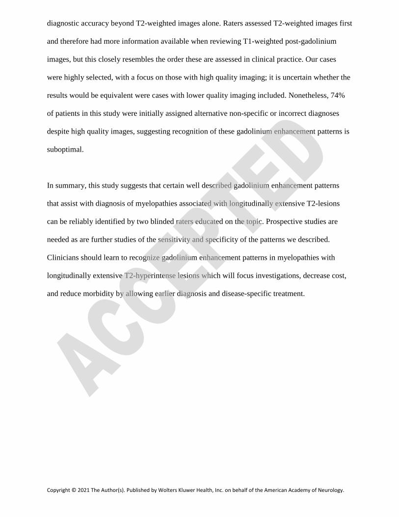
Table 5. Original alternative, incorrect, or non-specific diagnosis in clinical practice (n = 55) among patients with characteristic gadolinium enhancement patterns (n = 74). Abbreviations: ITM = idiopathic transverse myelitis; NMOSD = neuromyelitis optica spectrum disorders.
Final Confirmed Diagnosis Number of Patients
Original Incorrect or Non-specific Diagnosis
Number of Patients
Spinal cord infarct 4 ITM 4
Paraneoplastic myelopathy 4 ITM 2
Sarcoid myelopathy 1
Unspecified demyelinating disease
1
Spondylotic myelopathy 10 ITM 7
Primary spinal cord tumor 2
Multiple sclerosis 1
Sarcoid myelopathy 22 ITM 18
NMOSD 2
Multiple sclerosis 1
Unspecified demyelinating disease
1
AQP4-IgG positive NMOSD 10 ITM 8
Multiple sclerosis 1
Spinal cord infarct 1
Dural arteriovenous fistula 3 Lumbar spinal stenosis 2
Spinal cord infarct 1
Intramedullary metastasis 2 Acute inflammatory demyelinating polyradiculoneuropathy
1
ITM 1
Copyright © 2021 The Author(s). Published by Wolters Kluwer Health, Inc. on behalf of the American Academy of Neurology.

Acknowledgement:
We thank Dr. Sarosh Irani MA (Oxon), DPhil, MRCP (Nuffield Department of Clinical
Neurosciences, University of Oxford, Oxford OX3 9DU, United Kingdom) for his assistance
with the concept and design of this study.
Copyright © 2021 The Author(s). Published by Wolters Kluwer Health, Inc. on behalf of the American Academy of Neurology.

DOI 10.1212/CPJ.0000000000001036 published online January 25, 2021Neurol Clin Pract
Rafid Mustafa, Theodore J. Passe, Alfonso S. Lopez-Chiriboga, et al. T2-lesions
Utility of MRI Enhancement Pattern in Myelopathies with Longitudinally-extensive
This information is current as of January 25, 2021
ServicesUpdated Information &
ull.htmlhttp://cp.neurology.org/content/early/2021/01/25/CPJ.0000000000001036.fincluding high resolution figures, can be found at:
Subspecialty Collections
http://cp.neurology.org//cgi/collection/transverse_myelitisTransverse myelitis
http://cp.neurology.org//cgi/collection/mriMRI
http://cp.neurology.org//cgi/collection/all_spinal_cordAll Spinal Cordcollection(s): This article, along with others on similar topics, appears in the following
Permissions & Licensing
http://cp.neurology.org/misc/about.xhtml#permissionsentirety can be found online at:Information about reproducing this article in parts (figures,tables) or in its
Reprints
http://cp.neurology.org/misc/addir.xhtml#reprintsusInformation about ordering reprints can be found online:
2163-0402. Online ISSN: 2163-0933.Wolters Kluwer Health, Inc. on behalf of the American Academy of Neurology.. All rights reserved. Print ISSN:2011, it is now a bimonthly with 6 issues per year. Copyright Copyright © 2021 The Author(s). Published by
is an official journal of the American Academy of Neurology. Published continuously sinceNeurol Clin Pract


![arXiv:2006.12434v1 [eess.IV] 22 Jun 2020 · Cardiac Segmentation on Late Gadolinium Enhancement MRI: A Benchmark Study from Multi-Sequence Cardiac MR Segmentation Challenge Xiahai](https://img.dokumen.tips/doc/110x75/5f43858d7d44864654018f17/arxiv200612434v1-eessiv-22-jun-2020-cardiac-segmentation-on-late-gadolinium.jpg)