Embed Size (px)
Citation preview

Using Multiplex Assays and Assay Development to Enhance your Research
Program
James A. Lederer, PhD
Department of Surgery Brigham and Women’s Hospital (BWH), BWH Biomedical Research Institute (BWH-BRI) and
Harvard Medical School, Boston, MA 02115

Overview Presentation
1. Research interests and reasons for using multiplex assay.
2. Why and how we began using multiplex assays for our research.
3. Luminex assay basics and strategies for developing custom assays.
4. Examples of how we use and take advantage of multiplexing.

Our Research Projects 1. The effects of traumatic injuries on the immune system
• Complex response that involves multiple immune cell types and mediators
• Mouse and human studies • Systems Immunology
2. Treatments that enhance immunity and restore immune homeostasis after traumatic injuries and severe infections (sepsis).
• Immuno-monitoring by profiling immune cell phenotypes by flow cytometry and cytokine profiling.
• In vivo response to injury, infection, and treatments.

From Life Technologies Website
Increased knowledge requires increased data

“Old school” single-mediator assays are good, but multiplexing is more in tune with
current scientific needs 1. Difficult to interpret findings and publish results without
including more than one mediator or biomarker in dataset. • Inflammatory responses and T-cell immune regulation
for example
2. More financial demands on research funding requires increasing efficiency and data collection strategies.
3. We need to gather as much information as possible from expensive mouse models and limited amounts of human samples.

History: ELISA to Flow Cytometry Bead Assays to Luminex Assays
1. ELISA – basic principle behind all immunoassays • Plate-bound antibody – antigen – antibody
sandwiches with color absorbance detection • Usually a single analyte approach
2. Flow cytometry-based bead assays • Same principle as ELISA but on a bead with a specific
detection character for gating on a flow cytometer • Limited number of beads can be run as multiplex • Not automated or customizable
3. Luminex bead assays • Same principle as ELISA using beads with a specific
detection character • High number of specific beads for multiplexing • Customizable and automated
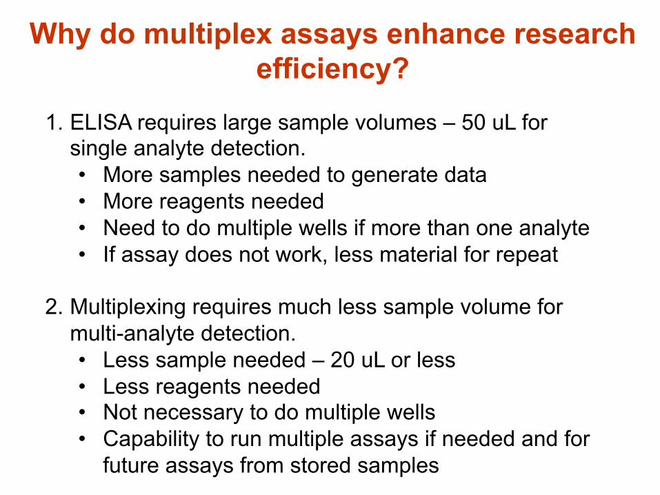
1. ELISA requires large sample volumes – 50 uL for single analyte detection. • More samples needed to generate data • More reagents needed • Need to do multiple wells if more than one analyte • If assay does not work, less material for repeat
2. Multiplexing requires much less sample volume for multi-analyte detection. • Less sample needed – 20 uL or less • Less reagents needed • Not necessary to do multiple wells • Capability to run multiple assays if needed and for
future assays from stored samples
Why do multiplex assays enhance research efficiency?

1. Purchased Luminex instrument in 2007 and found discrepancy in results between older technology (BD CBA) and Luminex assays. • Several cytokines that were detected by ELISA or
CBA were not detected in Luminex assays.
2. Tested several different available kits and found inconsistencies in cytokine detection. • Not surprising since assays are likely built with
different specific antibody pairs.
3. Some kits detected recombinant cytokines, but not natural cytokines
Reasons for developing “in-house” multiplex assays

1. Confidence that results will be accurate.
2. Reduce overall costs of performing Luminex assays for our immuno-phenotyping studies.
• Kit costs were $100-$200/cytokine/96-well plate. • Discovered that making in-house assays in bulk
would be less expensive. 3. Flexibility for future expansion of panels and for other
multiplex assay development – opens up capabilities to make a variety of different biomarker assays.
More reasons for developing “in-house” Luminex assays

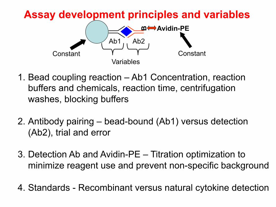
Assay development principles and variables
Immuno-assays based on sandwich ELISA principles
Ab1 Ab2
B
Avidin-PE
• High specificity due to dependence of signal on bi-molecular binding reagents
• Quality and specificity of assay reaction depends on the nature of Ab1 and Ab2. • Antibodies remain the most important component of these and other types of immunoassays
Ab1 Ab2
B
Avidin-PE
Constant Constant Variables
1. Bead coupling reaction – Ab1 Concentration, reaction buffers and chemicals, reaction time, centrifugation washes, blocking buffers
2. Antibody pairing – bead-bound (Ab1) versus detection (Ab2), trial and error
3. Detection Ab and Avidin-PE – Titration optimization to minimize reagent use and prevent non-specific background
4. Standards - Recombinant versus natural cytokine detection
Assay development principles and variables
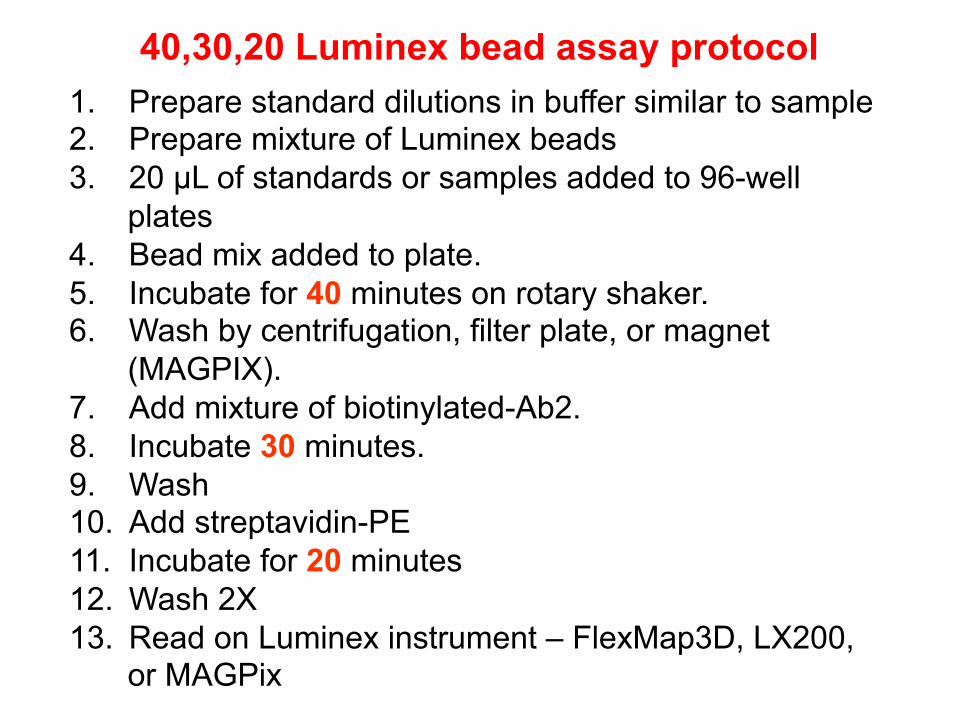
40,30,20 Luminex bead assay protocol 1. Prepare standard dilutions in buffer similar to sample 2. Prepare mixture of Luminex beads 3. 20 µL of standards or samples added to 96-well
plates 4. Bead mix added to plate. 5. Incubate for 40 minutes on rotary shaker. 6. Wash by centrifugation, filter plate, or magnet
(MAGPIX). 7. Add mixture of biotinylated-Ab2. 8. Incubate 30 minutes. 9. Wash 10. Add streptavidin-PE 11. Incubate for 20 minutes 12. Wash 2X 13. Read on Luminex instrument – FlexMap3D, LX200,
or MAGPix

Luminex assay principles and readout Ø 5.6 uM microsphere are dyed to create distinct colors and can be delineated based on red/infrared content
Ø Surface is modified to allow coupling of antibodies, proteins, peptides, or oligonucleotides
Ø Immunoassays are based on sandwich ELISA

SampleID Well Location MFI Std01 ( 0.62 pg/ml) A1 49 Std02 ( 1.25 pg/ml) B1 73 Std03 ( 2.50 pg/ml) C1 105 Std04 ( 5.00 pg/ml) D1 175 Std05 ( 10.00 pg/ml) E1 329 Std06 ( 20.00 pg/ml) F1 573 Std07 ( 39.00 pg/ml) G1 1027 Std08 ( 78.00 pg/ml) H1 1695 Std09 ( 156.00 pg/ml) A2 2593 Std10 ( 312.00 pg/ml) B2 4439 Std11 ( 625.00 pg/ml) C2 7267 Std12 (1250.00 pg/ml) D2 11025 Std13 (2500.00 pg/ml) E2 15787 Std14 (5000.00 pg/ml) F2 20073 Std15 (10000.00 pg/ml) G2 22369 Std16 (20000.00 pg/ml) H2 22839
1 A3 67 2 B3 1399 3 C3 175 4 D3 1209 5 E3 33 6 F3 25 7 G3 1341 8 H3 1447 9 A4 23
10 B4 1411
Data output example: Human IL-4
1. Standards run to calculate sample concentrations
2. Readout is mean fluorescence intensity (MFI)
3. Data output from instrument as .CSV files that input into MS Excel
4. Plotted to visualize data

Recent biomarker development project for lung disease diagnosis – COPD vs IPF
NIH development project to construct biomarker sets to distinguish patients with chronic obstructive pulmonary disease (COPD) vs. interstitial pulmonary fibrosis (IPF). Ivan Rosas and Fernanda Golzarri, BWH Pulmonary Critical Care and Lovelace Respiratory Research Institute

Steps to Building the COPD/IPF panel
1. Identify target analytes – list from literature
2. Search for the needed reagents – Ab1, Ab2, standard 3. Find standard
• Can be difficult for less studied proteins or complex proteins – e.g. surfactant protein A (SP-A)
4. “micro-batch” testing to optimize bead coupling reaction
5. Biotinylation optimization for Ab2, if needed

Examples: Cytokine Assays – “In-house” mouse cytokine assay
IL-1β
0.03125 1 32 1024 327680
500
1000
1500
2000
2500IL-1β TheirsIL-1β Ours
Concentration of Standard
MFI
IL-2
0.03125 1 32 1024 327680
5000
10000
15000IL-2 TheirsIL-2 Ours
Concentration of Standard
MFI
IL-4
0.03125 1 32 1024 327680
5000
10000
15000IL-4 TheirsIL-4 Ours
Concentration of Standard
MFI
IL-6
0.03125 1 32 1024 327680
5000
10000
15000IL-6 TheirsIL-6 Ours
Concentration of Standard
MFI
IL-10
0.03125 1 32 1024 327680
2500
5000
7500IL-10 TheirsIL-10 Ours
Concentration of Standard
MFI
IL-13
0.03125 1 32 1024 327680
2500
5000
7500
10000
12500IL-13 TheirsIL-13 Ours
Concentration of Standard
MFI
IL-17
0.03125 1 32 1024 327680
2500
5000
7500
10000
12500IL-17 TheirsIL-17 Ours
Concentration of Standard
MFI
TNF
0.03125 1 32 1024 327680
500
1000
1500
2000
2500
3000
3500TNF TheirsTNF Ours
Concentration of Standard
MFI
Standard Curve Plots Comparing Recombinant Cytokine Detection Assays (Mouse)

Detection of Naturally-Produced Mouse Cytokines IL-1β
0.0
2.5
5.0
7.5TheirsOurs
Dilution of Supernatant (1:2)
MFI
IL-2
0
10000
20000TheirsOurs
Dilution of Supernatant (1:2)
MFI
IL-4
0
50
100
150
200
250TheirsOurs
Dilution of Supernatant (1:2)
MFI
IL-6
0
500
1000
1500
2000
2500TheirsOurs
Dilution of Supernatant (1:2)
MFI
IL10
0100200300400500600700800900
TheirsOurs
Dilution of Supernatant (1:2)
MFI
IL13
0
1000
2000TheirsOurs
Dilution of Supernatant (1:2)
MFI
IL17
0
250
500
750TheirsOurs
Dilution of Supernatant (1:2)
MFI
TNFα
0
250
500
750
1000
1250TheirsOurs
Dilution of Supernatant (1:2)
MFI
Mouse cytokine assays

Human cytokine assay development and testing: Recombinant standards
IL-1a
0.1 1 10 100 1000 100000
5000
10000
15000
pg/ml
MFI
IL-1b
0.1 1 10 100 1000 100000
5000
10000
15000
pg/ml
MFI
IL-1ra
0.1 1 10 100 1000 100000
500
1000
1500
2000
2500
3000
3500
pg/ml
MFI
IL-4
0.1 1 10 100 1000 100000
5000
10000
15000
20000
25000
pg/ml
MFI
IL-5
0.1 1 10 100 1000 100000
10000
20000
pg/ml
MFI
IL-6
0.1 1 10 100 1000 100000
2500
5000
7500
10000
pg/ml
MFI
IL-7
0.1 1 10 100 1000 100000
100020003000400050006000700080009000
pg/ml
MFI
IL-8
0.1 1 10 100 1000 100000
5000
10000
15000
20000
25000
pg/ml
MFI
IL-10
0.1 1 10 100 1000 100000
10000
20000
pg/ml
MFI

IL-12p40
0.1 1 10 100 1000 100000
500
1000
1500
2000
2500
pg/ml
MFI
IL-12p70
0.1 1 10 100 1000 100000
1000
2000
3000
4000
5000
6000
7000
pg/ml
MFI
IL-13
0.1 1 10 100 1000 100000
1000
2000
pg/ml
MFI
GM-CSF
0.1 1 10 100 1000 100000
2500
5000
7500
10000
12500
pg/ml
MFI
IFNg
0.1 1 10 100 1000 100000
1000
2000
3000
4000
5000
pg/ml
MFI
TNFb
0.1 1 10 100 1000 100000
1000
2000
3000
4000
5000
6000
7000
pg/ml
MFI
TWEAK
0.1 1 10 100 1000 100000
500
1000
1500
2000
2500
pg/ml
MFI
More recombinant cytokine standard testing

Validate natural cytokine detection: Cytokine production by in vitro stimulated human PBMCs
natural human cytokine detectionIL
-1a
Il-1r
a
IL-1
b
IL-2
IL-3
IL-4
IL-5
IL-6
IL-7
IL-8
IL-9
IL-1
0
IL-1
2p40
IL-1
2p70
0
100
200
NoneLPSBLPAnti-CD3
10000
20000
MFI
natural human cytokine detection part2
IL-1
3
IL-1
7
IL-2
1
IL-3
2
fgf2
g-C
SF
gm-C
SF
IFN
g
MC
P-1
NG
F-1
TNFa
TNFb
TREM
-1
TWEA
K0
100
200
10000
20000 NoneLPSBLPAnti-CD3
MFI
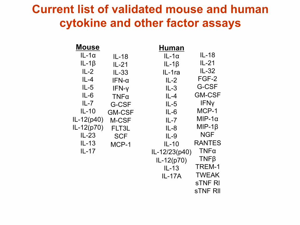
Mouse IL-1α IL-1β IL-2 IL-4 IL-5 IL-6 IL-7 IL-10
IL-12(p40) IL-12(p70)
IL-23 IL-13 IL-17
Human IL-1α IL-1β IL-1ra IL-2 IL-3 IL-4 IL-5 IL-6 IL-7 IL-8 IL-9 IL-10
IL-12/23(p40) IL-12(p70)
IL-13 IL-17A
IL-18 IL-21 IL-32
FGF-2 G-CSF
GM-CSF IFNγ
MCP-1 MIP-1α MIP-1β
NGF RANTES
TNFα TNFβ
TREM-1 TWEAK sTNF Rl sTNF Rll
IL-18 IL-21 IL-33 IFN-α IFN-γ TNFα G-CSF
GM-CSF M-CSF FLT3L SCF
MCP-1
Current list of validated mouse and human cytokine and other factor assays

Collaborative consumption
Dr. Andrew Lichtman, BWH and HMS – mouse models of T cell mediated inflammation Dr. Charles Serhan, BWH and HMS – inflammation research in mice and man Dr. Gerry Pier, BWH, Channing Lab – vaccine and infectious disease projects Dr. Pedram Hamrah, MGH – eye inflammation and infections Dr. Rachel Clark, BWH and HMS – human T cell biology in the skin Dr. Arlene Sharpe, HMS – mouse models of basic T cell activation mechanisms Dr. Mark Perrella, BWH and HMS – mouse stem cells and sepsis responses
• On campus Luminex assay collaborations with reagent replenishment support.
• A need for on-campus Luminex assay development and services – Harvard Catalyst or BWH-BRI

Examples of how to use of Luminex multiplexing to increase research efficiency
1. Micro-size experiments to gain more information from less cells
2. Test multiple stimulation conditions
3. Develop more efficient assays to save on reagent costs and sample cost – e.g. antibody-isotyping of vaccine assays by multiplex
4. Potential to assay any type of cell or tissue extract – e.g. human tears, organ extacts, exhaled breath condensate


Cytokine Levels in Tears Are Correlated with the Corneal
Nerve Density and Dendritic Cell Counts in Eyes with Infectious
Keratitis Takefumi Yamaguchi, Bernardo Cavalcanti, Pedram
Hamrah Mass Eye and Ear Infirmary
Shizu Ishikawa, Akinori Osuka, Kentaro Shimizu, James Lederer
Department of Surgery, Brigham and Women’s Hospital

0
5000
10000
15000
20000
25000
0.5 1.5 2.5 3.5 0
1500
3000
0.5 1.5 2.5 3.5
IL1b
Normal IK Contralateral eye 0
200
400
0.5 1.5 2.5 3.5
IL1Ra
Normal IK Contralateral eye
IL2
Normal IK Contralateral eye
0
15000
30000
0.5 1.5 2.5 3.5 0
200
400
0.5 1.5 2.5 3.5 0
2000
4000
0.5 1.5 2.5 3.5
IL6
Normal IK Contralateral eye
IL7
Normal IK Contralateral eye
IL8
Normal IK Contralateral eye
P=0.005 P=0.02
P < 0.001 P=0.04
P=0.002 P=0.03
NS NS

0
300
600
0.5 1.5 2.5 3.5 0
5000
10000
0.5 1.5 2.5 3.5 0
6000
12000
0.5 1.5 2.5 3.5
0
1000
2000
0.5 1.5 2.5 3.5 0
3000
6000
0.5 1.5 2.5 3.5 0
2500
5000
0.5 1.5 2.5 3.5
GMCSF
Normal IK Contralateral eye
MCP1
Normal IK Contralateral eye
IL10
Normal IK Contralateral eye
IL17a
Normal IK Contralateral eye
FGF2
Normal IK Contralateral eye
TREM1
Normal IK Contralateral eye
NS
NS
P=0.04 P=0.01 P=0.02
P=0.004 P=0.02
P=0.003 P=0.01

IgM
0
200
400
600 ShamCLP
MFI
IgM
IgG1
0
5000
10000
15000
MFI
IgG
1IgG2b
0
500
1000
1500
2000
1:50 1:450 1:1500 1:4500
Serum Dilution
MFI
IgG
2b
Multiplexing antigen- or immunogen-specific antibody assays for vaccine testing
Ab2 – anti-isotype Ab
B
Avidin-PE
Plasma or serum sample from immunized mouse (or human)
Immunogen

Ovalbumin-peptide induced cytokine production from TCR transgenic T cells, in situ detection with
MagPix beads IFN-γ
CTL sham CTL burn OVA shamOVA burn0
1000
2000
TNF
CTL sham CTL burn OVA shamOVA burn0
50
100
150
200
250IL6
CTL sham CTL burn OVA shamOVA burn0
500
1000
1500
2000
2500
3000
3500IL2
CTL sham CTL burnOVA shamOVA burn0
2500
5000
7500
10000
12500CTL shamCTL burnOVA shamOVA burn
IL4
CTL sham CTL burn OVA shamOVA burn0
100
200IL10
CTL sham CTL burn OVA shamOVA burn0
250
500
750
1000
IL13
CTL sham CTL burn OVA shamOVA burn0
100
200
300
400
500IL17
CTL sham CTL burn OVA shamOVA burn0
1000
2000
3000
4000
5000
6000
OVA

Summary
1. Development of custom multiplex assay is feasible and cost effective through careful optimization and validation.
2. Provides an opportunity to develop new assays that are not commercially available.
3. Opens opportunity for collaboration.
4. Recent development of VeloceBio LLC as a collaborative biomarker development company and potential partnering with Cambridge Biomedical to offer assay development and Luminex assay service.