Embed Size (px)
Citation preview

CLINICAL MICROBIOLOGY REVIEWS, Oct. 1994, p. 576-604 Vol. 7, No. 40893-8512/94/$04.O0+OCopyright © 1994, American Society for Microbiology
Uses of Flow Cytometry in VirologyJAMES J. McSHARRY*
Department of Microbiology, Immunology and Molecular Genetics,Albany Medical College, Albany, New York 12208
INTRODUCTION ............................................................. 576BRIEF DESCRIPTION OF FLOW CYTOMETRY ............................................................ 576SAMPLE PREPARATION AND PERMEABILIZATION METHODS ............................................................ 578STAINING CELL-ASSOCIATED VIRAL ANTIGENS AND NUCLEIC ACID .................................................580STUDIES OF VIRUS-CELL INTERACTIONS, USING FLOW CYTOMETRY ...............................................581Virus-Induced Programmed Cell Death ............................................................ 582Elfects of Virus Infection on Cellular Proteins ............................................................ 584Binding of Virus to Cells ............................................................ 585Detection of Viral Antigens on the Cell Surface ............................................................ 586Detection of Intracellular Viral Antigens ............................................................ 587SV40.............................................................587HCMV............................................................. 588unr can
HSV-1 and -2...........Miscellaneous Viruses
BTV...........................A.Qe'v
CI
..591
.59240.7.
REV-A ..593WHY ..593HCV..593BVDV..593FeLV..593F-MuLV..593EBV..594Autographa californica nuclear polyhedrosisvirus.........................................594INICAL VIROLOGY........................................594HCMV........................................594HIV........................................ 59
ANTIBODIES MEASURED BY FLOW CYTOMETRY.BACTERIA, FUNGI, AND PARASITES...........................FUTURE PROSPECTS.......................................................ANK?vn4---U1TI 1------iNT-
..597
..597
..597Coe
I'lsI J TILLUU.lL YO o* *v%J"I-iniLptylvir-illIa............................................................................................................................................7i~~~~~~~~~~~~~~~~~~~~~~~~~~~~~~~~~~~~~~~~~~....................................................
_ Tl7rMMl7kTf4M0c
INTRODUCTION
This review will focus on the uses of flow cytometry in thestudy of virus-cell interactions. This topic was last reviewed in1990 (88, 98, 105). With the exception of simian virus 40(SV40)-infected cells, there were few virus-cell systems thathad been analyzed by quantitative flow cytometry. Since thattime, a significant number of papers describing the use of thistechnology for basic, clinical, and applied research have beenpublished. These include studies on (i) methods for permeabi-lizing virus-infected cells; (ii) methods for staining viral anti-gens on the cell surface, in the cytoplasm, and in the nucleus;(iii) effects of virus infection on cellular constituents; (iv)attachment of virus to cells; (v) detection and quantitation ofvirus-infected cells in vitro and in vivo; and (vi) applications toclinical virology. A few virus-cell systems have been studied indetail by multiparametric quantitative flow cytometry, and
* Mailing address: Department of Microbiology, Immunology andMolecular Genetics, A-68, Albany Medical College, 47 New ScotlandAve., Albany, NY 12208. Phone: (518) 262-5174. Fax: (518) 262-5748.
these will be critically reviewed. The application of quantitativeflow cytometry in the analysis of many other virus-infected cellsystems is less well defined; however, that literature will bereviewed to demonstrate the broad applications of this tech-nology for detecting and quantitating virus-infected cells.
BRIEF DESCRIPTION OF FLOW CYTOMETRY
Cytometry can be defined as the measurement of physicaland/or chemical properties of cells. These measurements areoften performed with a light or fluorescence microscope.Microscopy is qualitative but, with the exception of computer-ized image analysis (77), seldom quantitative. Flow cytometryis the measurement of the physical and/or chemical character-istics of cells while they pass single file in a fluid stream througha measuring apparatus (185). The fluorescence-activated cellsorter (FACS) was developed in the early 1970s to studyisolated populations of viable cells for immunology. Theoriginal instruments were large, contained powerful dual lasersthat were water cooled, and were able to analyze and sort cells.Although flow cytometry continues to function in the field ofimmunology, its use in other fields such as cell biology,
576
-----------------------------------------------------------------------------------------V,.mREFERENCES .............................................................................................................................................................
Vano
on May 26, 2018 by guest
http://cmr.asm
.org/D
ownloaded from

FLOW CYTOMETRY IN VIROLOGY 577
ARGON ION LASER488nm 1SmW
Focusing Lens
Objective LensNA=0.8-0.9x1 6
REDFLUORESENCEPMT
1 4 7
8
NA=0.2x6.2
DiffuserI
FIGHT ANGLE GREENo emI*mTTe=I eIr
ORANGECa 1 lnncQ=r"r
Forward Ught bUA IcI M rLvCM U crcScatter Photodiode PMT PMT PMT
FIG. 1. Diagram of a flow cell with attached optical systems. As the cells pass through the flow cell (the small black square box in the centerleft), a laser beam intersects the stream of cells and scatters light. FALS passes through the forward light scatter lens, and the energy is collectedby the forward light scatter photodiode. RALS passes through the objective lens, the beam splitter, laser line filter, and diffuser, and the energyis collected by the right-angle scatter photomultiplier tube (PMT). If the cells are labeled with fluorescent molecules, the laser will excite thesemolecules, which emit light at higher energies. The emitted energies of different wavelengths pass through the objective lens and various filters andare collected and amplified by the various photomultiplier tubes. The amplified signals are converted into digital information and stored in acomputer for further analysis. The numbers refer to the following: 1, beam splitter; 2, laser line filter, 396- to 496-nm band pass; 3, diffuser; 4,dichroic mirror 1, 570-nm long-pass filter; 5, laser cut filter, 490-nm short cut; 6, green filter, 515- to 530-nm band pass; 7, dichroic mirror 2, 610-nmlong pass; 8, orange filter, 565- to 592-nm band pass; and 9, red filter, 660-nm long pass.
molecular biology, pathology, and diagnostic microbiology ison the increase. With the expanded use of flow cytometry forstudies that do not require the cell sorting capabilities of aFACS instrument, the machinery has been simplified to a morecompact, air-cooled, single-laser, analytical instrument. Duringanalysis, the sample is completely contained within the instru-ment, leading to a degree of operator safety. Currently avail-able multiparameter flow cytometers use a single laser beam toallow the simultaneous quantitative measurement of a numberof cellular properties, including cell number, cell size, cellulargranularity, and up to three different fluorochromes per cell. Inaddition to the number of fluorescently labeled cells, theamount of each fluorochrome associated with each cell can bemeasured by flow cytometry. Since a flow cytometer cananalyze cells at rates up to several thousand cells per second, itis possible to analyze a statistically significant number of cellsin a short time. Modern analytical flow cytometers with fixedoptical benches and internal computers allow the operator toset parameters for various analyses with ease and to store theseparameters in internal computers to be recalled when needed.Dual-laser flow cytometers can detect additional fluorescentmolecules, but they are not required for the types of analysispresented in this review.
Figure 1 illustrates the business end of a modern, single-laser, analytical flow cytometer. It is a diagram of the argon ionlaser and the rectangular flow cell (the small black square onthe left center of the diagram) with attached optical assembly,including lenses, mirrors, filters, and photomultiplier tubes. Allflow cytometers have four basic components: (i) a fluidicsystem to carry a suspension of single cells from the sampletube into a quartz flow cell containing sheath fluid; (ii) afocused light source, usually an argon ion laser with anexcitation energy of 488 nm; (iii) filters and photodetectors tocollect scattered light and light emitted from fluorescentlylabeled cells and processors to convert these light signals toanalog electrical impulses and then to digital signals; and (iv)a computer system to collect, store, and analyze the data. Cellsto be measured are introduced into the sheath fluid that keepsthe cells in the center of the quartz flow cell and forces the cellsto flow single file at uniform speed through a small orifice. Thecells pass by a point where the laser intersects the path of thefluid stream. As each cell passes this point, the laser light willbe scattered. The scattered light is collected as forward-anglelight scatter (FALS), which yields information on the numberand size of cells, and right-angle light scatter (RALS), whichyields information on the granularity of cells. If the cells are
SensingZone
VOL. 7, 1994
on May 26, 2018 by guest
http://cmr.asm
.org/D
ownloaded from

CLIN. MICROBIOL. REV.
stained with one or more fluorescent labels, the laser will excitethe fluorescent molecules that emit energy at higher wave-
lengths. For example, fluorescein has a peak emission spec-trum at 520 nm; phycoerythrin, at 580 nm; and propidiumiodide (PI), at 580 and 620 nm (149). The emitted energies ofdifferent wavelengths pass through filters (green filter, 515- to530-nm band pass; orange filter, 565- to 592-nm band pass; redfilter, 660-nm-long pass) that allow light of only a particularwavelength to pass; then they are collected and amplified byphotomultiplier tubes. The amplified signals are converted intodigital information which is stored in a computer for furtheranalysis and printed in graph form as a hard copy. For more
detailed information on the mechanics and practical uses of a
flow cytometer, the reader is referred to references 149, 185,208, and 212.With the advent of monoclonal antibodies, flow cytometry
began to be useful in the clinical laboratory, particularlyfor quantitating the various subsets of lymphocytes. Today,one of the major uses of flow cytometry in the clinicallaboratory is the determination of the number of CD4+ cells innormal and human immunodeficiency virus (HIV)-infectedpatients. Despite some uncertainties concerning the use ofCD4 cell counts as surrogate markers in HIV-infected individ-uals (99), the absolute number and percentage of CD4+cells are being used as surrogate markers for defining whichHIV-infected individuals will be classified as having AIDSand for determining those who will be treated with antire-troviral chemotherapy (28). Excellent reviews on the use offlow cytometry for phenotyping cells in health and diseasehave been published (28, 44, 64, 69, 101, 109, 137, 148,212).
SAMPLE PREPARATION AND PERMEABILIZATIONMETHODS
Intact single cells are the major requirement for flowcytometric analysis of virus-infected cells. Blood, bronchoal-veolar lavage (BAL), and urine specimens are ideal becausethey are single cell suspensions. Soft organs, lymph nodes, andspleens can be mechanically manipulated to release singlecells. Cells obtained from suspension cultures or cultured cellsremoved from solid surfaces with EDTA and/or trypsin andresuspended as single cells are suitable for analysis. Many ofthe cell surface proteins that serve as markers in flow cytom-
etry are removed with enzymatic treatment of cells in tissues or
cultured cells grown on solid supports, making it impossible to
stain cells for these surface markers. However, if only cytoplas-mic and/or nuclear antigens are to be detected and quanti-tated, treatment with proteolytic enzymes under conditionsthat yield intact cells can be useful for obtaining single cellsuspensions from cultures or tissues.When cytoplasmic and/or nuclear antigens or the nuclear
DNA content of cells is to be studied by flow cytometry,
methods to permeabilize the cell must be employed to allowmonoclonal antibodies and hydrophilic molecules, such as PI,to diffuse into the cell's interior. Most of the published work on
permeabilizing virus-infected cells for analysis by flow cytom-
etry has employed methanol. Jacobberger et al. (89) used 90%methanol to permeabilize SV40-transformed and spontane-
ously transformed Chinese hamster ovary (CHO) cells so thatthe cytoplasmic and nuclear compartments of the cell were
permeable to both antibodies and the nucleic acid binding dye,PI. Intact cells with 2 N (diploid) or greater DNA content were
identified by the amount of PI bound to the DNA after RNasetreatment, allowing for a distinction between intact cells and
debris. By using high-affinity monoclonal antibodies to theSV40 T antigen, affinity-purified fluorescently labeled secondantibodies, and 50% goat serum in all reactions, these re-searchers were able to reduce the fluorescent background sothat they could attain excellent coefficients of variation. Cellclumping was minimized by keeping the cells cold and addinggoat serum containing trace amounts of Triton X-100. Meth-anol permeabilization yields optimal staining of cellular DNAby PI and retains cytoplasmic and nuclear antigens in a state inwhich they remain reactive with polyclonal and monoclonalantibodies. A major advantage of this procedure is that cells in90% methanol can be stored at -70°C for more than a yearwithout detectable loss of internal antigens or DNA content.This permeabilization procedure has been used successfully formany studies on SV40-infected and -transformed cells as wellas on many other virus-infected cells including human lympho-cytes (13, 38, 54, 78, 80, 89, 96, 97, 103, 104, 106, 124, 125).Major disadvantages of the methanol fixation technique arethat it destroys the light scatter properties of cells, reduces theantigenicity of cell surface proteins, and causes destruction ofsome cells. Figure 2 illustrates the effect of methanol treatmenton the light scatter properties of leukocytes obtained from ahealthy person. Panel A depicts the FALS (FW-SC) and RALS(RT-SC) patterns of fresh leukocytes treated with Ortho-munelysing reagent (Ortho Diagnostic Systems, Inc., Raritan, N.J.)to lyse erythrocytes. Note the good separation of lymphocytes(L), monocytes (M), and granulocytes (G). Panel B shows theFALS (FW-SC) and RALS (RT-SC) patterns of leukocytesobtained from the same individual. In this case, the cells weretreated with methanol after erythrocyte lysis by Ortho-munelysing reagent. Note that the cells have lost their ability to beseparated by their light scatter properties in the flow cytometerand have fallen down to the bottom left and right portions ofthe graph.To overcome some of the disadvantages of methanol treat-
ment, Schimenti and Jacobberger (169) have compared anumber of combinations of paraformaldehyde and methanolfor permeabilizing CHO, HL60, and NIH 3T3 cells. Pretreat-ment of cells with paraformaldehyde before methanol treat-ment stabilized the cells, gave good coefficients of variation forDNA staining with PI, and allowed indirect immunofluores-cence staining of SV40 T antigen in the cell nucleus. However,in comparison to methanol treatment alone, the binding of PIto DNA was decreased and the amount of indirect immuno-fluorescence staining of T antigen in the nucleus was increased.Police et al. (154), who used 0.25% paraformaldehyde beforestaining with PI, also reported decreased binding of PI tocellular DNA.A recent report describes a permeabilizing reagent that
overcomes many of the disadvantages of the methanol proce-dure. Connelly et al. (33) used Permea-Fix (Ortho DiagnosticSystems) to permeabilize human leukocytes in whole blood.They report that, after Permea-Fix treatment, (i) leukocytesscatter light at least as well as untreated cells; (ii) CD3, CD4,and CD8 cell surface antigens can be labeled with fluorescentlylabeled monoclonal antibodies; and (iii) cytoplasmic antigens,such as vimentin or the HIV p24 core antigen, can be labeledwith specific monoclonal antibodies. HIV-infected and unin-fected H9 cells treated with Permea-Fix can be separated fromeach other by detection of intracytoplasmic p24 antigen la-beled with a fluorescein isothiocyanate (FITC)-conjugatedmonoclonal antibody. Furthermore, CD4+ human mononu-clear cells infected with HIV in vitro, treated with Permea-Fix,and stained with phycoerythrin (PE)-labeled OKT4A mono-clonal antibody and FITC-labeled monoclonal or polyclonalantibody to the HIV p24 antigen can be distinguished from
578 McSHARRY
on May 26, 2018 by guest
http://cmr.asm
.org/D
ownloaded from

FLOW CYTOMETRY IN VIROLOGY 579
A ORTHO-MUNE LYSING REAGENT
FM-SC250
200,
50 100 1I0 2OU 25CRT-SC
B METHANOL PERMEABILIZATION
FW-SC250
200
150
100
50
0U 5U 100 150
RT-SC
I-..A. . 200-. 25
FIG. 2. Effect of methanol treatment on light scatter properties of
lymphocytes. Whole blood was obtained from a healthy individual. (A)Contour map of whole blood cells treated with Ortho-mune lysingreagent followed by flow cytometric analysis of the leukocytes. Lym-phocytes (L), monocytes (M), and granulocytes (G) are separated on
the basis of their FALS (FW-SC) and RALS (RT-SC) properties. Note
the separation of lymphocytes, monocytes, and granulocytes. (B)Contour map of whole blood cells treated with Ortho-mune lysingreagent followed by 90% methanol and analysis of the FALS and
RALS properties of the cells by flow cytometry. Note that the cells
have lost their ability to be separated by FALS and RALS and are near
the bottom and sides of the graph.
other mononuclear cells on the basis of CD4 antigen stainingand from uninfected CD4+ cells on the basis of FITC-labeled
p24 antigen. The reagent can be used on whole blood and
seems to be considerably better than the other systems tested
for retaining the light scatter properties of human blood cells
while still allowing cell surface and cytoplasmic antigens to be
stained with monoclonal or polyclonal antibodies directed
against these antigens. Figure 3 illustrates the use of Permea-
Fix for determining light scatter, cell surface and cytoplasmiclabeling with monoclonal antibodies, and nuclear DNA label-
ing with PI of leukocytes obtained from an AIDS patient.One-tenth milliliter of blood was used for each determination.
Figure 3 shows that Permea-Fix allows separation of lympho-
cytes from other leukocytes in whole blood by light scatter; the
T3 cell surface marker can be stained with PE-labeled mono-
clonal antibody OKT3 (approximately 75% of the cells in the
lymphocyte gate stained with this monoclonal antibody); the
FITC-labeled isotype control antibody did not stain the PI
staining cells in the lymphocyte gate; and the monoclonalantibody to the HIV p24 antigen stained approximately 13% ofthe PIstaining cells in the lymphocyte gate. Thus, Permea-Fixcan be used to fix and permeabilize cells so that all of theparameters of the flow cytometer can be used for analysis ofvirus-infected cells.
Permea-Fix and some other reagents used to permeabilizecells contain formaldehyde or paraformaldehyde, which re-
duces the binding of PI to DNA. Since PIbinding to DNA isoften used to distinguish debris from cells and to gate on cellswith 2 N or higher DNA content for analysis of cell-associatedantigens, the effect of various fixatives on PI binding to DNAis an important consideration. Figure 4 illustrates a compari-son of four permeabilization reagents on the binding of PI tohuman mononuclear cells: methanol treatment (panel A)provides the highest fluorescence intensity due to PI binding,pretreatment with paraformaldehyde (panel B) reduced thefluorescence somewhat, and the other treatments yieldedconsiderably less binding of PI to DNA.
Other techniques for fixing and permeabilizing cells havebeen reported, but most have not been used for flow cytomet-ric analysis of virus-infected cells. These include pretreatmentof cells with paraformaldehyde followed by treatments with 0.1to 0.5% Triton X-100 (143), Tween 20 (79, 171), digitonin andTween 20 (100), or 0.74% N-octyl-,3-D-glucopyranoside (75).Some have used lysolecithin (47), FACS Lysing Reagent(Becton-Dickinson Immunocytometry Systems, Mountain View,Calif.) (197), or saponin (87) to permeabilize cell membranes.Although these techniques have allowed labeling of cell sur-
faces, with the exception of FACS Lysing Reagent, they do notresult in cell populations that scatter light as well as untreatedcells. These disadvantages could be overcome by labeling cellsurfaces to identify a specific population of cells that couldthen be analyzed for the presence of internal cellular or viralantigens without the need for cell separation by light scatter
(100). Permeabilized cells could be identified by binding of PIto DNA after RNase treatment and detecting viral antigenswith fluorescently labeled antibodies (54). Alternatively, one
could use presorted blood cells to study the internal viralantigens.A number of different procedures can be used to permeabi-
lize cells, and each investigator must choose the procedure thatis best suited to his or her needs. Table 1 summarizes threerecommended procedures for fixing and permeabilizing cells.The Permea-Fix procedure is excellent for cell surface, cyto-plasmic, and nuclear staining with retained light scatter prop-erties (33). FACS Lysing Reagent may also be effective forpermeabilizing cells while retaining light scatter properties(197), but no reports of its use on virus-infected cells have beenpublished. For labeling internal antigens and DNA withoutregard for cell surface antigen staining and light scatter
properties, the cells may be stabilized with 0.25% freshlyprepared paraformaldehyde followed by permeabilization with70 to 90% methanol (154, 169). For cell surface and internalantigen staining without concern for good light scatter prop-erties, theoretically, one could employ any of the detergentprocedures listed above (47, 75, 79, 87, 100, 143, 171); however,these have not been extensively used for virus-infected cells,and it is not clear how they will work in new situations. Each
procedure must be tried to determine how it satisfies individualrequirements. Depending on the virus-cell and antigen-anti-body combinations used, different procedures may yield differ-ent results. For ploidy studies, it is best to use only methanol,because paraformaldehyde impairs binding of PI to the DNA
(154, 169).
VOL. 7, 1994
200 250
on May 26, 2018 by guest
http://cmr.asm
.org/D
ownloaded from

CLIN. MICROBIOL. REV.580 McSHARRY
A i.vMPIocYTE GATE
Cell Number: 40,684 cells
2500
1200 .
RT-SC
r j:,j(:-,S,,,YPE ( 0N,'IROI,
Cell Number: 2,500 cells
RD-FL
101
1 021
GR-Fl.
B P E-OKT3
Cell Nuuber: 2,500 cells
OR-FL
D FITC'-P24 ANTIBODYCell Number: 2,500 cells
RD-FL
GR-FL
FIG. 3. Light scatter, cell surface, and internal labeling of lymphocytes permeabilized by Permea-Fix. Whole blood was obtained from a patientwith AIDS and permeabilized with Permea-Fix. One portion was treated with a PE-labeled OKT3 monoclonal antibody (Ortho DiagnosticSystems) and analyzed for light scatter and orange fluorescence by flow cytometry. (A) Graph showing separation of lymphocytes from other cellswith the lymphocytes gated; (B) graph showing that most of the lymphocytes (75%) in the gate stain with the monoclonal antibody to the T3 cellsurface marker. Another portion of the permeabilized cells was treated with an FITC-labeled isotype control (Chemicon International, Inc.,Temecula, Calif.) and PI and analyzed for light scatter and two-color fluorescence by flow cytometry. (C) Graph showing that essentially all of thecells stained with PI (RD-FL) but not with the FITC-labeled monoclonal isotype control antibody (GR-FL). A third portion of permeabilized bloodcells was treated with an FITC-labeled monoclonal antibody to the HIV p24 antigen (Chemicon International,) and PI and then analyzed for lightscatter and two-color fluorescence by flow cytometry. (D) Graph showing that essentially all of these cells are stained with PI (RD-FL) and aportion (13.2%) of the cells are stained with both PI (RD-FL) and FITC (GR-FL).
STAINING CELL-ASSOCIATED VIRAL ANTIGENSAND NUCLEIC ACID
Surface antigens on unfixed virus-infected cells can bestained with fluorescent-antibody molecules. To prevent non-specific binding of antibodies to cells, the cells are treated witha blocking reagent, such as normal serum, which binds to allantigens and blocks antibodies from binding to all except theirspecific antigen. This procedure works because specific anti-body of high avidity competes successfully for antigen-bindingsites with the nonspecific antibodies in normal serum. Nonspe-cific antigens remain blocked by the antibodies in normalserum. For detecting and quantitating cell surface markers,cells are treated with monoclonal or polyclonal antibodiesdirected against these molecules. For the antibodies to bedetected by the flow cytometer, they must be fluorescently
labeled. In the direct method, fluorescent molecules are co-valently attached to the antibody. In the indirect method,unlabeled antibody is first bound to the antigen in question andthen detected by reaction with fluorescently labeled antibodydirected against the bound unlabeled antibody. For example,FITC- or PE-conjugated goat anti-mouse immunoglobulin G(IgG) is used to detect an unlabeled mouse monoclonalantibody bound to the antigen in question. After any unreactedfluorescently labeled molecules are washed away, the stainedcells are detected and quantitated by flow cytometry. Up tothree different surface labels can be detected with a single-laserinstrument (101). Appropriate controls must be included torule out nonspecific binding of antibodies to cells. Theseinclude unlabeled cells (to rule out background fluorescence)and fluorescently labeled nonspecific antibodies of the same
on May 26, 2018 by guest
http://cmr.asm
.org/D
ownloaded from

FLOW CYTOMETRY IN VIROLOGY 581
10,000 cellsCounts
1200
0
720
480
240
10,001 cells
Counts
Graph No.2
50 IUUPI
Graph No.2
10,000 cells
Counts
10,000 cells
Counts
Graph No.2
Graph No.2
12C
4(3
24
10
20
3 5o tOn
Pt
B.
50 200 25t;l0
1 200
960
720U
480
240
I1I
D
. soPPI
FIG. 4. Effect of permeabilization procedures on binding of PI to DNA. Fresh blood was obtained from a healthy person, and PBMC were
isolated by centrifugation on Histopaque (Sigma Chemical Co., St. Louis, Mo.). The PBMC were divided into four portions, and each portion was
permeabilized by a different procedure, treated with RNase and PI, and analyzed for red fluorescence by flow cytometry. The gain of the redphotomultiplier tube was kept constant for the determination of PI binding by all four procedures. Shown is the fluorescence intensity of PBMCtreated with methanol (A), paraformaldehyde followed by methanol (B), FACS Lysing Reagent (C), and Permea-Fix (D). The data show thatmethanol treatment permitted the highest binding of PI to DNA (A); pretreatment with paraformaldehyde followed by methanol (B) gave slightlylower fluorescence intensity, and treatment with FACS Lysing Reagent (C) and Permea-Fix (D) gave the same fluorescence intensity, which was
approximately half that of methanol treatment alone.
isotype as the primary antibodies for the direct method or
unlabeled heterologous antibodies of the same isotype fol-lowed by fluorescently labeled second antibodies for the indi-rect method. Paraformaldehyde is often added to the cellsuspension after staining in an attempt to inactivate infectivityand to stabilize the cells before they are analyzed by flowcytometry (37, 141, 168). However, when virus-infected cul-tured cells or clinical specimens are examined, it is of utmostimportance to follow universal precautions (e.g., buttoned labcoat, gloves, eye protection, etc.) because none of the fixativeswill completely inactivate all viruses and other infectiousagents under all circumstances (136).
For labeling cytoplasmic or nuclear components, the cellsmust be permeabilized by one of the procedures describedabove and then treated with monoclonal or polyclonal anti-bodies to the antigens of interest. These components can alsobe stained by the direct or indirect antibody staining method(185). For labeling the DNA, permeabilized cells can betreated with hydrophilic dyes, such as PI. The binding of PI tothe cellular DNA is directly proportional to the amount ofDNA in the cells and is useful for determining the ploidy ofcells, i.e., 2 N, 4 N, or heteroploid. This reaction can be madespecific for DNA by treating the cells first with RNase todestroy the RNA, leaving only DNA to interact with the PI.
This procedure has been used extensively to identify cells inGO/G1, S, or G2/M phase of the cell cycle or anuploid cells on
the basis of the amount of PI bound to DNA (185). A usefulcombination of fluorescent labels for these studies is FITC-conjugated antibody to viral antigen and PI to stain the DNA,because their emission spectra do not interfere with each other(149). When using PI, one must avoid skin contact because PIis a carcinogen.
STUDIES OF VIRUS-CELL INTERACTIONS, USINGFLOW CYITOMETRY
Each week, papers reporting the use of flow cytometry tostudy virus-cell interactions are published. Thus, it would beboth impossible and inappropriate to review all of the paperson this subject. Instead, I will review only a small number ofpapers to illustrate the utility of flow cytometry for these kindsof studies. Analyses of virus-cell interactions by flow cytometryhave included (i) virus-induced programmed cell death, (ii)regulation of the expression of cellular antigens in response to
virus infection, (iii) interaction of fluorescently labeled viruseswith cells and identification of cell surface virus receptors, (iv)detection and quantitation of both viral and cell surface
A
VOL. 7, 1994
on May 26, 2018 by guest
http://cmr.asm
.org/D
ownloaded from

582 McSHARRY
TABLE 1. Recommended permeabilization procedures for virus-infected cells
Permeabilization procedure Advantages Disadvantages Reference(s)
Methanol Excellent permeabilization of plasma membrane Destroys light scatter properties of 89and nuclear envelope; excellent PI binding to cells, removes cell surfaceDNA. Cytoplasmic and nuclear antigens antigens, and often destroysretain their ability to react with antibody over cells.long periods of time when stored at -70°C.
Paraformaldehyde-methanol Excellent permeabilization of plasma membrane Destroys light scatter properties of 154, 169and nuclear envelope; increased stability of cells; removes cell surfacecells. Cytoplasmic and nuclear antigens retain antigens; and reduces binding oftheir ability to react with antibody over long PI to DNAperiods of time when stored at -70°C.
Permea-Fix Good permeabilization of plasma membrane Reduces binding of PI to DNA. 33and nuclear envelope. Cells retain lightscatter properties; the ability to label the cellsurface and to bind PI to DNA is retained.Both cytoplasmic and nuclear antigens retaintheir ability to react with antibody.
antigens on virus-infected cells, and (v) identification andquantitation of cytoplasmic and/or nuclear viral antigens.
Virus-Induced Programmed Cell Death
Virus infection usually leads to destruction of cells vianecrosis or persistant viral infection. However, some virus-cellinteractions have been shown to lead to programmed celldeath, or apoptosis. Table 2 lists the virus-cell interactionsleading to apoptosis that are discussed below.
Historically, apoptosis has been characterized by morpho-logical and molecular criteria (6, 7, 31). Apoptotic cells haveshrunken nuclei with condensed chromatin tightly appendingthe nuclear envelope and a ruffled plasma membrane thatremains intact. The DNA is fragmented due to endonucleolyticcleavage of the linker regions between nucleosomal cores (6).The fragmented DNA, isolated from apoptotic cells, can bedetected by electrophoresis on agarose gels, on which a laddereffect is seen after the gel is stained with ethidium bromide.
Recently, flow cytometry methods have been developed tomeasure apoptosis (1, 43, 115). Cells undergoing apoptosisleak some of the small DNA fragments (1). When apoptoticcells are permeabilized and treated with RNase and PI, theapoptotic cells exhibit a PI binding peak with less fluorescenceintensity than a normal Go/Gl peak (145). This is illustrated inFig. 5, where the AO peak represents the fluorescence intensityof the PI binding of cells undergoing apoptosis. This procedure
TABLE 2. Virus-cell interactions leading to apoptosis
Virus Apoptotic cells Reference(s)
HIV EBV transformed, lymphoblastoid 48HIV CD34+ bone marrow, progenitor 161, 213HIV CD8+ T, CD19+ B 27HIV gpl60 expressing CD4+ cells 112HIV PBMC 145HIV CD4+ 26Simian immuno- PBMC 46
deficiencyEBV, varicella-zos- CD45+RO+ 2
terInfluenza HeLa 200LCMV Murine T 160
is simple, rapid, inexpensive, and quantitative. It has been usedto measure the increase in the percentage of apoptotic cells inperipheral blood mononuclear cells (PBMC) obtained fromHIV-infected patients when the PBMC were incubated overtime (145). A second method has been developed to specifi-cally identify the fragmented DNA, using digoxigenin-dUTPand deoxynucleotidyl transferase to label the 3'-OH of theDNA segments and FITC-conjugated antibody to digoxigenin(83). The fluorescence intensity of the FITC-labeled DNA isquantitated by flow cytometry. A third method involves thedetection of increased cell surface expression of the Fasantigen on apoptotic cells. Some apoptotic cells express theFas antigen on their cell surfaces and have lower levels of bcl-2expression (31). Flow cytometry has been used to quantitatethe levels of the Fas antigen and bcl-2 expression in cellsundergoing apoptosis (48, 200). Figure 6 illustrates the en-hanced expression of the Fas antigen on gpl60-transfectedU937-2 cells (112). Both U937-2 cells (panel B) and the UE160clone that was transfected with the gene for HIV gpl60 (panelA) express the Fas antigen; however, a small percentage of thegpl6O-expressing cells have an enhanced expression of the Fasantigen, as indicated by the increased fluorescence intensity ofthat cell population (panel A). Thus, flow cytometric detectionof two- and three-color fluorescence is becoming the methodof choice for detecting apoptosis (172, 173).
In vitro and in vivo studies have implicated HIV in theinduction of apoptosis in lymphocytes and bone marrow cellsobtained from HIV-infected patients. In some cases, apoptosisis induced by viral antigens without the need for complete virusreplication. Oyaizu et al. (145) used flow cytometry to measurethe binding of PI to DNA to show that PBMC obtained fromHIV type 1 (HIV-1)-infected individuals undergo apoptosiswhen cultured in vitro. Carbonari et al. (27) used flow cytom-etry to identify apoptotic lymphocytes obtained from AIDSpatients. Apoptotic lymphocytes were identified on the basis oflight scatter properties and cell surface markers, which showedthat lymphocytes from AIDS patients undergo apoptosis whencultured in vitro, whereas those from healthy individuals donot. Phenotypic analysis of the lymphocytes undergoing apop-tosis in vitro showed that the apototic cells were CD8+ T cellsand CD19+ B cells, suggesting that the increased apoptosisseen in cultured lymphocytes from AIDS patients may notexplain the slow decrease in CD4+ T cells seen in HIV-
CLIN. MICROBIOL. REV.
on May 26, 2018 by guest
http://cmr.asm
.org/D
ownloaded from

FLOW CYTOMETRY IN VIROLOGY 583
(A)
.0Ec
0
(B)
Ec.0
IT I I.U .I I I I I I
Pi fluorescence
Pi fluorescenceFIG. 5. Detection of apoptotic cells by analysis of PI binding to
DNA by flow cytometry. Peripheral blood was obtained from HIV-seropositive and HIV-seronegative individuals, the mononuclear cellswere isolated by centrifugation over Ficoll-Hypaque density gradients,and the PBMC were cultured in RPMI 1640 medium supplementedwith 10% heat-inactivated fetal calf serum, L-glutamine, and antibiot-ics for 4 days. PBMC from each culture were permeabilized with 70%ethanol and then treated with RNase and PI. The red fluorescenceintensity was measured by single-color flow cytometry. Shown is thefluorescence intensity resulting from PI binding to DNA of PBMCfrom an HIV-seronegative individual (A) and that of an HIV-seropos-itive individual (B). A distinct cell cycle region, AO, with less fluores-cence intensity than the GOIGl peak was denoted in both histogramsbut is considerably larger in the PBMC obtained from the HIV-seropositive individual. Reprinted from reference 145 with permissionof the publisher.
infected individuals. Re et al. (161) showed that uninfectedhematopoietic progenitor cells (CD34+) obtained from AIDSpatients underwent apoptosis when cultured in vitro in thepresence of interleukin-3. No proviral DNA was present inthese cells as determined by PCR with primers that identifiedas few as five proviral genomes in 8E5/LAV cells, which areknown to contain only one HIV genome per cell. These resultssuggest that exposure of progenitor cells to other virus-infectedcells, such as CD4+ cells in the bone marrow, may induceapoptosis in the uninfected CD34+ cells. Zauli et al. (213)showed that 2-h exposure of CD34+ cells to HIV inducedapoptosis as measured by PI binding to DNA analyzed by flowcytometry.Cameron et al. (26) used flow cytometry to show apoptosis in
CD4+ T cells activated by HIV-infected dendritic cells. These
FIG. 6. Detection of the Fas antigen on cells undergoing apoptosis.The expression levels of surface Fas antigen on UE160 cells, whichwere transfected with the HIV envelope gene and express gpl60 (A),and U937-2 cells (B) as analyzed by flow cytometry are shown. Cellsuspensions from each sample were mixed with a mouse monoclonalantibody to the Fas antigen. Bound monoclonal antibody was revealedby FITC-conjugated mouse immunoglobulin antibodies and analysis byflow cytometry. The dotted lines represent the autofluorescence ofcells treated with FITC-conjugated anti-mouse immunoglobulin anti-body only. The darker solid line in each panel represents fluorescenceintensity of the FITC-labeled Fas antigen. Note that both the U937-2and the UE160 cells contain a significant portion of cells that express
the Fas antigen; however, a small portion of the UE160 cells express
the Fas antigen at higher fluorescence intensity than do the U937-2cells that do not express the gpl60 envelope gene of HIV. Reprintedfrom reference 112 with permission of the publisher.
results suggest a role for apoptosis in the decline of CD4+ Tcells early in HIV infection. De Rossi et al. (48) used flowcytometric analysis of PI binding to DNA and expression ofbcl-2 and morphological and molecular analyses of DNAladders to show that HIV-infected Epstein-Barr virus (EBV)lymphoblastoid B cells (Mt) expressing the gp120 cell surfaceantigen undergo apoptosis in culture whereas EBV lympho-blastoid B cells that do not express the gp120 surface antigendo not. These results implicate the HIV gpl20 envelopeprotein in apoptosis of activated cells, in this case an EBV-infected B-cell line. As described in the legend to Fig. 6, Lu etal. (112) showed that CD4+ cells expressing gp160 undergoapoptosis as measured by flow cytometric analysis of PIbinding to DNA and reaction with anti-Fas antibody. Theseresults suggest that lymphocytes that express HIV antigens,such as gp120 and gp160, can induce apoptosis in neighboringcells and could be a significant factor in the decline of CD4+cells in HIV-infected individuals.
del Llano et al. (46) showed that apoptosis in culturedmononuclear cells, as assessed by flow cytometric analysis of PIbinding to DNA with the formation of the apoptotic peak,along with mitochondrial metabolic activity and proliferativeresponses to pokeweed mitogen, provides a prognostic tool fordisease progression in simian immunodeficiency virus-infectedrhesus macaques. Razvi and Welsh (160) showed that murineT lymphocytes infected with lymphocytic choriomeningitisvirus (LCMV) undergo apoptosis after activation with anti-CD3 monoclonal antibody, suggesting a mechanism for theimmune deficiencies associated with these infections in mice.Takizawa et al. (200) showed that influenza virus infection of
Go/GI
Ao.:-I I
,. ,}
VOL. 7, 1994
IC
on May 26, 2018 by guest
http://cmr.asm
.org/D
ownloaded from

584 McSHARRY
TABLE 3. Analysis of the effect of virus infection on cell surface proteins by flow cytometry
Virus Cells Antigen Reference(s)
Pseudorabies L-929; Neuro 2A MHC class I expression decreased 130HSV Mouse embryo fibroblasts MHC class I expression decreased 91Cytomegalovirus Human foreskin fibroblasts MHC class I expression decreased 68Mouse hepatitis Cerebral endothelial MHC class II expression decreased 92West Nile Lewis Rat Schwann MHC class I & II expression increased 8HSV Endothelial GMP140 expression increased 56HIV T CD4 expression decreased 181, 198EBV B Bcl-2 expression decreased 120
HeLa cells induced the expression of the apoptosis-related Fasantigen, suggesting that influenza virus causes cell death viaapoptosis. Akbar et al. (2) showed that patients with acute viralinfections, such as those caused by EBV or varicella-zostervirus, have circulating CD45+RO+ cells with low levels ofbcl-2, which tended to undergo apoptosis in culture. Apoptosiscould be prevented by the addition of interleukin-2 to theculture medium. Thus, apoptosis seems to be a factor in thedemise of many virus-infected cells. With the aid of flowcytometry, it will be possible to examine the contributions ofviral and cellular antigens to this process.
A
EMz
0
0
*1>CV0cc
Effects of Virus Infection on Cellular Proteins
Virus infection often modulates the expression of cellularproteins. Table 3 lists a number of virus-cell systems in whichthe effect of virus infection on the expression of cellularproteins has been studied by flow cytometry.
In a defense against cell-mediated immunity, infection ofcells by a number of viruses results in the downregulation ofthe major histocompatability complex (MHC) antigens. Figure7 illustrates the effect of pseudorabies virus infection on theexpression of the class I MHC antigens of L929 and Neuro A
10
B Anti-K 2h 6h 12h
I0 10' 10' 10' 10' 10' 10' 10r
Fluorescence IntensityFIG. 7. Effect of virus infection on cell surface expression of mouse MHC antigens. L-929 (A) or Neuro A (B) cells were infected with
pseudorabies virus at a multiplicity of infection of 10. At various times postinfection, the cells were removed from the flasks and incubated with
mouse monoclonal antibodies, anti-H-2Kk and anti-H-2Dk, followed by FITC-labeled anti-mouse IgG. The fluorescence intensity of mock-infectedand infected cells was determined by flow cytometry. In both cell types (panels A and B), the mock-infected cells ( ) expressed MHC antigens
throughout the time course, whereas pseudorabies virus-infected cells (--- -) had reduced amounts of MHC on their cell surfaces. Backgroundfluorescence (... ) was determined on unstained mock-infected cells. Reprinted from reference 130 with permission of the publisher.
Anti-K 2h 6h 12h
Anti-D 2h 6h 12h
lo,,1'- ,:.l iol, 10, jflot. ip10' 10' 101 10' 10° 10' 101 10' 10° 10' 101 103
CLIN. MICROBIOL. REV.
. %V .,w v %F .s,F . s II
on May 26, 2018 by guest
http://cmr.asm
.org/D
ownloaded from

FLOW CYTOMETRY IN VIROLOGY 585
cells. As shown by fluorescence intensity, over a 12-h periodthe surface expression of class I Kk and Dk MHC antigens onpseudorabies virus-infected L929 cells decreased by 60 to 70%compared with uninfected L929 cells. Similar results are shownfor pseudorabies virus infection of Neuro 2A cells. The expres-sion of other surface antigens, such as the transferrin receptor,was not suppressed, suggesting a selective regulatory mecha-nism (130). Infection of B6/WT-3 mouse embryo fibroblastswith herpes simplex virus type 1 (HSV-1) or type 2 (HSV-2)resulted in the expression of HSV-1 or HSV-2 surface glyco-proteins on the plasma membrane but a reduction in theexpression of class I H-2K" and H-2Db (91). Furthermore, asshown by differences in fluorescence intensity, infection withHSV-2 resulted in a greater reduction of the MHC antigensthan infection with HSV-1. This observation was confirmed bydemonstrating that the cytolytic activity of HSV-2-specific,cytolytic T lymphocytes against HSV-2-infected cells was re-duced compared with the activity of HSV-1-specific cytolytic Tlymphocytes against HSV-1-infected target cells. In a similarmanner, flow cytometric analysis of human cytomegalovirus(HCMV)-infected human foreskin fibroblasts demonstrated areduction of the expression of the MHC antigens (68). Treat-ment of cerebral endothelial cells with gamma interferoninduces the MHC class II antigen complex, and infection ofthese cells with mouse hepatitis virus blocks the expression ofMHC class 11 (92). This effect was shown by using flowcytometry on surface-labeled cells, which indicated the loss ofthis antigen, and by Northern (RNA) analysis, which demon-strated the loss of mRNA for the MHC class II molecule. Theopposite effect was demonstrated when Schwann cells fromLewis rats were infected with West Nile virus: the expression ofboth MHC class I and class II cell surface proteins wasenhanced (8). The quantitative aspects of flow cytometryallowed these types of determinations, which would be impos-sible with fluorescence microscopy, to be made.
Infection of endothelial cells with HSV induces GMP140, anadhesion protein that is implicated in tissue injury and is notexpressed on the cell surfaces of uninfected cells (56). Flowcytometric analysis of HSV-infected endothelial cells showedthat expression of GMP140 is dependent on the expression ofthe gC glycoprotein of HSV.
In addition to the effects of virus infection on the expressionof MHC surface antigens, flow cytometry has been used todemonstrate the downregulation of the CD4 molecule onHIV-infected cells (181, 198). Martin et al. (120) showed thatinfection of B cells with the B95-8 strain of EBV increased theexpression of the cellular gene, bcl-2; however, the increasedexpression of bcl-2 had little effect on cell transformation,which is related to the EBV latent membrane protein geneexpression.The results of these studies demonstrate the utility of flow
cytometry in the measurement of the effect of virus infectionon the expression of particular cellular antigens. This type ofanalysis can be enhanced by staining individual cells for bothviral and cell surface antigen expression and by using themultiparametric analytical capabilities of the flow cytometer todetect these changes in small populations of infected cells.
Binding of Virus to Cells
A number of investigators have used flow cytometry todetect and quantitate cells that have fluorescently labeled virusattached to them. Both direct conjugation of FITC to virus andconjugation of biotin to virus followed by detection of biotiny-lated virus bound to cells by treatment with FITC- or PE-labeled avidin are methods that have been used to label virus
TABLE 4. Detection of cell-bound virus by flow cytometry
Virus Cells Receptor Reference(s)
EBV B lymphocytes CR2 76, 85EBV Human epithelial CR2 107SV40 CV-1 MHC class I 24SV40 Monkey kidney ND 14LCMV MC57, BHK, Vero, ND 20
HeLaHTLV T and B ND 49, 63Echovirus BK, Vero, P2002 ND 121Coxsackievirus A9 BK, Vero, P2002 ND 121Murine leukemia NIH 3T3, HeLa 94Measles Jurkat CD46 51, 139Poliovirus Human mononuclear CD14 60Sindbis Mouse neuronal ND 204HIV MT-4 CD4 176, 178
for these binding studies. As shown in Table 4, this techniquehas been used to identify cell surface receptors for a number ofviruses. Figure 8 illustrates the use of biotinylated EBV foridentifying PBMC that bind EBV. Binding of biotinylatedEBV to cells was detected by FITC-labeled avidin, and purifiedB cells were identified by the Bi monoclonal antibody thatreacts with B cells and FITC-conjugated goat anti-mouseantibody. EBV efficiently bound to all subpopulations of Blymphocytes but not to T cells. The receptor was identified asthe CR2 molecule, which is the receptor for the Cd3 comple-ment component as well as EBV (85).
In another study, FITC-labeled EBV was used to determinewhich human B-cell subpopulations were susceptible to virusinfection (76). The different subpopulations of B cells weredetermined by treating them with the following murine mono-clonal antibodies: (i) OKB7 to identify the Cd3 receptor; (ii)
A. B.
cc
m
2
-J-jLUJ0)wL
F.1wja:
400
300
200
100
0
I'llI0 I .lEa30f<4i I\_ I. 0
100I
101 102 10-3 1(4RELATIVE
FLUORESCENCE INTENSITYFIG. 8. Binding of fluorescently labeled virus to cells. (A) Biotiny-
lated EBV was incubated with purified peripheral blood B lymphocytesfollowed by incubation with FITC-labeled avidin, mounted underglycerol, and viewed under a fluorescence microscope. Three patternsof fluorescence are shown. (B) The solid line shows the flow cytometricanalysis of purified B cells exposed to biotinylated EBV followed byFITC-labeled avidin. (Inset) Flow cytometric analysis of purified Bcells treated with a B-cell-specific monoclonal antibody followed by anFITC-labeled F(ab')2 fragment of goat anti-mouse immunoglobulin.In each case, the dotted line represents purified B cells treated withFITC-labeled avidin or FITC-labeled goat anti-mouse IgG (inset).Note the similarity between the B-cell peak in the inset and the higherfluorescent peak in panel B, suggesting that EBV binds to B cells.Reprinted from reference 85 with permission of the publisher.
VOL. 7, 1994
on May 26, 2018 by guest
http://cmr.asm
.org/D
ownloaded from

CLIN. MICROBIOL. REV.
OKB9 to identify the transferrin receptor, an indicator ofB-cell activation; (iii) OKB10 to identify highly differentiated Bcells; and (iv) specific monoclonal antibodies to IgG, IgA, IgD,or IgM to identify B cells expressing surface immunoglobulins.The specifically labeled cells were treated with PE-labeled goatanti-mouse IgG as the second antibody, and two-color immu-nofluorescence was used to identify the cell type (PE-labeledcell surface receptors) and the cells that had virus attached tothem (FITC-labeled cells). Both resting B cells and B cellsexpressing surface IgM, IgG, IgD, and IgA were positive forFITC-labeled EBV, suggesting that these cells had the EBVreceptor on their cell surface. A portion of activated and highlydifferentiated B cells lacked binding by FITC-labeled EBV,suggesting that the expression of the EBV receptor is devel-opmentally regulated. To prove this point, the authors couldhave used different fluorescently labeled monoclonal antibod-ies to the Cd3 receptor, the transferrin receptor, and differen-tiation antigens to demonstrate that the activated and highlydifferentiated cells did not express the Cd3 receptor on theircell surfaces.EBV also replicates in human epithelial cells, leading to a
productive infection. The receptor for EBV on human epithe-lial cells is not known. To study the replication of EBV inhuman epithelial cells, Li et al. (107) transfected humanepithelial cell lines with an expression vector containing thegene for CR2 (the receptor for the Cd3 complement compo-nent and EBV). Successful transfection was measured byincubating the cells with a monoclonal antibody to CR2followed by FITC-labeled second antibody, and the fluores-cence intensity was quantitated by flow cytometry. Humanepithelial cells that expressed the CR2 receptor also boundEBV, as determined by incubating EBV-treated cells withhuman serum containing EBV antibodies followed by FITC-conjugated anti-human IgG antibody and quantitating fluores-cently stained cells by flow cytometry.A brief description of other virus-binding studies follows.
FITC-labeled SV40 was used in competition studies to confirmthat the class I major histocompatability proteins are an
essential component of the virus receptor on lymphoblastoidcell lines (24). The SV40 receptor was detected on polarizedmonkey kidney cells, using biotinylated SV40 and strepavidin-conjugated rhodamine to detect the bound virus (14). Flowcytometric analysis of polarized cells treated with the biotiny-lated SV40 showed that different populations of cells had low,medium, or high concentrations of SV40 receptors and that theexpression of the receptors was cell cycle dependent. BindingLCMV to HeLa, Vero, and BHK cells was detected by flowcytometry after biotinylated LCMV was incubated with thesecells and then treated with FITC-conjugated strepavidin (20).Using this technique, the authors showed that LCMV did notbind to murine lymphocytes. Biotinylated human T-cell leuke-mia virus type 1 (HTLV-1) was used to identify which cellswere infected with HTLV-1 (49). The results suggested thatboth T and B lymphocytes and monocytes bound the virus. Theunusual finding that B cells were infected with HTLV-1, a
T-cell tropic virus, was confirmed by nucleic acid hybridizationstudies with HTLV-1 probes. Further elucidation of the recep-
tor for HTLV-1 was obtained with monoclonal antibody 34-23,which inhibits biotinylated HTLV-1 but not HIV from bindingto activated PBMC (63). Biotinylated Echovirus 11 was used incompetition studies to determine the receptor for Echovirusesand coxsackievirus A9 (121). Binding of ecotropic or ampho-tropic murine leukemia viruses to NIH 3T3, HeLa, andCHO-Kl cells was assayed by flow cytometry (94). The differ-ent viruses were incubated with different cell types, and thenthe mixtures were treated with antibody to gp7O, the major
viral envelope protein shared by these viruses, followed byincubation with FITC-conjugated second antibody. Boundvirus was then quantitated by flow cytometry. The specificity ofthe binding was confirmed when resistant cells were madesusceptible to binding by transfection and expression of thereceptor gene from susceptible cells. The human membranecofactor protein, CD46, was shown to be the receptor formeasles virus by transfecting measles virus-resistant murinecells with an expression vector containing a cDNA isoform ofCD46 with exons 1 to 6, 9 to 12, and 14 and isolating cellsexpressing the CD46 cell surface protein. This was done byincubating the cells with a CD46-specific monoclonal antibodyfollowed by a fluorescently labeled second antibody and sortingby FACS (51, 139). Cells expressing the CD46 antigen wereincubated with measles virus and then treated with antibody tothe measles virus hemagglutinin, followed by FITC-labeledsecond antibody, and quantitated by flow cytometry. Theresults showed that cells that express CD46 bind measles viruswhereas those that do not express CD46 do not bind virus,suggesting that CD46 is a receptor for measles virus. Two-colorimmunofluorescence was used in conjunction with flow cytom-etry to detect the poliovirus receptor on the majority of CD14+mononuclear cells (60). The authors also showed that poliovi-rus will replicate in these cells, suggesting an extraneural sitefor poliovirus replication in humans. A possible receptor forSindbis virus on mouse neural cells was identified by using afluorescently labeled anti-idiotypic antibody to the neutralizingantibody for the viral E glycoprotein (204). This receptor isdevelopmentally regulated, being present at birth, but is re-duced in amounts shortly after birth. The loss of this receptormay explain the increased resistance of mice to Sindbis virusinfection with age.Many compounds inhibit virus replication by preventing
binding of virus to susceptible cells, and flow cytometry hasbeen used to study this effect. Schols et al. (176) used apolyclonal antibody to HIV-1 and FITC-labeled second anti-body in conjunction with flow cytometry to detect HIV-1bound to MT-4 cells (a transformed T-cell line). Inhibition ofbinding of HIV-1 to MT-4 cells by heparin, dextran sulfate, andpentosan polysulfate was quantified by flow cytometry. Neitherzidovudine (ZDV) nor dextran inhibited the binding of HIV-1to MT-4 cells. In further studies, Schols et al. (178) showedthat compounds that inhibit binding of HIV-1 to MT-4 cellsreact directly with gpl20 on the virus to block the interactionof gpl20 with the CD4 receptor on susceptible cells. Thus, flowcytometric analysis of the interaction of fluorescently labeledvirus with cells is a rapid method for quantitating the effect ofantiviral compounds on attachment of viruses to cells. Insummary, the data presented in these papers demonstrate theuse of one- or two-color immunofluorescence in conjunctionwith flow cytometry for studying the attachment of viruses tocells, identifying virus receptors on cells, and identifying thecells to which these viruses attach.
Detection of Viral Antigens on the Cell Surface
In recent years, a large number of papers that show the useof flow cytometry to detect the presence or absence of viralglycoproteins on the cell surface of virus-infected cells havebeen published. Table 5 lists some of the viruses and theirantigens and represents the kinds of studies that have beenperformed. Figure 9 illustrates the use of flow cytometry todetect feline immunodeficiency virus glycoprotein on the cellsurfaces of feline immunodeficiency virus persistently infectedFL-4 cells. In all of these studies, virus-infected cells weretreated with antibody to the viral antigen in question, followed
586 McSHARRY
on May 26, 2018 by guest
http://cmr.asm
.org/D
ownloaded from

FLOW CYTOMETRY IN VIROLOGY 587
TABLE 5. Detection of viral antigens on cell surfaces byflow cytometry
Virus Antigen(s) detected Reference(s)
HSV gB, gC, gD, gE 50, 90, 187, 194Bovine HSV gll; gIV 108, 205Duck hepatitis B Precore protein 170Japanese encephalitis E protein 167Mouse mammary tumor gp52 164Varicella-zoster gpI 192Vesicular stomatitis G protein 210Rabies G protein 209HCMV gB 15HIV gpl60/120 57, 155-157Influenza HA 114Parainfluenza F 114Feline immunodeficiency gpl2O 111Human herpesvirus 6 gpllO/60 58Measles H and F 175FLV env 162
by an FITC-labeled second antibody, and then surface glyco-protein expression on cells was detected and quantitated byflow cytometry. In addition to determining the presence or
absence of viral proteins on the cell surface, some of these flowcytometric studies include data on the kinetics of expression ofthe different viral glycoproteins, an analysis that would beimpossible to do by fluorescence microscopy (90). The abilityto readily quantitate the number of cells expressing surfaceviral antigens and the relative amount of antigen per celldemonstrates the advantage of using flow cytometry over
fluorescence microscopy, which yields only qualitative data.
Detection of Intracellular Viral Antigens
Detection and quantitation of viral antigens by flow cytom-etry have been extensively studied in three groups of viruses:the papovavirus, SV40; the lentiviruses, HIV-1, and -2; and theherpesviruses, HCMV, HSV-1, and HSV-2. The use of flowcytometry in studies of these viruses will be presented in detail,and the flow cytometric results will be compared with datafrom other techniques to give the reader some understandingof the potential use of flow cytometry in virology. Many otherviruses have been studied with flow cytometry, but since thereare so few papers on each virus, the experiments will beoutlined without any attempt at a critical comparison betweendifferent studies. Table 6 lists the viruses, permeabilizationprocedures, antigens, and antibodies that are covered in thissection.
SV40. One of the first and most thoroughly studied virus-cellsystems has been SV40-infected permissive and nonpermissivecells. Multiparameter flow cytometric analysis of SV40-in-fected permissive and nonpermissive cells has allowed elucida-tion of the role of the SV40 T antigen in the induction ofcellular DNA synthesis required to initiate cellular transfor-mation. In the initial studies using flow cytometry, the DNAcontent of SV40-infected Chinese hamster cells, which arenonpermissive for SV40 replication, was analyzed by flowcytometry (82). Infected and uninfected cells were treated withformalin, the DNA was stained with the acriflavine-Feulgenprocedure, and the DNA content was measured. The resultsshowed that SV40 induced these cells to undergo DNAsynthesis without mitosis, resulting in the production of cellswith a heteroploid DNA content. In a follow-up study, flowcytometry was used to detect and quantitate the amount ofSV40 T antigen in CV-1 cells productively infected with SV40
(81). The data showed that under these experimental condi-tions T antigen was not detected until 12 h postinfection andreached a maximum at 96 h postinfection. The quantitativeaspect of flow cytometric analysis allowed the measurement ofthe actual amounts of T antigen over time in a population ofvirus-infected cells. Jacobberger et al. (89) used multiparamet-ric flow cytometry to study the role of SV40 T antigen intransformation of Chinese hamster embryo cells. In this study,an SV40-transformed, T-antigen-positive cell line (A58-b) anda T-antigen-negative cell line (B1) were permeabilized with90% methanol, and two-color immunofluorescence was usedto detect and quantify the amount of cellular DNA and SV40T antigen in each SV40-infected cell by flow cytometry. Theintact cells were identified as those containing a 2 N DNAcontent as measured by the binding of PI; the number ofSV40-transformed cells was accurately quantitated, and theaverage content of SV40 T antigen in a population of SV40-transformed cells was estimated by flow cytometry. The resultsindicated that 97% of the cell-associated green fluorescencewas associated with T antigen and only 3% of the greenfluorescence was due to the nonspecific binding of the FITC-labeled secondary antibody. The low-fluorescence backgroundwas due to the use of high-affinity monoclonal antibodies andaffinity-purified secondary antibodies and the inclusion of goatserum in all reaction mixtures. In further studies, a multipara-metric analysis was undertaken to determine the cellular DNAcontent and the expression of SV40 T and V antigens inSV40-infected CV-1 cells, which are permissive for SV40infection (106). The cellular DNA content was quantitated bydetermining the binding of PI to the DNA, and the quantity ofSV40 T and V antigens was determined by the indirectimmunofluorescence technique. T antigen was detected in allphases of the cell cycle by 12 h postinfection, and at later timespostinfection there was an increased percentage of T-antigen-positive cells and tetraploid cells. This finding is in agreementwith earlier studies (81). V antigen, detected immediately afterinfection, was due to input virions; de novo synthesis of Vantigen began in late S and G2+M phases of the cell cycle, andit was expressed at a constant maximum level as the cellsbecame tetraploid. Multiparametric analysis of SV40-infectedpermissive cells allowed these investigators to monitor theappearance of each viral product and cellular DNA synthesisindependently over time.Lehman et al. (104) used multiparametric flow cytometry to
show that the level of SV40 T antigen correlates with theamount of cellular DNA during the cell cycle in both permis-sive and nonpermissive cells. In each case, the amount of Tantigen was highest at the G2 (or greater than G2) portion ofthe cell cycle. Permissive cells were characterized by T-antigenlevels in G2 that were more than twice those found in G1. Incontrast, in nonpermissive cells levels of T antigen in G2 wereless than twice those found in G1.
Multiparametric flow cytometric analysis of permissive cellsinfected with the temperature-sensitive mutant, tsA30, demon-strated that T-antigen function was required for induction oftetraploid cells (61). Confluent monkey kidney cells infectedwith tsA30 at the nonpermissive temperature of 40.5°C werestimulated into cellular DNA synthesis but did not becometetraploid. The infected cells accumulated in the G2 phase ofthe cell cycle, suggesting that the induction of the secondround of DNA synthesis was dependent on a T-antigenfunction that was not required for the first round of cellularDNA synthesis. This effect of SV40 infection on cellular DNAsynthesis, as measured by binding of PI to DNA, was recentlyconfirmed by using two other dyes that bind DNA, chromocyinA3 and mithromycin (103).
VOL. 7, 1994
on May 26, 2018 by guest
http://cmr.asm
.org/D
ownloaded from

588 McSHARRY
V1.U
.~~~~ ~ ~~~~ ~1@
I~~~~~~
Fluoraesence iotensityFIG. 9. Detection of viral antigens on the cell surface as assessed by flow cytometry. (A) FL4 cells persistently infected with feline
immunodeficiency virus P were incubated with anti-V3.3 serum ( ), preimmune serum (...), or a high-titer serum obtained from aspecific-pathogen-free cat infected with feline immunodeficiency virus M2 (- ) for 30 min on ice. The cells were then washed, incubated withFITC-conjugated goat-anti-cat IgG for 30 min, washed again, fixed in paraformaldehyde, and analyzed for fluorescence intensity by flow cytometry.(B) Results of a similar experiment using a nonreactive antipeptide antibody. In panel A, note that the antipeptide antibody, V3.3, bindssignificantly to the surface glycoprotein of feline immunodeficiency virus on the FL-4 cells but not as well as the polyclonal serum from an acutelyinfected cat. The results from panel B show that other antipeptide antibodies do not bind to the feline immunodeficiency virus persistently infectedcells. Reprinted from reference 111 with permission of the publisher.
Flow cytometry has been useful for studying the role of SV40gene products in cell transformation. In a multiparameter flowcytometric study of SV40-infected human diploid fibroblasts,cellular DNA content was correlated with the expression ofcellular p53, SV40 T antigen, and SV40 late V antigen (97).The results show a shift in the DNA content toward tetraploidywith a concomitant increase in the expression of cellular p53and SV40 T antigen. A multiparameter two-color flow cyto-metric analysis was used to study the interactions of p53 andthe SV40 T antigen in pre- and postcrisis normal humandiploid fibroblasts infected with SV40 (96). Flow cytometricdata, in combination with immunoprecipitation, suggest thatploidy, T antigen, p53, and T antigen-p53 complexes contributeto the formation of a stable SV40-transformed human cell line.Using a pSB3-neo plasmid that contains the genes coding forthe SV40 T antigen, Goolsby et al. (72) showed that large Tantigen and p53 gene expression increased during transforma-tion of a human cell line whereas small t antigen decreased.Similarly, Sladek and Jacobberger (188, 189) showed thatexpression of the large T antigen of SV40 decreased the G1phase and increased the G2 and M phases of the cell cycle inNIH 3T3 cells without affecting the S phase of the cell cycle.Taken together, these results suggest that large T antigen
pushes the cell cycle toward the G2/M phase, leading to morerapid or less controlled cell division, a characteristic of trans-formed cells.HCMV. Several groups have used flow cytometry to detect
and quantitate HCMV-infected cells. Elmendorf et al. (54)demonstrated that viral antigen and DNA content could bemeasured in HCMV-infected cell cultures by using two-colorfluorescent flow cytometry. The presence of HCMV earlyantigen could be detected within 1 h after infection (3 h afteraddition of virus to cells), and its fluorescence intensity in-creased with time after infection. The number of cells express-ing this antigen increased with increasing multiplicities ofinfection. HCMV infection of cell cultures caused a broaden-ing of the DNA profile, as measured by fluorescence intensityof PI bound to DNA, that could be attributable to increasedcellular DNA synthesis induced by virus infection, binding ofPI to newly synthesized viral DNA, or both.The ability of HCMV to infect PBMC in vitro has been
studied with flow cytometry (116, 193). PBMC were obtainedfrom healthy HCMV-seropositive and HCMV-seronegativeindividuals and infected in vitro with laboratory strains orclinical isolates of HCMV. The identity of the infected cellswas determined by using FITC-labeled monoclonal antibodies
CLIN. MICROBIOL. REV.
on May 26, 2018 by guest
http://cmr.asm
.org/D
ownloaded from

FLOW CYTOMETRY IN VIROLOGY 589
TABLE 6. Antigen detection of selected virus-infected cells by flow cytometry
Virus Permeabilization First antibody Second antibody Nucleic acid stain Location Referenceprocedure
SV40SV40
SV40
SV40
HCMV
HCMV
FormalinAcetone-methanol
Methanol
Methanol
Methanol
Ethanol
HCMV NA
HIV Methanol-acetone
HIV Methanol, para-formaldehyde,Triton X-100
HIV Methanol
HIV Methanol
HSV-1 Methanoland -2
BTV MethanolASFV NA
REV-A NA
WHV NA
HCV Formaldehyde,acetone
BVDV Saponin
FeLV Triton X-100
F-MuLV NA
EBV NA
AcMNPVb Acetone
NAaPolyclonal antibody to T anti-gen
Monoclonal antibody to Tantigen
Monoclonal antibody to Tantigen; polyclonal antibodyto V antigen
Monoclonal antibody to anearly HCMV antigen
Monoclonal antibodies to im-mediate early and lateHCMV antigens
Monoclonal antibodies to ma-jor immediate early andmatrix HCMV antigens
Monoclonal antibody to p24antigen
Monoclonal antibodies to p17,p24, and p55 antigens
Monoclonal antibody to p17,p24, nef, gpl20, andgpl60/41
Monoclonal antibodies to p24,p17, and gp41
Monoclonal antibodies toDNA binding protein;ICP4, and DNase
PE-labeled antibody to VP7Polyclonal antibodies toASFV
FITC-labeled monospecificantibodies to envelope pro-tein
FITC-labeled polyclonal anti-body to envelope and coreantigen
Monoclonal antibody to coreantigen
Biotinylated polyclonal anti-bodies to BVDV
Monoclonal antibody to p27antigen
Monospecific antibody togp7O
Monoclonal antibodies to cellsurface antigens
CAT-specific monoclonal anti-body'
NAFITC-labeled antibody
FITC-labeled antibody
FITC-labeled antibody
FITC-labeled antibody
FITC-labeled antibody
PE-labeled antibodies
FITC-labeled antibody
FITC-labeled antibody
FITC-labeled antibody
FITC-labeled antibody
FITC-labeled antibody
NAFITC-labeled antibodies
NA
NA
FITC-labeled antibody
FITC-labeled streptavi-den
FITC-labeled antibody
FITC-labeled antibodies
Fluorescently labeledantibodies
FITC-labeled antibody
Acriflavin-FeulgenNA
PI
PI
PI
NA
NA
NA
NA
PI Cytoplasm 125
NA
PI
NANA
NA
NA
NA
NA
NA
NA
NA
NA
a NA, not applicable.b AcMNPV; A. califomica nuclear polyhedrosis virus.c CAT, chloramphenicol acetyltransferase.
to cell surface markers and PE-labeled monoclonal antibodiesto immediate early and matrix HCMV antigens. The resultsshowed that up to 25% of the CD14+ monocytes and a smallpercentage of CD8+ T cells were infected whereas CD4+ Tcells and CD19+ B cells were not infected by HCMV in vitro.These results agree in part with published work on theinfection of human blood cells with HCMV in vivo by Gerna etal. (67), who used immunofluorescence microscopy to showthat polymorphonuclear leukocytes and monocytes were in-
fected with HCMV in vivo. However, Soderberg et al. (193)used mononuclear cells obtained from Ficoll-Hypaque gradi-
ents, which eliminate the majority of polymorphonuclear leu-kocytes for their analysis. This difference in techniques pre-vents a direct comparison with the in vivo studies reported byothers, which suggest that the polymorphonuclear leukocytesare the primary source of HCMV-infected cells in vivo (65-67).
Others have used flow cytometry to assay antiviral com-
pounds against HCMV in vitro (5, 140, 191). Andrei et al. (5)used a monoclonal antibody to a late HCMV antigen to
demonstrate that (S)-9-(3-hydroxy-2-phosphonylmethoxypro-pyl)-adenine (HPMPA), (S)-1-(3-hydroxy-2-phosphonylme-thoxypropyl)-cytosine (HPMPC), and ganciclovir (DHPG) in-
NucleusNucleus
Nucleus
Nucleus
Nucleus
Nucleus
Mem-brane
Cytoplasm
Cytoplasm
8281
89
106
54
116
193
35
36
Cytoplasm
Nucleus
78
124
133
203
29
22
159
45
135
134
138
CytoplasmMem-
braneMem-
brane
Mem-brane
Cytoplasm
Cytoplasm
Cytoplasm
Mem-brane
Mem-brane
Nucleus
VOL. 7, 1994
on May 26, 2018 by guest
http://cmr.asm
.org/D
ownloaded from

CLIN. MICROBIOL. REV.
hibit the replication of laboratory strains and clinical isolates ofHCMV. The quantitative aspects of flow cytometry allowed themeasurement of the effects of different concentrations of thesecompounds on the replication of HCMV in MRC-5 and HELcells. Ganciclovir was more effective than acyclovir or phos-phonoformate but less effective than HPMPC and HPMPA.These results were confirmed by plaque reduction assays thattook considerably longer to perform.
Neyts et al. (140) have used fluorescein diacetate, a nonfluo-rescent compound that becomes fluorescent upon hydrolysis bycytoplasmic esterases found in HCMV-infected cells, to detectand quantitate the number of HCMV-infected cells in culture.Uninfected cells exhibited less fluorescence than HCMV-infected cells in this assay. This assay was used to quantitatethe effect of antiviral compounds on HCMV-infected HELcells, and the results were compared with those obtained withfluorescence microscopy. The two systems gave similar results,but the flow cytometric assay was quantitative and much easierto use. Snoeck et al. (191) compared flow cytometry with theDNA hybridization technique to determine the effects ofHPMPC and HPMPA on HCMV replication. Both methodsshowed that the compounds were effective against HCMVinfection, but flow cytometry was more quantitative and gave
results faster than the nucleic acid hybridization technique.With the availability of many different monoclonal antibod-
ies to specific HCMV antigens, it is now possible to use flowcytometry to detect and quantitate the synthesis of immediateearly, early, and late HCMV antigens throughout the virusreplication cycle (128). The combination of these monoclonalantibodies and flow cytometry will be useful for determiningthe mode of action of antiviral drugs effective against HCMV.
HIV. The initial study describing the use of flow cytometry todetect HIV-infected cell lines was reported by Cory et al. (35).In this study, the authors compared permeabilization with a 1:1mixture of methanol-acetone with sequential fixation with100% methanol, 0.5% paraformaldehyde, and 0.5% TritonX-100. Uninfected and HIV-infected H9 cells were permeabi-lized, stained by the indirect immunofluorescence method, andanalyzed by flow cytometry. Cells were selected by FALS andRALS, and FITC-stained cells were quantitated on a log scale.The results showed little staining of uninfected H9 cells andsubstantial staining of infected H9 cells, with a minor peak oflower intensity and a major peak of higher intensity. Oneproblem with the methanol-acetone procedure was transfer offluorescence from the infected cells to uninfected cells whenthe two kinds of cells were stained together. This problem was
eliminated by using the methanol-paraformaldehyde-TritonX-100 permeabilization procedure. In this way, the authorswere able to determine the percentage of the cell populationthat was infected with HIV and the relative amount of p24antigen per cell. The sensitivity of this flow cytometric assay
allowed detection of one HIV-infected H9 cell in the presence
of 104 uninfected H9 cells. The assay was more rapid andsensitive than the reverse transcriptase assay in that virus-infected cells could be detected by flow cytometry 3 daysbefore reverse transcriptase could be detected in the superna-
tant. In a subsequent report, these investigators compared 10
monoclonal antibodies to HIV antigens to -monitor the repli-cation of HIV-1 in H9 cells (36). HIV-infected and uninfected
H9 cells were permeabilized with methanol, paraformalde-
hyde, and Triton X-100, treated with each of the various
monoclonal antibodies followed by FITC-conjugated second
antibody, and analyzed by flow cytometry. Monoclonal anti-
bodies to the capsid p24 antigen, the matrix p17 antigen, and
the precursor p55 antigen detected HIV-infected H9 cells,whereas monoclonal antibodies to envelope antigens were not
as useful. Using monoclonal antibodies to the p24 antigen, theauthors were able to monitor the kinetics of HIV replication inH9 cells by flow cytometry. Five to 6 days after infection, asignificant percentage of H9 cells appeared to be infected. Thetime to positivity was proportional to the amount of infectiousvirus added to the H9 cells. With this information, the infec-tious titer of a stock of HIV could be determined by thekinetics of the appearance of infected cells as measured by flowcytometry.The abilities of monoclonal antibodies to the HIV antigens
p24, p17, nef, gpl20, gp4l, and gpl60/41 to detect HIV-infected H9 cells were compared by indirect immunofluores-cence in conjunction with flow cytometry (125). Monoclonalantibodies to p24 and the nef antigens detected the samepercentage of HIV-1-infected H9 cells as a polyclonal anti-serum obtained from an HIV-infected individual which, on thebasis of immunoblot analysis, was known to contain antibodiesto at least five HIV antigens. At the concentrations used in thisstudy, monoclonal antibodies to p24 and nef antigens detected81% of the HIV-infected cells, those to the p17 antigendetected 21%, those to the gp4l antigen detected 12%, andthose to gpl20 antigen detected only 2% of the infected cells.Isotype controls demonstrated the specificity of the p24 andnef antibodies for HIV-infected H9 cells. This inability ofmonoclonal antibodies to gpl20 to detect HIV-infected H9cells was also reported by Cory et al. (36). Since methanol,which is known to destroy both cellular and viral antigens onthe surface of cells, was used in both studies to permeabilizethe cells before treatment with monoclonal antibodies, the lackof staining of the viral envelope antigens is not surprising.The replication of HIV-2ben in the U-937 clone 2 monocyte
cell line and the CEM and MOLT-4 lymphoid cell lines hasbeen studied by flow cytometry, reverse transcriptase assays,and syncytium formation (18). Unfixed cells were used for thisstudy. These investigators used the indirect immunofluores-cence method, employing polyclonal rabbit antiserum againstTween-ether-treated HIV-2ben as the first antibody and FITC-conjugated goat anti-rabbit IgG as the second antibody to labelviral antigens on the surface of virus-infected cells. The resultsindicated that virus replication occurred in all three cell lines,killing MOLT-4 and U-937 cells and causing a persistentinfection in CEM cells. The comparison of the three tech-niques showed that flow cytometry was more sensitive, quan-titative, and rapid than syncytium formation for detecting virusreplication in these three cell lines. The reverse transcriptaseassay failed to show virus replication in MOLT-4 and U-937cells but did show virus production in the persistently infectedCEM cell line. Since this study used polyclonal antibody thatwas directed against both the gag and env gene products andothers have shown that gag gene products are expressed on thesurface of HIV-infected cells (142, 183), it is not clear whichviral antigens were being detected on the cell surface by flowcytometry. This could be clarified by repeating the studies withmonoclonal antibodies to viral envelope antigens, gpl20 orgp4l, or gag gene products, p17 or p24, to determine whichantigens are being detected on the cell surface in this flowcytometric assay. Care must also be taken to ensure that theantibodies cannot penetrate the cell.Heynen and Holzer (78) used indirect immunofluorescence
and flow cytometry to study the expression of p24, pl7, andgp4l in CEM cells chronically infected with HIV-1. Theauthors demonstrated that monoclonal antibodies to all threeantigens detected HIV-infected cells at a level of at least 1infected cell among 103 uninfected cells. These studies are thefirst to show equivalent detection of p24, p17, and gp4l formethanol-fixed cells. The authors did not indicate the location
590 McSHARRY
on May 26, 2018 by guest
http://cmr.asm
.org/D
ownloaded from

FLOW CYTOMETRY IN VIROLOGY 591
of the immunofluorescence in the cell: the plasma membrane,the cytoplasm, or the nucleus. Possible explanations for apositive result with their monoclonal antibody to gp4l are thatit recognizes a carbohydrate-containing epitope that is re-tained in the cell after methanol treatment or that the fluores-cence is in the cytoplasm where the gp4l is synthesized. Wheneither CEM cells or PBMC were infected with HIV-1 in vitro,the time course of infection could be monitored by theproduction of p24 antigen as measured by flow cytometry.Measurement of released p24 antigen by enzyme-linked im-munosorbent assay (ELISA) or syncytium formation showedthat both assays became positive before a significant percent-age of HIV-infected cells was detected by flow cytometry. Thisresult may be due to the lack of quantitation of syncytiumformation and the fact that a very small percentage of HIV-1-infected cells can produce sufficient virus for detection by thesensitive ELISA for p24 antigen. After 4 to 5 days of infectionand a number of rounds of virus replication, a sufficientnumber of cells became infected to be detected by the quan-titative flow cytometric assay, which is similar to the timecourse seen by Cory et al. (36) with HIV-infected H9 cells.These results illustrate the ability of flow cytometry to quanti-tatively measure the number of virus-infected cells comparedwith other relatively nonquantitative assays such as syncytiumformation, which is difficult to quantitate, and ELISAs forreleased p24 antigen, which are very sensitive tests that candetect small amounts of p24 antigen being produced by arelatively small number of cells.
Others have used flow cytometry to assay the antiviral effectsof several compounds against HIV (10, 12, 131, 152, 176, 177,178, 180). Mercure et al. (131) used two-color flow cytometryto detect and quantitate HIV-infected CD8+ T cells that wereinfected in vitro. Then they used PCR and the production ofreleased p24 antigen measured by the ELISA to determinedthat ZDV and 2',3'-dideoxyinosine inhibit the production ofHIV in these cells. The effects of ZDV and 2',3'-dideoxycyti-dine on the replication of HIV-1 in MT-4 cells were measuredby indirect immunofluorescence and flow cytometry (152). Theresults compared favorably with assays for cytopathic effect,aggregate formation, and fluorescence as measured by fluores-cence microscopy. Schols et al. (180) used fluorescein diac-etate, an ester that becomes fluorescent when acted upon byesterases in viable MT-4 cells and fails to fluoresce in deadcells, to determine the effect of ZDV on HIV-1-infected MT-4cells. The presence or absence of fluorescence was detectedand quantitated by flow cytometry. They determined concen-trations of ZDV that prevented cell death, thereby leavingfluorescent MT-4 cells to be quantitated by flow cytometry. Inother studies, these authors (10, 177, 179) showed that flowcytometry can be used to detect syncytium formation inHIV-infected MOLT-4 cells and that syncytium formation canbe blocked by sulfated polysaccharides.
Recent information suggests that ZDV-resistant mutants ofHIV induce syncytium formation in MT-2 indicator cell lineswhereas ZDV-sensitive viruses do not (21, 95). This observa-tion suggests a rapid in vitro assay for determining which virusisolates are resistant to ZDV. Syncytia are large, multinucle-ated cells that are usually identified by light microscopy.However, for a ZDV resistance assay, MT-2 cells infected withHIV could be stained with PI in the presence of RNase, whichwould label the DNA in the multinucleated syncytium with afluorochrome. By using the FALS and fluorescence capabilitiesof the flow cytometer, the large multinucleated cells could thenbe detected and quantitated rapidly. When an HIV isolate iscultured in MT-2 cells in inhibiting concentrations of ZDV,cells infected with ZDV-resistant HIV would produce syncytia,
whereas MT-2 cells infected with ZDV-sensitive HIV wouldnot. The concentration of ZDV used and the percentage ofsyncytium-forming cells should be an indication of the resis-tance of the virus isolate. The ability to rapidly quantitate thesyncytia by flow cytometry may yield a procedure for monitor-ing disease progression and may be useful for determining achange in therapy before substantial amounts of ZDV-resis-tant HIV are produced in the host.HSV-1 and -2. Flow cytometry has been used to detect HSV
antigens in HSV-infected cultured cells. HSV-2-infected Verocells and HSV-2-transformed mouse embryo cells were ana-lyzed by scattered light and, for background immunofluores-cence, by flow cytometry (71). Little difference was notedbetween HSV-2-infected and -transformed cells when theywere studied by FALS and RALS; however, when the cellswere treated with polyclonal antibodies to HSV-2 raised ingoats as the primary antibody and FITC-labeled rabbit anti-goat antibody as the second antibody, there was a significantdifference between the fluorescently stained HSV-2-infectedcells, which exhibited high fluorescence intensity, and thetransformed cells, which did not. HSV-2-infected cells treatedwith normal goat serum and then with FITC-labeled rabbitanti-goat second antibody did not exhibit significant fluores-cence.HSV-1- and HSV-2-infected MRC-5 cells were treated with
a number of murine monoclonal antibodies that reacted withnuclear antigens of HSV-1, HSV-2, or both (201), and thefluorescence intensity was detected and quantitated by two-color immunofluorescence (124). Figure 10, which is a contourmap of log FITC-fluorescence intensity along the y axis andlinear red fluorescence intensity representing PI binding toDNA along the x axis, illustrates the results of treatinguninfected (panel A), or HSV-1 (panel B)- or HSV-2 (panelC)-infected MRC-5 cells with a monoclonal antibody specificfor the HSV-2 nuclear antigen and with PI to stain the cellularDNA. The antibody does not stain the uninfected (panel A) orthe HSV-1-infected (panel B) cells, whereas significant stain-ing of HSV-2-infected cells is evident (panel C). The PIbinding pattern is illustrated on the right half of each panel,where the relative cell number is along the y axis and fluores-cence intensity is along the x axis. Uninfected MRC-5 cells(panel A) exhibit a large narrow peak representing cells inGo/Gl and a small peak representing cells in G2/M portions ofthe cell cycle. Virus infection caused a change in the DNAprofile to a broader profile lacking Go, S, and G2 peaks (panelsB and C), which could be due to increased cellular DNAsynthesis, viral DNA synthesis, or both. In further studies withantibodies specific for HSV-1 or HSV-2 nuclear antigens,MRC-5 cells infected with HSV-1 could be distinguished fromMRC-5 cells infected with HSV-2. The sensitivity and speci-ficity of the assays were excellent. This report (124) shows thatflow cytometry can be used to rapidly and accurately detect andquantitate HSV-infected cells in culture and to monitor theeffect of viral replication on cellular DNA synthesis.Flow cytometry was used to study the effects of antiviral
drugs on HSV infection (165). Cellular DNA in HSV-infectedand uninfected cells was stained by PI binding, and the DNAprofiles were detected by flow cytometry. Uninfected cells gavea normal DNA profile consisting of GJG1, S, and G2/M peaks.In contrast, HSV-infected cells exhibited a dramatic broaden-ing of the DNA profile where the G0/Gl, S, and G2/M peaksmerged. Effective antiviral compounds, such as acyclovir, phos-phonoacetic acid, and a xanthate derivative, prevented thisbroadening of the DNA profile in HSV-infected cells. Thisvery simple, rapid, and relatively inexpensive procedure maybe useful for screening antiviral drugs against any of the
VOL. 7, 1994
on May 26, 2018 by guest
http://cmr.asm
.org/D
ownloaded from
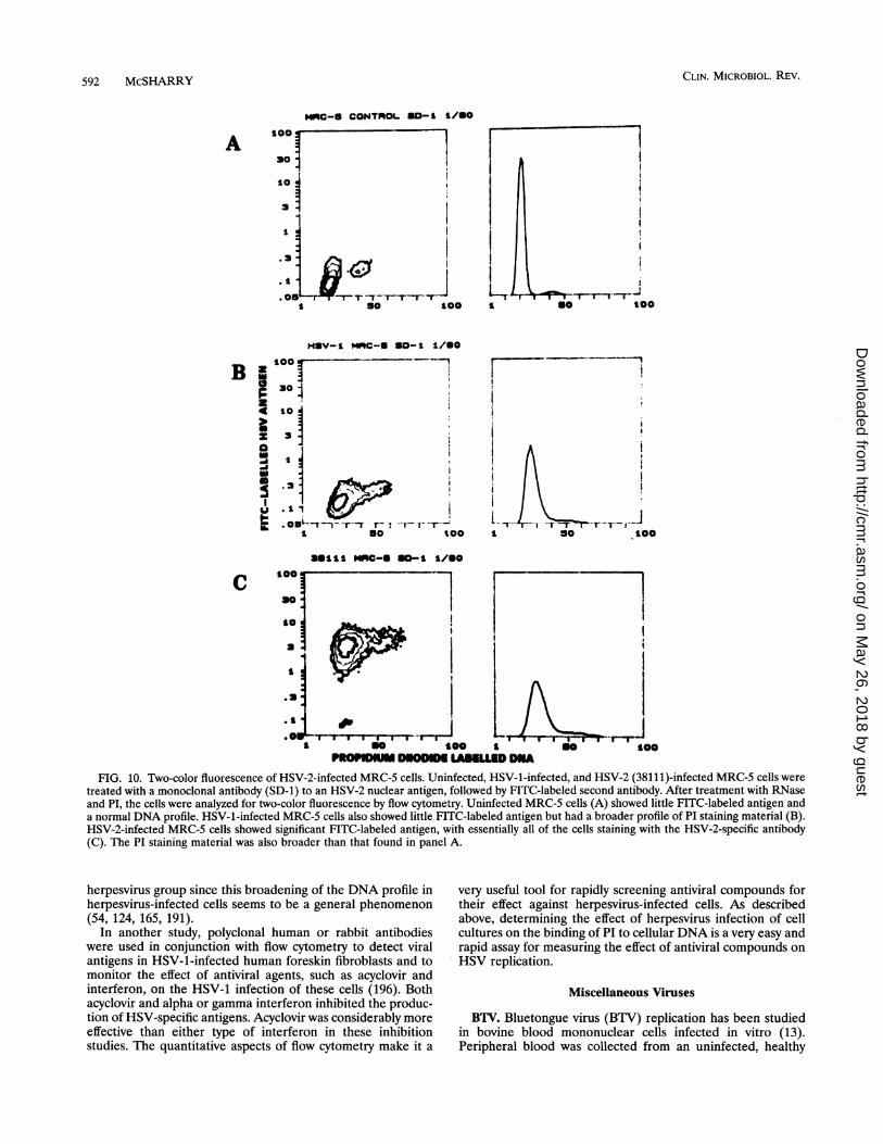
592 McSHARRY
MRC-5 CONTHOL 50-1 1/SO
A ---so
3
I
. O5 - -T -r- I rI so Soo
HIV-1 "MC-8 80-1 S/-Otoo ---
B z X-F3°i~~~~~~~~~~~~~~~~~~~~~~~~~~~~~~~~~~~~~~~~~~~~~~~~~~~~304
a .rs otooIa5I10
CLIN. MICROBIOL. REV.
t00
I
"I11 MRC-S SO-I 1/S0
so
a.
.a.
.01 5-I I 1 I Ura- I't so too s
n000 I1Dt00
FIG. 10. Two-color fluorescence of HSV-2-infected MRC-5 cells. Uninfected, HSV-1-infected, and HSV-2 (38111)-infected MRC-5 cells weretreated with a monoclonal antibody (SD-1) to an HSV-2 nuclear antigen, followed by FITC-labeled second antibody. After treatment with RNaseand PI, the cells were analyzed for two-color fluorescence by flow cytometry. Uninfected MRC-5 cells (A) showed little FITC-labeled antigen anda normal DNA profile. HSV-1-infected MRC-5 cells also showed little FITC-labeled antigen but had a broader profile of PI staining material (B).HSV-2-infected MRC-5 cells showed significant FITC-labeled antigen, with essentially all of the cells staining with the HSV-2-specific antibody(C). The PI staining material was also broader than that found in panel A.
herpesvirus group since this broadening of the DNA profile inherpesvirus-infected cells seems to be a general phenomenon(54, 124, 165, 191).
In another study, polyclonal human or rabbit antibodieswere used in conjunction with flow cytometry to detect viralantigens in HSV-1-infected human foreskin fibroblasts and tomonitor the effect of antiviral agents, such as acyclovir andinterferon, on the HSV-1 infection of these cells (196). Bothacyclovir and alpha or gamma interferon inhibited the produc-tion of HSV-specific antigens. Acyclovir was considerably moreeffective than either type of interferon in these inhibitionstudies. The quantitative aspects of flow cytometry make it a
very useful tool for rapidly screening antiviral compounds fortheir effect against herpesvirus-infected cells. As describedabove, determining the effect of herpesvirus infection of cellcultures on the binding of PI to cellular DNA is a very easy andrapid assay for measuring the effect of antiviral compounds onHSV replication.
Miscellaneous Viruses
BTV. Bluetongue virus (BTV) replication has been studiedin bovine blood mononuclear cells infected in vitro (13).Peripheral blood was collected from an uninfected, healthy
on May 26, 2018 by guest
http://cmr.asm
.org/D
ownloaded from

FLOW CYTOMETRY IN VIROLOGY 593
calf, and the mononuclear cells were obtained and cultured.After 4 h of incubation, nonadherent cells were removed andcultured separately. The nonadherent cells were stimulatedwith concanavalin A and interleukin-2 24 h before infectionwith BTV. Both adherent and nonadherent cells were infectedwith BTV, and infection was monitored by flow cytometry andinfectivity assays. Bovine T cells, B cells, and monocytes orgranulocytes were identified by staining cell surface markerswith FITC-conjugated monoclonal antibodies to their respec-tive antigens. With two-color fluorescence, BTV antigen wasdetected in adherent and mitogen-stimulated, nonadherentPBMC 2 to 4 h after infection; at 16 h postinfection, 10 to 12%of the cells were infected. Unstimulated, nonadherent cellsfailed to express the BTV VP7 antigen. Furthermore, virusreplication was evident by flow cytometry almost 24 h before itwas detected by the infectivity assay. The results suggest thatBTV replicates in bovine monocytes and mitogen-stimulatedCD4+ lymphocytes but not in unstimulated CD4+ cells. Theability to do multiparametric quantitative analysis of FITC-labeled surface antigens and PE-labeled internal antigens onthe same population of cells enabled these researchers toperform this experiment rapidly and quantitatively. Because ofthe small percentage of infected cells, this type of analysiswould be difficult to perform with the immunofluorescencemicroscope.ASFV. In in vitro experiments, hyperimmune polyclonal
antibody and monospecific antibodies raised in swine to Afri-can swine fever virus (ASFV) were used to monitor theexpression of ASFV glycoproteins on the surface of ASFV-infected monkey cells (3). Monkey cells were infected withASFV and analyzed for viral antigen expression at varioustimes ranging from 0 to 20 h postinfection. The results showedthat ASFV-specific proteins were expressed on the cell sur-faces within 6 h after infection, and maximal expressionoccurred at 12 h post-infection. The sensitivity of flow cytom-etry allowed these investigators to determine the number ofcells infected, the time course of the appearance of antigen onthe cell surfaces, and the amount of antigen on the surface ofeach cell. In addition to the ease and rapidity of the assay, thequantitative aspect of flow cytometry allowed measurementsthat would be difficult to make with fluorescence microscopy.REV-A. The gp9O envelope protein of reticuloendotheliosis-
associated virus (REV-A) was detected by flow cytometry onthe surface of virus-infected, transformed chicken bone mar-row cells (203). A fluorescein-labeled monospecific rabbitantibody to a peptide derived from the carboxy-terminalportion of the REV-A envelope protein was used to stainunfixed, REV-A-infected cells. Greater than 99% of REV-A-infected bone marrow cells expressed gp9O antigen on their cellsurfaces, whereas uninfected cells treated with the same FITC-labeled antibody showed little staining. In the presence ofantibody to gp9O and complement, bone marrow cells express-ing this protein on their cell surface were lysed, suggesting thatgp9O is exposed on the cell surface of REV-A-infected cellsand that its carboxyl terminus is exposed to site-directedantibodies that cause cytotoxicity by activating the complementsystem.WHY. Woodchuck hepatitis virus (WHV) has been detected
by flow cytometry in PBMC obtained from experimentallyinfected animals (29). Polyclonal antibodies to WHV surfaceor core antigens were obtained from either infected wood-chucks or a human hepatitis B virus chronic carrier with a hightiter of anti-hepatitis B virus core antibody. These antibodieswere conjugated with FITC for direct staining and used to labelWHV antigens on the surface of the PBMC obtained fromWHV-infected animals. Both WHV surface and core antigens
were detected on the surface of infected PBMC obtained fromthese animals. The interaction of fluorescently labeled anti-WHV antibodies with WHV antigens expressed on the surfaceof infected cells could be inhibited by pretreating the infectedcells with unlabeled antibody to surface and core antigens. Theresults of these blocking studies suggest that the reaction isspecific and that, under these labeling conditions, both WHVsurface and core antigens are exposed on the surface of thecell.HCV. A monoclonal antibody directed against the first 75
amino acids of the hepatitis C virus (HCV) core antigen wasused in conjunction with flow cytometry to detect and quanti-tate HCV-infected cells in PBMC obtained from a patient withchronic hepatitis (22). Neither the surface of the monocytesnor the lymphocytes stained with monoclonal antibody to thisepitope on the core antigen, suggesting that the reaction wasspecific for the internal core antigen. Others have demon-strated the presence of HCV core antigen and virus in thecirculation by ELISA and by PCR (199), suggesting that HCVdoes circulate in the peripheral blood of patients with non-A,non-B chronic hepatitis.BVDV. In in vivo experiments, bovine viral diarrhea virus
(BVDV) was detected by flow cytometry in blood samplesfrom cattle persistently infected with the virus (159). Hepa-rinized peripheral blood was obtained from persistently in-fected and uninfected cattle, and the erythrocytes were lysed,fixed with 1% paraformaldehyde, and permeabilized with 1%saponin. Biotinylated polyclonal antibody against BVDV orbiotinylated normal serum was used, and specifically boundantibodies were detected with FITC-conjugated strepavidin byflow cytometry. Of 143 samples tested, 37 obtained frompersistently infected cattle were positive for BVDV by flowcytometry, whereas only 34 of these 37 samples were positiveby the standard infectivity assay. The results suggest that theflow cytometric assay is more sensitive and considerably morerapid than the standard infectivity assay for BVDV.FeLV. Feline leukemia virus (FeLV)-infected lymphoid,
erythroid, and myeloid lineage marrow cells in cats werestudied by flow cytometry (45). Bone marrow cells wereobtained from FeLV-infected cats and fixed with 3%paraformaldehyde followed by 0.5% Triton X-100 to perme-abilize the plasma membrane. Indirect immunofluorescencewith a monoclonal antibody to the p27 viral core antigen wasused to detect FeLV-infected cells. The cells were separatedinto myeloid, erythroid, and lymphoid populations by lightscatter, and the percentage of p27 antigen-positive cells in eachpopulation was determined by flow cytometry. The resultssuggested that all three cell populations are infected withFeLV. This interesting finding was made possible by using themultiparametric properties of the flow cytometer, includinglight scatter to separate the lymphoid, erythroid, and myeloidcell populations and immunofluorescence detection to detectFeLV-infected cells. This type of analysis would be difficultwith a light or fluorescence microscope.F-MuLV. In one study, the gp7O surface antigen of Friend
murine leukemia virus (F-MuLV) was detected on the plasmamembrane of infected cells and the number of infected cellsexpressing this antigen was quantitated by flow cytometry(135). Mouse erythroleukemia cells or splenocytes explantedfrom F-MuLV-infected mice were stained for the presence ofthe F-MuLV gp7O surface antigen by indirect immunofluores-cence and analyzed by flow cytometry. The results showed thatapproximately 10% of the splenocytes were expressing F-MuLV gp7O on their cell surfaces. However, the F-MuLV-infected mice were only 1% as susceptible as uninfected miceto superinfection with other retroviruses, such as spleen focus-
VOL. 7, 1994
on May 26, 2018 by guest
http://cmr.asm
.org/D
ownloaded from

CLIN. MICROBIOL. REV.
forming virus, that use the same receptor. Similar results werefound with mice incapable of making antibody or lackingmature T cells, suggesting that the immune response does notcontribute to this interference phenomenon. This flow cyto-metric assay established that F-MuLV-infected mice are resis-tant to challenge by spleen focus-forming virus because thegp7O antigens of F-MuLV on infected cells are occupying thecell receptor for the spleen focus-forming virus. This is thebasis for the interference phenomenon. The ability of flowcytometry to accurately detect small percentages of virus-infected cells has made this type of analysis possible.EBV. Miller et al. (134) used flow cytometry to determine
calcium mobilization and cell surface expression of MHC classII, CD19, CD22, and IgM on lymphoblastoid cells transfectedwith the gene for LMP2, the latent membrane protein ex-pressed in EBV-transformed cells. The results suggested thatthese surface proteins were expressed to the same extent in thetransfected cells as in control cells. Antibody to IgM, MHCclass II, or CD19 cross-link these proteins on the cell surfaceand induce calcium mobilization in normal lymphoid cells.Expression of LMP2 in the transfected cells inhibited thecalcium mobilization. In this case, calcium mobilization wasmeasured by flow cytometry of indol-1-loaded cells.Autographa californica nuclear polyhedrosis virus. Morris
and Miller (138) used flow cytometry to study the expression ofA. californica nuclear polyhedrosis virus gene products in avariety of cultured insect cells. The expression of early, late,and very late promoters in various cell lines was studied bymeasuring the amount of chloramphenicol acetyltransferaseactivity. Cell cultures were infected with recombinant virusescontaining chloramphenicol acetyltransferase reporter genesunder the transcriptional control of viral early, late, and verylate promoters. The expression of the chloramphenicol acetyl-transferase gene attached to these promoters was measured bytreating the transfected cells with a mouse monoclonal anti-body to the chloramphenicol acetyltransferase gene productfollowed by FITC-conjugated goat anti-mouse IgG as thesecond antibody and analysis by flow cytometry. The datasuggest that productive infection occurred in Spodoptera frugi-perda cells, with almost 100% of the cells infected, whereas alower percentage of cells was infected in the other cell linestested. Nonpermissive cells were blocked in viral DNA repli-cation.
In summary, this brief survey of several virus-cell systemsthat have been analyzed by flow cytometry should give thereader some idea of the potential of multiparameter flowcytometric analysis for the study of virus-cell interactions. Withthe ability to differentiate individual antigens stained by asmany as three fluorescent labels, it will be possible to expandthese kinds of studies in the future.
CLINICAL VIROLOGY
Flow cytometry has been used as a rapid test for the directdetection of virus-infected cells obtained from patients in-fected with HCMV or HIV-1.
HCMV
In the studies of Elmendorf et al. (54), HCMV-infectedBAL samples obtained from patients with HCMV pneumoniawere permeabilized, stained by the indirect immunofluores-cence method, and analyzed by two-color quantitative flowcytometry. The data indicated that HCMV-infected BAL cellscould be distinguished from uninfected BAL cells and thatinfected BAL cells could be detected and quantitated by
indirect immunofluorescence in conjunction with flow cytom-etry. The flow cytometric technique for detecting HCMV-infected BAL cells was as accurate as the standard infectivityassays and gave a quantitative answer within 24 h after thespecimen was received. This rapid technique for diagnosingHCMV infection is considerably faster and more quantitativethan standard cell culture assays, which take weeks to com-plete, or the shell vial technique for amplification of HCMV incell culture followed by detection of immediate early antigenby immunofluorescence (40, 70).
Indirect immunofluorescence in conjunction with flow cy-tometry can be used to detect and quantitate HCMV-infectedPBMC obtained from immunosuppressed patients with acuteHCMV infection (126). PBMC, obtained from healthy controlsor immunocompromised patients with acute HCMV infection,were permeabilized, stained by the indirect immunofluores-cence method, and analyzed for two-color fluorescence by flowcytometry. Figure 11 illustrates the flow cytometric analysis ofPBMC obtained from a healthy control subject and thatobtained from an immunocompromised patient with acuteHCMV disease. Panel A shows the gating of PBMC with a 2 Ncontent of DNA on the basis of FALS and PI binding. Panel Bshows the lack of staining of PBMC for the FITC-labeledimmediate early antigen in cells obtained from a normalindividual. Note that very few PBMC are labeled in this controlsample. Panel C illustrates the staining of PBMC expressingthe HCMV immediate early antigen in cells obtained from apatient with acute HCMV disease. Note that, compared withhealthy individuals, a significant percentage of the PBMCobtained from this patient is expressing the immediate earlyantigen, indicating that these cells are actively infected withHCMV. Table 7 shows the range of values obtained from theflow cytometric analysis of PBMC obtained from uninfectedhealthy adults (controls) and 10 immunocompromised patientswith acute HCMV disease. The data show that a very smallpercentage of PBMC obtained from healthy adults (controls,0.1 to 0.3% positive cells) expresses the HCMV immediateearly antigen, whereas patients with acute HCMV disease, asconfirmed by an infectivity assay, have much higher percent-ages (0.6 to 8%) of PBMC expressing the HCMV immediateearly antigen. Furthermore, the results obtained from flowcytometry were available within 24 h, while the infectivityassays took considerably longer. These results compare favor-ably with the recently developed HCMV antigenemia assaythat is being used to detect HCMV in peripheral blood ofimmunosuppressed transplant patients with suspected HCMVdisease (65-67) and with PCR assay data from patients withHCMV disease (186). The flow cytometric technique is morerapid and much easier to perform and gives more accuratequantitative data than other techniques that attempt to assayHCMV directly from specimens obtained in vivo. This analysiscan be performed on freshly isolated PBMC and yields adiagnostic result within 8 h from the time the clinical sample isobtained. Future studies will attempt to use multiparameterflow cytometric analysis of triple-stained leukocytes to deter-mine which blood cells are infected with HCMV in vivo.Flow cytometry has been used for monitoring the effect of
the antiviral drug ganciclovir on virus gene expression inHCMV-infected individuals (126). Table 8 shows the effect ofganciclovir treatment on the expression of HCMV immediateearly antigen in PBMC obtained from a kidney transplantpatient with acute HCMV disease. Peripheral blood wasobtained during the acute stage of the patient's disease andapproximately each week thereafter. Whole blood was sedi-mented over Ficoll-Hypaque, and the PBMC were isolated,permeabilized, stained for the HCMV immediate early antigen
594 McSHARRY
on May 26, 2018 by guest
http://cmr.asm
.org/D
ownloaded from
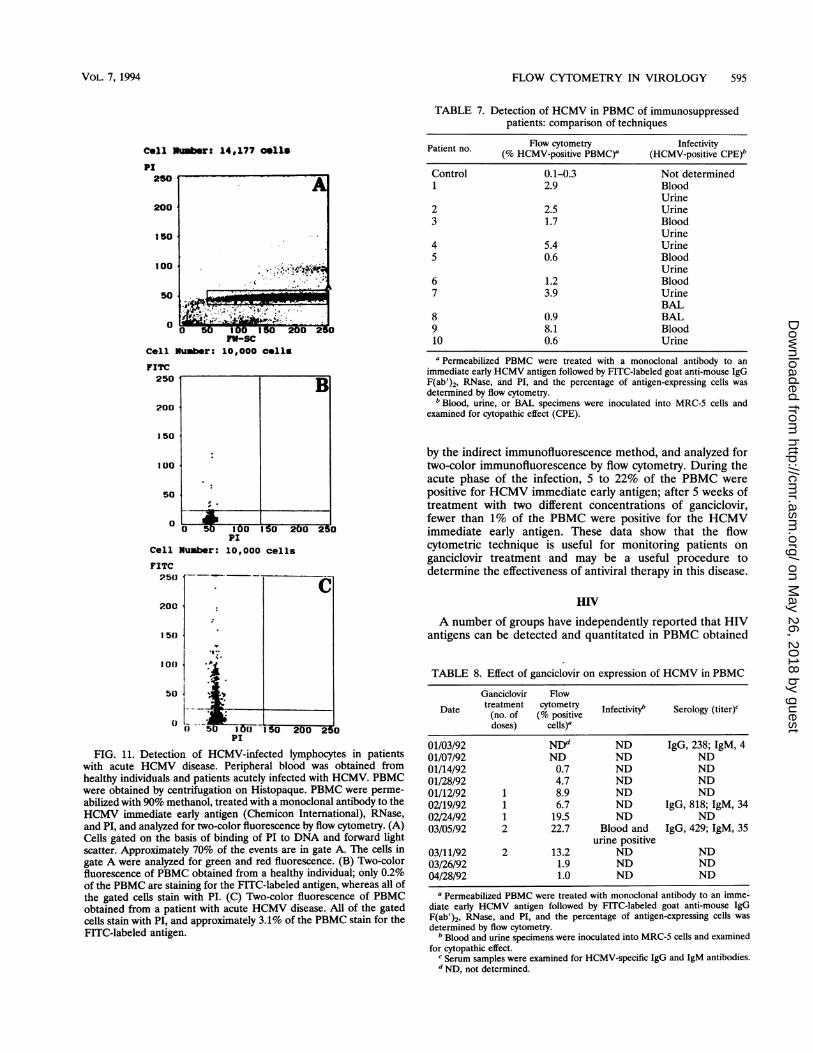
FLOW CYTOMETRY IN VIROLOGY 595
cell M
PI250
200
150
100
50
0
Cell I
FITC250
200
150
100
50
0
luber: 14,177 Cells
00 150202TV-SC
lumber: 10,000 cells
B
;;00I 2
Cell Number:
FITC250
200:
150
.I..100
50
(1 - -5
PI10,000 cells
10UPi
I C
Iau 0uu 25u
FIG. 11. Detection of HCMV-infected lymphocytes in patientswith acute HCMV disease. Peripheral blood was obtained fromhealthy individuals and patients acutely infected with HCMV. PBMCwere obtained by centrifugation on Histopaque. PBMC were perme-abilized with 90% methanol, treated with a monoclonal antibody to theHCMV immediate early antigen (Chemicon International), RNase,and PI, and analyzed for two-color fluorescence by flow cytometry. (A)Cells gated on the basis of binding of PI to DNA and forward lightscatter. Approximately 70% of the events are in gate A. The cells ingate A were analyzed for green and red fluorescence. (B) Two-colorfluorescence of PBMC obtained from a healthy individual; only 0.2%of the PBMC are staining for the FITC-labeled antigen, whereas all ofthe gated cells stain with PI. (C) Two-color fluorescence of PBMCobtained from a patient with acute HCMV disease. All of the gatedcells stain with PI, and approximately 3.1% of the PBMC stain for theFITC-labeled antigen.
TABLE 7. Detection of HCMV in PBMC of immunosuppressedpatients: comparison of techniques
Patient no. Flow cytometry Infectivity(% HCMV-positive PBMC)Q (HCMV-positive CPE)b
Control 0.1-0.3 Not determined1 2.9 Blood
Urine2 2.5 Urine3 1.7 Blood
Urine4 5.4 Urine5 0.6 Blood
Urine6 1.2 Blood7 3.9 Urine
BAL8 0.9 BAL9 8.1 Blood10 0.6 Urine
a Permeabilized PBMC were treated with a monoclonal antibody to animmediate early HCMV antigen followed by FITC-labeled goat anti-mouse IgGF(ab')2, RNase, and PI, and the percentage of antigen-expressing cells wasdetermined by flow cytometry.bBlood, urine, or BAL specimens were inoculated into MRC-5 cells and
examined for cytopathic effect (CPE).
by the indirect immunofluorescence method, and analyzed fortwo-color immunofluorescence by flow cytometry. During theacute phase of the infection, 5 to 22% of the PBMC werepositive for HCMV immediate early antigen; after 5 weeks oftreatment with two different concentrations of ganciclovir,fewer than 1% of the PBMC were positive for the HCMVimmediate early antigen. These data show that the flowcytometric technique is useful for monitoring patients onganciclovir treatment and may be a useful procedure todetermine the effectiveness of antiviral therapy in this disease.
HIV
A number of groups have independently reported that HIVantigens can be detected and quantitated in PBMC obtained
TABLE 8. Effect of ganciclovir on expression of HCMV in PBMC
Ganciclovir Flow
Date treatment cytometry Infectivityb Serology (titer)cDae (no. of (% positivedoses) cells)'
01/03/92 NDd ND IgG, 238; IgM, 401/07/92 ND ND ND01/14/92 0.7 ND ND01/28/92 4.7 ND ND01/12/92 1 8.9 ND ND02/19/92 1 6.7 ND IgG, 818; IgM, 3402/24/92 1 19.5 ND ND03/05/92 2 22.7 Blood and IgG, 429; IgM, 35
urine positive03/11/92 2 13.2 ND ND03/26/92 1.9 ND ND04/28/92 1.0 ND ND
I Permeabilized PBMC were treated with monoclonal antibody to an imme-diate early HCMV antigen followed by FITC-labeled goat anti-mouse IgGF(ab')2, RNase, and PI, and the percentage of antigen-expressing cells was
determined by flow cytometry.b Blood and urine specimens were inoculated into MRC-5 cells and examined
for cytopathic effect.Serum samples were examined for HCMV-specific IgG and IgM antibodies.NND, not determined.
VOL. 7, 1994
k
on May 26, 2018 by guest
http://cmr.asm
.org/D
ownloaded from

596 McSHARRY
from HIV-seropositive patients. In the initial report, PBMCobtained from uninfected and HIV-infected individuals wereisolated on Ficoll-Hypaque, permeabilized, and analyzed fortwo-color fluorescence by flow cytometry (125). The resultsshowed that HIV p24 or nef antigens could be detected inPBMC obtained from patients with clinical AIDS. The major-ity of HIV-seropositive individuals who were asymptomatic(Centers for Disease Control [CDC] group II) did not expressthe HIV p24 antigen in significant numbers of their PBMC,whereas the majority of those with AIDS (CDC group IV) did.These results suggested that this technique can be used tomeasure disease progression. Furthermore, the percentage ofp24 antigen-positive PBMC was inversely proportional to thetotal CD4+ cell count, indicating that the percentage of p24antigen-positive cells correlates with a frequently used surro-gate marker for HIV infection (99). PBMC obtained fromHIV-seronegative individuals showed no significant stainingwith either the p24 or the nef monoclonal antibodies. In asmall sample, patients on ZDV therapy showed decreasedpercentages of p24 antigen in their PBMC, suggesting that flowcytometry may be used to monitor patients on antiretroviralchemotherapy. This technique has been used to monitor virusload in patients on various antiretroviral therapies (129, 163).
Ohlsson-Wilhelm et al. (143) permeabilized PBMC and thenused a double-labeling technique employing PE-labeled mono-clonal antibody to CD14, which labeled the monocytes, andmouse monoclonal antibody to p24 antigen followed by FITC-labeled second antibody, which labeled the HIV-infected cells.With this procedure, the PE-labeled CD14+ monocytes wereexcluded from the lymphocyte population so that only thelymphocyte population was analyzed for the presence ofinternal p24 antigen. The results demonstrated that PBMCobtained from both asymptomatic (mean, 2%) and symptom-atic (mean, 4.2%) HIV-seropositive patients had significantnumbers of p24 antigen-positive blood cells, whereas most ofthose on ZDV therapy and all normal controls did not expressp24 antigen in this assay. The percentage of p24 antigen-positive PBMC was inversely related to the total number ofCD4+ cells. When cells from patients with higher percentagesof p24 antigen-positive PBMC were tested for virus infectivityby coculture with mitogen-stimulated control lymphocytes, thecultures became positive for p24 antigen sooner than thosecocultured with PBMC obtained from patients with lowerpercentages of p24 antigen-positive PBMCs.
In a similar study, Costigliola et al. (38) showed that p24antigen could be detected in CD4+ PBMC obtained fromHIV-seropositive patients at all stages of the disease. Those inCDC stage II had lower percentages of p24 antigen-positiveCD4+ cells, and those in CDC stage IV had higher percentagesof p24 antigen-positive CD4+ cells. Antiretroviral therapy withZDV reduced the percentage of p24 antigen-positive cells.Holzer et al. (80) used flow cytometric analysis of CD4+ cellsobtained from HIV-infected individuals to show that theCD4+ cell number declined and the percentage of HIV-p24antigen-positive CD4+ cells increased as the disease pro-gressed. Finally, Gadol et al. (62) reported on the use of frozenPBMC obtained from HIV-seropositive patients for the detec-tion of p24 and p17 antigens by flow cytometry. They compareda number of different fixation techniques, each of whichyielded slightly different results. However, they did show thatasymptomatic, HIV-seropositive individuals seldom had signif-icant numbers of p24 antigen-positive PBMC, whereas symp-tomatic individuals were frequently positive for p24 antigen inup to 10% of their PBMC. From the results of these fiveindependent studies, it is clear that by using the rapid andsensitive quantitative flow cytometric technique, cell-associ-
ated HIV p24 antigen expression can be detected most of thetime in patients with AIDS but less frequently in asymptomaticHIV-seropositive individuals. Furthermore, when HIV anti-gens are detected in PBMC from asymptomatic patients, alower percentage of antigen-positive cells is detected. Thequantitative aspects of this technique allow this differentiationto be made rapidly and with ease. This procedure has beenused to monitor disease progression in HIV-seropositive pa-tients and to ascertain the effect of antiviral therapy on virusreplication in these patients (38, 62, 80, 125, 143). With theability to detect both cell surface and internal antigens by usingfixatives, such as Permea-Fix and possibly FACS Lysing Re-agent, it will be possible to identify and quantitate the periph-eral blood cells that are infected with HIV by two- or three-color multiparameter flow cytometric analysis of PBMCobtained from HIV-infected patients without the need to sortcells.
This flow cytometric technique has been used successfully todetect p24 antigen-positive cells obtained from HIV-seropos-itive children 6 months to 12 years of age (127). There is acritical need for an accurate method to identify HIV-infectedbabies shortly after birth. Recently, a number of investigatorshave demonstrated the ability to detect free p24 antigen inplasma of newborn babies by the immune complex-dissociatedp24 antigenemia assay (9, 25, 84, 133). Since there is sufficientfree p24 antigen in the plasma of newborn babies for detectionby ELISA, it should be possible to detect cell-associated p24antigen in PBMC from these babies by direct or indirectimmunofluorescence in conjunction with flow cytometry. Withthese two rapid, quantitative, complementary techniques, itshould be possible to detect cell-free and cell-associated HIVantigens in HIV-infected babies born to HIV-seropositivemothers within days to weeks after birth. This time frame isuseful for therapeutic intervention for those babies who areinfected and for identification of those HIV-seropositive ba-bies who are not HIV infected. In addition, these two antigendetection systems are more rapid and less expensive than thevery sensitive, but time-consuming and costly, quantitativeinfectivity assays (4, 113).One of the major criticisms of the flow cytometric technique
is that the number of p24 antigen-positive cells detected seemsto be very high (13 to 25% in earlier studies and 1 to 10% inmore recent studies), the reasons for which vary. They includethe point at which the cutoff value for a negative sample is set,the lack of isotype controls in many experiments, and highnonspecific binding of antibodies to cells in some of the clinicalsamples. Percentages of p24 antigen-positive cells as high as10% are possible in individuals with advanced HIV infection orAIDS. In one report, essentially all patients with 1 to 10% oftheir PBMC positive for the p24 antigen were also positive forinfectivity in coculture assays (146). Quantitative cocultureassays will be required to better compare these two assays.That this technique identifies p24 antigen-positive cells inasymptomatic individuals is probably correct for the followingreasons: (i) PCR has detected both HIV proviral DNA andmRNA expression in PBMC at all stages of disease (11, 23, 34,55, 86, 93, 132, 153, 174, 195, 211); (ii) immune complex-dissociated p24 antigen assays have detected free p24 inplasma at all stages of disease (9, 25, 133, 158); and (iii)infectious virus has been detected in PBMC and plasma fromHIV-seropositive patients at all stages of HIV infection (4, 42,84, 146, 147). An additional criticism of flow cytometry for thisuse has been the possibility that p24 antibodies are detectingp24 antigen in cells that have phagocytized free viral antigen.The only way to address this possibility is to use antibodies
CLIN. MICROBIOL. REV.
on May 26, 2018 by guest
http://cmr.asm
.org/D
ownloaded from

FLOW CYTOMETRY IN VIROLOGY 597
against HIV antigens that accumulate in the nucleus, such asRev and Tat (41).
In sum, the data indicate that flow cytometry should be auseful, rapid, and relatively inexpensive technique to measurevirus load in HIV-infected individuals and to monitor the effectof antiviral therapy on viral load in vivo. However, it must beremembered that these flow cytometric techniques will onlydetect cells that are expressing the antigen to which thefluorescently labeled antibody is directed. Cells latently in-fected with HIV will not be detected with this assay.
ANTIBODIES MEASURED BY FLOW CYTOMETRY
In addition to detecting and quantitating virus-infected cells,antibodies to viruses, cells, and bacteria have been detectedand quantitated by flow cytometry. McHugh et al. (122, 123),Scillian et al. (182), and Sligh et al. (190) demonstrated thatantibodies to HCMV, HSV, and HIV can be detected bybinding to microspheres coated with viral antigens. Antibodiesto different viruses could be distinguished by binding each virusto microspheres of different sizes that could be separated bylight scatter. Once the antibody attached to its specific micro-sphere, a fluorochrome-labeled antibody directed against thebound antibody was added to the microspheres, and thelabeled spheres were analyzed for fluorescence and lightscatter by flow cytometry. Gorse et al. (73) used uninfected H9cells, HIV-infected H9 cells, and H9 cells expressing therecombinant gpl60 gene to measure antibody to recombinantgp160 vaccine. After the human sera reacted with the cells, theantibody-bearing cells were treated with a fluorochrome-la-beled antibody to human IgG, and then the fluorescence wasanalyzed by flow cytometry. This technology has been used todetect antilymphocyte antibodies bound to peripheral bloodlymphocytes in patients with AIDS, using FITC-conjugatedgoat antibody to human IgG (19). Antibodies to bacteria havealso been detected by flow cytometry. A multicomponentantigen of Helicobacter pylon was bound to microspheres andused to test sera for the presence of antibodies to this organism(17). Bound antibodies were detected with FITC-conjugatedgoat anti-human IgG, and the fluorescence was quantitated byflow cytometry. Antibodies to Toxoplasma gondii in humanserum have been measured by using fluorescently labeledantibodies against human IgG, IgM, or IgA after these anti-bodies were attached to fixed tachyzoites (39). The amount ofeach antibody bound was quantitated by measuring fluores-cence intensity by flow cytometry. Thus, it is possible to use thistechnology for rapid, quantitative assays for both antibodiesand cells expressing antigens derived from patients.
BACTERIA, FUNGI, AND PARASITES
Although this review deals with the use of flow cytometry forthe detection of viruses in cells and clinical samples, briefmention will be given to the use of flow cytometry for thedetection and quantitation of bacteria, yeasts, and parasites.This subject was recently reviewed (52, 184). Phagocytosis andkilling of Candida albicans and Staphylococcus aureus havebeen quantitated by flow cytometry (117-119). The organismswere treated with a nonfluorescent derivative of carboxyfluo-rescein that becomes fluorescent when activated by esterases inviable cells. The phagocytic cells in dextran-enriched plasmawere labeled with a PE-conjugated monoclonal antibody. Afterthe organisms were mixed with the phagocytic cells andincubated, two-color fluorescence was detected and quanti-tated by flow cytometry. Phagocytic activity was monitored bythe increase of green fluorescence in a constant amount of
orange cells, and cell killing was monitored by a decrease in theamount of green fluorescence in these orange cells. Green etal. (74) used the permeability to PI of C. albicans and otherorganisms and analysis of cell-associated red fluorescenceintensity by flow cytometry to determine the minimal fungi-cidal concentration of various antifungal compounds.The viability of Pneumocystis carinii has been detected in
athymic nude F344 rat lung homogenates and human BALspecimens by flow cytometric analysis of PI-stained cells (102).Only nonviable organisms take up the PI, making it possible todetect and quantitate nonviable organisms. This proceduremay be useful for rapid diagnosis and for testing anti-infectiveagents against P. carinii.The fluorescent nucleic acid-binding dye, ethidium bromide,
was used to label bacteria, and the light scatter properties ofthe bacteria and the fluorescent dye were used to determinethe antibiotic effect of amikacin on a variety of gram-negativeorganisms, using flow cytometry (30). Ordonez and Wehman(144) used the fluorescent dye 3,3'-diphenyloxacarbocyanineto develop a rapid test to measure the effect of antibiotics onthe induction of membrane potential changes in S. aureus.Changes in fluorescence due to antibiotic action were moni-tored by flow cytometry, and tests could be performed in 90min compared with overnight culture for standard assays. Theeffect of immune sera and complement on the killing of theLyme disease parasite, Borrelia burgdorfen, was quantitated byflow cytometry (166). The organisms were treated with amurine monoclonal antibody, anti-OspA, followed by FITC-conjugated second antibody, or PI was used to stain theorganism. The organisms were detected by a combination oflight scatter and fluorescence. Specific antibody and comple-ment killed the organisms, reducing the number detected byflow cytometry. This assay was rapid and sensitive. Finally,Plasmodium falciparum has been detected in intact and lysederythrocytes of infected individuals by using flow cytometry(150, 206). This brief summary of some applications of flowcytometry for the detection of other microorganisms shouldgive the reader a sense of the wide applicability of thistechnology in microbiology.
FUTURE PROSPECTS
I trust that this review has given the reader the sense thatflow cytometry has a place in the analysis of virus-cell interac-tions. Four major areas on which flow cytometry will likelyhave an impact are summarized below.
(i) Flow cytometry will be used for rapid detection ofvirus-infected cells and other organisms in body fluids ifmonoclonal or polyclonal antibodies that specifically react withcell surface or intracellular viral antigens are available. Exam-ples are cells infected with viruses, such as HTLV-1 and -2(16), hepatitis B virus (29), HCV (199), human herpesvirus 6(32, 53), varicella-zoster virus (192), and measles virus (59), inblood and microbes, such asP. carinii (102) and Mycobacteriumtuberculosis, in pulmonary lavage fluids. Using flow cytometryfor direct detection and possible quantitation of these mi-crobes will save time and expense in the clinical laboratory.
(ii) Flow cytometry will be used for rapid detection of virusesafter amplification in cell culture. For many viruses that are notcell associated in body fluids, or when there are too few cellsfor a statistically accurate determination by flow cytometry, it ispossible to amplify the number of cells expressing the viralantigens by overnight incubation in cell culture and then topermeabilize, stain, and analyze the virus-infected cells by flowcytometry. Again, this will save considerable time in the clinicallaboratory, particularly when those viruses that take several
VOL. 7, 1994
on May 26, 2018 by guest
http://cmr.asm
.org/D
ownloaded from

598 McSHARRY
days to produce cytopathic effects in tissue culture are beingstudied.
(iii) As has been demonstrated above, flow cytometricanalysis of virus-infected cells is an excellent way to monitorthe effects of antiviral or anti-infective agents in vitro and invivo. Further application of this technology will allow screeningof numerous compounds for their antiviral and anti-infectiveactivities. Flow cytometry will continue to be useful in moni-toring patients on antiviral chemotherapy to determine the invivo effects of therapeutic agents on their infection.
(iv) With the development of fluorescent in situ hybridiza-tion in suspension, it will be possible to use nucleic acidhybridization methods to detect viruses, bacteria, and othermicroorganisms in cells and quantitate them by flow cytometry(110, 151, 202, 207). Since flow cytometric analysis of cellsstained for viral antigens will detect only cells that are activelyexpressing viral proteins, the combination of in situ hybridiza-tion and flow cytometry will allow detection of latently infectedcells. In addition, the combination of flow cytometry and PCRwill provide the sensitivity required to detect and quantitateviruses in individual cells.Of course, flow cytometry will continue to be used to
monitor lymphocytes in healthy and diseased people, detectDNA ploidy in tumor cells, and serve as an excellent tool forbasic research. Increased use and familiarization with thispowerful technology will give the clinician, the laboratorian,and the researcher the ability to move from qualitative scienceto a more quantitative aspect of their work. In addition, theability of flow cytometry to measure multiple parameterssimultaneously will allow those who are interested in studyingthe interactions of different cellular and viral componentswithin the cell to perform these experiments. The multiparam-eter flow cytometric analysis of double- and triple-stained cellswill give investigators the opportunity to identify which cellsare infected as well as to quantitate the number of virus-infected cells. For example, assuming that the CD4+ T lym-phocyte is the primary target for HIV, peripheral blood cellscould be stained with one or more fluorescently labeled cellsurface reagents, and the cells could be permeabilized andtreated with one or more fluorescently labeled reagents di-rected against internal viral antigens. These types of experi-ments should enable one to identify the peripheral blood cellsthat are infected with HIV or any virus of interest.
ACKNOWLEDGMENTS
I thank Scot Remick and Tom Friedrich for reviewing the manu-
script and for helpful discussion, Ann Ogden-McDonough and BettyOlson for technical assistance, and JoAnn D'Annibale and Karla Rollfor secretarial support.
This work was supported in part by grants AI30883 and A132367from the National Institutes of Health.
REFERENCES
1. Afanasyev, V. N., B. A. Korol, N. P. Matylevich, V. A. Pechatni-kov, and S. R. Umansky. 1993. The use of flow cytometry for theinvestigation of cell death. Cytometry 14:603-609.
2. Akbar, B. A., N. Borthwick, M. Salmon, W. Gombert, M. Bofill,N. Shamsadeen, D. Pilling, S. Pett, J. E. Grundy, and G. Janossy.1993. The significance of low bcl-2 expression by CD45RO T cellsin normal individuals and patients with acute viral infections. Therole of apoptosis in T cell memory. J. Exp. Med. 178:427-438.
3. Alcaraz, C., A. Alvarez, and J. M. Escribano. 1992. Flow cyto-
metric analysis of African swine fever virus-induced plasmamembrane proteins and their humoral immune response ininfected pigs. Virology 189:266-273.
4. Alimenti, A., M. O'Neill, J. L. Sullivan, and K. Luzuriaga. 1992.
Diagnosis of vertical human immunodeficiency virus type 1infection by whole blood culture. J. Infect. Dis. 166:1146-1148.
5. Andrei, G., R. Snoeck, D. Schols, P. Goubau, J. Desmyter, and E.De Clercq. 1991. Comparative activity of selected antiviral com-pounds against clinical isolates of human cytomegalovirus. Eur. J.Clin. Microbiol. Infect. Dis. 10:1026-1033.
6. Arends, M. J., R. G. Morris, and A. H. Wyllie. 1990. Apoptosis.The role of the endonuclease. Am. J. Pathol. 136:593-608.
7. Arends, M. J., and A. H. Wyllie. 1991. Apoptosis mechanisms androles in pathology. Int. Rev. Exp. Pathol. 32:223-254.
8. Argall, K. G., P. J. Armati, N. J. C. King, and M. W. Douglas.1991. The effect of West Nile virus on major histocompatibilitycomplex class I and II molecule expression in Lewis rat Schwanncells in vitro. J. Neuroimmunol. 35:273-284.
9. Ascher, D. P., C. Roberts, and A. Fowler. 1992. Acidificationmodified p24 antigen capture assay in HIV seropositives. J.Acquired Immune Defic. Syndr. 5:1080-1083.
10. Baba, M., D. Schols, R. Pauwels, H. Nakashima, and E. DeClercq. 1990. Sulfated polysaccharides as potent inhibitors ofHIV-induced syncytium formation: a new strategy towards AIDSchemotherapy. J. Acquired Immune Defic. Syndr. 3:493-499.
11. Bagasra, O., T. Seshamma, J. W. Oakes, and R. J. Pomerantz.1993. Frequency of cells positive for HIV-1 sequences assessed byin situ polymerase chain reaction. AIDS 7(Suppl.):S7-S10.
12. Balzarini, J., M.-J. Perez-Perez, A. San-Felix, D. Schols, C.-F.Perno, A.-M. Vandamme, M.-J. Camarasa, and E. De Clercq.1992. 2',5'-Bis-O-(tert-butyldimethylsilyl)-3'-spiro-5"-(4"-amino-1',2"-oxathiole-2",2"-dioxide) pyrimidine (TSAO) nucleoside an-alogues: highly selective inhibitors of human immunodeficiencyvirus type 1 that are targeted at the viral reverse transcriptase.Proc. Natl. Acad. Sci. USA 89:4392-4396.
13. Barratt-Boyes, S. M., P. V. Rossitto, J. L. Stott, and N. N.MacLachlan. 1992. Flow cytometric analysis of in vitro blue-tongue virus infection of bovine mononuclear cells. J. Gen. Virol.73:1953-1960.
14. Basak, S., H. Turner, and R. W. Compans. 1992. Expression ofSV40 receptors on apical surfaces of polarized epithelial cells.Virology 190:393-402.
15. Basgoz, N., I. Qadri, D. Navarro, A. Sears, E. Lennette, J.Youngblom, and L. Pereira. 1992. The amino terminus of humancytomegalovirus glycoprotein B contains epitopes that varyamong strains. J. Gen. Virol. 73:983-988.
16. Beilke, M. 1992. Detection of HTLV-I in clinical specimens. J.Virol. Methods 40:133-144.
17. Best, L. M., S. J. 0. V. van Zanten, G. S. Bezanson, D. J. M.Haldane, and D. A. Malatjalian. 1992. Serological detection ofHelicobacter pylon by a flow microsphere immunofluorescenceassay. J. Clin. Microbiol. 30:2311-2317.
18. Bohm, D., S. Nick, G. Voss, and G. Hunsmann. 1992. Detectionof viral surface antigens on HIV-2ben infected human tumor celllines by flow cytometry. Cytometry 13:259-266.
19. Bonner, B. C., and T. A. Poulton. 1991. Cytofluorometric analysisof anti-lymphocyte antibodies in AIDS. FEMS Microbiol. Immu-nol. 89:33-40.
20. Borrow, P., and M. B. A. Oldstone. 1992. Characterization oflymphocytic choriomeningitis virus-binding protein(s): a candi-date cellular receptor for the virus. J. Virol. 66:7270-7281.
21. Boucher, C., J. Lange, F. Miedema, G. Weverling, M. Koot, J.Mulder, B. Larder, and M. Tersmette. 1992. HIV-1 biologicalphenotype and development of resistance in relation to diseaseprogression in asymptomatic individuals during treatment. AIDS6:1259-1264.
22. Bouffard, P., P. H. Hayashi, R. Acevedo, N. Levy, and J. B. Zeldis.1992. Hepatitis C virus is detected in a monocyte/macrophagesubpopulation of peripheral blood mononuclear cells of infectedpatients. J. Infect. Dis. 166:1276-1280.
23. Brandt, C. D., T. A. Rakusan, A. V. Sison, S. H. Josephs, E. S.Saxena, K. D. Herzog, R. H. Parrott, and J. L. Sever. 1992.Detection of human immunodeficiency virus type 1 infection inyoung pediatric patients by using polymerase chain reaction andbiotinylated probes. J. Clin. Microbiol. 30:36-40.
24. Breau, W. C., W. J. Atwood, and L. C. Norkin. 1992. Class I majorhistocompatibility proteins are an essential component of the
CLIN. MICROBIOL. REV.
on May 26, 2018 by guest
http://cmr.asm
.org/D
ownloaded from

FLOW CYTOMETRY IN VIROLOGY 599
simian virus 40 receptor. J. Virol. 66:2037-2045.25. Burgard, M., M.-J. Mayaux, S. Blanche, A. Ferroni, M.-L.
Guihard-Moscato, M.-C. Allemon, N. Ciraru-Vigneron, G. Fir-ton, C. Floch, F. Guillot, E. Lachassine, M. Vial, C. Griscelli, C.Rouzioux, and the HBI Infection in Newborns French Collabo-rative Study Group. 1992. The use of viral culture and p24antigen testing to diagnose human immunodeficiency virus infec-tion in neonates. N. Engl. J. Med. 327:1192-1197.
26. Cameron, P. U., M. Pope, S. Gezelter, and R. M. Steinman. 1994.Infection and apototic cell death of CD4+ T cell during animmune response to HIV-1 pulsed dendritic cells. AIDS Res.Hum. Retroviruses 10:61-71.
27. Carbonari, M., M. Cibati, M. Cherchi, D. Sbarigia, A. M. Pesce,L. Dell'Anna, A. Modica, and M. Fiorilli. 1994. Detection andcharacterization of apoptotic peripheral blood lymphocytes inhuman immunodeficiency virus infection and cancer chemother-apy by a novel flow immunocytometric method. Blood 83:1268-1277.
28. Centers for Disease Control. 1992. Guidelines for the perfor-mance of CD4+ T-cell determinations in persons with humanimmunodeficiency virus infection. Morbid. Mortal. Weekly Rep.41:RR8.
29. Chemin, I., I. Baginski, C. Vermot-Desroches, 0. Hantz, C.Jacquet, D. Rigal, and C. Trepo. 1992. Demonstration of wood-chuck hepatitis virus infection of peripheral blood mononuclearcells by flow cytometry and polymerase chain reaction. J. Gen.Virol. 73:123-129.
30. Cohen, C. Y., and E. Sahar. 1989. Rapid flow cytometric bacterialdetection and determination of susceptibility to amikacin in bodyfluids and exudates. J. Clin. Microbiol. 27:1250-1256.
31. Cohen, J. J. 1993. Apoptosis. Immunol. Today 14:126-130.32. Cone, R. W., M.-L. W. Huang, R. Ashley, and L. Corey. 1993.
Human herpesvirus 6 DNA in peripheral blood cells and salivafrom immunocompetent individuals. J. Clin. Microbiol. 31:1262-1267.
33. Connelly, M. C., J. J. McSharry, and P. E. Rao. A simple andrapid flow cytometric method for the detection of HIV infectedcells and their immunophenotype, abstr. POA2363, p. A63.Abstr. VIII Int. Conf. AIDS/STD World Conf., Amsterdam,19-24 July 1992.
34. Connor, R., H. Mohri, Y. Cao, and D. D. Ho. 1993. Increased viralburden and cytopathicity correlate temporally with CD4+ T-lym-phocyte decline and clinical progression in human immunodefi-ciency virus type 1-infected individuals. J. Virol. 67:1772-1777.
35. Cory, J. M., B. M. Ohlsson-Wilhelm, E. J. Brock, N. A. Shaeffer,M. E. Steck, M. E. Eyster, and F. Rapp. 1987. Detection ofhuman immunodeficiency virus-infected lymphoid cells at lowfrequency by flow cytometry. J. Immunol. Methods 105:71-78.
36. Cory, J. M., B. M. Ohlsson-Wilhelm, M. E. Steck, M. D.Smithgall V. Rozday, M. E. Eyster, and F. Rapp. 1989. Kineticsof infected cell appearance as a determinant of number of humanimmunodeficiency virus-1 infectious units. AIDS Res. Hum.Retroviruses 5:97-106.
37. Cory, J. M., F. Rapp, and B. M. Ohlsson-Wilhelm. 1990. Effectsof cellular fixatives on human immunodeficiency virus produc-tion. Cytometry 11:647-651.
38. Costigliola, P., F. Tumietto, E. Ricchi, and F. Chiodo. 1992.Detection of circulating p24 antigen-positive cells during HIVinfection by flow cytometry. AIDS 6:1121-1125.
39. Cozon, G., C. Roure, G. Lizard, T. Greenland, D. Larget-Piet, F.Gandilhon, and F. Peyron. 1993. An improved assay for thedetection of Toxoplasma gondii antibodies in human serum byflow cytometry. Cytometry 14:569-575.
40. Crawford, S. W., R. A. Bowden, R. C. Hackman, C. A. Gleaves,J. D. Meyers, and J. G. Clark. 1988. Rapid detection of cytomeg-alovirus pulmonary infection by bronchoalveolar lavage andcentrifugation culture. Ann. Intern. Med. 108:180-185.
41. Cullen, B. R. 1991. Regulation of HIV-1 gene expression. FASEBJ. 5:2361-2368.
42. Daar, E. S., T. Moudgil, R. D. Meyer, and D. D. Ho. 1991.Transient high levels of viremia in patients with primary humanimmunodeficiency virus type 1 infection. N. Engl. J. Med. 324:961-964.
43. Darzynkiewicz, Z., S. Bruno, G. Del Bino, W. Gorczyca, M. A.Hotz, P. Lassota, and F. Traganos. 1992. Features of apoptoticcells measured by flow cytometry. Cytometry 13:795-808.
44. Darzynkiewicz, Z., and H. A. Crissman (ed.). 1990. Methods incell biology, vol. 33, p. 411-450.
45. Dean, G. A., P. M. Groshek, J. I. Mullins, and E. A. Hoover. 1992.Hematopoietic target cells of anemogenic subgroup C versusnonanemogenic subgroup A feline leukemia virus. J. Virol.66:5561-5568.
46. del Llano, A. M., J. P. Amieiro-Puig, E. N. Kraiselburd, M. J.Kessler, C. A. Mlaga, and J. A. Lavergne. 1993. The combinedassessment of cellular apoptosis, mitochondrial function andproliferative response to pokeweek mitogen has prognostic valuein SIV infection. J. Med. Primatol. 22:147-153.
47. Dent, G. A., M. C. Leglise, K. B. Pryzwansky, and D. W. Ross.1989. Simultaneous paired analysis by flow cytometry of surfacemarkers, cytoplasmic antigens or oncogene expression with DNAcontent. Cytometry 10:192-198.
48. De Rossi, A., L. Ometto, S. Roncella, E. D'Andrea, C. Menin, F.Calderazzo, M. Rowe, M. Ferrarini, and L. Ghieco-Bianchi. 1994.HIV-1 induces down regulation of bcl-2 expression and death byapoptosis of EBV-immortalized B cells: a model for a persistent"self-limiting" HIV-1 infection. Virology 198:234-244.
49. Dhawan, S., H. Z. Streicher, L. M. Wahl, N. Miller, A. T. Louie,I. S. Goldfarb, W. L. Jackson, P. Casali, and A. L. Notkins. 1991.Model for studying virus attachment. II. Binding of biotinylatedhuman T cell leukemia virus type 1 to human blood mononuclearcells potential targets for human T cell leukemia virus type 1infection. J. Immunol. 147:102-108.
50. Dolter, K. E., W. F. Goins, M. Levine, and J. Glorioso. 1992.Genetic analysis of type specific antigenic determinants of herpessimplex virus glycoprotein C. J. Virol. 66:4864-4873.
51. Dorig, R. E., A. Marcil, A. Chopra, and C. D. Richardson. 1993.The human CD46 moleculae is a receptor for measles virus(Edmonston strain). Cell 75:295-305.
52. Edwards, C., J. Porter, J. R. Saunders, J. Diaper, J. A. W.Morgan, and R. W. Pickup. 1992. Flow cytometry and microbi-ology. Soc. Gen. Microbiol. Q. 19:105-108.
53. Eizuru, Y., and Y. Minamishima. 1992. Evidence for putativeimmediate early antigens in human herpesvirus 6-infected cells. J.Gen. Virol. 73:2161-2165.
54. Elmendorf, S., J. J. McSharry, J. A. Lalin, D. Fogleman, andJ. M. Lehman. 1988. Detection of an early cytomegalovirusantigen with two-color quantitative flow cytometry. Cytometry9:254-260.
55. Embretson, J., M. Zupancic, J. L. Ribas, A. Burke, P. Racz, K.Tenner-Racz, and A. Haase. 1993. Massive covert infection ofhelper T lymphocytes and macrophages by HIV during theincubation period of AIDS. Nature (London) 362:359-362.
56. Etingin, 0. R., R. L. Silverstein, and D. Hajjar. 1991. Identifica-tion of a monocyte receptor on herpesvirus-infected endothelialcells. Proc. Natl. Acad. Sci. USA 88:7200-7203.
57. Farmer, J. L., R. G. Hampton, and E. Boots. 1989. Flowcytometric assays for monitoring production of recombinantHIV-1 gpl60 in insect cells infected with a baculovirus expressionvector. J. Virol. Methods 26:279-290.
58. Flamand, L., I. Stefanescu, D. V. Ablashi, and J. Menezes. 1993.Activation of the Epstein-Barr virus replicative cycle by humanherpesvirus 6. J. Virol. 67:6768-6777.
59. Forthal, D. N., Aarnaes, S., J. Blanding, L. de la Maza, and J. G.Tilles. 1992. Degree and length of viremia in adults with measles.J. Infect. Dis. 166:421-424.
60. Freistadt, M., H. B. Fleit, and E. Wimmer. 1993. Poliovirusreceptor on human blood cells: a possible extraneural site ofpoliovirus replication. Virology 195:798-803.
61. Friedrich, T. D., J. Laffin, and J. M. Lehman. 1992. Simian virus40 large T antigen function is required for induction of tetraploidDNA content during lytic infection. J. Virol. 66:4576-4579.
62. Gadol, N., G. J. Crutcher, and M. P. Busch. 1994. Detection ofintracellular HIV in lymphocytes by flow cytometry. Cytometry15:359-370.
63. Gavalchin, J., N. Fan, M. J. Lane, L. Papsidero, and B. J. Poiesz.1993. Identification of a putative cellular receptor for HTLV-1 by
VOL. 7, 1994
on May 26, 2018 by guest
http://cmr.asm
.org/D
ownloaded from
CLIN. MICROBIOL. REV.
a monoclonal antibody, Mab34-23. Virology 194:1-9.64. Gelman, R, S.-C. Cheng, P. Kidd, M. Waxdal, and J. Kagan.
1993. Assessment of the effects of instrumentation, monoclonalantibody, and fluorochrome on flow cytometric immunopheno-typing: a report based on 2 years of the NLAID DAIDS flowcytometry quality assessment program. Clin. Immunol. Pathol.66:150-162.
65. Gerna, G., E. Percivalle, M. G. Revello, and F. Morini. 1993.Correlation of quantitative human cytomegalovirus pp65-, p72-and plS0-antigenemia, viremia and circulating endothelial giantcells with clinical symptoms and antiviral treatment in immuno-compromised patients. Clin. Diagn. Virol. 1:47-59.
66. Gerna, G., M. G. Revello, E. Percivalle, and F. Morini. 1992.Comparison of different immunostaining techniques and mono-
clonal antibodies to the lower matrix phosphoprotein (pp65) foroptimal quantitation of human cytomegalovirus antigenemia. J.Clin. Microbiol. 30:1232-1237.
67. Gerna, G., D. Zipeto, E. Percivalle, M. Parea, M. G. Revello, R.Maccario, G. Peri, and G. Milanesi. 1992. Human cytomegalo-virus infection of the major leukocyte subpopulations and evi-dence for initial replication in polymorphonuclear leukocytesfrom viremic patients. J. Infect. Dis. 166:1236-1244.
68. Gilbert, M. J., S.R Riddell, C.-R Li, and P. D. Greenberg. 1993.Selective interference with class I major histocompatibility com-
plex presentation of the major immediate-early protein followinginfection with human cytomegalovirus. J. Virol. 67:3461-3469.
69. Giorgi, J., H.-L. Cheng, J. B. Margolick, K. D. Bauer, J. Ferbas,M. Waxdal, I. Schmid, L. Hultin, A. L. Jackson, L. Park, J. M. G.Taylor, and the Multicenter AIDS Cohort Study Group. 1990.Quality control in the flow cytometric measurement of T-lym-phocyte subsets: the multicenter AIDS cohort study experience.Clin. Immunol. Immunopathol. 55:173-186.
70. Gleaves, C. A., T. F. Smith, E. A. Shuster, and G. R Pearson.1984. Rapid detection of cytomegalovirus in MRC-5 cells inocu-lated with urine specimens by using low-speed centrifugation andmonoclonal antibody to an early antigen. J. Clin. Microbiol.19:917-919.
71. Goolsby, C., H. Gray, J. J. Docherty, and P. Todd. 1988. Flowcytometric detection of herpes simplex virus type 2 infected andtransformed cells by immunoenzymatic and by indirect immuno-fluorescence staining. Cytometry 9:126-130.
72. Goolsby, C., M. Steiner, and J. Nemeth. 1991. Viral and cellularoncogene expression during progressive malignant transforma-tion of SV40 transformed human fibroblasts. Cytometry 12:748-756.
73. Gorse, G. J., S. E. Frey, F. K. Newman, R. B. Belshe, and theAIDS Vaccine Clinical Trials Network. 1992. Detection of bind-ing antibodies to native and recombinant human immunodefi-ciency virus type 1 envelope glycoproteins following recombinantgpl60 immunization measured by flow cytometry and enzyme
immunoassays. J. Clin. Microbiol. 30:2606-2612.74. Green, L., B. Petersen, L. Steimel, P. Haeber, and W. Current.
1994. Rapid determination of antifungal activity by flow cytom-
etry. J. Clin. Microbiol. 32:1088-1091.75. Hallden, G., U. Andersson, J. Hed, and S. G. 0. Johansson. 1989.
A new membrane permeabilization method for the detection ofintracellular antigens by flow cytometry. J. Immunol. Methods124:103-109.
76. Harabuchi, Y., S. Koizumi, T. Osato, N. Yamanaka, and A.Kataura. 1988. Flow cytometric analysis of Epstein-Barr virusreceptor among the different B-cell subpopulations using simul-taneous two-color immunofluorescence. Virology 165:278-281.
77. Hart, L., R M. Donovan, E. Goldstein, and F. P. Brady. 1990.Detection of human immunodeficiency virus in infected CEMcells using fluorescent DNA probes and a laser based computer-
ized image cytofluorometry system. Anal. Quant. Cytol. Histol.12:127-134.
78. Heynen, C. A., and T. J. Holzer. 1992. Evaluation of a flowcytometric model for monitoring HIV antigen expression in vitro.J. Immunol. Methods 152:25-33.
79. Hollander, Z., and M. R Loken. 1988. Simultaneous analysis ofDNA content and surface antigens in human bone marrow.
Cytometry 9:485-490.
80. Holzer, T. J., C. A. Heynen, R M. Novak, D. L. Pitrak, and G. J.Dawson. 1993. Frequency of cells positive for HIV-1 p24 antigenassessed by flow cytometry. AIDS 7(Suppl.):S3-S5.
81. Horan, M., P. K. Horan, and C. A. Williams. 1975. Quantitativemeasurement of SV40 T antigen production. Exp. Cell Res.91:247-252.
82. Horan, P. K., J. H. Jett, A. Romero, and J. M. Lehman. 1974.Flow microfluorometry analysis of DNA content in chinesehamster cells following infection with simian virus 40. Int. J.Cancer 14:514-521.
83. Hotz, M. A., J. Gong, F. Traganos, and Z. Darzynkiewicz. 1994.Flow cytometric detection of apoptosis: comparison of the assaysof in situ DNA degradation and chromatin changes. Cytometry15:237-244.
84. Ikeda, M. K., W. A. Andiman, J. L. Mezger, E. D. Shapiro, and G.Miller. 1993. Quantitative leukoviremia and immune complex-dissociated antigenemia as predictors of infection status in chil-dren born to mothers infected with human immunodeficiencyvirus type 1. J. Pediatr. 122:524-531.
85. Inghirami, G., M. Nakamura, J. E. Balow, A. L. Notkins, and P.Casali. 1988. Model for studying virus attachment: identificationand quantitation of Epstein-Barr virus-binding cells by usingbiotinylated virus in flow cytometry. J. Virol. 62:2453-2463.
86. Innocenti, P., M. Ottmann, P. Morand, P. LeClercq, and J.-M.Seigneurin. 1992. HIV-1 in blood monocytes: frequency ofdetection of proviral DNA using PCR and comparison with thetotal CD4 count. AIDS Res. Hum. Retroviruses 8:261-268.
87. Jacob, M. C., M. Favre, and J.-C. Bensa. 1991. Membrane cellpermeabilization with saponin and multiparametric analysis byflow cytometry. Cytometry 12:550-558.
88. Jacobberger, J. W. 1991. Cell cycle expression of nuclear proteins,p. 305-326. In A. Yen (ed.), Flow cytometry: advanced researchand clinical applications, vol. 1. CRC Press Inc., Boca Raton, Fla.
89. Jacobberger, J. W., D. Fogleman, and J. M. Lehman. 1986.Analysis of intracellular antigens by flow cytometry. Cytometry7:356-364.
90. Jennings, S. R, P. A. Lippe, K. J. Pauza, P. G. Spear, L. Pereira,and S. S. Tevethia. 1987. Kinetics of expression of herpes simplexvirus type 1-specific glycoprotein species on the surfaces ofinfected murine, simian, and human cells: flow cytometric anal-ysis. J. Virol. 61:104-112.
91. Jennings, S. R., P. L. Rice, E. D. Kloszewski, R W. Anderson,D. L. Thompson, and S. S. Tevethia. 1985. Effect of herpessimplex virus types 1 and 2 on surface expression of class 1 majorhistocompatibility complex antigens on infected cells. J. Virol.56:757-766.
92. Joseph, J., R. L. Knobler, F. D. Lublin, and M. N. Hart. 1991.Mouse hepatitis virus (MHV-4, JHM) blocks gamma interferon-induced major histocompatibility complex class II antigen expres-sion on murine cerebral endothelial cells. J. Neuroimmunol.33:181-190.
93. Jurriaans, S., J. T. Dekker, and A. de Ronde. 1992. HIV-1 viralDNA load in peripheral blood mononuclear cells from serocon-verters and long-term infected individuals. AIDS 6:635-641.
94. Kadan, M. J., S. Sturm, W. F. Anderson, and M. A. Eglitis. 1992.Detection of receptor-specific murine leukemia virus binding tocells by immunofluorescence analysis. J. Virol. 66:2281-2287.
95. Koot, M., I. Keet, A. Vos, R. de Goede, M. Roos, R. Coutinho, F.Miedema, S. Schellekens, and M. Tersmette. 1993. Prognosticvalue of HIV-1 syncytium-inducing phenotype for rate of CD4+cell depletion and progression to AIDS. Ann. Intern. Med.118:681-688.
96. Kuhar, S. G., and J. M. Lehman. 1991. T antigen and p53 in pre-and post-crisis simian virus 40-transformed human cells. Onco-gene 6:1499-1506.
97. Laffin, J., D. Fogleman, and J. M. Lehman. 1989. Correlation ofDNA content, p53, T antigen and V antigen in simian virus40-infected human diploid cells. Cytometry 10:205-213.
98. Laflin, J., and J. M. Lehman. 1990. Detection of intracellularvirus and viral proteins. Methods Cell Biol. 33:271-284.
99. Lagakos, S. W. 1993. Surrogate markers in AIDS clinical trials:conceptual basis, validation and uncertainties. Clin. Infect. Dis.16:S22-S25.
600 McSHARRY
on May 26, 2018 by guest
http://cmr.asm
.org/D
ownloaded from

FLOW CYTOMETRY IN VIROLOGY 601
100. Landay, A., C. Jennings, M. Forman, and R. Raynor. 1993.Whole blood method for simultaneous detection of surface andcytoplasmic antigens by flow cytometry. Cytometry 14:433-440.
101. Landay, A., B. Ohlsson-Wilhelm, and J. V. Giorgi. 1990. Appli-cation of flow cytometry to the study of HIV infection. AIDS4:479-497.
102. Lapinsky, S. E., D. Glencross, N. G. Car, J. M. Kallenbach, andS. Zwi. 1991. Quantitation and assessment of viability of Pneu-mocystis carinii organisms by flow cytometry. J. Clin. Microbiol.29:911-915.
103. Lehman, J. M., T. Friedrich, and J. Laffin. 1992. Flow cytometryof DNA increase after simian virus 40 infection of CV-1 cells. InVitro Cell. Dev. Biol. 28A:306-308.
104. Lehman, J. M., T. D. Friedrich, and J. Laffin. 1993. Quantitationof simian virus 40 T antigen correlated with the cell cycle ofpermissive and non-permissive cells. Cytometry 14:401-410.
105. Lehman, J. M., and J. W. Jacobberger. 1990. Virus-cell interac-tions analyzed with flow cytometry, p. 623-631. In M. R. Mel-amed, T. Lindmo, M. L. Mendelsohn (ed.), Flow cytometry andsorting, 2nd ed. Wiley-Liss, Inc., New York.
106. Lehman, J. M., J. Laflin, J. W. Jacobberger, and D. Fogleman.1988. Analysis of simian virus 40 infection of CV-1 cells byquantitative two-color fluorescence with flow cytometry. Cytom-etry 9:52-59.
107. Li, Q. X., L. S. Young, G. Niedobitek, C. W. Dawson, M.Birkenbach, F. Wang, and A. B. Rickinson. 1992. Epstein-Barrvirus infection and replication in a human epithelial system.Nature (London) 356:347-350.
108. Liang, X., M. Tang, T. J. Zamb, L. A. Babiuk, J. Kowalski, andM. L. Tykocinski. 1993. Expression of glycoprotein gIII-humandecay-accelerating factor chimera on the bovine herpesvirus 1virion via a glycosyl phosphatidylinositol-based membrane an-chor. J. Virol. 67:4896-4904.
109. Liu, C.-M., K. A. Muirhead, S. P. George, and A. L. Landay. 1989.Flow cytometric monitoring of human immunodeficiency virus-infected patients. Simultaneous enumeration of five lymphocytesubsets. Am. J. Clin. Pathol. 92:721-728.
110. Lizard, G., M. C. Chignol, Y. Chardonnet, C. Souchier, M. Bordes,D. Schmitt, and J. P. Revillard. 1993. Detection of human papillo-mavirus DNA in CaSki and HeLa cells by flow cytometry in situhybridization. Analysis by flow cytometry and confocal laser scan-ning microscopy. J. Immunol. Methods 157:31-38.
111. Lombardi, S., C. Garzelli, C. La Rosa, L. Zaccaro, S. Spector, G.Malvaldi, F. Tozzini, F. Esposito, and M. Bendinelli. 1993.Identification of a linear neutralization site within the thirdvariable region of the feline immunodeficiency virus envelope. J.Virol. 67:4742-4749.
112. Lu, Y.-Y., Y. Koga, K. Tanaka, M. Sasaki, G. Kimura, and K.Nomoto. 1994. Apoptosis induced in CD4+ cells expressing gpl60of human immunodeficiency virus type 1. J. Virol. 68:390-399.
113. Luzuriaga, K., P. McQuilken, A. Alimenti, M. Somasundaran,R. A. Hesselton, and J. L. Sullivan. 1993. Early viremia andimmune responses in vertical human immunodeficiency virustype 1 infection. J. Infect. Dis. 167:1008-1013.
114. Lydy, S. L., and R. W. Compans. 1993. Role of the cytoplasmicdomains of viral glycoproteins in antibody-induced cell surfacemobility. J. Virol. 67:6289-6294.
115. Lyons, A. B., K. Samuel, A. Sanderson, and A. H. Maddy. 1992.Simultaneous analysis of immunophenotype and apoptosis ofmurine thymocytes by single laser flow cytometry. Cytometry13:809-821.
116. Maciejewski, J. P., E. E. Bruening, R. E. Donahue, S. E. Sellers,C. Carter, N. S. Young, and S. St. Jeor. 1993. Infection ofmononuclear phagocytes with human cytomegalovirus. Virology195:327-336.
117. Martin, E., and S. Bhakdi. 1991. Quantitative analysis of op-sonophagocytosis and of killing of Candida albicans by humanperipheral blood leukocytes by using flow cytometry. J. Clin.Microbiol. 29:2013-2023.
118. Martin, E., and S. Bhakdi. 1992. Flow cytometric assay forquantifying opsonophagocytosis and killing of Staphylococcusaureus by peripheral blood leukocytes. J. Clin. Microbiol. 30:2246-2255.
119. Martin, E., A. Stuben, A. Gorz, U. Weller, and S. Bhakdi. 1994.Novel aspect of amphotericin B action: accumulation in humanmonocytes potentiates killing of phagocytosed Candida albicansAntimicrob. Agents Chemother. 38:13-22.
120. Martin, J. M., D. Veis, S. J. Korsmeyer, and B. Sugden. 1993.Latent membrane protein of Epstein-Barr virus induces cellularphenotypes independently of expression of Bcl-2. J. Virol. 67:5269-5278.
121. Mbida, A. D., B. Pozzetto, 0. Sabido, Y. Akono, F. Grattard, M.Habib, and 0. G. Gaudin. 1991. Competition binding studies withbiotinylated echovirus 11 in cytofluorimetry analysis. J. Virol.Methods 35:169-176.
122. McHugh, T. M., R. C. Miner, L. H. Logan, and D. P. Stites. 1988.Simultaneous detection of antibodies to cytomegalovirus andherpes simplex virus by using flow cytometry and a microsphere-based fluorescence immunoassay. J. Clin. Microbiol. 26:1957-1961.
123. McHugh, T. M., D. P. Stites, C. H. Casavant, and M. J. Fulwyler.1986. Flow cytometric detection and quantitation of immunecomplexes using human Clq coated microspheres. J. Immunol.Methods 95:57-63.
124. McSharry, J. J., R. Costantino, M. B. McSharry, R. A. Venezia,and J. M. Lehman. 1990. Rapid detection of herpes simplex virusin clinical samples by flow cytometry after amplification in tissueculture. J. Clin. Microbiol. 28:1864-1866.
125. McSharry, J. J., R. Costantino, E. Robbiano, R. Echols, R.Stevens, and J. M. Lehman. 1990. Detection and quantitation ofhuman immunodeficiency virus-infected peripheral blood mono-nuclear cells by flow cytometry. J. Clin. Microbiol. 28:724-733.
126. McSharry, J. J., D. Herman, D. Conti, N. Lempert, and S.Remick 1992. A flow cytometric analysis of the effects ofganciclovir on the expression of cytomegalovirus (CMV) imme-diate early antigen in peripheral blood mononuclear cells(PBMC) obtained from patients suspected of CMV relateddisease, abstr. 146, p. 120. Abstr. Vth Int. Conf. Antiviral Res.,Vancouver, 8-13 March 1992.
127. McSharry, J. J., and J. M. Lehman. 1990. Flow cytometricdetection of p24 antigen positive peripheral blood mononuclearcells obtained from HIV-seropositive children, abstr. C-7. Abstr.90th Annu. Meet. Am. Soc. Microbiol. 1990. American Societyfor Microbiology, Washington, D.C.
128. McSharry, J. J., A. C. Ogden-McDonough, B. A. Olson, D.Dembrow, and C. B. Toorkey. 1993. Flow cytometric evaluationof a panel of monoclonal antibodies for the in vitro and in vivodetection of cytomegalovirus-infected cells, abstr. S-26. Abstr.93rd Gen. Meet. Am. Soc. Microbiol. 1993. American Society forMicrobiology, Washington, D.C.
129. McSharry, J. J., S. Remick, S. Szebenyi, D. Herman, J. Bills, J.Slaga, P. Gorman, A. Ogden-McDonough, E. Dickerson, andJ. M. Lehman. 1991. Use of flow cytometry to monitor theexpression of HIV p24 antigen in PBMC obtained from HIV-seropositive individuals undergoing antiviral chemotherapy, ab-str. 154, p. 126. Abstr. IVth Int. Conf. Antiviral Res., NewOrleans, 21-26 April 1991.
130. Mellencamp, M. W., P. M. O'Brien, and J. R. Stevenson. 1991.Pseudorabies virus-induced suppression of major histocompati-bility complex class I antigen expression. J. Virol. 65:3365-3368.
131. Mercure, L., B. J. Brenner, D. Phaneuf, C. Tsoukas, and M. A.Wainberg. 1994. Effect of 3'-azido-3'-deoxythymidine and 2',3',-dideoxyinosine on establishment of human immunodeficiencyvirus type 1 infection in cultured CD8+ lymphocytes. Antimicrob.Agents Chemother. 38:986-990.
132. Michael, N. L., M. Vahey, D. S. Burke, and R R Redfield. 1992.Viral DNA and mRNA expression correlate with the stage ofhuman immunodeficiency virus (HIV) type 1 infection in hu-mans: evidence for viral replication at all stages of HIV disease.J. Virol. 66:310-316.
133. Miles, S. A., E. Balden, L. Magpantay, L. Wei, A. Leiblein, D.Hofheinz, G. Toedter, E. R Stiehm, Y. Bryson, and the SouthernCalifornia Pediatric AIDS Consortium. 1993. Rapid serologictesting with immune-complex-dissociated HIV p24 antigen forearly detection of HIV infection in neonates. N. Engl. J. Med.328:297-302.
VOL. 7, 1994
on May 26, 2018 by guest
http://cmr.asm
.org/D
ownloaded from

602 McSHARRY
134. Miller, C. L., R. Longnecker, and E. Kieff. 1993. Epstein-Barrvirus latent membrane protein 2A blocks calcium mobilization inB lymphocytes. J. Virol. 67:3087-3094.
135. Mitchell, T., and R. Risser. 1992. Interference established in miceby infection with Friend murine leukemia virus. J. Virol. 66:5696-5702.
136. Montefiori, D. C., W. E. Robinson, Jr., A. Modliszewski, andW. M. Mitchell. 1990. Effective inactivation of human immuno-deficiency virus with chlorhexidine antiseptics containing deter-gents and alcohol. J. Hosp. Infect. 15:279-282.
137. Moody, D. J., C. H. Casavant, M. J. Fulwyler, T. McHugh, andD. P. Stites. 1988. Multiparameter flow cytometric analysis ofmononuclear cells from HIV-infected individuals. Cytometry3:44-47.
138. Morris, T. D., and L. K. Miller. 1993. Characterization ofproductive and non-productive AcMNPV infection in selectedinsect cell lines. Virology 197:339-348.
139. Naniche, D., G. Varior-Krishnan, F. Cervoni, T. F. Wild, B.Rossi, C. Raourdin-Combe, and D. Gerlier. 1993. Human mem-
brane cofactor protein (CD46) acts as a cellular receptor formeasles virus. J. Virol. 67:6025-6032.
140. Neyts, J., R. Snoeck, D. Schols, B. Himpens, and E. De Clercq.1991. Sensitive, reproducible and convenient fluorometric assay
for the in vitro evaluation of anti-cytomegalovirus agents. J.Virol. Methods 35:27-38.
141. Nicholson, J., S. W. Browning, S. L. Orlof, and J. S. McDougal.1993. Inactivation of HIV-infected H9 cells in whole bloodpreparations by lysing/fixing reagents used in flow cytometry. J.Immunol. Methods 160:215-218.
142. Nishino, Y., K. Ohki, T. Kimura, S. Morikawa, T. Mikami, andK. Ikuta. 1992. Major core proteins, p24s, of human, simian andfeline immunodeficiency viruses are partly expressed on thesurface of the virus-infected cells. Vaccine 10:677-683.
143. Ohlsson-Wilhelm, B. M., J. M. Cory, H. A. Kessler, M. E. Eyster,F. Rapp, and A. Landay. 1990. Circulating human immunodefi-ciency virus (HIV) p24 antigen-positive lymphocytes: a flowcytometric measure of HIV infection. J. Infect. Dis. 162:1018-1024.
144. Ordonez, J. V., and N. M. Wehman. 1993. Rapid flow cytometricantibiotic susceptibility assay for Staphylococcus aureus. Cytom-etry 14:811-818.
145. Oyaizu, N., T. W. McCloskey, M. Coronesi, N. Chirmule, V. S.Kalyanaraman, and S. Pahwa. 1993. Accelerated apoptosis inperipheral blood mononuclear cells (PBMCs) from human im-munodeficiency virus type-1 infected patients and in CD4 cross-
linked PBMCs from normal individuals. Blood 82:3392-3400.146. Pan, L.-Z., A. Werner, and J. A. Levy. 1993. Detection of plasma
viremia in human immunodeficiency virus-infected individuals at
all clinical stages. J. Clin. Microbiol. 31:283-288.147. Pantaleo, G., C. Graziosi, J. F. Demarest, L. Butini, M. Mon-
troni, C. H. Fox, J. M. Orenstein, D. P. Kotler, and A. S. Fauci.1993. HIV infection is active and progressive in lymphoid tissueduring the clinically latent stage of disease. Nature (London)362:355-358.
148. Parker, J. W., B. Adelsberg, S. P. Azen, D. Boone, M. A. Fletcher,G. F. Gjerset, J. Hasset, J. Kaplan, J. C. Niland, T. Odom-Maryon, E. A. Operskalski, H. Prince, D. Scott, D. P. Stites, J. W.Mosely, and the Transfusion Safety Study Group. 1990. Leuko-
cyte immunophenotyping by flow cytometry in a multisite study:standardization, quality control and normal values in the trans-
fusion safety study. Clin. Immunol. Immunopathol. 55:187-220.
149. Parks, D. R., L. A. Herzenberg, and L. A. Herzenberg. 1989. Flow
cytometry and fluorescence-activated cell sorting, p. 781-801. In
W. E. Paul (ed.), Fundamental immunology, 2nd ed. Raven Press
Ltd., New York.150. Pattanapanyasat, K., R. Udomsangpetch, and H. K. Webster.
1993. Two-color flow cytometric analysis of intraerythrocytic
malaria parasite DNA and surface membrane-associated antigen
in erythrocytes infected with Plasmodium falciparum. Cytometry
14:449-454.151. Patterson, B. K., M. Till, P. Otto, C. Goolsby, M. R. Furtado, L. J.
McBride, and S. M. Wolinsky. 1993. Detection of HIV-1 DNA
and messenger RNA in individual cells by PCR-driven in situ
hybridization and flow cytometry. Science 260:976-979.152. Pauwels, R., E. de Clercq, J. Desmyter, J. Balzarini, P. Goubau,
P. Herdewijn, H. Vanderhaeghe, and M. Vandeputte. 1987.Sensitive and rapid assay on MT-4 cells for detection of antiviralcompounds against the AIDS virus. J. Virol. Methods 16:171-185.
153. Piatak, M., M. S. Saag, L. C. Yang, S. J. Clark, J. C. Kappes,K.-C. Luk, B. H. Hahn, G. M. Shaw, and J. D. Lifson. 1993. Highlevels of HIV-1 in plasma during all stages of infection deter-mined by competitive PCR. Science 259:1749-1754.
154. Police, A. A., J. P. McCoy, S. E. Shackney, C. A. Smith, J.Agarwal, D. R. Burholt, L. E. Janocko, F. J. Hornicek, S. G.Singh, and R. J. Hartsock. 1992. Sequential paraformaldehydeand methanol fixation for simultaneous flow cytometric analysisof DNA, cell surface proteins, and intracellular proteins. Cytom-etry 13:432-444.
155. Posner, M. R., H. S. Elboim, T. Cannon, L. Cavacini, and T.Hideshima. 1992. Functional activity of an HIV-1 neutralizingIgG human monoclonal antibody: ADCC and complement me-diated lysis. AIDS Res. Hum. Retroviruses 8:553-558.
156. Posner, M. R, T. Hideshima, T. Cannon, M. Mukherjee, K. H.Mayer, and R. A. Byrn. 1991. An IgG human monoclonalantibody that reacts with HIV-1/gpl20, inhibits virus binding tocells, and neutralizes infection. J. Immunol. 146:4325-4332.
157. Potts, B. J., K. G. Field, Y. Wu, M. Posner, L. Cavacini, and M.White-Scharf. 1993. Synergistic inhibition of HIV-1 by CD4binding domain reagents and V3-directed monoclonal antibod-ies. Virology 197:415-419.
158. Quinn, T. C., R Kline, M. W. Moss, R A. Livingston, and N.Hutton. 1993. Acid dissociation of immune complexes improvesdiagnostic utility of p24 antigen detection in perinatally acquiredhuman immunodeficiency virus infection. J. Infect. Dis. 167:1193-1196.
159. Qvist, P., H. Houe, B. Aasted, and A. Meyling. 1991. Comparisonof flow cytometry and virus isolation in cell culture for identifi-cation of cattle persistently infected with bovine viral diarrheavirus. J. Clin. Microbiol. 29:660-661.
160. Razvi, E. S., and R. M. Welsh. 1993. Programmed cell death of Tlymphocytes during acute viral infection: a mechanism for virus-induced immune deficiency. J. Virol. 67:5754-5765.
161. Re, M. C., G. Zauli, D. Gibellini, G. Furlini, E. Ramazzotti, P.Monari, S. Ranieri, S. Capitani, and M. La Placa. 1993. Unin-fected haematopoietic progenitor (CD34+) cells purified fromthe bone marrow of AIDS patients are committed to apoptoticcell death in culture. AIDS 7:1049-1055.
162. Reinhart, T. A., A. K. Ghosh, E. A. Hoover, and J. I. Mullins.1993. Distinct superinfection interference properties yet similarreceptor utilization by cytopathic and noncytopathic feline leu-kemia viruses. J. Virol. 67:5153-5162.
163. Remick, S. C., J. J. McSharry, B. C. Wolf, C. G. Blanchard, A. Y.Eastman, H. Wagner, E. Portuese, T. Wighton, D. Powell, T.Pearce, J. Horton, and J. C. Ruckdeschel. 1993. Novel oralcombination chemotherapy in treatment of intermediate-gradeand high-grade AIDS-related non-Hodgkin's lymphoma. J. Clin.Oncol. 11:1691-1702.
164. Ritzi, E. M. 1992. Quantitative flow cytometry of mouse mam-mary tumor virus envelope glycoproteins (gpS2): alternativemeasures of hormone-mediated change in a viral cell surfaceantigen. J. Virol. Methods 40:11-30.
165. Rosenthal, K. S., C. M. Hodnichak, and J. L. Summers. 1987.Flow cytometric evaluation of anti-herpes drugs. Cytometry8:392-395.
166. Sachsenmeier, K. F., K. Schell, L. W. Morrissey, D. R Pennell,R. M. West, S. M. Callister, and R. F. Schell. 1992. Detection ofborreliacidal antibodies in hamsters by using flow cytometry. J.Clin. Microbiol. 30:1457-1461.
167. Sato, T., C. Takamura, A. Yasuda, M. Miyamoto, K. Kamagawa,and K. Yasui. 1993. High level expression of the Japaneseencephalitis virus E protein by recombinant vaccina virus andenhancement of its extracellular release by the NS3 gene product.Virology 192:483-490.
168. Sattar, S. A., and V. S. Springthorpe. 1991. Survival and disin-fectant inactivation of the human immunodeficiency virus: a
CLIN. MICROBIOL. REV.
on May 26, 2018 by guest
http://cmr.asm
.org/D
ownloaded from
FLOW CYTOMETRY IN VIROLOGY 603
critical review. Rev. Infect. Dis. 13:430-447.169. Schimenti, K J., and J. W. Jacobberger. 1992. Fixation of
mammalian cells for flow cytometric evaluation of DNA contentand nuclear immunofluorescence. Cytometry 13:48-59.
170. Schlicht, H.-J. 1991. Biosynthesis of the secretory core protein ofduck hepatitis B virus: intracellular transport, proteolytic pro-cessing, and membrane expression of the precore protein. J.Virol. 65:3489-3495.
171. Schmid, I., C. H. Uittenbogaart, and J. V. Giorgi. 1991. A gentlefixation and permeabilization method for combined cell surfaceand intracellular staining with improved precision in DNA quan-tification. Cytometry 12:279-285.
172. Schmidt, I., C. H. Uittenbogaart, and J. Giorgi. 1994. Sensitivemethod for measuring apoptosis and cell surface phenotype inhuman thymocytes by flow cytometry. Cytometry 15:12-20.
173. Schmidt, I., C. H. Uittenbogaart, B. Keld, and J. V. Giorgi. 1994.A rapid method for measuring apoptosis and dual-color immu-nofluorescence by single laser flow cytometry. J. Immunol. Meth-ods 170:145-157.
174. Schnittman, S. M., M. C. Psallidopoulos, H. C. Lane, L Thomp-son, M. Baseler, F. Massari, C. H. Fox, N. P. Salzman, and A. S.Fauci. 1989. The reservoir of HIV-1 in human peripheral bloodis a T cell that maintains expression of CD4. Science 245:305-308.
175. Schnorr, J.-J., S. Schneider-Schaulies, A. Simon-Jodicke, J.Pablovic, M. A. Horisberger, and V. Ter Meulen. 1993. MxA-dependent inhibition of measles virus glycoprotein synthesis in astably transfected human monocytic cell line. J. Virol. 67:4760-4768.
176. Schols, D., M. Baba, R. Pauwels, and E. De Clercq. 1989. Flowcytometric method to demonstrate whether anti-HIV-1 agentsinhibit virion binding to T4+ cells. J. Acquired Immune Defic.Syndr. 2:10-15.
177. Schols, D., R. Pauwels, M. Baba, J. Desmyter, and E. De Clercq.1989. Syncytium formation and destruction of bystander CD4+cells cocultured with T cells persistently infected with humanimmunodeficiency virus as demonstrated by flow cytometry. J.Gen. Virol. 70:2397-2408.
178. Schols, D., R. Pauwels, J. Desmyter, and E. De Clercq. 1990.Dextran sulfate and other polyanionic anti-HIV compoundsspecifically interact with the viral gpl20 glycoprotein expressedby T-cells persistently infected with HIV-1. Virology 175:556-561.
179. Schols, D., R. Pauwels, J. Desmyter, and E. De Clercq. 1990. Flowcytometric method to monitor the destruction of CD4+ cellsfollowing their fusion with HIV-infected cells. Cytometry 11:736-743.
180. Schols, D., R Pauwels, F. Vanlangendonck, J. Balzarini, and E.De Clercq. 1988. A highly reliable, sensitive, flow cytometric/fluorometric assay for the evaluation of the anti-HIV activity ofantiviral compounds in MT-4 cells. J. Immunol. Methods 114:27-32.
181. Schwartz, O., Y. Riviere, J.-M. Heard, and 0. Danos. 1993.Reduced cell surface expression of processed human immunode-ficiency virus type 1 envelope glycoprotein in the presence of Nef.J. Virol. 67:3274-3280.
182. Scillian, J. J., T. M. McHugh, M. P. Busch, M. Tam, M. J.Fulwyler, D. Y. Chien, and G. N. Vyas. 1989. Early detection ofantibodies against rDNA-produced HIV proteins with a flowcytometric assay. Blood 73:2041-2048.
183. Shang, F., H. Huang, K. Revesz, H.-C. Chen, R Herz, and A.Pinter. 1991. Characterization of monoclonal antibodies againstthe human immunodeficiency virus matrix protein, pl78ar: iden-tification of epitopes exposed at the surface of infected cells. J.Virol. 65:4798-4804.
184. Shapiro, H. M. 1990. Flow cytometry in laboratory microbiology:new directions. ASM News 56:584-588.
185. Shapiro, H. M. 1991. Practical flow cytometry, 3rd ed. Alan R.Liss, New York.
186. Shibata, D., W. J. Martin, M. D. Appleman, D. M. Causey, J. M.Leedom, and N. Arnheim. 1988. Detection of cytomegalovirusDNA in peripheral blood of patients infected with humanimmunodeficiency virus. J. Infect. Dis. 158:1885-1889.
187. Skoff, A. M., and T. C. Holland. 1993. The effect of cytoplasmicdomain mutations on membrane anchoring and glycoproteinprocessing of herpes simplex virus type 1 glycoprotein C. Virol-ogy 196:804-816.
188. Sladek, T. L., and J. W. Jacobberger. 1992. Simian virus 40 largeT-antigen expression decreases the G, and increases the G2 + Mcell cycle phase durations in exponentially growing cells. J. Virol.66:1059-1065.
189. Sladek, T. L., and J. W. Jacobberger. 1992. Dependence of SV40large T antigen cell cycle regulation on T antigen expressionlevels. Oncogene 7:1305-1313.
190. Sligh, J. M., S. T. Roodman, and C. C. Tsai. 1989. Flowcytometric indirect immunofluorescence assay with high sensitiv-ity and specificity for detection of antibodies to human immuno-deficiency virus (HIV). Am. J. Clin. Pathol. 91:210-214.
191. Snoeck, R, D. Schols, G. Andrei, J. Neyts, and E. De Clercq.1991. Antiviral activity of anti-cytomegalovirus agents (HPMPC,HPMPA) assessed by flow cytometric method and DNA hybrid-ization technique. Antiviral Res. 16:1-9.
192. Snoeck, R, D. Schols, C. Sadzot-Delvaux, J. M. Cloes, G. Andrei, E.De Clercq, J. Peitte, and R Rentier. 1992. Flow cytometric methodfor the detection of gpl antigens of varicella zoster virus andevaluation of anti-VZV agents. J. Virol. Methods 38:243-254.
193. Soderberg, C., S. Larsson, S. Bergstedt-Lindqvist, and E. Moller.1993. Definition of a subset of human peripheral blood mono-nuclear cells that are permissive to human cytomegalovirusinfection. J. Virol. 67:3166-3175.
194. Sodora, D. L., R J. Eisenberg, and G. H. Cohen. 1991. Charac-terization of a recombinant herpes simplex virus which expressesa glycoprotein D lacking aspargine-linked oligosaccharides. J.Virol. 65:4432-4441.
195. Spear, G. T., C.-Y. Ou, H. A. Kessler, J. L. Moore, G. Schochet-man, and A. L. Landay. 1990. Analysis of lymphocytes, mono-cytes, and neutrophils from human immunodeficiency virus(HIV) infected persons for HIV DNA. J. Infect. Dis. 162:1239-1244.
196. Steele-Mortimer, 0. A., H. Meier-Ewert, R. Loser, and M. J.Hasmann. 1990. Flow cytometric analysis of virus-infected cellsand its potential use for screening antiviral agents. J. Virol.Methods 27:241-252.
197. Syrjala, M. T., M. Tiirikainen, S.-E. Jansson, and T. Krusius.1993. Flow cytometric analysis of terminal deoxynucleotidyltransferase: a simplified method. Am. J. Clin. Pathol. 99:298-303.
198. Taddeo, B., M. Ederico, F. Titti, G. B. Rossi, and P. Verani. 1993.Homologous superinfection in both producer and non-producerHIV-infected cells is blocked at a late retrotranscription step.Virology 194:441-452.
199. Takahashi, K., H. Okamoto, S. Kishimoto, E. Munekata, K.Tachibana, Y. Akahane, H. Yoshizawa, and S. Mishiro. 1992.Demonstration of a hepatitis C virus-specific antigen predictedfrom the putative core gene in the circulation of infected hosts. J.Gen. Virol. 73:667-672.
200. Takizawa, T., S. Matsukawa, Y. Higuchi, S. Nakamura, Y.Nakanishi, and R Fukuda. 1993. Induction of programmed celldeath (apoptosis) by influenza virus infection in tissue culturecells. J. Gen. Virol. 74:2347-2355.
201. Thomas, M. S., M. Gao, D. M. Knipe, and K. L. Powell. 1992.Association between the herpes simplex virus major DNA-binding protein and alkaline nuclease. J. Virol. 66:1152-1161.
202. Timm, E. A., and C. C. Stewart. 1992. Fluorescent in situhybridization en suspension (FISHES) using digoxigenin-labeledprobes and flow cytometry. BioTechniques 12:362-367.
203. Tsai, W.-P., and S. Oroszlan. 1988. Site-directed cytotoxic anti-body against the C-terminal segment of the surface glycoproteingp9O of avian reticuloendotheliosis virus. Virology 166:608-609.
204. Ubol, S., and D. E. Griftfin. 1991. Identification of a putativealphavirus receptor on mouse neural cells. J. Virol. 65:263-271.
205. van den Hurk, S., M. D. Parker, D. R Fitzpatrick, T. J. Zamb,J. V. van den Hurk, M. Campos, R. Harland, and L. A. Babiuk.1991. Expression of bovine herpesvirus 1 glycoprotein gIV byrecombinant baculovirus and analysis of its immunogenic prop-erties. J. Virol. 65:263-271.
206. van Vianen, P. H., A. van Engen, S. Thaithong, M. van der Keur,
VOL. 7, 1994
on May 26, 2018 by guest
http://cmr.asm
.org/D
ownloaded from

CLIN. MICROBIOL. REV.
H. J. Tanke, H. J. van der Kaay, B. Mons, and C. J. Janse. 1993.Flow cytometric screening of blood samples for malaria parasites.Cytometry 14:276-280.
207. Wallner, G., R. Amann, and W. Beisker. 1993. Optimizingfluorescent in situ hybridization with rRNA-targeted oligonucle-otide probes for flow cytometric identification of microorganisms.Cytometry 14:136-143.
208. Watson, J. V. 1991. Introduction to flow cytometry. CambridgeUniversity Press, New York.
209. Whitt, M. A., L. Buonocore, C. Prehaud, and J. K. Rose. 1991.Membrane fusion activity, oligomerization and assembly of therabies virus glycoprotein. Virology 185:681-688.
210. Whitt, M. A., and J. K. Rose. 1991. Fatty acid acylation is not
required for membrane fusion activity or glycoprotein assemblyinto VSV variant. Virology 185:875-878.
211. Wood, R., H. Dong, D. A. Katzenstein, and T. C. Merigan. 1993.Quantification and comparison of HIV-1 proviral load in periph-eral blood mononuclear cells and isolated CD4+ T cells. J.Acquired Immune Defic. Syndr. 6:237-240.
212. Yen, A. (ed.). 1990. Flow cytometry: advanced research andclinical applications, vol. 1. CRC Press, Boca Raton, Fla.
213. Zauli, G., M. Vitale, M. C. Re, G. Furlini, L. Zamai, E. Falcieri,D. Gibellini, G. Visani, B. R. Davis, S. Capitani, and M. La Placa.1994. In vitro exposure to human immunodeficiency virus type 1induces apoptic cell death of the factor-dependent TF-1 hema-topoietic cell line. Blood 83:167-175.
604 McSHARRY
on May 26, 2018 by guest
http://cmr.asm
.org/D
ownloaded from