Embed Size (px)
Citation preview

LSBioTM
Human IL27
ELISA Kit
Catalog No. LS-F1000
User Manual
Please Read the Manual Carefully
Before Starting your Experiment
For research use only. Not approved for use in humans or for clinical diagnosis.

\Assay Biotechnology Company
CONTENTS PAGE
Introduction………………………………………………..…………….………..3
Assay Principles……………………………………………………….…………4
Assay Format………………………………………………………………..……5
Assay Restrictions……………………………………………………….……….6
Materials Included……………………………………………….……………….6
Additional Materials Required…………………………………………….…….7
Health and Safety Precautions…………………………………………….…...7
Storage Information………………………………………………………….…..8
Sample Preparation and Storage……………………………………………….9
Sample Experiment Layout…………………………………………………....10
Immunoassay Protocol……………………………………….…………….…..11
Summarized Protocol………………………………………………………......16
Sensitivity………………………………………………………………………..17
Cross Reactivity and Specificity……………………………………….…...…17
Technical Support……………………………………………….……………...18
ELISA Plate Template……………………………………………………..…...19
Notes…………………………………………………………….……………….20
INTRODUCTION IL-27 is a cytokine with pro- and anti-inflammatory properties that can regulate TH development, suppress T-cell proliferation, stimulate TC activity, induce isotype switching in B-cells, and has diverse effects on innate immune cells. Among its target cells are CD4 TH cells which can differentiate in type 1 effector cells (TH1), type 2 effector cells (TH2) and IL17 producing helper T-cells (TH17). In addition, it drives rapid clonal expansion of naive but not memory CD4 T-cells. IL-27 also strongly synergizes with IL-12 to trigger IFN-gamma production of naive CD4 T-cells, binding to the cytokine receptor WSX-1/TCCR which appears to be required but not sufficient for IL-27-mediated signal transduction. IL-27 potentiates the early phase of TH1 response and suppresses TH2 and TH17 differentiation while it induces the differentiation of TH1 cells via two distinct pathways, p38 MAPK/TBX21- and ICAM1/ITGAL/ERK-dependent pathways. This protein also induces STAT1, STAT3, STAT4 and STAT5 phosphorylation and activates TBX21/T-Bet via STAT1 with resulting IL12RB2 up-regulation, an event crucial to TH1 cell commitment. It suppresses the expression of GATA3, the inhibitor TH1 cells development. In CD8 T-cells, it activates STATs as well as GZMB. IL-27 reveals to be a potent inhibitor of TH17 cell development and of IL-17 production. Indeed, IL-27 subunit p28 alone is also able to inhibit the production of IL17 by CD4 and CD8 T-cells. While IL-27 suppressed the development of proinflammatory Th17 cells via STAT1, it inhibits the development of anti-inflammatory inducible regulatory T-cells, iTreg, independently of STAT1. IL-27 also has an effect on cytokine production, suppressing proinflammatory cytokine production such as IL2, IL4, IL5 and IL6 and activating suppressors of cytokine signaling such as SOCS1 and SOCS3. Moreover, IL-27 antagonizes the effects of some cytokines such as IL6 through direct effects on T cells. Another important role of IL-27 is its antitumor activity as well as activation of production of anti-angiogenic chemokines such as IP-10/CXCL10 and MIG/CXCL9. In vein endothelial cells, it induces IRF1 and increase the expression of MHC class II transactivator/CIITA with resulting up-regulation of MHC class II. IL-27 also demonstrates antiviral activity with inhibitory properties on HIV-1 replication. Source: Entrez Gene; Swiss-Prot

ASSAY PRINCIPLES The LSBio™ Human IL-27 ELISA Kit contains the components necessary for quantitative determination of natural or recombinant Human IL-27 concentrations within any experimental sample including cell lysates, serum and plasma. This particular immunoassay utilizes the quantitative technique of a “Sandwich” Enzyme-Linked Immunosorbent Assay (ELISA) where the target protein (antigen) is bound in a “sandwich” format by the primary capture antibodies coated to each well-bottom and the secondary detection antibodies added subsequently by the investigator. The capture antibodies coated to the bottom of each well are specific for a particular epitope on Human IL-27 while the user-added detection antibodies bind to epitopes on the captured target protein. Amid each step of the procedure, a series of wash steps must be performed to ensure the elimination of non-specific binding between proteins to other proteins or to the solid phase. After incubation and “sandwiching” of the target antigen, a peroxidase enzyme is conjugated to the constant heavy chain of the secondary antibody (either covalently or via Avidin/Streptavidin-Biotin interactions), allowing for a colorimetric reaction to ensue upon substrate addition. When the substrate TMB (3, 3’, 5, 5’-Tetramethylbenzidine) is added, the reaction catalyzed by peroxidase yields a blue color that is representative of the antigen concentration. Upon sufficient color development, the reaction can be terminated through addition of Stop Solution (2 N Sulfuric Acid) where the color of the solution will turn yellow. The absorbance of each well can then be read by a spectrophotometer, allowing for generation of a standard curve and subsequent determination of protein concentration.

ASSAY FORMAT

ASSAY RESTRICTIONS
This ELISA kit is intended for research purposes only, NOT diagnostic or clinical procedures of any kind.
Materials included in this kit should NOT be used past the expiration date on the kit label.
Reagents or substrates included in this kit should NOT be mixed or substituted with reagents or substrates from any other kits.
Variations in pipetting technique, washing technique, operator laboratory technique, kit age, incubation time or temperature may cause differences in binding affinity of the materials provided.
The assay is designed to eliminate interference and background by other cellular macromolecules or factors present within any biological samples. However, the possibility of background noise cannot be fully excluded until all factors have been tested using the assay kit.
MATERIALS INCLUDED
Component Quantity Per Plate Container
Microstrips Coated w/ Capture Antibody
12 x 8-Well Microstrips -
Protein Standard Lyophilized (4.4 ng) Red
Biotinylated Detection Antibody Lyophilized Yellow
400x Streptavidin-HRP 30 μl Blue
Wash Buffer (10x) 50 ml Clear
Assay Diluent 50 ml Clear
Ready-to-Use Substrate 12 ml Brown
Stop Solution 12 ml Clear
Adhesive Plate Sealers 4 Sheets -
Technical Manual 1 Manual -

ADDITIONAL MATERIALS REQUIRED The following materials and/or equipment are NOT provided in this kit but are necessary to successfully conduct the experiment:
Microplate reader able to measure absorbance at 450 nm (with correction wavelength set to 540 nm or 570 nm)
Micropipettes with capability of measuring volumes ranging from 1 μl to 1 ml
Deionized or sterile water
Squirt bottle, manifold dispenser, multichannel pipette reservoir or automated microplate washer
Graph paper or computer software capable of generating or displaying logarithmic functions
Absorbent paper or vacuum aspirator
Test tubes or microfuge tubes capable of storing ≥1 ml
Bench-top centrifuge (optional)
Bench-top vortex (optional)
Orbital shaker (optional)
HEALTH AND SAFETY PRECAUTIONS
Reagents provided in this kit may be harmful if ingested, inhaled or absorbed through the skin. Please carefully review the MSDS for each reagent before conducting the experiment.
Stop Solution contains 2 N Sulfuric Acid (H2SO4) and is an extremely corrosive agent. Please wear proper eye, hand and face protection when handling this material. When the experiment is finished, be sure to rinse the plate with copious amounts of running water to dilute the Stop Solution prior to disposing the plate.

STORAGE INFORMATION Note: If used frequently, reagents may be stored at 4°C. Unopened Kits: Store at 4°C for 6 months.
Component Storage Time Storage
Information
Microstrips Coated w/ Capture Antibody
6 Months 4°C
400x Streptavidin-HRP
Wash Buffer (10x)
Assay Diluent
Ready-to-Use Substrate
Stop Solution
Protein Standard Lyophilized: 6 Months
Reconstituted: 1 Month 4°C Biotinylated Detection
Antibody
Adhesive Plate Sealers - -
Technical Manual - -

SAMPLE PREPARATION AND STORAGE If samples are to be used within 24 hours, aliquot and store at 4°C. If samples are to be used over a long period of time, aliquot and store between -20°C and -80°C, depending on the duration of storage. Note: Samples containing a visible precipitate or pellet must be clarified prior to use in the assay. Caution: Avoid repeated freeze/thaw cycles to prevent loss of biological activity of proteins in experimental samples. Cell Lysate and Supernatants Remove large cell components via centrifugation and perform the assay. Cell lysates and supernatants require a dilution using Assay Diluent. A serial dilution may be performed to determine a suitable dilution factor for the sample. For future use of the sample, follow the sample storage guidelines stated above. Serum Allow samples to clot in a serum separator tube (SST) for 30 minutes. After sufficient clotting, centrifuge at 1000 x g for 15 minutes and remove serum from SST in preparation for the assay. Serum samples require at least a 1:50 dilution using Assay Diluent. For future use of the sample, follow the storage guidelines above. Plasma Use heparin, citrate or EDTA as an anticoagulant to gather plasma from original biological sample. After collection of the plasma, centrifuge for 15 minutes at 1000 x g. This step must be performed within 30 minutes of plasma collection. Plasma samples require at least a 1:50 dilution using Assay Diluent. Afterwards, perform the assay or for future use of the sample, follow the storage guidelines stated above.

SAMPLE EXPERIMENT LAYOUT
1 2 3 4 5 6
A Standard
(High Point) Standard
(High Point) Standard
(High Point) Sample Sample Sample
B Standard
(1:2) Standard
(1:2) Standard
(1:2) Sample Sample Sample
C Standard
(1:4) Standard
(1:4) Standard
(1:4) Sample Sample Sample
D Standard
(1:8) Standard
(1:8) Standard
(1:8) Sample Sample Sample
E Standard
(1:16) Standard
(1:16) Standard
(1:16) Sample Sample Sample
F Standard
(1:32) Standard
(1:32) Standard
(1:32) Sample Sample Sample
G Standard
(1:64) Standard
(1:64) Standard
(1:64) Sample Sample Sample
H Negative Control
Negative Control
Negative Control
Sample Sample Sample

IMMUNOASSAY PROTOCOL Note: If possible, all incubation steps should be performed on an orbital shaker to equilibrate solutions when added to the microplate wells. Also, all provided solutions should be at ambient temperature prior to use. Note: Avoid adding solutions into wells at an angle, always keep pipette tip perpendicular to plate bottom. Reconstitution of Provided Materials
1. Reconstitute the Biotin-Conjugated Detection Antibody in 55 µl of ddH₂O for a concentration of 72 µg/ml.
2. Reconstitute the Protein Standard in 28 µl of ddH₂O for a concentration of 160 ng/ml.
3. Dilute the 50 ml of 10x Wash Buffer in 450 ml of ddH2O for 500 ml of 1x Wash Buffer.
Addition of Known Standard and Unknown Sample to Immunoassay The LSBio™ Human IL-27 ELISA Kit allows for the detection and quantification of endogenous levels of natural and/or recombinant Human IL-27 proteins within the range of 156-10000 pg/ml.
1. Dilute the known standard sample from 10000 pg/ml to 0 pg/ml in a series of microfuge tubes. Mix each tube thoroughly by inverting several times or by vortexing lightly to ensure proper equilibration. Add 100 μl of each serial dilution step into the wells of a specified row or column of the 96-well microtiter plate in duplicate or triplicate and incubate at room temperature for 2 hours. Unknown samples of interest can be serial diluted with Assay Diluent to concentrations within the detection range of this assay kit and added to the plate at 100 μl per well. Seal the microplate air-tight using one of the microplate adhesive seals provided in this kit or Parafilm if readily available. See Appendix for serial dilution diagram.

To obtain serial dilution high point, dilute reconstituted Protein Standard to the maximum concentration for serial dilution by adding n μl reconstituted Protein Standard to serial dilution high point tube and then raising the volume to 200 μl. Shown below is a diagram illustrating a hypothetical 2-fold serial dilution on a given reconstituted Protein Standard.
For samples of unknown protein concentrations, serial dilute the experimental sample using Assay Diluent to determine range of detection and acceptable dilutions. Shown below is a diagram illustrating a 10-fold serial dilution on a given Sample of Interest.

Addition of Detection Antibody to Capture Antibody-Bound Samples
1. Aspirate the protein standard solution out of the microplate wells. If your lab does not have a vacuum-based aspirator, you may dump the solutions from the microplate into a waste container and blot 3-4 times on a stack of paper towels until most or all of the liquid is removed from the wells. Dilute the 10x Wash Buffer to 1x using pure H2O. Add 300-400 μl of 1x Wash Buffer to each well being used and gently shake for 5-7 minutes on an orbital shaker. Perform this wash step 4 times consecutively.
2. After the 4th wash step, dilute the detection antibody solution 1:180 in
Assay Diluent to a concentration of 0.4 μg/ml. Mix the test tube either by inverting several times or vortexing to ensure proper equilibration. Ensure that there is enough detection antibody solution for all wells being used. Add 100 μl of the diluted detection antibody solution into each well, seal the plate and incubate at room temperature for 2 hours.
Conjugation of Streptavidin-HRP to Biotinylated Detection Antibody
1. Remove the detection antibody solution out of the microplate wells by either vacuum-based aspirator or paper towel blotting. Perform 4 consecutive wash steps with gentle shaking between each wash.
2. Dilute the 400x Streptavidin-HRP by 1:400 using Assay Diluent to a
1x Streptavidin-HRP solution.
3. After the 4th wash step, add 100 μl of 1x Streptavidin-HRP solution into each well and incubate at room temperature for 30 minutes.
Application of Liquid Substrate for Colorimetric Reaction
1. Remove the 1x Streptavidin-HRP solution out of the microplate wells by either vacuum-based aspirator or paper towel blotting. Prepare the Ready-to-Use Substrate by bringing it to room temperature without exposure to fluorescent or UV light as these may degrade the substrate. Perform 4 consecutive wash steps with gentle shaking between each wash.

2. After the 4th wash step, add 100 μl of Ready-to-Use Substrate solution into each well and incubate at room temperature for color development. The microplate should be kept out of direct light by either covering with an opaque object or putting it into a dark room. Closely monitor the color development as some wells may turn blue very quickly depending on analyte and/or detection antibody-HRP concentrations. Once the blue color has ceased to develop further, immediately add 100 μl of Stop Solution to each well being used. The color in the wells should immediately change from blue to yellow.
3. The microplate is now ready to be read by a microplate reader. Within
30 minutes of adding the Stop Solution, determine the optical density (absorbance) of each well by reading the plate with the microplate reader set to 450 nm. If wavelength correction is available, set to 540 nm or 570 nm. If wavelength correction is not available, subtract readings at 540 nm or 570 nm from the readings at 450 nm. Caution: Readings made directly at 450 nm without correction may be higher and less accurate.
Generation of Standard Curve and Interpretation of Data
1. Average the duplicate or triplicate readings for each standard, control and sample and subtract the average zero standard optical density.
2. Generate a standard curve by using Microsoft Excel or other
computer software capable of establishing a 4-Parameter Logistic (4-PL) curve fit. If using Excel or an alternative graphing tool, plot the average optical density values in absorbance units (y-axis) against the known standard concentrations in pg/ml (x-axis). Note: Only use the values in which a noticeable gradient can be established. Afterwards, generate a best fit curve or “trend-line” through the plotted points via regression analysis. Note: Shown on the next page is an example of typical data produced by analysis of the standard sample.
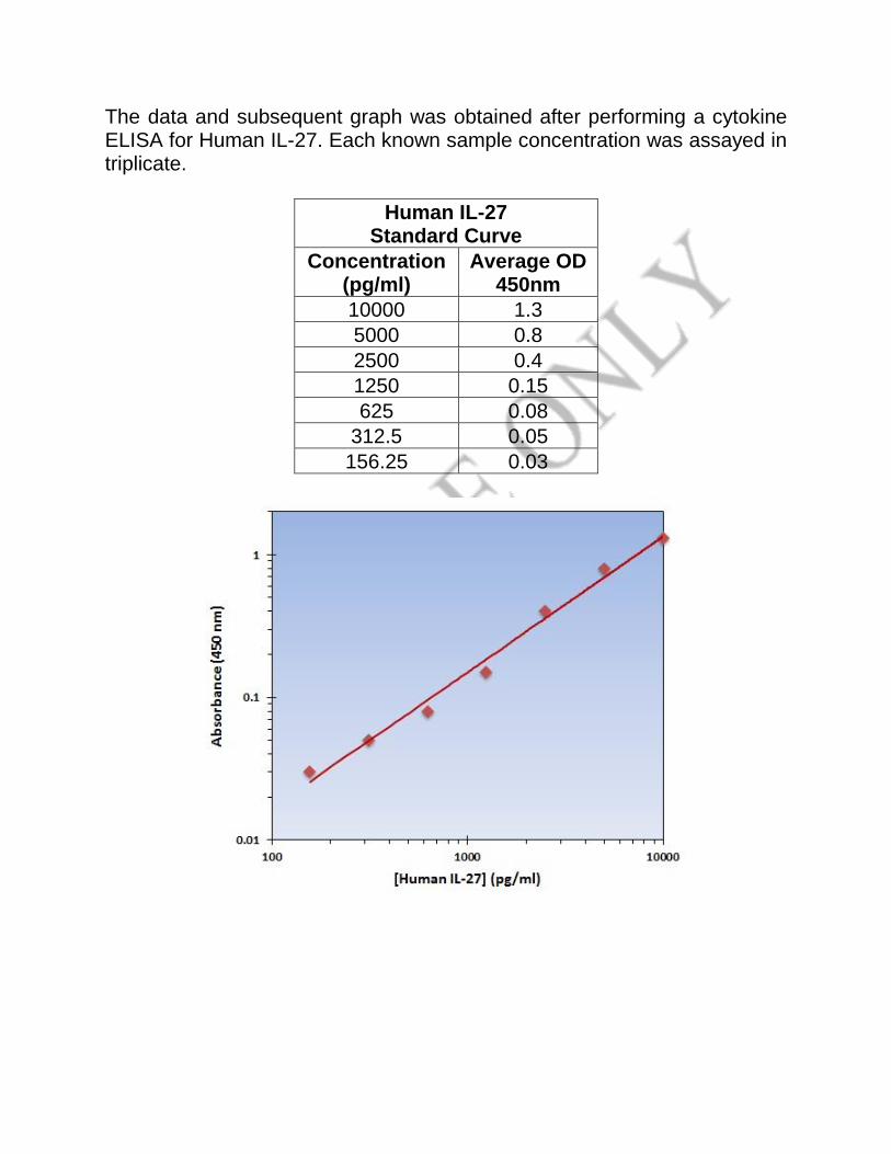
The data and subsequent graph was obtained after performing a cytokine ELISA for Human IL-27. Each known sample concentration was assayed in triplicate.
Human IL-27 Standard Curve
Concentration (pg/ml)
Average OD 450nm
10000 1.3
5000 0.8
2500 0.4
1250 0.15
625 0.08
312.5 0.05
156.25 0.03

SUMMARIZED PROTOCOL

SENSITIVITY
The Human IL-27 ELISA Kit allows for the detection and quantification of endogenous levels of natural and/or recombinant Human IL-27 proteins within the range of 157-10000 pg/ml.
CROSS REACTIVITY AND SPECIFICITY The Human IL-27 ELISA is capable of recognizing both recombinant and naturally produced Human IL-27 proteins. The antigens listed below were tested at 50 ng/ml and did not exhibit significant cross reactivity or interference. • Human: IL-12, IL-12/IL-23 p40 • Murine: IL-27


Important Note: During shipment, small volumes of product will occasionally become entrapped in the seal of the product vial. We recommend briefly centrifuging the vial to dislodge any liquid in the container's cap prior to opening. Warning: This reagent may contain sodium azide and sulfuric acid. The chemical, physical, and toxicological properties of these materials have not been thoroughly investigated. Standard Laboratory Practices should be followed. Avoid skin and eye contact, inhalation, and ingestion. Sodium azide forms hydrazoic acid under acidic conditions and may react with lead or copper plumbing to form highly explosive metal azides. On disposal, flush with large volumes of water to prevent accumulation. Returns, Refunds, Cancelations: Any problems with LifeSpan products must be reported to LifeSpan within 10 days of product receipt. The customer must obtain written authorization from LifeSpan before returning items. To request that goods be returned, please contact LifeSpan Technical Support. If an error by LifeSpan BioSciences results in shipment of an incorrect order, LifeSpan will, at its option, either ship a replacement order at no charge, or credit the customer's account for the original product shipped in error. Returns and cancelations may be subject to a 30% restocking fee. Conditions & Warranty: All LifeSpan products are intended for Research Use Only and are not for use in human therapeutic or diagnostic applications. The information supplied with each product is believed to be accurate, but no warranty or guarantee is offered for the products, because the ultimate conditions of use are beyond LifeSpan's control. The information supplied with each product is not to be construed as a recommendation to use this product in violation of any patent, and LifeSpan will not be held responsible for any infringement or other violation that may occur with the use of its products. Under no event will LifeSpan be responsible for any loss of profit or indirect consequential damage, including, but not limited to, personal injuries resulting from use of these products. LifeSpan's liability to any user of Products for damages that do not result from any fault of the user, will be limited to replacement of the Product(s) only, and in no event shall LifeSpan's liability exceed the actual price received by LifeSpan for the Product(s) at issue. LifeSpan shall not be liable for any indirect, special, incidental or consequential damages. LIFESPAN FURTHER DISCLAIMS ANY AND ALL EXPRESS AND IMPLIED OR STATUTORY WARRANTIES WITH RESPECT TO THE PRODUCTS, INCLUDING BUT NOT LIMITED TO ANY IMPLIED WARRANTIES OF MERCHANTABILITY, FITNESS FOR A PARTICULAR PURPOSE. LifeSpan disclaims any and all responsibility for any injury or damage which may be caused by the fault of the user.
For research use only. Not approved for use in humans or for clinical diagnosis.
2401 Fourth Avenue Suite 900 Seattle, WA 98121
Tel: 206-464-1554 Fax Toll Free (North America): 866-206-6909
Fax International Or Local: 206-577-4565 [email protected]


![Sample Manual - lsbio.com fileA S S A Y S P E C IF IC A T IO N S Target : [Target Common Name] Synonyms : [Synonyms] Specificity : This kit is for the detection of [Target Species]](https://img.dokumen.tips/doc/110x75/5ca8c72188c9939f3e8b5ca0/sample-manual-lsbiocom-s-s-a-y-s-p-e-c-if-ic-a-t-io-n-s-target-target-common.jpg)


![User Manual - lsbio.com · Instruction Manual 1 K I T S T O R A G E h }v ] Z l] Z}µo } ð£ ](]v v (} µ Á] Z]v 24 hours. Otherwise the Assay Plate, Standard, Detection Reagent](https://img.dokumen.tips/doc/110x75/6061b2804941776e717020f9/user-manual-lsbiocom-instruction-manual-1-k-i-t-s-t-o-r-a-g-e-h-v-z-l-zo.jpg)


