Embed Size (px)
Citation preview
952
Use of costic acid, a natural extract from Dittrichia viscosa,for the control of Varroa destructor, a parasite of theEuropean honey beeKalliopi Sofou1, Demosthenis Isaakidis1, Apostolos Spyros1, Anita Büttner2,3,Athanassios Giannis*2 and Haralambos E. Katerinopoulos*1
Full Research Paper Open Access
Address:1Department of Chemistry, University of Crete, Voutes, Heraklion,71003, Crete, Greece, 2Institut für Organische Chemie, UniversitätLeipzig, Johannisallee 29, 04103 Leipzig, Germany and 3recentaddress: Department of Chemistry and Food Chemistry, TU Dresden,01062, Dresden, Germany
Email:Athanassios Giannis* - [email protected];Haralambos E. Katerinopoulos* - [email protected]
* Corresponding author
Keywords:Apis mellifera; Dittrichia viscosa; natural products; Varroa destructor;varroosis
Beilstein J. Org. Chem. 2017, 13, 952–959.doi:10.3762/bjoc.13.96
Received: 13 March 2017Accepted: 24 April 2017Published: 18 May 2017
Editor-in-Chief: P. H. Seeberger
© 2017 Sofou et al.; licensee Beilstein-Institut.License and terms: see end of document.
AbstractCostic acid has been isolated from the plant Dittrichia viscosa and its efficacy against Varroa destructor, a parasite of Apis
mellifera, the European honey bee, has been studied. Costic acid exhibited potent in vivo acaricidal activity against the parasite.
Initial experiments showed that the compound is not toxic for human umbilical vein endothelial cells (HUVEC) at concentrations of
up to 230 micromolar (μM), indicating that costic acid could be used as a safe, low-cost and efficient agent for controlling varroosis
in honey bee colonies.
952
IntroductionThe honey bee ectoparasite Varroa destructor (Anderson &
Trueman) was discovered by Oudemans in 1904 [1]. The mite
was located on the island of Java and was infesting Apis cerana,
the Asiatic bee, which does not exhibit any symptoms when
infested by this parasite. Oudemans named the mite Varroa
jacobsoni. After extensive studies on mtDNA Co-I gene se-
quences and comparison of the morphological characters on nu-
merous populations of V. jacobsoni world-wide, Anderson
and Trueman came to the conclusion that the mite belongs
to two species: a) Varroa jacobsoni s.s., located in the
Malaysia–Indonesia region, infesting Apis cerana F. and
b) Varroa destructor, Anderson & Trueman, infesting both,
Apis cerana in Asia and Apis mellifera worldwide [2]. Given
that A. mellifera is not tolerant to the mite, varroa can attack and
Beilstein J. Org. Chem. 2017, 13, 952–959.
953
eliminate whole bee colonies within a period of a few years
[3,4]. The decimation of the bee population entails a negative
impact on the global economy, given that honey bees are
considered the economically most important pollinators of crop
monocultures worldwide [5]. A recent article on the protection
of pollinators stresses the need for “statistically robust monitor-
ing programs for native bees”, especially when taking into
account that some species have been designated as endangered
[6]. In times when the global pollinator decline is apparent
[7,8], control of varroosis has a significant impact on the main-
tenance of wild plant diversity, ecosystem stability, and crop
production.
In Europe, many beekeepers used acaricides such as coumaphos
and synthetic pyrethroids to keep mite populations under
control [9,10]. Literature data indicated that a number of pesti-
cide residues have been detected in honey samples [11,12].
Given that the parasite has developed significant resistance to
synthetic acaricides, alternatives such as oxalic acid [13-15]
were proposed and extensively studied for their effects against
varroosis. The use of oxalic acid as acaricide against V.
destructor was first proposed in 1989 [16]. Subsequent studies
indicated the potential of the method [17-22], which has been
applied with better results during the broodless period. Applica-
tion of the method does not increase the amount of oxalic acid
in honey [23-25] and has no toxic effects on bees. However,
later studies indicated that the utilization of oxalic acid by either
trickling or spraying has a detrimental effect on brood develop-
ment when open brood is present, and is therefore not as safe as
it has been assessed in the past [26,27]. Recent findings support
the hypothesis that V. destructor associates with bacteria
capable of degrading oxalic acid [28].
Crete is a Greek island located in the south-east of Europe.
Given the great diversity in the plant species of the flora of
Crete, the area is an excellent feeding place for bee colonies,
and beekeeping has been a profitable practice of the local popu-
lation since ancient years. Dittrichia viscosa (L.) W. Greuter
(syn. Inula viscosa (L.) Aiton), an invading ruderal species of
the Asteraceae family [29], is a herbaceous perennial plant
which is widespread in the Mediterranean region, known in folk
medicine to possess anti-inflammatory [30], antipyretic, and
gastric antiulcerous effects [31,32]. Due to lack of competing
flowers in Autumn, the plant attracts many pollinating insects
including bees, wasps and some butterflies to feed on, an indi-
cation that the plant volatiles do not act as repellents on bees.
The fact that D. viscosa, called by the natives “akoniza”, has
been used by the local beekeepers as a means of controlling
varroosis, prompted us to analyze the components of the plant
in order to identify the active agent and study its efficacy
against the parasite [33,34]. The use of natural products against
varroosis is an eco-friendly approach to this severe problem.
Field tests on the use of methanolic extracts of Lepidium lati-
folium and Zataria multiflora indicated that these preparations
exhibited acaricidal activity against the mites [35]. However,
there are no preceding data on the action of costic acid against
varroa. In this publication we report the isolation and structure
identification of costic acid as a component of D. viscosa, as
well as in vivo and field studies providing strong evidence of
the acaricidal action of the acid against V. destructor.
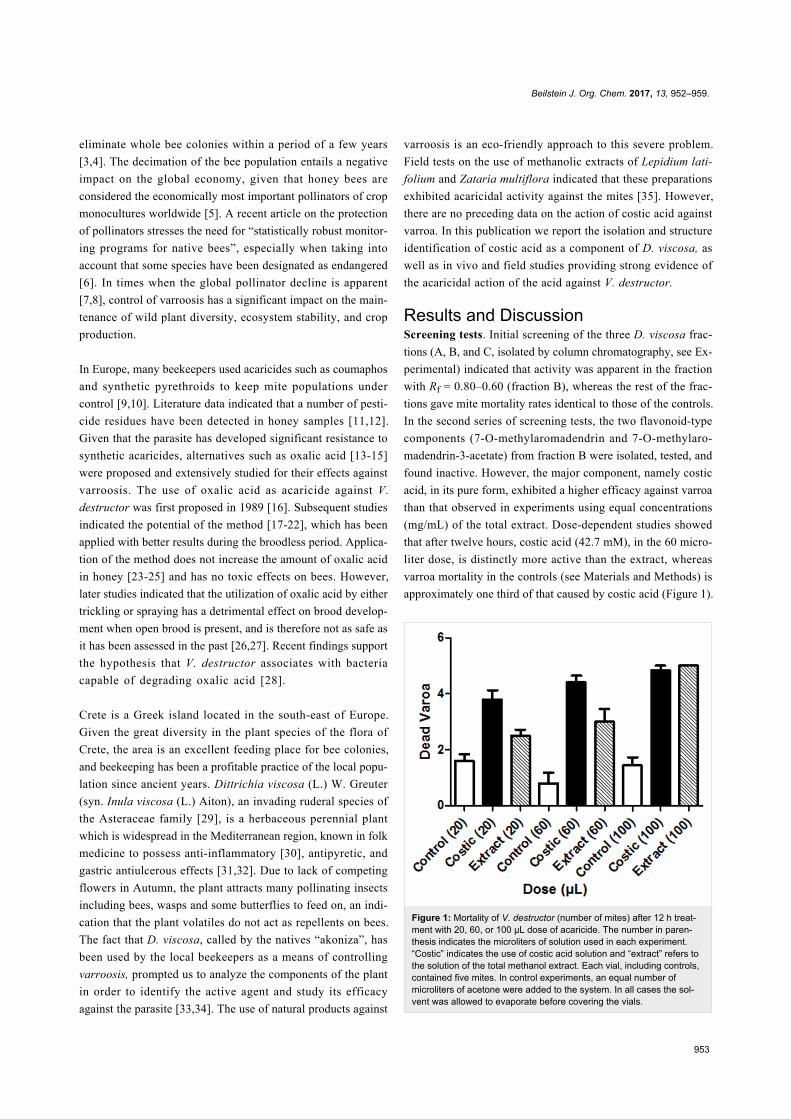
Results and DiscussionScreening tests. Initial screening of the three D. viscosa frac-
tions (A, B, and C, isolated by column chromatography, see Ex-
perimental) indicated that activity was apparent in the fraction
with Rf = 0.80–0.60 (fraction B), whereas the rest of the frac-
tions gave mite mortality rates identical to those of the controls.
In the second series of screening tests, the two flavonoid-type
components (7-O-methylaromadendrin and 7-O-methylaro-
madendrin-3-acetate) from fraction B were isolated, tested, and
found inactive. However, the major component, namely costic
acid, in its pure form, exhibited a higher efficacy against varroa
than that observed in experiments using equal concentrations
(mg/mL) of the total extract. Dose-dependent studies showed
that after twelve hours, costic acid (42.7 mM), in the 60 micro-
liter dose, is distinctly more active than the extract, whereas
varroa mortality in the controls (see Materials and Methods) is
approximately one third of that caused by costic acid (Figure 1).
Figure 1: Mortality of V. destructor (number of mites) after 12 h treat-ment with 20, 60, or 100 μL dose of acaricide. The number in paren-thesis indicates the microliters of solution used in each experiment.“Costic” indicates the use of costic acid solution and “extract” refers tothe solution of the total methanol extract. Each vial, including controls,contained five mites. In control experiments, an equal number ofmicroliters of acetone were added to the system. In all cases the sol-vent was allowed to evaporate before covering the vials.
Beilstein J. Org. Chem. 2017, 13, 952–959.
954
Figure 3: Field test results on application of extract B, bayvarol and oxalic acid to bee populations.
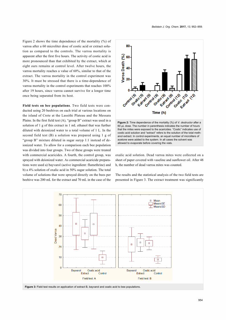
Figure 2 shows the time dependence of the mortality (%) of
varroa after a 60 microliter dose of costic acid or extract solu-
tion as compared to the controls. The varroa mortality is
apparent after the first five hours. The activity of costic acid is
more pronounced than that exhibited by the extract, which at
eight ours remains at control level. After twelve hours, the
varroa mortality reaches a value of 60%, similar to that of the
extract. The varroa mortality in the control experiment was
30%. It must be stressed that there is a time-dependence of
varroa mortality in the control experiments that reaches 100%
after 19 hours, since varroa cannot survive for a longer time
once being separated from its host.
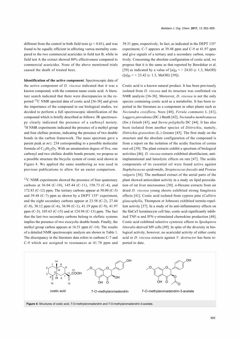
Field tests on bee populations. Two field tests were con-
ducted using 20 beehives on each trial at various locations on
the island of Crete at the Lassithi Plateau and the Messara
Plains. In the first field test (A), “group B” extract was used in a
solution of 1 g of this extract in 1 mL ethanol that was further
diluted with deionized water to a total volume of 1 L. In the
second field test (B) a solution was prepared using 1 g of
“group B” mixture diluted in sugar suryp 1:1 instead of de-
ionized water. To allow for a comparison each bee population
was divided into four groups. Two of these groups were treated
with commercial acaricides. A fourth, the control group, was
sprayed with deionized water. As commercial acaricide prepara-
tions were used a) bayvarol (active ingredient: flumethrine) and
b) a 4% solution of oxalic acid in 50% sugar solution. The total
volume of solutions that were sprayed directly on the bees per
beehive was 200 mL for the extract and 70 mL in the case of the
Figure 2: Time dependence of the mortality (%) of V. destructor after a60 μL dose. The number in parenthesis indicates the number of hoursthat the mites were exposed to the acaricides. “Costic” indicates use ofcostic acid solution and “extract” refers to the solution of the total meth-anol extract. In control experiments, an equal number of microliters ofacetone were added to the system. In all cases the solvent wasallowed to evaporate before covering the vials.
oxalic acid solution. Dead varroa mites were collected on a
sheet of paper covered with vaseline and sunflower oil. After 48
h, the number of dead varroa mites was counted.
The results and the statistical analysis of the two field tests are
presented in Figure 3. The extract treatment was significantly
Beilstein J. Org. Chem. 2017, 13, 952–959.
955
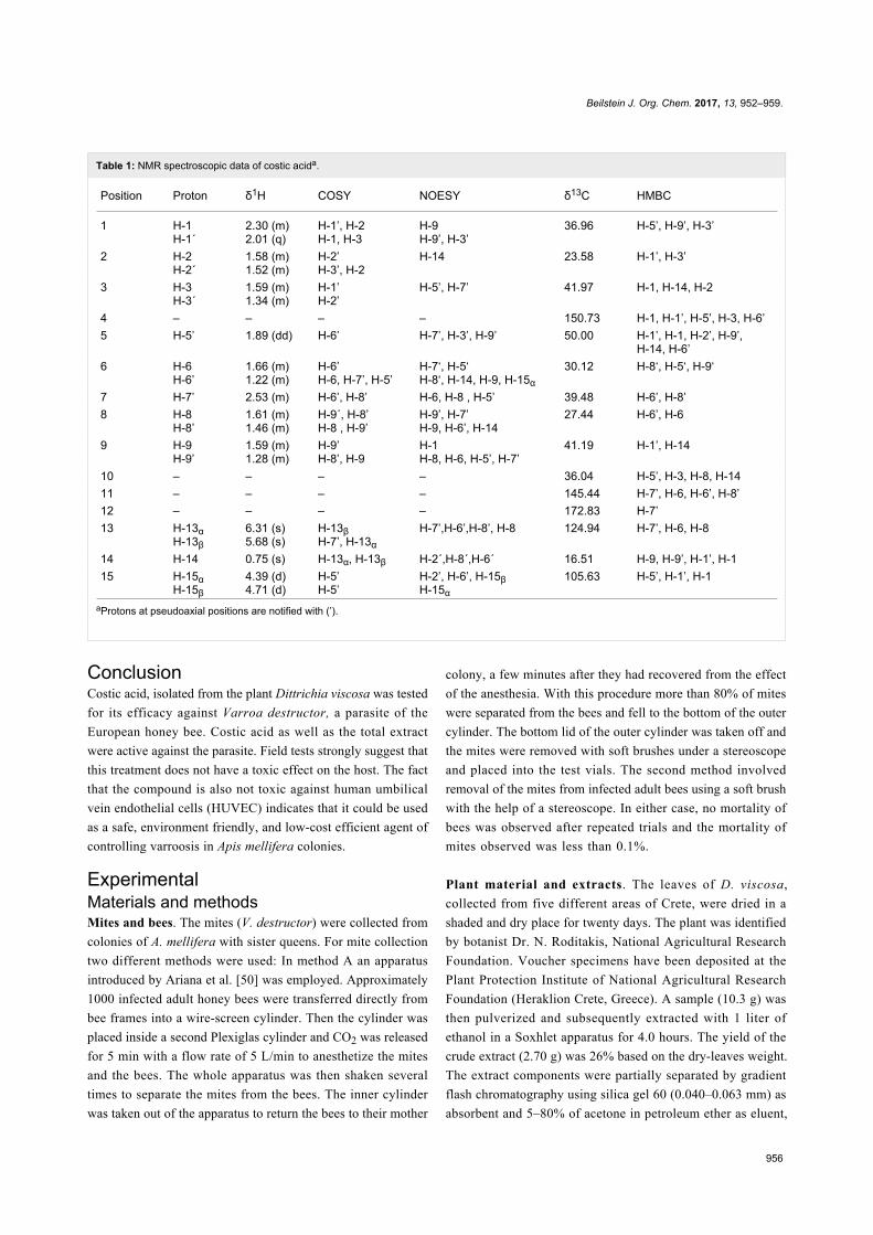
Figure 4: Structures of costic acid, 7-O-methylaromadendrin and 7-O-methylaromadendrin-3-acetate.
different from the control in both field tests (p < 0.01), and was
found to be equally efficient in affecting varroa mortality com-
pared to the two commercial acaricides in field test B, while in
field test A the extract showed 80% effectiveness compared to
commercial acaricides. None of the above mentioned trials
caused the death of treated bees.
Identification of the active component. Spectroscopic data of
the active component of D. viscosa indicated that it was a
known compound, with the common name costic acid. A litera-
ture search indicated that there were discrepancies in the re-
ported 13C NMR spectral data of costic acid [36-38] and given
the importance of the compound in our biological studies, we
decided to perform a full spectroscopic identification of the
compound which is briefly described as follows: IR spectrosco-
py clearly indicated the presence of a carboxyl moiety.1H NMR experiments indicated the presence of a methyl group
and four olefinic protons, indicating the presence of two double
bonds in the carbon framework. The mass spectrum gave a
parent peak at m/z: 234 corresponding to a possible molecular
formula of C15H22O2. With an unsaturation degree of five, one
carbonyl and two olefinic double bonds present, we propose as
a possible structure the bicyclic system of costic acid shown in
Figure 4. We applied the same numbering as was used in
previous publications to allow for an easier comparison.
13C NMR experiments showed the presence of four quaternary
carbons at 36.04 (C-10), 145.44 (C-11), 150.73 (C-4), and
172.83 (C-12) ppm. The tertiary carbons appear at 50.00 (C-5)
and 39.48 (C-7) ppm as shown by a DEPT 135° experiment,
and the eight secondary carbons appear at 23.58 (C-2), 27.44
(C-8), 30.12 ppm (C-6), 36.96 (C-1), 41.19 ppm (C-9), 41.97
ppm (C-3), 105.63 (C-15) and at 124.94 (C-13) ppm. The fact
that the last two secondary carbons belong to olefinic systems
implies the presence of two exocyclic double bonds. Finally, the
methyl group carbon appears at 16.51 ppm (C-14). The results
of a detailed NMR spectroscopic analysis are shown in Table 1.
The discrepancy in the literature data refers to carbons C-7 and
C-9 which are assigned to resonances at 41.78 ppm and
39.31 ppm, respectively. In fact, as indicated in the DEPT 135°
experiment, C-7 appears at 39.48 ppm and C-9 at 41.97 ppm
and give signals of a tertiary and a secondary carbon, respec-
tively. Concerning the absolute configuration of costic acid, we
propose that it is the same as that reported by Bawdekar et al.
[39] as indicated by a value of [α]D = + 24.03 (c 1.3, MeOH)
([α]D = + 23.42 (c 1.3, MeOH) [39]).
Costic acid is a known natural product. It has been previously
isolated from D. viscosa and its structure was confirmed via
NMR analysis [36-38]. Moreover, D. viscosa is not the only
species containing costic acid as a metabolite. It has been re-
ported in the literature as a component in other plants such as
Nectandra cissiflora, Nees [40], Ferula communis L [41],
Laggera pterodonta (DC.) Benth [42], Nectandra membranacea
(Sw.) Griseb [43], and Stevia polyphylla DC [44]. It has also
been isolated from another species of Dittrichia, namely,
Dittrichia graveolens (L.) Greuter [45]. The first study on the
structure and the absolute configuration of the compound is
from a report on the isolation of the acidic fraction of costus
root oil [39]. The plant extracts exhibit a spectrum of biological
activities [46]. D. viscosa extracts exhibited abortifacient, anti-
implantational and luteolytic effects on rats [47]. The acidic
components of its essential oil were found active against
Staphylococcus epidermidis, Streptococcus foecalis and Proteus
vulgaris [36]. The methanol extract of the aerial parts of the
plant showed antioxidant activity in a study on lipid peroxida-
tion of rat liver microsomes [30]. n-Hexane extracts from air
dried D. viscosa young shoots exhibited strong fungitoxic
effects [41]. Costic acid isolated from cypress pine (Callitris
glaucophylla, Thompson et Johnson) exhibited termite-repel-
lent activity [37]. In a study of its anti-inflammatory effects on
the HaCaT keratinocyte cell line, costic acid significantly inhib-
ited TNF-α and IFN-γ-stimulated chemokine production [48].
Costic acid exhibited selective cytotoxic effects to Spodoptera
littoralis-derived Sf9 cells [49]. In spite of the diversity in bio-
logical activity, however, no acaricidal activity of either costic
acid or D. viscosa extracts against V. destructor has been re-
ported to date.
Beilstein J. Org. Chem. 2017, 13, 952–959.
956
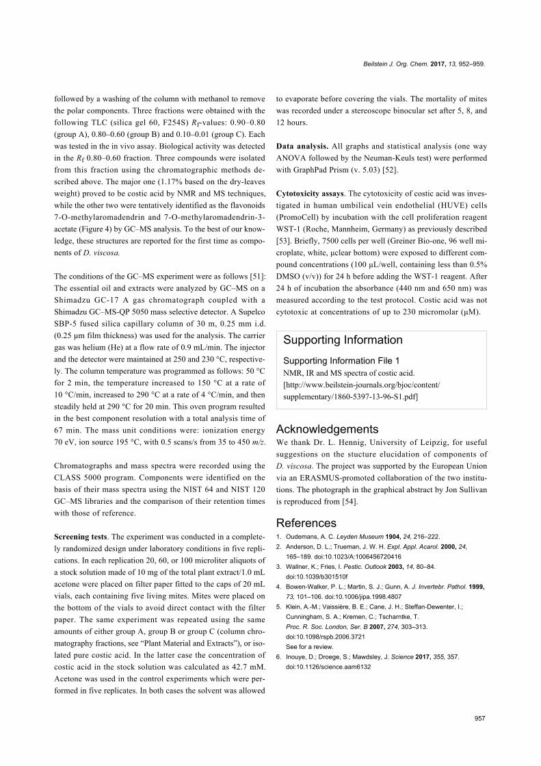
Table 1: NMR spectroscopic data of costic acida.
Position Proton δ1H COSY NOESY δ13C HMBC
1 H-1H-1΄
2.30 (m)2.01 (q)
H-1’, Η-2H-1, Η-3
H-9H-9’, Η-3’
36.96 Η-5’, Η-9’, Η-3’
2 H-2H-2΄
1.58 (m)1.52 (m)
Η-2’Η-3’, Η-2
Η-14 23.58 Η-1’, Η-3’
3 H-3H-3΄
1.59 (m)1.34 (m)
Η-1’Η-2’
Η-5’, Η-7’ 41.97 Η-1, H-14, H-2
4 – – – – 150.73 H-1, H-1’, Η-5’, Η-3, Η-6’5 H-5’ 1.89 (dd) Η-6’ Η-7’, Η-3’, Η-9’ 50.00 Η-1’, Η-1, Η-2’, Η-9’,
Η-14, Η-6’6 H-6
H-6’1.66 (m)1.22 (m)
Η-6’Η-6, Η-7’, Η-5’
Η-7‘, Η-5‘Η-8‘, Η-14, H-9, H-15α
30.12 Η-8‘, Η-5‘, Η-9‘
7 H-7’ 2.53 (m) Η-6’, Η-8’ Η-6, Η-8 , Η-5’ 39.48 Η-6’, Η-8’8 H-8
H-8’1.61 (m)1.46 (m)
Η-9΄, Η-8’Η-8 , Η-9’
Η-9’, Η-7’Η-9, Η-6’, Η-14
27.44 Η-6’, Η-6
9 H-9H-9’
1.59 (m)1.28 (m)
Η-9’Η-8’, Η-9
Η-1Η-8, Η-6, Η-5’, Η-7’
41.19 Η-1’, Η-14
10 – – – – 36.04 Η-5’, Η-3, Η-8, Η-1411 – – – – 145.44 Η-7’, Η-6, Η-6’, Η-8’12 – – – – 172.83 Η-7’13 H-13α
H-13β6.31 (s)5.68 (s)
Η-13βΗ-7’, Η-13α
Η-7’,Η-6’,Η-8’, H-8 124.94 Η-7’, Η-6, Η-8
14 H-14 0.75 (s) Η-13α, Η-13β Η-2΄,Η-8΄,Η-6΄ 16.51 Η-9, Η-9’, Η-1’, Η-115 H-15α
H-15β4.39 (d)4.71 (d)
Η-5’Η-5’
Η-2’, Η-6’, Η-15βH-15α
105.63 Η-5’, Η-1’, Η-1
aProtons at pseudoaxial positions are notified with (’).
ConclusionCostic acid, isolated from the plant Dittrichia viscosa was tested
for its efficacy against Varroa destructor, a parasite of the
European honey bee. Costic acid as well as the total extract
were active against the parasite. Field tests strongly suggest that
this treatment does not have a toxic effect on the host. The fact
that the compound is also not toxic against human umbilical
vein endothelial cells (HUVEC) indicates that it could be used
as a safe, environment friendly, and low-cost efficient agent of
controlling varroosis in Apis mellifera colonies.
ExperimentalMaterials and methodsMites and bees. The mites (V. destructor) were collected from
colonies of A. mellifera with sister queens. For mite collection
two different methods were used: In method A an apparatus
introduced by Ariana et al. [50] was employed. Approximately
1000 infected adult honey bees were transferred directly from
bee frames into a wire-screen cylinder. Then the cylinder was
placed inside a second Plexiglas cylinder and CO2 was released
for 5 min with a flow rate of 5 L/min to anesthetize the mites
and the bees. The whole apparatus was then shaken several
times to separate the mites from the bees. The inner cylinder
was taken out of the apparatus to return the bees to their mother
colony, a few minutes after they had recovered from the effect
of the anesthesia. With this procedure more than 80% of mites
were separated from the bees and fell to the bottom of the outer
cylinder. The bottom lid of the outer cylinder was taken off and
the mites were removed with soft brushes under a stereoscope
and placed into the test vials. The second method involved
removal of the mites from infected adult bees using a soft brush
with the help of a stereoscope. In either case, no mortality of
bees was observed after repeated trials and the mortality of
mites observed was less than 0.1%.
Plant material and extracts. The leaves of D. viscosa,
collected from five different areas of Crete, were dried in a
shaded and dry place for twenty days. The plant was identified
by botanist Dr. N. Roditakis, National Agricultural Research
Foundation. Voucher specimens have been deposited at the
Plant Protection Institute of National Agricultural Research
Foundation (Heraklion Crete, Greece). A sample (10.3 g) was
then pulverized and subsequently extracted with 1 liter of
ethanol in a Soxhlet apparatus for 4.0 hours. The yield of the
crude extract (2.70 g) was 26% based on the dry-leaves weight.
The extract components were partially separated by gradient
flash chromatography using silica gel 60 (0.040–0.063 mm) as
absorbent and 5–80% of acetone in petroleum ether as eluent,
Beilstein J. Org. Chem. 2017, 13, 952–959.
957
followed by a washing of the column with methanol to remove
the polar components. Three fractions were obtained with the
following TLC (silica gel 60, F254S) Rf-values: 0.90–0.80
(group A), 0.80–0.60 (group B) and 0.10–0.01 (group C). Each
was tested in the in vivo assay. Biological activity was detected
in the Rf 0.80–0.60 fraction. Three compounds were isolated
from this fraction using the chromatographic methods de-
scribed above. The major one (1.17% based on the dry-leaves
weight) proved to be costic acid by NMR and MS techniques,
while the other two were tentatively identified as the flavonoids
7-O-methylaromadendrin and 7-O-methylaromadendrin-3-
acetate (Figure 4) by GC–MS analysis. To the best of our know-
ledge, these structures are reported for the first time as compo-
nents of D. viscosa.
The conditions of the GC–MS experiment were as follows [51]:
The essential oil and extracts were analyzed by GC–MS on a
Shimadzu GC-17 A gas chromatograph coupled with a
Shimadzu GC–MS-QP 5050 mass selective detector. A Supelco
SBP-5 fused silica capillary column of 30 m, 0.25 mm i.d.
(0.25 μm film thickness) was used for the analysis. The carrier
gas was helium (He) at a flow rate of 0.9 mL/min. The injector
and the detector were maintained at 250 and 230 °C, respective-
ly. The column temperature was programmed as follows: 50 °C
for 2 min, the temperature increased to 150 °C at a rate of
10 °C/min, increased to 290 °C at a rate of 4 °C/min, and then
steadily held at 290 °C for 20 min. This oven program resulted
in the best component resolution with a total analysis time of
67 min. The mass unit conditions were: ionization energy
70 eV, ion source 195 °C, with 0.5 scans/s from 35 to 450 m/z.
Chromatographs and mass spectra were recorded using the
CLASS 5000 program. Components were identified on the
basis of their mass spectra using the NIST 64 and NIST 120
GC–MS libraries and the comparison of their retention times
with those of reference.
Screening tests. The experiment was conducted in a complete-
ly randomized design under laboratory conditions in five repli-
cations. In each replication 20, 60, or 100 microliter aliquots of
a stock solution made of 10 mg of the total plant extract/1.0 mL
acetone were placed on filter paper fitted to the caps of 20 mL
vials, each containing five living mites. Mites were placed on
the bottom of the vials to avoid direct contact with the filter
paper. The same experiment was repeated using the same
amounts of either group A, group B or group C (column chro-
matography fractions, see “Plant Material and Extracts”), or iso-
lated pure costic acid. In the latter case the concentration of
costic acid in the stock solution was calculated as 42.7 mM.
Acetone was used in the control experiments which were per-
formed in five replicates. In both cases the solvent was allowed
to evaporate before covering the vials. The mortality of mites
was recorded under a stereoscope binocular set after 5, 8, and
12 hours.
Data analysis. All graphs and statistical analysis (one way
ANOVA followed by the Neuman-Keuls test) were performed
with GraphPad Prism (v. 5.03) [52].
Cytotoxicity assays. The cytotoxicity of costic acid was inves-
tigated in human umbilical vein endothelial (HUVE) cells
(PromoCell) by incubation with the cell proliferation reagent
WST-1 (Roche, Mannheim, Germany) as previously described
[53]. Briefly, 7500 cells per well (Greiner Bio-one, 96 well mi-
croplate, white, μclear bottom) were exposed to different com-
pound concentrations (100 μL/well, containing less than 0.5%
DMSO (v/v)) for 24 h before adding the WST-1 reagent. After
24 h of incubation the absorbance (440 nm and 650 nm) was
measured according to the test protocol. Costic acid was not
cytotoxic at concentrations of up to 230 micromolar (μM).
Supporting InformationSupporting Information File 1NMR, IR and MS spectra of costic acid.
[http://www.beilstein-journals.org/bjoc/content/
supplementary/1860-5397-13-96-S1.pdf]
AcknowledgementsWe thank Dr. L. Hennig, University of Leipzig, for useful
suggestions on the stucture elucidation of components of
D. viscosa. The project was supported by the European Union
via an ERASMUS-promoted collaboration of the two institu-
tions. The photograph in the graphical abstract by Jon Sullivan
is reproduced from [54].
References1. Oudemans, A. C. Leyden Museum 1904, 24, 216–222.2. Anderson, D. L.; Trueman, J. W. H. Expl. Appl. Acarol. 2000, 24,
165–189. doi:10.1023/A:10064567204163. Wallner, K.; Fries, I. Pestic. Outlook 2003, 14, 80–84.
doi:10.1039/b301510f4. Bowen-Walker, P. L.; Martin, S. J.; Gunn, A. J. Invertebr. Pathol. 1999,
73, 101–106. doi:10.1006/jipa.1998.48075. Klein, A.-M.; Vaissière, B. E.; Cane, J. H.; Steffan-Dewenter, I.;
Cunningham, S. A.; Kremen, C.; Tscharntke, T.Proc. R. Soc. London, Ser. B 2007, 274, 303–313.doi:10.1098/rspb.2006.3721See for a review.
6. Inouye, D.; Droege, S.; Mawdsley, J. Science 2017, 355, 357.doi:10.1126/science.aam6132
Beilstein J. Org. Chem. 2017, 13, 952–959.
958
7. Potts, S. G.; Biesmeijer, J. C.; Kremen, C.; Neumann, P.;Schweiger, O.; Kunin, W. E. Trends Ecol. Evol. 2010, 25, 345–353.doi:10.1016/j.tree.2010.01.007
8. Gallai, N.; Salles, J.-M.; Settele, J.; Vaissière, B. E. Ecol. Econ. 2009,68, 810–821. doi:10.1016/j.ecolecon.2008.06.014
9. Fries, I.; Wallner, K.; Rosenkranz, P. J. Apic. Res. 1998, 37, 85–90.doi:10.1080/00218839.1998.11100959
10. Rickli, M.; Imdorf, A.; Kilchenmann, V. Apidologie 1991, 22, 417–421.doi:10.1051/apido:19910406
11. Eissa, F.; El-Sawi, S.; Zidan, N. E.-H. Pol. J. Environ. Stud. 2014, 23,1573–1580.
12. Notardonato, I.; Avino, P.; Cinelli, G.; Russo, M. V. RSC Adv. 2014, 4,42424–42431. doi:10.1039/C4RA06822J
13. Rademacher, E.; Harz, M. Apidologie 2006, 37, 98–120.doi:10.1051/apido:2005063See for a review on the use of oxalic acid for the control of varroosis.
14. Rashid, M.; Wagchoure, E. S.; Mohsin, A. U.; Raja, S.; Sarwar, G.J. Anim. Plant Sci. 2012, 22, 72–76.
15. Toomemaa, K.; Martin, A. J.; Williams, I. H. Apidologie 2010, 41,643–653. doi:10.1051/apido/2010029
16. Popov, E. T.; Melnik, V. N.; Matchinev, A. N. Application of oxalic acidin varroatosis. In Proc. XXXII Int. Congr. Apimondia, Rio de Janeiro;Apimondia Publ. House: Bucharest, 1989; p 149.
17. Gregorc, A.; Poklukar, J. Vet. Parasitol. 2003, 111, 351–360.doi:10.1016/S0304-4017(02)00408-9
18. Gregorc, A.; Planinc, I. Vet. J. 2002, 163, 306–310.doi:10.1053/tvjl.2001.0675
19. Gregorc, A.; Planinc, I. Apidologie 2001, 32, 333–340.doi:10.1051/apido:2001133
20. Milani, N. Apidologie 2001, 32, 127–138. doi:10.1051/apido:200111821. Aliano, N. P.; Ellis, M. D.; Siegfried, B. D. J. Econ. Ent. 2006, 99,
1579–1582. doi:10.1093/jee/99.5.157922. Bacandritsos, N.; Papanastasiou, I.; Saitanis, C.; Nanetti, A.;
Roinioti, E. Vet. Parasitol. 2007, 148, 174–178.doi:10.1016/j.vetpar.2007.06.001
23. Moosbeckhofer, R.; Pechhacker, H.; Unterweger, H.; Bandion, F.;Heinrich-Lenz, A. Eur. Food Res. Technol. 2003, 217, 49–52.doi:10.1007/s00217-003-0698-z
24. Bogdanov, S.; Charriere, J.-D.; Imdorf, A.; Kilchenmann, V.; Fluri, P.Apidologie 2002, 33, 399–409. doi:10.1051/apido:2002029
25. Mutinelli, F.; Baggio, A.; Capolongo, F.; Piro, R.; Prandin, L.; Biasion, L.Apidologie 1997, 28, 461–462. doi:10.1051/apido:19970612
26. Hatjina, F.; Haristos, L. J. Apic. Res. 2005, 44, 172–174.doi:10.1080/00218839.2005.11101174
27. Higes, M.; Meana, A.; Suárez, M.; Llorente, J. Apidologie 1999, 30,289–292. doi:10.1051/apido:19990404
28. Maddaloni, M.; Pascual, D. W. Lett. Appl. Microbiol. 2015, 61,411–417. doi:10.1111/lam.12486
29. Stavrianakou, S.; Liakopoulos, G.; Karabourniotis, G. Envir. Expl. Bot.2006, 56, 293–300. doi:10.1016/j.envexpbot.2005.03.007
30. Schinella, G. R.; Tournier, H. A.; Prieto, J. M.;Mordujovich De Buschiazzo, P.; Rios, J. L. Life Sci. 2002, 70,1023–1033. doi:10.1016/S0024-3205(01)01482-5
31. Alarcón de La Lustra, C.; López, A.; Motilva, V. Planta Med. 1993, 59,497–501. doi:10.1055/s-2006-959747
32. Maoz, M.; Kashman, Y.; Neeman, I. Planta Med. 1999, 65, 281–282.doi:10.1055/s-2006-960780
33. Sofou, K. Isolation, structure elucidation and use of components fromthe plant Dittrichia viscosa against the bee parasite Varroa destructor.M.Sc. Thesis, University of Crete, Greece, 2007.
34. Katerinopoulos, H. E.; Isaakidis, D.; Sofou, K.; Spyros, A. Use OfCostic Acid or Extracts of Dittrichia viscosa against Varroa Destructor.EU Pat. Appl. EP2346328 A1, July 27, 2011.
35. Razavi, S. M.; Asadpour, M.; Jafari, A.; Malekpour, S. H.Parasitol. Res. 2015, 114, 4233–4238. doi:10.1007/s00436-015-4661-2
36. Blanc, M.-C.; Bradesi, P.; Casanova, J. Phytochem. Anal. 2005, 16,150–154. doi:10.1002/pca.834
37. Watanabe, Y.; Mihara, R.; Mitsunaga, T.; Yoshimura, T. J. Wood Sci.2005, 51, 514–519. doi:10.1007/s10086-004-0683-6
38. Zheng, Q.; Xu, Z.; Sun, X.; Yao, W.; Sun, H.; Cheng, C. H. K.; Zhao, Y.Phytochemistry 2003, 63, 835–839.doi:10.1016/S0031-9422(03)00370-4
39. Bawdekar, A. S.; Kelkar, G. R. Tetrahedron 1965, 21, 1521–1528.doi:10.1016/S0040-4020(01)98315-2
40. Garcez, F. R.; Garcez, W. S.; Hamerski, L.; de M. Miranda, A. C.Quim. Nova 2010, 33, 1739–1742.doi:10.1590/S0100-40422010000800022
41. Mamoci, E.; Cavoski, I.; Simeone, V.; Mondelli, D.; Al-Bitar, L.;Caboni, P. Molecules 2011, 16, 2609–2625.doi:10.3390/molecules16032609
42. Xiao, Y.; Zheng, Q.; Zhang, Q.; Sun, H.; Guéritte, F.; Zhao, Y.Fitoterapia 2003, 74, 459–463. doi:10.1016/S0367-326X(03)00106-0
43. Wu, X.; Vogler, B. Nat. Prod. Commun. 2006, 1, 465–468.44. Zdero, C.; Bohlmann, F.; King, R. M.; Robinson, H. Phytochemistry
1988, 27, 2835–2842. doi:10.1016/0031-9422(88)80673-345. Abou-Douh, A. M. Chem. Pharm. Bull. 2008, 56, 1535–1545.
doi:10.1248/cpb.56.153546. Parolin, P.; Ion Scotta, M.; Bresch, C. Phyton 2014, 83, 251–262.
See for a recent review.47. Al-Dissi, N. M.; Salhab, A. S.; Al-Hajj, H. A. J. Ethnopharmacol. 2001,
77, 117–121. doi:10.1016/S0378-8741(01)00261-648. Lim, H.-S.; Jin, S.-E.; Kim, O.-S.; Shin, H.-K.; Jeong, S.-J.
Phytother. Res. 2015, 29, 1088–1096. doi:10.1002/ptr.535449. González-Coloma, A.; Guadaño, A.; Tonn, C. E.; Sosa, M. E.
Z. Naturforsch., C: J. Biosci. 2005, 60, 855–861.doi:10.1515/znc-2005-11-1207
50. Ariana, A.; Ebadi, R.; Tahmasebi, G. Expl. Appl. Acarol. 2002, 27,No. 319. doi:10.1023/A:1023342118549
51. Katerinopoulos, H. E.; Pagona, G.; Afratis, A.; Stratigakis, N.;Roditakis, N. J. Chem. Ecol. 2005, 31, 111–122.doi:10.1007/s10886-005-0978-0
52. One-way ANOVA followed by Neuman-Keuls multiple comparisons testwas performed using GraphPad Prism version 6.00 for Windows,GraphPad Software, La Jolla, California, USA,http://www.graphpad.com.
53. Seifert, K.; Büttner, A.; Rigol, S.; Eilert, N.; Wandel, E.; Giannis, A.Bioorg. Med. Chem. 2012, 20, 6465–6481.doi:10.1016/j.bmc.2012.08.026
54. Honey bee. https://en.wikipedia.org/wiki/Honey_bee (accessed April 7,2017).
Beilstein J. Org. Chem. 2017, 13, 952–959.
959
License and TermsThis is an Open Access article under the terms of the
Creative Commons Attribution License
(http://creativecommons.org/licenses/by/4.0), which
permits unrestricted use, distribution, and reproduction in
any medium, provided the original work is properly cited.
The license is subject to the Beilstein Journal of Organic
Chemistry terms and conditions:
(http://www.beilstein-journals.org/bjoc)
The definitive version of this article is the electronic one
which can be found at:
doi:10.3762/bjoc.13.96