Embed Size (px)
Citation preview
Use of a Vascular Doppler in Diabetics
with PAD
Dr. Anita Kharbteng,Clinical Support Manager,
Asia Pacific, Cardinal Health
GLOBALLEADERSHIP

GLOBALLEADERSHIP Peripheral Arterial Disease (PAD)
• PAD is a condition characterised by atherosclerotic occlusive disease of the lower extremities.
• The most common symptom of PAD is intermittent claudication (IC). More extreme presentations of PAD include rest pain, tissue loss, or gangrene; collectively termed ‘Critical Limb Ischaemia’ (CLI).
• The symptoms are often subtle and hence physicians need to be proactive in looking for PAD, realising that it may often be asymptomatic.

GLOBALLEADERSHIP RISK FACTORS
Murabito JM et al. Circulation 1997;96:44–49; Laurila A et al. Arterioscler Throm Vasc Biol 1997;17:2910–2913;Malinow MR et al. Circulation 1989;79:1180–1188; Brigden ML. Postgrad Med 1997;101:249–262.
Gender (male) Increasing Age Smoking High blood pressure Diabetes High Cholesterol Family history of
heart attack or stroke Fibrinogen Homocysteinaemia
PAD
AtherosclerosisArteriosclerosisAtherothrombosis
Ischaemicstroke
Myocardialinfarction
Diabetes and smoking are the strongest risk factor for PAD.

GLOBALLEADERSHIP Statistics
Use of ABI has shown the prevalance of Diabetes in PAD individuals:
40yrs old is 20% 50yrs old is 29%
”Peripheral Arterial Disease Detection, Awareness and Treatment in Primary Care”Alan T. Hirsch, MD et al. JAMA, 2001;286-1317-1324

GLOBALLEADERSHIP PAD a Walking Time Bomb!
Patients with PAD have a higher 5-year mortality rate than breast cancer
~40% of amputees die within 2 years of amputation
Vascular Disease Foundation and the American Cancer Society

GLOBALLEADERSHIP PAD in Diabetes
• Diabetes is the strongest risk factor for PAD• PAD in diabetes is different in its biology, clinical
presentation, and management.• In contrast to the focal and proximal atherosclerotic
lesions of PAD in other high risk patients, in diabetics the lesions are more diffuse and distal - tibial vessels below the knee.
• PAD in diabetes is usually accompanied by peripheral neuropathy with impaired sensory feedback. Thus they may not present with the classic symptom of IC. They have subtle symptoms like leg fatigue and slow walking velocity and thus they are simply attributed to old age.
• Diabetic pts with PAD experience worse lower-extremity function than non-diabetics
• These Pts are more prone to sudden ischemia of arterial thrombosis leading to CLI and risk of amputation.
• This lends urgency to the task of diagnosing PAD in those apparently asymptomatic individuals with diabetes.
GLOBALLEADERSHIP Impact of PAD
• PAD sufferers have a five-fold risk of death from heart attack or stroke. Diabetic patients are at an even higher risk.
• Approx 27% of patients with PAD show progression of symptoms over a 5-year period with limb loss in ~4%.
• The majority of patients remain stable in their lower limb symptoms but there is a striking excess in cardiovascular events in the same 5-year period with 20% sustaining nonfatal events (MI and stroke)and a 30% mortality rate.
• Patients with CLI have worse outcomes:– 30% have amputations– 20% die within 6 months

GLOBALLEADERSHIP Impact of PAD
• PAD in diabetes also adversely affects quality of life, contributing to long-term disability and functional impairment that is often severe
• There are significant economic costs of health care, reduced productivity, and personal expenses associated with a chronic manifestation of PAD.
• Identifying a patient with subclinical disease and instituting preventative measure may possibly avoid acute, limb-threatening iscemia, MI and stroke.

GLOBALLEADERSHIP
Diagnosed PAD Undiagnosed PAD
Symptomatic PAD
60%40%
Diagnosed PAD Undiagnosed PAD
Asymptomatic PAD
95%
5%
PAD is still under-diagnosed!
Source: US PAD Market Assessment Study – May 2003
Diagnosis of PAD

GLOBALLEADERSHIP Diagnosis of PAD in Diabetes
• Medical History and Physical Examination
• Palpation of peripheral pulses (high degree of false-positive and false-negative)
• Ankle Brachial Index (ABI): a reproducible, reasonably accurate, noninvasive measurement of PAD and the determination of disease severity. It has been validated against angiogram and found to be 95% sensitive and almost 100% specific.

GLOBALLEADERSHIP The ANKLE-BRACHIAL INDEX (ABI)
• The ABI is a simple (5 mins) and inexpensive test that can identify patients with PAD
• The ABI test is a systolic blood pressure comparison between the arms (brachial) and ankles (dorsalispedis and posterior tibial)
• You only need to use standard blood pressure cuffs and a Nicolet Doppler to listen and look at the blood flow.

GLOBALLEADERSHIP Why do an ABI Exam?
It is a fast, effective tool for screening for PAD It is non-invasive, easy , and affordable Recommended in patients with:
• 65+ years old • diabetic• High blood pressure • overweight• Inactive or bedridden • high cholesterol• Family history of heart attack or stroke

Performing the ABI Exam
Using your Nicolet Vascular Using your Nicolet Vascular DopplerDoppler
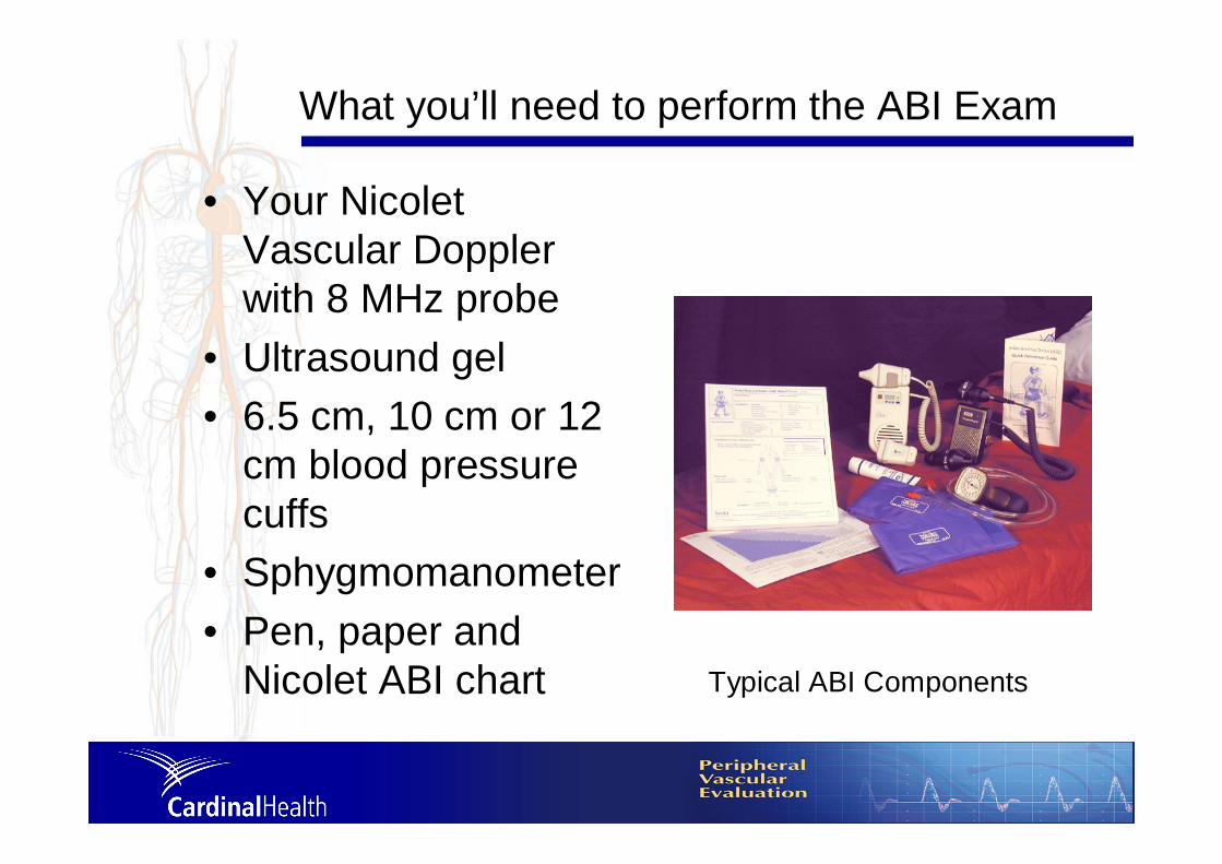
GLOBALLEADERSHIP What you’ll need to perform the ABI Exam
• Your Nicolet Vascular Doppler with 8 MHz probe
• Ultrasound gel• 6.5 cm, 10 cm or 12
cm blood pressure cuffs
• Sphygmomanometer• Pen, paper and
Nicolet ABI chart Typical ABI Components

GLOBALLEADERSHIPWhat You’ll Need to Perform the ABI Exam
• Nicolet VersaLab w/8 MHz probe
• Ultrasound gel• Proper size cuffs• Sphygmomanometer • Pen, paper and Nicolet
ABI chart
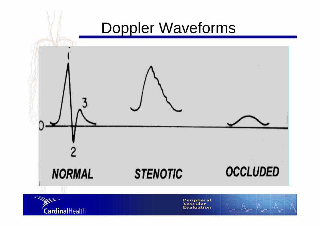
GLOBALLEADERSHIP Doppler Waveforms

GLOBALLEADERSHIP Choose the proper cuff
• The AHA recommends the cuff bladder size be 20% wider than the limb diameter.
• Generally, a 10 cm cuff is fine for use at both the ankle and arm sites.
• Use a 12 cm cuff for patients with larger limbs or a 6.5 cm cuff for smaller limbs.
• Be sure to use the same size cuffs on both the arms and ankles.

GLOBALLEADERSHIP Obtaining the Correct Brachial Pressure
• Have the patient lie supine for a few minutes prior to test.
• Place the appropriate size blood pressure cuff about midway up the patient’s upper arm above the elbow.

GLOBALLEADERSHIPObtaining Brachial PressuresObtaining Brachial Pressures
• Attach the sphygmomanometer.• Place ultrasound gel at the right arm
brachial location.• Hold the probe as if it were a pen or
pencil, angling it so it points up the patient’s arm. The angle will be 45-60 degrees for most probes.
• Slowly move the probe across the brachial area until the pulsating, “whooshing” sound is heard.

GLOBALLEADERSHIP Distinguish Arterial Sounds
• It is very important to distinguish arterial from venous sounds.
• Arterial sounds:– are synchronized with every
heartbeat.– have a rhythmic “whooshing” sound.
• Make sure that you always use an artery to take blood pressures!
GLOBALLEADERSHIP Distinguish Venous Sounds
• It is very important to distinguish venous from arterial sounds.
• Venous sounds are significantly different from arterial sounds, they:– are spontaneous and vary with
respiration.– sound like the wind blowing through the
trees. • Make sure that you always use an
artery to take blood pressures!
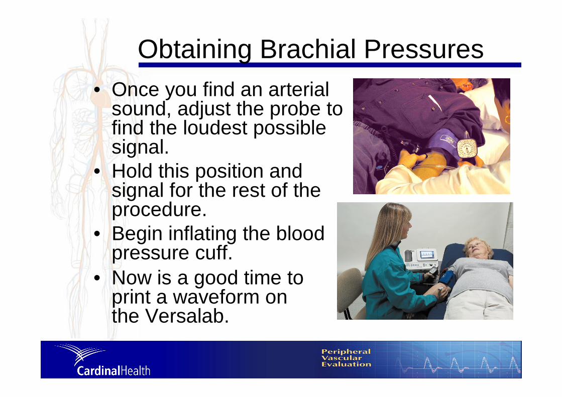
GLOBALLEADERSHIP Obtaining Brachial Pressures
• Once you find an arterial sound, adjust the probe to find the loudest possible signal.
• Hold this position and signal for the rest of the procedure.
• Begin inflating the blood pressure cuff.
• Now is a good time to print a waveform on the Versalab.

GLOBALLEADERSHIP Sample Brachial Waveforms
Simple Waveform Trace Example.Waveform with Spectral Analysis.
Note smooth, uninterrupted pulsatile trace.

GLOBALLEADERSHIP Obtaining Brachial Pressures
• Continue inflating, the flow sound will cease as the cuff pressure begins to stop the blood flow.
• Stop inflating at about 10 - 20 mmHG past this point.
Elite with 8 MHz probe
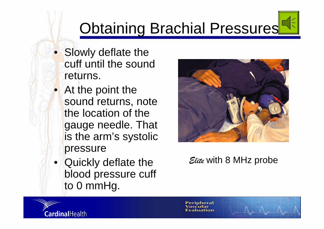
GLOBALLEADERSHIP Obtaining Brachial Pressures
• Slowly deflate the cuff until the sound returns.
• At the point the sound returns, note the location of the gauge needle. That is the arm’s systolic pressure
• Quickly deflate the blood pressure cuff to 0 mmHg.
Elite with 8 MHz probe

GLOBALLEADERSHIP Recording Pressures
• Record the pressure on your Nicolet ABI report form shown at the right or on the VersaLabprintout.
• We recommend attaching the printout to the back of the ABI form.
• Repeat the procedure for the other arm.
ABI Report Form (Catalog #ABI12)

GLOBALLEADERSHIP Obtaining Ankle Pressures
• There are two locations suitable for obtaining the ankle pressure:
Posterior tibial artery Dorsalis pedis artery

GLOBALLEADERSHIP Obtaining Ankle Pressures
• Most clinicians try to obtain the posterior tibial pressure first.
• Use the dorsalis pedis pressure if posterior tibial pressure can’t be obtained.
• If values for both sites are obtained, use the highest value for the index.

GLOBALLEADERSHIP Position the Ankle Cuff
• Use the appropriate size cuff.
• Wrap the cuff snugly around the patient’s ankle, just above the foot.
• Attach the sphygmomanometerto the cuff.
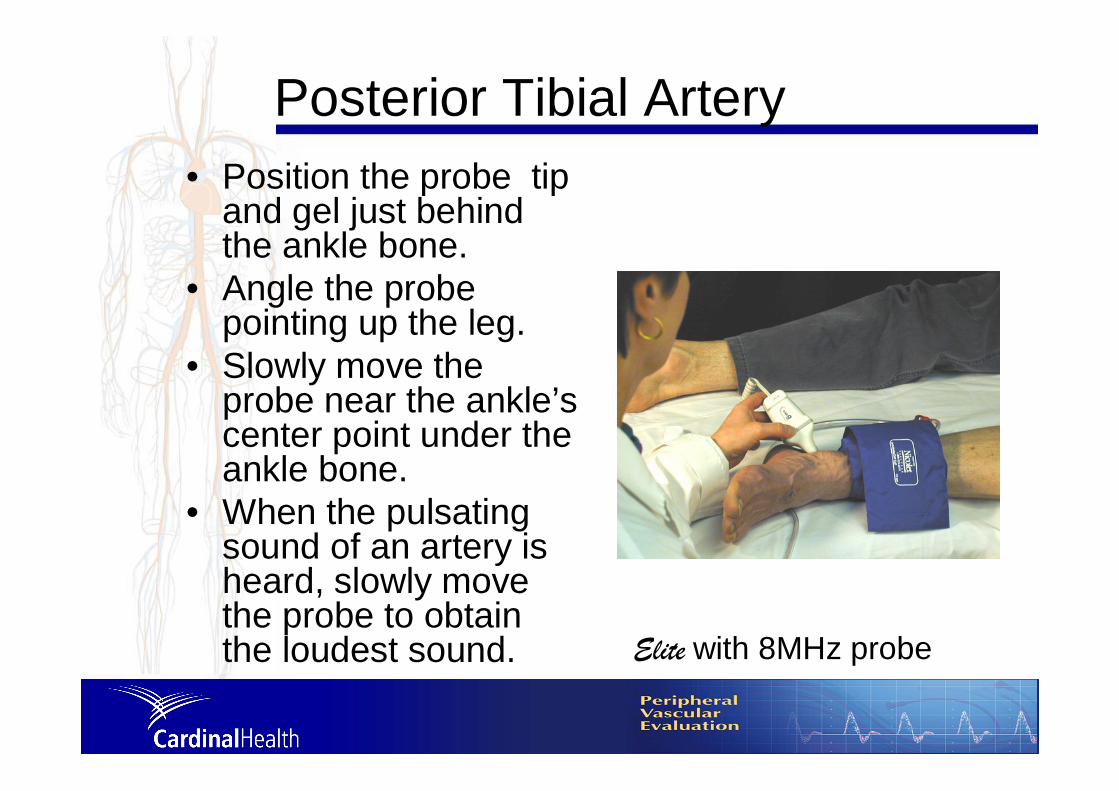
GLOBALLEADERSHIP Posterior Tibial Artery
• Position the probe tip and gel just behind the ankle bone.
• Angle the probe pointing up the leg.
• Slowly move the probe near the ankle’s center point under the ankle bone.
• When the pulsating sound of an artery is heard, slowly move the probe to obtain the loudest sound. Elite with 8MHz probe

GLOBALLEADERSHIP Using the Dorsalis Pedis Artery
• To take an ankle pressure using the dorsalis pedis artery, position the probe on top of the foot in between the big toe and second toe, and halfway up the foot.
• Press lightly because this artery can be easily compressed against the bone, stopping the flow of blood.
GLOBALLEADERSHIP Obtaining Ankle Pressures
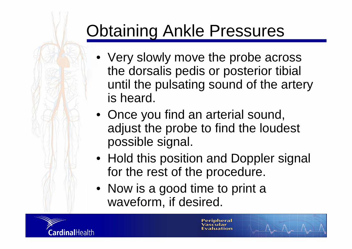
• Very slowly move the probe across the dorsalis pedis or posterior tibialuntil the pulsating sound of the artery is heard.
• Once you find an arterial sound, adjust the probe to find the loudest possible signal.
• Hold this position and Doppler signal for the rest of the procedure.
• Now is a good time to print a waveform, if desired.

GLOBALLEADERSHIP Obtaining Ankle Pressures
• Begin inflating the blood pressure cuff by squeezing the sphygmomanometer bulb.
• Continue inflating, the flow sound will cease as the cuff pressure stops the blood flow.
• Stop inflating 10-20 mmHg past this point.

GLOBALLEADERSHIP Obtaining Ankle Pressures
• Now slowly deflate the cuff until the sound returns.
• Note the location of the gauge needle at the point the sound returns. That is the ankle’s systolic pressure.
• Quickly deflate the blood pressure cuff to 0 mmHg.

GLOBALLEADERSHIP Obtaining Ankle Pressures
• If desired, press the PRINT button on the VersaLab for a waveform print out.
• Record the pressure on your Nicolet ABI report form or the VersaLabprintout.
• We recommend attaching the printout to the back of the ABI form.
• Repeat the procedure for the other ankle.
GLOBALLEADERSHIP Calculating the ABI
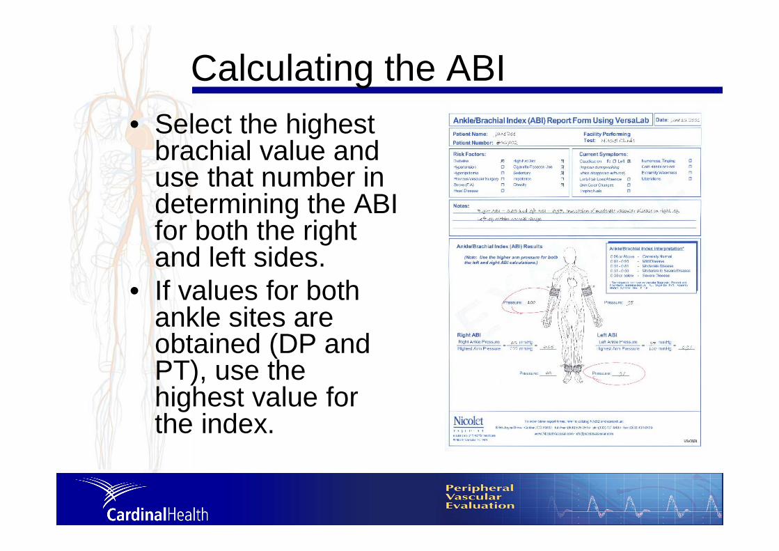
• Select the highest brachial value and use that number in determining the ABI for both the right and left sides.
• If values for both ankle sites are obtained (DP and PT), use the highest value for the index.

GLOBALLEADERSHIP Calculating the Right Side ABI
• In the left column of the ABI chart, find the brachial pressure closest to the one you selected.
• Then in the row at the top of the ABI chart, find the closest right ankle pressure.
• The box where the column and row intersect shows the Ankle/Brachial Index for the right side.
GLOBALLEADERSHIP Calculating the Right Side ABI
• Or using a calculator, divide the pressure for the right ankle by the highest brachial pressure.
• Right Ankle Pressure Highest Brachial Pressure = Right ABI

GLOBALLEADERSHIP Calculating the Left Side ABI
• In the left column of the ABI chart, find the brachial pressure closest to the one you selected.
• Then in the row at the top of the ABI chart, find the closest left ankle pressure.
• The box where the column and row intersect shows the Ankle/Brachial Index for the left side.
GLOBALLEADERSHIP Calculating the Left Side ABI (#2)
• Or using a calculator, divide the pressure for the left ankle by the highest brachial pressure.
• Left Ankle Pressure Highest Brachial Pressure = Left ABI

GLOBALLEADERSHIP Resting ABI values
• 0.91-1.30 = Normal • 0.70-0.90 = Mild obstruction • 0.40-0.69 = Moderate obstruction• < 0.40 = Severe obstruction • > 1.30* = Poorly compressible
(due to medial arterial calcification)
An ABI (Ankle-Brachial Index) of 0.78 portends an approximately 30% 5-year risk of heart attack, stroke and vascular death.

GLOBALLEADERSHIP ADA Recommendation
• Perform a screening ABI in patients with diabetes who are >50 years of age, and, if normal, repeat the test every 5 years.
• Consider a screening ABI in patients with diabetes who are <50 years of age and have other PAD risk factors (e.g., smoking, hypertension, hyperlipidemia, or duration of diabetes >10 years).
• Perform a diagnostic ABI in any patient with symptoms of PAD.

GLOBALLEADERSHIP Additional Evaluation
• Vascular lab evaluation, segmental pressures and pulse volume recordings to assess location and severity of PAD: for pts with abnormal ABI, or for pts with poorly compressible vessels (ABI >1.3) or for pts with normal ABI but high suspicion of PAD
• Treadmill functional testing: to diagnose pts with atypical symptoms or to diagnose pts with a normal ABI with symptoms of claudication.
• Other studies: toe pressure, transcutaneouspartial pressure of oxygen, sonography or MRA: to make decisions for revascularization
• X-ray angiography: for patients where a revascularisation procedure is intended. It is an invasive test and should not be done for diagnosing PAD.
GLOBALLEADERSHIP Treatment
Treatment of the patient with diabetes and PAD should be twofold:1) primary and secondary cardiovascular disease risk factor modification and2) treatment of PAD symptoms (claudication and CLI) and limiting progression of disease.
GLOBALLEADERSHIP Conclusions
• ABI screening for PAD in patients with diabetes is enormously productive.
• Routine screening of individuals with diabetes of > 50 yrs of age with the ABI test can identify PAD in nearly a third of the individuals; e.g highly effective identification.
• Identifying PAD before it has progressed to its more severe stages allows effective treatment to be offered.
• These therapies may arrest PAD development and, perhaps reverse its advance, while reducing cardiovascular risk (MI and stroke).

GLOBALLEADERSHIP In Conclusion
• PAD is a common complication of pts with diabetes. It is more commonly asymptomatic and thus patients present with more severe disease with greater risk of amputation, MI and stroke.
• Diagnosis of PAD in diabetes is thus very important and justifies screening with the simple ABI test.
• The use of a simple ABI test has been proven as an accurate means of identifying, treating and managing your at risk patients.

THANK YOUTHANK YOU