Embed Size (px)
Citation preview

Urogenital system II
1Lufukuja G.

DEVELOPMENT OF THE GONADS
Lufukuja G. 2
Development of the gonads• Although the sex of the embryo is determined
genetically at the time of fertilization, the gonads do not acquire male or female morphological characteristics until the seventh week of development.
• Gonads appear initially as a pair of longitudinal ridges, the genital or gonadal ridges. They are formed by proliferation of the epithelium and a condensation of underlying mesenchyme. Germ cells do not appear in the genital ridges until the sixth week of development.
Lufukuja G. 3
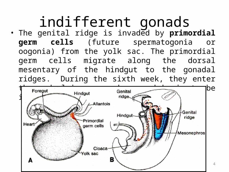
indifferent gonads• The genital ridge is invaded by primordial germ cells (future
spermatogonia or oogonia) from the yolk sac. The primordial germ cells migrate along the dorsal mesentary of the hindgut to the gonadal ridges. During the sixth week, they enter the underlying mesenchyme and begin to be incorporated into the sex cords.
Lufukuja G. 4
Lufukuja G. 5
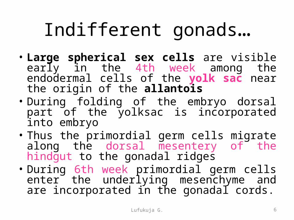
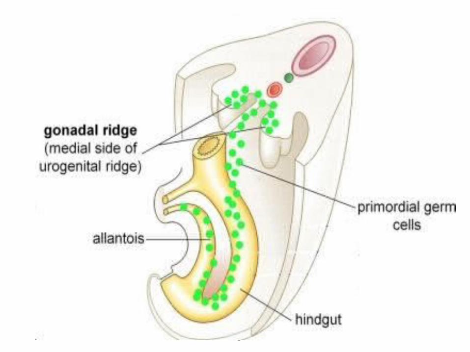
Indifferent gonads…• Large spherical sex cells are visible early in
the 4th week among the endodermal cells of the yolk sac near the origin of the allantois
• During folding of the embryo dorsal part of the yolksac is incorporated into embryo
• Thus the primordial germ cells migrate along the dorsal mesentery of the hindgut to the gonadal ridges
• During 6th week primordial germ cells enter the underlying mesenchyme and are incorporated in the gonadal cords.
Lufukuja G. 6

Indifferent gonads…• They migrate by amoeboid movement along the dorsal
mesentery of the hindgut, arriving at the primitive gonads at the beginning of the fifth week and invading the genital ridges in the sixth week.
• If they fail to reach the ridges, the gonads do not develop. Hence the primordial germ cells have an inductive influence on development of the gonad into ovary or testis.
Lufukuja G. 7
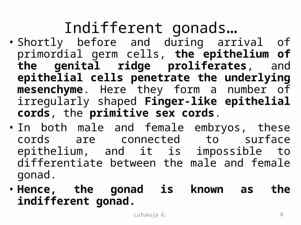
Indifferent gonads…• Shortly before and during arrival of primordial
germ cells, the epithelium of the genital ridge proliferates, and epithelial cells penetrate the underlying mesenchyme. Here they form a number of irregularly shaped Finger-like epithelial cords, the primitive sex cords.
• In both male and female embryos, these cords are connected to surface epithelium, and it is impossible to differentiate between the male and female gonad.
• Hence, the gonad is known as the indifferent gonad.
8Lufukuja G.

Lufukuja G. 9
Indifferent gonad

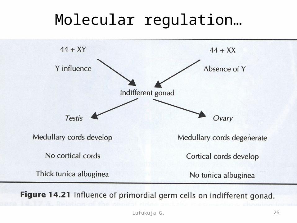
Development of testes• If the embryo is genetically male, the
primordial germ cells carry an XY sex chromosome complex
• Under influence of the SRY gene on the Y chromosome, which encodes the testis-determining factor (TDF), the primitive sex cords continue to proliferate and penetrate deep into the medulla to form the testis or medullary cords.
Lufukuja G. 10

Lufukuja G. 11
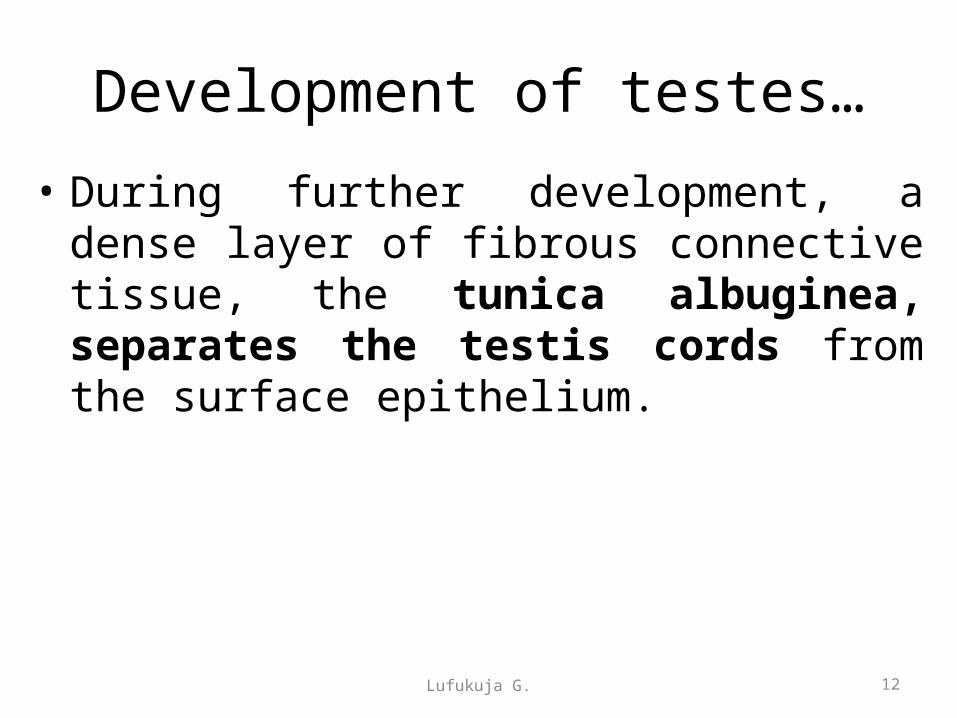
Development of testes…
• During further development, a dense layer of fibrous connective tissue, the tunica albuginea, separates the testis cords from the surface epithelium.
Lufukuja G. 12
Lufukuja G. 13

Development of testes…• Under the influence of TDF, cells in the medullary region of
the primitive sex cords differentiate into sustentacular (pre-Sertoli) cells, while those in the cortical region degenerate.
• Pre-Sertoli cells secrete Müllerian inhibiting hormone (MIH) (also called Müllerian inhibiting substance or anti-Müllerian hormone).
• Testis cords remain solid untill puberty; when they are canalyzed forming seminiferous tubules
• Once the seminiferous tubules are canalized, they join the rete testis tubules, which in turn enter the ductuli efferentes.
Lufukuja G. 15
Development of testes…
• Interstitial cells of Leydig derived from the original mesenchyme of the gonadal ridge begin development shortly after onset of differention of primitive sex cords
• Leydig cells lie between the testis cords and begin testosterone production by 8th week of gestation.Thus the testis is able to influence sexual differentiation of the genital ducts and ext genitalia
16Lufukuja G.
Genital duct development in male
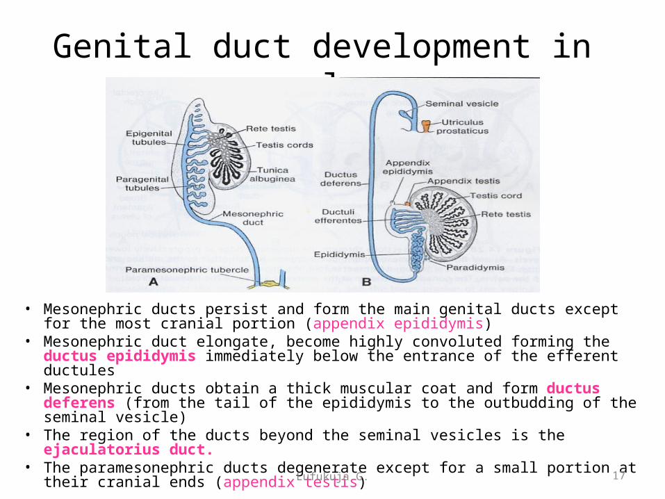
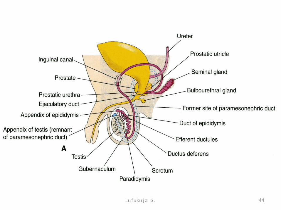
• Mesonephric ducts persist and form the main genital ducts except for the most cranial portion (appendix epididymis)
• Mesonephric duct elongate, become highly convoluted forming the ductus epididymis immediately below the entrance of the efferent ductules
• Mesonephric ducts obtain a thick muscular coat and form ductus deferens (from the tail of the epididymis to the outbudding of the seminal vesicle)
• The region of the ducts beyond the seminal vesicles is the ejaculatorius duct.• The paramesonephric ducts degenerate except for a small portion at their
cranial ends (appendix testis) 17Lufukuja G.
Mesonephric duct• In the male the mesonephric (Wolffian) duct persist
and participate in formation of the genital system, but they disappear in the female. The duct gives rise in male to paradidymis; appendix of epididymis; duct of epididymis; ductus deferens; ureter; renal pelvis; calices; collecting tubules; ejaculatory duct and seminal gland. The duct in female gives rise to duct of epoophron; duct of Gartner; ureter ; pelvis; calices and collecting tubules.
18Lufukuja G.
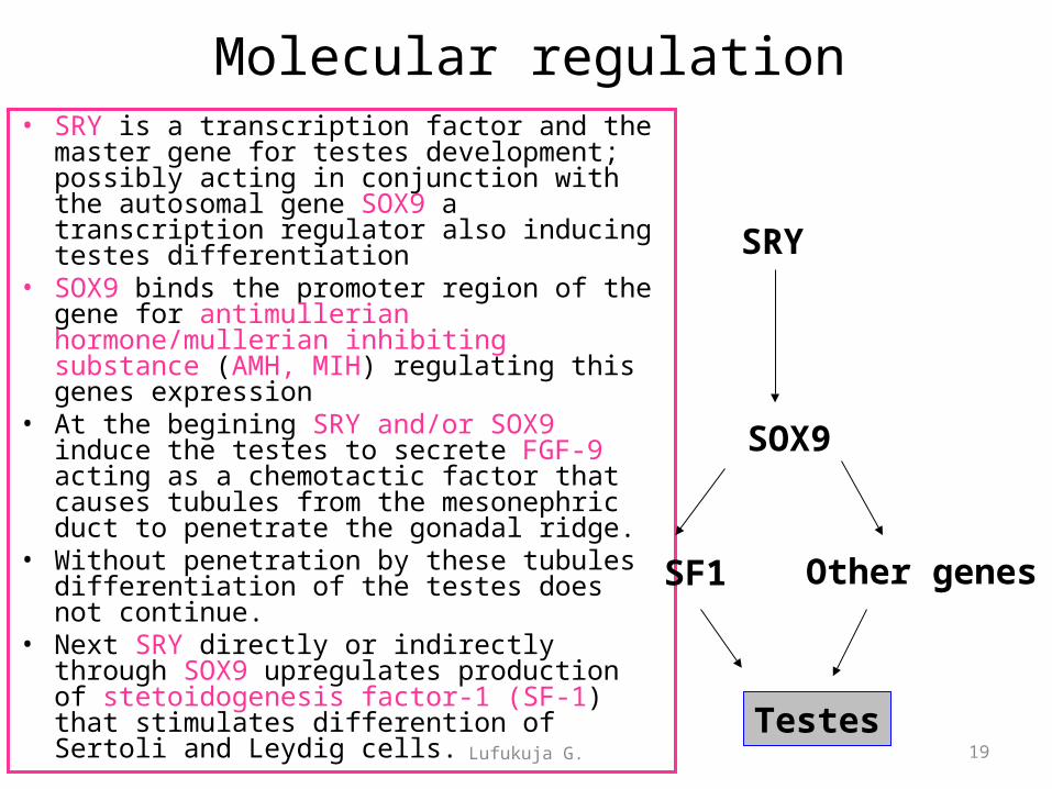
Molecular regulation• SRY is a transcription factor and the master gene
for testes development; possibly acting in conjunction with the autosomal gene SOX9 a transcription regulator also inducing testes differentiation
• SOX9 binds the promoter region of the gene for antimullerian hormone/mullerian inhibiting substance (AMH, MIH) regulating this genes expression
• At the begining SRY and/or SOX9 induce the testes to secrete FGF-9 acting as a chemotactic factor that causes tubules from the mesonephric duct to penetrate the gonadal ridge.
• Without penetration by these tubules differentiation of the testes does not continue.
• Next SRY directly or indirectly through SOX9 upregulates production of stetoidogenesis factor-1 (SF-1) that stimulates differention of Sertoli and Leydig cells.
SRY
SOX9
SF1 Other genes
Testes19Lufukuja G.

DEVELOPMENT OF OVARIES
Lufukuja G. 20
Development of ovaries
• In female embryos with an XX sex chromosome complement and no Y chromosome, primitive sex cords dissociate into irregular cell clusters
• These clusters, containing groups of primitive germ cells, occupy the medullary part of the ovary. Later they disappear and are replaced by a vascular stroma that forms the ovarian medulla
Lufukuja G. 21
Development of ovaries• Surface epithelium of the female gonad (unlike
that of the male) continues to proliferate giving rise to a second generation of cords (cortical cords) in the 7th week.
• Cortical cords penetrate the underlying mesenchyme but remain close to the surface
• In the 4th month cortical cords split into isolated cell clusters with each surrounding one or more primitive germ cells
• Germ cells develop into oogonia, surrounding epithelial cells, descendants of the surface epithelium form follicular cells.
22Lufukuja G.
Lufukuja G. 23
Development of ovaries…
24Lufukuja G.

Molecular regulation• WNT4 is the ovary determining gene;
upregulating DAX1 which is a member of the nuclear hormone receptor family
• DAX1 inhibits the function of SOX9• WNT regulates expression of other
genes (TAFII105....TATA binding protein for RNA polimerase in ovarian follicular cells) responsible for ovarian differentiation
• Estrogens are involves in sexual differentiation; under their influence paramesonephric (mullerian) ducts are stimulated to form ext genitalia
WNT4
DAX1 Other genesTAFII 105
Ovaries
25Lufukuja G.
26Lufukuja G.
Molecular regulation…

Development of female genital ducts
Lufukuja G. 27
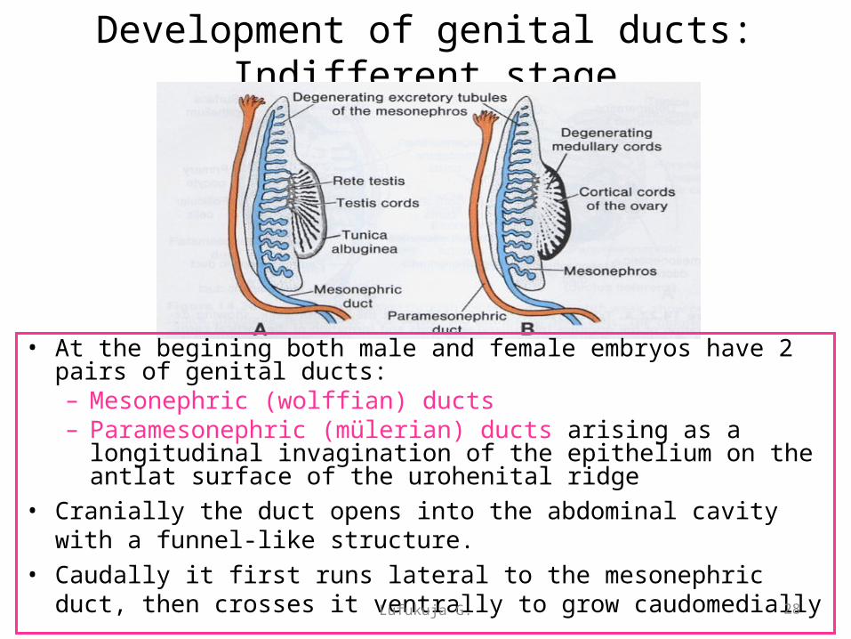
Development of genital ducts: Indifferent stage
• At the begining both male and female embryos have 2 pairs of genital ducts:– Mesonephric (wolffian) ducts – Paramesonephric (mülerian) ducts arising as a longitudinal
invagination of the epithelium on the antlat surface of the urohenital ridge
• Cranially the duct opens into the abdominal cavity with a funnel-like structure.
• Caudally it first runs lateral to the mesonephric duct, then crosses it ventrally to grow caudomedially
28Lufukuja G.
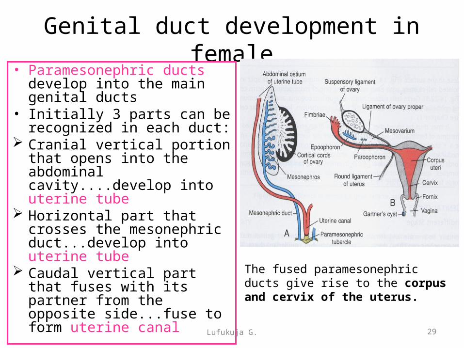
Genital duct development in female• Paramesonephric ducts
develop into the main genital ducts
• Initially 3 parts can be recognized in each duct:
Cranial vertical portion that opens into the abdominal cavity....develop into uterine tube
Horizontal part that crosses the mesonephric duct...develop into uterine tube
Caudal vertical part that fuses with its partner from the opposite side...fuse to form uterine canal
29Lufukuja G.
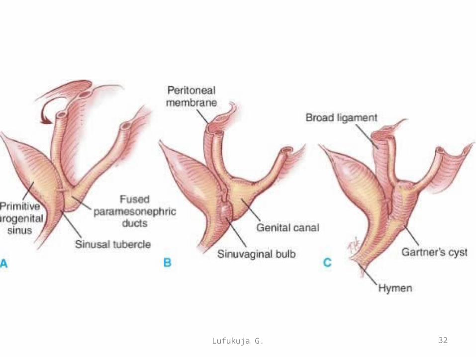
The fused paramesonephric ducts give rise to the corpus and cervix of the uterus.
Genital duct development in female
• After the ducts fuse in the midline, a broad transverse pelvic fold is established. This fold, which extends from the lateral sides of the fused paramesonephric ducts toward the wall of the pelvis, is the broad ligament of the uterus.
30Lufukuja G.

Lufukuja G. 31
Lufukuja G. 32

DESCENT OF THE TESTES
Lufukuja G. 33
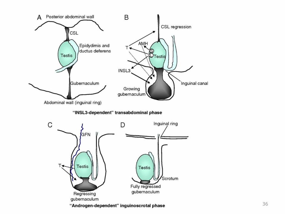
Descent of the testes• Toward the end of the second month, the urogenital
mesentery attaches the testis and mesonephros to the posterior abdominal wall. With degeneration of the mesonephros the attachment serves as a mesentery (suspensory ligament) for the gonad
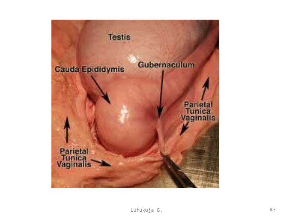
• Caudally it becomes ligamentous and is known as the caudal genital ligament. Also extending from the caudal pole of the testis is a mesenchymal condensation rich in extracellular matrices, the gubernaculum connects the gonads to the the scrotal swellings.
Lufukuja G. 34
Descent of the testes…• Regression of the CSL and the outgrowth and
subsequent regression and inversion of the gubernaculum mediates transfer of the testes into the scrotum.
Lufukuja G. 35

Lufukuja G. 37

38Lufukuja G.

39Lufukuja G.
40Lufukuja G.
41Lufukuja G.

42Lufukuja G.
Lufukuja G. 43
44Lufukuja G.
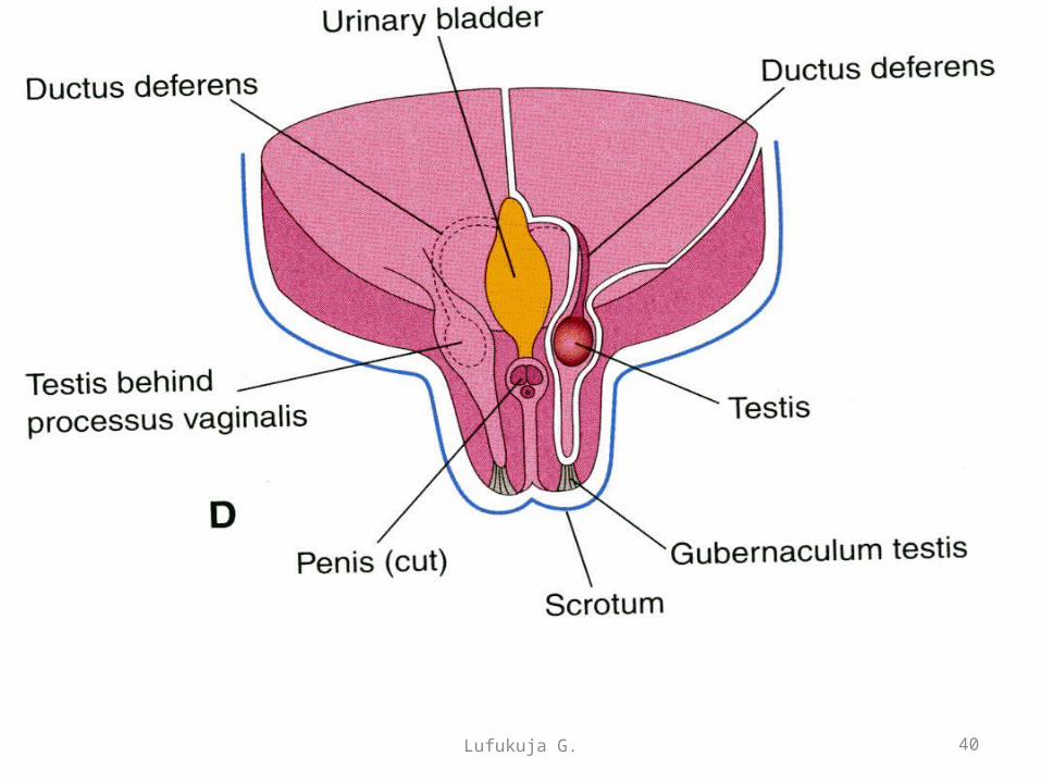
Descent of the testes
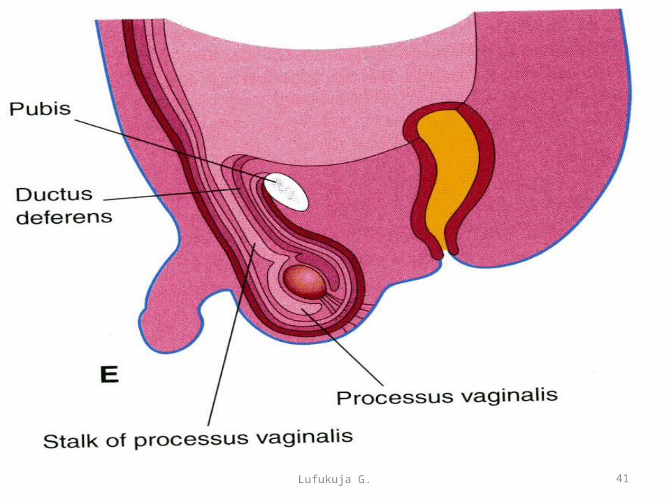
• Normally, the testes reach the inguinal region by approximately 12weeks gestation, migrate through the inguinal canal by 28 weeks, and reach the scrotum by 33 weeks Descent may take 2-3 days and the inguinal canal contracts after they enter the scrotum
• As the testis and the ductus deferens descend they are enshetaed by the facial extensions of the abdominal wall
45Lufukuja G.

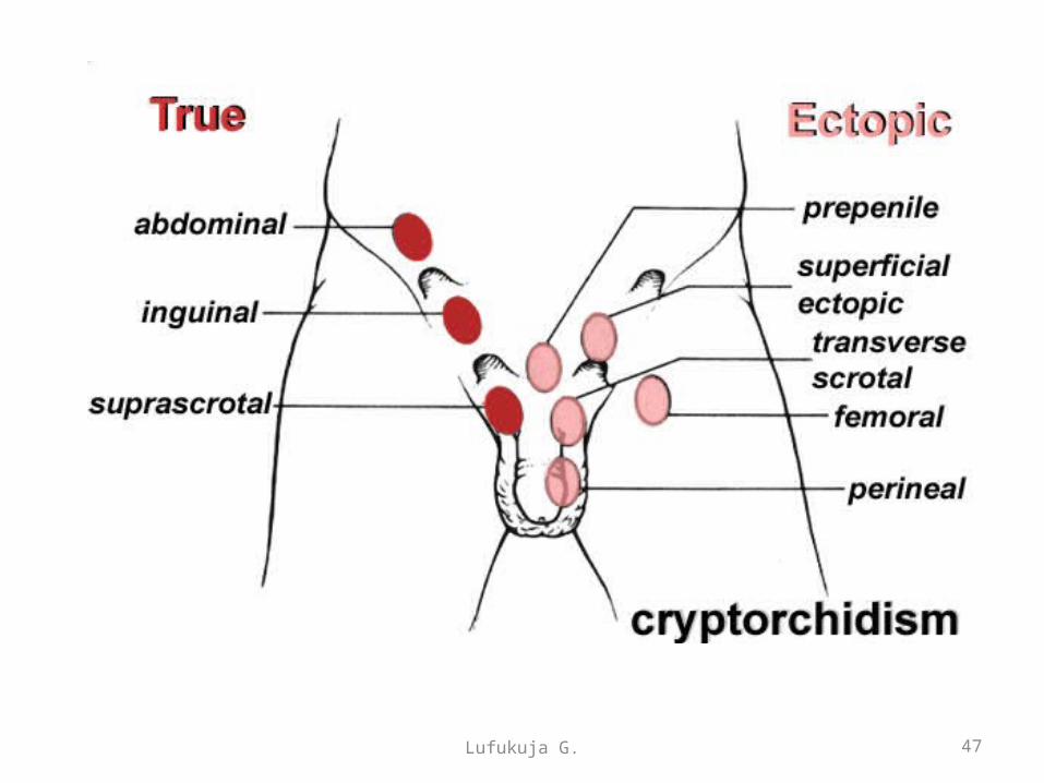
• Cryptorchidism or undescended testis: occurs in 30 % of premature, 3-4% of full-term males. It may be uni or bilateral. Failure of descent in the first year causes atrophy of testes. It may be in the abdominal cavity or anywhere along the descent path, usually in the inguinal canal. It may be caused by teh defficiency of androgen production in testes.
• Ectopic testes: After traversing the inguinal canal, the testis may deviate from its usual path of descent and lodge in various abnormal locations.
Congenital anomalies of descent of the testesCongenital anomalies of descent of the testes
46Lufukuja G.
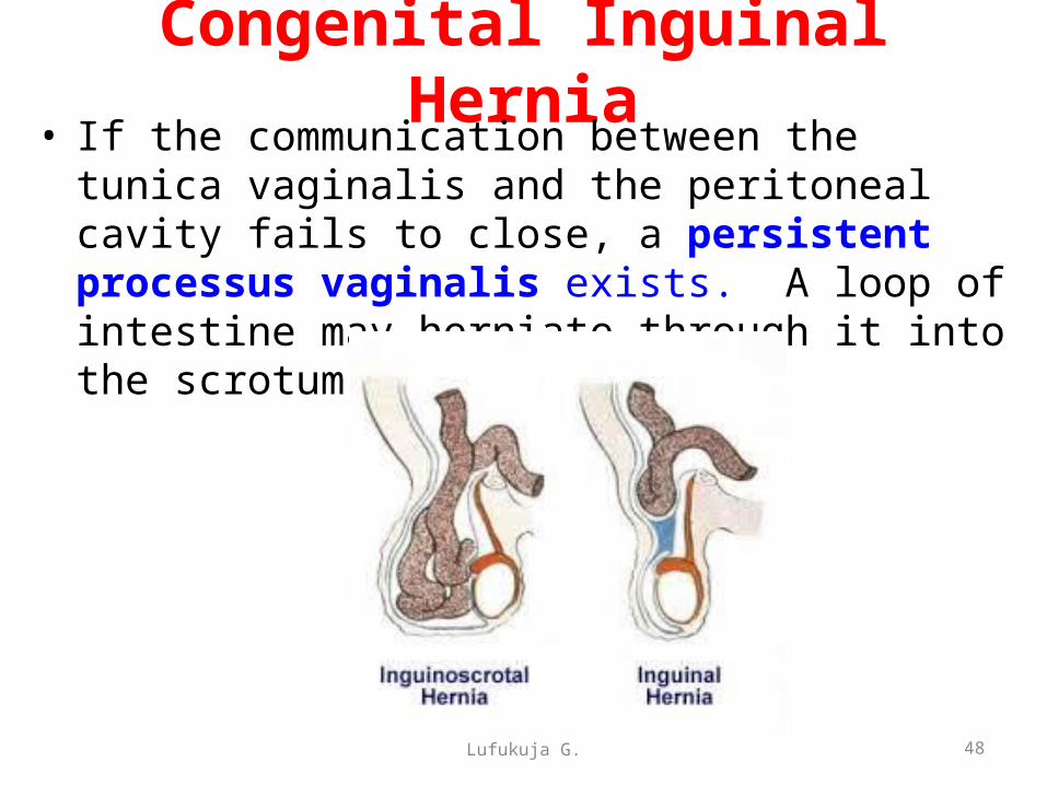
Congenital Inguinal Hernia• If the communication between the tunica vaginalis
and the peritoneal cavity fails to close, a persistent processus vaginalis exists. A loop of intestine may herniate through it into the scrotum or labium majus.
Lufukuja G. 48

Congenital Hydrocele• Occasionally the abdominal end of the
processus vaginalis remains open but is too small to permit herniation of intestine
• Peritoneal fluid passes into the patent processus vaginalis and forms hydrocele of the testis
Lufukuja G. 49

DESCENT OF THE OVARIES
Lufukuja G. 50
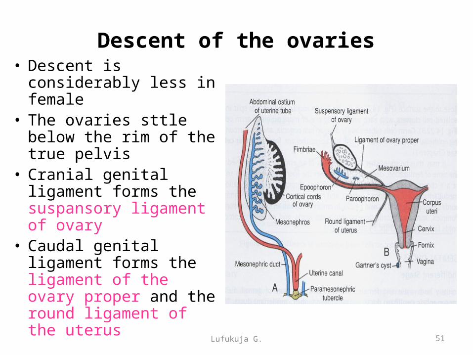
Descent of the ovaries• Descent is considerably
less in female• The ovaries sttle below
the rim of the true pelvis• Cranial genital ligament
forms the suspansory ligament of ovary
• Caudal genital ligament forms the ligament of the ovary proper and the round ligament of the uterus
51Lufukuja G.
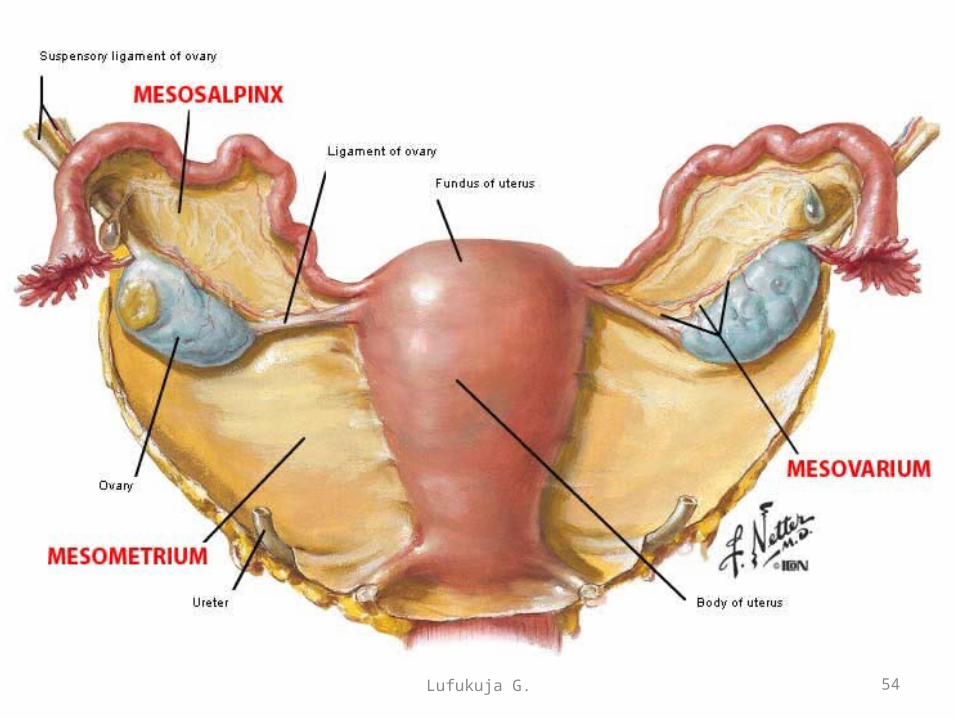
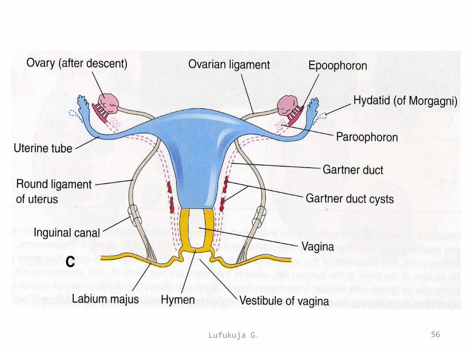
The broad ligament• The mesovarium is the portion of the broad ligament of
the uterus that suspends the ovaries. The ovary is not covered by the mesovarium, rather it is covered by germinal epithelium.
• At first the mesonephros and genital ridge are suspended by a common mesentery, but as the embryo grows the genital ridge gradually becomes pinched off from the mesonephros, with which it is at first continuous, though it still remains connected to the remnant of this body by a fold of peritoneum. In the male this is the mesorchium, and in the female, this is the mesovarium.
Lufukuja G. 52

Lufukuja G. 53
Lufukuja G. 54
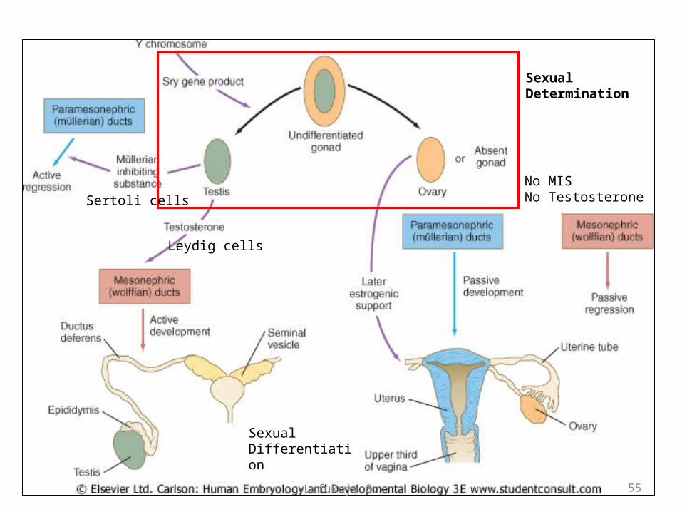
Sertoli cells
Leydig cells
No MISNo Testosterone
Sexual Determination
Sexual Differentiation
55Lufukuja G.
Lufukuja G. 56

Lufukuja G. 57