Embed Size (px)
Citation preview

J. Exp. Biol. (1968), 48, 159-174 I egWith 11 text-figures
Printed in Great Britain
URINE FORMATION BY THE MALPIGHIANTUBULES OF CALLIPHORA
I. CATIONS
BY M. J. BERRIDGE*
Department of Biology, University of Virginia, Charlottesville,Virginia, and Developmental Biology Center, Western Reserve
University, Cleveland, Ohio
{Received 10 August 1967)
INTRODUCTION
The current concept of fluid transport in vertebrates is that water moves passivelydown the osmotic gradient created by an active transport of solute. Earlier suggestionsthat these tissues could transport water in the absence of a concomitant movement ofsolute have been rejected (Curran, 1965; Diamond, 1965; Robinson, 1965, 1966).Interest is now focused on the mechanism by which water movement is coupled toactive solute transport. A simple explanation would be that active solute transportacross an epithelium might increase the total solute concentration on one side and thusprovide the osmotic gradient for a passive flow of water. This simple hypothesis,however, cannot account for water transport against an osmotic gradient. Curran(i960) extended the hypothesis by postulating that active solute transport takes placeinto an enclosed compartment within which a large osmotic pressure gradient couldbe maintained. Long narrow channels (e.g. lateral intercellular spaces and basalinfoldings), which are a constant feature of secretory or reabsorptive epithelia, appearto be the structural analogues of the enclosed compartment in Curran's model(Diamond & Tormey, 1966a, b; Kaye, Wheeler, Whitlock & Lane, 1966; Berridge &Gupta, 1967; Davis & Schmidt-Nielsen, 1967). A mathematical treatment of solute-linked water transport indicates that standing osmotic gradients within long narrowchannels can adequately account for the coupling of water to solute transport(Diamond & Bossert, 1967).
Urine formation by the Malpighian tubules of insects resembles fluid transport invertebrate epithelia in that water flow depends on an active secretion of potassium(Ramsay, 1953, 1955 b, 1956). However, details of the exact linkage between solute andwater transport have not been determined. This paper attempts to extend Ramsay'sobservations on the nature of cation transport and to consider the structural basis forthe mechanism of solute-linked water transport in Malpighian tubules.
• Present address: Department of Biology, Western Reserve University, Cleveland, Ohio 44106,U.S.A.

160 M. J. BERRIDGE
MATERIAL AND METHODS
Adult female Calliphora erythrocephala were reared as described previously(Berridge, 19666). The Malpighian tubules of 3-day-old females were set up inartificial media kept under liquid paraffin (Berridge, 19666). Rate of urine productionwas always measured 2 hr. after setting up the tubules.
Table 1. Composition of artificial media: the final concentrationof phosphate in medium C was 15 niMJl.
CaCl2MgCl26HaOTrehaloseGlucoseProlineGlutaminea-AlanineGlycinePenicillinStreptomycin
Fumaric acidCitric acidMalic acidK O H
NaClKC1SucroseNaH2PO4H2ONa.HPO,
O-22-oi-8i-8o-6o-80-4
o-S0-03o - i
—
—
—
VariableVariableVariable
VariableVariable
i -8i-8o-6o-80 4
o-S0-03
o - i
i - o
i - o
i - o
2-25
Variable——
O-2
2-O
1-8i-8o-6o-80 4
o-S0-03o-i
i -oi -oi -o2-25
4-8
—VariableVariable
The composition of the artificial media used is shown in Table 1; the pH of allmedia (except C) was 7-2 and the osmotic pressure was adjusted to give a depressionof the freezing point of approximately 0*7° C. To test the effect of ionic strength andosmotic pressure on the process of urine formation, different media were prepared byadding various amounts of sodium, potassium or sucrose to medium A. Medium Bwas used to test the ability of divalent cations to support urine formation. This mediumcontained a number of organic acids which helped to keep these divalent cations insolution, especially when they were present in high concentrations. Solutions ofsimilar osmotic pressure were obtained by balancing the concentration of the divalentions with sodium. The effect of pH on the activity of tubule cells was tested withmedium C; different hydrogen ion concentrations were obtained by means of aphosphate buffer.
The sodium and potassium concentrations of urine and artificial media weremeasured by means of a Beckman flame-photometer. Micropipettes were used toobtain fluid samples from under liquid paraffin.
Osmotic pressure determinations were made by the cryoscopic method of Ramsay& Brown (1955).

Urine formation by the malpighian tubules of Calliphora 161
RESULTS
Effect of potassium concentration on urine formation at constant osmotic pressure
{a) Potassium transport in the presence of sodium
Rate of urine formation by the Malpighian tubules of Calliphora is markedlyaffected by the external potassium concentration (Fig. i). In the absence of potassium(i.e. when sodium was the major cation), urine formation occurred at a very slow rate(0-9 mm.3 x icr3/min.), but was considerably accelerated as potassium was pro-gressively replaced by sodium. The increase occurred as a two-stage process; replace-ment of only 8 mM/1. sodium with an equivalent amount of potassium caused a largeincrease of urine flow, but thereafter the increase became asymptotic. The maximumrate of urine formation was recorded when potassium completely replaced sodium inthe bathing medium.
0 20 40 60 80 100 120 140
Potassium concentration in medium (mM/1.)
Fig. 1. Effect of potassium concentration on rate of urine formation when osmoticpressure was kept constant with (a) sodium (#), and (6) sucrose (O).
Transport of sodium and potassium independently of each other is shown moreclearly when the concentration of these two ions in the urine is compared with theirconcentration in the bathing medium (Fig. 2). The slow rate of urine secretion in apotassium-free medium (Fig. 1) resulted from a secretion of sodium which appearedin the urine at a concentration similar to that in the bathing medium (Fig. 2). Replace-ment of a small quantity of sodium with an equivalent amount of potassium in thebathing medium produced a sudden shift in the potassium and sodium concentrationof the urine; potassium almost completely replaced sodium. Appearance of potassiumin the urine coincided with the large increase in rate of urine production (Fig. 1) andemphasizes further the extraordinary sensitivity of the tubule cells to potassium.
11 Exp. Biol. 48, 1

162 M. J. BERRIDGE
Potassium concentration in the urine (100-140 mM/1.) was always much higher thanits concentration in the bathing medium, especially at low concentrations of the latter(i.e. 8-60 mM/1.). Sodium apparently does not interfere with the carrier mechanism forpotassium, since potassium was transported against a large gradient even with a highconcentration of sodium in the medium (Fig. 2).
Since tubules were capable of producing urine in a potassium-free medium bysecreting sodium, this might indicate that at least one component of the cation trans-port mechanism is relatively unspecific. Therefore the specificity of cation transportwas explored further by using different cations (Table 2). Potassium and rubidiummaintained a high rate of urine production, whereas caesium and sodium were less
-o ^^—Q__20I
40I
60I
80 100I
1201
140 [K+]
140 120 100 80 60 40 20 0 [Na+]
Concentration in medium (mM/1).
Fig. 2. Concentration of sodium and potassium in the urine collected during the experimentsillustrated in Fig. m (•—•, potassium; O—O, sodium) and 16 (O—O, potassium con-centration when sucrose replaced sodium in the bathing medium).
Table 2. The ability of individual cations to support a flow of urine: each cationwas dissolved in medium A to give a final concentration of 140 ntMJl.
Cations
PotassiumRubidiumCaesiumSodiumAmmoniumLithiumCholine
Rate ofurine
production(mm.3x io~3/min.)
13810-9
2-S0 4O
OO

Urine formation by the malpighian tubules of Calliphora 163
effective. Ammonium, lithium and choline were not able to maintain a flow of urine.These results demonstrate that tubule cells can secrete urine in the presence of asingle univalent cation.
(b) Potassium transport in the presence of sucrose
The next series of experiments tested the ability of Malpighian tubules to secreteurine at variable pdtassium concentrations when sucrose replaced sodium (Fig. 1). Ifa non-transportable molecule is used to adjust the osmotic pressure, rate of urineformation is linearly related to the external potassium concentration (Fig. 1). Undersuch conditions, potassium concentration of the urine was again much higher than itsconcentration in the bathing medium and remained relatively constant throughout therange of external potassium concentration (Fig. 2). However, potassium concentrationin the urine was slightly higher than that secreted by tubules in a sodium + potassiummedium.
50
I
30
I 20
10
0 100 200 300 400 S00 600
Potassium concentration in medium (tnM/1.)
Fig. 3. Effect of potassium concentration on rate of urine formation when theosmotic pressure is not adjusted.
Effect of osmotic pressure on urine formation
(a) Effect of osmotic pressure on urine formation at variable potassium concentration
Rate of urine formation by the Malpighian tubules of insects is directly related tothe external osmotic pressure (Ramsay, 1954; Berridge, 1966 a); in the previousexperiments by Ramsay and Berridge the osmotic pressure of serum was modified togive hypotonic or hypertonic solutions by adding distilled water or sucrose respec-tively. However, such a procedure introduces a complicating factor, because, althoughosmotic pressure is continuously variable, the potassium concentration is decreasedby dilution but remains constant in the hypertonic media. Therefore, in the presentstudy a range of osmotic pressure was obtained by adding different amounts of potas-sium chloride to medium A (Table 1). Consequently, both potassium concentrationand osmotic pressure were continuously variable; Fig. 3 illustrates the change in the

164 M. J. BERRIDGE
rate of urine formation under such conditions. A maximum rate of urine formationwas recorded at the lowest potassium concentration and hence the lowest osmoticpressure. An increase in both these parameters resulted in a consistent decrease inrate of urine formation. The tubules in hypotonic medium were considerably swollenbut became progressively shrunken as the outside osmotic pressure increased. Clearly,the tubules of Calliphora can function over a wide range of potassium concentrationand osmotic pressure.
"s"B,uc
un
c
ion
trat
ceni
§u
E3tnCOC3
OOH
700
600
500
400
300
200
100
-
/
/
if
/
y%
1 1 1 1 1 1
0 100 200 300 400 500 600
Potassium concentration in medium (mM/1.)
Fig. 4. Concentration of potassium in the urine secreted during the experimentillustrated in Fig. 3.
The potassium concentration of the urine remained consistently higher than thatof the bathing medium throughout the range of external potassium concentrationemployed (Fig. 4). The average difference of [K+]urlne - [K+]medlum for all the obser-vations was 37 ITIM/I. Combination of the data in Fig. 3 and 4 showed that rate ofpotassium secretion (Fig. 5) is inversely related to the external potassium concen-tration. When the osmotic pressure was kept constant with sucrose, however, rate ofpotassium secretion (obtained from the data in Figs. 1 and 2) was directly related tothe external potassium concentration (Fig. 5).
The osmotic pressure of urine was slightly, but consistently, hypertonic to thebathing medium throughout the range of osmotic pressures employed (Fig. 6). Theaverage value of the o.p.urlne — o.p.medlum was 0-066° C. At low osmotic pressures, rateof urine formation is increased (Fig. 3) because more water molecules must followeach ion in order to maintain this constant osmotic pressure relationship (i.e. urineosmotic pressure 0-066° C hypertonic to the medium).
(b) Effect of osmotic pressure on urine formation at constant potassium concentration
Progressive addition of sucrose to medium A containing 140 mM/1. of potassiumchloride caused a regular decrease in rate of fluid formation (Fig. 7). The presence ofsucrose in the bathing medium also had a pronounced effect on the potassium

Urine formation by the malpighian tubules of Calliphora 165
concentration of urine (Fig. 8). The curve in Fig. 8 represents the calculated concen-tration of potassium in the urine required to exactly counteract the increase in osmoticpressure of the bathing medium due to the presence of sucrose. Most of the observedvalues (open circles) lie very close to this curve, therefore the concentration of potas-sium in the urine increased by 1 nw/1. for each 2 mM/1. of sucrose added to thebathing medium. In rabbit gall bladder, a similar increase in concentration of thetransported cation (in this case sodium) was observed when impermeant non-electro-lytes such as raffinose and sucrose were added to the bathing solution (Diamond,1964).
8.5Si278 222 xa S
3
30
25
20
15
10
5
•ao
20
1-5
10
0-5
_ /
7
/ \ i i
\/
y
100 200 300 400 500 600 0-5 1-0 1-5 20
Osmotic pressure of medium (A° C.)Potassium concentration inmedium (mM/1.)
Fig 5. Fig 6.
Fig. 5. Rate of potassium secretion when (a) osmotic pressure is unbalanced (#; data takenfrom Figs. 3 and 4), and (6) osmotic pressure is balanced with sucrose (O; data taken fromFigs. 1 and 2).Fig. 6. The relationship between osmotic pressure of the medium and that of the urinesecreted in the experiment illustrated in Fig. 3. The point where the two solutions are isosmolaris indicated by the straight line. Most of the points lie above the line, indicating that theurine is slightly hypertonic.
Effect of divalent cations on urine formation
The divalent cations, calcium and magnesium, cannot replace the univalent cationsin the process of urine formation. Nevertheless, these divalent ions are known to playan important role in active transport mechanisms. These ions were tested on tubulesprovided with potassium so that they could secrete urine. In the complete absence ofthese divalent cations, urine production occurred at a slow rate but was acceleratedafter addition of either magnesium or calcium (Fig. 9). The optimal concentration ofthese cations was about 10 mM/1. Higher concentrations tended to depress urineproduction with calcium showing a greater inhibitory effect than magnesium. Theisolated Malpighian tubules of Calliphora reacted to high concentrations of mag-nesium and calcium in much the same way as do the tubules of Dixippus (Ramsay,I956)-

166 M. J. BERRIDGE
Effect on pH and inhibitors on urine production
The ability of Malpighian tubules to secrete urine in the presence of a single mono-valent cation is unusual and might suggest that the transport mechanism is electro-genie. Such a suggestion cannot be taken seriously until it is possible to excludehydrogen as an exchange partner for potassium in the form of a coupled potassium-hydrogen pump.
I50 100 150 200 250 300
330
310
290
270
250
230
210
190
17050 100 150 200 250 300
Sucrose concentration in medium (tmi/l.) Sucrose concentration in medium (mM/1.)
Fig. 7 Fig. 8Fig. 7. The effect of adding sucrose to the bathing medium (medium A+ 140 mM/1. KCl)onrate of urine formation.Fig. 8. Increase in the potassium concentration of the urine caused by the progressiveaddition of sucrose to the bathing medium. • , Calculated values; O, observed values. Seetext for further details.
.5
o
3•ao
I20 10040 60 80
Concentration (mM/1.)
Fig. 9. Effect of magnesium (•) and calcium (O) on rate of urine production.

Urine formation by the malpighian tubules of Calliphora 167
The inhibitors acetazolamide (Diamox) and sulphanilamide were tested on isolatedMalpighian tubules but were found to have no effect. In certain vertebrate epitheliawhich secrete hydrogen these agents inhibit the enzyme carbonic anhydrase whichmakes hydrogen ions available to the transport mechanism (Berliner, 1963; Potts,1965). Further studies determined the effect of pH on rate of urine production.If potassium secretion is linked to a movement of hydrogen in the opposite direction,an increase in hydrogen-ion concentration in the external medium would be expectedto drive the pump in the opposite direction and result in a slowing down of urine flow.This did not occur, however, since the results (Fig. 10) showed that rate of urineproduction is little affected by hydrogen ion concentration despite the wide range ofpH employed (5-5-9-2).
u3S
S 2
-H- •f
50 6 0 70
pH of medium
80 90
Fig. 10. Effect of pH on rate of urine production. (Vertical lines represent + twicethe standard error of each mean.)
The ion-transport mechanism of the Malpighian tubules of Calliphora was com-pletely insensitive to the cardiac glycoside ouabain, which is a specific inhibitor ofactive ion transport in a large number of tissues (Glynn, 1964). Ouabain appears toact directly on the sodium-potassium activated adenosine triphosphatase which maybe intimately involved with the carrier mechanism itself. Evidence for this has comefrom studies which showed that the isolated enzyme is affected by ions and cardiacglycosides in much the same way as the intact cell. The intimate relationship betweendrug action and ionic environment is emphasized by the observation that an increasein potassium concentration can antagonize the inhibitory effect of ouabain on sodiumtransport. Therefore, ouabain was tested on Malpighian tubules maintained in threemedia which differed markedly with respect to their potassium and sodium content.The first medium was sodium-free, the second contained both ions with a K/Na ratioof 0-4 and the third was a potassium-free medium. When ouabain (1 x IO~3M) wastested on Malpighian tubules maintained in any one of these three media, noinhibitory effect was observed (Fig. 11).

i68 M. J. BERRIDGE
DISCUSSION
The experiments described here confirm the observations of Ramsay (1955 b) on theMalpighian tubules of Dixippus, that rate of urine production is critically dependenton the concentration of potassium ions. The tubules of Calliphora, like those ofDixippus, are capable of concentrating potassium in the urine, thus confirming Ramsay'stheory ' that the secretion of potassium is the prime mover in generating the flow ofurine' (Ramsay, 1956). In Calliphora, potassium secretion is relatively non-specificbecause urine formation is possible with related cations such as rubidium, caesiumand sodium. Moreover, these experiments also indicate that tubule cells can secreteurine in the presence of a single univalent cation. The implications of this observationare worth considering further.
Active transport of a cation across a membrane is often accompanied by an obligatetransfer of a different cation species in the opposite direction, and such a coupledpump is thought to be responsible for maintaining the high intracellular potassiumconcentration found in most cells (Ussing, i960). It is postulated that sodium, whichdiffuses passively into the cell, is pumped out in exchange for potassium. In the caseof reabsorptive epithelia, deployment of such coupled sodium-potassium pumps on
12
1 10
co•§•g 68aa>3
(a)
(c)
0 1 2 3 4 5 6Time (hr.)
Fig. 11. Effect of ouabain on rate of urine production at different concentrations of sodiumand potassium in the bathing medium, (a) Sodium-free medium (medium A+i4OmM/l.potassium); (6) medium A + sodium (84 mM/1.) + potassium (56mM/l.); (c) potassium-freemedium (medium A+140 mM/1. sodium). • , Control; O, artificial medium + ouabain(1 x IO~SM).

Urine formation by the malpighian tubules of Calliphora 169
one side of the cell, such as on the basal plasma membrane of kidney cells (Schmidt-Nielsen, 1965) or on the lateral membrane of frog skin (Farquhar & Palade, 1966),can result in a directional movement of sodium. An important characteristic of thesepumps is their dependence on the simultaneous presence of both cations for maximalactivity. Removal of one ion from the system usually results in a decreased flux ofits partner. For example, efflux of sodium from the nerve cord of Periplaneta is con-siderably suppressed in a potassium-free solution, suggesting a coupling of sodium andpotassium transport (Treherne, 1961). Consequently, the ability of the Malpighiantubules of Calliphora to function in the presence of single univalent cations suggeststhat at least one component of potassium transport is not a coupled pump. Insensi-tivity of tubule cells to a wide range of pH seems to exclude effectively the possibilityof a coupling between hydrogen and potassium ions as is thought to occur in the midgutof Hyalophora cecropia (Harvey & Nedergaard, 1964; Haskell, Clemons & Harvey,1965). This component of potassium transport might be electrogenic. On the otherhand, sodium can markedly stimulate the secretion of potassium, especially at lowexternal concentration of the latter (Fig. 1). Choline (Berridge, 1967a) or sucrose,however, have no such effect, and, in their presence, potassium secretion is linearlyrelated to the external potassium concentration. The following hypothesis has beendevised in an effort to explain these observations.
It is postulated that potassium secretion is achieved by ion pumps situated on boththe basal and apical surface. The pump on the basal plasma membrane is thought toresemble a linked sodium-potassium pump, whereas that on the apical surface maybe the electrogenic pump mentioned earlier. Since urine is continuously secreted, thefluid in the lumen cannot function as a reservoir of ions to participate in an exchangepump on the apical surface. Therefore, the apical membrane must achieve a net secre-tion of potassium from the cell into the lumen without the concomitant movement ofanother cation in the opposite direction. Conceivably, the pump could be an electri-cally neutral cation-anion pump similar to that found in the gall bladder (Diamond,1962); however, evidence will be presented elsewhere suggesting that such a closecoupling is absent in Malpighian tubules (Berridge, 19676). The existence of anelectrogenic potassium pump on the apical plasma membrane is therefore worthserious consideration. Electrogenic pumps have been reported in muscle (Kernan,1962, 1967), nerves (Kerkut & Thomas, 1965), frog skin (Bricker, Biber & Ussing,1963), toad bladder (Frazier & Leaf, 1963) and the gastric intima (Rehm, 1964).
Effectiveness of the electrogenic pump will probably depend on the rate at whichpotassium can enter the cell across the opposite surface. When the external potassiumconcentration is low, the sodium-potassium exchange pump on the basal surfaceprobably facilitates potassium entry into the cell so that a high intracellular concen-tration of potassium can be maintained. If sodium is removed from the outside mediumand replaced with choline (Berridge, 1967 a) or sucrose, this coupled pump becomesineffective and potassium entry into the cell will then depend upon diffusion. Such anexplanation is consistent with the observation that, under these conditions, rate ofpotassium secretion is linearly related to the external potassium concentration. Satur-ation of the potassium transport mechanism in the presence of sodium probablyrepresents saturation of the exchange pump on the basal surface. However, it mustbe stressed that the nature of potassium transport across the two surfaces of the

170 M. J. BERRIDGE
cell is not certain, and the model outlined above will provide a basis for futurestudy.
There is considerable cytological and histochemical evidence suggesting that bothbasal and apical surfaces of tubule cells are involved in the process of urine formation.The surface area of the tubule cells of Calliphora is greatly enlarged. The basal plasmamembrane is extensively infolded to form a complex system of tubular channels;numerous large mitochondria lie within the cytoplasmic compartments between theseinfolded membranes. The surface area of the apical membrane is increased by theformation of regular microvilli, many of which contain long thin mitochondria.Furthermore, a Mg2+-activated adenosine triphosphatase has been localized on thecytoplasmic surface of both these membranes (unpublished observation).
Fluid transport by Malpighian tubules depends not only on potassium secretioninto the lumen but also on the simultaneous transfer of an anion to neutralize thepositive charge. If chloride, for example, is replaced in the bathing medium by a non-transportable anion such as sulphate, or if the permeability of the cell membrane toanions is reduced by the presence of trace amounts of copper, net solute transportstops and rate of urine flow falls to zero (Berridge, 19676). A wide variety of anionscan accompany the secretion of potassium, apparently ruling out the possibility of aclose coupling of cation and anion transport similar to that found in the gall bladder(Diamond, 1962; Dietschy, 1966). It is more likely that the active secretion of potas-sium generates an electrical gradient for the movement of anions so that the over-allprocess would be a net transport of neutral salt. A lag between the primary transferof potassium and the resulting movement of anions, however, could result in ameasurable potential gradient similar to that already recorded across the tubule cellsof Pieris, Tenebrio, Dytiscus and Dtxippus (Ramsay, 1953).
In order to understand fully how water movement is coupled to active solutetransport, it is necessary to locate the sites of ion transport and the route of water flowthrough the cell. Considerable information is now available concerning fluid transportby reabsorptive epithelia such as gall bladder (Diamond, 1964; Diamond & Tormey,1966a, b; Dietschy, 1966; Kaye et al. 1966), vertebrate ileum (Curran, 1965) and therectum of insects (Phillips, 1965; Berridge & Gupta, 1967). A consistent feature of allthese epithelia is the presence of complex intercellular spaces, and Diamond & Tormey(1966 a, b) have suggested that active solute transport into these spaces establishes astanding osmotic gradient which promotes a passive flow of water. Furthermore, thesestructural devices also ' prevent actively transported solute from diffusing away beforewater can follow osmotically' (Diamond & Tormey, 19666). Reabsorptive epithelia,therefore, appear to function by maintaining a standing osmotic gradient within anintercellular compartment which communicates with that surface of the cell towardswhich fluid transport is directed. The model proposed by Diamond & Tormey(19660,6) is applicable only to water movement across one surface of the cell (i.e thelateral plasma membrane) and does not attempt to explain how water movement occursacross the apical surface; however, water transport across the luminal surface is prob-ably also an active process and Diamond & Bossert (1967) have suggested that micro-villi may function in isotonic water-to-solute coupling across the apical membrane.
The mechanism of fluid transport by secretory epithelia is not as well understoodas the reabsorptive mechanisms described above. For example, the exact location of
Urine formation by the malpighian tubules of Calliphora 171
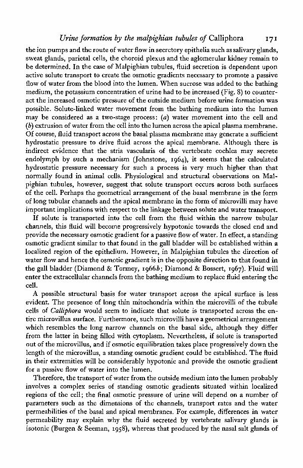
the ion pumps and the route of water flow in secretory epithelia such as salivary glands,sweat glands, parietal cells, the choroid plexus and the aglomerular kidney remain tobe determined. In the case of Malpighian tubules, fluid secretion is dependent uponactive solute transport to create the osmotic gradients necessary to promote a passiveflow of water from the blood into the lumen. When sucrose was added to the bathingmedium, the potassium concentration of urine had to be increased (Fig. 8) to counter-act the increased osmotic pressure of the outside medium before urine formation waspossible. Solute-linked water movement from the bathing medium into the lumenmay be considered as a two-stage process: (a) water movement into the cell and(b) extrusion of water from the cell into the lumen across the apical plasma membrane.Of course, fluid transport across the basal plasma membrane may generate a sufficienthydrostatic pressure to drive fluid across the apical membrane. Although there isindirect evidence that the stria vascularis of the vertebrate cochlea may secreteendolymph by such a mechanism (Johnstone, 1964), it seems that the calculatedhydrostatic pressure necessary for such a process is very much higher than thatnormally found in animal cells. Physiological and structural observations on Mal-pighian tubules, however, suggest that solute transport occurs across both surfacesof the cell. Perhaps the geometrical arrangement of the basal membrane in the formof long tubular channels and the apical membrane in the form of microvilli may haveimportant implications with respect to the linkage between solute and water transport.
If solute is transported into the cell from the fluid within the narrow tubularchannels, this fluid will become progressively hypotonic towards the closed end andprovide the necessary osmotic gradient for a passive flow of water. In effect, a standingosmotic gradient similar to that found in the gall bladder will be established within alocalized region of the epithelium. However, in Malpighian tubules the direction ofwater flow and hence the osmotic gradient is in the opposite direction to that found inthe gall bladder (Diamond & Tormey, 19666; Diamond & Bossert, 1967). Fluid willenter the extracellular channels from the bathing medium to replace fluid entering thecell.
A possible structural basis for water transport across the apical surface is lessevident. The presence of long thin mitochondria within the microvilli of the tubulecells of Calliphora would seem to indicate that solute is transported across the en-tire microvillus surface. Furthermore, such microvilli have a geometrical arrangementwhich resembles the long narrow channels on the basal side, although they differfrom the latter in being filled with cytoplasm. Nevertheless, if solute is transportedout of the microvillus, and if osmotic equilibration takes place progressively down thelength of the microvillus, a standing osmotic gradient could be established. The fluidin their extremities will be considerably hypotonic and provide the osmotic gradientfor a passive flow of water into the lumen.
Therefore, the transport of water from the outside medium into the lumen probablyinvolves a complex series of standing osmotic gradients situated within localizedregions of the cell; the final osmotic pressure of urine will depend on a number ofparameters such as the dimensions of the channels, transport rates and the waterpermeabilities of the basal and apical membranes. For example, differences in waterpermeability may explain why the fluid secreted by vertebrate salivary glands isisotonic (Burgen & Seeman, 1958), whereas that produced by the nasal salt glands of
172 M. J. BERRIDGE
marine birds is considerably hypertonic (Schmidt-Nielsen, i960). Although the intra-cellular osmotic pressure of the tubule cells of Calliphora was not determined, theosmotic pressure of the urine was slightly hypertonic to the bathing medium through-out a wide range of osmotic pressures of the latter. Similarly, Ramsay (1952) foundthat the urine secreted by the distal tubules of Rhodnius was also hypertonic. In thecase of Dixippus, however, Ramsay (1954) has pointed out that a slight hypotonicityof the urine apparently argues against the hypothesis outlined above. But the Mal-pighian tubules of Dixippus do show some degree of regional differentiation (Ramsay,1955 a, b), and hypotonicity of the urine may have resulted from solute reabsorptionin the proximal part of the tubules.
SUMMARY
1. Rate of urine formation is very sensitive to potassium concentration.2. Potassium is concentrated in the urine by a mechanism which is independent of
other monovalent cations. Rubidium, caesium and sodium are also capable of main-taining a flow of urine. At low external potassium concentrations, sodium stimulatespotassium secretion.
3. Rate of urine secretion is stimulated by low osmotic pressures; the osmoticpressure of urine was slightly hypertonic throughout the range of external osmoticpressure employed. Addition of sucrose depresses rate of urine secretion; the potas-sium concentration of the urine increased by 1 mM/1. for each 2 mM/1. of sucrose addedto the bathing medium.
4. Urine formation is insensitive to sulphanilamide, acetazolamide, ouabain and awide variation of pH.
5. These observations are discussed in relation to the hypothesis that potassiumsecretion takes place across both surfaces of the cell. The pump on the basal surfacemay be a coupled sodium-potassium pump, whereas that on the apical surface may beelectrogenic.
6. Microvilli at the apical surface or channels formed by a complex infolding of thebasal plasma membrane may represent structural devices by which standing osmoticgradients can be established during solute-linked water transport across the cells ofMalpighian tubules.
The author would like to thank Drs J. M. Diamond, J. L. Oschman and B. J. Wallfor valuable criticism of the manuscript. This study was supported by NIH grantHD 29-03 and NSF grant GB 4847 awarded to Prof. D. Bodenstein and by NIHgrant GM 09960 awarded to Prof. M. Locke.
REFERENCES
BERLINER, R. W. (1963). Effect of drugs on renal transport mechanisms. In Drugs and Membranes (edC. Adrian, M. Hogben), pp. 145-55. New York: Macmillan.
BERRIDGE, M. J. (1966a). The physiology of excretion in the cotton stainer, Dysdercus fasciatus,Signoret. IV. Hormonal control of excretion. J. exp. Biol. 44, 553-66.
BERRIDGE, M. J. (19666). Metabolic pathways of isolated Malpighian tubules of the blowfly functioningin an artificial medium. J. Insect. Pkysiol. iz, 1523-38.
BERRIDGE, M. J. (1967a). Ion and water transport across epithelia. (In preparation.)BERRIDGE, M. J. (19676). Urine formation by the Malpighian tubules of Calliphora. II. Anions. (In
preparation.)

Urine formation by the malpighian tubules of Calliphora 173BERRIDGE, M. J. & GUPTA, B. L. (1967). Fine-structural changes in relation to ion and water transport
in the rectal papillae of the blowfly, Calliphora. J. Cell Sci. 2, 89-112.BBICKER, N. S., BIBER, T. & USSING, H. H. (1963). Exposure of the isolated frog skin to high potassium
concentrations at the internal surface. I. Bioelectric phenomena and sodium transport. J. din.Invest. 42, 88-99.
BORGEN, A. S. V. & SEEMAN, P. (1958). The role of the salivary duct system in the formation of thesaliva. Can. J. Biochem. Physiol. 36, 119-43.
CURRAN, P. F. (i960). Na, Cl, and water transport by rat ileum in vitro. J. gen. Physiol. 43, 1137-48.CURRAN, P. F. (1965). Ion transport in intestine and its coupling to other transport processes. Fedn.
Proc. Fedn Am. Socs exp. Biol. 24, 993-9.DAVIS, L. E. & SCHMIDT-NIELSEN, B. (1967). Ultrastructure of the crocodile kidney (Crocodylus acutus)
with special reference to electrolyte and fluid transport. J. Morph. 121, 255-76.DIAMOND, J. M. (1962). The mechanism of solute transport by the gall-bladder. J. Physiol., Lond. 161,
474-502.DIAMOND, J. M. (1964). The mechanism of isotonic water transport. J. gen. Physiol. 48, 15-42.DIAMOND, J. M. (1965). The mechanism of isotonic water absorption and secretion. Symp. Soc. exp.
Biol. 19, 329-47-DIAMOND, J. M. & BOSSERT, W. H. (1967). Standing-gradient osmotic flow: a mechanism for coupling
of water and solute transport in epithelia. J. gen. Physiol. 50, 2061-83.DIAMOND, J. M. & TORMEY, J. McD. (1966a). Role of long extracellular channels in fluid transport
across epithelia. Nature, Lond. 210, 817—20.DIAMOND, J. M. & TORMEY, J. McD. (19666). Studies on the structural basis of water transport across
epithelial membranes. Fedn. Proc. Fedn Am. Socs exp. Biol. 25, 1458-63.DIETSCHY, J. M. (1966). Recent developments in solute and water transport across the gall bladder
epithelium. Gastroenterology 50, 692—707.FARQUHAR, M. G. & PALADE, G. E. (1966). Adenosine triphosphatase localization in amphibian epi-
dermis. J. Cell. Biol. 30, 359-79.FRAZIER, H. S. & LEAF, A. (1963). The electrical characteristics of active sodium transport in the toad
bladder. J. gen. Physiol. 46, 491-503.GLYNN, I. M. (1964). The action of cardiac glycosides on ion movements. Pharmac. Rev. 16, 381-407.HARVEY, W. R. & NEDERGAARD, S. (1964). Sodium-independent active transport of potassium in the
isolated midgut of the Cecropia silkworm. Proc. natn. Acad. Sci. U.S.A. 51, 757-65.HASKELL, J. A., CLEMONS, R. D. & HARVEY, W. R. (1965). Active transport by the Cecropia midgut. I.
Inhibitors, stimulants, and potassium-transport. J. cell. comp. Physiol. 65, 45-56.JOHNSTONE, B; M. (1964). Endolymph and endocochlear potentials. In Transcellular Membrane Poten-
tials and Ionic Fluxes, ed. F. M. Snell and W. K. Noell. New York: Gordon and Breach.KAYE, G. I., WHEELER, H. O., WHITLOCK, R. T. & LANE, N. (1966). Fluid transport in the rabbit
gallbladder. J. Cell Biol. 30, 237-68.KERKUT, G. A. & THOMAS, R. C. (1965). An electrogenic sodium pump in snail nerve cells. Comp.
Biochem. Physiol. 14, 167—83.KERNAN, R. P. (1962). The role of lactate in the active excretion of sodium by frog muscle. J. Physiol.,
Lond. 162, 129-37.KERNAN, R. P. (1967). Electrogenic potassium pump related to generation of endplate potentials in
muscles. Nature, Lond. 214, 725-6.PHILLIPS, J. E. (1965). Rectal absorption and renal function in insects. Trans. R. Soc. Can. 3, 237-54.POTTS, A. M. (1965). The effects of drugs upon the eye. In Physiological Pharmacology, vol. 11 (ed.
W. S. Root and F. G. Hofmann), pp. 329-97. New York: Academic Press.RAMSAY, J. A. (1952). The excretion of sodium and potassium by the Malpighian tubules of Rhodnius.
J. exp. Biol. 29, 110-26.RAMSAY, J. A. (1953). Active transport of potassium by the Malpighian tubules of insects. J. exp. Biol.
30, 358-69.RAMSAY, J. A. (1954). Active transport of water by the Malpighian tubules of the stick insect, Dixippus
morosus (Orthoptera, Phasmidae). J. exp. Biol. 31, 104-13.RAMSAY, J. A. (1955a). The excretory system of the stick insect, Dixippus morosus (Orthoptera, Phasmi-
dae). J. exp. Biol. 32, 183-99.RAMSAY, J. A. (19556). The excretion of sodium, potassium and water by the Malpighian tubules of the
stick insect, Dixippus morosus (Orthoptera, Phasmidae). J. exp. Biol. 32, 200-16.RAMSAY, J. A. (1956). Excretion by the Malpighian tubules of the stick insect, Dixippus morosus (Ortho-
ptera, Phasmidae): calcium, magnesium, chloride, phosphate and hydrogen ions. J. exp. Biol. 33,697-708.
RAMSAY, J. A. & BROWN, R. H. J. (1955). Simplified apparatus and procedure for freezing-pointdeterminations upon small volumes of fluid. J. Sci. Instrum. 32, 372-5.
REHM, W. S. (1964). Gastric potential and ion transports. In Transcellular Membrane Potentials andIonic Fluxes, ed. F. M. Snell and W. K. Noell. New York: Gordon and Breach.

174 M. J. BERRIDGE
ROBINSON, J. R. (1965). Water regulation in mammalian cells. Symp. Soc. exp. Biol. 19, 237-58.ROBINSON, J. R. (1966). Binding of potassium in cells; water transport: is there a case for active trans-
port? Fedn. Proc. Fedn Am. Socs exp. Biol. 25, 1108-11.SCHMIDT-NIELSEN, B. (1965). Comparative morphology and physiology of excretion. In Ideas in Modern
Biology, ed. J. A. Moore. New York: The Natural History Press.SCHMIDT-NIELSEN, K. (i960). The salt-secreting gland of marine birds. Circulation 21, 955-67.TREHERNE, J. E. (1961). The movement of sodium ions in the isolated abdominal nerve cord of the cock-
roach, Periplaneta americana. J. exp. Biol. 38, 629-36.USSING, H. H. (i960). The Alkali Metal Ions in Biology. Berlin: Springer-Verlag.