Embed Size (px)
Citation preview
UPTAKE AND DISTRIBUTION OF INHALATIONAL ANESTHETICS
Dr.J.Edward Johnson.M.D.(Anaes),D.C.H.
Asst.Professor,
Kanyakumari Govt. Medical College Hospital.
INTRODUCTION
The modern anesthetist expeditiously develops and then sustains anesthetic concentrations in the
central nervous system that are sufficient for surgery with agents and techniques that usually
permit rapid recovery from anesthesia. Understanding the factors that govern the relationship
between the delivered anesthetic and brain concentrations enhances the optimum conduct of
anesthesia.
I. UPTAKE OF INHALED ANESTHETICS.
A. Factors raising the alveolar concentration, assuming a constant inspired anesthetic
concentration, and no uptake by blood:
1. The inspired concentration (FI).
a. The rate of rise is directly proportional to the inspired concentration.
2. The alveolar ventilation (Valveolar)
a. The larger the minute alveolar ventilation (Valveolar), the more rapid the rise in alveolar
concentration (FA) .
b. Inspired gas is diluted by the FRC, so the larger the FRC, as a fraction of Valveolar, the slower the
alveolar rise in anesthetic concentration.
c. For ventilatory rates over 4 breaths, the ventilatory rate does not make any difference at the same
Valveolar.
3. The time constant
The time required for flow through a container to equal the capacity of the container.
Time constant = volume (capacity)/flow
Time Constant % washin/washout
1 63%
2 86%
3 95%
4 98%
For example;
If 10 liter box is initially filled with oxygen and 5 l/min of nitrogen flow into box then, the
TC is volume (capacity)/flow.
TC = 10 / 5 = 2 minutes.
So, the nitrogen concentration at end of 2 minutes is 63%.
The time constant for the lungs is FRC/Valveolar.

Uptake and Distribution Page 2
The time constant for the anesthesia circuit is circuit capacity/FGF.
4. The larger the FRC, the slower the washin of a new gas.
5. The rate of rise of the alveolar concentration (FA/FI ) is greatly slowed by anesthetic uptake by the
blood.
Factors that Increase or Decrease the Rate of Rise of FA/FI
Barash: Handbook of Clinical Anesthesia, 6th Edition
B. Factors determining uptake by blood.
1. Solubility in blood:
a. The blood/gas partition coefficient.
b. The relative capacity per unit volume of two solvents (e.g. gas and blood) to hold the
anesthetic gas.( ie. blood-gas partition coefficient of 1.4 means that each milliliter of blood
holds 1.4 times as much isoflurane as a milliliter of alveolar gas does)
c. The relative molar amount in equal volumes of blood and gas when the partial pressures
are equal.( “Equilibrium” means that no difference in partial pressure exists)
Gas Blood/Gas Brain/Bld. Muscle/bld Fat/Bld
Nitrous Oxide 0.47 1.1 1.2 2.3
Isoflurane 1.4 1.6 2.9 45
Enflurane 1.8 1.4 1.7 36
Halothane 2.5 1.9 3.4 51
Desflurane 0.45 1.3 2.0 27
Sevoflurane 0.65 1.7 3.1 48
Diethyl Ether 12 2.0 1.3 5
Methoxyflurane 15 1.4 1.6 38
References: Eger EI II, Uptake and Distribution in Miller
Increase Decrease Comment
Low blood solubility High blood solubility
As the blood solubility decreases,
the rate of rise in FA/FI increases.
Low cardiac output High cardiac output The lower the cardiac output, the
faster the rate of rise in FA/FI
High minute ventilation Low minute ventilation The higher the minute ventilation,
the faster the rate of rise in FA/FI
High pulmonary arterial to
venous partial venous partial
Low pulmonary arterial to
venous partial venous partial
"At the beginning of induction, PV
is zero but increases rapidly (thus
[PA-PV] falls rapidly) and FA/FI
increase rapidly. Later during
induction and maintenance, PV
rises more slowly so FA/FI rises
more slowly."

Uptake and Distribution Page 3
d. Other things equal, the more soluble the anesthetic, the more drug will be taken up by the
blood, and the slower the rise in alveolar concentration.
2. Cardiac Output:
a. The flow of blood through the lungs determines the amount of blood available to remove
anesthetic gas.
b. The greater the cardiac output, the slower the rise in alveolar concentration.
c. Mathematically, changes in cardiac output have exactly the same influence on anesthetic
uptake from the lungs as changes in solubility, since both influence exactly the same process:
the size of the storage capacity of the blood for anesthetic agent over a given time interval.
3. The mixed venous anesthetic concentration:
a. The higher the mixed venous concentration, the slower the anesthetic uptake.
b. Initially 0.
c. At equilibrium, the venous partial pressure = arterial partial pressure = alveolar partial
pressure (e.g. uptake = 0).
d. The uptake from the lung, in liters of gas/minute:
e. The rate of rise of the mixed venous concentration depends on the tissue uptake of the
anesthetic.
4. Tissue uptake of anesthetic:
a. The tissue uptake (including blood) equals the uptake from the lungs.
b. The same factors which govern uptake by the blood from the lungs govern uptake by the
tissues from the blood:
1. The tissue/blood partition coefficient (tissue solubility)
2. The tissue blood flow.
3. The tissue anesthetic concentration (analogous to the mixed venous tissue concentration).
c. Tissue uptake, in liters of gas/minute =
d. The rate of rise in tissue anesthetic concentration is proportional to tissue blood flow.
e. The rate of rise in tissue anesthetic concentration is inversely proportional to the tissue
capacity.
1. The tissue capacity is:tissue volume x tissue solubility
f. Just as discussed for the lungs, the tissues have a time constant:
1. solubility volume
time constant = flow
alveolar venousblood solubility cardiac output ( - )P Puptake from lung =
atmospheric pressure
arterial tissuetissue solubility tissue blood flow ( - )P Ptissue uptake =
atmospheric pressure

Uptake and Distribution Page 4
Thus, eventually fat governs the uptake of all anesthetics, until equilibrium is reached (at several
days)
g. The contribution of each tissue to the mixed venous partial pressure is the tissue anesthetic
partial pressure * the flow to that tissue.
h. The body can be roughly divided into 4 tissue groups, vessel rich group (brain, heart, lungs,
kidney, splanchnic bed, glands), the muscle group, the fat group, and the vessel poor group
(bones, cartilage, ligaments):
Group % MASS %CO
VRG 10 75
MG 50 18
FG 20 5.5
VPG 20 1.5
C. Unifying the above concepts:
1. Wash in of gas to the FRC occurs very fast (within 1 minute), so we will ignore it.
2. The alveolar/inspired partial pressure ratio approaches 1 with time.
3. VxF
U-1 =
F
F
alveolarinspired
Lung
inspired
alveolar
Where ULung = uptake from lung and
inspired alveolarF xV is the rate of drug delivery to the lungs,
while ULung = B x Q x ((PA-Pvenous)
Barometric pressure (Blood-gas solubility (λ), cardiac output (Q), and alveolar-to-venous partial pressure difference (PA - Pv))
- is the rate of drug leaving the lung.
D. Metabolism
a. About 10-20% of halothane is eliminated through metabolism.
i. May be related to halothane hepatotoxicity
The time constants
for the brain
(in Minutes)
The time constants
for the muscle
(in Minutes)
The time constants
for the fat
(in Hours)
Gas 1 TC 2 TC 3 TC 1 TC 2 TC 3 TC 1 TC 2 TC 3 TC
Nitrous Oxide 1.1 2.2 3.3 40 80 120 1.3 2.6 3.8
Isoflurane 1.6 3.2 4.8 97 193 290 25 50 75
Enflurane 1.4 2.8 4.2 57 113 170 20 40 60
Halothane 1.9 3.8 5.7 113 227 340 28 57 85
Desflurane 1.3 2.6 3.9 67 133 200 15 30 45
Sevoflurane 1.7 3.4 5.1 103 207 310 27 53 80
Diethyl Ether 2 4 6 43 87 130 3 6 8
Methoxyflurane 1.4 2.8 4.2 53 107 160 21 42 63

Uptake and Distribution Page 5
b. About 2.5% of enflurane is metabolized.
c. No significant metabolism of isoflurane, nitrous oxide, or desflurane.
d. Sevoflurane is broken down into Compound A by CO2 absorbants (baralyme and soda lime).
Compound A is nephrotoxic in large doses.
i. Most studies to date suggest that this is NOT a clinical issue with
sevoflurane, even at low flow.
II . Hydraulic Model (intuitive model)
A. The model consists of 5 upright cylinders.
1. The cylinders represent the inspired reservoir (mouth), the alveolar gas, vessel rich
group, the muscle group, and the fat group.
2. The arrangement of the cylinders is as follows :( Fig 1)
(Fig 1) Mapleson Hydraulic Model
3. The cross sectional surface of each cylinder corresponds to its capacity
(volume * tissue solubility)
4. The four peripheral cylinders are connected to the alveolar gas cylinder by
pipes at their bases. The diameter of the pipes correlates to the blood/gas
partition coefficient times the cardiac output to each group, (except for the
connection between the inspired reservoir (mouth), where the diameter of the
pipe represents alveolar ventilation.)

Uptake and Distribution Page 6
5. The height of the column of fluid in each cylinder corresponds to the partial
pressure of the anesthetic in that cylinder.
B. Mapleson’s analogy for inhaled anaesthetics of varying solubility:
1. The cross-sectional area of the vessels must be proportional, not only to the volume
of the tissues but also to the solubility of the anaesthetic concerned in the tissues.
2. If two anaesthetics of different solubility are to be represented, two versions of
model are needed.
3. For a low solubility anaesthetic (Fig 2)
All the compartments are small
The pipes are represented as small because the low solubility of anaesthetic
is less carried by given blood flow
To achieve equilibrium for least soluble of anaesthetic small quantity of
anaesthetic has to go in to the system
(Fig 2)
4. For a high solubility anaesthetic (Fig 3)
All the compartments are large
The pipes are represented as larger because the high solubility of anaesthetic is more carried
by given blood flow
To achieve equilibrium for high solubility of anaesthetic large quantity of anaesthetic has to
go in to the system through the same sized ventilation pipe and therefore it takes longer time.

Uptake and Distribution Page 7
(Fig 3)
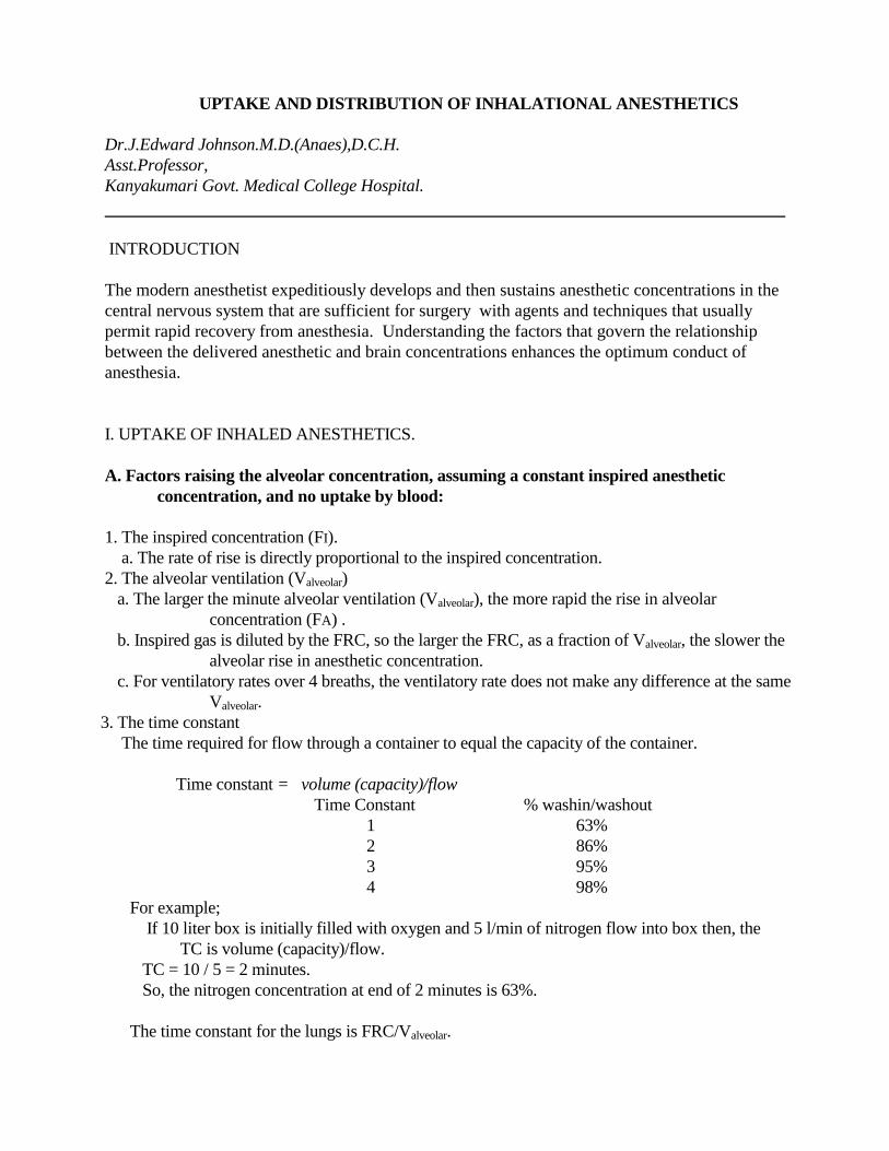
III. The concentration and second gas effects:
A. The concentration effect
1. The higher the inspired concentration, the more rapid the rise in alveolar concentration.
Explanations:
a. The concentrating effect: As gas is taken up by the blood, the remaining gas is present in
a smaller volume, which diminishes the change in partial pressure that might
otherwise be expected.
b. The ventilation effect: As gas is taken up, more gas is brought in to the lungs to replace
the lost volume, which both increases Valveolar and lowers the influence of uptake on
reducing Falveolar
2. Examples:
Example 1: We instantaneously fill a 4 liter box (the lungs) with 50% nitrous oxide. The
concentrating effect tells us that if half of the nitrous oxide is taken up (1 liter),
we will have 1 liter of nitrous in a 3 liter box, for a concentration of 33%, not
25% as might be expected. If we consider the ventilation effect: the 1 liter of
nitrous taken up will be replaced with another liter of 50% nitrous (the
inspired gas), which will add another .5 liters of nitrous to the box. This
results in 1.5 liters of nitrous in a 4 liter box, for a final concentration of 38%.
(Fig 4)
Uptake and Distribution Page 8
(Fig 4)
Example 2: We start with a 4 liter box the lungs filled with 100% nitrous oxide (don't try
this), connected to gas supply that is also 100% nitrous oxide. No matter how
much nitrous is taken up, the concentration cannot fall below 100%.
3. The concentration effect is only significant for gases present in high concentrations (e.g.
nitrous). It is negligible for the potent agents.
B. The second gas effect (applies to the effects of nitrous oxide on the uptake of another gas):
1. Analogous to the concentration effect, but relates instead to the use of a potent agent
concurrently with a second gas present in large quantity, usually nitrous oxide.
2. The higher the inspired concentration of the second gas (nitrous), the more rapid the rise in
alveolar concentration. Explanations:
a. The concentrating effect: As the second gas (nitrous) is taken up in significant volumes,
all remaining gases (including the potent agent, which is the "first" gas) are
concentrated in the remaining volume.
b. The ventilation effect: As the second gas is taken up in significant volumes, additional
fresh gas in brought into the lungs. This increases Valveolar, which increases the rate
of rise of anesthetic concentration.
3. Example:
We instantaneously fill a 4 liter box with 50% nitrous oxide and 40 cc (1%) of isoflurane.
The concentration effect tells us that if half of the nitrous oxide is taken up, the
concentration of isoflurane will increase from 1% to 1.33% (40cc / 3 liters). The
ventilation effect tells us that another liter of fresh gas, containing 500 mls of nitrous, and
10mls of isoflurane will be brought into the lungs, resulting in a concentration of 1.25%.
Although the ventilation effect has lowered the concentration of isoflurane slightly, it has
increased Valveolar, which will more than offset the change. (Fig 5)

Uptake and Distribution Page 9
(Fig 5)
IV. Changes in Ventilation and Circulation
A. Changes in Ventilation
1. Increasing ventilation increases the rate of rise of alveolar concentration.
2. The change is greatest for more soluble anesthetics:
A doubling of ventilation increases the methoxyflurane concentration at 10 minutes of
anesthetic administration by 75%, increases the isoflurane concentration by 18%, and
increases the desflurane concentration by only 6%. (Fig 6 )
(Fig 6 )

Uptake and Distribution Page 10
3. Anesthetics depress Valveolar, which slows the rate of rise of the alveolar concentration.
However, the concentration only stops rising towards the inspired concentration when
the patient stops breathing entirely.
a. This depression is sets an upper limit on the alveolar concentration which can be obtained
in a spontaneously breathing patient. For halothane, patients stop breathing entirely
when the concentration in the brain reaches 2.5%.
b. There is a delay caused by the time required for transport of the anesthetic from the lungs
to the brain, and so in the first few minutes, patients may breath spontaneously with
alveolar concentrations higher than 2.5%.
4. Hyperventilation reduces cerebral blood flow.
The effect on induction time is function of solubility:
a. For nitrous oxide, the reduction in cerebral blood flow more then compensates for the
increased rate of rise of the alveolar concentration with hyperventilation, so induction
time is actually slower in the hyperventilating patient (remember hyperventilation
didn't help that much in the first place).
b. For halothane, the reduction in cerebral blood flow almost exactly balances the effect of
the increased rate of rise of alveolar concentration.
c. For ether, the increased rate of rise of alveolar concentration more than compensates for
the reduction in cerebral blood flow, so induction is faster.
B. Changes in Cardiac Output
1. Increasing cardiac output lowers the alveolar anesthetic concentration, and slows
the rate of rise of alveolar anesthetic concentration.
2. The change is greatest for more soluble anesthetics (as was the influence of changes
in ventilation)
a. Nitrous rises quite fast, regardless of cardiac output.
b. The potent agents are highly affected by cardiac output, with halothane the
most affected, desflurane the least.
3. If the cardiac output is lowered, the effect depends on the distribution of cardiac
output.
a. If the cerebral circulation is less, then induction will be slower, even though
the alveolar concentration will rise more quickly.
b. If the cardiac output is reduced, but cerebral circulation is maintained (the
usual situation), then the alveolar concentration will rise more rapidly,
and this rapid rise will be quickly reflected in the brain anesthetic
concentration.
c. Thus, patients in hypovolemic shock will have very rapid rises in cerebral
anesthetic concentration.
d. Patients in septic shock may have fairly slow rises in cerebral anesthetic
concentration.
4. Anesthetics may reduce cardiac output, which will increase the rate of rise of
anesthetic concentration.

Uptake and Distribution Page 11
V. Recovery from an inhalational anesthetic
A. Overall, it is the reverse process of the anesthetic induction:
1. The rate of fall in alveolar concentration determines anesthetic recovery.
2. Increased solubility slows recovery
3. Increased cardiac output slows recovery
4. Increasing ventilation may help the recovery from potent agents, but with
hyperventilation, the increased rate of fall in alveolar concentration is nearly
balanced by the reduced cerebral blood flow.
5. Hyperventilation probably delays recovery from nitrous oxide, because there is
little improvement in pulmonary wash-out, while wash-out from the brain is
delayed from the reduced cerebral blood flow.
6. There is no concentration effect on emergence, as there was on induction,
because the gas in high concentrations (nitrous oxide) is not being drawn from
an infinite reservoir, as it was on induction.
B. Diffusion Hypoxia
1. Related to the large outpouring of nitrous oxide diluting the inspired oxygen at
the conclusion of a case.
2. Only a risk for the first 3-5 minutes after terminating the nitrous oxide.
3. Easily managed (and now almost never seen) with supplemental oxygen for a
few minutes following termination of the nitrous.
C. The low partial pressure of anesthetics in most peripheral tissues (especially fat)
means that during the initial recovery from anesthesia, anesthetic is continuing to pass
into these tissues, rather than being released from these tissues. Thus, recovery is
enhanced by redistribution of the inhalational anesthetic into fat and muscle, just like
it is for most intravenous anesthetics.
References:
1. Steven L. Shafer, M.D : Inhalational Anesthetics : Uptake and Distribution , July 24, 2007.
2. Read Eger's The Pharmacology of Inhaled Anesthetics.
3. Miller's Anesthesia, Seventh Edition.
4. Barash : Handbook of Clinical Anesthesia (6th Ed. 2009)
5. Anesthesia and Anesthesiology Teaching Site: http://www.anesthesia2000.com/
6. Edmond I Eger II, MD : Illustrations of Inhaled Anesthetic Uptake, Including Intertissue Diffusion to
and from Fat. Anesth Analg 2005;100:1020–33.
7. B.Korman and W.W.Mapleson: Concentration and second gas effects: Can the accepted
explanations be improved? British Journal of Anaesthesia 1997;78:618-625.

Uptake and Distribution Page 12