Embed Size (px)
Citation preview

Research ArticleUpregulation of Cardiac IL-10 and Downregulation of IFN-γ inBalb/c IL-4−/− in Acute Chagasic Myocarditis due to ColombianStrain of Trypanosoma cruzi
Marcos Vinicius da Silva ,1 Vera Lúcia de Almeida,2 Wendyson Duarte de Oliveira,2
Natália Carasek Matos Cascudo,2 Pollyana Guimarães de Oliveira,2
Crislaine Aparecida da Silva ,3 Aline Cristina Souza da Silva ,3
Maria Luíza Gonçalves dos Reis Monteiro ,3 Rosana Rosa Miranda Correa,3
Milton Adriano Pelli Oliveira ,2 Ruy de Sousa Lino-Júnior,2 Mara Rúbia Nunes Celes ,2
and Juliana Reis Machado 3
1Department of Parasitology, Institute of Natural and Biological Sciences, Federal University of Triângulo Mineiro, 38025-180 Uberaba, MG, Brazil2Institute of Tropical Pathology and Public Health of Federal University of Goiás, Federal University of Goiás,74605-050 Goiânia, Brazil3General Pathology, Institute of Natural and Biological Sciences, Federal University of Triângulo Mineiro, 38025-180 Uberaba,MG, Brazil
Correspondence should be addressed to Juliana Reis Machado; [email protected]
Received 15 July 2018; Accepted 5 November 2018; Published 28 November 2018
Academic Editor: Daniela Novick
Copyright © 2018Marcos Vinicius da Silva et al. This is an open access article distributed under the Creative Commons AttributionLicense, which permits unrestricted use, distribution, and reproduction in anymedium, provided the original work is properly cited.
Inflammatory response in Chagas disease is related to parasite and host factors. However, immune system regulation has not beenfully elucidated. Thus, this study is aimed at evaluating IL-4 influence on acute phase of Trypanosoma cruzi experimental infectionthrough dosage of cytokine levels in cardiac homogenate of infected Balb/c WT and Balb/c IL-4−/− as well as its histopathologicalrepercussions. For such purpose, mice were divided into two groups: an infected group with 100 forms of the Colombian strain andan uninfected group. After 21 days of infection, animals were euthanized and the blood, spleen, and heart were collected. The spleenwas used to culture splenic cells in 48 h. Subsequently, cytokines TNF-α, IL-12p70, IL-10, IFN-γ, and IL-17 were measured in theblood, culture supernatant, and heart apex by ELISA. The base of the heart was used for histopathological analysis. From theseanalysis, infected Balb/c IL-4−/− mice showed milder inflammatory infiltrate compared to Balb/c WT, but without changes innest density and collagen deposition. IL-4 absence culminated in lower cardiac tissue IFN-γ production, although it did notaffect TNF-α expression in situ. It also decreased TNF-α systemic production and increased IL-10, both systemically and in situ.In addition, IL-4 absence did not influence IL-17 expression. Splenocytes of IL-4-deficient mice produced higher amounts ofIFN-γ, TNF-α, and IL-17 and lower amounts of IL-10. Thus, IL-4 absence in acute phase of experimental infection with T. cruziColombian strain reduces myocarditis due to lower IFN-γ production and greater IL-10 production in situ and this pattern isnot influenced by splenocyte general repertoire.
1. Introduction
Chagas disease, also known as American trypanosomiasis, isa chronic systemic infectious disease caused by the protozoan
Trypanosoma cruzi (T. cruzi), an important public healthproblem in American continent. According to the WorldHealth Organization (WHO), the disease affects around 7.5millionpeopleworldwide, themajority inLatinAmerica [1, 2].
HindawiMediators of InflammationVolume 2018, Article ID 3421897, 9 pageshttps://doi.org/10.1155/2018/3421897
Murine experimental model reproduces human Chagas’disease infections, and genetic background has influencesboth, triggering a series of reactions that evolve from an acutesymptomatic phase to chronic phase [3]. Some variables inter-fere in Chagas’ disease pathogenicity such as the infecting strainand the mouse lineage. Balb/c strain mice are known to be sus-ceptible to T. cruzi infection, present a high parasitemia and thevast majority succumb early to acute infection [4]. Colombianstrain has been used in several studies with the purpose of eval-uating cardiac function due to its evident myotropism [5].Prominent myocardial lesions with intense inflammatory pro-cess, evolution in parasitemia with peak between 21 and 25days of postinfection, distinct myotropism with involvementof the skeletal musculature resulting in extensive lesions inthe skeletal muscle fibers, electrocardiographic alterations,and resistance to chemotherapeutics such as benznidazoleand nifurtimox are characteristics of this strain. [6, 7].
Knockout model is widely used in studies that evaluatedirect or indirect implication of certain cytokines in resistanceto T. cruzi infection. IL-10 KO mice infected with T. cruzi Ystrain had lower serum and tissue parasite load and higherIFN-gamma and nitric oxide production by spleen cells thanwild-typemice [8]. Furthermore, IL-10KOmice infectedwiththe Tulahuen strain, in addition to lower serum parasite load,also showed higher levels of serum TNF-α, IL-12, and IFN-gamma compared with infected IL-10+/+ mice [9]. Knockoutmodels for other molecules as B2-microglobulin [10], CD4orCD8 [11], gammadeltaTcells [12], andFas ligand [13]werealso used in studies on Chagas disease.
In acute phase, Chagas disease has variable clinical pre-sentations, from asymptomatic to symptomatic and evenfatal in some cases. Parasite proliferation and disseminationby blood or lymphatic vessels characterized this phase, witha parasitemia peak that regresses in some days and tends tobe undetectable in chronic phase. Acute phase disseminationaffects several cellular types, especially cardiac muscle fibers,with amastigotes nest formation [14], myocarditis, and sub-sequent collagen deposition between cardiac fibers, causingprogressive functional damage. Excessive immune systemactivation with proinflammatory cytokine production ofTh1-type IL-12, TNF-α, and IFN-γ characterizes this phase.On the other hand, cytokines such as IL-10 and TGF-β areactivated to modulate immune response with consequentreduction of tissue damage [15, 16].
IL-4 is described as an anti-inflammatory cytokine, pro-totypic of Th2 profile, and related to susceptibility to proto-zoa infection, such as cutaneous leishmaniasis [17, 18],although even in models of polarized clinical manifestations,its relation with Th1 response still raises major controversies[19]. IL-4 reduces IFN-γ production [20] controlling Th1response and preventing excessive tissue inflammation [21].IL-4 knockout animals tend to have better response to infec-tion by intracellular parasites, and when infected by T. cruzi,they increase Th1-type immune response, resulting indecreased parasitemia and mortality and increased cardiacinflammatory reaction, although these effects are observedin late stages of acute or in chronic phase [21, 22]. On thebasis of in vitro testing, two contrasting roles for IL-4 havebeen described: enhancement of intracellular T. cruzi
destruction by macrophages [23] and inhibition of IFN-γ-mediated trypanocidal activity [24].
Our group had already pointed out that infected andreinfected animals with Colombian strain present a modula-tion of immune response leading to higher production ofproinflammatory cytokines, such as IFN-γ and TNF-α,resulting in marked myocarditis and lower survival rate[25]. As Balb/c mice are usually good IL-4 producers [26]and due to IL-4 relevance in modulating response againstinfectious and parasitic agents, including chagasic cardiomy-opathy, IL-4−/− knockout animals were used to analyze if thiscytokine has any influence on immune response modulationin mice infected with T. cruzi Colombian strain, which hasmarked myotropism in acute phase. We demonstrate thatin early stages of acute phase, IL-4−/− knockout animals pre-sented milder myocarditis with lower IFN-γ production andhigher in situ IL-10 production, despite Th1 cells increase insplenocyte general repertoire. Our results open perspectivesof IL-4 role in chagasic myocarditis initial events in a morecomplex frame than the dichotomy Th1/Th2.
2. Material and Methods
2.1. Animals and Infection. We evaluated male animals ofBalb/c WT and Balb/c IL-4−/− lineages infected with T. cruziColombian strain (n = 8) and an uninfected group (n = 6),both aged 10 weeks and weighing 20-25 g. Infections wereperformed using trypomastigote forms obtained at peak ofparasitemia from previously infected Swiss mice (Waynforthand Flecknell, 1992). Briefly, Swiss mice were euthanized andperipheral blood was collected by cardiac puncture. Fivemicroliters of blood was used to quantify parasite numberin a hematocytometer, counted in fifty microscopic fields ata final magnification of ×400. Inoculum was adjusted to1000 trypomastigotes/ml. One hundred (100) trypomastigoteforms were subcutaneously inoculated. Animals were eutha-nized on the 21st day of infection for acute phase. Next, nec-ropsy was performed and the heart was collected through aventral incision in thoracic cavity. This research wasapproved by CEUA/UFG.
2.2. Histological Analysis. To evaluate inflammatory infiltrate,slides of cardiac tissue (ventricle) stained with hematoxylinand eosin (HE) were used. Qualitative analysis classifiedinfiltrate as predominantly mononuclear (macrophagesand lymphocytes) or polymorphonuclear (neutrophils andeosinophils), according to the cellular type observed in morethan 50% of the infiltrate. Semiquantitative analysis classi-fied the inflammatory infiltrate in mild (involvement< 25%of the tissue), moderate (25%–50% of the tissue), or severe(involvement> 50% of the tissue) [25].
Tissue parasitism was quantified under light microscopyby the number of myocardium T. cruzi nests using threeslides with serial slices. All infected animals had nests.
For collagen quantification, we used the heart sectionsstained with sirius red. Slides were analyzed in polarized lightmicroscope at a final magnification of ×400 with a semiauto-matic interactive image analyzer system, ImageJ® (NationalInstitutes of Health, Bethesda, EUA).
2 Mediators of Inflammation

2.3. Immunological Analysis
2.3.1. Cardiac Tissue Homogenate Preparation. The hearttissue sections were immersed in PBS solution containingcomplete protease inhibitor (Sigma, St. Louis, MO, USA)and Nonidet-P40. After that, they were submitted to tissuehomogenizer. The homogenate obtained was centrifuged at14,000×g for 10 minutes, and the supernatant was main-tained for quantification of cytokines and total proteins [25].
2.3.2. Culture of Splenocytes. Mouse splenocytes were col-lected, maintained in RPMI 1640 medium (GE Healthcare,Uppsala, Sweden) and macerated for cell individualization.These suspended cells were washed three times by centrifu-gation at 400×g for 15min at 8°C in RPMI 1640. Then, theywere counted in a Neubauer chamber and resuspended to2× 106 cells/ml in RPMI 1640 medium with addition of50mM Hepes (Gibco, Grand Island, NY, USA), 5% of inac-tivated fetal bovine serum (Gibco, USA), 2mM L-glutamine(Gibco, USA), 0.05mM 2β-mercaptoethanol (Gibco, USA),and 40μg/ml gentamicin (Neoqumica, Anápolis, GO, BR).Then, 2× 10⁶ cells were incubated without stimulus or with10μg/ml of concanavalin A in 24-well culture plates (BDPharmingen, San Diego, CA, USA). Cultures were kept ina moist incubator with 5% CO2 at 37°C for 48 hours.Supernatants were collected and maintained at −70°C untilanalysis [25].
2.3.3. TNF-α, IL-12p70, IL-10, IFN-γ, and IL-17Quantification. Cytokine quantification was performed oncardiac homogenate, spleen culture, and serum cells byELISA (enzyme-linked immunosorbent assay), using anti-body pairs from BD commercial kit (Biosciences, USA) andfollowing manufacturer’s instructions. Reaction was devel-oped using 3,3′,5,5′-tetramethylbenzidine (TMB) peroxidasesubstrate and read at 450nm. For cardiac homogenate,results were normalized to total protein concentration, deter-mined by Bradford assay (Bio-Rad, Hercules, CA, USA) ofeach heart and expressed as picogram of cytokine per gramof tissue (pg/g of tissue).
2.4. Statistical Analyses. The GraphPad Prism 6.0 software(GraphPad Software,USA)was used. Student’s t-testwas usedfor analysis between two groups with normal distribution.
Qualitative variables were expressed as percentageand associations were analyzed using the chi-square(χ2) test. Results were considered statistically significantwhen p < 0 05.
3. Results
3.1. Balb/c IL-4−/− Mice Infected with T. cruzi StrainPresented Less Intense Inflammatory Infiltrate in AcutePhase of Infection. Inflammatory cardiac infiltrate in Balb/cWT and Balb/c IL-4−/− mice infected with Colombian strainin acute phase of experimental Chagas disease was analyzed.IL-4 absence had an impact on inflammatory infiltratereduction, as Balb/c IL-4−/− had predominantly moderateinflammatory infiltrate (p = 0 04, chi-square test, Figure 1).Regarding quantification of heart fibrosis and amastigotes
nests, no difference was observed in group comparison(Figure 1).
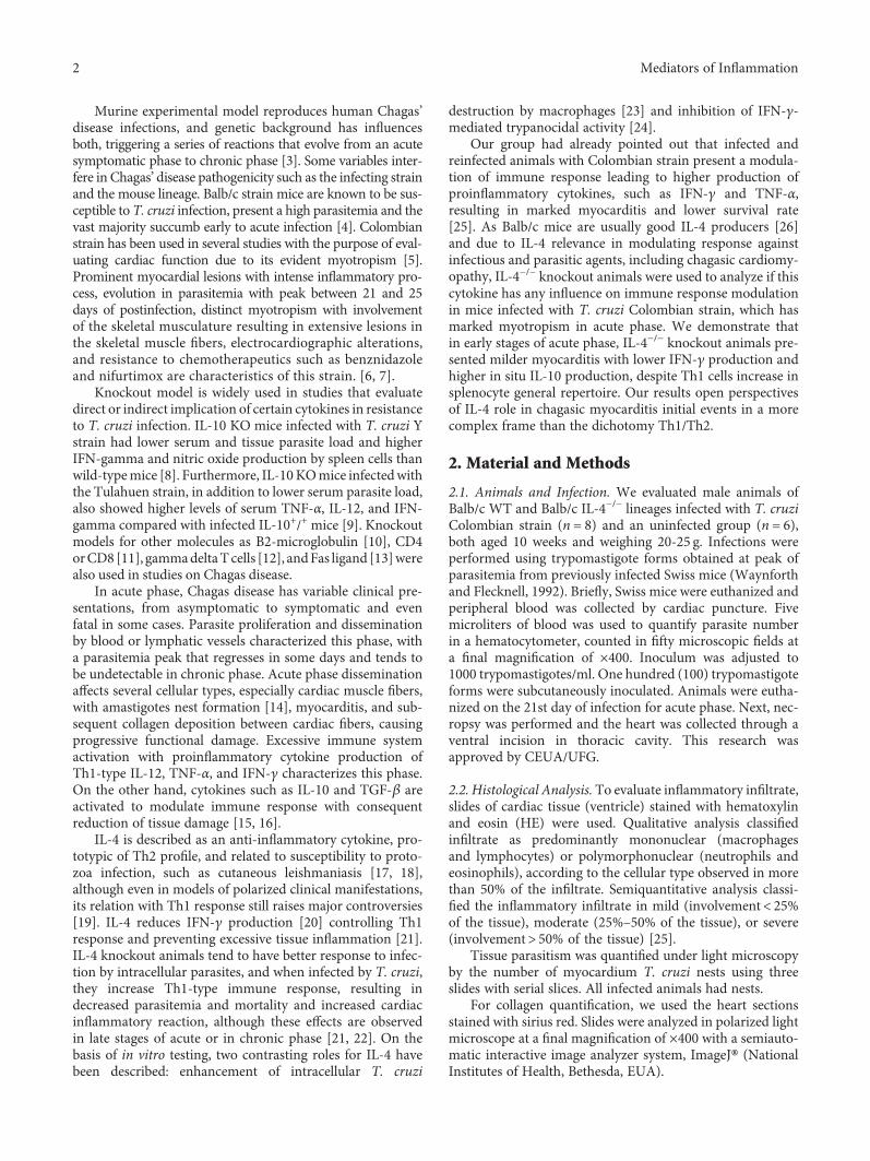
3.2. Absence of IL-4 Culminates in Lower IFN-γ CardiacTissue Production with Similar Expression of TNF-α. Onceinfection with T. cruzi Colombian strain leads to lowerinflammatory infiltrate in IL-4 knockout animals, we evalu-ated immune response quality in situ through cardiachomogenate and systemically through serum dosages. Asexpected, wild-type animal infection triggered increasedIFN-γ production in cardiac tissue (noninfected WT vs.infected WT, p = 0 02, t = 2 611). However, in IL-4 absence,IFN-γ expression was significantly reduced, either basal (NIWT vs. NI IL-4−/−, p = 0 009, t = 3 315) or after infectionwith Colombian strain (Inf WT vs. Inf IL-4−/−, p = 0 001,t = 4 43). No statistically significant difference was observedin IFN-γ serum levels (data not shown).
However, despite significant IFN-γ reduction in car-diac tissue, TNF-α expression was similar between WT andIL-4−/−, both in uninfected mice (NI WT vs. NI IL-4−/−,p > 0 17) and after infection (Inf WT vs. Inf IL-4−/−, p >0 5). In both groups of animals, infection with Colombianstrain induced a significant increase in TNF-α (NI WT vs.Inf WT, p = 0 02, t = 2 61 and NI IL-4−/− vs. Inf IL-4−/−,p = 0 002, t = 4 74). Interestingly, TNF-α systemic levelswere significantly elevated in WT-infected animals com-pared to IL-4−/− animals (p = 0 01, t = 3 08).
To investigate if nonproduction of IFN-γwas due to non-expression of Th1-inducing cytokines, we evaluated in situexpression of IL-12p70. In both groups, infection inducedsignificantly increased IL-12p70 expression in cardiac tissue(NI WT vs. Inf WT, p = 0 04, t = 2 32 and NI IL-4−/− vs. InfIL-4−/−, p = 0 04, t = 2 39), but with no difference betweenWT and IL-4−/−, in situ or systemically (p > 0 05, Figure 2).
3.3. Lower Inflammation and Lower IFN-γ Production inCardiac Tissue of IL-4−/− Animals Infected with T. cruziColombian Strain Are due to Increased IL-10 Production.Once we observed Balb/c mice with IL-4 absence andinfected with T. cruzi cardiotropic strain had a significantIFN-γ expression reduction in cardiac tissue, and that thisreduction was not due to a decrease in innate immunity cyto-kine production related to Th1 cell differentiation or func-tion, such as TNF-α and IL-12p70, we evaluated a possibledifferential production of IL-10, classically implicated inanti-inflammatory mechanisms.
Our results indicate that T. cruzi Colombian strain infec-tion induced significant IL-10 production, both in WT mice(NI WT vs. Inf WT, p = 0 007, t = 5 29) and Balb/c IL-4−/−
(NI IL-4−/− vs. Inf IL-4−/−, p = 0 001, t = 4 93). Furthermore,in IL-4 absence, significantly higher IL-10 levels wereobserved in cardiac tissue following infection (Inf WT vs.Inf IL-4−/−, p = 0 004, t = 3 42). A significant increase inserum IL-10 between WT and IL-4 animals−/− (p > 0 05)was not observed, although both had higher systemic levelsafter infection (NI WT vs. Inf WT, p = 0 002, t = 4 25 andNI IL-4−/− vs. Inf IL-4−/−, p = 0 001, t = 4 89) (Figure 3).
Finally, considering that IL-4 absence could imply alter-ations of other non-Th1 inflammatory profiles, we assessed
3Mediators of Inflammation

Th17 profile through IL-17 in situ expression. No statisticaldifferences were observed in IL-17 cytokine expressionbetween groups, which shows that nonactivation of Th2 pro-file by IL-4 absence did not lead to Th17 profile activation ina compensatory fashion (Figure 3).
3.4. Cytokine Pattern in Cardiac Tissue Is Not due toDifference in Splenocyte General Repertoire. To assess ifobserved differences in cardiac tissue cytokine expression ofIL-4−/− animals were due to a global difference in lymphocyterepertoire, we stimulated splenocytes with 10μg/ml of con-canavalin A for 48h and calculated cytokine production(expressed in fold increment related to nonstimulated sple-nocytes). We demonstrate here that IL-4−/− mouse spleno-cytes produce significantly increased amounts of IFN-γ(Inf WT vs. Inf IL-4−/−, p = 0 02, t = 2 79), TNF-α (Inf WTvs. Inf IL-4−/−, p = 0 009, t = 3 39), IL-17 (Inf WT vs. InfIL-4−/−, p = 0 006, t = 5 47), and lower IL-10 production
(Inf WT vs. Inf IL-4−/−, p = 0 007, t = 5 76) (Figure 4). Thisresult shows a clear difference between repertoire generatedby T. cruzi Colombian strain infection and cytokines expressedat the infection site, more clearly demonstrated by radar plots(Figure 5). These results were calculated using themean expres-sion of each cytokine in cardiac tissue homogenate or CON-A-stimulated splenocytes from Balb/c IL-4−/−-infected mice andexpressed as fold change relative to Balb/c WT-infected mice.
4. Discussion
This study is aimedat evaluating IL-4 role in immune responsemodulation of mice infected with T. cruzi Colombian strainin acute phase of infection. Our results point out the relation-ship between IL-4 and Th1 cells, classically described asantagonistic. In acute myocarditis triggered by cardiotropicstrain of T. cruzi, IL-4 absence implies a general polarizationfor Th1 in the spleen, but in cardiac tissue, inflammatory
WT Inf IL-4-/- Inf0.00
0.05
0.10
0.15
Am
astig
ote n
est/m
m2
(a)
Mild infiltrate
Moderate infiltrate
020406080
100
WT InfIL-4-/- Inf
p = 0.04
Mic
e (%
)
(b)
WT
NI
IL-4
-/-N
I
WT
Inf
IL-4
-/-In
f0
2
4
6
8
Colla
gen
(%)
(c)
(d) (e) (f)
(g) (h) (i)
Figure 1: Morphological analysis of the heart of Balb/c WT and Balb/c IL-4−/−mice infected and not infected with the Colombian strain of T.cruzi in the acute phase of experimental Chagas’ disease. (a) Density of amastigote nests in cardiac tissue of infected WT and IL-4−/− mice.Bars represent the mean, and vertical lines represent the standard error. (b) Intensity of cardiac inflammatory infiltrate in infected WTand IL-4−/− mice (p = 0 04, chi-square test). (c) Percentage of collagen fibers in cardiac tissue of infected and uninfected WT and IL-4−/−
mice. Bars represent the mean, and vertical lines represent the standard error. Histological sections of the WT and IL-4−/−-infected miceheart. (d, g) HE-stained amastigote nests. (e) Mild inflammatory infiltrate. (h) Moderated inflammatory infiltrate. (f, i) Collagen fibersstained red by sirius red.
4 Mediators of Inflammation

balance is significantly regulated by an increase in IL-10, trig-gering a lower inflammatory infiltrate.
The profile of cytokines released during T. cruzi infectionmay be associated with a protective or disease susceptibleprofile. Th1-type response is characterized by IL-2 andIFN-γ secretion, leading to activation of macrophagesand cell-mediated response, whereas in Th2 response, syn-thesis of IL-4, IL-5, and IL-10 is observed, culminating inrelevant humoral response. Balance between these cyto-kines is fundamental to determine which response has tobe developed [27, 28].
In our study, IL-4 absence at 21 days of infection did notdetermine a change in amastigotes nest density in cardiac tis-sue, using 100 forms of Colombian strain of T. cruzi, a refer-ence strain for chagasic myocarditis studies due to its highheart tropism in Balb/c mice [29, 30]. These results conflictwith other studies that demonstrated the potentiating IL-4effect on infection, since IL-4−/− mice would better controltissue parasitism [21, 31]. However, here and in both studies,nest density was small, even though several serial cuts wereevaluated. Another study using greater inoculum with Ystrain found no difference in parasite load as did our study[32]. Taken together, all these studies show in the early eventsof T. cruzi infection that intense myocarditis has no directrelation with the amount of amastigote nests that will persistin chronic phase.
In the present study, Balb/c IL-4−/−-infected mice hadmilder inflammatory infiltration compared to WT Balb/c.No significant differences were observed in intensity ofdiffuse and focal inflammation in cardiac tissue comparedIL-4 KO and WT animals [31]; however, this work evaluatedlate acute phase in 30 days of infection, where parasitemia isreduced and transition to chronic infection begins, while weevaluate the moment when Colombian strain presents itsparasitemia peak, that is, greater parasitic circulation [25].
Fibrosis is a fundamental substrate of Chagas’ heart dis-ease and progression to heart failure [33]. However, there isno consensus on the exact moment process takes place. Inface of a continuous inflammatory process, it is believed thatextracellular matrix increases collagen production evolvingto fibrosis, especially secondary to tissue damage. In thepresent study, difference in collagen deposition was notfound, possibly due to the time infection was evaluated,when myocarditis is prominent and fibrosis is not yet fullyinstalled. In chronic phase, it is well established that induc-tion of inflammatory response and death of cardiac fiberscontribute to continuous deposition of collagen and conse-quently fibrosis [1, 34].
IL-4 absence culminated in lower cardiac tissue IFN-γproduction; this fact justifies decrease of in situ infiltrate.However, IFN-γ expression is extremely relevant for immu-nity against intracellular pathogens, including T. cruzi [35].
WT NI IL-4-/- NI WT Inf IL-4-/- Inf0
200
400
600
800
Δ ΔCa
rdia
c IFN
-γ(p
g/g
of ti
ssue
)
⁎
(a)
WT NI IL-4-/- NI WT Inf IL-4-/- Inf0
50
100
150
200
250
Card
iac T
NF-𝛼
(pg/
g of
tiss
ue)
⁎
⁎
(b)
Δ
WT NI IL-4-/- NI WT Inf IL-4-/- Inf0
50
100
150
Seru
mTN
F-𝛼
(pg/
g of
tiss
ue)
(c)
WT NI IL-4-/- NI WT Inf IL-4-/- Inf0
100
200
300
400
Card
iac I
L-12
p70
(pg/
g of
tiss
ue)
⁎⁎
(d)
Figure 2: Expression of proinflammatory cytokines in cardiac tissue and serum of Balb/c WT and Balb/c IL-4−/− mice infected and notinfected with the Colombian strain of T. cruzi in the acute phase of experimental Chagas’ disease. (a) Tissue IFN-γ production (pg/g). (b)Tissue TNF-α production (pg/g). (c) Serum levels of TNF-α (pg/ml). (d) Tissue IL12p70 production (pg/g). Student’s t-test. Bars representthe mean, and vertical lines represent the standard error. ∗Significant differences between infected versus uninfected animals. ΔSignificantdifferences between the WT versus IL-4−/− group.
5Mediators of Inflammation

We believe systemic production of IFN-γ suppressed localdecrease of this cytokine, since its levels were well expressedin splenocytes culture in the IL-4−/− group. However,increased IFN-γ and nitric oxide production was demon-strated in mice infected with T. cruzi Tulahuen strain withIL-4 suppression, showing that IL-4 absence is related to agreater proinflammatory state activation in T. cruzi infec-tion [36].
IL-4 absence did not alter TNF-α in situ production,despite IFN-γ local reduction. TNF-α is especially synthe-sized by macrophages, T lymphocytes, and NK cells, witha great diversity of functions including recruitment and acti-vation of macrophages, which stimulate nitric oxide produc-tion and intracellular destruction of protozoan, activelyparticipating in the initial proinflammatory response of thedisease [37, 38]. Other studies have shown that increasedTNF-α is also associated with increased cardiac damage[39, 40], although treatment with TNF-α inhibitors, suchas etanercept, in chronic phase, aggravates chagasic myocar-ditis [41], demonstrating that its presence in cardiac tissuehas more complex repercussions than conceptual simplifica-tion increase is equal to damage. It is important to empha-size that splenocyte production capacity was positivelyimpacted by IL-4 absence, suggesting cardiac tissue-specificcontrol mechanisms.
WT NI IL-4-/- NI WT Inf IL-4-/- Inf0
100
200
300
Card
iac I
L-10
(p
g/g
of ti
ssue
)
Δ
⁎
⁎
(a)
WT NI IL-4-/- NI WT Inf IL-4-/- Inf0
500
1000
1500
2000
Seru
m IL
-10
(pg/
g of
tiss
ue) ⁎
⁎
(b)
WT NI IL-4-/- NI WT Inf IL-4-/- Inf0
5
10
15
20Ca
rdia
c IL-
17
(pg/
g of
tiss
ue)
(c)
Figure 3: Cytokine expression with regulatory profile in cardiac tissue and serum of Balb/c WT and Balb/c IL-4−/− mice infected and notinfected with the T. cruzi Colombian strain in the acute phase of experimental Chagas’ disease. (a) Production of IL-10 in cardiac tissue(pg/g) and (b) serum levels of IL-10 (pg/ml). (c) Production of IL-17 in cardiac tissue (pg/g). Student’s t-test. Bars represent the mean,and vertical lines represent the standard error. ∗Significant differences between infected versus uninfected animals. ΔSignificant differencesbetween the WT versus IL-4−/− group.
1 10 100Fold increase (CON-A/medium)
B alb IL-4-/-B alb WT
IFN-γ
TNF-𝛼
IL12p70
IL-10
IL-17
⁎
⁎
⁎
⁎
Figure 4: Cytokine production in splenocytes of WT Balb/c andIL-4−/− Balb/c mice infected with Colombian strain of T. cruzistimulated with 10μg/ml of concanavalin A in the acute phase ofexperimental Chagas’ disease. Fold change between nonstimulatedversus CON-A stimulated splenocytes. Student’s t-test. Barsrepresent the mean, and vertical lines represent the standard error.∗Significant differences between infected versus uninfected animals.
6 Mediators of Inflammation

Among infected animals, there was no difference in IL-12p70 cytokine expression in IL-4 absence. It is known thatthis cytokine is a fundamental mediator of innate immuneresponse, secreted by mononuclear phagocytes and dendriticcells, and important in stimulating IFN-γ production by NKcells and T lymphocytes [42, 43]. It can be inferred that, inthis study, at the beginning of infectious process, the abilityto produce IL-12p70 contributes to a Th1 response profileinfluenced by pathogen itself.
In fact, animals in the present study had no histologicalevidence of cardiac involvement. We believe this is due toimmune response control represented in our study by in situincrease in IL-10 and decrease in IFN-γ. Our data also pointout that immune response local regulation associated with agood repertoire of systemic Th1 immune response may havebeen sufficient to maintain the effectiveness of the responseto T. cruzi infection. These data become more consistent,since we observed increased serum and in situ IL-10 withreduced systemic repertoire production, as demonstrated instimulated culture.
IL-4 absence did not influence IL-17 expression in situ;however, the repertoire in the ex vivo culture increased. IL-17 plays an important role in resolution of T. cruzi protozoaninfection, and this cytokine is associated with protective andnonpathogenic responses [44–46]. In chagasic patients withheart disease, greater serum IL-17 levels were related toimprovement of organ function; therefore, it has protectiveeffect [47]. A study in IL-17−/− animals demonstrated thatthis cytokine actively participates in inflammatory responsein initial phase of disease and its absence during T. cruziinfection results in a reduction in recruitment of defensecells, which favors parasitemia [48]. However, researchesare still recent and limited on true role of this cytokine inChagas’ disease.
The relationship between IL-4 functions and Th1 cell dif-ferentiation is classically described as antagonistic and widelyreported in several models of T lymphocyte differentiation[49–52]. However, we demonstrated in acute myocarditistriggered by T. cruzi cardiotropic strain that IL-4 absenceimplies a repertorial polarization for Th1, but in cardiactissue, inflammatory balance is strongly regulated by anincrease in IL-10, triggering a lower inflammatory infiltrate.Our results open perspectives of IL-4 role in initial eventsof chagasic myocarditis in a more complex frame than thedichotomy Th1/Th2.
Abbreviations
CON-A: Concanavalin AELISA: Enzyme-linked immunosorbent assayIFN-γ: Interferon gammaIL-4: Interleukin 4IL-10: Interleukin 10IL-17: Interleukin 17IL-12p70: Interleukin 12p70KO: Knockout for the geneT. cruzi: Trypanosoma cruziTNF-α: Tumor necrosis factor-alphaTMB: 3,3′,5,5′-TetramethylbenzidineWT: Wild type.
Data Availability
The datasets generated during and/or analyzed during thecurrent study are available from the corresponding authoron reasonable request.
0.5
1
2
4IFN-γ
TNF-𝛼
IL-12p70
Cardiac tissue
IL-10
IL-17
Balb WTBalb IL-4-/-
(a)
0
1
2
3
4
IL-12p70IL-10
IL-17
Balb WTBalb IL-4-/-
IFN-γ
TNF-𝛼
Splenocytes
(b)
Figure 5: Radar plot representation of cytokine profile in cardiac tissue and splenocytes. The lines highlight the fold change in cytokineproduction in IL-4−/− Balb/c (gray line) in relation to WT Balb/c mice (black line). Data were obtained by calculating the ratio betweenthe mean concentrations of each cytokine in the IL-4−/− Balb/c-infected group and WT Balb/c-infected mice.
7Mediators of Inflammation

Conflicts of Interest
The authors declare that they have no conflicts of interest.
Authors’ Contributions
Marcos Vinicius da Silva and Vera Lúcia de Almeida equallycontributed to this paper.
References
[1] J. A. Marin-Neto, É. Cunha-Neto, B. C. Maciel, and M. V.Simões, “Pathogenesis of chronic Chagas heart disease,” Circu-lation, vol. 115, no. 9, pp. 1109–1123, 2007.
[2] World Health Organization (WHO), Preventing Mother-to-Child Transmission of Chagas disease: from Control to Elimina-tion, WHO: Neglected tropical disease, 2018.
[3] B. A. Burleigh and N. W. Andrews, “The mechanisms of Try-panosoma cruzi invasion of mammalian cells,” Annual Reviewof Microbiology, vol. 49, no. 1, pp. 175–200, 1995.
[4] T. C. de Araújo-Jorge and S. L. de Castro, Doença de Chagas:Manual Para Experimentação Animal, Editora FIOCRUZ,Rio de Janeiro, 2000.
[5] E. E. Federici, W. H. Abelmann, and F. A. Neva, “Chronic andprogressive myocarditis and myositis in C3H mice infectedwith Trypanosoma cruzi,” The American Journal of TropicalMedicine and Hygiene, vol. 13, no. 2, pp. 272–280, 1964.
[6] S. G. Andrade and J. B. Magalhães, “Biodemes and zymodemesof Trypanosoma cruzi strains: correlations with clinical dataand experimental pathology,” Revista da Sociedade Brasileirade Medicina Tropical, vol. 30, no. 1, pp. 27–35, 1997.
[7] S. G. Andrade, A. R. Pimentel, M. M. de Souza, and Z. A.Andrade, “Interstitial dendritic cells of the heart harbor Try-panosoma cruzi antigens in experimentally infected dogs:importance for the pathogenesis of chagasic myocarditis,” TheAmerican Journal of Tropical Medicine and Hygiene, vol. 63,no. 1, pp. 64–70, 2000.
[8] I. A. Abrahamsohn and R. L. Coffman, “Trypanosoma cruzi:IL-10, TNF, IFN-γ, and IL-12 regulate innate and acquiredimmunity to infection,” Experimental Parasitology, vol. 84,no. 2, pp. 231–244, 1996.
[9] C. A. Hunter, L. A. Ellis-Neyes, T. Slifer et al., “IL-10 isrequired to prevent immune hyperactivity during infectionwith Trypanosoma cruzi,” The Journal of Immunology,vol. 158, pp. 3311–3316, 1997.
[10] R. L. Tarleton, B. H. Koller, A. Latour, andM. Postan, “Suscep-tibility of β2-microglobulin-deficient mice to Trypanosomacruzi infection,” Nature, vol. 356, no. 6367, pp. 338–340, 1992.
[11] M. E. Rottenberg, M. Bakhiet, T. Olsson et al., “Differentialsusceptibilities of mice genomically deleted of CD4 andCD8 to infections with Trypanosoma cruzi or Trypanosomabrucei,” Infection and Immunity, vol. 61, no. 12, pp. 5129–5133, 1993.
[12] E. C. Santos Lima and P. Minoprio, “Chagas’ disease is atten-uated in mice lacking γδ T cells,” Infection and Immunity,vol. 64, no. 1, pp. 215–221, 1996.
[13] M. F. Lopes, M. P. Nunes, A. Henriques-Pons et al.,“Increased susceptibility of Fas ligand-deficient gld mice toTrypanosoma cruzi infection due to a Th2-biased hostimmune response,” European Journal of Immunology, vol. 29,no. 1, pp. 81–89, 1999.
[14] M. d. L. Higuchi, “Chronic chagasic cardiopathy: the productof a turbulent host-parasite relationship,” Revista do Institutode Medicina Tropical de São Paulo, vol. 39, no. 1, pp. 53–60,1997.
[15] J. S. Silva, D. R. Twardzik, and S. G. Reed, “Regulation of Try-panosoma cruzi infections in vitro and in vivo by transforminggrowth factor β (TGF-β),” The Journal of Experimental Medi-cine, vol. 174, no. 3, pp. 539–545, 1991.
[16] W. Savino, D. M. S. Villa-Verde, D. A. Mendes-da-Cruz et al.,“Cytokines and cell adhesion receptors in the regulation ofimmunity to Trypanosoma cruzi,” Cytokine & Growth FactorReviews, vol. 18, no. 1-2, pp. 107–124, 2007.
[17] R. Hurdayal and F. Brombacher, “The role of IL-4 and IL-13 incutaneous leishmaniasis,” Immunology Letters, vol. 161, no. 2,pp. 179–183, 2014.
[18] J. Alexander and K. Bryson, “T helper (h)1/Th2 and Leish-mania: paradox rather than paradigm,” Immunology Letters,vol. 99, no. 1, pp. 17–23, 2005.
[19] J. Alexander and F. Brombacher, “T helper1/T helper2 cellsand resistance/susceptibility to Leishmania infection: is thisparadigm still relevant?,” Frontiers in Immunology, vol. 3,p. 80, 2012.
[20] O. M. Martinez, R. S. Gibbons, M. R. Garovoy, and F. R.Aronson, “IL-4 inhibits IL-2 receptor expression and IL-2-dependent proliferation of human T cells,” The Journal ofImmunology, vol. 144, pp. 2211–2215, 1990.
[21] M. B. P. Soares, K. N. Silva-Mota, R. S. Lima, M. C. Bellintani,L. Pontes-de-Carvalho, and R. Ribeiro-dos-Santos, “Modula-tion of chagasic cardiomyopathy by interleukin-4: dissociationbetween inflammation and tissue parasitism,” The AmericanJournal of Pathology, vol. 159, no. 2, pp. 703–709, 2001.
[22] C. Hölscher, G. Köhler, U. Müller, H. Mossmann, G. A.Schaub, and F. Brombacher, “Defective nitric oxide effectorfunctions lead to extreme susceptibility of Trypanosomacruzi-infected mice deficient in gamma interferon receptor orinducible nitric oxide synthase,” Infection and Immunity,vol. 66, no. 3, pp. 1208–1215, 1998.
[23] J. J. Wirth, F. Kierszenbaum, and A. Zlotnik, “Effects of IL-4on macrophage functions: increased uptake and killing ofa protozoan parasite (Trypanosoma cruzi),” Immunology,vol. 66, no. 2, pp. 296–301, 1989.
[24] J. M. Golden and R. L. Tarleton, “Trypanosoma cruzi: cytokineeffects on macrophage trypanocidal activity,” ExperimentalParasitology, vol. 72, no. 4, pp. 391–402, 1991.
[25] J. Reis Machado, M. V. Silva, D. C. Borges et al., “Immuno-pathological aspects of experimental Trypanosoma cruzi rein-fections,” BioMed Research International, vol. 2014, ArticleID 648715, 9 pages, 2014.
[26] S. R. Paludan, “Interleukin-4 and interferon-γ: the quintes-sence of a mutual antagonistic relationship,” ScandinavianJournal of Immunology, vol. 48, no. 5, pp. 459–468, 1998.
[27] M.-J. Pinazo, M. C. Thomas, J. Bustamante, I. C. Almeida,M. C. Lopez, and J. Gascon, “Biomarkers of therapeuticresponses in chronic Chagas disease: state of the art and futureperspectives,” Memórias do Instituto Oswaldo Cruz, vol. 110,no. 3, pp. 422–432, 2015.
[28] W. O. Dutra, C. A. S. Menezes, L. M. D. Magalhães, and K. J.Gollob, “Immunoregulatory networks in human Chagas dis-ease,” Parasite Immunology, vol. 36, no. 8, pp. 377–387, 2014.
[29] E. Camandaroba, T. S. Thé, D. H. Pessina, and S. G. Andrade,“Trypanosoma cruzi: clones isolated from the Colombian
8 Mediators of Inflammation

strain, reproduce the parental strain characteristics, with ubiq-uitous histotropism,” International Journal of ExperimentalPathology, vol. 87, no. 3, pp. 209–217, 2006.
[30] L. O. Andrade, C. R. S. Machado, E. Chiari, S. D. J. Pena, andA. M. Macedo, “Trypanosoma cruzi: role of host genetic back-ground in the differential tissue distribution of parasite clonalpopulations,” Experimental Parasitology, vol. 100, no. 4,pp. 269–275, 2002.
[31] V. Michailowsky, N. M. Silva, C. D. Rocha, L. Q. Vieira,J. Lannes-Vieira, and R. T. Gazzinelli, “Pivotal role ofinterleukin-12 and interferon-γ axis in controlling tissue para-sitism and inflammation in the heart and central nervous sys-tem during Trypanosoma cruzi infection,” The AmericanJournal of Pathology, vol. 159, no. 5, pp. 1723–1733, 2001.
[32] I. A. Abrahamsohn, A. P. G. da Silva, and R. L. Coffman,“Effects of interleukin-4 deprivation and treatment onresistance to Trypanosoma cruzi,” Infection and Immunity,vol. 68, no. 4, pp. 1975–1979, 2000.
[33] E. R. Lopes, E. Chapadeiro, W. L. Tafuri, A. O. Almeida, andD. Abraão, “Peso do coração e tipo de morte no chagásico,”Revista do Instituto de Medicina Tropical de São Paulo,vol. 12, pp. 293–297, 1970.
[34] J. A. Torreão, B. M. Ianni, C. Mady et al., “Myocardial tissuecharacterization in Chagas’ heart disease by cardiovascularmagnetic resonance,” Journal of Cardiovascular MagneticResonance, vol. 17, no. 1, p. 97, 2015.
[35] M. d. L. Higuchi, T. de Brito, M. Martins Reis et al., “Correla-tion between Trypanosoma cruzi parasitism and myocardialinflammatory infiltrate in human chronic chagasic myocardi-tis: light microscopy and immunohistochemical findings,”Cardiovascular Pathology, vol. 2, no. 2, pp. 101–106, 1993.
[36] K. Hiyama, S. Hamano, T. Nakamura, K. Nomoto, and I. Tada,“IL-4 reduces resistance of mice to Trypanosoma cruzi infec-tion,” Parasitology Research, vol. 87, no. 4, pp. 269–274, 2001.
[37] J. A. Langermans, M. E. van der Hulst, P. H. Nibbering, andR. van Furth, “Endogenous tumor necrosis factor alpha isrequired for enhanced antimicrobial activity against Toxo-plasma gondii and Listeria monocytogenes in recombinantgamma interferon-treated mice,” Infection and Immunity,vol. 60, no. 12, pp. 5107–5112, 1992.
[38] A. A. Rodrigues, A. F. O. Notário, T. L. Teixeira et al., “A highthroughput analysis of cytokines and chemokines expressionduring the course of Trypanosoma cruzi experimental oralinfection,” Acta Tropica, vol. 157, pp. 42–53, 2016.
[39] A. Haensel, P. J. Mills, R. A. Nelesen, M. G. Ziegler, and J. E.Dimsdale, “The relationship between heart rate variabilityand inflammatory markers in cardiovascular diseases,” Psy-choneuroendocrinology, vol. 33, no. 10, pp. 1305–1312, 2008.
[40] E. Cunha-Neto, L. G. Nogueira, P. C. Teixeira et al., “Immuno-logical and non-immunological effects of cytokines andchemokines in the pathogenesis of chronic Chagas disease car-diomyopathy,” Memórias do Instituto Oswaldo Cruz, vol. 104,Suppl 1, pp. 252–258, 2009.
[41] A. M. B. Bilate, V. M. Salemi, F. J. Ramires et al., “TNFblockade aggravates experimental chronic Chagas diseasecardiomyopathy,” Microbes and Infection, vol. 9, no. 9,pp. 1104–1113, 2007.
[42] L. Rogge, L. Barberis-Maino, M. Biffi et al., “Selective expres-sion of an interleukin-12 receptor component by human Thelper 1 cells,” The Journal of Experimental Medicine,vol. 185, no. 5, pp. 825–832, 1997.
[43] M. S. Cardoso, J. L. Reis-Cunha, and D. C. Bartholomeu, “Eva-sion of the immune response by Trypanosoma cruzi duringacute infection,” Frontiers in Immunology, vol. 6, p. 659, 2015.
[44] Y. Miyazaki, S. Hamano, S. Wang, Y. Shimanoe, Y. Iwakura,and H. Yoshida, “IL-17 is necessary for host protection againstacute-phase Trypanosoma cruzi infection,” The Journal ofImmunology, vol. 185, no. 2, pp. 1150–1157, 2010.
[45] P. M. da Matta Guedes, F. R. S. Gutierrez, F. L. Maia et al.,“IL-17 produced during Trypanosoma cruzi infection playsa central role in regulating parasite-induced myocarditis,”PLoS Neglected Tropical Diseases, vol. 4, no. 2, articlee604, 2010.
[46] D. A. Bermejo, S. W. Jackson, M. Gorosito-Serran et al., “Try-panosoma cruzi trans-sialidase initiates a program indepen-dent of the transcription factors RORγt and Ahr that leads toIL-17 production by activated B cells,” Nature Immunology,vol. 14, no. 5, pp. 514–522, 2013.
[47] G. R. Sousa, J. A. S. Gomes, M. P. S. Damasio et al., “The role ofinterleukin 17-mediated immune response in Chagas disease:high level is correlated with better left ventricular function,”PLoS One, vol. 12, no. 3, article e0172833, 2017.
[48] J. Tosello Boari, M. C. Amezcua Vesely, D. A. Bermejo et al.,“IL-17RA signaling reduces inflammation and mortality dur-ing Trypanosoma cruzi infection by recruiting suppressiveIL-10-producing neutrophils,” PLoS Pathogens, vol. 8, no. 4,article e1002658, 2012.
[49] C. A. Lazarski, J. Ford, S. D. Katzman, A. F. Rosenberg, andD. J. Fowell, “IL-4 attenuates Th1-associated chemokineexpression and Th1 trafficking to inflamed tissues and limitspathogen clearance,” PLoS One, vol. 8, no. 8, article e71949,2013.
[50] M. López-Bravo, M.Minguito de la Escalera, P. M. Domínguezet al., “IL-4 blocks TH1-polarizing/inflammatory cytokinegene expression during monocyte-derived dendritic cell differ-entiation through histone hypoacetylation,” The Journal ofAllergy and Clinical Immunology, vol. 132, no. 6, pp. 1409–1419.e13, 2013.
[51] T. R. Mosmann, H. Cherwinski, M. W. Bond, M. A. Giedlin,and R. L. Coffman, “Two types of murine helper T cell clone.I. Definition according to profiles of lymphokine activitiesand secreted proteins,” The Journal of Immunology, vol. 136,pp. 2348–2357, 1986.
[52] E. A. Kurt-Jones, S. Hamberg, J. Ohara, W. E. Paul, and A. K.Abbas, “Heterogeneity of helper/inducer T lymphocytes. I.Lymphokine production and lymphokine responsiveness,”The Journal of Experimental Medicine, vol. 166, no. 6,pp. 1774–1787, 1987.
9Mediators of Inflammation

Stem Cells International
Hindawiwww.hindawi.com Volume 2018
Hindawiwww.hindawi.com Volume 2018
MEDIATORSINFLAMMATION
of
EndocrinologyInternational Journal of
Hindawiwww.hindawi.com Volume 2018
Hindawiwww.hindawi.com Volume 2018
Disease Markers
Hindawiwww.hindawi.com Volume 2018
BioMed Research International
OncologyJournal of
Hindawiwww.hindawi.com Volume 2013
Hindawiwww.hindawi.com Volume 2018
Oxidative Medicine and Cellular Longevity
Hindawiwww.hindawi.com Volume 2018
PPAR Research
Hindawi Publishing Corporation http://www.hindawi.com Volume 2013Hindawiwww.hindawi.com
The Scientific World Journal
Volume 2018
Immunology ResearchHindawiwww.hindawi.com Volume 2018
Journal of
ObesityJournal of
Hindawiwww.hindawi.com Volume 2018
Hindawiwww.hindawi.com Volume 2018
Computational and Mathematical Methods in Medicine
Hindawiwww.hindawi.com Volume 2018
Behavioural Neurology
OphthalmologyJournal of
Hindawiwww.hindawi.com Volume 2018
Diabetes ResearchJournal of
Hindawiwww.hindawi.com Volume 2018
Hindawiwww.hindawi.com Volume 2018
Research and TreatmentAIDS
Hindawiwww.hindawi.com Volume 2018
Gastroenterology Research and Practice
Hindawiwww.hindawi.com Volume 2018
Parkinson’s Disease
Evidence-Based Complementary andAlternative Medicine
Volume 2018Hindawiwww.hindawi.com
Submit your manuscripts atwww.hindawi.com