Embed Size (px)
Citation preview

Upper limbLecture one
Dr Amal Albtoosh

LECTURE
NUMBER
TOPIC
1 Introduction, Bones of the shoulder and arm, forearm, hand
2 Fascial planes and muscles
3 brachial Plexus

What is the limb?• Limb: a leg or arm of a human being
The upper limb is a
multijointed lever
that is freely
movable on the trunk
at the shoulder joint.
At the
distal end of the
upper limb is the
important organ,
the hand.
PROXIMAL
DISTAL

Scapula:
flat bone
inverted triangle
3 borders
Superior border
medial border
lateral border
3 angles
superiomedial angle
Superiolateral angle
Inferior angle
3 Fossae
supraspinous fossa
infraspinous fossa
Subscapular fossa
3 processes
spine
Acromion
coracoid
2 surfaces
anterior
posterior

The superior border has a
Suprascapular notch
the lateral border has the
glenoid cavity
above the cavity - supra-
glenoid tubercle
below it infra-glenoid tubercle


Clavicle
The clavicle is the only long bone that lies in a horizontal
position in the body
The clavicle has three regions:
the medial end: sternal end of the clavicle
the lateral end: acromial end of the clavicleIt also
serves as an attachment point for two ligaments:
Conoid tubercle – attachment point of the conoid ligament,
the medial part of the coracoclavicular ligament.
Trapezoid line – attachment point of the trapezoid
ligament, the lateral part of the coracoclavicular ligament.
and the shaft: acts as a point of attachment for several
muscles – deltoid, trapezius, Subclavius, Pectoralis major,
sternocleidomastoid and sternhyoid

The following landmarks are found on the humerus:
Head. A ball-shaped structure that articulates with the glenoid
cavity.
■ Anatomical neck. Formed by a narrow constriction immediately
distal to the head of the humerus.
■ Surgical neck. Lies distal to the anatomical neck and tubercles
of the humerus.
The axillary nerve and the posterior humeral circumflex artery
course into the posterior compartment of the arm, deep to the
surgical neck.
The humerus is the longest bone of the arm
and is characterized by many distinct features
that help to allow the upper extremity to move
through a significant range of motion
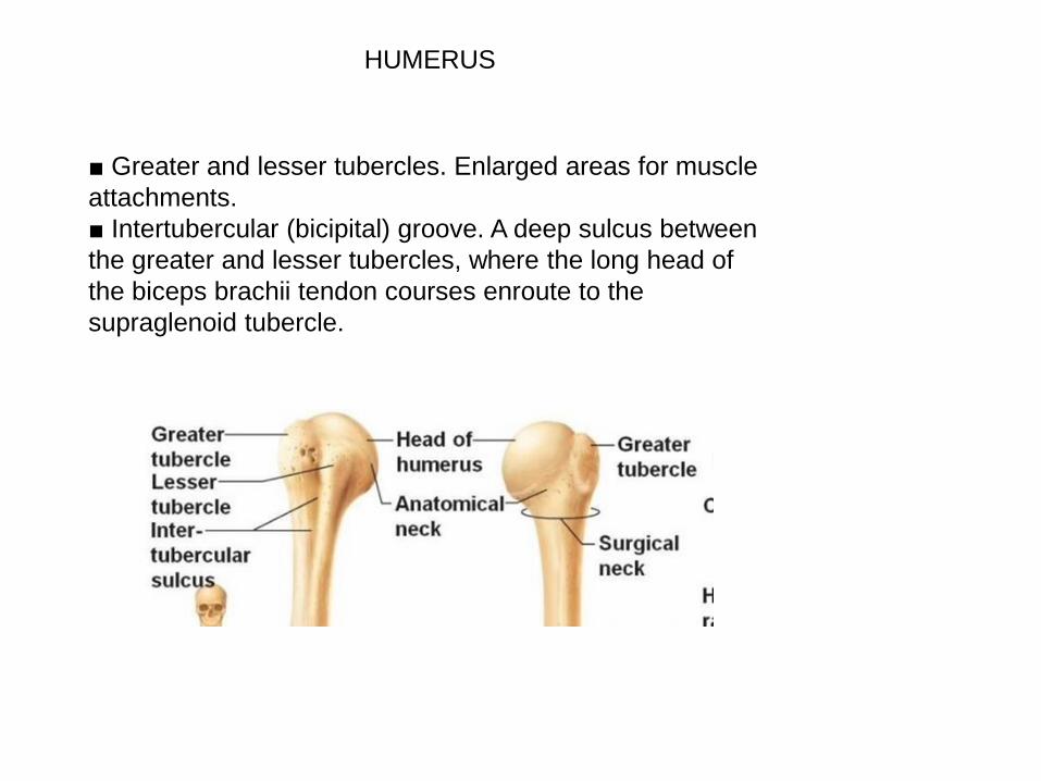
■ Greater and lesser tubercles. Enlarged areas for muscle
attachments.
■ Intertubercular (bicipital) groove. A deep sulcus between
the greater and lesser tubercles, where the long head of
the biceps brachii tendon courses enroute to the
supraglenoid tubercle.
HUMERUS

■ Radial (spiral) groove. A distinct
groove on the posterior surface of
the humerus, where the radial nerve
and the deep
brachial artery course.
■ Deltoid tuberosity. A large V-
shaped protrusion on the lateral
surface of the humerus, midway
along its length where the deltoid
muscle attaches.
■ Lateral epicondyle. Located on
the distal lateral end of the humerus
and provides an attachment surface
for the posterior
forearm muscles (extensors).
HUMERUS

■ Medial epicondyle. Located on the
distal medial end of the
humerus and provides an attachment
surface for the anterior
forearm muscles (flexors).
■ Trochlea. Characterized by a pulley
shape; it helps to guide
the hinge joint. The trochlea of the
humerus articulates with
the trochlear notch of the ulna.
HUMERUS

■ Capitulum. Characterized by
its oval, convex shape for
articulation with the radial head.
■ Coronoid fossa. Located on
the distal anterior surface of the
humerus, where the coronoid
process of the ulna articulates.
■ Olecranon fossa. Located on
the distal posterior surface of the
humerus, where the olecranon
process of the ulna articulates.
HUMERUS


The forearm (antebrachium) :consists of TWO BONES: the radius and ulna.Proximally, the forearm articulates with the humerus through the elbow complex (humeroulnar and humeroradial joints).Distally, the forearm articulates with the carpal bones through the wrist complex, enabling a wide array of actions. The muscles of the forearm that act upon the elbow, wrist complex, and the digital joints Muscles are organized into two fascial compartments, similar to those of the arm muscles..
THE FOREARM


ULNA FEATURES
Long Bone
looks like a wrench مفتاح انجليزي
It lies medially and parallel to the radius
The proximal end
Olecranon
Coronoid process
Trochlear notch
Radial notch Tuberosity of ulna

Shaft of the Ulna
As it moves distally, it decreases in
width.
HAS THREE SURFACES:
Anterior
Posterior
Medial
HAS THREE BORDERS:
Posterior – palpable along the entire
length of the forearm posteriorly
Interosseous
Anterior
Distal end
The distal end of the ulna is much
smaller in diameter than the proximal end.
It is mostly unremarkable, terminating in
rounded HEAD,
the ulnar styloid process.

The radius
Long bone
It lies laterally and parallel to ulna.
Proximal end of the Radius
head,
neck
radial tuberosity (bicipital tuberosity)
Shaft of the Radius
The radial shaft EXPANDS in diameter
as it moves distally.
Much like the ulna, it has three borders
and three surfaces.
The Distal End
The lateral side projects distally as
the styloid process.
ulnar notch
The distal surface of the radius has two
facets, for articulation with
the scaphoid and lunate carpal bones.
This makes up the wrist joint.

8 carpal bonesProximal row:
Scaphoid
Lunate
Triquetrum
Pisiform
Distal row:Trapezium
Trapezoid
Capitate
Hamate
5 Metacarpals
14 phalanges
The Bony Structure of The Hand