Embed Size (px)
Citation preview
UPPER GASTROINTESTINAL BLEEDING
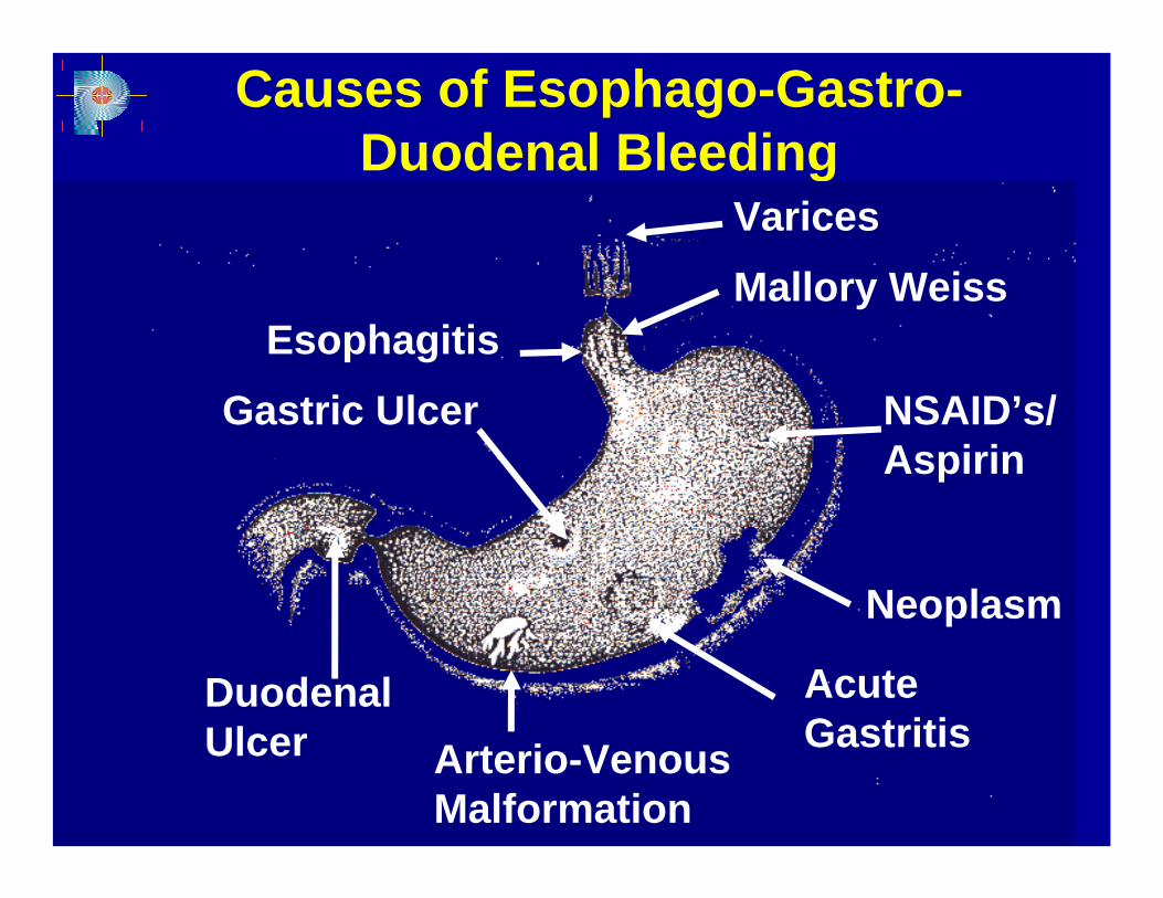
Causes of Esophago-Gastro-Duodenal Bleeding
VaricesMallory Weiss
NSAID’s/Aspirin
Neoplasm
Acute GastritisArterio-Venous
Malformation
Duodenal Ulcer
Gastric UlcerEsophagitis
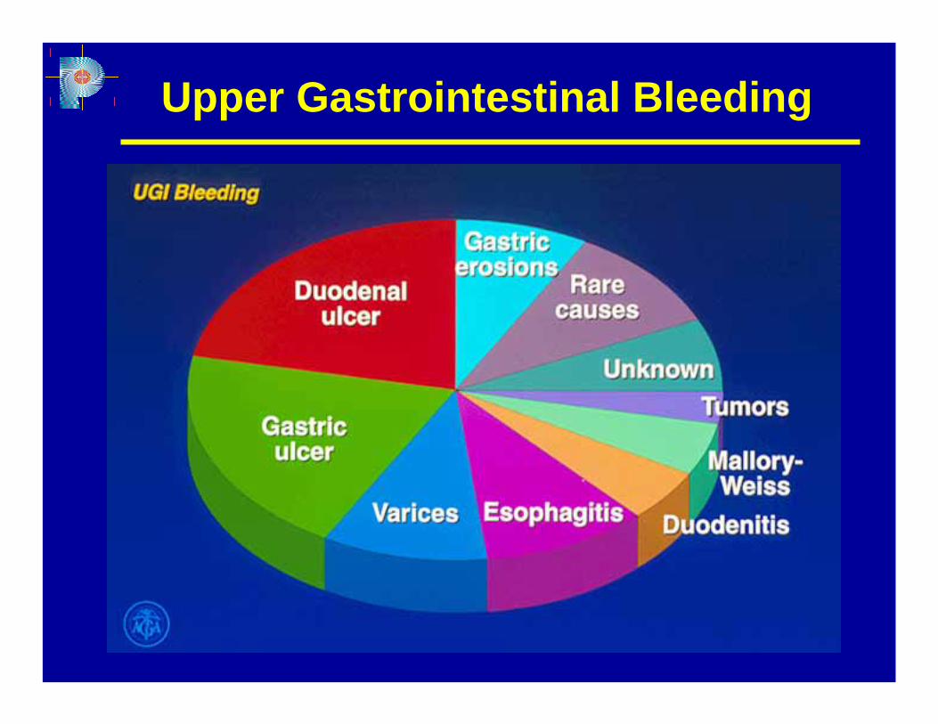
Upper Gastrointestinal Bleeding
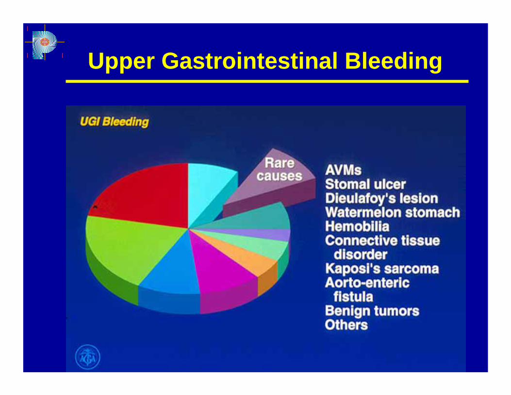
Upper Gastrointestinal Bleeding
Upper Gastrointestinal Bleeding
Severe UGIH is a commonand
serious medico-surgical problem
Upper Gastrointestinal Bleeding
Despite a decreased incidence of ulcer
disease and improvements in the
management of acute upper GI bleeding,
mortality remains at + 6-7 % in most series
in the literature for the past 30 years.
Upper Gastrointestinal Bleeding
Endoscopic hemostatic therapy
has been demonstrated to be the
mainstay of management.
Upper Gastrointestinal Bleeding
At intragastric pH < 7, coagulation is
deficient due to ineffective function
of clotting factors and platelets
Upper Gastrointestinal Bleeding
Maintenance of a high intragastric
pH > 6 during management of upper
G I Bleeding is warranted.
IV PPI’s are able to maintain gastric
pH > 6 for 24 hours a day.

Upper Gastrointestinal Bleeding
Recent clinical trial data support the
use of PPI’s to decrease the rate of
re-bleeding and the need for surgery.
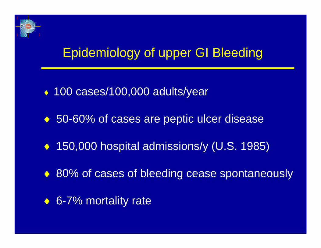
Epidemiology of upper GI Bleeding
♦ 100 cases/100,000 adults/year
♦ 50-60% of cases are peptic ulcer disease
♦ 150,000 hospital admissions/y (U.S. 1985)
♦ 80% of cases of bleeding cease spontaneously
♦ 6-7% mortality rate
Upper GI bleeding
Pathophysiology
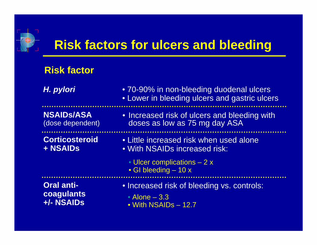
Risk factors for ulcers and bleeding
Risk factor
H. pylori • 70-90% in non-bleeding duodenal ulcers• Lower in bleeding ulcers and gastric ulcers
NSAIDs/ASA (dose dependent)
• Increased risk of ulcers and bleeding with doses as low as 75 mg day ASA
Corticosteroid + NSAIDs
• Little increased risk when used alone• With NSAIDs increased risk:
• Ulcer complications – 2 x• GI bleeding – 10 x
Oral anti-coagulants +/- NSAIDs
• Increased risk of bleeding vs. controls:• Alone – 3.3• With NSAIDs – 12.7
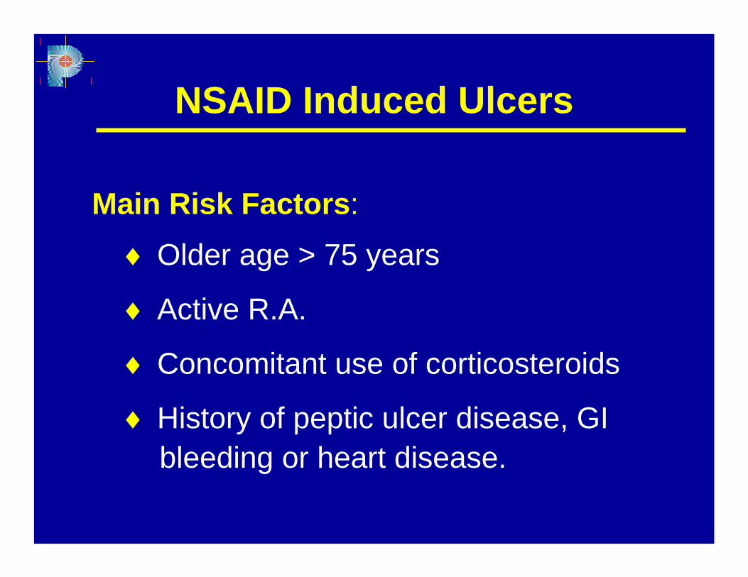
NSAID Induced Ulcers
Main Risk Factors:
♦ Older age > 75 years
♦ Active R.A.
♦ Concomitant use of corticosteroids
♦ History of peptic ulcer disease, GIbleeding or heart disease.
Prognostic Factors
Clinical:♦ Haemodynamic instability
♦ Fresh red blood in the emesis
♦ Haematochezia
♦ Increasing number of units transfused
Prognostic Factors
♦ Age > 60 years
♦ Concurrent illness - Cardiovascular, pulmonary and Diabetes Mellitus
♦ Onset while hospitalised for other reasons
♦ Recurrent bleeding
Prognostic Factors
Urgent Endoscopy:
♦ Patients with coffee-ground vomiting with melena
♦ Haematemesis with or without melena
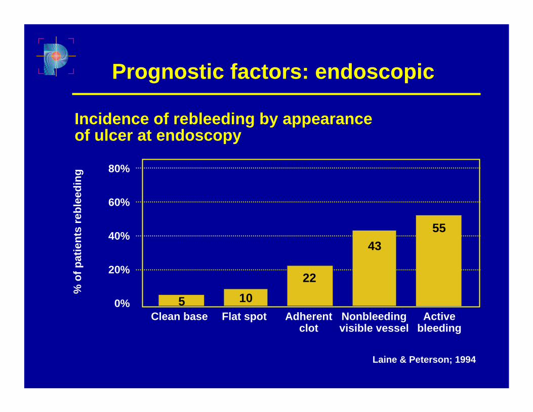
Prognostic factors: endoscopic
80%
60%
40%
20%
0%Clean base Flat spot Adherent
clot
% o
f pat
ient
s re
blee
ding
Laine & Peterson; 1994
Incidence of rebleeding by appearance of ulcer at endoscopy
Nonbleeding visible vessel
Active bleeding
5 1022
4355
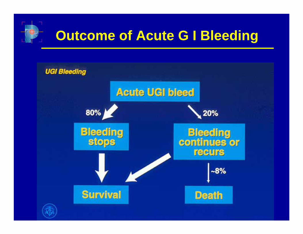
Outcome of Acute G I Bleeding
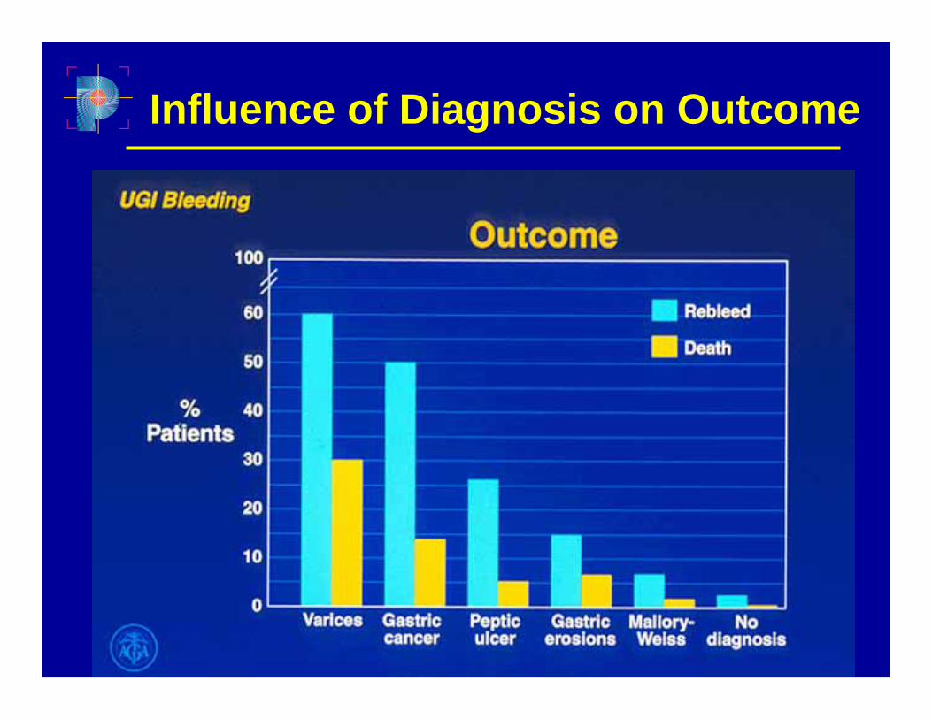
Influence of Diagnosis on Outcome
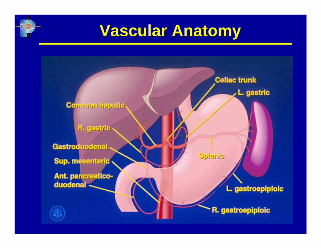
Vascular Anatomy
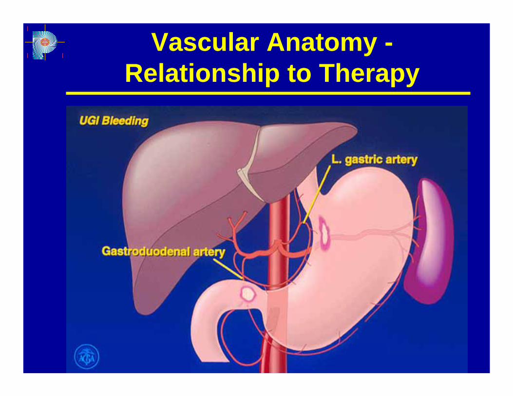
Vascular Anatomy -Relationship to Therapy
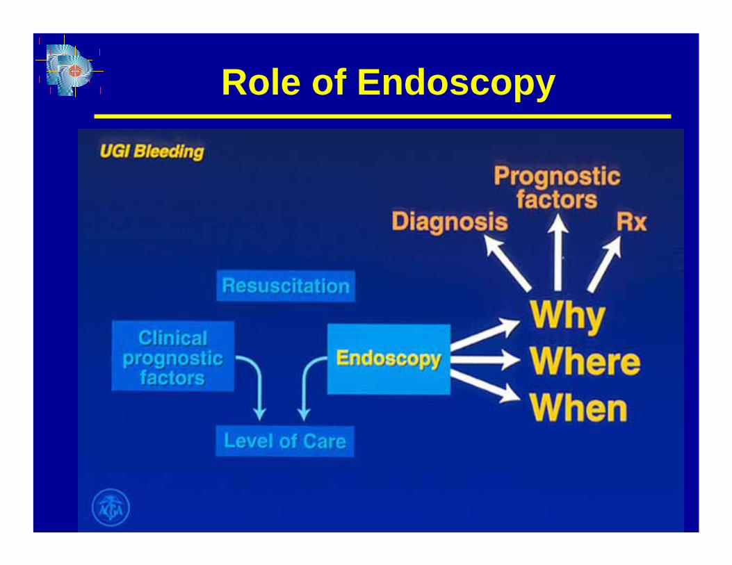
Role of Endoscopy
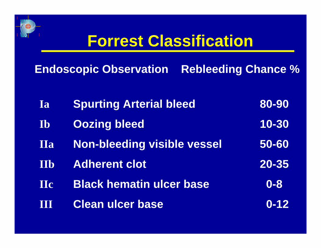
Forrest ClassificationEndoscopic Observation Rebleeding Chance %
Ia Spurting Arterial bleed 80-90
Ib Oozing bleed 10-30
IIa Non-bleeding visible vessel 50-60
IIb Adherent clot 20-35
IIc Black hematin ulcer base 0-8
III Clean ulcer base 0-12
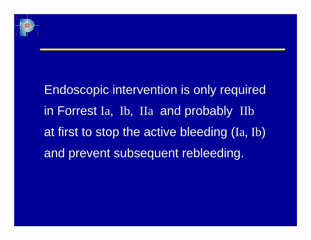
Endoscopic intervention is only required
in Forrest Ia, Ib, IIa and probably IIb
at first to stop the active bleeding (Ia, Ib)
and prevent subsequent rebleeding.
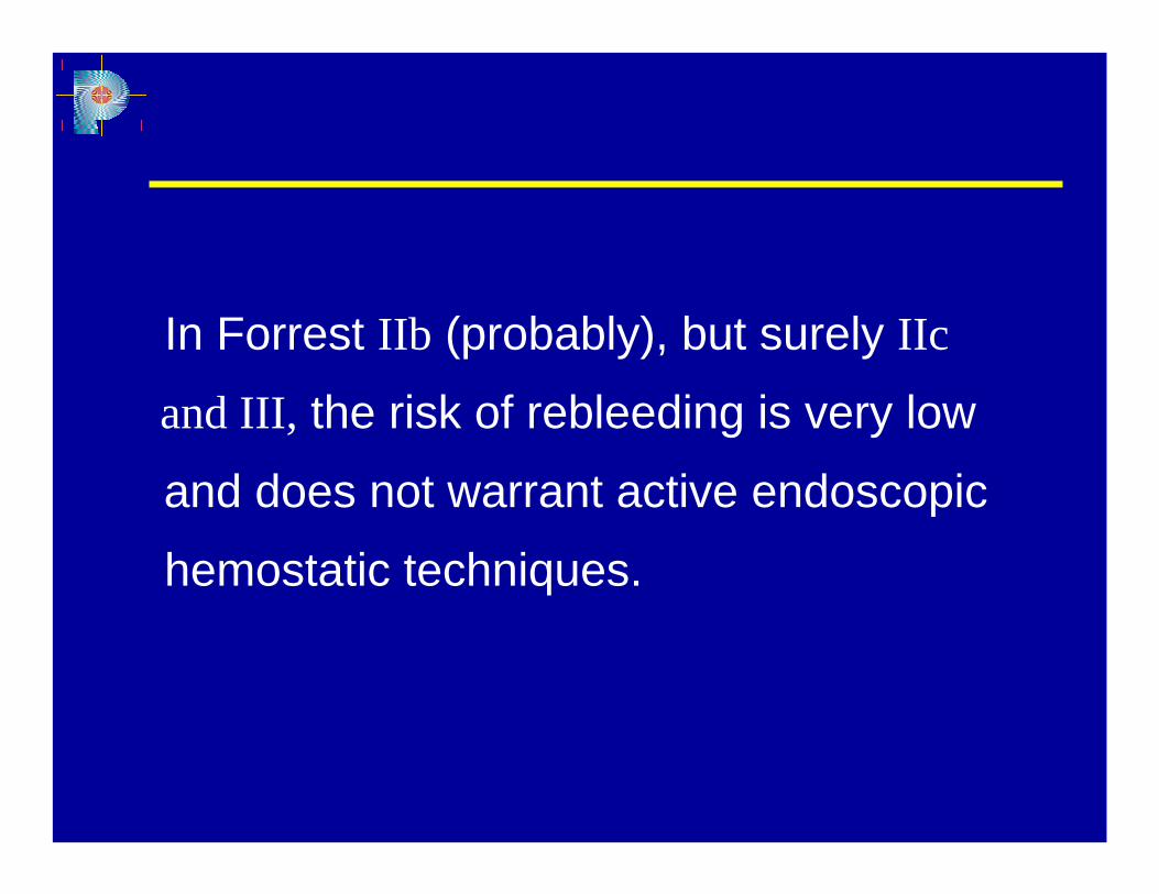
In Forrest IIb (probably), but surely IIc
and III, the risk of rebleeding is very low
and does not warrant active endoscopic
hemostatic techniques.
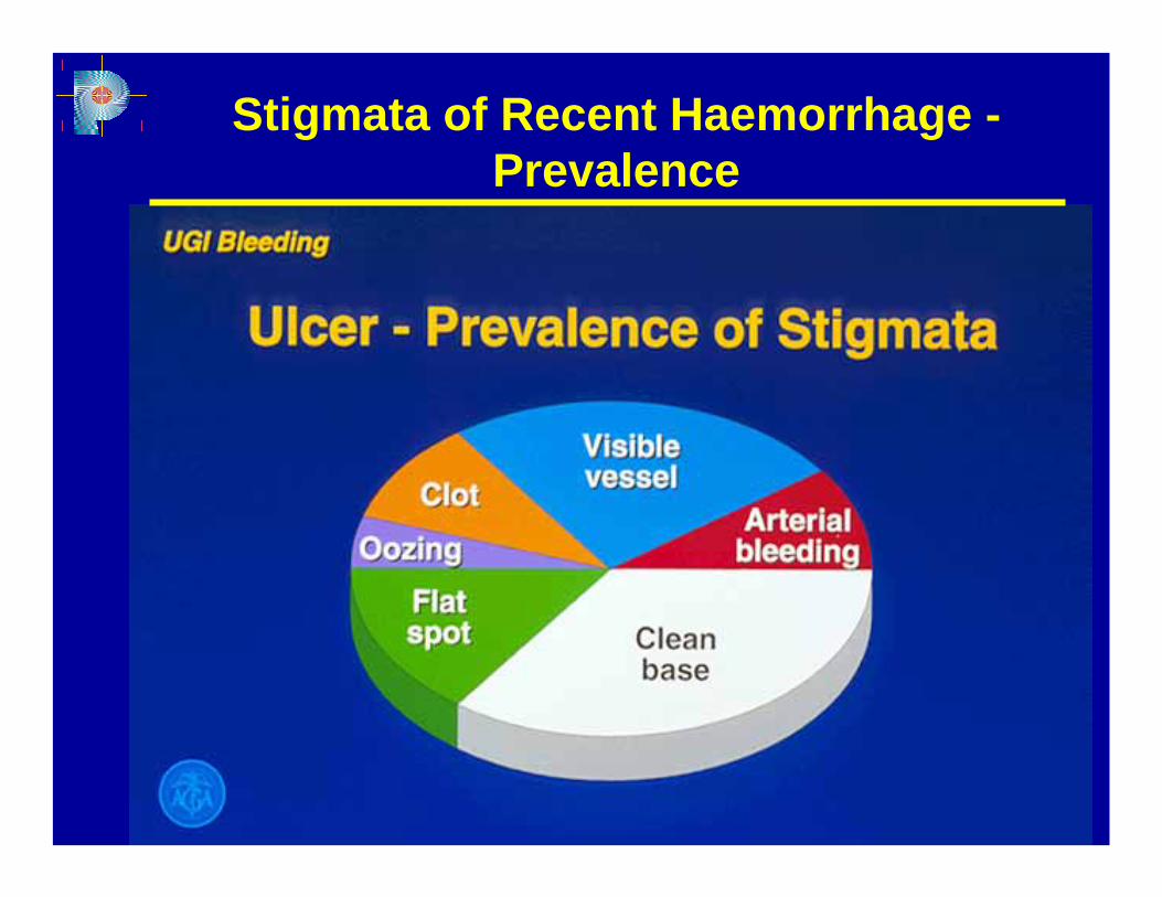
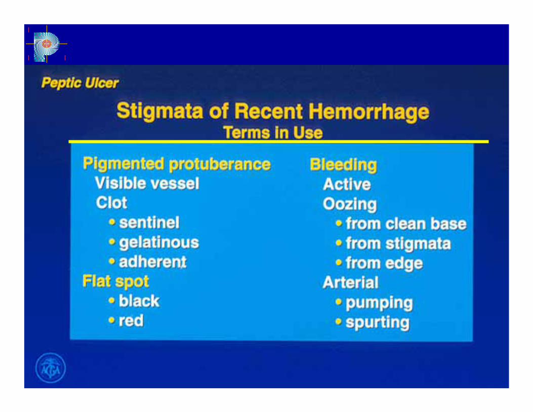
Stigmata of Recent Haemorrhage -Prevalence
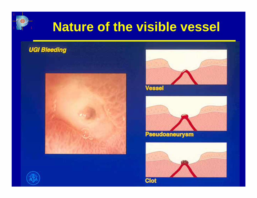
Nature of the visible vessel
Overview of management
♦ Initial management
♦ Endoscopic therapy
♦ Surgical therapy
♦ Pharmacological therapy
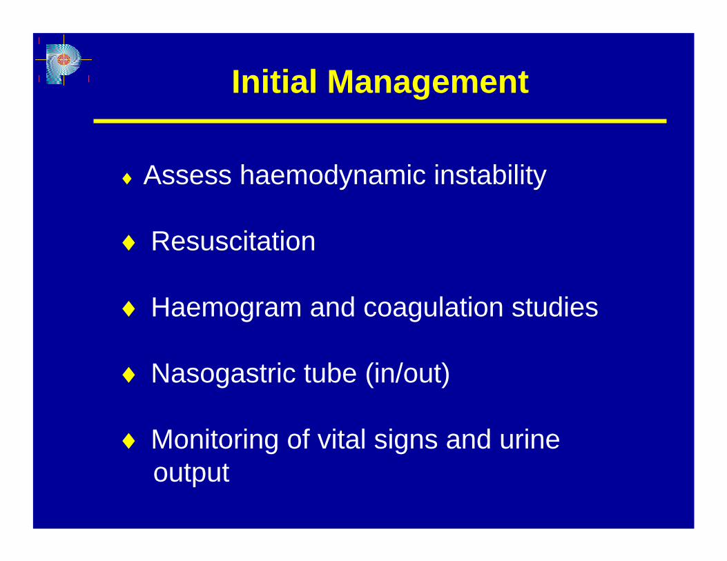
Initial Management
♦ Assess haemodynamic instability
♦ Resuscitation
♦ Haemogram and coagulation studies
♦ Nasogastric tube (in/out)
♦ Monitoring of vital signs and urineoutput
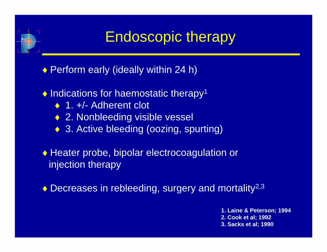
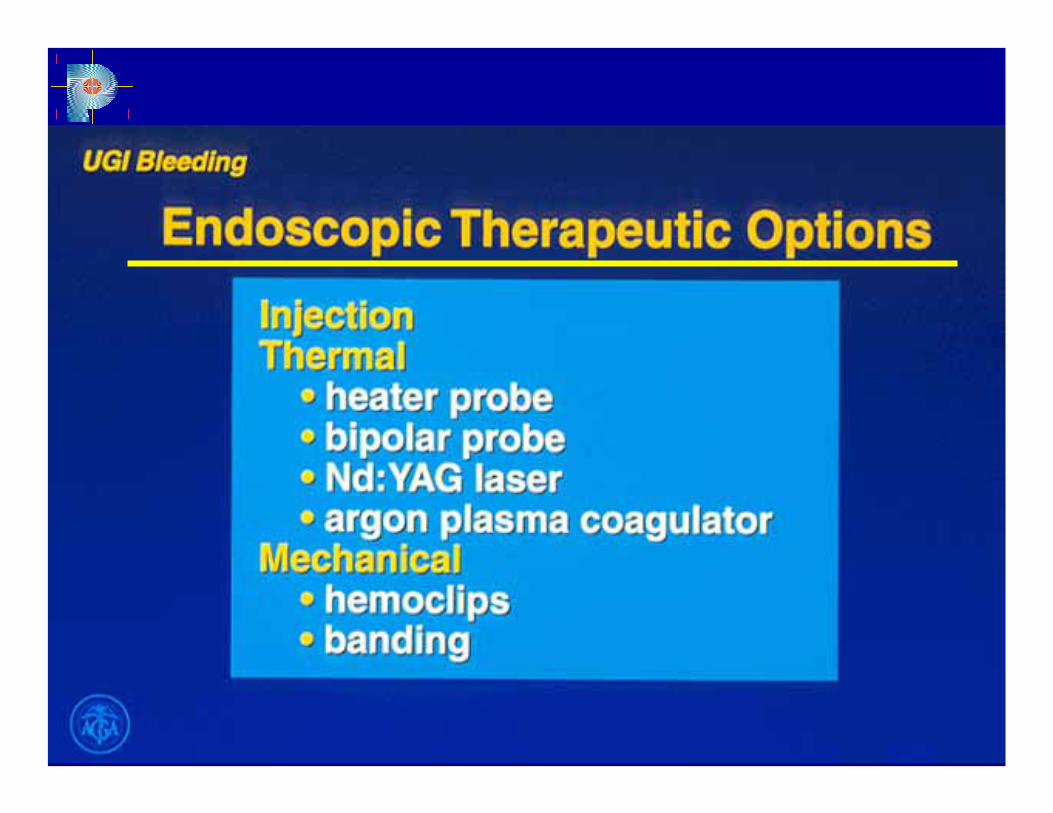
Endoscopic therapy
♦Perform early (ideally within 24 h)
♦Indications for haemostatic therapy1
♦ 1. +/- Adherent clot♦ 2. Nonbleeding visible vessel♦ 3. Active bleeding (oozing, spurting)
♦Heater probe, bipolar electrocoagulation or injection therapy
♦Decreases in rebleeding, surgery and mortality2,3
1. Laine & Peterson; 19942. Cook et al; 19923. Sacks et al; 1990
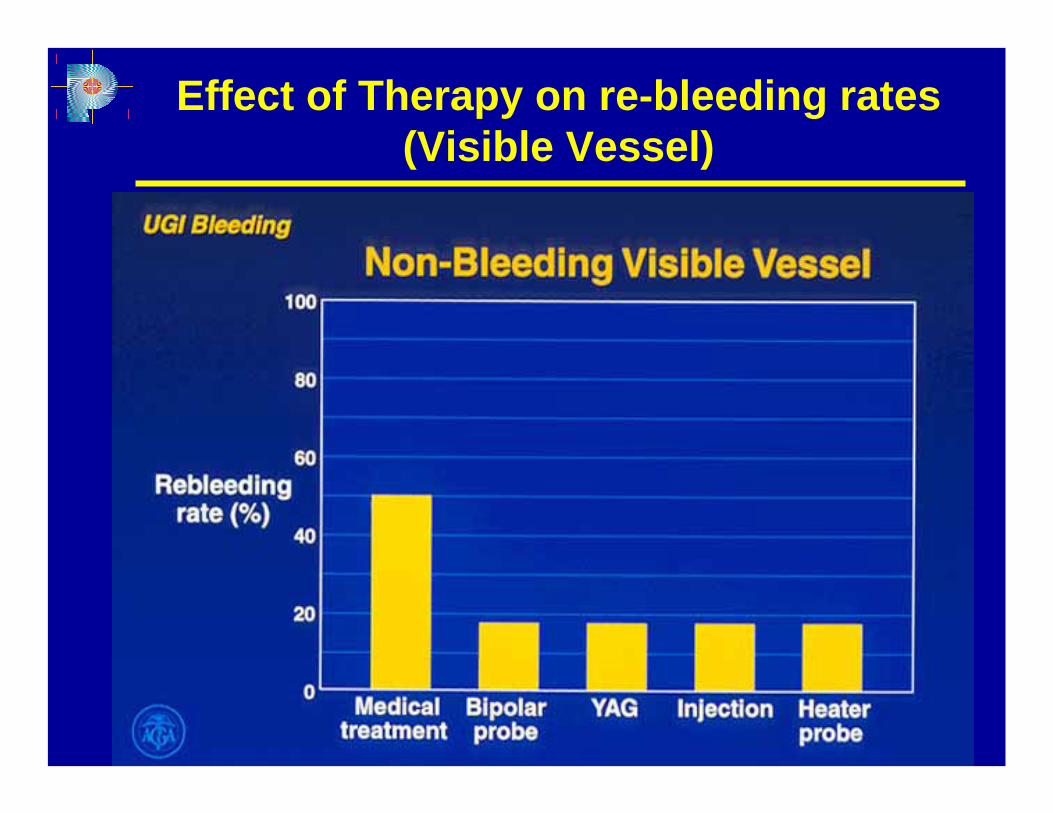
Effect of Therapy on re-bleeding rates (Visible Vessel)
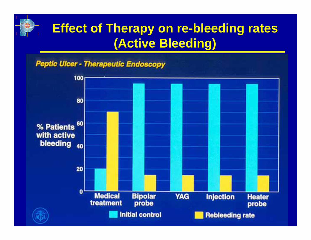
Effect of Therapy on re-bleeding rates (Active Bleeding)
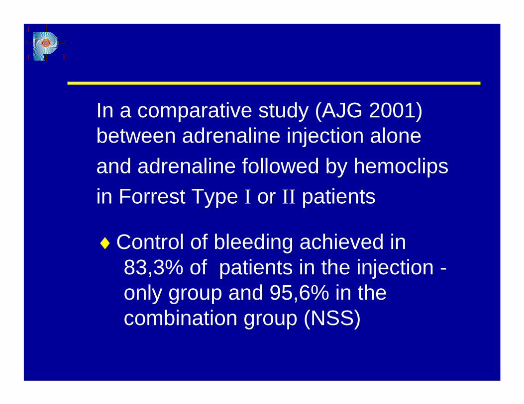
In a comparative study (AJG 2001) between adrenaline injection aloneand adrenaline followed by hemoclipsin Forrest Type I or II patients
♦Control of bleeding achieved in83,3% of patients in the injection -only group and 95,6% in thecombination group (NSS)
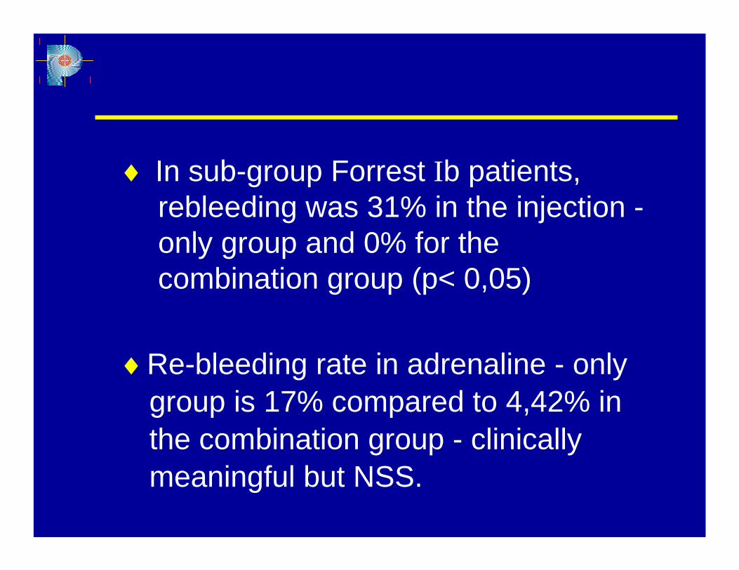
♦ In sub-group Forrest Ib patients, rebleeding was 31% in the injection -only group and 0% for the combination group (p< 0,05)
♦Re-bleeding rate in adrenaline - only group is 17% compared to 4,42% in the combination group - clinically meaningful but NSS.
Endoscopic therapy may not be
possible in up to 12% of bleeding
duodenal ulcers and at least 1% of
bleeding gastric ulcers because of
inaccessibility of the lesion or massive
hemorrhage.
Patients who do not have active
bleeding, non-bleeding visible vessels,
or adherent clots are low risk for further
bleeding.
Bleeding from a P.U. recurs after initial endoscopic hemostasis in 15-20% of patients.
Endoscopic re-treatment reduces the need for surgery without increasing the risk of death and is associated with fewer complications than surgery
Hypotension and ulcer size of at least 2cm are independent factors predictive of the failure of endoscopic re-treatment.
Patients with larger ulcers and therefore heavier bleeding, surgery may be a better choice than endoscopic re-treatment.
Salvage surgery for recurrent bleeding is associated with a mortality rate
ranging from 15-25%.
Surgical therapy
♦Endoscopic management failure
♦Other extenuating circumstances
♦Patient survival improved by optimal timing
♦Individualized by clinical context, endoscopic and surgical expertise
- lowers splanchnic blood pressure- induces vasoconstriction- high rate of complications
Pharmacological Therapy
♦Vasopressin
- Lower toxicity- additional effects of decreasing
gastric acid secretion and increasingduodenal bicarbonate secretion
- decreased risk of re-bleedingcompared to H2RAs
Pharmacological Therapy
♦ Somatostatin and Octreotide
- appears to decrease mortality
- increased risk of thrombo-embolic events
Pharmacological Therapy
♦ Tranexamic acid - Antifibrinolytic agent
♦ Acid suppressing agents
- H2 Receptor Antagonists
- Proton Pump Inhibitors
Pharmacologic Therapy
♦ Aggressive acid suppression with PPI’s reduce the rate of recurrent bleeding, theneed for transfusions, and the need for surgery.
They represent an important adjunct toendoscopic therapy.
Pharmacologic Therapy
Role of acid in haemostasis
♦Impairs clot formation– Impairs platelet aggregation and causesdisaggregation
♦Accelerates clot lysis- Predominantly acid-stimulated pepsin
♦May impair integrity of mucus/bicarbonate barrier
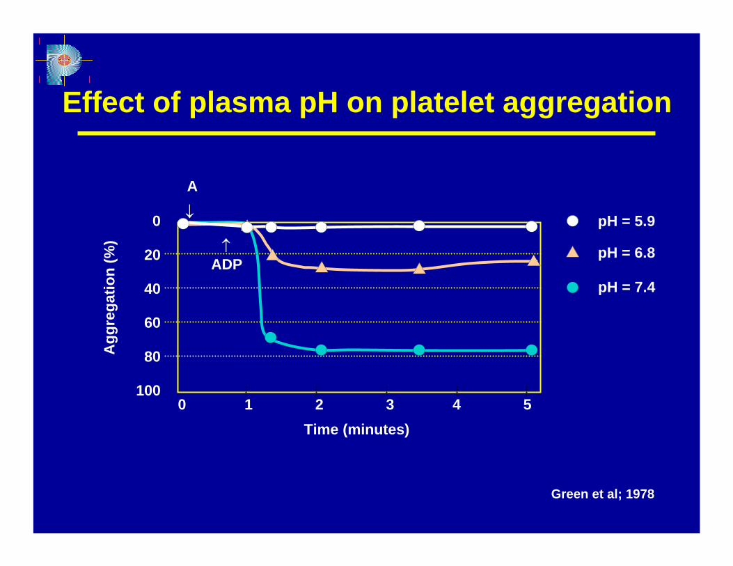
pH = 7.4
Agg
rega
tion
(%)
Effect of plasma pH on platelet aggregation
Green et al; 1978
Time (minutes)
0
20
40
60
80
1000 1 2 3 4 5
pH = 5.9
pH = 6.8
A
↑ADP
↓
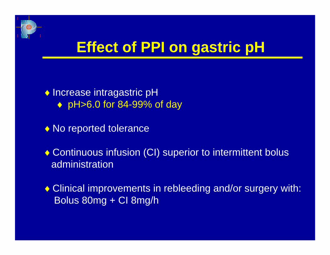
Effect of PPI on gastric pH
♦Increase intragastric pH♦ pH>6.0 for 84-99% of day
♦No reported tolerance
♦Continuous infusion (CI) superior to intermittent bolusadministration
♦Clinical improvements in rebleeding and/or surgery with:Bolus 80mg + CI 8mg/h
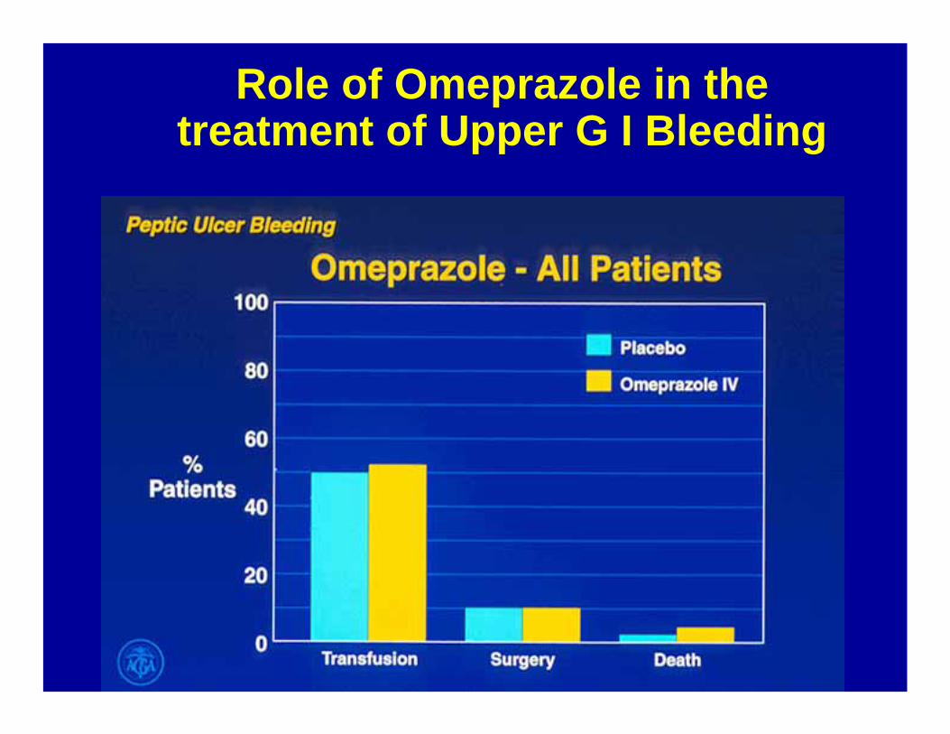
Role of Omeprazole in the treatment of Upper G I Bleeding
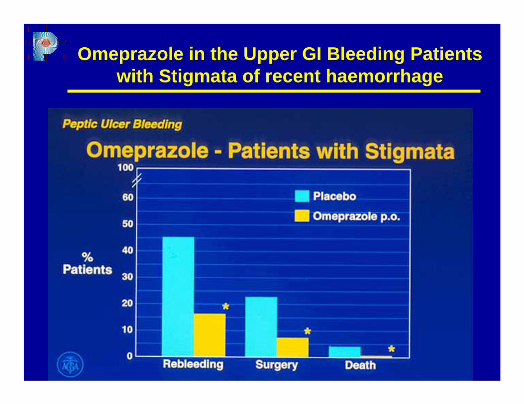
Omeprazole in the Upper GI Bleeding Patients with Stigmata of recent haemorrhage
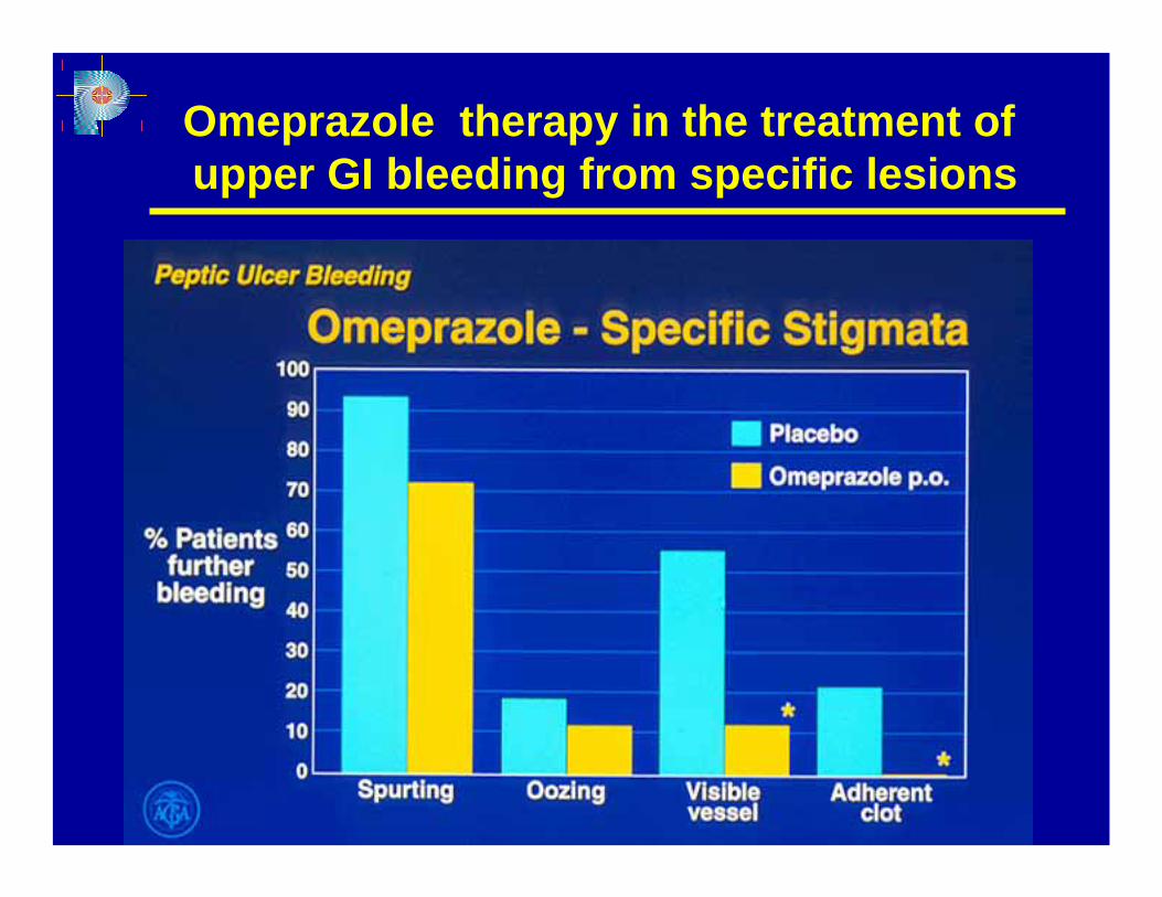
Omeprazole therapy in the treatment ofupper GI bleeding from specific lesions
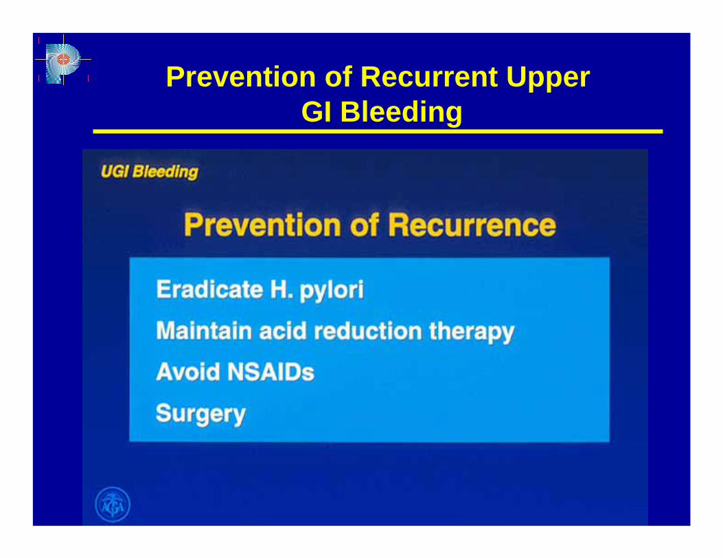
Prevention of Recurrent UpperGI Bleeding
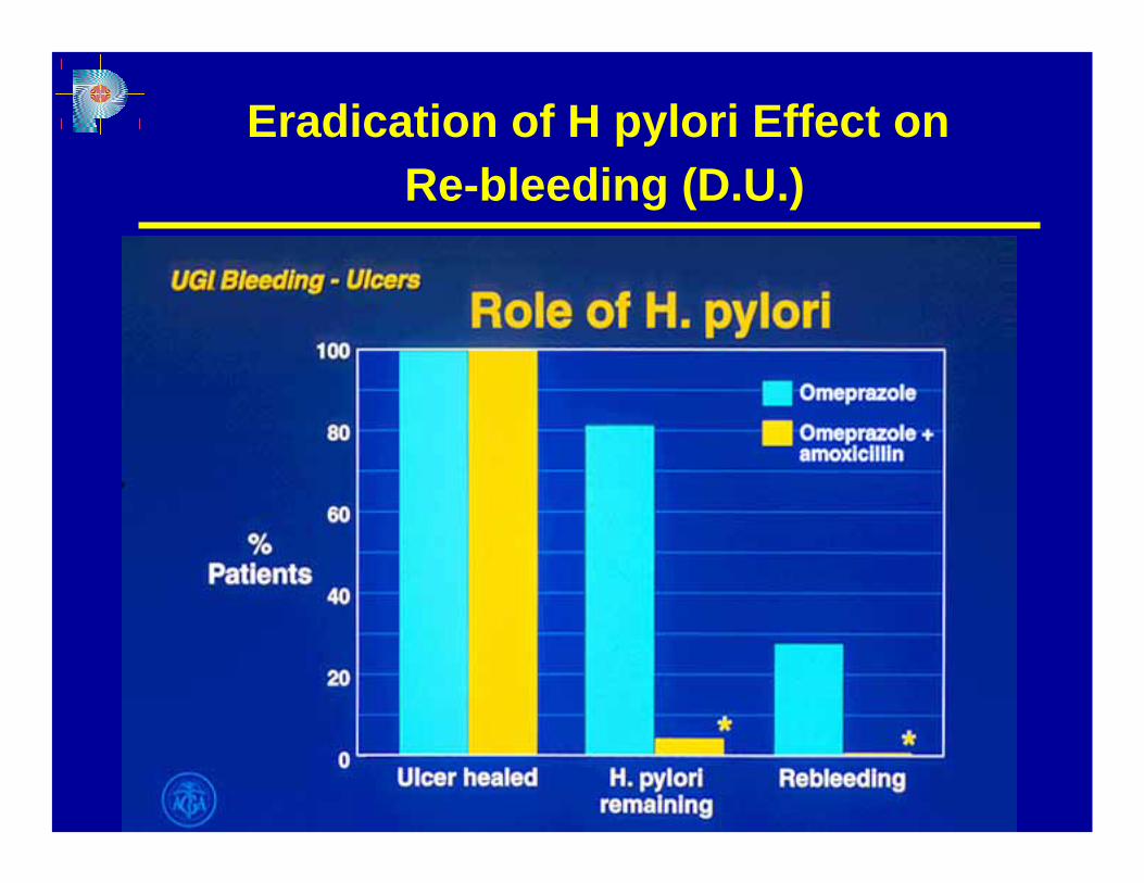
Eradication of H pylori Effect onRe-bleeding (D.U.)
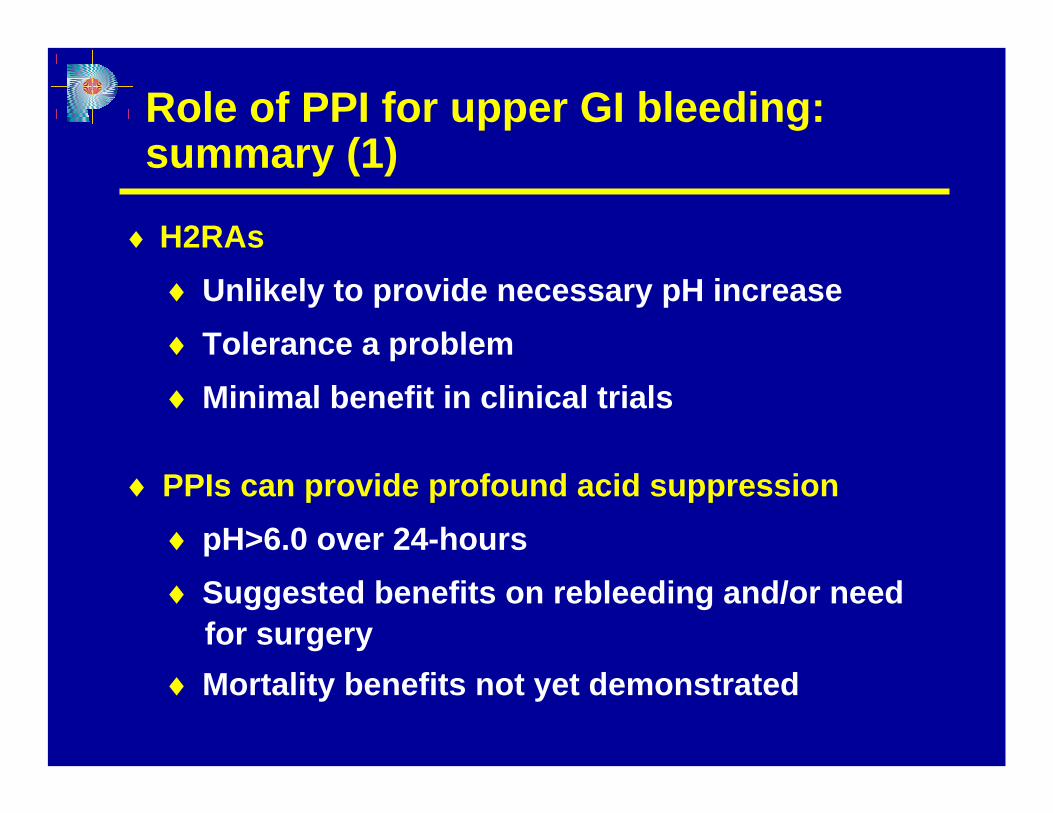
Role of PPI for upper GI bleeding: summary (1)
♦ H2RAs♦ Unlikely to provide necessary pH increase♦ Tolerance a problem♦ Minimal benefit in clinical trials
♦ PPIs can provide profound acid suppression♦ pH>6.0 over 24-hours♦ Suggested benefits on rebleeding and/or need
for surgery♦ Mortality benefits not yet demonstrated
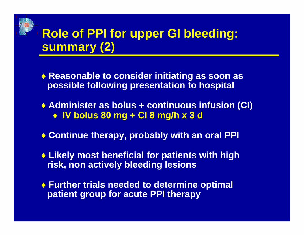
♦Reasonable to consider initiating as soon as possible following presentation to hospital
♦Administer as bolus + continuous infusion (CI)♦ IV bolus 80 mg + CI 8 mg/h x 3 d
♦Continue therapy, probably with an oral PPI
♦Likely most beneficial for patients with high risk, non actively bleeding lesions
♦Further trials needed to determine optimal patient group for acute PPI therapy
Role of PPI for upper GI bleeding: summary (2)
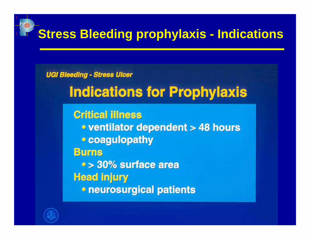
Stress Bleeding prophylaxis - Indications
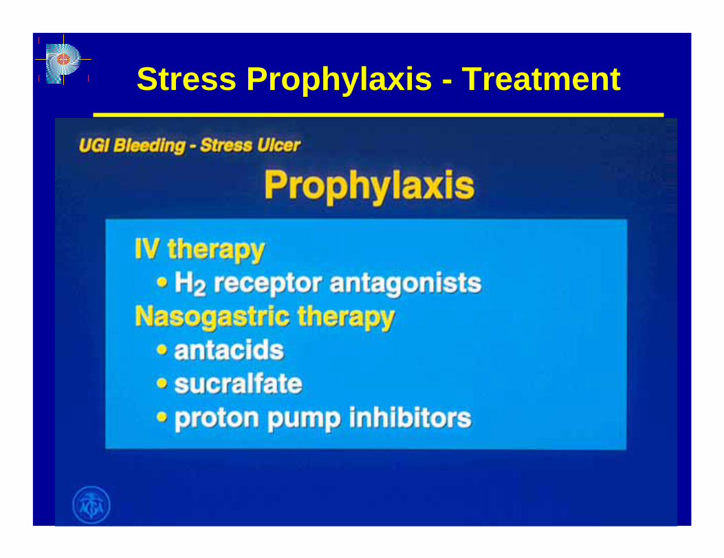
Stress Prophylaxis - Treatment
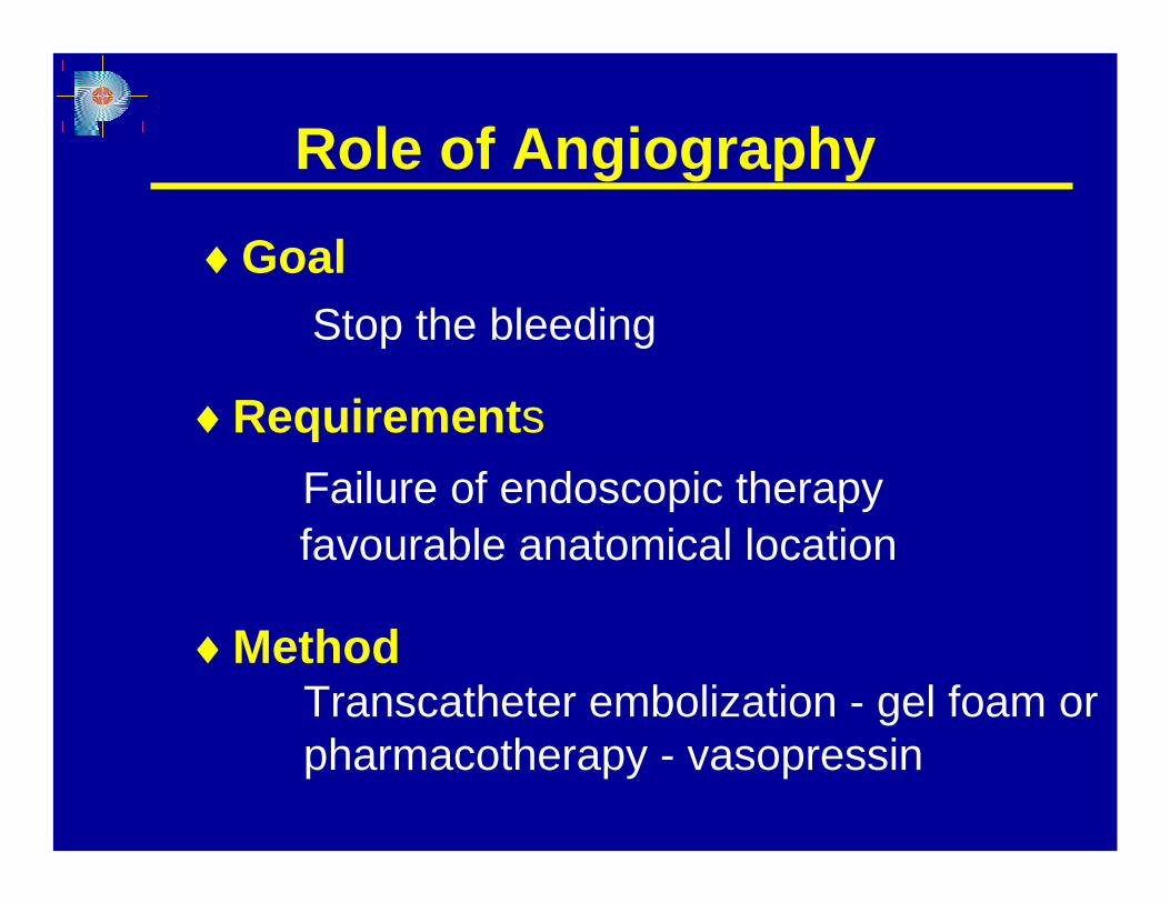
♦MethodTranscatheter embolization - gel foam orpharmacotherapy - vasopressin
Role of Angiography
♦GoalStop the bleeding
♦RequirementsFailure of endoscopic therapyfavourable anatomical location
♦ Oesophageal varices cause + 10% of
cases of acute upper GI bleeding
admitted to hospitals
Variceal Haemorrhage
♦ Mortality rate 30-50%
♦Gastro-oesophageal varices are presentin + 50% of cirrhotic patients. Their presence correlates with severity of liverdisease
Variceal Haemorrhage
♦Bleeding from oesophageal varicesceases spontaneously in up to 40% ofpatients
♦ Control of hemorrhage (24 hour bleeding free period within first48 hours after therapy)
Treatment of Acute VaricealHemorrhage
♦ Prevention of early recurrence
High rate of major complications
Pharmacotherapy
♦ Vasoactive therapy - Vasopressin
Conflicting results with Terlipressin and Nitroglycerin
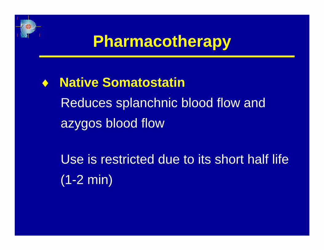
♦ Native SomatostatinReduces splanchnic blood flow and azygos blood flow
Use is restricted due to its short half life(1-2 min)
Pharmacotherapy
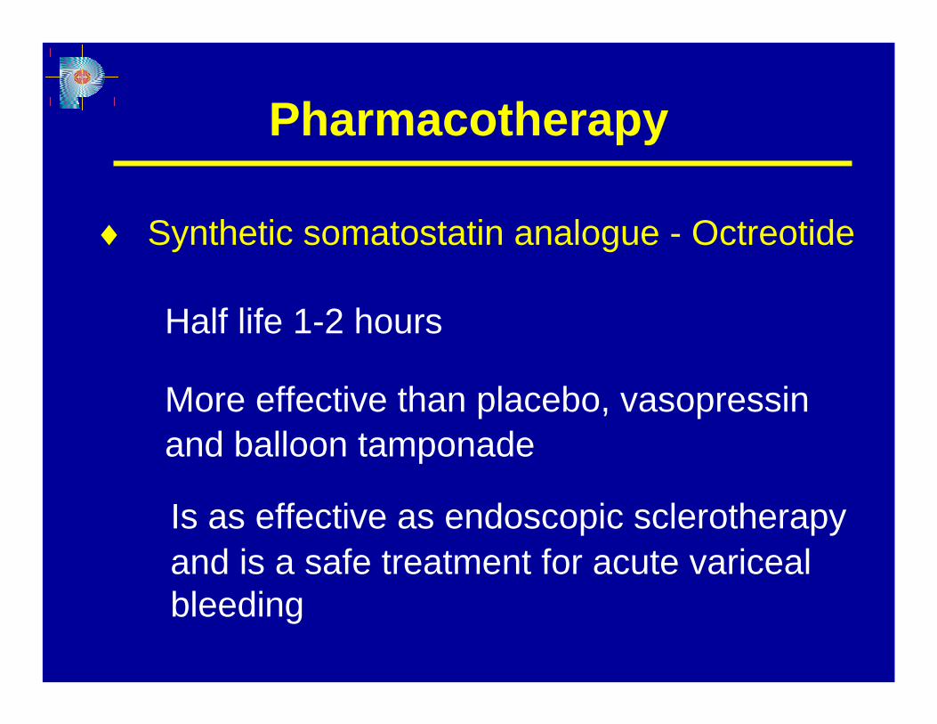
Is as effective as endoscopic sclerotherapyand is a safe treatment for acute variceal bleeding
Pharmacotherapy
♦ Synthetic somatostatin analogue - Octreotide
Half life 1-2 hours
More effective than placebo, vasopressinand balloon tamponade
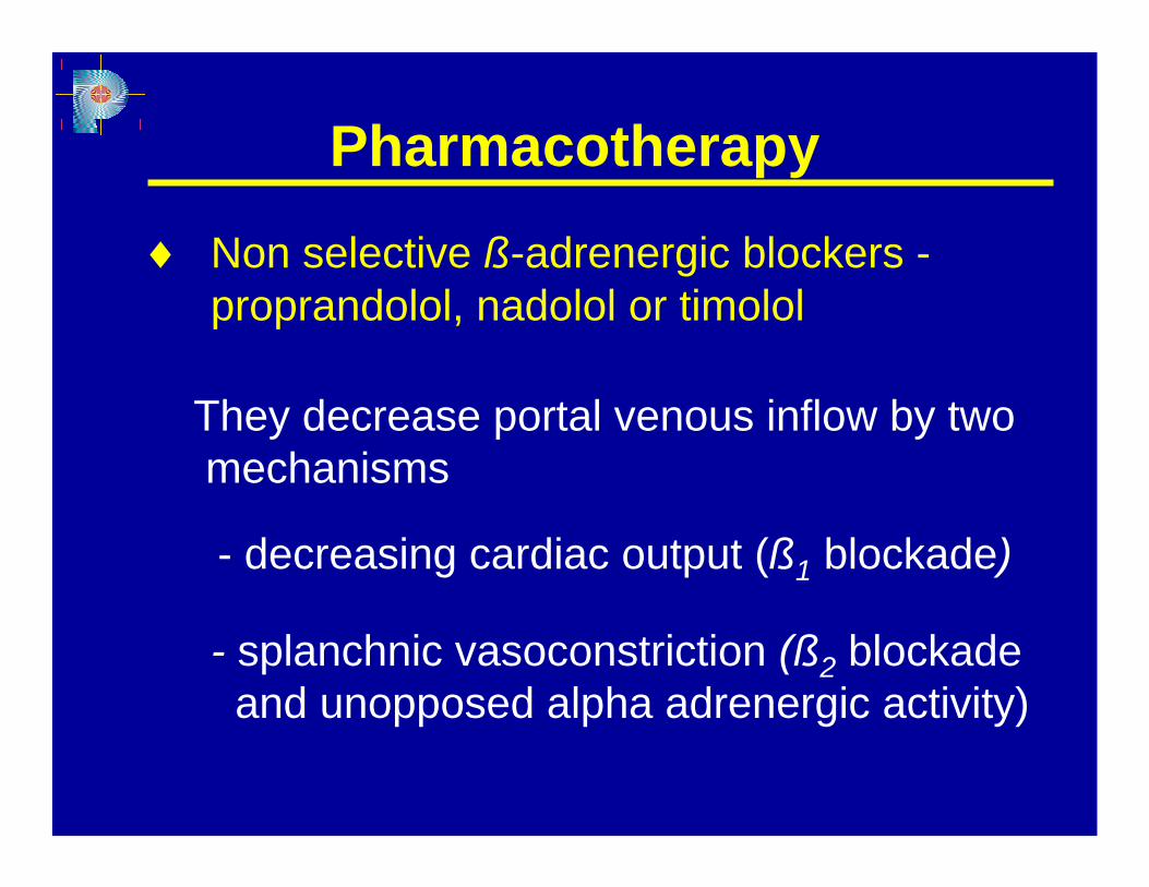
♦ Non selective ß-adrenergic blockers -proprandolol, nadolol or timolol
Pharmacotherapy
They decrease portal venous inflow by twomechanisms
- decreasing cardiac output (ß1 blockade)
- splanchnic vasoconstriction (ß2 blockade and unopposed alpha adrenergic activity)

♦ Antibiotic prophilaxis is mandatory
Pharmacotherapy
- Reduces rate of bacterial infections- Increases survival
♦ Avoid intravascular over expansion
♦ Blood replacement to target Hematocrit of25-30%
♦ Octreotide as adjunct to endoscopic therapy appears to be the most promising approach in the treatment of acute variceal hemorrhage
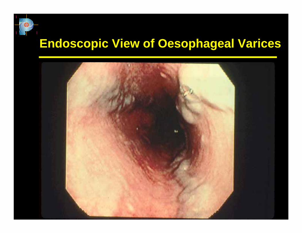
Endoscopic View of Oesophageal Varices
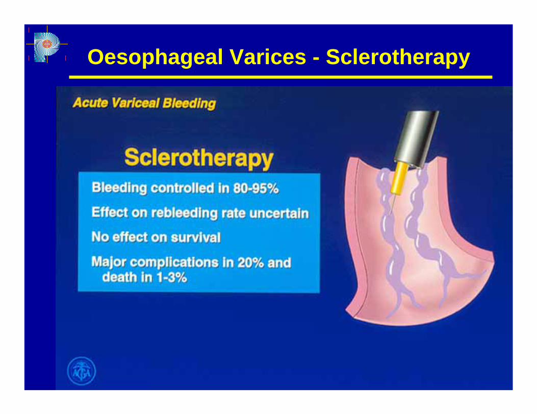
Oesophageal Varices - Sclerotherapy
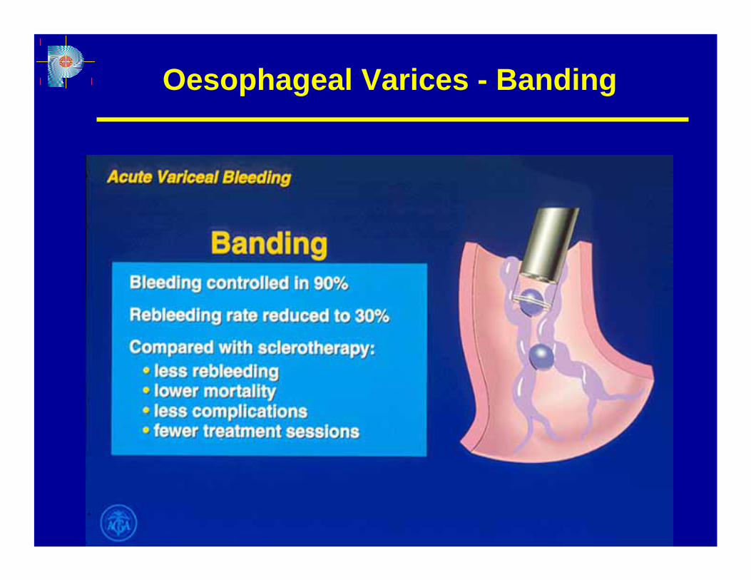
Oesophageal Varices - Banding
♦ Shunt surgery (distal spleno-renal) in well compensated liver disease (Child A) or TIPS are of provenclinical efficacy as salvage therapy for patients not responding to endoscopic or pharmacologic therapy
Shunt Therapy

♦ prevents rebleeding
Shunt Surgery
♦ increases risk of portosystemicencephalopathy
♦ no effect on survival
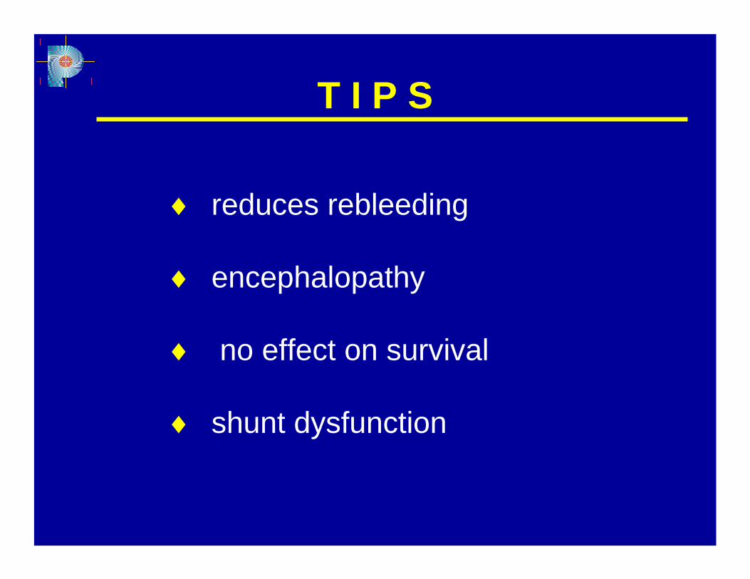
♦ reduces rebleeding
♦ encephalopathy
♦ no effect on survival
♦ shunt dysfunction
T I P S