Embed Size (px)
Citation preview

Basic Ultrasound-Guided
Upper Extremity Blocks
Christian R. Falyar, CRNA, DNAP

Objectives • Discuss the patient and surgical indications for
ultrasound-guided regional anesthesia (UGRA) of
the upper extremity
• Describe specific ultrasound landmarks for each of
the upper extremity blocks
• Review the transducer axis, needle insertion plane,
and local anesthetic requirements for each of the
upper extremity blocks
• Explain potential side-effects and complications
related to specific upper extremity blocks
Indications • The choice to use UGRA is determined by many
factors such as patient comorbidities, suitability of
the technique for the proposed surgery, provider
comfort in performing the procedure, as well the
mental status of the patient. UGRA has many
indications, including:
• Primary anesthetic
• Pain Management
• History of severe PONV or risk of MH
• Patient is too ill for general anesthesia
• Physician (surgeon) preference

Contraindications
• There are certain instances where under no
circumstances should regional anesthesia be
considered. These are known as absolute
contraindications. They include:
• Patient refusal
• Local infection at the site of the proposed block
• Active bleeding an anticoagulated patient
• Proven allergy to a local anesthetic

Contraindications
• Most contraindications to regional anesthesia are
relative. The provider must determine the risk vs.
benefit before proposing any procedure.
• Respiratory compromise
• Inability to cooperate/understand procedure
• An anesthetized patient (adult population)
• Bleeding diathesis secondary to an anticoogulant or
genetic defect
• Bloodstream infection
• Preexisting peripheral neuropathy

Complications
• Although uncommon, regional anesthesia can
result in complications such as:
• Local anesthetic toxicity
• Intra-arterial injection
• Respiratory compromise
• Parathesias and nerve damage
• Prior to performing any regional anesthetic, the risks
and benefits should be discussed with the patient,
allowing them to make an informed decision.

Prior to any procedure…
• Verify the correct patient
• Obtain informed consent
• Verify the correct procedure
• Verify the correct extremity
• Gather all necessary equipment
• Obtain baseline vital signs and monitor during the
procedure
• Administer proper/adequate sedation

The Brachial Plexus
• The brachial plexus consists of ventral rami of the
C5 – T1 nerve roots and extends from the neck to
axilla
• From central to the periphery, the roots exit the
vertebral foramen and converge into trunks,
divisions, cords, and branches
• With a few exceptions, the brachial plexus supplies
sensory and motor innervation to the upper
extremity

The Brachial Plexus

Interscalene
• The interscalene block is performed at the root level
• It is the primary upper extremity block for
procedures involving the shoulder and proximal
upper arm
• Nerve roots C5 - 7 are found in the interscalene
groove between the anterior and middle scalene
muscles near the level of the cricoid cartilage (C6)
posterior the sternocleidomastoid muscle
• A catheter is usually placed for procedures
involving the rotator cuff

Interscalene
Posterior Scalene
Brachial Plexus
Phrenic Nerve
Anterior Scalene

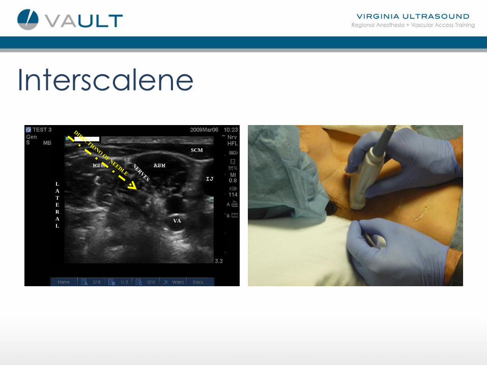
Interscalene • Patient is placed in supine position with head turned
to the non-operative side
• Transducer is placed either superior to clavicle in
the mid fossa and moved cephalad, or at the level
of the cricoid cartilage and moved laterally
• High frequency linear array transducer
• Short-axis, in-plane image
• 3 to 4 hypoechoic circles located between the
anterior and middle scalene muscles
• 5cm needle is used
• 20 – 30cc’s of local anesthetic injected

Interscalene • Short-axis, in-plane
image with posterior
needle insertion
• Interscalene anatomy
is variable, and the
nerve roots may not
appear in the
interscalene groove
• For shoulder surgery the
needle should pass
between the C5-6 roots

Interscalene
Suprascapular Nerve
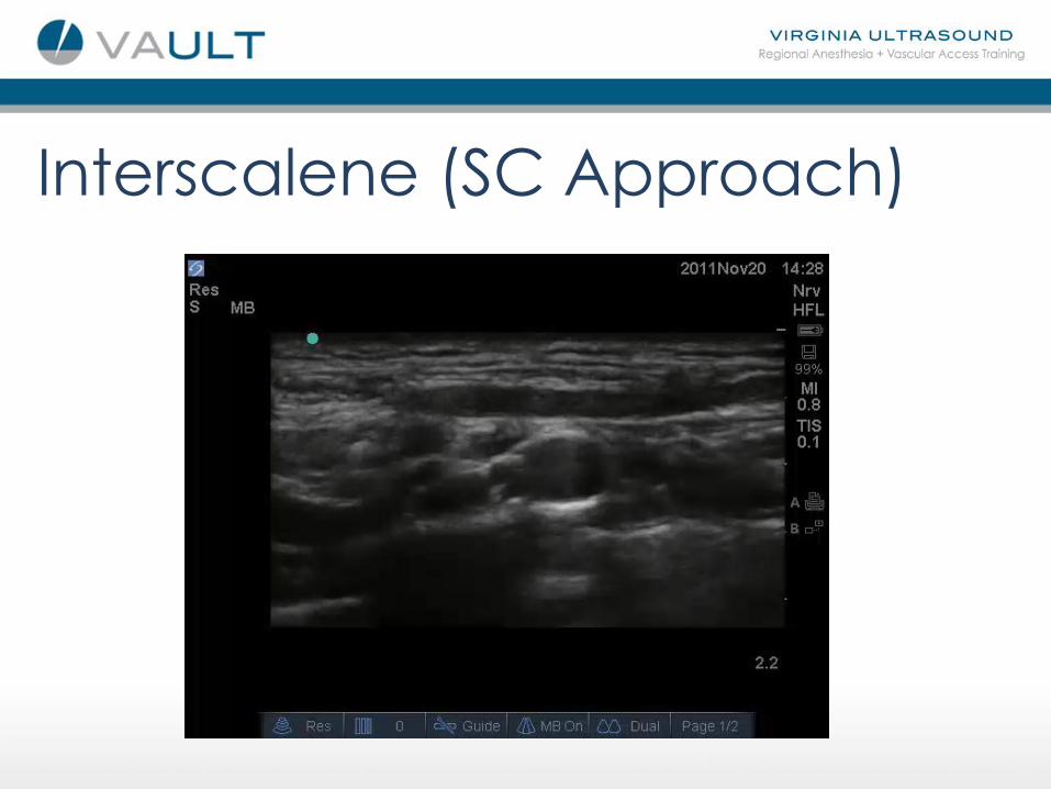
Interscalene (SC Approach)

Interscalene (IJ Approach)
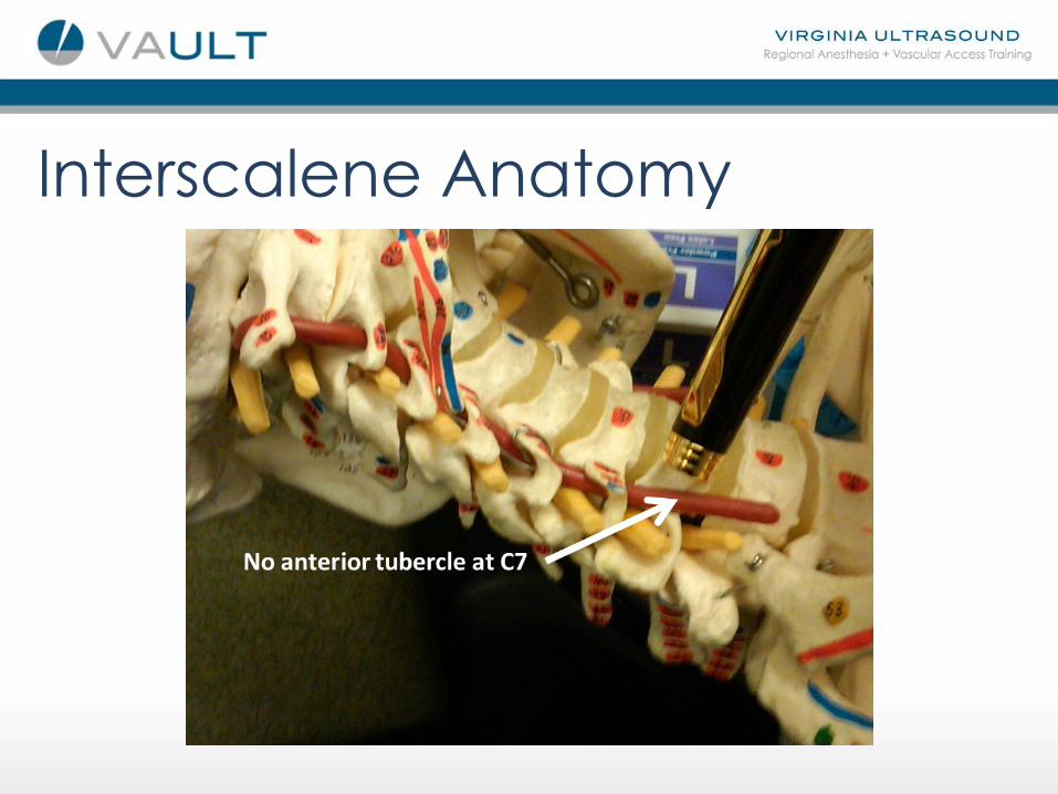
Interscalene Anatomy
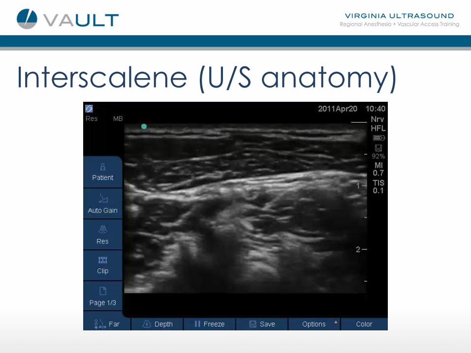
Interscalene (U/S anatomy)

Interscalene (U/S anatomy) “Normal” “Abnormal”
Interscalene
VA
SCM
L
A
T
E
R
A
L

Interscalene Injection

Interscalene • Because the phrenic nerve also lies in the
interscalene groove, it is also frequently blocked, resulting in hemiparesis of the diaphragm
• It is important to avoid injecting local anesthetic immediately adjacent to the transverse process because of the risk of unintentional epidural or spinal injection
• Horner’s syndrome (myosis, ptosis, and anhidrosis) may occur because of the close proximity of the stellate ganglion
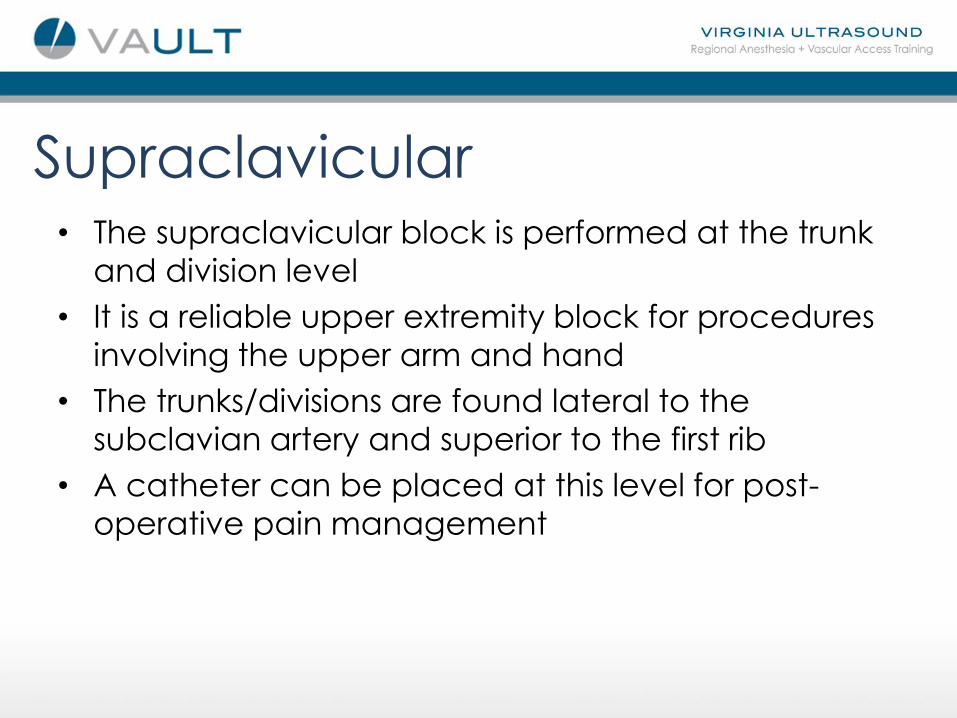
Supraclavicular • The supraclavicular block is performed at the trunk
and division level
• It is a reliable upper extremity block for procedures
involving the upper arm and hand
• The trunks/divisions are found lateral to the
subclavian artery and superior to the first rib
• A catheter can be placed at this level for post-
operative pain management

Supraclavicular
http://warwickphysio.com

Supraclavicular • Patient is placed in supine position with their head
turned to the non-operative side
• Transducer placed supraclavicular fossa
• High frequency linear array transducer
• SAX in-plane image
• Nerves appear as a group of hypoechoic circles
lateral to subclavian artery, superior to first rib
• 5cm needle is used
• 20 – 30cc’s of local anesthetic injected

Supraclavicular
• With ultrasound, it is an
excellent choice for
surgeries of the upper
extremity that do not
involve the shoulder
• Once placed in the
fossa, the transducer is
rocked until 1st rib lies
inferior to the nerves
and artery, and
superior to the pleura

Supraclavicular (anatomy)

Supraclavicular • SAX In-plane image,
needle enter from
lateral position
• The most complete
block is achieved when
local anesthetic is
placed above the first
rib and below the
nerve bundle
M
E
D
I
A
L

Supraclavicular Block

Supraclavicular • As with the Interscalene block there is increased risk
of phrenic nerve paralysis and stellate ganglion
block
• Because of the proximity of the subclavian artery
and lung, there is the possibility for inadvertent
arterial puncture and pneumothorax

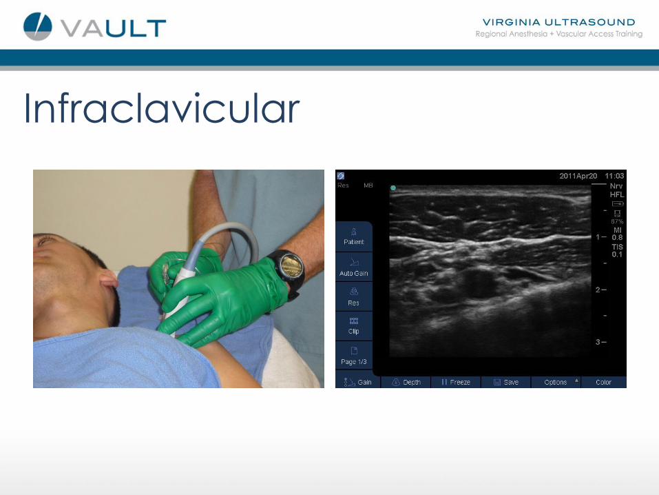
Infraclavicular
• The infraclavicular block is performed at the cord level
• It is an good alternative to the supraclavicular
block, and may be preferred in patients with severe chronic obstructive pulmonary disease
(COPD) or respiratory insufficiency
• The lateral, posterior and medial cords are
arranged superior and lateral, posterior, and posterior and medial around the axillary artery

Infraclavicular

Infraclavicular
• Patient is placed in supine position with their head
turned to the non-operative side
• Transducer is placed perpendicular to the clavicle
just medial to the coracoid plexus
• Mid frequency linear or curved array transducer
• SAX in-plane image
• Nerves are arranged around the axillary artery
• 5 –10cm needle is used
• 20 – 30cc’s of local anesthetic injected

Infraclavicular • It is good for
procedures in the distal upper arm and hand, while at the same time sparing the phrenic nerve
• This block should be judiciously reserved for patients suffering from severe chronic obstructive pulmonary disease (COPD)

Infraclavicular
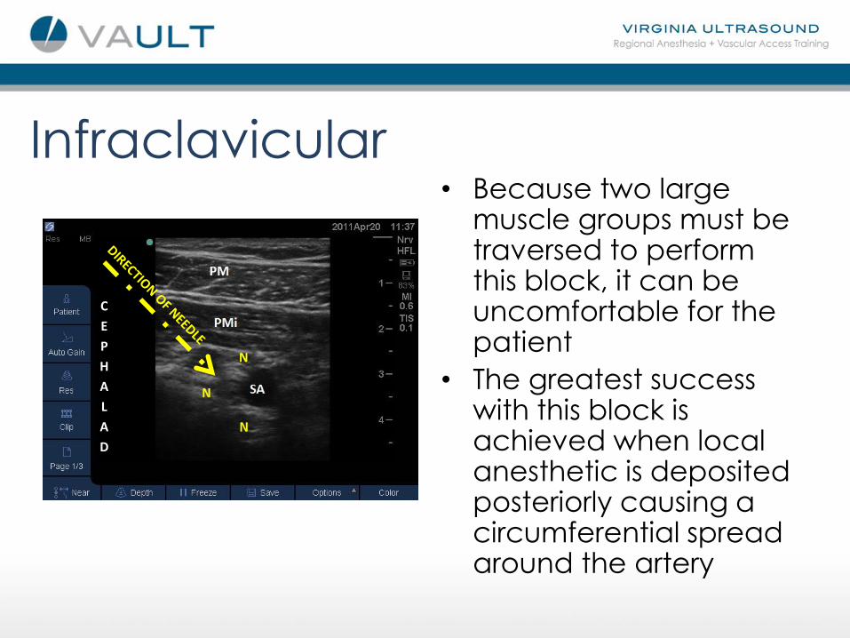
Infraclavicular • Because two large
muscle groups must be traversed to perform this block, it can be uncomfortable for the patient
• The greatest success with this block is achieved when local anesthetic is deposited posteriorly causing a circumferential spread around the artery
C
E
P
H
A
L
A
D
N
Infraclavicular
• Sliding the needle medially increased the potential
for pneumothorax and hemothorax
• The thoraco-acromial artery and pectoral veins
pass between the pectoral muscles. Doppler may
be used to help identify these to prevent
inadvertent puncture

Infraclavicular
Infraclavicular

Axillary • The axillary block is directed at the terminal
branches of the brachial plexus
• It is an excellent block for procedures below the
elbow
• Once a mainstay of regional anesthesia for the
upper extremity, ultrasound has made it less
attractive because other blocks can be done as
efficiently with minimal complications

Axillary • Patient is placed in the supine position with head
turned to the non-operative side, operative arm
abducted and rotated externally
• Transducer is placed perpendicular to the axillary
artery in the crease formed by the biceps muscle
and pectoris major
• High frequency linear array transducer
• Short-axis, in-plane image

Axillary • The radial, ulnar and median nerves are
located around the axillary artery. The
musculo-cutaneous nerve has already left the
sheath and lies within the coracobrachialis
muscle
• 5cm needle is used
• 20 – 30cc’s of local anesthetic injected around
the four nerves. Circumferential spread around the axillary artery is a reliable sign of a
successful block

Axillary • Image the axillary
artery in short-axis. The
needle can be inserted
from either side of the
transducer
• A nerve stimulator may
be helpful in identifying
specific nerves
• There are multiple veins
located around the
artery. Be cautious

Axillary • Compressing the veins
may decrease the risk of vascular puncture
• It is advantageous to block the radial nerve first because it tends to lie deeper than the median and ulnar
• Slide the transducer distally to appreciate each of the nerves, then follow them proximally to their origin
MC
R
M U

Axillary (Ulnar)

Axillary (Median)

Axillary (Radial)

Axillary (Musculocutaneous)

Axillary
• Complications of an axillary block are not common,
however there is an increased risk of vascular
puncture because several needle sticks are
required to achieve adequate local anesthetic
distribution
• Repeated parathesias from multiple needle
punctures may result in neuropathies

Questions ?

References • Chan V., & Pollard B.; An Introductory Cirriculum for Ultrasound-Guided
Regional Anesthesia; 2009, University of Toronto Press.
• Chan, Vincent; Ultrasound Imaging for Regional Anesthesia: A Practical Guide; 3rd Edition; 2010, Toronto Printing Company.
• Gray, Andrew; Atlas of Ultrasound-Guided Regional Anesthesia; 2007, Saunders/Elsevier.
• Hadzic, Admir; Textbook of Regional Anesthesia and Acute Pain Management; 2007, McGraw-Hill Medical.
• Morgan, G., & Mikhail, M.; Clinical Anesthesiology; 4th Edition; 2006, McGraw-Hill Medical.
• Sites, B., & Spence, B.; Ultrasound Guidance in Regional Anesthesia: Techniques for Upper-Extremity and Lower-Extremity Nerve Blocks; 2008, McMahon Publishing.
• Zwiebel, W., & Pellerito, J.; Introduction to Vascular Ultrasonography; 5th Edition; 2005, Elsevier Saunders.