Embed Size (px)
Citation preview

1
Version 1 August 2014
Prevention of Intravascular Catheter-related Infection in Ireland
Update of 2009 National Guidelines1
September 2014 1 HSE Health Protection Surveillance Centre. Prevention of Intravascular Catheter-related Infection in Ireland
SARI Prevention of Intravascular Catheter-related Infection Sub-Committee, December 2009, Updated
February 2010. ISBN 978-0-9551236-6-5
2
Version 1 August 2014
Contents Foreword 2014 ............................................................................................................................ 4
Foreword 2009 ............................................................................................................................ 5
Section 1: Recommendations and Definitions ............................................................................... 6
Summary of 2014 Updated and New Recommendations ........................................................... 7
A: GENERAL INFECTION PREVENTION AND CONTROL PRINCIPLES .............................................. 8
B: CENTRAL INTRAVASCULAR CATHETERS (CVC) ........................................................................ 9
B 1: PREVENTION OF INFECTION ASSOCIATED WITH CVCs ...................................................... 9
B 2: SURVEILLANCE OF INFECTION ASSOCIATED WITH CVCs ................................................. 13
B 3: MANAGEMENT OF CVC-RELATED INFECTION ................................................................. 14
Figure 1: Management of CRBSI associated with non-tunnelled CVCs. .................................. 16
Figure 2: Management of CRBSI associated with tunnelled CVCs or ports (CVC/P) ................ 17
C: PERIPHERAL INTRAVASCULAR CATHETERS (PIVC)................................................................. 18
D: DIAGNOSIS OF INTRAVASCULAR CATHETER-RELATED INFECTION ......................................... 19
E: PREVENTION OF CRBSI IN SPECIFIC SETTINGS ....................................................................... 20
F: IMPLEMENTION OF THESE GUIDELINES................................................................................ 21
Clinical Definitions for Catheter-related Infections 4 ................................................................ 24
Surveillance Definitions 1 ........................................................................................................ 25
Section 2: Rationale for Recommendations ................................................................................. 26
1. Introduction ....................................................................................................................... 26
2. General Infection Prevention and Control Principles ............................................................ 30
3. Central Vascular Catheters (CVCs) ....................................................................................... 33
3.1 Prevention of CVC Infection ........................................................................................... 33
3.2. Surveillance ................................................................................................................. 50
3.3. Management of CVC-related infection 4;15;60 .................................................................. 55
4. Peripheral vascular catheters (PIVCs) .................................................................................. 64
4.1 Prevention of PIVC Infection: Hand Hygiene, Aseptic Technique and Skin Asepsis ........... 64
4.1.2 Selection of PIVC Type ................................................................................................ 65
4.1.3 Selection of PIVC Site ................................................................................................. 65

3
Version 1 August 2014
4.1.4 Procedure for PIVC Insertion, PIVC Fixation and Maintenance of Patency .................... 66
4.1.5 PIVC Removal and Replacement ................................................................................. 67
4.1.6 PIVC Care Bundles (Appendix 16) ................................................................................ 71
4.1.7 PIVC Infection ............................................................................................................ 71
5. Diagnosis of Catheter associated or related infections ......................................................... 72
5.1 Clinical Diagnosis .......................................................................................................... 72
5.2 Laboratory Diagnosis ..................................................................................................... 72
6. Considerations for Specific Settings ..................................................................................... 78
6.1 The Emergency Department .......................................................................................... 78
6.2 Haemodialysis ............................................................................................................... 78
6.3 Critical Care .................................................................................................................. 83
Section 3: Appendices and Reference List ................................................................................... 85
Disclaimer
The clinical advisory group’s (Appendix 1a) expectation is that healthcare staff will use clinical judgment, medical, nursing and clinical knowledge in applying the general principles and recommendations contained in this document. Recommendations may not be appropriate in all circumstances and the decision to adopt specific recommendations should be made by the practitioner taking into account the individual circumstances presented by each patient/resident and available resources. Therapeutic options should be discussed with a clinical microbiologist or infectious disease physician on a case-by-case basis as necessary.

4
Version 1 August 2014
Foreword 2014
In December 2009, the Strategy for the Control of Antimicrobial Resistance in Ireland (SARI) Prevention of Intravascular Catheter-related Infection Sub-Committee published National Guidelines for the Prevention of Intravascular Catheter-related Infection in Ireland (ISBN 978-0-9551236-6-5).
Aspects of these guidelines are updated in these revised guidelines as described below.
This update is integrated with the original recommendations and evidence from the 2009 guidelines.
The updated recommendations are clearly marked as ‘Update 2014’ and highlighted in the text.
Rationale for the update: In January 2014, the Clinical Advisory Group of the National Clinical Programme for the Prevention of Healthcare-Associated Infection (HCAI) and Antimicrobial Resistance (AMR) (Appendix 1a) identified that certain recommendations of the 2009 guidelines for the prevention of intravascular (IV) catheter-related infection in Ireland required updating as did the corresponding national care bundles. To ensure the rapid assessment and implementation of emerging evidence in this important area, a partial review of the 2009 Irish guidelines was undertaken. This review was lead by Dr. Joanne O Gorman in conjunction with the members of the multidisciplinary clinical advisory group.
As other international groups had recently reviewed the evidence base, it was agreed not to repeat this process, rather review the 2009 guidelines in relation to these recent publications. The review focused on the prevention of IV catheter infection and incorporated aspects of the following publications that are acknowledged as the most authoritative reference guidelines currently available;
o epic3: National Evidence-Based Guidelines for Preventing Healthcare-Associated Infections in NHS hospitals in England. (National Institute for Health and Clinical Excellence (NICE) accredited) 2014.
o Infection: prevention and control of healthcare-associated infections in primary and community care (NICE Clinical Guideline) 2012.
o Guidelines for Prevention of Intravascular Catheter-Related Infection (Centre for Disease Control /Healthcare Infection Control Practices Advisory Committee (CDC/HICPAC)) 2011.
o IBTS - National Blood Users Group. Guidelines for the Administration of Blood and Blood Components. 2004.
Limitations: A review of published literature beyond that cited in the aforementioned documents was not undertaken and evidence grading was not applied. The update does not include a review of the 2009 guidelines in relation to; management of intravascular catheter related infection (Sections B3 and 3.3), diagnosis of infection (Sections D and 5.0) or implementation of the guidelines (Section F). A partial review of section B2 was performed. Since publication of the 2009 guidelines, European case definitions for catheter-related infection were agreed by the European Centre for Disease Prevention and Control (ECDC). The clinical advisory group recommend that these definitions are used for surveillance of catheter-related infection. A partial review of section E: prevention of CRBSI in specific settings (Emergency Department and Haemodialysis) was performed to ensure content was updated where applicable.
Consultation: The updated recommendations were widely circulated for consultation. (Appendix 2a) Feedback from the consultation exercise was discussed by the clinical advisory group and the updated guidelines approved in August 2014. The updated guidelines and care bundles are available for download from the Health Protection Surveillance Centre (HPSC) website.

5
Version 1 August 2014
Foreword 2009 Subcommittee details: The Strategy for the Control of Antimicrobial Resistance in Ireland (SARI) National Committee established a subcommittee to produce national guidelines on the prevention of intravascular catheter–related infection. Nominations were requested from the Intensive Care Society of Ireland (ICSI), Infectious Diseases Society of Ireland (IDSI), Irish Nephrology Society (INA), Infection Prevention Society (IPS), Irish Society of Clinical Microbiologists (ISCM), Royal College of Surgeons in Ireland (RCSI) Faculty of Radiologists and the Surveillance Scientists Association of Ireland (SSAI). In addition, individuals with an interest in the field were invited to participate in the group. The membership of the subcommittee is outlined in Appendix 1 The committee first meet in July 2008. Members agreed the terms of reference as listed below. A draft document was sent for circulation to a wide range of professional groups (Appendix 2) in February 2009. This document represents the expert opinion of the sub-committee following a literature review and consultative process. It was not possible for the sub-committee to grade the evidence available in the literature as outlined by the Scottish Intercollegiate Guidelines Network (SIGN) due to the heterogeneity of evidence available, the lack of good quality evidence available for SIGN recommendations and other work commitments of sub-committee members, which precluded a more detailed literature review.
Terms of Reference: To review international best evidence and to make recommendations for the prevention, surveillance, diagnosis and clinical management of intravascular catheter-related infection in Ireland.
This document is aimed at healthcare professionals and outlines recommendations for the prevention, surveillance, diagnosis and clinical management of intravascular catheter-related infection in Ireland. Abbreviations used in this document are outlined in Appendix 3
While we accept that some aspects of the recommendations may be difficult to implement initially due to a lack of facilities or insufficient personnel, we strongly believe that these guidelines represent best practice
Where there are difficulties, these should be highlighted to senior management of the healthcare facility, the Health Services Executive (HSE) and the Department of Health and Children (DoHC) so that measures are taken to ensure implementation, including the provision of appropriate resources and personnel
The Committee recommends that these guidelines are reviewed and updated in 3-5 years

6
Version 1 August 2014
Section 1: Recommendations and Definitions This update to the National Guidelines for the Prevention of Intravascular (IV) Catheter Related Infection in Ireland is integrated with the original recommendations and evidence from the 2009 SARI Guidelines. The recommendations made in this update are clearly marked as ‘Update 2014’ and highlighted in the text.
Recommendations are divided into six sections as follows:
Section Subsection Recommendation Number
General Infection Prevention and Control Principles
General Principles
Hand Hygiene
Aseptic technique
Educations of healthcare workers & patients
1 2 3 4
Central Intravascular Catheters
Skin asepsis
Maximal Barrier Precautions
CVC Insertion Protocols
Selection of CVC type & insertion site
Prophylaxis: Antimicrobial Ointments, Antiseptic & Antimicrobial Locks
CVC Care and Maintenance
Daily Review of CVCs
CVC replacement
CRBSI surveillance
Denominators for surveillance
Management of CVC related infection
5 6 7 8
9
10 11 12 13 14 15
16
Peripheral Intravascular Catheters
17
Diagnosis of Intravascular Catheter related infection
18
Prevention of CRBSI in specific settings
The Emergency Department
Haemodialysis
19 20
Implementation of these guidelines
Responsibility for implementation of these guidelines
21

7
Version 1 August 2014
Summary of 2014 Updated and New Recommendations
CENTRAL INTRAVASCULAR CATHETERS (CVC) Recommendation 5: Skin Asepsis
- Updated recommendation on chlorhexidine allergy Recommendation 8: Selection of CVC Type and Insertion Site
- Updated recommendation for patients requiring regular or continuous IV access - Updated recommendation on antiseptic/antimicrobial impregnated CVCs
Recommendation 10: CVC Care and Maintenance
- New recommendation on chlorhexidine sponge dressings - Updated recommendation on daily skin cleansing with chlorhexidine in adult
patients with CVCs - Updated recommendation on administration sets (IV giving sets)
Recommendation 14: Case definitions for CRBSI surveillance updated PERIPHERAL INTRAVASCULAR CATHETERS Recommendation 17: Updated recommendations on replacement of peripheral intravascular catheters PREVENTION OF CRBSI IN SPECIFIC SETTINGS Recommendation 19: The Emergency Department
- Updated recommendation on replacement of IV catheters APPENDIX 16: UPDATED PERPIHERAL INTRAVASCULAR CATHETER CARE BUNDLE

8
Version 1 August 2014
Summary of Recommendations
A: GENERAL INFECTION PREVENTION AND CONTROL PRINCIPLES Recommendation 1:
Intravascular catheters should only be inserted when there is a clear clinical indication for their use. When the clinical indication is no longer present, the catheter must be removed.
Recommendation 2: Hand Hygiene
Hand hygiene is the single most important procedure in the prevention of intravascular catheter-associated or related infections. Hands must be decontaminated before and after accessing or dressing an intravascular catheter.
Hands can be decontaminated by washing with an antimicrobial liquid soap and water, or if hands are physically clean, by an alcohol based hand rub. Hands that are visibly soiled or contaminated with dirt or organic material must be washed with liquid soap and water before using an alcohol hand rub.
Recommendation 3: Aseptic Technique
Aseptic technique should be used by all healthcare workers during insertion and maintenance of intravascular catheters. Aseptic (no touch) technique is a term used to describe a technique that maintains asepsis and is non-touch in nature – the susceptible site should not come into contact with any item that is not sterile. (Appendix 6)
Following hand hygiene, clean gloves and an aseptic (no touch) technique should be used when accessing an intravascular catheter when the luer* lock is not disconnected from the catheter (e.g., intravenous drug administration, blood sampling or connecting or disconnecting intravenous fluids).
Sterile gloves in addition to aseptic (no touch) technique should be used when a luer needleless connector is disconnected (e.g., manipulation of a catheter, haemodialysis).
Sterile gloves and aseptic (no touch) technique must be used for changing total parenteral nutrition (TPN) and central venous catheter (CVC) insertion site dressing change.
Each facility should develop and implement a standardised protocol for aseptic (no touch) technique.
*Luer connection systems are the standard way of attaching syringes, catheters, hubbed needles, IV tubes, and so on to each other. They consist of round male and female interlocking tubes, they can either be ‘luer slip’, or can have an additional outer rim of threading called a ‘luer lock’, allowing them to be more secure.
Recommendation 4: Education of Healthcare Workers and Patients
Infection prevention and control, including the principles of prevention of catheter-related bloodstream infection (CRBSI), must be an essential component of the core curriculum of training programmes of medical and nursing students at both undergraduate and postgraduate level.
Following training, HCWs must be assessed and documented as competent in using and consistently adhering to appropriate infection prevention and control practices when inserting or maintaining intravascular catheters. Ideally a national competency

9
Version 1 August 2014
document would ensure standardisation of training and allow for interchange between healthcare facilities (due to staff movement); however, this would need an appropriate infrastructure in terms of project management, IT and education.
Only competent, trained staff (or training staff supervised by competent staff) should insert and maintain intravascular catheters.
Before discharge from a healthcare facility, patients with an intravascular catheter and their carers must be educated by a member(s) of the patient’s clinical multidisciplinary team with respect to the procedures necessary to safely manage their catheter and to prevent infection. This should include education on the signs of infection and a relevant information leaflet. (Appendix 7)
Ongoing quality assurance/improvement, risk management and surveillance programmes should be in place to monitor the incidence of infection associated with intravascular catheters, to evaluate the response to patient and staff education, and to identify future educational needs. Monitoring compliance with care bundles are important process measures for evaluation of a CRBSI preventative programme. (Appendix 10, 11 and 16) These results should be reviewed and fed back to relevant ward areas and senior management at regular intervals.
B: CENTRAL INTRAVASCULAR CATHETERS (CVC)
B 1: PREVENTION OF INFECTION ASSOCIATED WITH CVCs
Recommendation 5: Skin Asepsis
Individual single use sachets of antiseptic solution or individual packages of single use antiseptic-impregnated swabs or wipes should be used to disinfect the CVC insertion site. Skin must be allowed to air dry prior to further manipulation. If the skin is visibly dirty, it should be washed with soap and water prior to skin asepsis.
In adults and children ≥ 2 months (assuming normal gestation at birth), a single patient use application of alcoholic chlorhexidine gluconate solution (preferably 2% chlorhexidine gluconate in 70% isopropyl alcohol if compatible with the CVC) should be used and must be allowed to air dry; o For skin disinfectant prior to the insertion of a CVC. o To disinfect the CVC insertion site during dressing changes. o Prior to accessing the CVC hub or injection port.
0.5-1% chlorhexidine is the optimal range for neonatal (< 2 months) skin asepsis; however randomised controlled trials are required to clarify this range.
An aqueous solution of 2% chlorhexidine gluconate should be used if the CVCs manufacturer’s recommendations prohibit the use of alcohol with their product.
Update 2014
Healthcare providers should be aware of the risk of chlorhexidine allergy including anaphylaxis. Single patient use application of alcoholic povidone-iodine solution should be used for patients with a history of chlorhexidine sensitivity if available. Alternatives include tincture of iodine, an iodophor (such as 10% aqueous povidone iodine or povidone iodine alcoholic tincture) or 70% alcohol.

10
Version 1 August 2014
HCW should ensure that CVC site care is compatible with CVC materials (e.g., tubing, hubs, injection ports, luer needleless connectors and extensions) and carefully check compatibility with the manufacturer’s recommendations. This assessment must be performed in advance of purchasing the CVC/materials. If the CVC/materials are incompatible with 2% chlorhexidine gluconate in 70% isopropyl alcohol, there should be a clear clinical benefit to purchasing the CVC/materials. If not, an alternative CVC/materials should be sought.
Recommendation 6: Maximal Barrier Precautions
Maximal barrier precautions are recommended for insertion of all CVCs and when exchanging a CVC over a guidewire and must be used by the operator and any person who enters the sterile field to assist in the procedure.
These precautions include: o Strict compliance with hand hygiene must be practiced by the operator placing the
CVC and staff assisting in the procedure. o Covering the patient with sterile drape(s) from head to toe with an appropriate
opening for the site of insertion. o The operator and staff assisting in the procedure wearing the following: cap, (should
cover all hair), mask (should cover the nose and mouth tightly), protective eyewear, sterile gown and sterile gloves.
Recommendation 7: CVC Insertion Protocols
It is recommended that each healthcare facility has a written CVC insertion procedure guideline that is updated regularly. (Appendix 8)
CVC insertion packs containing all the necessary items for CVC insertion are recommended. (Appendix 9)
It is recommended that a CVC checklist is used to ensure adherence to infection prevention and control practices at the time of CVC insertion. (Appendix 10) This checklist is used to ensure and document compliance with aseptic technique. CVC insertion should be observed by a HCW who has received appropriate education to ensure that aseptic technique is maintained. The observer will assist in identifying breaches in aseptic technique, which if observed should result in the procedure being aborted and restarted.
Recommendation 8: Selection of CVC Type and Insertion Site Patients should be assessed prior to CVC insertion as to the appropriate number of
lumens that are likely to be required. If a multi-lumen CVC is used, one port should be identified and designated exclusively for TPN (if required).
In selecting an appropriate insertion site, the risks for infection should be assessed against the risks of mechanical complications.
For patients likely to require long term renal replacement, early consideration of the future vascular access plan is essential prior to CVC insertion (including future arteriovenous (AV) fistula site). In these patients the subclavian site should be avoided because of the frequent development of subclavian stenosis which interferes with long term provision of vascular access.

11
Version 1 August 2014
Portable ultrasound imaging may be considered for selected patients at high risk of complications (e.g., known vascular anomaly) or where vascular access is likely to be difficult (e.g., children).
The use of implantable ports is recommended for patients who require long term, intermittent vascular access.
Recommendation 9: Prophylaxis: Antimicrobial Ointments, Antiseptic and Antimicrobial Locks
The application of antimicrobial ointment to the CVC placement site prior to insertion is not recommended.
Antimicrobial lock solutions may be used for the prevention of CRBSI in certain subgroups of patients, notably those who require long term vascular access (e.g., haemodialysis, short bowel syndrome) and who have had multiple episodes of CRBSI and have developed these infections despite strict adherence to all other preventative measures. Ongoing surveillance for the emergence of resistant organisms should be performed where antimicrobial lock therapy (ALT) is used.
The decision to use antimicrobial lock prophylaxis and the choice of antimicrobial agent to be used will need to be decided on a individual patient basis, based on the previous positive microbiology and in conjunction with the medical microbiologist / infectious diseases physician.
The administration of prophylactic antimicrobials prior to CVC insertion is not recommended.
Recommendation 10: CVC Care and Maintenance
It is recommended that each healthcare facility has a written CVC care and maintenance guideline that is updated regularly/as new evidence becomes available.
Hand hygiene, aseptic technique and decontamination of the CVC hub/injection port should be performed as in Recommendations 2, 3 and 5.
Manipulations of the CVC, including replacement of dressings should be documented.
A sterile, transparent semipermeable dressing should be used to cover the CVC insertion site and should be changed every seven days or sooner if it is no longer intact or if moisture collects under the dressing. If a sterile gauze dressing is used (e.g., if a patient has profuse perspiration or if the insertion site is bleeding or oozing) it should be replaced by a transparent semipermeable dressing as soon as possible.
Update 2014
In units or patient populations that have a high CRBSI rate despite compliance with basic CRBSI prevention practices, antiseptic or antimicrobial impregnated CVCs should be used in adults whose catheter is expected to remain in place >5 days.
Update 2014
The use of chlorhexidine impregnated sponge dressing should be considered in adult patients with temporary short term CVCs.

12
Version 1 August 2014
Dressings used on tunnelled or implanted CVC insertion sites should be replaced every seven days until the insertion site has healed, unless there is an indication to change them sooner.
A sterile 0.9% sodium chloride solution should be used to flush and lock CVC lumens. When recommended by the manufacturer, implanted ports or opened-ended CVC lumens should be flushed and locked with heparin sodium flush solutions. Routine use of systemic anticoagulants is not recommended to prevent CRBSI. The committee have omitted heparin dosage information in these guidelines. This is because policy may differ between healthcare facilities and patient groups. It is suggested that on adoption of these guidelines, the use of heparin is supported with in-house guidelines which take into account dosage and product formulation. In addition, special provision should be made for patients with a history of heparin induced thrombocytopenia, as heparin should not be used in such a scenario.
Recommendation 11: Daily Review of CVCs
All CVCs should be reviewed daily, documented as reviewed and those that are no longer clinically indicated promptly removed.
The insertion site should be examined daily for drainage, tenderness, pain, redness, swelling, suture integrity and CVC position and all findings documented. Site appearance should not be used as the only indicator of infection. The patient should also be examined for fever or other signs of sepsis (e.g., tachycardia, tachypnoea, hypotension).
Patients should be encouraged (where possible) to report any changes in their CVC site or any new discomfort.
Patients transferring from other healthcare facilities with a CVC in situ must have the device reviewed upon arrival for evidence of any infectious or mechanical complications.
Recommendation 12: CVC Replacement
Management of CVC replacement in the context of CVC infection is outlined in Recommendation 16.
Update 2014
Consider the use of daily skin cleansing with chlorhexidine in adult patients with a CVC.
Update 2014
Administration sets (IV giving sets) in continuous use do not need to be replaced more frequently than every 96 hours unless they become disconnected, or the intravascular access device is replaced.
Blood administration sets should be changed after a maximum of 6 hours.
Administration sets in continuous use for lipid containing parenteral nutrition should be changed 24 hours after initiating the infusion.
Replace tubing used to administer propofol infusions every 6 or 12 hours, when the vial is changed, per the manufacturer’s recommendation.

13
Version 1 August 2014
If the CVC is fractured, it should be replaced and a new CVC inserted ideally at a different site.
Because breaches in sterile technique are more likely during emergency procedures, CVCs inserted during a medical emergency must be replaced as soon as possible.
Routine replacement of CVCs that are functioning and have no evidence of causing local or systemic complications (including scheduled guidewire exchanges of CVCs) as a method to reduce CRBSI is not recommended.
Guidewire techniques should not be used to replace CVCs in patients suspected of having CVC infection. Guidewire assisted CVC exchange to replace a malfunctioning CVC or to exchange an existing CVC should be used only if there is no infection at the CVC site or no suspicion of CRBSI. If after a guidewire exchange, investigations reveal CRBSI, the newly inserted CVC should be removed and if still required reinserted at a different site. In selected patients with tunnelled haemodialysis CVCs and bacteraemia, CVC exchange over a guidewire, in combination with antibiotic therapy, might be an alternative as a salvage strategy in patients with limited venous access.
For guidewire exchanges, the same meticulous aseptic technique and use of full sterile barriers are mandatory as outlined in Recommendations 2-3 and 5-9.
B 2: SURVEILLANCE OF INFECTION ASSOCIATED WITH CVCs
Recommendation 13: CRBSI Surveillance
Healthcare managers must support surveillance activities, including surveillance of CRBSI.
Surveillance must start and end with the patient in order to improve patient care. A CRBSI surveillance programme should be introduced in an healthcare facility as dictated by the specialities and requirements of that healthcare facility and the resources available for surveillance, to determine healthcare associated (HCA) CRBSI rates, monitor trends in rates, and assist in identifying lapses in infection prevention and control practices. Areas that may be involved might include ICU/HDU, dialysis units, haematology/oncology units, TPN services and interventional radiology units. The committee have provided sample forms for CRBSI surveillance. (Appendices 12-13) These forms represent a template and can be used to guide healthcare facilities in the design of their own forms. Each healthcare facility may wish to include additional questions in the template form so that local needs can be met.
A local multidisciplinary steering committee should be established with representatives from the relevant area(s) in which surveillance is to commence (e.g., ICU, haemodialysis, medical microbiology, infectious diseases, infection prevention and control and senior management) to help drive the surveillance project, encourage compliance and advise the relevant area(s) and healthcare facility management based on surveillance results.
CRBSI rates must be fed back to the relevant area(s) and healthcare facility management on a regular basis, ideally monthly, but at least quarterly.
All clusters of HCA CRBSI and all episodes of HCA CRBSI due to S. aureus must be investigated.
The introduction of new intravascular catheters should be monitored for an increase in the occurrence of infection.

14
Version 1 August 2014
Recommendation 14: Case Definitions for CRBSI Surveillance
CRBSI protocols must be standardised and adhere to other international frameworks (e.g., HELICS) for comparative analysis of CRBSI incidence rates.2
Recommendation 15: Denominators for Surveillance
The CRBSI rate should be expressed as the number of CRBSIs per 1000 CVC days.
B 3: MANAGEMENT OF CVC-RELATED INFECTION
Recommendation 16:
Management of CVC-related infection depends on the type of CVC involved, the infecting organism, and the associated complications.
When a CVC-related infection is documented and a specific pathogen is identified, systemic antimicrobial therapy should be adjusted according to antimicrobial susceptibility.
Duration of treatment will depend on the organism identified, presence of bacteraemia, presence of complications and whether the line has been removed.
When denoting duration of antibiotic therapy for treatment of BSI, day one is the first day on which negative blood cultures are obtained.
Exit site infection: Empiric therapy with an appropriate antibiotic should be commenced after blood cultures are taken and involvement of the tunnel/port pocket outruled (if a tunnelled CVC is present). CVC removal is recommended if antibiotic treatment fails. Exchange of the CVC over a guidewire in the presence of an exit site infection is not recommended. If blood cultures are positive, then treatment for CRBSI is indicated.
Tunnel infection: Successful therapy of tunnel infections without CVC removal is very unlikely. In the absence of bacteraemia 7-10 days of antibiotics may suffice. If associated with bacteraemia, the patient should be considered to have complicated CRBSI.
CRBSI: o In patients with BSI and an indwelling CVC, it is important to rule out other sources of
infection to avoid unnecessary CVC removal. Where a patient has a single blood culture for coagulase-negative Staphylococcus spp. additional blood cultures (peripheral and through the CVC) should be obtained.
o Empiric intravenous antimicrobial therapy should be considered, after cultures are obtained. In general a glycopeptide antibiotic is recommended for empirical therapy in
2 2 ECDC - European Surveillance of Healthcare-associated Infections in Intensive Care Units – HAIICU
Protocol Version 1.1
http://www.ecdc.europa.eu/en/aboutus/calls/Procurement%20Related%20Documents/5_ECDC_HAIICU_proto
col_v1_1.pdf
Update 2014
The HELICS case definitions for catheter-related infection as outlined by the European Centre for Disease Prevention and Control (ECDC) are the recommended case definitions for intravascular catheter-related infection surveillance.

15
Version 1 August 2014
health care settings in which MRSA is prevalent. Additional gram- negative coverage is indicated in patients who are neutropenic or severely ill with sepsis or for suspected infections involving femoral catheters. Antifungal agents (choice depending on local susceptibility patterns) should be considered for empirical treatment when fungaemia is suspected.
o Patients with complicated CRBSI will require 4-6 weeks of IV antibiotics. This includes patients with suppurative thrombophlebitis, endocarditis, metastatic seeding, or persistent bacteraemia (> 72 hours despite appropriate antibiotics) after removal of the catheter.
o Management of CRBSI when the infecting organism is known is outlined in Figures 1 and 2.
o Repeat blood cultures to document clearance of bacteraemia are recommended. o In uncomplicated CRBSI due to organisms other than S. aureus, P. aeruginosa, fungi,
mycobacteria, Micrococcus spp., Proprionobacterium or Bacillus spp., CVC salvage may be attempted in situations where there is limited vascular access. If bacteraemia is persistent (>72 hours) this should prompt reassessment of the ability to salvage the CVC. ALT should be used when CVC salvage is being attempted, however this should always be administered with systemic antibiotic therapy.

16
Version 1 August 2014
Figure 1: Management of CRBSI associated with non-tunnelled CVCs.

17
Version 1 August 2014
Figure 2: Management of CRBSI associated with tunnelled CVCs or ports (CVC/P)

18
Version 1 August 2014
C: PERIPHERAL INTRAVASCULAR CATHETERS (PIVC)
Recommendation 17:
Only competent, trained staff (or training staff supervised by competent staff) should insert and maintain PIVCs.
In order to prevent contamination of PIVC sites and subsequent BSI, hand hygiene and aseptic technique as outlined in Recommendations 2 and 3 must be performed each time: o Before PIVC insertion (both before and after palpating the PIVC insertion site). o Before PIVC access or maintenance (e.g., dressing manipulations, palpating the PIVC). Following hand hygiene, clean gloves and an aseptic technique must be employed. Hand hygiene must also be performed immediately after removing gloves and after each episode of patient care. All sharps must be disposed of carefully into an approved sharps container.
In adults and children ≥ 2 months (assuming normal gestation at birth), a single patient use application of alcoholic chlorhexidine gluconate solution (preferably 2% chlorhexidine gluconate in 70% isopropyl alcohol if compatible with the PIVC) should be used and allowed to air dry; o For skin disinfection prior to the insertion of a PIVC. o To disinfect the PIVC insertion site during dressing changes. o Prior to accessing the PIVC hub.
0.5-1% chlorhexidine is the optimal range for neonatal (< 2 months) skin asepsis; however, randomised controlled trials are required to clarify this range. (Section 3.1.2.i)
The PIVC site should not be re-palpated after skin asepsis.
Select the PIVC and insertion site with the lowest risk for complications for the anticipated type and duration of IV therapy.
A sterile, transparent semipermeable dressing should be used to cover the PIVC insertion site. Routine dressing change is not recommended unless the dressing is no longer intact or moisture collects under the dressing
When adherence to aseptic technique cannot be ensured (i.e., when PIVCs are inserted during a medical emergency), the PIVC should be replaced as soon as possible.
All PIVCs should be reviewed daily, and those that are no longer needed should be promptly removed. Details of the review and the decision to remove or not should be clearly documented.
All PIVCs must be removed promptly when there is clinical evidence that the PIVC is infected.
Update 2014
The PIVC insertion site should be visually inspected at least twice daily (on every shift) for evidence of complications. This assessment should be clearly documented.
PIVC should be re-sited when clinically indicated and not routinely.
Update 2014
Patients transferring from other healthcare facilities with a PIVC in situ should have this device reviewed upon arrival to ensure it is still needed. PIVC

19
Version 1 August 2014
D: DIAGNOSIS OF INTRAVASCULAR CATHETER-RELATED INFECTION
Recommendation 18:
Clinical findings alone are unreliable for establishing a diagnosis of intravascular catheter–related infection, because of their poor specificity and sensitivity.
Two sets of blood cultures should be taken using aseptic technique from all patients with suspected intravascular catheter-related infection. For CVCs either through the CVC and peripherally or through different lumens of the CVC if blood cultures cannot be drawn from a peripheral vein. Blood cultures should be taken prior to initiation of antimicrobial therapy. The bottles should be appropriately marked to reflect the site the cultures were drawn from.
Routine culturing of intravascular catheter tips is not recommended. However, CVC tips should always be sent for culture if the CVC is removed and catheter-related infection is suspected. It is essential that every CVC is removed using aseptic technique.
For suspected pulmonary artery catheter infection, the introducer tip should be cultured.
If an implantable port is removed for suspected CRBSI, the catheter tip and the port should be sent for qualitative culture of the port reservoir contents.
If pus is present at the catheter exit site, the site must be swabbed for culture and removal of the catheter considered. (Recommendations 16 and 17)
Growth of >15 CFU from a segment of the catheter tip by semiquantitative (roll-plate) culture or growth of >102CFU from a catheter by quantitative (sonication) broth culture reflects catheter colonisation. All such isolates from CVC tips are potentially significant and should be identified to genus level and to species level, if clinically indicated. Antimicrobial susceptibility should be performed on all clinically significant isolates.
The choice of the precise microbiological method for CRBSI diagnosis may vary locally and should be made according to technical availability and after discussion between clinicians and medical microbiologists. In addition, economic considerations, such as cost-effectiveness, may also be taken into account.
Blood culture results that are positive for S. aureus, coagulase-negative staphylococci, or Candida spp., in the absence of any other identifiable source of infection, should increase the suspicion for CRBSI.
For diagnosis of CRBSI the following criteria should be met: Bacteraemia or fungaemia in a patient who has an intravascular device and >1 positive blood culture obtained from the peripheral vein, clinical manifestations of infection (e.g., fever, chills, and/or hypotension), and no apparent source for BSI (with the exception of the catheter).
One of the following should be present: o A positive result of semiquantitative (>15 CFU/catheter segment) or quantitative
(>102 CFU /catheter segment) catheter culture, whereby the same organism (spp.) is isolated from a catheter segment and a peripheral blood culture.
o Simultaneous quantitative cultures of blood with a ratio of > 3 : 1 CFU/ml of blood (catheter versus peripheral blood); differential time to positivity (Growth in a blood culture drawn through catheter hub is detected by an automated blood culture system at least 2 hours earlier than a simultaneously drawn, peripheral blood culture of equal volume).

20
Version 1 August 2014
E: PREVENTION OF CRBSI IN SPECIFIC SETTINGS
Recommendation 19: The Emergency Department
Only appropriately trained staff (or trainee staff supervised by competent staff) should insert percutaneous CVCs in Emergency Departments. (Recommendation 4)
There should be strict adherence to hand hygiene, skin asepsis and aseptic insertion technique. (Recommendations 2-3 and 5-9)
Ultrasound-guided central venous access should be considered.
Accurate documentation and record keeping is required for all instances of CVC insertion in the Emergency Department. A CVC Insertion Checklist (Appendix 10) may be used to ensure patient safety, auditing of clinical practice, and the tracking of infective complications.
Recommendation 20: Haemodialysis
Haemodialysis patients should whenever possible and practical have a primary arteriovenous (AV) fistula created for vascular access. If it is not possible to achieve a functioning AV fistula a polytetrafluoroethylene (PTFE) graft is in general preferable to long term cuffed catheters.
Renal units need to have adequate access to vascular surgeons in order to ensure the timely creation of primary vascular access.
Patients with progressive renal failure should have a primary AV fistula created when the eGFR is between 17 and 12 aiming to start such patients with their first dialysis through a functioning fistula.
Each unit should keep records of primary fistula prevalence, PTFE graft prevalence and cuffed catheter prevalence.
Units should review bacteraemia rates for patients with and without catheters on a regular basis. When an episode of bacteraemia develops in a dialysis patient a root cause analysis should be undertake to identify the source of infection and potentially modifiable risk factors.
All patients should be screened for prevalence of MRSA colonisation regularly (e.g., three monthly) and patients managed as per national guidelines2.
When CVC infection is suspected in haemodialysis patients, two sets of blood cultures should be taken using aseptic technique (either through the CVC and peripherally, or through different lumens of the CVC if peripheral blood cultures cannot be taken). Peripheral blood cultures should be obtained from vessels not intended for future use in creating a dialysis fistula. When a peripheral blood culture cannot be obtained, blood cultures should be drawn during haemodialysis from bloodlines connected to the CVC.
Empiric antibiotic therapy can be discontinued in patients with suspected CRBSI if both sets of blood cultures are negative and no other source of infection is identified. If a peripheral blood culture cannot be obtained and no clinical evidence for an alternate source of infection, then a positive catheter-drawn blood culture in a symptomatic
Update 2014
PIVC which have been inserted using aseptic technique in the Emergency Department do not need to be removed if there is no evidence of complications.

21
Version 1 August 2014
haemodialysis patient should lead to continuation of antimicrobial therapy for possible CRBSI.
The infected CVC should be removed in patients with haemodialysis CRBSI due to S. aureus, Pseudomonas or Candida spp. and a temporary (non-tunnelled catheter) inserted into another anatomical site. A long-term haemodialysis catheter can be placed once repeat blood cultures are negative. Guidewire exchange is recommended only if no alternative sites are available for CVC insertion.
For CRBSI due to other pathogens (e.g., Gram negative bacilli other than Pseudomonas spp. or coagulase-negative staphylococci), a patient can be started on empiric intravenous antibiotics without immediate catheter removal (provided patient clinically stable). If symptoms persist or there evidence of a metastatic infection, the catheter should be removed.
Surveillance blood cultures should be obtained one week after completing an antibiotic course for CRBSI if the catheter has been retained. If the blood cultures are positive, the catheter should be removed and a new, long-term dialysis catheter should be placed after a repeat blood cultures are negative.
F: IMPLEMENTION OF THESE GUIDELINES
Recommendation 21: Responsibility for the implementation of these guidelines
Prevention of HCAI should be prioritised by the Department of Health (DHC), the Health Services Executive (HSE) and all healthcare staff in order to improve patient care and safety and to reduce all HCAI, including CRBSI.
Implementation of the National Standards for the Prevention and Control of HCAI3 will be a key aspect of the prevention and control of intravascular catheter-related infection. Standard 8 (invasive medical device-related infection) outlines the specific key criteria that will be assessed in this regard.
The following infrastructural requirements are recommended to institute a programme to prevent CRBSI: o An adequately staffed infection prevention and control programme responsible for
identifying patients with CRBSI, including a surveillance coordinator with appropriate administrative support.
o Information technology to collect and calculate catheter- days as a denominator for computing rates of CRBSI and patient-days to allow calculation of CVC utilisation; Catheter-days from information systems should be validated against a manual method.
o Resources to provide appropriate education and training. o Adequate laboratory support for timely processing of specimens and reporting of
results.
Implementation of these guidelines may require ring-fenced funding to assist healthcare facilities to meet these recommendations, specifically surveillance, laboratory, infection prevention and control infrastructure and personnel.

22
Version 1 August 2014
Update 2014
It is essential that all healthcare staff understand and appreciate that they are responsible for the prevention and control of HCAI which includes intravascular catheter-related infection in all areas of their responsibility.
This must be supported by clear lines of accountability which include systems that can detect and correct lapses in infection prevention and control practice on a timely basis and increases in intravascular catheter-related infection incidence.
Patients can also play a role, expecting the highest standards of healthcare quality and safety and ensuring that healthcare facilities assure them that there is an effective intravascular catheter-related infection control programme in place.
Roles and Responsibilities: Each healthcare staff member has a role to play in the prevention and control of healthcare-associated infection, which includes intravascular catheter-related infection by adhering to best practice as outlined in these guidelines.
This guideline should be reviewed by the healthcare facilities senior management teams in conjunction with the relevant specialists to plan implementation of the recommendations.
This will enable the facility to ensure that the prevention and control of intravascular catheter-related infection is a key patient/resident safety issue for the facility.
Organisational responsibility: Within each healthcare facility the CEO/General Manager has corporate and clinical responsibility for implementation of this guideline.
All healthcare staff:
Comply with this guideline and related policies, procedures and protocols.
Adhere to their code of conduct and scope of practice guidelines as appropriate to their role and responsibilities
Maintain competency in the prevention and control of intravascular catheter-related infection
In using this guideline be aware of the role of appropriate delegation. The following are examples of audit criteria to monitor implementation of these guidelines:

23
Version 1 August 2014
Outcome Measures:
- Intravascular catheter-related infection rates
- Bloodstream infection associated with intravascular catheters (central and peripheral)
Process Measures:
- CVC Insertion checklist compliance
- Maintenance Care Bundle compliance
- Hand hygiene compliance score (%) When adherence to aseptic technique cannot be ensured (i.e. catheters inserted during a medical emergency), replace the intravascular catheter

24
Version 1 August 2014
Clinical Definitions for Catheter-related Infections 4
Definition
Catheter Colonisation
Significant growth of one or more microorganisms in a quantitative or semiquantitative culture of the catheter tip, subcutaneous catheter segment, or catheter hub (Section 5)
Phlebitis Induration or erythema, warmth, and pain or tenderness along the tract of a catheterised or recently catheterised vein
Exit site infection o Microbiological
Exudate at catheter exit site yields a microorganism with or without concomitant bloodstream infection (BSI)
o Clinical
Erythema, induration, and/or tenderness within 2 cm of the catheter exit site; may be associated with other signs and symptoms of infection, such as fever or purulent drainage emerging from the exit site, with or without concomitant BSI
Tunnel Infection Tenderness, erythema, and/or induration >2 cm from the catheter exit site, along the subcutaneous tract of a tunnelled catheter (e.g., Hickman or Broviac catheter), with or without concomitant BSI
Pocket Infection
Infected fluid in the subcutaneous pocket of a totally implanted intravascular device; often associated with tenderness, erythema, and/or induration over the pocket; spontaneous rupture and drainage, or necrosis of the overlying skin, with or without concomitant BSI
Bloodstream infection o Infusate-Related
Concordant growth of a microorganism from infusate and cultures of percutaneously-obtained blood cultures with no other identifiable source of infection
o Catheter-Related
Bacteraemia or fungaemia in a patient who has an intravascular device and >1 positive blood culture obtained from the peripheral vein, clinical manifestations of infection (e.g., fever, chills, and/or hypotension), and no apparent source for BSI (with the exception of the catheter). One of the following should be present:
A positive result of semiquantitative (>15 CFU/catheter segment) or quantitative (>102 CFU /catheter segment) catheter culture, whereby the same organism (species.) is isolated from a catheter segment and a peripheral blood culture
Simultaneous quantitative cultures of blood with a ratio of > 3 : 1 CFU/ml of blood (catheter vs. peripheral blood); differential time to positivity (Growth in a blood culture drawn through catheter hub is detected by an automated blood culture system at least 2 hours earlier than a simultaneously drawn, peripheral blood culture of equal volume).
Note: This definition differs from the definition of central line-associated BSI used for surveillance activities.

25
Version 1 August 2014
Surveillance Definitions 1
The HELICS case definitions for catheter-related infection as outlined by the European Centre for Disease Prevention and Control (ECDC) are the recommended case definitions for intravascular catheter-related infection surveillance
At the time of publication of the 2009 guidelines European surveillance definitions for intravascular catheter-related infection had not been agreed and at that stage
the CDC surveillance definitions were recommended1
Since then, ECDC have recommended the HELICS case definitions as outlined in the protocol for intensive care unit surveillance.3 These definitions were used by Irish hospitals that participated in the 2012 prevalence survey of hospital-acquired infection.4
Further information is available on the ECDC and HPSC websites.
3
http://www.ecdc.europa.eu/en/aboutus/calls/Procurement%20Related%20Documents/5_ECDC_HAIICU_proto
col_v1_1.pdf 4 http://www.hpsc.ie/A-
Z/MicrobiologyAntimicrobialResistance/InfectionControlandHAI/Surveillance/HospitalPointPrevalenceSurveys
/2012/

26
Version 1 August 2014
Section 2: Rationale for Recommendations
1. Introduction
A major feature of healthcare-associated infection (HCAI) in the last 20 years has been its association with medical devices such as intravascular catheters. Though essential for the care of patients, intravascular catheters represent an avenue by which microorganisms can gain entry to the body. Intravascular catheter-related bloodstream infections (CRBSI) have become a leading cause of health-care-associated (HCA) bloodstream infections (BSI) and are associated with substantial morbidity and mortality. CRBSI represent 10–20% of all nosocomial infection and may complicate the stays of up to 10% of intensive care unit (ICU) patients.5 CRBSI independently increase hospital cost and length of stay.6-9 Over 250 000 CRBSI occur annually in the US with an attributable mortality ranging from 12% to 25% in critically ill patients, with an added cost ranging from US$3000 to $56 167.6;10;11 Intravascular catheters represent potentially modifiable HCAI risk factors, therefore a focus on infection prevention is essential to ensure appropriate practice during the insertion and subsequent optimal care. Preventative strategies to reduce the prevalence of CRBSI have been
effective in other countries and include; education of health-care workers (HCWs) on correct catheter insertion and maintenance, routine monitoring of healthcare facility CRBSI rates, adherence to hand hygiene, the use of a dedicated infusion therapy team, use of sterile semipermeable dressings and removing the intravascular catheter as soon as possible. (Sections 2-4) Preventative programmes, including institution of appropriate surveillance programmes not only reduce catheter-related infection, but also have significant cost savings. In one Irish hospital, the introduction of a dedicated total parenteral nutrition (TPN) surveillance coordinator resulted in a decrease of 9.8 CRBSI per year, representing a minimum saving of €78,300 per annum.12 1.1 Types of Intravascular Catheters A large variety of intravascular catheters exist which can be broadly divided into central vascular catheters (CVC) and peripheral vascular catheters (PIVC). CVCs are intravascular catheters that terminate at or close to the heart or in one of the great vessels and are used for infusion, withdrawal of blood or hemodynamic monitoring. The tip of a CVC is placed close to a site feeding a large deep systemic vein (Swan Ganz CVCs are placed in pulmonary arteries) where there is a large vessel lumen and high flow state limiting vessel injury and thrombosis. These vessels include internal jugular (IJ), subclavian (SC) and femoral vein (FV) placement. In exceptional circumstances, CVCs may be placed translumbar into the inferior vena cava, in hepatic veins and through large collaterals in those with central venous obstruction. A number of different CVCs exist which vary with respect to insertion technique, size, number of lumens and intravascular catheter materials. (Appendix 4 and 5) In contrast, the tip of a PIVC is placed in a superficial small systemic vein, typically basilic, cephalic, forearm, hand or foot veins. PIVCs may rarely be placed in other superficial veins or collateral veins. 1.2 Clinical Presentation and Diagnosis of Catheter-related Infection All intravascular catheters are associated with a risk of infection. This risk varies with the type of catheter, insertion site, experience and education of the catheter inserter, frequency of accessing the catheter, duration of catheter placement, the use of infection prevention

27
Version 1 August 2014
and control strategies and characteristics of the catheterised patient.10 Any patient with an intravascular catheter is potentially at risk for intravascular catheter-related infection however certain populations of patients are at higher risk. These patients include:
Patients in the ICU - frequent insertion of multiple intravascular catheters that are repeatedly accessed, often required for prolonged periods and may be inserted in emergency situations.
Non-ICU patients with CVCs, including haemodialysis patients and haematology/ oncology patients. For patients with CVCs, factors associated with increased risk of infection include; prolonged hospitalisation before catheterisation, prolonged duration of catheterisation, heavy microbial colonisation at the insertion site or CVC hub, internal jugular catheterisation, neutropaenia, prematurity, TPN and substandard care of the catheter (e.g., excessive manipulation of the catheter or reduced nurse-to-patient ratio).13
CVC-related infections can present with local or systemic symptoms. Local infections include exit site infection, tunnel infection, and pocket infection. (Section 3.3) Symptoms may include induration, erythema, warmth, and pain or tenderness at or around the intravascular catheter exit site. Local infections can be associated with systemic symptoms including CRBSI. CRBSI should be considered when a patient with a CVC presents with bacteraemia/fungaemia in the presence of signs and symptoms of systemic infection (e.g., fever, rigors, hypotension). Probable CRBSI can be diagnosed by one or more positive blood cultures obtained from a peripheral vein, when there is no apparent source for the BSI except the intravascular catheter. However, the diagnosis of CRBSI remains a major challenge. Local catheter site inflammation has poor sensitivity, while the presence of systemic symptoms such as fever is not specific enough.14;15 Therefore, microbiological evidence implicating the catheter as a source of the BSI is necessary for establishing a diagnosis of CRBSI. These diagnostic approaches which can be divided into two major groups (those that require catheter removal and those that do not) will be discussed in further detail in Section 5.
PIVCs are the devices most frequently used for vascular access. Although the incidence of BSI is low, serious complications can produce considerable morbidity. . PIVCs may be complicated by phlebitis, extravasation and colonisation, all of which increase the risk of PIVC infection and BSI. Phlebitis is associated with prolonged placement of a PIVC (>72 hours).
1.3 Pathogenesis The microorganisms most commonly associated with CRBSI include coagulase negative staphylococci, Staphylococcus aureus, aerobic gram- negative bacilli, and Candida spp. Important pathogenic determinants of catheter-related infection are the material of which the device is made and the intrinsic virulence factors of the infecting organism. Catheters made of polyvinyl chloride or polyethylene are likely less resistant to the adherence of microorganisms than are catheters made of PTFE, or silicone elastomer.10 Certain materials are more thrombogenic than others, which may predispose to catheter colonisation. In addition, adherence properties and biofilm formation by a given microorganism is also important in the pathogenesis of infection.

28
Version 1 August 2014
The pathogenesis of CVC infection also varies with the type of CVC. Infection of non-tunneled CVC is due to either extra luminal CVC colonisation (which originates most frequently from the skin and less commonly from haematogenous seeding of the tip), or intraluminal CVC colonisation of the hub and lumen.15 In contrast, contamination of the CVC hub and intraluminal infection is the most common route of infection of tunneled CVCs or implantable devices. In addition to skin, there is evidence that mucosal colonisation is an important source of coagulase-negative staphylococcal bacteraemia.16 With respect to PIVCs, phlebitis is associated with prolonged placement (>72 hours). Migration of skin organisms at the insertion site into the cutaneous PIVC tract with colonisation of the tip is the most common route of infection. Occasionally organisms enter intraluminally following contamination of the PIVC hub. Once microorganisms enter, biofilm forms on the lumen surface and as a consequence, the PIVC becomes infected. 1.4 Irish Epidemiology 1.4.1 North-South MRSA Study 199917 The 1999 North-South Study evaluated the epidemiology and management of meticillin resistant S. aureus (MRSA) cases identified in Irish laboratories. The prevalence of MRSA was higher in the South (14.0 per 100 000 population) than in the North (11.4 per 100 000 population). While the majority of cases represented MRSA colonisation, 5% (North) and 10% (South) of cases had invasive infection. Patients with invasive infection were more likely to have a history of PIVC or CVC than those with colonisation only. 1.4.2 Enhanced EARSS Surveillance The European Antimicrobial Resistance Surveillance System (EARSS) comprises a network of over 800 microbiological laboratories serving some 1200 hospitals in 30 countries that collects routinely-generated antimicrobial susceptibility testing data on invasive infections caused by seven important bacterial pathogens: Staphylococcus aureus, Streptococcus pneumoniae, Escherichia coli, Enterococcus faecalis, Enterococcus faecium, Klebsiella pneumoniae and Pseudomonas aeruginosa. The HPSC coordinates national collation of EARSS data. As of quarter 1 2009, 42 Irish laboratories serving 61 acute hospitals (public and private) participate in EARSS representing approximately 97% coverage of the Irish population. In the first quarter of 2009, 30% of S. aureus were meticillin resistant compared with 31% in the last quarter of 2008.18 The annual trend decreased from approximately 42% in 2006 to 39% in 2007 and 34% in 2008. This is the lowest annual proportion since surveillance began in 1999. In addition, HPSC has collected enhanced surveillance data since 2004. The enhanced programme involves voluntary participation by laboratories that provide data on invasive pathogens causing BSI. CVCs have been recorded as the most common source of S. aureus BSI and are equally relevant to both meticillin resistant and sensitive isolates. (Table 1.1) A smaller but significant proportion of S. aureus BSI was associated with PIVCs.

29
Version 1 August 2014
1.4.3 Hospital Infection Society (HIS) HCAI Prevalence Survey 2006 Of the 75 694 UK and Irish patients surveyed during the 2006 HIS HCAI prevalence survey, 5743 (7.6%) had a HCAI. 449 patients had a primary BSI, 184(41%) of which were CVC- related.19 The presence of a CVC on the day of the survey or within the last seven days was significantly associated with primary BSI, with odds ratios of 14.6 and 4.14 respectively.20 Significantly more patients in the Republic of Ireland had intravascular catheters in situ (PIVC (p<0.001) or CVC (p=0.030)), when compared with patients in Northern Ireland, though there was no significant difference in prevalence rates of HCAI, device-related HCAI or HCAI associated with secondary BSI. There was however, a significant difference in MRSA-associated HCAI.21 As in other countries, presence of a CVC in Irish patients was associated with a HCAI.
1.5 Existing International Guidelines and Purpose of this Document A number of existing international guidelines for prevention and management of intravascular catheter-related infection are routinely used by healthcare professionals in Ireland, including CDC10 (2002 – due to be updated by 2010 – personal communication to chair), The Institute for Healthcare Improvement, IDSA,15 EPIC-222 and the National Kidney Foundation- Kidney diseases outcomes quality initiative (NKF-K/DOQI) (http://www.kidney.org/professionals/KDOQI/) The purpose of this document is to review existing guidelines, update where evidence is available and produce a single document for use by Irish healthcare professionals caring for patients with intravascular catheters.
Update 2014 EARSS has been renamed EARS-net since the publication of the 2009 guidelines. Updated information on the enhanced EARS-net protocol is available on the HPSC website at the following link: http://www.hpsc.ie/A-Z/MicrobiologyAntimicrobialResistance/EuropeanAntimicrobialResistanceSurveillanceSystemEARSS/EnhancedBacteraemiaSurveillance/
Update 2014 In 2012 a second national prevalence survey took place in Irish Hospitals. Reports from this survey are available on the HPSC website at the following link: http://www.hpsc.ie/A-Z/MicrobiologyAntimicrobialResistance/InfectionControlandHAI/Surveillance/HospitalPointPrevalenceSurveys/2012/

30
Version 1 August 2014
2. General Infection Prevention and Control Principles
Intravascular catheters should only be inserted when there is a clear clinical indication for their use. When the clinical indication is no longer present, the catheter must be removed. Hand hygiene is the single most important procedure in prevention of intravascular catheter-associated or related infections.10;23 Education-based preventive programmes, the use of aseptic technique, the optimal insertion site, skin preparation and appropriate intravascular catheter care and replacement also play an important role. 2.1 Hand Hygiene Hands must be decontaminated by washing with an antimicrobial liquid soap and water, or if hands are physically clean, applying an alcohol based hand rub.24 Hands must be decontaminated before and after accessing or dressing an intravascular catheter. 2.2 Aseptic Technique Aseptic technique should be used by all HCW during insertion and maintenance of intravascular catheters.(Appendix 6) Aseptic (no-touch) technique is a term used to describe a technique that maintains asepsis and is non-touch in nature.25 The susceptible site should not come in contact with any item that is not sterile; therefore unsterile gloves can be used (e.g., for reconstitution of medication), but the key parts of the device must not be touched or come in contact with any unsterile material.25;26 The underlying principles of aseptic (no-touch) technique are:
Update 2014 As outlined in the 2014 foreword, the 2014 review focused on the prevention of IV catheter infection and incorporated aspects of the following publications that are acknowledged as the most authoritative reference guidelines currently available;
o epic3: National Evidence-Based Guidelines for Preventing Healthcare-Associated Infections in NHS hospitals in England. (National Institute for Health and Clinical Excellence (NICE) accredited) 2014.
o Infection: prevention and control of healthcare-associated infections in primary and community care (NICE Clinical Guideline) 2012.
o Guidelines for Prevention of Intravascular Catheter-Related Infection (Centre for Disease Control /Healthcare Infection Control Practices Advisory Committee (CDC/HICPAC)) 2011.
o IBTS - National Blood Users Group. Guidelines for the Administration of Blood and Blood Components. 2004.
Update 2014 Further information on hand hygiene is available at the following links; www.hse.ie/handhygiene and http://www.hpsc.ie/A-Z/Gastroenteric/Handwashing/

31
Version 1 August 2014
Always perform hand hygiene effectively.
Never contaminate ‘key parts’.
Touch non-key parts with confidence.
Take appropriate infective precautions. The principle of aseptic (no-touch) technique operates on the basis of identifying and protecting ‘key parts’ of equipment, which if touched either directly or indirectly could result in infection. This is achieved by preventing direct and indirect contact of ‘key parts’ by a non-touch method. Only sterile equipment and fluids are used and parts of the components that should remain sterile are not touched or allowed to come into contact with non-sterile surfaces (e.g., the tip of intravenous connectors). In intravenous therapy the key parts are usually those which come into contact with the liquid infusion (e.g., needles, syringe tips, IV line connections, exposed CVC lumens). Effective hand hygiene is the most significant procedure in preventing cross infection. Gloves are not a replacement for good hand hygiene; therefore, staff must decontaminate their hands before donning and after removing gloves as described in Section 2.1.
As with any standardised practice, it is essential that standardised protocols (for use in all units where patients have intravascular catheters in situ) are developed by healthcare facilities detailing the components of aseptic (no-touch) technique. Staff should be educated and deemed competent before introduction of the protocol. After implementation compliance should be monitored and audited on a regular basis. The Committee recommends that aseptic technique should be used by all healthcare workers during insertion and maintenance of intravascular catheters. Following hand hygiene, clean gloves and an aseptic (no touch) technique should be used when accessing an intravascular catheter if the luer* lock access device is not disconnected from the catheter (e.g., intravenous drug administration, blood sampling or connecting or disconnecting intravenous fluids). Sterile gloves in addition to aseptic (no touch) technique should be used if the luer* lock access device is disconnected (e.g., manipulation of a line, haemodialysis). Sterile gloves and non touch technique must be used for changing TPN and CVC insertion site dressing change. *Luer connection systems are the standard way of attaching syringes, catheters, hubbed needles, IV tubes, and so on to each other. They consist of round male and female interlocking tubes, they can either be ‘luer slip’, or can have an additional outer rim of threading called a ‘luer lock’, allowing them to be more secure.
2.3 Education of Healthcare Workers (HCW) and Patients Infection prevention and control, including the principles of prevention of CRBSI, must be an essential component of the core curriculum of training programmes of medical and nursing students at both undergraduate and postgraduate level. HCW caring for a patient with an intravascular catheter (CVC and PIVC) should be trained in:
Standard precautions (including formal hand hygiene training).
Aseptic (no touch) technique.
Indications for intravascular catheter use.
Appropriate insertion technique (if relevant).
Appropriate catheter care and maintenance.
CRBSI: risks, diagnosis and management.

32
Version 1 August 2014
Following training, HCWs must be assessed and documented as competent in using and consistently adhering to appropriate infection prevention and control practices when inserting or maintaining intravascular catheters. Ideally a national competency document would ensure standardisation of training and allow for interchange between healthcare facilities (due to staff movement); however this would need an appropriate infrastructure in terms of project management, IT and education. It is well recognised that insertion or maintenance of intravascular catheters by inexperienced staff increase the potential for colonisation and BSI. Only competent staff (or training staff supervised by competent staff) should insert and maintain intravascular catheters. There is a higher rate of infection in haemodialysis patients when new or inexperienced dialysis staff manipulate the patient’s vascular access.27 Specialised IV teams have shown effectiveness in reducing the incidence of PIVC-related infections.28 It is recommended that HCW are periodically assessed with respect to their knowledge of and adherence to preventive measures.13
Patient and carer education also plays a role in the prevention of catheter-related infection; Appendix 7 outlines a patient information leaflet that may be useful in this regard. Before discharge from a healthcare facility, patients with an intravascular catheter and their carers should be educated by a member(s) of the patient’s clinical multidisciplinary team with respect to procedures necessary to safely manage their device and to prevent infection and on the signs of infection. This training should be documented in the patient’s records and the patient/carer should sign that they have understood the principles of prevention of intravascular catheter infection. In haemodialysis patients, poor personal hygiene is a risk factor for vascular access site infections29 and is certainly true for all patients with CVCs. Therefore, patients with poor personal hygiene habits should be taught how to improve and maintain their personal hygiene.
Educational programs that provide, monitor, evaluate and feedback are essential. Tracking the occurrence of infections (e.g., CRBSI surveillance, Section 3.2) can help identify the source and allow corrective action to be taken. More recently, the development and implementation of care bundles has increased awareness, adherence to guidelines and reduced the incidence of catheter-related infections, however education of HCW is key to the success of implementing and maintaining a care bundle programme. (Sections 3.1.8 and 4.1.6) Ongoing quality assurance/improvement, risk management or surveillance programmes should be in place to monitor the incidence of infection associated with intravascular catheters, to evaluate the response to patient and staff education, to identify gaps in practice that will need remedial action and to identify future educational needs.

33
Version 1 August 2014
3. Central Vascular Catheters (CVCs)
3.1 Prevention of CVC Infection
3.1.1 Hand Hygiene and Aseptic Technique Hand hygiene and an aseptic technique are essential to prevent contamination of CVC sites and subsequent BSI. (Sections 2.1 and 2.2) 3.1.2 Skin Asepsis The epidemiology of CVC-related infections clearly shows a predominance of gram-positive organisms. There is a worldwide consensus on the use of chlorhexidine as the optimum antiseptic for skin preparation prior to CVC insertion. The concentration of chlorhexidine used in different studies has varied from 0.5% to 4%. The lowest concentration, 0.5% has typically been used in neonatal patient cohorts. This lower concentration would have similar efficacy to povidone iodine solutions. The 2% chlorhexidine solution was most commonly selected in a range of studies, although a number of authors admit that a 1% solution was not regularly available at the commencement of their trial. There is a strong argument to combine 2% aqueous chlorhexidine with alcohol, as alcohol has an instant effect and provides better cover for a range of gram-negative organisms or gram-positive organisms with relatively high MIC values for chlorhexidine (e.g., Bacillus spp.). Indeed chlorhexidine has no activity against Bacillus spearothermophilus, ATCC 7953 and an MIC of 10,000mg/L against Bacillus subtilis ATCC 9372.30
Direct comparison of aqueous versus alcohol solutions of chlorhexidine for prevention of CVC-related infection has not been performed. Intellectually the argument for the addition of alcohol seems persuasive and hence the EPIC guideline recommendation for 2% chlorhexidine gluconate in 70% isopropyl alcohol.22
Update 2014 A recomendation in relation to chlorhexidine sponge dressings has been updated. See section 3.1.6 v

34
Version 1 August 2014
The Committee recommend single patient use of 2% chlorhexidine gluconate in 70% isopropyl alcohol in adults and children ≥ 2 months (assuming normal gestation at birth) as follows:
Skin asepsis prior to the insertion of a CVC.
To disinfect the CVC insertion site during dressing changes.
To disinfect CVC hub or injection port. Most modern CVCs are generally alcohol-resistant, i.e., they are not damaged by contact with alcohol. However, alcohol and other organic solvents, oil-based ointments and creams may damage some types of polyurethane and silicon CVC tubing (e.g., some CVCs used in haemodialysis). HCW should therefore ensure that CVC-site care is compatible with CVC materials (tubing, hubs, injection ports, luer connectors and extensions) and carefully check compatibility with the manufacturer’s recommendations. The manufacturer’s recommendations for only using disinfectants that are compatible with specific CVC materials must be followed. This assessment must be performed in advance of purchasing the CVC/materials. If the CVC/materials are incompatible with 2% chlorhexidine gluconate in 70% isopropyl alcohol, there should be a clear clinical benefit to purchasing the CVC/materials. If not, alternative CVC/materials should be sought. An aqueous solution of chlorhexidine gluconate should be used if the manufacturer’s recommendations prohibit the use of alcohol with their product.
Update 2014 Licensed preparations containing chlorhexidine 2% / isopropyl alcohol 70% designed for skin asepsis prior to IV catheter insertion are now commercially available in Ireland.
Update 2014 Healthcare providers should be aware of the risk of chlorhexidine allergy including anaphylaxis. Single patient use application of alcoholic povidone-iodine solution should be used for patients with a history of chlorhexidine sensitivity if available. Alternatives include tincture of iodine, an iodophor (such as 10% aqueous povidone iodine or povidone iodine alcoholic tincture) or 70% alcohol. Chlorhexidine is a potential allergic antiseptic. In susceptible individuals, initial contact will cause minor hypersensitivity reaction that although not severe should not go undocumented as subsequent exposures to chlorhexidine may lead to anaphylaxis.29 If there is a contraindication to chlorhexidine CDC/HICPAC recommend that a tincture of iodine, an iodophor or 70% alcohol can be used as alternatives. 1 EPIC3 reviewers recommend povidone iodine in alcohol for patients with sensitivity to chlorhexidine and cite a 2004 study where the use of 5% povidone iodine solution in 70% ethanol was shown to be associated with a substantial reduction in CVC related colonization and infection compared with 10% aqueous povidone iodine solution 30 The manufacturers recommendations for only using disinfectants that are compatible with specific CVC materials should be followed.

35
Version 1 August 2014
3.1.2. i Neonatal Skin Asepsis Neonatal skin is known to be fragile with premature birth cohorts being particularly vulnerable. A study of 705 neonates, where a chlorhexidine impregnated sponge was used for CVC site care showed that 15% of 98 very low birth weight infants developed contact dermatitis, while only 1.5% of 237 neonates weighing > 1000 grams developed this complication.31 Absorption of chlorhexidine or alcohol is another concern. Chlorhexidine absorption was investigated in 1970s and 1980s with variable results but generally premature neonates did absorb detectable amounts (range 13 to 1021ng/ml). The upper level of serum chlorhexidine that can be considered safe is unknown. The potential for absorption appears to be reduced when chlorhexidine is applied in aqueous or other non-ethanol based formulations. Wilson et al suggests that the highest tolerable concentration for newborn skin cleansing is 1% chlorhexidine.32 Other side effects reported include, transient bradycardia (in a breastfed infant where the maternal breast was sprayed with chlorhexidine) and burns, some sufficiently severe to require skin grafting have also been reported from neonatal units. Currently different skin antiseptics are being used in neonatal units across Ireland. A UK survey of 50 tertiary-level neonatal intensive care units (NICUs) on cutaneous antisepsis prior to insertion of central venous and umbilical catheters revealed a lack of uniformity across the NICUs with regard to the type or concentration of antiseptic solutions currently being used. Antiseptic solutions used included 0.05% chlorhexidine in aqueous solution (27 NICUs), 0.015% chlorhexidine and 0.15% cetrimide in aqueous solution (8 NICUs), 10% povidone-iodine in aqueous solution (6 ICUs), 0.5% chlorhexidine in 70% alcoholic solution (5 NICUs), 1% chlorhexidine in aqueous solution(3 NICUs) and 70% alcohol (one NICU).33 On balance, it appears that 0.5-1% chlorhexidine is the optimal range for neonatal skin asepsis; however, randomised controlled trials are required to clarify this range. 3.1.3 Maximal Barrier Precautions Maximal barrier precautions clearly decrease the odds of developing CRBSI. Two studies show that the odds of developing a CVC infection were higher if maximal barrier precautions were not used.34;35 The components of maximal barrier precautions are outlined in Fig 3.1. These precautions are the same as for any other surgical procedure that carries a risk of infection and must be performed by the operator and any person who enters the sterile field to assist before placing a CVC (including guidewire exchanges).
Fig 3.1 Maximal Barrier Precautions
Hand hygiene: Strict compliance with hand hygiene by the operator placing the CVC and for
those assisting in the procedure (antimicrobial soap or alcohol-based hand rub as outlined in
Section 2.1)
Covering the patient from head to toe with a sterile drape with a small opening for the site of
insertion.
The operator must wear:
o Cap (the cap should cover all hair)
o Mask (the mask should cover the nose and mouth tightly)
o Protective eyewear
o Sterile gown
o Sterile gloves

36
Version 1 August 2014
3.1.4 Selection of CVC, Insertion Site and CVC Placement The indications for CVC insertion may include:
Infusion of cardiovascular supports.
Haemodynamic monitoring.
High volume fluid resuscitation.
Administration of TPN.
Haemodialysis.
Poor venous access.
Intravenous administration of hyperosmolar and irritating solutions and solutions of acidic or alkaline pH, which may cause endothelial damage and subsequent phlebitis and thrombus formation (e.g., chemotherapy, vesicants, TPN). The site of CVC placement can influence the risk of subsequent BSI. Potential sites are outlined in Appendix 4. It is recommended to use the insertion site associated with the least likelihood of injury (jugular, femoral, subclavian) and to consider portable ultrasound imaging for selected patients at high risk of complications (e.g., known vascular anomaly) or where vascular access is likely to be difficult (e.g., young children). In a large randomized trial of ultrasound versus the landmark technique for insertion of jugular CVCs, significantly fewer infections were found in the ultrasound group, possibly due to the fewer skin punctures required when ultrasound was used.36 Mermel et al. demonstrated that the great majority of infections develop at the insertion site; other risk factors were use of the jugular insertion site over the subclavian site.34 A similar effect was demonstrated for CVCs used for TPN.37 Recent US guidance recommends avoiding using the femoral vein for central venous access in adult patients on the basis that the femoral access site is associated with greater risk of infection and deep venous thrombosis in adults.13 The increased risk of infection associated with femoral catheters in adults may however be limited to overweight patients (body mass index higher than 28.4).38 In selecting an appropriate insertion site, the risks for infection should be assessed against the risks of mechanical complications. Recent prospective evidence shows that subclavian, jugular and femoral sites have similar CRBSI rates in critically ill patients.38-41 When CVCs are inserted in dialysis patients that are likely to require long term renal replacement; the subclavian site should be avoided because of the frequent development of subclavian stenosis which interferes with long term provision of vascular access. A large variety of CVC types are available as outlined in Appendix 5. The risk of infection with peripherally inserted CVCs (PICCs) in ICU patients is similar to CVCs placed in the subclavian and internal jugular veins.13 A single or double-lumen CVC is recommended unless multiple ports are essential for the management of a patient. If a multi-lumen CVC is used, one port should be identified and designated exclusively for TPN (if required). The use of implantable ports is recommended for patients who require long-term, intermittent vascular access.

37
Version 1 August 2014
Update 2014 For patients requiring regular or continuous access, a tunnelled CVC is preferred. A PICC may be considered for patients in whom medium term (6 weeks to 6 months) intermittent access is required. The authors of epic3 acknowledge that PICCs are increasingly used and based on a review of the evidence have issued a recommendation to use a PICC for patients in whom medium- term intermittent access is required. (6 weeks to 6 months). The committee acknowledges that there “is little recent robust evidence regarding comparison of rates of CR-BSI in PICCs vs other long-term central venous access devices.”2 CDC/HICPAC (2011) recommend the use of a midline catheter or PICC, instead of a short peripheral catheter, when the duration of IV therapy will likely exceed six days.1 No recommendation is made in relation to the period of time for which a PICC is considered appropriate however, PICCs should not be routinely replaced to prevent catheter related infections.1 The CVC care bundle (See section 3.1.8) should be used for the maintenance of PICC and Midline Catheters.
Update 2014 In units or patient populations that have a high CRBSI rate despite compliance with basic CRBSI prevention practices, antiseptic/antimicrobial impregnated CVCs should be used in adults whose catheter is expected to remain in place >5 days. The 2010 Irish guidelines recommendation was based on the 2008 CDC/SHEA publication which suggested that antimicrobial/antiseptic impregnated catheters should be considered for use in the following scenarios
“Units or patient populations that have a CRBSI rates higher than the healthcare facility goal despite compliance with basic CRBSI prevention practices.
Patients with limited venous access and a history of recurrent CRBSI
Patients that are at heightened risk for severe sequelae from a CRBSI (e.g patients with recently implanted intravascular devices, such as prosthetic heart valve or aortic graft” 4
Following an extensive review of the evidence, the 2011 updated version of these guidelines by CDC/HICPAC recommend using “a chlorhexidine/silver sulfadiazine or minocycline/rifampin -impregnated CVC in patients whose catheter is expected to remain in place >5 days if, after successful implementation of a comprehensive strategy to reduce rates of CLABSI, the CLABSI rate is not decreasing. The comprehensive strategy should include at least the following three components: educating persons who insert and maintain catheters, use of maximal sterile barrier precautions, and a >0.5% chlorhexidine preparation with alcohol for skin antisepsis during CVC insertion [106–113]. Category IA”.1The authors acknowledge the recommendation should be balanced against concern for emergence of resistant pathogens and the cost of implementing this strategy.

38
Version 1 August 2014
The recommendation to use antimicrobial impregnated catheters and cuffs has been endorsed by the authors of the epic3 guidelines which state as follows; “Use an antimicrobial-impregnated central venous access device for adult patients whose central venous catheter is expected to remain in place for >5 days if catheter-related bloodstream infection rates remain above the locally agreed benchmark, despite the implementation of a comprehensive strategy to reduce catheter-related bloodstream infection Class A”. 2
A key pieces of evidence reviewed by the authors of epic3 was the recently published Cochrane review on the efficacy of catheter impregnation, coating or bonding for reducing central venous catheter-related infections in adults.5
The main results of this review were as follows;
56 studies with 16,512 catheters and 11 types of antimicrobial impregnations were identified. There were low or unclear risks of bias in the included studies, except for blinding, which was impossible in most studies due to different appearances between the catheters assessed.
Overall, catheter impregnation significantly reduced CRBSI, with an Actual Risk Reduction (ARR) of 2% (95% CI 3% to 1%), Relative Risk (RR)of 0.61 (95% CI 0.51 to 0.73) and Number Needed To Benefit (NNTB) of 50. (41 studies)
Catheter impregnation also reduced catheter colonization, with an ARR of 10% (95% CI 13% to 7%), RR of 0.66 (95% CI 0.58 to 0.75) and NNTB of 10.
However, catheter impregnation made no significant difference to the rates of clinically diagnosed sepsis (RR 1.0 (95% CI 0.88 to 1.13)) and all-cause mortality (RR 0.88 (95% CI 0.75 to 1.05)). These outcomes were less often assessed than CRBSI and catheter colonization. (9 studies all cause mortality ) (12 studies clinically diagnosed sepsis)
In a subgroup analysis for the outcome of catheter colonization, catheter impregnation conferred significant benefit in studies conducted in intensive care units (ICUs) (RR 0.68 (95% CI 0.59 to 0.78)) but not in studies conducted in haematological and oncological units (RR 0.75 (95% CI 0.51 to 1.11)) or those receiving long-term total parenteral nutrition (TPN) (RR 0.99 (95% CI 0.74 to 1.34)). However subgroup analysis did not identify the same benefit in terms of CRBSI.
There were no significant differences between the impregnated and non-impregnated groups in the rates of adverse effects.
The review concluded that there was evidence for the effectiveness of antimicrobial CVCs in improving such outcomes as CRBSI and catheter colonization. However, the magnitude of benefits in catheter colonisation varied according to the setting and therefore caution in recommending the routine use of antimicrobial-impregnated CVC’s was advised.

39
Version 1 August 2014
It is recommended that each healthcare facility has a written CVC insertion guideline that is updated regularly/as new evidence becomes available. An example of such a guideline is provided in Appendix 8. CVC insertion packs containing all necessary items for aseptic CVC insertion are also recommended. (Appendix 9) These packs should be easily accessible in all units where CVCs are inserted. As recommended previously, CVC should only be inserted by either experienced HCW (educated and trained in the proper procedures for insertion and assessed as competent in using and consistently adhering to appropriate infection prevention and control practices) or less experienced HCW under the direct supervision of an experienced HCW. (Section 2.3)
Once the CVC is inserted it is recommended that CVC placement is confirmed with chest radiology. If accidental insertion of wide-bore CVC into subclavian artery or femoral artery above inguinal ligament occurs the catheter should be left in situ and vascular surgery/interventional radiology consulted for possible endovascular repair with closure device. 3.1.4. i Image Guided Placement and Interventional Radiology Image guided placement is performed in a radiology/angiography suite with sonography, duplex and angiography/fluoroscopic facility. A variey of procedures as outlined in Fig 3.2 can be carried out. The interventional team consists of radiologist, radiographer and specialist nurse. The team should be capable of standard and complex line placement and equipped to deal with complications of placement or of long term use. CVC insertion should take place in a certified ventilation unit with air exchange in keeping with a procedure unit, containing a scrub and preparation area and designated clean and dirty utility areas. Prior to CVC insertion, the team should perform full sterile preparation including surgical scrub and sterile table preparation. Equipment and disposables used for the procedure must be sterile. Vessels are identified and targeted with fluoroscopy, venography or vascular sonography and the line course and tip position confirmed with fluoroscopy. Radiology report of procedure with surgical note becomes part of the patient record. Patient monitoring and sedation or general anaesthetic is recorded in the procedure note. Anaesthetic notes may be included in addition if required. An image is recorded of access and final line position. As previously recommended, each healthcare facility should have a written CVC insertion guideline, that is updated regularly as new evidence becomes available. An example of such a guideline is provided in Appendix 8 and of CVC insertion packs in Appendix 9. Fig 3.2 Peripheral / Central Line Techniques in an Interventional Unit
Standard and complex line placement
Tip replacement if dislodged or migrates
Tunneled line removal
Tunneled line tract bleeding management
Line exchange
Long term line stripping if fibrin sheath
Venography for non functioning lines
Line fracture-repair and and retrieval of migrated fragments
Thrombolysis
Diagnosis and managment of complications such as arterial and venous injury

40
Version 1 August 2014
3.1.5 Antimicrobial Ointments, Locks and Prophylaxis 3.1.5. i Antimicrobial Ointments The EPIC group22 reviewed several studies examining the application of antimicrobial ointments to the CVC site, either at the time of CVC insertion, or during routine dressing changes, to reduce microbial contamination of CVC insertion sites. Reported efficacy in preventing catheter-related infections by this practice yielded contradictory findings. There was also concern that the use of polyantibiotic ointments that were not fungicidal could significantly increase the rate of colonisation by Candida spp. Recent guidelines recommend the application of povidone-iodine or polysporin ointment to haemodialysis catheter insertion sites in patients with a history of recurrent S. aureus CRBSI. Mupirocin ointment is not recommended due to the risks of mupirocin resistance and damage to polyurethane catheters.13 3.1.5. ii Antibiotic Antimicrobial Locks An antimicrobial lock solution consists of an antimicrobial agent, frequently mixed with an anticoagulant, which is used to fill the lumen of the intravascular catheter. A variety of antimicrobials (both single agent and in combination) have been studied to evaluate their effectiveness in the prevention of CRBSI. Concerns that their widespread use would lead to the selection of resistant organisms (especially vancomycin resistant organisms) have thus far limited their widespread recommendation. A meta-analysis of randomized controlled trials evaluating the use of vancomycin for prevention of CRBSI showed a statistically significant reduction in the number of CRBSI with vancomycin lock solution. However, this analysis consisted of only seven studies (five studies with cancer patients, one with neonates and one with cancer and neonates with parenteral nutrition). Antimicrobial flushes were not shown to cause a statistically significant reduction in CRBSI. The authors did not find any report of colonisation or BSI with vancomycin-resistant organisms and concluded that it is highly unlikely that microorganisms in a patient's microflora would develop resistance to vancomycin from the very low dosage of vancomycin used in the antimicrobial locks.42 In a retrospective study, Feely et al. explored the efficacy of antibiotic lock therapy in high-risk haemodialysis patients and identified a subgroup of patients with three or more documented BSIs over two years, in whom lock solutions (gentamicin-heparin, minocycline-ethylenediaminetetraacetic acid (EDTA) or vancomycin-heparin) decreased the rate of catheter infections from 9.1 to 1.04 episodes per 1000 patient-days.43 Doxycline-EDTA is a possible alternative to minocycline-EDTA, which is no longer available on the market. Previously, prophylactic use of a vancomycin-heparin lock solution in high-risk neonates with long-term CVCs was shown to reduce CRBSI.44
A recent meta-analysis evaluating the use of antimicrobial lock solutions for the prevention of CRBSI in haemodialysis patients showed that the use of antimicrobial lock therapy was associated with a reduction in rates of both CRBSI and catheter removal. However, while the authors conclude that antimicrobial lock therapy does indeed result in a statistically significant reduction in CRBSI, a final recommendation as to which antimicrobial lock solution to choose (a number of different antimicrobial agents were used in the trials included in the meta-analysis: vancomycin, gentamicin, cefazolin, minocycline, cefotaxime) and for how long, was not addressed (range of duration 1-15 weeks). Additional

41
Version 1 August 2014
preventative measures (nasal decontamination with mupirocin, topical chlorhexidine, iodine dressings) used in conjunction with antimicrobial lock therapy were also necessary to prevent CRBSI.45 On the basis of the available evidence, there appears to be a role for antimicrobial lock therapy in prevention of CRBSI, most notably with respect to eradication of gram-positive organisms. Anecdotal and case reports only are available regarding the use of antimicrobial lock prophylaxis for the prevention of gram-negative CRBSI and therefore the use of lock therapy prophylaxis cannot be recommended for this category of patients at present. Antimicrobial lock therapy is not recommeded for the prevention of CRBSI due to Candida spp. and other fungi, due to mycotic seeding and the potential for endocarditis. A recommendation for the use of antimicrobial lock solutions for the prevention of CRBSI can be made for certain subgroups of patients, notably those who require indefinite vascular access (e.g., haemodialysis, short bowel syndrome) and who have had multiple episodes of CRBSI and and have developed these infections despite strict adherence to all other preventative measures. Antimicrobial lock therapy should only be considered for use with long term CVCs. Ongoing surveillance for the emergence of resistant organisms should be performed where antimicrobial lock therapy is used. The decision to use antimicrobial lock prophylaxis and the choice of antimicrobial agent to be used will need to be decided on a individual patient basis, based on their previous positive microbiology and in conjunction with the medical microbiologist / infectious diseases physician. 3.1.5. iii Non Antibiotic Antimicrobial Locks The rational behind the use of anticoagulants in the prevention of CVC infection is that thrombin and fibrin deposited on CVCs might serve as nidus for microbial colonisation. Anticoagulant flush solutions are used widely to prevent CVC thrombosis. The majority of heparin solutions contain preservatives with antimicrobial activity, therefore it is impossible to ascertain whether any decrease in CRBSI is as a result of decreased thrombus formation, antimicrobial activity or both. Investigation of tetrasodium-EDTA, in vitro and ex vivo (with explanted infected haemodialysis catheters) has shown promise in reducing CVC-associated biofilms of clinically relevant microorganisms (including MRSA, S. epidermidis, P. aeruginosa, and C. albicans). Tetrasodium-EDTA is also a potent anticoagulant that could replace the use of heparin and eliminate the risk of heparin-induced thrombocytopenia.46 Other non-antibiotic antimicrobial CVC lock solutions include; tauroline, citrate and combinations of both these agents. Taurolidine is a broad spectrum agent with in vitro activity against gram negative and gram positive organisms and Candida spp. and a number of reports appear promising.47-49 Likewise, the use of ethanol as a lock has been reported.50;51 There is currently insufficient evidence to warrant routine use of taurolidine or other non-antibiotic antimicrobial locks, however, studies are encouraging. Further study is therefore required before these agents can be routinely recommended to prevent CRBSI.
3.1.5. iv Systemic Antimicrobial Prophylaxis Systemic antimicrobial prophylaxis should not be used routinely to prevent CVC colonisation or CRBSI, either before insertion or during the use of a CVC. There are no studies demonstrating that the use of oral or intravenous antimicrobials decrease the incidence of

42
Version 1 August 2014
CRBSI in adults. Of two recent Cochrane reviews, one concluded that there is no evidence to administer antibiotics prophylaxis to prevent CVC–related gram-positive infections in oncology patients52 and the other concluded that the use of prophylactic systemic antimicrobials in neonates with CVCs reduced the rate of proven or suspected BSI but did not result in any significant difference in overall mortality and therefore their use cannot be recommended.53 3.1.6 CVC Exit Site Care The safe maintenance of a CVC and relevant care of the insertion site are essential components of a comprehensive strategy for preventing CVC-related infections. This includes good practice in caring for the patient’s CVC hub and connection port, the use of an appropriate exit site dressing regimen and the use of flush solutions to maintain patency of the CVC. CVCs should be maintained by experienced HCW educated and trained in the proper procedures for maintenance and assessed as competent in using and consistently adhering to appropriate infection prevention and control practices (Section 2.3). It is recommended that each healthcare facility has a written CVC maintenance guideline that is updated regularly/as new evidence becomes available. 3.1.6. i Hand Hygiene and Aseptic Technique Strict adherence to hand hygiene and aseptic technique is the cornerstone for preventing CVC-related infection (Sections 2.1 and 2.2). 3.1.6. ii Management of IV accessories (e.g., hub/needless devices/bungs, administration sets) Contamination of the CVC hub is an important contributor to intraluminal microbial colonisation of CVCs, particularly long-term CVCs.10 Frequent CVC hub manipulation increases the risk for microbial contamination. During prolonged catherisation, CVC hubs are accessed more frequently, increasing the likelihood of a CRBSI emanating from a colonised CVC hub rather than the insertion site. Consequently, hubs and sampling ports must be disinfected before they are accessed. In adults and children ≥ 2 months (assuming normal gestation at birth), it is recommended that prior to accessing the system, hand hygiene must be performed and catheter hub / injection port should be thoroughly decontaminated with a single patient use application of alcoholic chlorhexidine gluconate solution (preferably 2% chlorhexidine gluconate in 70% isopropyl alcohol), once compatible with the CVC and allowed to dry. (Section 3.1.2) If needleless devices are used, the manufacturer’s recommendations for changing the needleless components should be followed. When needleless devices are used, HCWs should ensure that all components of the system are compatible and secured, to minimise leaks and breaks in the system.

43
Version 1 August 2014
Infusion therapy should not be disconnected from the hub unless clinically indicated. Once disconnected, both the solution and administration set must be replaced. Administration sets used for intermittent infusions should be discarded after each use.54 3.1.6. iii Antimicrobial Ointments Local application of antimicrobial ointment to the CVC insertion sites has no role in routine CVC site care and is not recommended. 3.1.6. iv Choosing the Correct Dressing Following CVC placement, a dressing is used to protect the insertion site. Occlusive dressings trap moisture on the skin, and provide an ideal environment for the rapid growth of microorganisms, therefore dressings for insertion sites must be permeable to water vapour.
Update 2014
Administration sets (IV giving sets) in continuous use do not need to be replaced more frequently than every 96 hours unless they become disconnected, or the intravascular access device is replaced.
Blood administration sets should be changed after a maximum of 6 hours.
Administration sets in continuous use for lipid containing parenteral nutrition should be changed 24 hours after initiating the infusion
Replace tubing used to administer propofol infusions every 6 or 12 hours, when the vial is changed, per the manufacturer’s recommendation.
The recommendation in relation to administration sets has been updated by CDC/HICPAC in 2011 to read “in patients not receiving blood, blood products or fat emulsions, replace administration sets that are continuously used, including secondary sets and add-on devices, no more frequently than at 96-hour intervals, but at least every 7 days”.1 The 96 hour interval is also endorsed by the epic3 group “unless device-specific recommendations from the manufacturer indicate otherwise, they become disconnected or the intravascular access device is replaced”2,13 Although the epic3 group, CDC/HICPAC and the Nice Clinical Guidelines for preventing HCAI in primary and community care suggest longer time period can be observed in relation to blood administration sets, the recommendation of six hours is consistent with the current Irish Blood Transfusion Service - National Blood Users Group. Guidelines for the Administration of Blood and Blood Components. 1,2,19,20
Administration sets in continuous use for lipid containing parenteral nutrition should be changed 24 hours after initiating the infusion 1,2 Replace tubing used to administer propofol infusions every 6 or 12 hours, when the vial is changed, per the manufacturer’s recommendation.This recommendation is consistent with CDC/HICPAC recommendations. The use of propofol is not specifically addressed in the epic3 guidelines. 1

44
Version 1 August 2014
The two most common types of dressings used for insertion sites are: Sterile, transparent, semipermeable polyurethane dressings coated with a layer of an
acrylic adhesive. Sterile dry gauze and tape dressings. Sterile, transparent, semipermeable polyurethane dressings have become a popular means of dressing CVC insertion sites. They reliably secure the CVC, permit continuous visual inspection of the CVC site, allow patients to bathe and shower without saturating the dressing, and require less frequent change than that required for standard gauze and tape dressings. Transparent semipermeable dressings should be permeable to water vapour and oxygen and impermeable to microorganisms. There is no difference between the various types of dressings with respect to protection against infection, therefore the choice of dressing is a matter of preference.10 Semipermeable dressings should be changed every seven days or sooner if they are no longer intact or moisture collects under the dressing. If blood is oozing from the CVC insertion site or the patient has profuse perspiration, a gauze dressing might be preferred. Gauze dressings are not waterproof and require frequent changing in order to inspect the CVC site. They are rarely useful in patients with long term CVCs. The need for a gauze dressing should be assessed daily and changed when inspection of the insertion site is necessary or when the dressing becomes damp, loosened or soiled. A gauze dressing should be replaced by a transparent semipermeable dressing as soon as possible. The Committee recommend that sterile, transparent semipermeable dressings are used for CVC dressing and are changed every seven days or sooner if they are no longer intact or moisture collects under the dressing. If sterile gauze dressing is used (e.g., if a patient has profuse perspiration or if the insertion site is bleeding or oozing) it should be replaced by a transparent semipermeable dressing as soon as possible. Dressings used on tunnelled or implanted intravascular catheter insertion sites should be replaced every seven days until the insertion site has healed, unless there is an indication to change them sooner.

45
Version 1 August 2014
Update 2014 The use of chlorhexidine impregnated sponge dressing should be considered in adult patients with temporary short term CVCs. CDC/HPCPAC recommend the “use of chlorhexidine-impregnated sponge dressing for temporary short-term catheters in: patients older than 2 months of age if the CLABSI rate is not decreasing despite adherence to basic prevention measures, including education and training, appropriate use of chlorhexidine for skin antisepsis, and maximum sterile barrier precautions”.1 Evidence assessed included two RCT’s in adults which showed a reduction in CRBSI in patients using impregnated sponge dressings vs standard dressings.6,7 However a meta- analysis that included eight randomized controlled trials demonstrated that chlorhexidine impregnated sponge dressings are associated with a reduction of vascular and epidural catheter exit site colonization but no significant reduction in CRBSI.8 The epic3 (2014) guidelines also make a recommendation to “consider the use of a chlorhexidine impregnated sponge dressing in adult patients with a CVC as a strategy to reduce catheter related bloodstream infection”.2 The recommendation is based on the studies identified by the CDC/HPCPAC and three more recent publications.
1. A 2012 RCT of 1879 patients where investigators reported the CR BSI rate was 60% lower in the chlorhexidine gluconate dressing group than with non chlorhexidine dressings. (0.5 vs 1.3 per 1000 catheter- days, HR 0.402, 95% Cl 0.186–0.868, p=0.02). Highly adhesive dressings decreased the detachment rate to 64.3% versus 71.9% (P < 0.0001) and the number of dressings per catheter to two (one to four) versus three (one to five) (P < 0.0001) but increased skin colonization (P < 0.0001) and catheter colonization (HR, 1.650; 95% CI, 1.21-2.26; P = 0.0016) without influencing catheter related infection or CR-BSI rates.9
2. A 2012 systematic review and meta-analysis of five studies in which the investigators concluded chlorhexidine gluconate -impregnated sponge dressings are effective for the prevention of CR-BSI (OR 0.43, 95% CI 0.29–0.64) and catheter colonisation (OR 0.43, 95% CI 0.36–0.51). Of note four of the five studies in this meta-analysis were sponsored by the manufacturers of the product. Two of the studies were in patients in haematology/oncology ICUs and the remainder in surgical and medical ICUs14,10
3. An economic evaluation of the use of chlorhexidine gluconate sponge dressings and the non-inferiority of dressing changes at 3 and 7 days. The authors concluded that the major cost avoided by the use of chlorhexidine gluconate sponge dressings and 7-day dressing changes rather than 3-day dressing changes was the increased length of stay of 11 days associated with CR-BSI. Chlorhexidine- impregnated sponge dressings remained cost saving for any value where the cost per CR-BSI was >$4400 and the baseline rate of CRBSI was >0.35%.11
The use of chlorhexidine impregnated sponges in paediatric patients >2 months of age is included in the HPCPCAC/CDC (2011) guidelines. The evidence presented is based on two RCTs which found a reduction in catheter colonisation but not CR-BSI.12,13 chlorhexidine gluconate impregnated sponge dressings have been associated with localised contact dermatitis when used for infants of very low birth weight.14

46
Version 1 August 2014
3.1.6. v Maintenance of CVC Patency A sterile 0.9% sodium chloride injection should be used to flush and lock CVC lumens. When recommended by the manufacturer, implanted ports or opened-ended lumens should be flushed and locked with heparin sodium flush solutions. Systemic anticoagulants should not be used routinely to prevent CRBSI.22 3.1.6. vi In-line Filters Although in-line filters reduce the incidence of infusion-related phlebitis, there is no evidence that they prevent infections associated with CVCs.10;22 Filtration of medications or infusates in the pharmacy is a more practical and less costly way to remove the majority of particulates. 3.1.7 CVC Replacement 3.1.7. i Daily Review Daily review of CVC necessity will prevent unnecessary delays in removing CVCs that are no longer clearly necessary in the care of the patient. Many times, CVCs remain in place simply because of their reliable access and because HCW have not considered removing the line. However, it is clear that the risk of infection increases over time as the line remains in place and that the risk of infection is decreased if removed. All CVCs should be reviewed daily and those that are no longer clearly needed should be promptly removed. The insertion site should be examined daily (or at each dressing change if gauze is used) for erythema, drainage, tenderness, pain, redness, swelling, suture integrity and CVC position. Site appearance should not be used as the only indicator of infection as local inflammation may not be present. The patient should also be examined for fever or
Consider the use of daily skin cleansing with chlorhexidine in adult patients with a CVC.
CDC/HICPAC recommend the “use of a 2% chlorhexidine wash for daily skin cleansing to reduce CRBSI”.1 The recommendation was made on the basis of three RCTs15-17 Subsequently, these three RCTs were included in a 2012 systematic review and meta analysis of 12 publications in relation to the efficacy of either 2% chlorhexidine gluconate -impregnated cloths or 4% chlorhexidine gluconate solution for daily skin cleansing in adult acute care settings, mostly ICUs.18 The authors concluded that among ICU patients, daily chlorhexidine gluconate bathing with liquid (OR 0.47, 95% CI 0.31–0.71) or cloths (OR 0.41, 95% CI 0.25–0.65) reduced the risk of CR-BSI. Similar benefit was obtained regardless of whether chlorhexidine gluconate cloths or liquid preparation was used (OR 0.44, 95% CI 0.44– 0.59). The review was not generalisable to paediatric care.
On the basis of the evidence to date, the authors of the epic3 guidelines have recommended as follows; “Consider the use of daily cleansing with chlorhexidine daily in adult patients with a CVC as a strategy to reduce catheter-related bloodstream infection”.2

47
Version 1 August 2014
other signs of sepsis (e.g., tachycardia, tachypnoea, hypotension). Patients should be encouraged (where possible) to report any changes in their CVC site or any new discomfort. Patients transferring from other healthcare facilities with a CVC in situ must have this device reviewed upon arrival for infectious and mechanical complications. 3.1.7. ii Replacement of Non Tunnelled CVCs All CVCs should be replaced promptly when there is clinical evidence that the CVC is the source of infection (e.g., purulence at the insertion site) and a new CVC inserted, ideally at a different site. CVCs can be replaced by either removing the CVC and placing a new CVC at another site or placing a new CVC over a guidewire at the existing site. Because breaches in sterile technique are more likely during emergency procedures, CVCs inserted during a medical emergency must be replaced as soon as possible and after no longer than 48 hours. All fluid administration tubing and connectors should be replaced when the CVC is replaced. Routine replacement of CVCs that are functioning and have no evidence of causing local or systemic complications (including scheduled guidewire exchanges of CVCs) as a method to reduce CRBSI has not lowered rates.10 CVCs should be replaced only on clinical indications (i.e., clinical infection/purulence at the insertion site). Pulmonary artery CVCs are inserted through a Teflon® introducer and typically remain in place an average of three days. Studies have shown an increased risk for CRBSI after five days (0/442 CRBSI before five days versus 5/442 CSBSI after five days, p< 0.001) and in those left in place longer than seven days. As with other nontunneled CVCs, no studies indicate that pulmonary artery CVC replacement at scheduled time intervals is an effective method to reduce CRBSI.10 In patients who continue to require hemodynamic monitoring, pulmonary artery CVCs do not need to be changed more frequently than every seven days. No specific recommendation can be made regarding routine replacement of pulmonary artery CVCs that need to be in place for greater than seven days. CVC replacement over a guidewire has become an accepted technique for replacing a malfunctioning CVC or exchanging a pulmonary artery CVC for a CVC when invasive monitoring is no longer needed and is associated with less discomfort and a significantly lower rate of mechanical complications than are those percutaneously inserted at a new site.10 However, replacement of temporary CVCs over a guidewire in the presence of BSI/suspected BSI is not an acceptable replacement strategy, because the source of infection is usually colonisation of the skin tract from the insertion site to the vessel.10 Guidewire assisted CVC exchange to replace a malfunctioning CVC or to exchange an existing CVC should therefore be used only if there is no infection at the CVC site or no suspicion of CRBSI. If after a guidewire exchange, investigations reveal CRBSI, the newly inserted CVC should be removed and if still required reinserted at a different site. Guidewire exchanges should not be used routinely for percutaneous CVCs to prevent infection. The exception may be early failure of the device in a situation where a new central venous puncture would be hazardous to the patient. For guidewire exchanges, the same meticulous aseptic technique and use of full sterile barriers are mandatory during insertion of the new CVC. After skin asepsis, inserting the guide-wire, removing the old CVC, and further skin asepsis, the operator must re-glove and

48
Version 1 August 2014
re-drape the site, as the original gloves and drapes are likely to have become contaminated from manipulation of the old CVC. (Sections 3.1.2 to 3.1.3) 3.1.7. iii Replacement of Tunnelled CVCs Tunnelled CVCs should be replaced only on clinical indications (i.e., clinical infection and/or purulence at the insertion site). Guidewire-assisted CVC exchange is not advised for cuffed tunnelled CVCs when it may be technically easier and safer to insert a new CVC into a clean site. In selected patients with tunnelled haemodialysis CVCs and bacteraemia, where venous access is limited, CVC exchange over a guidewire in combination with antibiotic therapy might be an alternative as a salvage strategy.10 3.1.7. iv Implantable Ports Fully implanted CVCs (implantable ports) are more suitable for less frequent accessing but long-term use, whereas skin-tunnelled CVCs are recommended for intensive access. The maximum time a port can remain in place has not yet been determined but have been reported to be used for as long as five years or up to 2 000 needle punctures. Ports should be replaced only on clinical indications (i.e., clinical infection) and all fluid administration tubing and connectors replaced when the port is replaced. 3.1.7. v Replacement of PICCs PICCs should be replaced only on clinical indications, (i.e., clinical infection +/- purulence at the insertion site). All fluid administration tubing and connectors should be replaced when the PICC is replaced. Guidewire exchanges should not be used routinely for PICC to prevent infection. Guidewire exchanges of PICCs are not recommended in the presence of BSI. 3.1.8 CVC Care Bundles Care bundles are groupings of evidence-based best practices with respect to a disease process that individually improve care, but when applied together result in substantially greater improvement. The science supporting the bundle components is sufficiently established to be considered standard of care. The CVC bundle is a group of evidence-based interventions for patients with CVCs, that when implemented together result in better outcomes than when implemented individually. All the elements of a care bundle must be adhered to for every patient, every time the procedure is performed (e.g., CVC insertion or CVC maintenance). The key components of the CVC bundle are outlined in Table 3.1. Healthcare facilities may wish to monitor CVC insertion and maintenance separately. Use of a CVC insertion checklist should be encouraged to ensure that all processes related to CVC placement are executed for each CVC placement thereby leading to consistency in CVC insertion. This checklist includes a list of activities that are considered standard before, during, and after the procedure, as well as a safety checklist. The elements of the checklist and its implementation should be agreed in advance by all relevant HCW involved in inserting and caring for CVCs. CVC insertion should be observed by a HCW who has received appropriate education to ensure that aseptic technique is maintained. The observer will assist in identifying breaches in aseptic technique, which if observed should result in the procedure being aborted and restarted.

49
Version 1 August 2014
Compliance with a CVC bundle is defined as the percentage of patients with CVCs for whom all elements of the CVC bundle are documented. This measure is an assessment of how well the unit/ward is adhering to the CVC bundle. Therefore, it is important to measure compliance with the entire bundle, not just parts of it. It is worthwhile noting that this is an ‘all or nothing’ indicator. On a given day, all patients with CVCs in the unit/ward being studied are selected and assessed for compliance with the CVC bundle. If one element of the bundle is missing, the case is not in compliance with the bundle and scores zero. If all elements of the bundle are performed, the case is in compliance and scores one. For example, if there are seven patients with CVCs and six have all five bundle elements completed then there is 86 percent (six divided by seven) compliance with the CVC bundle. If all seven patients had all five elements completed, compliance would be 100 percent. If all seven were missing even a single bundle item, compliance would be 0 percent. The sample should include all patients with CVCs in the unit/ward being studied. Only patients with all five elements of the CVC bundle in place are recorded as being compliant. If a bundle element is contraindicated for a particular patient and this is documented appropriately on the checklist, then the patient can still be considered compliant with regard to this measure. Table 3.1 CVC care bundle components55
Element of CVC care bundle
Hand Hygiene 1. Include hand hygiene as part of your checklist for CVC placement.
2. Keep soap/alcohol-based hand washing dispensers prominently placed and make Standard Precautions equipment, such as gloves, only available near hand hygiene equipment.
3. Post signs at the entry and exits to the patient room as reminders.
4. Initiate a campaign using posters including photos of local HCW recommending hand hygiene.
5. Create an environment where reminding each other about hand hygiene is encouraged.
6. Signs often become ‘invisible’ after just a few days. Try to alter them weekly or monthly (colour, shape size).
Maximal Barrier Precautions upon insertion
1. Include maximal barrier precautions as part of your checklist for CVC placement.
2. Keep equipment ready stocked in a cart for CVC placement to avoid the difficulty of finding necessary equipment to institute maximal barrier precautions.
Chlorhexidine skin antisepsis
1. Include chlorhexidine antisepsis as part of your checklist for CVC placement.
2. Include chlorhexidine antisepsis kits in carts storing CVC equipment.
3. Ensure that solution dries completely before an attempted line insertion.
Optimal CVC site selection
Include optimal site selection as part of your checklist for CVC placement with room for appropriate contraindications (e.g., bleeding risks).

50
Version 1 August 2014
Daily review of CVC necessity
1. Include daily review of CVC necessity as part of your multidisciplinary rounds.
2. Include assessment for removal of CVCs part of your daily record.
3. Record time and date of line placement for record keeping purposes and evaluation by staff to aid in decision making.
3.2. Surveillance
HCAI surveillance is a key requirement under SARI and a requirement under European Commission decision 2119/98/. Without HCAI surveillance, the true burden of HCAI is unknown. Development of a high quality surveillance system is essential to monitor HCAI including CRBSI and identify areas for improvement. Such an initiative will save public monies and is an essential component under the quality and safety of patient care. There is a large variation in the incidence of CRBSI depending on the type of intravascular catheter used, the frequency of catheter manipulation and the patient’s underlying risk factors of disease and severity of illness. The incidence of infections associated with PIVCs (the most frequent used device for vascular access) is usually low, however serious infectious complications result in high morbidity rates due to the high frequency of use. The majority of serious intravascular catheter-related infections are associated with CVCs, especially in ICU patients. A study in an 18-bed medical ICU of a large teaching healthcare facility in Geneva reported an incidence rate of 5.8/1000 central-line days for microbiologically documented BSIs, with dramatic decreases occurring following implementation of a programme targeted at vascular-access care.56 This same study reported an incidence rate of 19.8/1000 central-line days if clinical sepsis surveillance was also included, reflecting the importance of establishing accurate surveillance definitions at the outset of a programme and not changing them during the programme. 3.2.1 Surveillance in Other Countries The United States have been collecting data using the CDC's National Nosocomial Infection Surveillance System (NNIS) on the incidence and aetiology of CRBSI in over 300 US healthcare facilities since the 1970s. The majority of healthcare facility-acquired BSIs are associated with the use of a CVC, with higher BSI rates observed in patients with CVCs compared to those without CVCs.10 Incidence rates of 5 per 1000 central-line days have been reported;57 however, the rate of CVC-associated BSI varies considerably depending on healthcare facility size, patient type, ward/unit type and type of CVC. In 2005, The National Healthcare Safety Network (NHSN) was established to integrate three CDC surveillance systems (the NNIS system, the Dialysis Surveillance Network, and the National Surveillance System for Healthcare Workers).58 The NHSN has both ‘Patient Safety’ and ‘Healthcare Personnel Safety’ surveillance components. Within the ‘Patient Safety’ component, data are collected using CDC standardised methods and definitions and are grouped into specific module protocols
(device-associated, procedure-associated and medication-associated). The
modules may be used singly or simultaneously, but, once selected they must be used for a minimum of one calendar month. Similar to the NNIS system, participating NHSN facilities
51
Version 1 August 2014
voluntarily report their HCAI surveillance data for aggregation into a single national database. The device-associated module includes surveillance of CVC-associated primary BSI in both adult and paediatric settings and may also be used by facilities other than healthcare facilities, including outpatient dialysis centres. In NICUs, data is collected on central line-associated and umbilical catheter-associated primary BSI. In 2006-2007, ICU rates of CVC-associated BSI ranged from 5.6 (in a burns critical care unit) to 1.0 (in a paediatric medical critical care unit) BSIs per 1000 CVC days, inpatient ward rates from 2.4 (adult step-down post critical care) to 0.5 (rehabilitation) BSIs per 1000 CVC days and CVC-associated BSI rates in permanent lines were 3.9 (bone marrow transplantation) and 1.7 (haematology-oncology).59 A number of European countries have established national nosocomial surveillance programmes; for example, the Nosocomial Infection National Surveillance Scheme (NINSS) in England, the Krankenhaus-Infektions-Surveillance System (KISS) in Germany and the PREventie van ZIEkenhuisinfecties door Surveillance (PREZIES) in the Netherlands. These are not mandatory surveillance schemes and target different infections/patient types. No national CRBSI data collection scheme exists in the United Kingdom. Speciality-specific BSI rates/1000 patient-days are provided in England and there are plans that the National Patient Safety Agency in England will run a dedicated national patient safety initiative to tackle CVC-related BSI commencing 2009. 3.2.2 Setting up Surveillance CRBSI can be prevented by appropriate insertion and maintenance (Section 3.1) and by monitoring CRBSI rates with a surveillance programme. In the Republic of Ireland, healthcare facilities differ in the types of surveillance resources available to them as well as in their needs for surveillance of categories of HCAI. HCAI surveillance including CRBSI surveillance must start and end with the patient in order to improve patient care. Local CRBSI surveillance programmes must be relevant to the needs of their patients and local priorities, therefore a CRBSI surveillance programme should be introduced in a healthcare facility as dictated by the specialities and requirements of that facility and the resources available for surveillance. This programme will determine HCA CRBSI rates, monitor trends in rates and assist in identifying lapses in infection control practices. Areas that may be included in a CRBSI surveillance programme are: o ICU/NICU, o Specialty care areas (e.g., haematology/oncology, transplant, dialysis, long term acute care, interventional radiology, TPN), o Any other inpatient location in the healthcare facility where denominator data can be collected (e.g., surgical or medical wards). 3.2.3 Surveillance Infrastructure Healthcare managers must support surveillance activities, including surveillance of CRBSI. In order to implement a CRBSI programme both locally and nationally, ring-fenced funding will need to be assigned to fill gaps in surveillance infrastructure (IT and personnel). Recent guidelines recommend the following infrastructural requirements to prevent CRBSI:13
An adequately staffed infection prevention and control programme responsible for identifying patients with CRBSI.

52
Version 1 August 2014
Information technology to collect and calculate catheter-days as a denominator for computing rates of CRBSI and patient-days to allow calculation of CVC utilisation. Catheter-days from information systems should be validated against a manual method.
Resources to provide appropriate education and training.
Adequate laboratory support for timely processing of specimens and reporting of results. In addition to the above, the Committee recommend the following for each healthcare facility in order to establish a CRBSI surveillance programme. Many of these resources will also support other surveillance activities:
A local multidisciplinary steering committee should be established with representatives from the relevant area(s) in which surveillance is to commence (e.g., ICU, haemodialysis, medical microbiology, infectious diseases, infection prevention and control and senior management) to help drive the surveillance project, encourage compliance and advise the relevant area(s) and healthcare facility management based on the results of surveillance data.
Appointment of a dedicated surveillance coordinator has been demonstrated to be crucial to the success of surveillance in Irish healthcare facilities with pre-existing surveillance programmes. This would be a full-time position with responsibilities in coordinating the process, training staff, following up on surveillance forms, liaising with the analysis team and feeding data back to the relevant units. For smaller healthcare facilities this post might be combined with another role or shared between two smaller healthcare facilities.
Administrative support for the surveillance coordinator. 3.2.4 Case Definitions and Denominators The Committee recommend that internationally comparable case definitions and protocols are employed. The most common CRBSI definitions used in Europe are CDC and HELICS definitions. CRBSI protocols should be standardised and adhere to other international frameworks for comparative analysis of CRBSI incidence rates. This will enable comparison of rates with other healthcare facilities and/or published data.
Update 2014 The HELICS case definitions for catheter-related infection as outlined by the European Centre for Disease Prevention and Control (ECDC) are the recommended case definitions for intravascular catheter-related infection surveillance
At the time of publication of the 2009 guidelines European surveillance definitions for intravascular catheter-related infection had not been agreed and at
that stage the CDC surveillance definitions were recommended1
Since then, ECDC have recommended the HELICS case definitions as outlined in the protocol for intensive care unit surveillance. These definitions were used by Irish hospitals that participated in the 2012 prevalence survey of hospital-acquired infection.

53
Version 1 August 2014
The CDC and the Joint Commission on Accreditation of Healthcare Organisations recommend that the rate of CVC-associated BSIs is expressed as the number of CVC associated BSIs per 1000 CVC days.10 3.2.5 Data Collection Forms and Protocol A CRBSI surveillance programme requires active, patient-based, prospective surveillance of CVC-associated infections and their corresponding denominator data by data collectors trained in surveillance definitions and methodology. The data collector seeks out infections during a patient’s stay by screening a variety of data sources, such as laboratory, pharmacy, admission/discharge/transfer, radiology/imaging, pathology databases and patient charts (including history and physical examination notes, nurses/physicians notes, temperature charts). Laboratory-based surveillance should not be used alone, unless all possible criteria for identifying an infection are solely determined by laboratory evidence. Retrospective chart reviews should be used only when patients are discharged before all information can be gathered. When denominator data are available from electronic databases, these sources should be used.
The Committee have provided some examples of forms for CRBSI surveillance. (Appendices 12-13) The forms represent a template that can be used to guide healthcare facilities in the design of their own forms. Individual healthcare facilities may wish to include additional questions to the template form so that local needs can be met. It is strongly recommended that forms are designed using form-recognition software (e.g., Teleform, Formic) to ensure high quality data. Not all healthcare facilities have scanning resources, therefore, surveillance data could be collated, scanned, validated and analysed at a regional or national level (with appropriate resourcing) in order to reduce duplication of work and resources (i.e., having multiple scanners in neighbouring healthcare facilities) or if there are existing IT systems within the healthcare facility that could be employed for surveillance, these could also be used for CRBSI surveillance rather than scanning, where feasible.
Form 1 (Appendix 12) is a daily count of all CVCs in the area under surveillance. Data should be collected at a specified time each day. This count will provide the denominator value when determining the number of catheter related infections per line days. The hospital code on this form refers to the EARSS code as supplied by the HPSC (this can be useful if data were be returned and analysed at a national level). Healthcare facilities may wish to adapt Appendix 12 to collect more detailed denominator data such as CVC types and site of insertion.
Form 2 (Appendix 13) is filled out if a CRBSI is suspected, and contains clinical data collected by the clinical staff at the area under surveillance in conjunction with the medical microbiology/infectious diseases team. Form 2 also collects the laboratory findings which are used in conjunction with the clinical data to conclude CRBSI as defined in the case definitions. Healthcare facilities may wish to collect additional information such as isolates associated with CRBSI. An isolate coding system may be used such as the WHONET database (a surveillance system provided by the World Health Organisation), available for download at http://www.who.int/drugresistance/whonetsoftware/en/.
The ECDC protocol for surveillance of nosocomial infection in ICUs, contains a section on surveillance of CVC-related infection, in addition to surveillance of other nosocomial

54
Version 1 August 2014
infections.5 Surveillance in outpatient units such as haemodialysis units is outlined in Section 6.2.2.i. 3.2.6 Examples - Calculation of Device-associated Infection Fig 3.3 outlines a procedure that may be useful in identifying patients with CRBSI. he following examples may provide useful for calculation of device-associated infection rate.59
Decide on the time period for your analysis (e.g., a month, a quarter, a year).
Select the patient population for analysis (e.g., the type of location or a birth-weight category in a NICU).
Select the infections (i.e., CRBSI) to be used in the numerator. They must be site-specific and must have occurred in the selected patient population. Their date of onset must be during the selected time period.
Determine the number of device-days, which is used as the denominator of the rate. Device-days are the total number of days of exposure to the device (e.g., CVC, umbilical catheter) by all of the patients in the selected population during the selected time period.
Example: Five patients on the first day of the month had one or more CVCs in place; 5
on day 2; 2 on day 3; 5 on day 4; 3 on day 5; 4 on day 6; and 4 on day 7. Adding the number of patients with CVCs on days 1 through 7 (5 + 5 +2 +5 + 3 +4 +4) = 28 CVC-days for the first week. If continued for the entire month, the number of CVC-days for the month is the sum of the daily counts.
Calculate the device-associated infection rate (per 1000 device-days) using the following formula:
Device-associated infection rate = Number of device-associated infections for an infection site X 1000 Number of device-days
Example: CRBSI rate per 1000 CVC-days = Number of CRBSI X 1000 Number of CVC-days
5
http://www.ecdc.europa.eu/en/aboutus/calls/Procurement%20Related%20Documents/5_ECDC_HAIICU_proto
col_v1_1.pdf

55
Version 1 August 2014
Fig 3.3 Identifying patients with CRBSI and calculating a CRBSI rate
3.2.7 Feedback of Surveillance Results CRBSI rates must be fedback to the relevant area(s) and healthcare facility management on a regular basis, ideally monthly, but at least quarterly. This will enable the steering group to advise the relevant area(s) and healthcare facility management based on the results of surveillance data and to monitor the effectiveness of preventative programmes. It is recommended that all clusters of HCA CRBSI and all episodes of HCA CVC/PIVC-related S. aureus BSI are investigated (e.g., by systems analysis) to identify potentially modifiable risk factors for infection that require improvement. In addition, the introduction of new intravascular catheters that includes needleless devices should be monitored for an increase in the occurrence of intravascular catheter-associated infection.
3.3. Management of CVC-related infection 4;15;60
3.3.1 Management of CVC Exit site Infection For patients with exit site infection, blood cultures should be taken, the exit site exudate (if present) sent for culture and empiric therapy with a glycopeptide antibiotic (e.g., vancomycin) commenced. In healthcare facilities with a low rate of MRSA, flucloxacillin is an acceptable alternative. However as many Irish healthcare facilities have meticillin resistance rates of 35-40% in S. aureus isolates, it is recommended that glycopeptide empiric therapy is used. The CVC should be removed if treatment with systemic antibiotics fails. Exchange of the CVC over a guidewire in the presence of an exit site infection may result in bacteraemia and septic emboli and is not recommended. For patients with tunnelled CVC exit site infection, it is important to establish that the infection has not spread to the tunnel or pocket of the port as this is an additional indication for removal and more prolonged antibiotic therapy. If there is no associated bacteraemia, the patient should be managed as for a cellulitis or soft tissue infection. If blood cultures are positive, then treatment as for CRBSI is indicated. (Section 3.3.4 and Figs 3.5 and 3.6)

56
Version 1 August 2014
3.3.2 Management of Tunnel Infections/Implantable Port Abscess Successful treatment of tunnel infections/port abscess without CVC removal is very unlikely. In the absence of BSI, management involves CVC removal, incision and drainage if indicated (sending appropriate specimens for culture) and 7-10 days of antimicrobial therapy. However many of these infections are associated with BSI and management is as for complicated CRBSI (i.e., CVC removal, incision and drainage if indicated and antimicrobial therapy continued for a prolonged duration). (Section 3.3.4 and Fig 3.6) 3.3.3 Management of Positive CVC Tips CVC tips cultures should only be performed when there is clinical suspicion of a CRBSI. In patients whose CVC tip cultures reveal significant growth (Section 5.2) in the absence of positive blood cultures, antimicrobial therapy should not be given on the basis of a positive CVC tip alone. Rather, the decision to consider antimicrobial therapy will depend on clinical and microbiological findings. For example, if the patient is afebrile and a low virulence organism isolated (or a mixed culture), this suggests either CVC colonisation (without systemic infection) or contamination of the line during removal and antimicrobial therapy would not necessarily be indicated. In contrast, S. aureus or Candida spp. colonisation of an intravascular catheter is more likely to be associated with CRBSI than other organisms and CRBSI due to S. aureus and Candida spp. are more likely to cause metastatic and complicated infections.61-64Recent guidelines recommend that patients whose CVC tip grows S. aureus but whose initial peripheral blood cultures are negative should receive a 5-7 day antibiotic course and close monitoring for signs & symptoms of ongoing infection with repeat blood cultures accordingly.4 3.3.4 Management of CRBSI In the initial management of the patient with suspected CRBSI, it is important to ensure that the patient has a true CRBSI rather than contaminated blood cultures or fever from another source. As CVCs are intravascular, infected catheters may cause intravascular infections such as endocarditis, septic thrombophlebitis or bacteraemia which may result in distant seeding of the infection resulting in e.g., osteomyelitis or psoas abscess. Short course treatment will only cure infections that have not seeded and where an intravascular infection has not been established. Serial blood cultures with documentation of the duration and ultimate clearance of bacteraemia, tranoesophageal echocardiogram (TOE) and other investigations may be required to complete a patient assessment. Even if the CVC has been removed, persistent bacteraemia/fungaemia or a lack of clinical improvement, especially if greater than 72 hours after CVC removal and initiation of appropriate antimicrobial therapy mandates an aggressive workup for a complicated infection. Fig 3.4 Risk factors for complicated CRBSI The management of CRBSI therefore involves making decisions related to:
Underlying valvular heart disease.
Presence of indwelling vascular prosthesis.
Prolonged duration of the bacteraemia/fungaemia.
Presence of systemic complications.
CRBSI due to S. aureus or Candida spp.

57
Version 1 August 2014
3.3.4. i Empiric Antimicrobial Therapy Empiric therapy should begin promptly as delays are associated with increased morbidity and mortality.7 Empiric antimicrobial treatment should be initiated after appropriate cultures are obtained. The antimicrobial(s) should be given intravenously and the choice of antimicrobial(s) should take into account the severity of illness, the site of CVC insertion, the most likely pathogen(s) (including gram-positive cocci) and local epidemiological factors including antimicrobial susceptibility data. Knowledge of the local epidemiology is essential when choosing an empiric antimicrobial as the presence of a high proportion of e.g., vancomycin-resistant enterococci may influence the initial choice of empiric therapy. It is recommended that local antimicrobial guidelines address empiric therapy of CRBSI to assist doctors in making appropriate empiric choices for that healthcare facility. Although there are no data that support the use of specific empirical antimicrobial therapy for CRBSI, glycopeptides (e.g., vancomycin) are usually recommended in those healthcare facilities with an increased prevalence of MRSA. Otherwise penicillinase-resistant penicillins (e.g., flucloxacillin) should be used. For healthcare facilities with a preponderance of MRSA isolates with vancomycin MIC values >2 µg/ml, alternative agents such as daptomycin are indicated.4 It is not recommended that linezolid be used as preliminary data showed increased mortality in patients with CVC infections receiving this agent, however, it appears that this increase in mortality was due to inadequate gram-negative cover.65 Septic and immunocompromised patients should receive additional gram-negative cover with the addition of a β lactam (such as pipericillin/tazobactam or ceftazidime), an aminoglycoside or a fluoroquinolone (the choice of second agent will be governed by local antimicrobial susceptibility data). Antifungal agents (choice depending on local epidemiology of Candida spp.)66 should be considered for empirical treatment when fungaemia is suspected. Once antimicrobial susceptibility data are available targeted treatment with potential de-escalation should occur. (Section 3.3.4.ii) The duration of antimicrobial therapy will be determined by the organism identified, the presence of complications and whether the CVC has been removed and is outlined below. In general, a more prolonged course of antimicrobial therapy (duration 4–6 weeks) should be considered if there is:
Prolonged or persistent bacteraemia/fungaemia after CVC removal (i.e., occurring >72 hours after removal).
Evidence of endocarditis.
Evidence of suppurative thrombophlebitis.
Clinical evidence of a metastatic focus of infection.
Osteomyelitis in paediatric patients (6-8 weeks of therapy is recommended for treatment of osteomyelitis in adults).
3.3.4. ii Definitive Antimicrobial Therapy CVC tip and blood cultures results should identify the infecting organism, determine if there is an associated BSI and give information on antimicrobial susceptibilities. This allows for targeted antimicrobial therapy and may assist in the assessment of the need for CVC removal. Figs 3.5 and 3.6 outline recommendations for CRBSI management when the organism is known.

58
Version 1 August 2014
Figure 3.5 Management of CRBSI associated with non-tunnelled CVCs

59
Version 1 August 2014
Figure 3.6 Management of CRBSI associated with tunnelled CVCs or ports (CVC/P)

60
Version 1 August 2014
3.3.4. ii. a Coagulase-negative staphylococci Coagulase-negative staphylococci are the most common cause of CRBSI. However they are also the most common blood culture contaminants and accurate diagnosis of CRBSI is of particular importance.60 If a single positive blood culture grows coagulase-negative staphylococci, blood cultures should be repeated (through the CVC and from a peripheral vein) before initiation of antimicrobial therapy and/or CVC removal to ensure that the patient has a true CRBSI. Microbiologic data suggestive of true CRBSI rather than contamination include the following;
Multiple positive blood cultures drawn from different sites.
Isolation of the same organism from a CVC tip and a peripheral blood culture.
CVC blood culture positive at least two hours earlier than the blood drawn from a peripheral vein.
Severe sepsis is rare. Fever and inflammation at the CVC exit-site are more common clinical manifestations of coagulase-negative staphylococcal CRBSI.15 The one exception is CRBSI due to Staphylococcus lugdunensis which is associated with endocarditis and metastatic infection and should be managed similar to S. aureus.4 (Section 3.3.4.ii.b) There are no randomised trials evaluating treatment of coagulase-negative staphylococcal CRBSI. Management includes:
Consideration of CVC removal (non tunnelled CVCs).
Treatment with appropriate antibiotics; o CVC removed: Such infections may resolve with CVC removal alone and some
recommend no antibiotic therapy in patients without intravascular or orthopaedic prosthesis/devices unless fever and/or bacteraemia persist after CVC removal. However, others recommend antibiotic therapy for 5-7 days if the CVC is removed.4
o CVC retained: Treat for 10-14 days with consideration of antibiotic lock therapy. (Section 3.3.4.iii)
If there is clinical deterioration or persisting bacteraemia, the CVC should be removed (if still in situ) and complicated infection outruled.
The choice of antibiotics used should be based on the local antimicrobial susceptibility patterns - for meticillin resistant isolates, a glycopeptide could be considered; for meticillin sensitive isolates a penicillinase-resistant penicillin (e.g., flucloxacillin) or first-generation cephalosporin is an appropriate choice. 3.3.4. ii. b Staphylococcus aureus S. aureus is associated with a high rate of deep-seated metastatic infections, including septic thrombosis and endocarditis. Removal of the CVC in S. aureus CRBSI (including uncomplicated cases) is associated with a more rapid response to therapy and a lower relapse rate.60 Non-tunnelled CVCs should be removed immediately for S. aureus CRBSI.4 For S. aureus CRBSI involving tunnelled CVCs/ports, the CVC/port should be removed unless there are major contraindications. Relative contraindications to CVC removal include lack of alternative venous access and profound thrombocytopenia. In these cases consideration can be given to catheter salvage and antibiotic lock therapy in conjunction with systemic antibiotic therapy for 4 weeks.4;60 For patients with CRBSI in whom CVC salvage is

61
Version 1 August 2014
attempted, repeat blood cultures should be obtained and the CVC removed if blood cultures (e.g., two sets of blood cultures on a given day) remain positive when drawn 72 hours after initiation of appropriate therapy.4 Patients with uncomplicated S. aureus CRBSI should have the infected CVC removed and receive 4 to 6 weeks of antimicrobial therapy unless they are suitable for a shorter duration of therapy as outlined in Fig 3.7.4 Patients who are being considered for a shorter duration of therapy should have a TOE performed ideally 5-7 days after onset of bacteraemia and other investigations performed to outrule metastatic infection. Identifying patients without risk factors for haematogenous complications and pursuing a full evaluation for metastatic infection is important before proceeding to short-course therapy. (Fig 3.7) Predictors of haematogenous complications include positive blood cultures 72 hours after initiation of appropriate antimicrobial therapy and CVC removal, community-acquired infection, skin changes consistent with septic emboli and failure or delay in removing the CVC. Patients with prosthetic devices and those on haemodialysis, or patients who are diabetic or immunosuppressed are also at higher risk of haematogenous complications. Fig 3.7 Patients with S. aureus CRBSI that can be considered for a shorter duration of antimicrobial therapy (i.e., a minimum of 14 days therapy) A repeat TOE should be performed in patients with persistent fever or BSI 72 hours or more after CVC removal and initiation of appropriate antibiotics, if they had an earlier TOE without evidence of endocarditis and in whom there’s no evidence of an undrained metastatic infection. The choice of antibiotics used should be based on the local susceptibility patterns of S. aureus. For MSSA a semi synthetic penicillinase-resistant penicillin or first-generation cephalosporin is the first choice. For MRSA, several options can be considered, including glycopeptides or daptomycin (for MRSA isolates with vancomycin MIC values >2 µg/ml; vancomycin has a lower clinical success rate in treating MRSA bacteraemia if the MIC is >2 µg/ml).
The infected CVC is removed and
Fever and bacteraemia resolve within 72 hours of initiating appropriate antimicrobial therapy.
The patient has no prosthetic intravascular device (e.g., pacemaker, recently placed vascular graft).
There is no evidence of endocarditis or suppurative thrombophlebitis on TOE and ultrasound, respectively.
There is no clinical evidence of metastatic infection.
The patient is not diabetic, not immunosuppressed (i.e., not receiving systemic steroids or other immunosuppressive drugs such as those used for transplantation and is not neutropenic).

62
Version 1 August 2014
3.3.4. ii. c Enterococcus spp. In 2008 in Ireland, CVCs represented the primary source of 23% (14/61) of vancomycin resistant and 14% (29/199) of vancomycin susceptible enterococcal bacteraemia.67 Removal of the infected non-tunnelled CVC is recommended in enterococcal CRBSI. In the case of tunnelled CVCs/ports, blood cultures should be repeated and the CVC retained. However CVC removal is recommended if there is insertion site or pocket infection, suppurative thrombophlebitis, sepsis, endocarditis, persistent bacteraemia or metastatic infection. For uncomplicated enterococcal CRBSI 7-14 days of antibiotic therapy is recommended. A TOE should be performed if endocarditis is clinically suspected, if the patient has prolonged bacteraemia or fever despite appropriate antimicrobial therapy, if there is radiographic evidence of septic pulmonary emboli or the patient has a prosthetic valve or other endovascular device in situ. For patients with CRBSI in whom CVC salvage is attempted, repeat blood cultures should be obtained and the CVC removed if blood cultures (e.g., two sets of blood cultures on a given day) remain positive when drawn 72 hours after initiation of appropriate therapy.4 Ampicillin is recommended for treatment of ampicillin-sensitive enterococcal CRBSI and a glycopeptide (e.g., vancomycin) should be used if the isolate is ampicillin-resistant. The role of combination therapy (i.e., a cell wall-active antimicrobial and an aminoglycoside) for treating enterococcal CRBSI without endocarditis is unresolved. In cases of CRBSI due to ampicillin- and vancomycin-resistant Enterococcus spp., linezolid or daptomycin may be used based on antibiotic susceptibility results. Antibiotic lock therapy may be considered in addition to systemic antibiotics if CVC salvage is attempted. 3.3.4. ii.d Gram-negative bacilli Gram-negative bacteraemia usually arises from a non CVC-related source, such as urinary tract or intra-abdominal infection. In patients with gram-negative CRBSI, failure to remove the CVC is associated with a significantly higher rate of treatment failure and bacteraemia recurrence.60 There is limited data on the use of antibiotic lock therapy. Therefore, if the gram-negative bacillary bacteraemia is judged to be a CRBSI, then it is prudent to remove the CVC and treat with a 7-14 day course of appropriate antibiotics guided by antimicrobial susceptibility results. If the patient has a tunnelled CVC / port and CVC salvage is attempted (systemic antibiotics with or without antibiotic lock), blood cultures should be repeated and if the patient has persistent bacteraemia despite appropriate therapy or severe sepsis, the CVC should be removed and complicated infection outruled. 3.3.4. ii. e Candida spp. CVCs are the leading source of candidaemia and antifungal therapy is recommended in all cases of CVC-related fungaemia.15 CVC removal is associated with improved outcome in non-neutropenic patients with candidaemia. In a retrospective study, multivariate analysis showed that CVC retention for more than 72 hours was associated with a poorer outcome (decreased response to antifungal agents, and increased morbidity, and mortality).68 Antifungal therapy is recommended in all cases of Candida spp. CRBSI, including patients in whom clinical manifestations of infection and/or candidaemia resolve after CVC removal prior to initiation of antifungal therapy. Fluconazole is recommended for azole-susceptible strains and echinocandins or amphotericin B for isolates with decreased susceptibility to

63
Version 1 August 2014
azoles. There is limited data on the use of antifungal lock therapy. The duration of therapy for uncomplicated CVC-related candidaemia should be 2 weeks from the first negative blood culture.4 3.3.4. iii Antibiotic Lock Therapy (ALT) ALT involves the instillation of an antibiotic containing solution into the lumen of a CVC in a volume sufficient to fill the lumen. The solution is then allowed to dwell for up to 24 hours. This provides very high concentrations of antimicrobial agents at the site of infection with a low incidence of systemic toxicity of these antibiotics. While the use of ALT therapy has been studied for prevention of CVC-related infection (Sections 3.1.5.ii and 3.1.5.iii), there is limited data looking at the usefulness of ALT in the treatment of CRBSI. A recent review evaluated three comparative studies (systemic antimicrobial therapy ± ALT) and 25 non comparative studies (mainly case series, ALT used ± systemic antimicrobial therapy) for treatment of CRBSI.69 In one comparative study there was a significant benefit of addition of ALT to systemic antimicrobial therapy in terms of longer catheter survival in haemodialysis patients.70 However, no significant difference was found with respect to treatment success of CRBSI between systemic antimicrobial therapy with and without ALT in the two other comparative studies.71;72 Treatment success as high as 75% was reported in the non comparative studies, however there was considerable variability between studies even when they evaluated similar patient populations.69 While there appears to be a trend towards the benefit of ALT, there is a need for well-designed large comparative studies using standardised definitions to examine if the addition of ALT to CRBSI management is indeed of benefit. Situations that appear to be associated with lower treatment success are infection with S. aureus and Candida spp., infections of totally implanted devices and in patients with underlying HIV infection.69 Therefore, CVC removal is recommended for S. aureus and Candida spp. CRBSI rather than CVC salvage with systemic and lock antimicrobials, unless there are unusual extenuating circumstances (e.g., no alternative CVC insertion site). ALT may be a useful adjunct to systemic antibiotics in situations where removal of the CVC is particularly difficult or where venous access is limited (i.e., CVC salvage). ALT should always be used in conjunction with systemic antibiotic therapy. In selecting patients consideration needs to be given to the CVC infection (i.e., tunnel/port pocket infections should not be considered for CVC salvage), the identified organism, selected antibiotic and dosing and the need for a sufficient dwell time. Recent guidelines recommend that if CVC salvage is attempted and ALT cannot be used, administration of systemic antibiotics should be considered through the colonised CVC.4 3.3.5 CVC Removal and Guidewire Exchange In patients with BSI and an indwelling CVC, there can be a tendency to assume the diagnosis of a CRBSI. However, as discussed previously, it is important to rule out other sources to avoid unnecessary CVC removal. Frequently, the development of clinical sepsis without a primary source of infection leads to the suspicion of a CVC-related infection.10 In such situations, a catheter-related infection will be microbiologically documented in only 20-30% of cases. As discussed above, only a minority of CVCs associated with a BSI can be retained and the decision to retain an infected CVC should be based on an individualised risk-benefit assessment, which should include; consideration of the type of CVC, the ongoing need for the CVC and feasibility of placing alternative vascular access. In general, the decision to maintain the CVC should only be considered in patients with no evidence of sepsis or in

64
Version 1 August 2014
patients with potential technical difficulties in inserting a CVC at a new site. The benefit of maintaining a CVC needs to be balanced with the potential serious complications, such as endovascular and or metastatic infections. For patients with CRBSI in whom CVC salvage is attempted, repeat blood cultures should be obtained and the CVC removed if blood cultures (e.g., two sets of blood cultures on a given day) remain positive when drawn 72 hours after initiation of appropriate therapy.4
In cases without local signs of infection at the insertion site and in the absence of or pending positive blood cultures, guidewire exchange of the device has become a standard practice in some healthcare facilities.73 Despite the absence of strong evidence supporting this practice, it is recommended by some experts and guidelines.10 However, as previously discussed in this document, guidewire techniques should not be used to replace CVCs in patients suspected of having CVC-related infection. Guidewire-assisted CVC exchange to replace a malfunctioning CVC or to exchange an existing CVC should be used only if there is no infection at the CVC site or no suspicion of CRBSI. If after a guidewire exchange, investigations reveal CRBSI, the newly inserted CVC should be removed and if still required reinserted at a different site. In selected patients with tunnelled haemodialysis CVCs and bacteraemia, CVC exchange over a guidewire, in combination with antibiotic therapy, might be an alternative as a salvage strategy in patients with limited venous access.
4. Peripheral vascular catheters (PIVCs)
4.1 Prevention of PIVC Infection: Hand Hygiene, Aseptic Technique and Skin Asepsis
In order to prevent contamination of PIVC sites and subsequent BSI, hand hygiene and aseptic technique as outlined in Sections 2.1 and 2.2 must be performed:
Before PIVC insertion (both before and after palpating the PIVC insertion site).
Before PIVC access or maintenance (e.g., dressing manipulations). If the skin is visibly dirty, it should be washed prior to skin asepsis. In adults and children ≥ 2 months (assuming normal gestation at birth), a single patient use application of alcoholic chlorhexidine gluconate solution (preferably 2% chlorhexidine gluconate in 70% isopropyl alcohol if compatible with the PIVC) should be used; For skin disinfection prior to the insertion of a PIVC. To disinfect the PIVC insertion site during dressing changes. Prior to accessing the PIVC hub. Skin should be allowed to air dry prior to further manipulation. 0.5-1% chlorhexidine is the optimal range for neonatal (< 2 months) skin asepsis; however, randomised controlled trials are required to clarify this range (Section 3.1.2.i).

65
Version 1 August 2014
4.1.2 Selection of PIVC Type
In general, it is recommended that the smallest gauge cannula for the treatment that is required should be used. For infusions of viscous fluids such as blood and for rapid infusions, the largest PIVC (14 – 16 gauge) should be used. Smaller sizes (18 – 20 gauge) suffice for crystalloids. The smallest PIVCs (20 – 24 gauge) are adequate for the intermittent administration of drugs, except those given by rapid infusion. Steel needles should not be used due to the risk of extravasation and needlestick injury. PIVC and steel-winged infusion sets (if used) should be equipped with a safety device with engineered sharps injury protection. Table 4.1 Peripheral venous cannula gauge sizes, common uses and average flow rates (using H2O)74
Gauge Sizes Flow Rate (H2O) Uses
24G yellow 24ml/min Fragile veins, paediatrics
22G blue 35ml/min Most medications, blood and fluids
20G pink 62 ml/min Large volumes fluids , blood transfusion
18G green 104ml/min Large volumes fluids, stem cell, blood transfusion
16G grey 215ml/min Large volumes fluids, resuscitation, anaesthetics
14G orange 350ml/min Large Volumes fluids, resuscitation anaesthetics
4.1.3 Selection of PIVC Site
The risk of PIVC infection is related to the risk for phlebitis and the density of skin flora at the PIVC site. Specific patient factors should be assessed in advance such as; pre-existing PIVCs, anatomic deformity, site restrictions (e.g., mastectomy, AV fistula or graft), the relative risk of mechanical complications and the risk of infection. Fig 4.1 outlines guidance
Licensed preparations containing chlorhexidine 2% / isopropyl alcohol 70% designed for skin asepsis prior to IV catheter insertion are now commercially available in Ireland.

66
Version 1 August 2014
for selecting a site for PIVC insertion. The use of a short extension set attached to the PIVC can also reduce complications and is recommended. Fig 4.1 Selection of PIVC site
4.1.4 Procedure for PIVC Insertion, PIVC Fixation and Maintenance of Patency
Hand hygiene, aseptic technique and skin asepsis must be performed as outlined in Section 4.1.1 for insertion and during all manipulations of the PIVC. Prophylactic antibacterial or antifungal agents are not recommended at the time of insertion or during use of a PIVC to prevent infection. It is recommended that each healthcare facility has a written PIVC insertion guideline that is updated regularly/as new evidence becomes available. An example of such a guideline is provided in Appendix 14. The PIVC should be stabilised with a sterile transparent semipermeable dressing and sterile adhesive tape to prevent dislodgement. The ability to visualise the PIVC site and surrounding tissues must not be obscured with adhesive tape. Non-sterile adhesive tape should not be applied under the transparent semipermeable dressing. Adhesive tape should not be placed directly on the PIVC-skin junction site. Flushing is recommended to promote and maintain patency and prevent the mixing of incompatible medications and solutions. The optimal volume and frequency of flushing of PIVCs used for intermittent injections or infusions is unclear. It is recommended that;
PIVCs are flushed with a minimum of 2ml solution: o After placement. o Prior to and after fluid infusion or injection. o Or at least every 12 hours.
Non-dominant forearm is preferred
Avoid areas of flexion and bony prominences
The basilic or cephalic veins on the posterior forearm are the preferred site. The metacarpal veins on the dorsum of the hand are easiest to visualise but are more liable to block, difficult to stabilise, and prone to infusate or medication induced vessel damage.
The antecubital fossa veins should be reserved for emergency use
The dorsum of the hand should be used in patients with chronic renal failure. The use of the anterior (ventral) forearm veins (particularly the cephalic veins) is not recommended in patients with impending need for dialysis in whom preservation of upper extremity veins is needed for fistula implantation. When venipuncture of the arm veins is necessary, sites should be rotated
PIVCs inserted into the lower limbs have a greater risk of thrombophlebitis and thrombosis than the upper limbs and should only be used for the short term or in emergencies
Initial sites should be in the distal areas of the upper extremities; subsequent PIVCs should be proximal to the previous PIVC

67
Version 1 August 2014
Sterile 0.9% sodium chloride for injection is used to flush a PIVC.
Only single-dose solutions are used. A 10mL (or larger) syringe should be used to avoid excessive pressure (syringes smaller than 10mL can produce higher pressure in the PIVC).
Flush in a pulsatile (push-pause or start-stop-start) motion.
The flush solution and flushing intervals is documented. Management of IV accessories (hub/needless devices/bungs, administration sets etc) is outlined in Section 3.1.6.
4.1.5 PIVC Removal and Replacement
4.1.5. i Daily Review All PIVCs should be reviewed at least daily, and those that are no longer needed promptly removed. The insertion site should be visually inspected for phlebitis, tenderness, PIVC position and infiltration. PIVC assessment should be clearly documented. A visual infusion phlebitis score may be used to assess for signs of phlebitis and to offer guidance as to whether PIVC removal should be considered.75 (Appendix 15) Patients should be encouraged to report any discomfort such as pain, burning, swelling or bleeding. The following procedure is recommended for removal of PIVCs:
Perform hand hygiene and don non-sterile gloves.
Clean site thoroughly with alcoholic 2% chlorhexidine and allow to dry prior to removal.
Digital pressure with sterile gauze should be applied until haemostasis is achieved.
Cover site with a sterile dressing; remove the dressing in 24 hours.
PIVC sites should be observed for 48 hours after device removal to detect post-infusion phlebitis.
The PIVC insertion site should be visually inspected at least twice daily (on every shift) for evidence of complications. The assessment should be clearly documented.

68
Version 1 August 2014
4.1.5. ii PIVC Replacement – Update 2014
PIVCs should be re-sited when clinically indicated and not routinely. Rationale A 2013 Cochrane review found no evidence to support changing PIVCs every 72-96hours.21 The finding was based on a review of seven randomized control trials (Table 1) however two of the trials were not included in the final analyses. Three of the authors of the Cochrane Review were investigators in five of the trials analysed. This conflict was acknowledged and individual investigators did not assess their own work. All of the trials analysed were performed in Australia. The studies assessed differed in the methodologies of IV line insertion, insertion was performed by dedicated IV line teams, by general medical/nursing staff or in some studies by a mixture of both. The Cochrane review group commented that they found no suggestion that insertion by an IV team explains the inefficacy of routine replacement to prevent complications.
CRBSI was assessed in five trials (4806 patients) however; three trials reported no incidents of CRBSI. In the remaining two trials there was no significant between group difference in the CRBSI rate (clinically-indicated 1/2365; routine change 2/2441). The RR was 0.61 but the CI was wide, creating uncertainty around the estimate (95% CI 0.08 to 4.68; p = 0.64).
Phlebitis was assessed in five trials. No difference in phlebitis rates was found whether catheters were changed according to clinical indications or routinely (clinically-indicated 186/2365; 3-day change 166/2441; RR 1.14, 95% CI 0.93 to 1.39). This result was unaffected by whether infusion through the catheter was continuous or intermittent.
No differences in occurrence of phlebitis between groups were observed when data was analysed by device days (RR 1.03, 95% CI 0.84 to 1.27; p = 0.75).
On the basis of the Cochrane review, the authors of the epic3 guidelines recommended that PIVCs should only be replaced when clinically indicated..2 The benefits cited includes significant cost savings and the avoidance of unnecessary pain for patients. CDC/HICPAC were unable to make a recommendation on the replacement of PIVCs in adults only when clinically indicated, stating it was an unresolved issue.1 As of February 1st 2014 no update to the CDC/HICPAC 2011 guidelines has been made.

69
Version 1 August 2014
Investigators
Location No of patients
IV line insertion Outcome Comment
Baker et al 2004 22
England 47 patients Junior Doctors/Clinical support workers Catheters changed every 48 hours
Recommended routine change
Low number of patients Classification of phlebitis unclear Not included in the metaanalysis of studies
Nishanth et al 2009 23
India
42 patients
Residents/Ward Nurses under supervision of investigators Catheters changed every 48 hours
Elective re siting at 48 hours recommended
Low number All patients whose PIVC’s were changed only when clinically indicated developed phlebitis – an extreme result unlikely to occur by chance Not included in the metanalysis of studies
Rickard et al 201024
Australia
362 patients (2.090 device days)
Medical/Nursing staff NO dedicated IV line team Catheters changed every 72-96 hours in control group
Routine replacement not recommended
No reported CRBSI All cause complication rates 68/1000 device days (dd) in clinically indicated, 66/1000 dd routine replacement (P=0.86) Nurses recorded outcomes/research nurse oversaw
Rickard et al 201225
Australia
3283 patients (17,412 device days)
Three hospital sites, mix of insertion by dedicated teams and medical staff 40% of intravenous catheters placed by intravenous insertion teams Catheters changed every 72-96 hours in control group Assessed by research nurse/study
Routine replacement not recommended.
Only trial powered to report on phlebitis alone. 7% rate in control and 7% rate in intervention group 1 patient had CRBSI (routine replacement group) Included PIVC inserted in the emergency department The dedicated IV insertion teams did no post-

70
Version 1 August 2014
Table 4.2 (new 2014): Evidence assessed in Cochrane Review of clinically indicated replacement versus routine replacement of PIVCs.
manager insertion care. Outcomes assessed by research nurse 30% of PIVC’s had some sort of failure occlusion/infiltration/accidental removal.
Van Donk et al 200926
Australia 316 patients in the HOME setting (1,208 device days)
IVs were inserted by emergency department or family doctors and emergency department and/or Hospital in the Home nurses. Catheters changed every 72-96 hours
Routine replacement not recommended
No reported CRBSI Outcome Phlebitis and/or occlusion =76.8 events/1000 dd Vs 87.3/1000 dd (p=.71) The longest IV dwell time for a case patient was 19 days, 52% of the IVs were removed by 96 hours and 85% by 7 days.
Webster et al 200727
Australia 206 patients
IV Unit Team inserted all canulae and reviewed insertion site Catheters changed every 72-96 hours
Routine replacement not recommended
Outcome: unplanned cannula removal rate = no significant difference Cost Reduction in clinically indicated group Rate of phlebitis 1.5%
Webster et al 200828
Australia 755 patients (4,613 device days)
Dedicated IV line team and clinical staff (74% inserted by dedicated IV line team) catheters changed every 72 hours.
Routine replacement not recommended
Outcome: infilitration/phlebitis 4% vs 3% control group – no significant difference Reason for removal of catheter reported by nursing staff

71
Version 1 August 2014
When adherence to aseptic technique cannot be ensured (i.e., when PIVCs are inserted during a medical emergency), PIVCs must be replaced as soon as possible.
Peripheral arterial catheters are usually inserted into the radial or femoral artery and permit continuous blood pressure monitoring and blood gas measurements. The rate of CRBSI is comparable to that of temporary CVCs (2.9 versus 2.3 per 1,000 PIVC days).10 Peripheral arterial catheters demonstrated no difference in infection rates between changing at scheduled times and changing on an as-needed basis.10 As the risk for CRBSI is likely similar to that of short-term CVCs, arterial catheters should be approached in a similar way. 4.1.6 PIVC Care Bundles (Appendix 16) As with the CVC bundle, compliance with the PIVC bundle is defined as the percentage of patients with PIVCs for whom all elements of the PIVC bundle are documented.
4.1.7 PIVC Infection
All PIVCs should be clinically indicated. As recommended previously, the need for a PIVC should be assessed daily (e.g., could the therapy be given by the oral route instead and/or is it still required) and the PIVC removed promptly if no longer clinically indicated. PIVCs associated with pain, induration, erythema or exudates should be removed promptly; any exudates swabbed and blood cultures taken if the patient is systemically unwell. For patients with PIVC exit site infection, blood cultures should be taken; the exit site exudate swabbed and sent for culture (if present) and the PIVC removed. If the patient is febrile or unstable and PIVC-related infection is suspected, empiric therapy with a glycopeptide antibiotic (e.g., vancomycin) should be commenced. In healthcare facilities with a low rate of MRSA, flucloxacillin is an acceptable alternative. If there is no associated bacteraemia, antibiotics may be given orally and the patient managed as for a cellulitis or soft tissue infection. If blood cultures are positive, then treatment as for CRBSI is indicated. (Section 3.3.4)
Update 2014 Patients transferring from other healthcare facilities with PIVC in situ should have this device reviewed upon arrival to ensure it is still needed. PIVC It is acknowledged that re-siting a PIVC only when clinically indicated achieves savings in equipment, staff time and patient discomfort.1,2,21 CDC/HICPAC and epic3 do not specifically address this issue of patients who transfer from other healthcare facilities with a PIVC in-situ. CDC/HICPAC suggests that when adherence to aseptic technique cannot be ensured (i.e. catheters inserted during a medical emergency), the catheter should be replaced as soon as possible, i.e., within 48 hours.
Update 2014 The PIVC care bundle has been updated to reflect the 2014 updated recommendations on replacement of PIVCs. The link to the updated care bundle is outlined in Appendix 16

72
Version 1 August 2014
5. Diagnosis of Catheter associated or related infections
5.1 Clinical Diagnosis
Infections linked to the use of intravascular catheters include; exit-site infections, intravascular catheter colonisation and both catheter-associated and catheter-related infections. Intravascular catheter-associated infections include; primary BSI and clinical sepsis, which are epidemiologically associated with the use of catheters.10 Clinical findings alone are unreliable for establishing a diagnosis of intravascular catheter–related infection, because of their poor specificity and sensitivity. The most sensitive clinical findings, such as fever with or without chills, have poor specificity and inflammation or purulence around the intravascular catheter and BSI have greater specificity but poor sensitivity.15 Blood culture results that are positive for S. aureus, coagulase negative staphylococci, or Candida spp., in the absence of any other identifiable source of infection, should increase the suspicion for CRBSI. In the absence of device culture, defervescence after removal of an implicated CVC/PIVC from a patient with primary BSI is considered indirect evidence of CRBSI. In general, the diagnosis of infection associated with or related to intravascular catheters relies on clinical suspicion in conjunction with relevant laboratory findings.
5.2 Laboratory Diagnosis Many microbiological methods have been described to diagnose intravascular catheter-related infections. There is, however, no consensus on a true gold standard and the accuracy of numerous microbiological methods has generated debate among experts. In addition, the variability in the definitions used over the past decades has not simplified the understanding of the literature.76 In this context, the distinction between device-associated and device-related infections proposed in the CDC guidelines provides a useful tool.10 Laboratory methods for the diagnosis of infection may be divided into two categories and are outlined in Table 5.1: these categories are methods requiring device removal and those not requiring device removal. 15;77-92 1. Methods requiring catheter removal:
Quantitative catheter tip culture
Semiquantitative catheter tip culture
Qualitative catheter segment culture 2. Methods not requiring catheter removal:
Paired quantitative blood cultures
Unpaired quantitative blood culture
Differential time to positivity
Acridine-orange leukocyte cytospin on blood drawn through the device
Unpaired qualitative blood culture
Paired qualitative blood cultures
Endoluminal brush
Culture of swabs of skin insertion site and of the hub

73
Table 5.1 Microbiological techniques for diagnosis of CRBSI73
Technique Description Criteria for positivity Sensitivity
(%)
Specificity
(%)
Methods requiring device removal
Quantitative catheter tip culture The most accurate method
A distal tip segment of the removed device is flushed with broth or sonicated or vortexed in broth that is further incubated
≥1000 CFU 78-88 87-91
Semi-quantitative catheter tip culture
A 3–4cm distal tip segment of the removed device is rolled across an agar plate and incubated overnight.
Unable to culture intraluminal organisms
≥15 CFU 81-89 85-87
Qualitative Catheter segment culture
Incubation of a segment of the removed device in broth media Any growth 79-96 72-78
Methods not requiring device removal
Paired quantitative blood cultures
Paired blood cultures obtained through the device and from a separate venipuncture. Most accurate, but labour intensive and costly
Positive cultures from both sites and concentration of micro-organisms from the device 5 to10-fold higher than from the peripheral venipuncture
74-84 98-100
Unpaired quantitative blood culture
Blood cultures obtained through the device ≥100 CFU 80-93 83-89
Differential time to positivity Concomitant conventional qualitative blood cultures obtained from the device and from a separate venipuncture continuously monitored until growth of microorganisms. Currently available with most automated blood culture systems Hard to interpret when patient is taking antibiotics through the CVC
Blood culture drawn through the device turns positive ≥120 min before those obtained from venipuncture
86–92 79–87
Acridine-orange leukocyte Cytospin on blood drawn through the device
Staining with acridine orange of a slide from 50µl blood and examined under ultraviolet light. Accuracy may be improved if performed on specimen obtained by endoluminal
Any microorganism within the cellular monolayer in a
80–96 89–97

74
brushing minimum of 100 high-power field
Unpaired qualitative blood culture Blood cultures obtained through the device
Any growth 84-98 83-89
Paired qualitative blood cultures Paired blood cultures obtained through the device and from a separate venipuncture
Any growth 51-65 78-95
Endoluminal brushing Culture of sonicated and vortexed brush passed down the internal lumen to the device distal tip May induce bacteraemia, arrhythmias, embolisation ≥100 CFU 92-100 84-98
Culture of swabs of skin insertion site and of the hub Semi quantitative cultures on agar plate Any growth 96-100 67-71
75
5.2.1.i Specimen Collection93 Two sets of blood cultures should be taken using aseptic technique (in the case of CVCs, either through the CVC and peripherally or through different lumens of the CVC if blood cultures cannot be drawn from a peripheral vein), from all patients with suspected CRBSI. Blood cultures should be taken prior to initiation of antimicrobial therapy. The bottles should be appropriately marked to reflect the site the cultures were drawn from. A sufficient volume of blood collected per set and inoculated into both aerobic and anaerobic media should allow the identification of 99% of detectable bacteraemia. If pus is present at the intravascular catheter exit site, the site must be swabbed prior to cleaning, the swab sent for culture and removal of the catheter considered as outlined previously in this document. Routine culturing of intravascular catheter tips is not recommended. However, CVC tips should always be sent for culture if the CVC is removed and catheter-related infection is suspected. In these cases it is essential that the CVC is removed aseptically. When a catheter segment is submitted for culture, it is adequate to culture only the catheter tip and not the subcutaneous portion of the catheter.94 On removal of the CVC, the tip (a segment of 4cm) should be sent for culture. For suspected pulmonary artery catheter infection, culture of the introducer tip is recommended as it provides a higher yield, in comparison with the pulmonary artery catheter tip.15 For implantable ports, culture of the material inside the port reservoir is more sensitive than culture of the catheter tip for diagnosis of CRBSI.95
5.2.2. ii Culture Techniques –Catheter Tips and Catheter Exudate Swabs73;93 As discussed above, culture of catheters should be done only when CRBSI is suspected. The culture methods used after removal of a catheter can be of a quantitative or semi quantitative nature. (Table 5.1) If available, acridine orange leukocyte cytospin may be considered for rapid diagnosis of CVC infection. The semi-quantitative (roll plate) method where only the outside of the tip is cultured is used in many laboratories. A recently inserted catheter (i.e., indwelling < 14 days) is most commonly colonised from a skin microorganism along the external surface of the catheter; therefore the roll-plate method has high sensitivity. The criterion of positivity for this method is >15 CFU from a segment of the catheter tip. Intraluminal spread of microbes from the catheter hub into the bloodstream is important for long-term catheters (i.e., indwelling > 14 days).96 There is some concern that the roll-plate method is less sensitive than other methods that also sample the internal surface of catheter,62;96 though this has not been confirmed in other studies.97 The most accurate method of quantitative catheter tip culture is a distal tip segment of the removed device flushed with broth or sonicated or vortexed in broth that is further incubated. (Tables 5.1 and 5.2) A criterion of positivity with this method is >102 CFU/ segment. This method gives information on both the inner and outer surface of the tip but is time-consuming. Table 5.2 summarises culture of intravascular catheters and catheter exudate swabs and potentially significant (target) organisms that may be cultured. The use of antimicrobial coatings on intravascular catheters may lead to false-negative culture results.98;99 It is thought that the antimicrobial effects of antiseptic-impregnated catheters wane within several days of placement98 It has been suggested that the addition

76
of inhibitors of silver sulfadiazine-chlorhexidine to media may be prudent especially when culturing antimicrobial coated catheters removed after short indwelling times.99 Table 5.2 Laboratory culture media / incubation / target organism Specimen Media Incubation Read
cultures Target Organism
Intravascular catheter tip
Blood agar
35-37oC in 5-10% CO
2
24-48hours Daily Any Organism
Swabs Blood agar
35-37oC in 5-10% CO
2
24-48hours
Daily Coagulase-negative staphylococci Corynebacterium spp. Enterobacteriaciae spp. Enterococci spp. Pseudomonas spp. Streptococci spp. S. aureus Yeasts
All isolates from CVC tips are potentially significant and should be identified to genus level and to species level if clinically indicated. Antimicrobial susceptibility should be performed on all clinically significant isolates. Coagulase-negative staphylococci are the most frequent causes of catheter-related infections. However, these organisms are commonly isolated as contaminants from blood cultures, which makes interpretation of their clinical significance difficult. 5.2.2. iii Comparison of Microbiological Methods A number of prospective cohort studies have evaluated laboratory methods for CRBSI diagnosis that enable the CVC to remain in situ:
When Gram stain and acridine-orange leukocyte cytospin, tip-roll, tip-flush, and endoluminal brush methods were compared, Gram stain and acridine-orange leukocyte cytospin had a sensitivity of 96% and specificity of 92%. By comparison, the tip-roll, tip-flush, and endoluminal-brush methods had sensitivities of 90, 95, and 92%, respectively, with specificities of 55, 76, and 98%, respectively. The authors concluded that the Gram stain and acridine-orange leukocyte cytospin test are simple and rapid methods for the diagnosis of CRBSI, which compare favourably with other methods.88
In another study, the sensitivities of the endoluminal brush, of quantitative culture blood cultures, and of the differential time to positivity were reported as 100, 89, and 72%, respectively, with corresponding specificities of 89, 97, and 95%, respectively. Blood could be directly aspirated from only 74% of all lumens; however, the authors concluded that the differential time to positivity was the simplest technique to perform. As a result of the high specificity of the method, they recommended its use as a first-line approach, with the endoluminal brush technique reserved for cases in which blood cannot be obtained from the CVC.100
A third study reported that the sensitivity and specificity of swab cultures from the insertion site and from the hub were 78.6 and 92.0%, respectively; for differential quantitative blood cultures, 71.4 and 97.7%, respectively; and for the differential time to positivity, 96.4 and 90.3%, respectively. The authors argued that convenience in different medical contexts, the use of resources, and expertise should determine the choice of a

77
technique. As a result of the ease of performance, low cost, and wide availability, they recommended combining semi quantitative superficial cultures and peripheral vein blood cultures for the screening of catheters suspected of causing infection, and to use differential quantitative blood cultures as a confirmatory method.101
These studies suggest that the choice of a precise microbiological method, or of the eventual combinations of some of them, should be made according to technical availability and after discussion between clinicians and medical microbiologists. In addition, economic considerations, such as cost-effectiveness, may also be taken into account. PCR to target bacterial 16s ribosomal DNA is sensitive and specific for diagnosing catheter-related infection.102 The use of this technique has the potential to reduce the unnecessary removal of CVCs but is not routinely used at present in medical microbiology laboratories.
Experts have proposed algorithms taking into account most of these difficulties to help clinicians in the diagnosis of catheter-related infections. Some authors have suggested obtaining two sets of paired blood cultures drawn through the catheter and peripherally.103 A sufficient volume of blood collected per set and inoculated into both aerobic and anaerobic media should allow the identification of 99% of detectable bacteraemia. In cases in which clinical judgement mandates the removal of the catheter, catheter cultures should provide information likely to confirm the diagnosis. If the intravascular catheter is not removed, the differential time to positivity is then recommended as the first-line method, followed by quantitative blood cultures. Alternatively, if only qualitative blood cultures are available, the authors strongly recommend performing additional tests, such as culture of the device, to improve the sensitivity of the method. In any cases of positive microbiological cultures, the authors recommend applying more strict criteria in the presence of coagulase-negative staphylococci likely to reflect only contamination.103 The committee therefore recommend that for diagnosis of CRBSI, the following criteria should be met: Bacteraemia or fungaemia in a patient who has an intravascular device and > 1 positive blood culture obtained from the peripheral vein, clinical manifestations of infection (e.g., fever, chills and/or hypotension) and no apparent source for BSI (with the exception of the catheter). One of the following should be present:
A positive result of semiquantitative (> 15 CFU/catheter segment) or quantitative (> 102 CFU /catheter segment) catheter culture, whereby the same organism (spp.) is isolated from a catheter segment and a peripheral blood culture.
Simultaneous quantitative cultures of blood with a ratio of > 3: 1 CFU/ml of blood (catheter vs. peripheral blood).
Differential time to positivity: Growth in a blood culture drawn through catheter hub is detected by an automated blood culture system at least 2 hours earlier than a simultaneously drawn, peripheral blood culture of equal volume.
Note this definition differs from the definition of central line-associated BSI used for infection control surveillance activities.

78
6. Considerations for Specific Settings
6.1 The Emergency Department
CVCs, PIVCs and peripheral arterial catheters inserted in the Emergency Department (ED) have higher rates of bacterial contamination and colonisation than those inserted in other hospital settings41;104-106 including the critical care setting.41;104 Some authors have recommended that CVC insertion is postponed until the patient is transferred from the ED to the ICU or operating theatre.107 The committee does not necessarily support this approach; however its existence does indicate the problems associated with ED insertion of CVCs. Rather, as previously recommended, CVCs inserted in the ED in critically ill patients should be removed/replaced as early as possible, once the patient is clinically stable.106
As previously discussed, in selecting an appropriate CVC insertion site, the risks for infection should be assessed against the risks of mechanical complications. (Section 3.1.4) Recent prospective evidence shows that subclavian, jugular and femoral sites have similar CRBSI rates in critically ill patients though others have shown that the subclavian route is associated with lower rates of catheter-related infection in the acute setting.41;108Therefore, it is recommended that the subclavian site should be the route of choice for CVC insertion in the ED, 106unless the patient is likely to require long-term renal replacement when the subclavian site should be avoided (Section 6.2.1). Although the evidence base is relatively weak, some authors suggest use of antiseptic/antibiotic coated CVCs in preference to uncoated catheters for CVC insertion in ED patients, due to the higher rates of CVC bacterial contamination and colonisation associated with insertion in the ED.109 However, recent guidelines advise considering their use in specific circumstances only and the committee supports these recommendations. (Section 3.1.4)
6.2 Haemodialysis
The delivery of maintenance haemodialysis requires access to the circulation so that up to 500 mls/min of blood can be purified three to four times a week. Currently there are four
Update 2014
When adherence to aseptic technique cannot be ensured (i.e. catheters inserted during a medical emergency), replace the intravascular catheter as soon as possible. PIVC which have been inserted using aseptic technique in the Emergency Department do not need to be removed if there is no evidence of complications.
In the absence of complications routine replacement of peripheral IV canulae is not recommended.2, 20 However, if adherence to aseptic technique at the time of insertion cannot be ensured e.g. in an emergency situation, the catheter should be replaced as soon as possible.1

79
major forms of vascular access, Primary arteriovenous (AV) fistula, polytetrafluoroethylene (PFTE) grafts, tunnelled cuffed venous CVCs and temporary non-cuffed CVCs. The choice of access depends on many factors, however, a primary AV fistula if it can be created is always the most preferable access. It provides the most durable long term access with the least complications or interventions required to maintain patency. (Table 6.1) Table 6.1 Types of vascular access and infection rates. Type of access CRBSI rate
Temporary non-cuffed CVC 5 episodes/1000 intravascular catheter days Tunnelled cuffed CVC 3.5 episodes/1000 intravascular catheter days PTFE graft 0.2 episodes /patient year Primary AV fistula 0.05 episodes /patient-year
Approximately 30% of patients who present with chronic renal failure will not have been seen by a nephrologist previously and therefore will not have had the opportunity to have had definitive vascular access created prior to the initiation of haemodialysis. In these circumstances there will be no alternative but to employ either a cuffed tunnelled intravascular catheter or a temporary line. NKF-K/DOQI and other guidelines recommend that patients with progressive renal decline should have a primary AV fistula fashioned when the GFR is less than15mls/min where possible. Such strategies of pre-emptive management of vascular access results in dramatic long term survival advantages for patients and dramatic reductions in bacteraemia rates.110 The Committee recommend that haemodialysis patients should have a primary AV fistula created for vascular access whenever possible and practical. If it is not possible to achieve a functioning AV fistula, a PTFE graft is in general preferable to long term cuffed catheters. Renal units will therefore need adequate access to vascular surgeons in order to ensure the timely creation of primary vascular access. 6.2.1 Choice of Access Site for Acute Haemodialysis Options for placement of temporary haemodialysis lines will include the jugular, subclavian or femoral sites. For patients that are likely to require long term renal replacement the subclavian site should be avoided because of the frequent development of subclavian stenosis which interferes with long term provision of vascular access. The opportunity for life threatening complications to develop including carotid puncture or pneumothorax is much higher for jugular line insertion when compared to femoral access. It is generally reported that femoral access is associated with the highest rates of bacteraemia, although this has recently been questioned.38 For short term temporary vascular access, the femoral site may be considered. If access is going to be required for more than 5-7 days, insertion of a cuffed jugular CVC under radiological guidance is recommended. For dialysis patients, early consideration of the long term vascular access plan is essential prior to CVC insertion (including future AV fistula sites). To preserve veins for vascular access, it is recommended to avoid venepuncture and insertion of PIVCs in the forearm and elbow, especially the cephalic veins of the non-dominant arm.

80
6.2.2 Prevention of Infection Both haemodialysis and the presence of a CVC were important risk factors for S. aureus bacteraemia in the enhanced EARSS surveillance scheme. From 2004 to the end of 2008, 11% (159/1428) of patients with MSSA bacteraemia and 13% (143/1103) of patients with MRSA bacteraemia had haemodialysis. Therefore, prevention of S. aureus bacteraemia and CVC infection in haemodialysis patients represents important modifiable risk factors. Measures to prevent infections associated with CVCs have been outlined in Section 3.1. Specific prevention in dialysis patients requires meticulous exit-site care, both for vascular access and peritoneal catheters. (Section 3.1.6) Dialysis units should develop written protocols describing aseptic technique for CVC insertion and maintenance (e.g., Appendices 8-11) and all dialysis staff should be adequately trained in these techniques in addition to training in hand hygiene and aseptic technique as outlined in Sections 2.1 and 2.2. CVC care bundles are outlined in Section 3.1.8. Each unit should keep records of primary fistula prevalence, PTFE graft prevalence and cuffed catheter prevalence. Units should review bacteraemia rates for patients with and without catheters on a regular basis. When an episode of bacteraemia develops in a dialysis patient, a root cause analysis should be undertaken to identify the source of infection and to identify potentially modifiable risk factors for future preventative strategies. 6.2.2. i Surveillance CRBSI surveillance is outlined in Section 3.2, however as haemodialysis patients are outpatient based, it is frequently difficult to obtain accurate denominator data in this setting. The CDC NHSN system conducts infection surveillance for outpatient haemodialysis facilities (available at http://www.cdc.gov/nhsn/psc_da_de.html). Numerator data is collected on each patient with a hospitalisation, patients commenced on outpatient IV antimicrobial therapy, or patients with a positive blood culture. Denominator data is derived from the number of chronic haemodialysis patients with each access type who received haemodialysis at the centre during the first two working days of the month. These data are used to estimate the number of patient-months. Only chronic haemodialysis outpatients are included in the denominator. This system allows healthcare facilities to categorise haemodialysis patients by vascular access type and assess several outcomes including access-related infections, antimicrobial starts and hospitalisations. Using this information, healthcare facilities can calculate risk-stratified rates and compare against national risk-stratified rates and can also assess process measures such as catheter and fistula prevalence. 6.2.2. ii Antimicrobial Ointments and Locks Rifampicin therapy to decrease nasal carriage of S. aureus has been reported to be associated with fewer CVC-related infections,111suggesting that a large number of S. aureus infections in dialysis patients are related in part to a high rate of nasal S. aureus carriage. However, the emergence of resistance with chronic antibiotic use has limited the widespread adoption of this technique and it is not recommended. Another approach is the use of topical antiseptics (e.g., povidone-iodine, chlorhexidine) or antibiotics (e.g., mupirocin) at CVC entry sites. A recent meta analysis reported that topical antibiotics compared with no antibiotic therapy, lowered the bacteraemia rate, exit-site infection rate, requirement for CVC removal and hospitalisation for infection.112 However, the emergence of resistance to topical antimicrobial agents is a definite risk of such therapies, specifically

81
the emergence of mupirocin resistance, which has been reported in Ireland.113 Although isolation of antibiotic-resistant isolates was not observed in the studies that included surveillance of same in the recent meta analysis, longer follow-up periods may be required to allow for resistance to be detected in study settings. One study in patients on chronic peritoneal dialysis reported a rate of 15% mupirocin resistance in S. aureus isolates at the end of four years.114 Mupirocin is widely used for nasal decolonisation and emergence of resistance would significantly compromise MRSA decolonisation programmes. Recent guidelines recommend the application of povidone-iodine or polysporin ointment to haemodialysis catheter insertion sites in patients with a history of recurrent S. aureus CRBSI. Mupirocin ointment is not recommended due to the risks of mupirocin resistance and damage to polyurethane catheters.13 (Section 3.1.5.i) Further studies are required to evaluate the potential for development of antibiotic resistance with long term use of topical agents. Recently, much interest has focused on the efficacy of ALT using vancomycin, gentamicin and citrate in preventing bacteraemia in haemodialysis patients. The incidence of tunnelled CVC infection was significantly lower in the 33 patients assigned to ALT than in the 30 control patients. These approaches have recently been the subject of a meta analysis which demonstrated strong benefit from using CVC lock solutions.112 However, it is also possible that preventative antibiotic treatment may favour resistance. ALT for prevention (Section 3.1.5.ii and iii) and treatment (Section 3.3.4.iii) of CVC infection is discussed elsewhere in this document. 6.2.2. iii MRSA Screening The current guidelines on the control and prevention of MRSA in hospitals emphasise the importance of the early detection of patients colonised with MRSA so that they can be isolated/cohorted appropriately and decolonisation commenced as soon as possible.2 Universal screening of all hospital patients is not currently recommended, but hospitals are advised to screen those patients at high risk, e.g., patients previously known to be MRSA positive, transfers from other hospitals or patients admitted to critical care units such as ICU. There is no specific mention of screening haemodialysis patients, nor is the effect of MRSA screening in renal patients evaluated extensively in the literature. Results from the interim report of an NHS Scotland project evaluating the feasibility of universal MRSA screening suggested that while there was no evidence to date to support universal screening, MRSA screening may be appropriate in high prevalence specialities which included nephrology.115 The European Renal Association recommend screening for nasal colonisation (and decolonisation of those colonised) all high-risk patients, such as those with a past history of S. aureus infection and those dialysed through a CVC.116 One study evaluating the effects of a contact isolation program for MRSA colonisation/infection in a haemodialysis unit, showed a benefit in terms of reduction of MRSA infection.117 As previously discussed in this document, patients with CVCs represent a significant proportion of patients with S. aureus BSI (both MSSA and MRSA) and renal patients represent a significant proportion of patients with CVCs. It could therefore be argued that renal patients represent high-risk patients for both MRSA cross-infection and bacteraemia and that in order to reduce the prevalence of MRSA BSI and to identify patients with MRSA earlier and break the chain of transmission, renal patients should be screened for MRSA colonisation regularly (e.g., three-monthly) and decolonised as per national guidelines.2 Units may also

82
wish to consider screening also for MSSA, however, this will have implications for the laboratory and would need resourcing.
6.2.3 Management of CVC Infection When CVC infection is suspected in haemodialysis patients, peripheral blood cultures should be obtained from vessels not intended for future use in creating a dialysis fistula. However, this is frequently not feasible as the peripheral veins may have been exhausted as a result of multiple failed dialysis fistulas or grafts. When a peripheral blood culture cannot be obtained, blood cultures should be drawn during haemodialysis from bloodlines connected to the CVC.
A significant proportion of dialysis patients with CRBSI are treated successfully in the outpatient setting, with hospitalisation if severe sepsis or metastatic infection. CRBSI in dialysis patients are most often due to coagulase-negative staphylococci or S. aureus.70 Empiric antibiotic therapy of CRBSI is outlined in Section 3.3.4.i. Recently published guidelines recommend that empiric antibiotic therapy can be discontinued in patients with suspected CRBSI if both sets of blood cultures are negative and no other source of infection is identified. If a blood culture cannot be obtained, there is no drainage to culture from the insertion site and no clinical evidence for an alternate source of infection, then a positive catheter-drawn blood culture in a symptomatic haemodialysis patient should lead to continuation of antimicrobial therapy for possible CRBSI.4
CRBSI involving long-term catheters in haemodialysis patients is of concern as the infected CVC is the vascular access for ongoing dialysis. The prognosis of S. aureus and other gram- positive bacteraemia in dialysis patients is severe with mortality ranging from 8 to 30%.
Infective endocarditis is a serious complication. In a study on dialysis patients with infective
endocarditis, the overall mortality was 49%; more patients who had valvular heart surgery survived than patients who did not. Metastatic infection, discitis, osteomyelitis and myocardial abscess are less frequent but serious complications. The risk of recurrent bacteraemia is frequent, particularly when CVCs with abnormal exit sites are not removed. Administration of intravenous antibiotics alone is unsatisfactory as BSI recurs in the majority of patients once the course of antibiotics has been completed.4 For example in one study the use of tunneled CVC salvage and S. aureus were found to be risk factors for treatment failure of CRBSI.118 In patients whose symptoms resolve after 2-3 days of intravenous antibiotics and who do not have evidence of metastatic infection, guidewire exchange of the catheter is associated with comparable cure rates as immediate removal with delayed placement of a new catheter.4 Recent guidelines advise that the infected CVC should always be removed in patients with haemodialysis CRBSI due to S. aureus, Pseudomonas spp., or Candida spp. and a temporary (non-tunnelled catheter) inserted into another anatomical site. A long-term haemodialysis catheter can be placed once repeat blood cultures are negative. Guidewire exchange is recommended only if no alternative sites are available for
Update 2014 The updated National Clinical Guideline No 2: Prevention and Control of MRSA published in 2013 recommend that patients requiring renal dialysis are screened for MRSA. The guidelines can be downloaded from the HPSC website at http://bit.ly/mrsa2013

83
CVC insertion. For CRBSI due to other pathogens (e.g., Gram-negative bacilli other than Pseudomonas spp., or coagulase-negative staphylococci), a patient can be started on empiric intravenous antibiotics without immediate catheter removal. If symptoms persist or there evidence of a metastatic infection, the catheter should be removed. If symptoms resolve within 2-3 days and there is no metastatic infection, then the infected catheter can be exchanged over a guidewire for a new, long-term haemodialysis catheter or alternatively the catheter can be retained and an antibiotic lock used as adjunctive therapy after each dialysis session for 10-14 days. Surveillance blood cultures should be obtained one week after completing an antibiotic course for CRBSI if the catheter has been retained. If the blood cultures are positive, the catheter should be removed and a new, long-term dialysis catheter should be placed after a repeat blood cultures are negative
6.3 Critical Care
Safe CVC use is essential for effective multi-organ support in critically ill patients and is associated with survival. CRBSI and other HCAIs (e.g., ventilator-associated pneumonia, surgical-site infections) are not infrequent in critically ill patients and are associated with significant morbidity and mortality.119 The causative organism may be multi-drug resistant (MDR) and MDR-HCAIs are associated with further increased mortality in critically ill patients.120 Recent prospective evidence shows that subclavian, jugular and femoral sites have similar CRBSI rates in critically ill patients.38-41 As previously discussed, when selecting an appropriate insertion site, the risks for infection should be assessed against the risks of mechanical complications. (Section 3.1.4) It has been suggested that in patients with severe hypoxia or haemostasis disorders, the femoral approach is associated with an acceptable rate of complications, especially when the catheter is inserted under strict aseptic conditions.121 For individual critically ill patients, the intensive care consultant selects the safest insertion site. CRBSI prevention guidelines have been published.10;122 These guidelines have been simplified for ease of implementation into a CVC bundle (www.ihi.org/IHI) of five interventions (hand hygiene, using full-barrier precautions during the insertion of CVCs, skin asepsis, avoiding the femoral site if possible and removing unnecessary CVCs). (Section 3.1.8) In a single centre trial the CVC bundle was associated with decreased CRBSI.123 However, this was not observed in a multi-centre ICU trial.64 There is evidence that training and education increase compliance with the CRBSI prevention bundle.65 In addition to an education programme, a ‘tick-box’ CVC insertion procedure may be used to promote CVC bundle compliance, an example of such is provided in Appendix 10. Increased compliance with infection control practices involves behavioural change in HCW. Behavioural sciences provide models of (HCW) behavioural change for infection prevention and control practices e.g., hand washing. “Successful strategies to improve infection control practices result from their multidimensional aspect” and “multimodal intervention strategies have more chance of success than single approaches or promotion programmes focusing on one or two elements alone”.124 There is no evidence, however, that a direct supervision programme prevents CRBSI per se. Scheduled CVC

84
replacement is not associated with a decreased incidence of CRBSI. (Section 3.1.7) In the severely critically ill patient, guidewire exchange may be safer than new CVC insertion in terms of mechanical complications. For example, a mechanical complication e.g., pneumothorax is associated with significant increased mortality in a critically ill patient.119 When a critically ill patient is pyrexic and CRBSI is part of the differential diagnosis, blood cultures should be taken, (peripherally and through the CVC), in addition to other appropriate cultures and either CVC replacement or a meticulous guidewire exchange with culture of the old CVC tip performed. The patient’s critical illness may be of such severity that an unnecessary line insertion complication may be lethal. However, as previously discussed in Section 3.1.7, guidewire exchange should be used only if there is no CVC exit site infection or high suspicion of CRBSI. If after a guidewire exchange, investigations reveal CRBSI, the newly inserted CVC should be removed and if still required reinserted at a different site. For guidewire exchanges, the same meticulous aseptic technique and use of full sterile barriers are mandatory during the insertion of any new CVC. After skin asepsis, inserting the guide-wire, removing the old CVC, and further skin asepsis, the operator must re-glove and re-drape the site, as the original gloves and drapes are likely to have become contaminated from manipulation of the old CVC. Empiric therapy of CRBSI is as outlined in Section 3.3.4.i and management of CRBSI when the organism is known is as outlined in Section 3.3.4.ii.
Update 2014 The unit acquired bloodstream infection surveillance protocol for critical care was published in 2013 and is available for download at http://www.hpsc.ie/A-Z/MicrobiologyAntimicrobialResistance/InfectionControlandHAI/Surveillance/UABSISurveillanceProtocolforIreland/

85
Section 3: Appendices and Reference List

86
Appendix 1: Committee Membership and Acknowledgements 2009 Dr. Fidelma Fitzpatrick, Consultant Microbiologist, Health Protection Surveillance Centre (HPSC) &
Beaumont Hospital, Dublin (Chair)
Prof. Peter Conlon, Consultant Nephrologist, Beaumont Hospital, Dublin (representing INA)
Ms. Nuala Doyle, CNM 2 Dialysis Services, St James’s Hospital, Dublin
Ms. Margaret Fitzpatrick, Medical Scientist, Mater Hospital, Dublin
Dr. Catherine Fleming, Consultant Infectious Diseases Physician, UCHG, Galway (representing IDSI)
Ms. Ann Flynn, Assistant Director of Nursing – Infection Prevention and Control, St. Vincent’s Hospital, Dublin (representing IPS)
Dr. Sinead Kelly, Consultant Microbiologist, Adelaide and Meath Hospital, Dublin Incorporating the NCH, Dublin (representing ISCM)
Dr. Leo Lawler, Consultant Radiologist, Mater Hospital, Dublin (representing RCPI Faculty of Radiologists)
Dr. Maureen Lynch, Consultant Microbiologist, Mater and Cappagh Hospital, Dublin (representing ISCM)
Dr. Margaret Morris-Downes, Surveillance Scientist Beaumont Hospital, Dublin (representing SSAI)
Mr. Eddie Mc Cullagh, Surveillance Scientist, Adelaide and Meath Hospital, Dublin Incorporating the National Children’s Hospital , Dublin (representing SSAI)
Ms. Margaret McCann, Lecturer, School of Nursing and Midwifery, Trinity College Dublin
Dr. Deirdre O’Brien, Specialist Registrar in Microbiology, Beaumont Hospital, Dublin
Dr. Niamh O’Sullivan, Consultant Microbiologist, Our Ladys Children’s Hospital and Coombe Hospital, Dublin (representing ISCM)
Dr. Dermot Phelan, Consultant in Intensive Care Medicine, Mater Hospital, Dublin (representing ICSI)
Dr. Micheal Power, Consultant Anaesthetist, Beaumont Hospital, Dublin (representing ICSI)
Mr Toney Thomas, Assistant Director of Nursing – Infection Prevention and Control, Beaumont Hospital, Dublin (representing IPS)
Subsequent to the consultation process the following representatives were invited to join the Committee
Mr. Sean Egan, Antimicrobial Pharmacist, The Adelaide and Meath Hospital, Incorporating the National Children’s Hospital Dublin (representing the Irish Antimicrobial Pharmacists Group)
Mr. Abel Wakai, Locum Consultant in Emergency Medicine, St. James's Hospital, Dublin (representing the Irish Association for Emergency Medicine)
Ms. Sheila Donlon, Infection Prevention and Control Manager, HPSC The Committee wishes to acknowledge the assistance of Ms. Rebecca Rush, Surveillance Scientist, Our Ladys Children’s Hospital Dublin with drafting the surveillance forms in Appendices 12-13, of Mr. Ajay Oza, Surveillance Scientist, HPSC for enhanced EARSS data and of the Infection Prevention and Control team, Galway University Hospitals for providing us with their patient information leaflet. (Appendix 7) The chair wishes to acknowledge Dr. Karen Burns, Ms. Orla Bannon and Mr. Maurice Kelly for proof reading the document.

87
Appendix 1a: Membership of the RCPI Clinical Advisory Group of the National Clinical Programme for HCAI & AMR Prevention, August 2014
Institution Represented Nominee
Chair of Clinical Advisory Group Dr. Niamh O'Sullivan
National Clinical Lead on HCAI/ AMR Dr. Fidelma Fitzpatrick
Programme Manager Ms. Roisin Breen
RCPI Programme Administration Ms. Anita Nicholson
Academy of Medical Laboratory Science
- Surveillance Scientist Association Ms. Anne-Marie Meenan Ms. Karen Logan
Hospital Pharmacist Association of Ireland
- General Pharmacist Ms. Deirdre Lynch
- Antimicrobial Pharmacist Ms. Marie Philbin
Department of Agriculture, Food & Fisheries Ms. Hazel Sheridan Dr. Caroline Garvan
Department of Health & Children Dr. Eibhlin Connolly
Faculty of Pathology, RCPI Dr Deirbhile Keady Dr. Brian O’Connell
Faculty of Public Health, RCPI Dr. Phil Jennings
Health Information & Quality Authority
Mr. Sean Egan
HSE- HPSC (Health Protection Surveillance Centre)
- Microbiologist Dr. Karen Burns
- Microbiologist Dr. Robert Cunney
- Infection Control Nursing Manager Ms. Sheila Donlon
HSE- Patient Safety & Quality
- Assistant National Director for Health Protection Dr. Kevin Kelleher
- Public Health Specialist Dr. Anne Sheehan
Irish Association of Directors of Nursing & Midwifery
- Director of Nursing Ms. Sarah McMickan (Feb 2014)
- Infection Prevention & Control Ms. Mairead Holland (Feb 2014)
Irish College of GP's Dr. Nuala O'Connor
Irish Patients Association Mr. Stephen McMahon
Principal Dental Surgeon To be confirmed
Royal College of Surgeons in Ireland Dr. Gillian Paul (Feb 2014)
Royal College of Physicians of Ireland
- ID Physician Dr. Paddy Mallon
- Geriatric - Physician Dr. Lorraine Kyne
UCD Faculty of Veterinary Medicine Prof. Barbara Kirby
Irish Pharmacy Union Ms. Pamela Logan
Community Infection Control Nurse Manager Ms. Margaret Moran
Specialist Registrar Representative Dr. Karina O Connell

88
Appendix 2: Consultation Process 2009 The draft document was placed on the HSE and HPSC websites for general consultation in February 2009. In addition, a draft of this document was sent to the following groups for consultation Academy of Medical Laboratory Science Cystic Fibrosis Registry of Ireland HSE HCAI Governance Group HSE Nurse Practice Development Units HSE Directors of Nursing Haematology Association of Ireland Irish Antimicrobial Pharmacists Group Irish Association of Critical Care Nurses Irish Association for Emergency Medicine Irish Association for Nurses in Oncology Irish Association for Paediatric Nursing Intensive Care Society of Ireland Irish College of General Practitioners Infectious Diseases Society of Ireland Irish Nephrology Association Irish Nephrology Nurses Association Irish Society of Clinical Microbiologists Irish Society of Medical Oncology Irish Patients Association Infection Prevention Society Public Health Medicine Communicable Disease Group Royal College of Physicians of Ireland (RCPI) RCPI Faculty of Pathology RCPI Faculty of Paediatrics Royal College of Surgeons in Ireland (RCSI) RCSI Faculty of Radiologists SARI National Committee SARI Regional Committees Surveillance Scientists Association of Ireland

89
Appendix 2a: Consultation Process 2014
A draft of the document with updated recommendation was sent to the following groups for
consultation.
A draft of the updated recommendations with explanatory rationale was sent to the
following groups for consultation on 13 th March 2014.
A four week period was given for comments. The results of the consultation process were
discussed by the clinical advisory group on 29th May 2014 and the updated guidelines (full
version and summary document) and care bundles were approved in September 2014.
Members of the Prevention of Intravascular Catheter Related infection in Ireland Guidelines
Committee (2009 Composition)
Academy of Medical Laboratory Science
Haematology Association of Ireland
Hospital Pharmacists Association of Ireland
HSE Clinical Leads & Programme Managers
HSE Directors of Nursing
HSE Nurse Practice Development Units
Infectious Diseases Society of Ireland
Intensive Care Society of Ireland
Irish Antimicrobial Pharmacists Group
Irish Association of Critical Care Nurses
Irish Association for Emergency Medicine
Irish Association for Nurses in Oncology
Irish Association for Paediatric Nursing
Irish College of General Practitioners
Irish Nephrology Society
Irish Nephrology Nurses Association
Irish Patients Association
Infection Prevention Society
Irish Society of Clinical Microbiologists
Irish Society of Medical Oncology
Public Health Medicine Communicable Disease Group
Royal College of Physicians of Ireland (RCPI) clinical advisory group on HCAI and AMR
RCPI Faculty of Pathology
RCPI Faculty of Paediatrics
Royal College of Surgeons in Ireland (RCSI)
RCSI Faculty of Radiologists
Surveillance Scientists Association
90
Appendix 3: Abbreviations ALT Antibiotic lock therapy AMR Antimicrobial resistance AV Arteriovenous BSI Bloodstream infection CDC Centres for Disease Control & Prevention, US CRBSI Catheter-related bloodstream infection CFU Colony forming unit CIDR Computerised Infectious Disease Reporting CVC Central intravascular catheter CVC-VASC Cardiovascular system infection – arterial or venous infection (CDC surveillance definition) DoHC Department of Health and Children EARSS European Antimicrobial Resistance Surveillance System ED Emergency Department FV Femoral vein HCA Healthcare-associated HCAI Healthcare-associated infection HCF Healthcare facility HCW(s) Healthcare worker(s) HDU High dependency unit HELICS Hospitals in Europe Link for Infection Control through Surveillance HIS Hospital Infection Society, UK HPS Health Protection Scotland HPSC Health Protection Surveillance Centre HSE Health Services Executive ICSI Intensive care society of Ireland ICT Infection prevention and control team ICGP Irish College of General Practitioners ICN Infection prevention and control nurse ICU Intensive care unit IDSI Infectious Diseases Society of Ireland IDSA Infectious Diseases Society of America INA Irish Nephrology Association IPS Infection Prevention Society incorporating IPCNA ISCM Irish Society of Clinical Microbiologists IJ Internal Jugular IV Intravascular K/DOQI Kidney diseases outcomes quality initiative KISS Krankenhaus-Infektions-Surveillance System LCBI Laboratory confirmed bloodstream infection (CDC surveillance definition) MIC Minimum Inhibitory Concentration MDR Multi-drug resistant MRSA Meticillin-resistance S. aureus MSSA Meticillin-sensitive S. aureus NICU Neonatal intensive care unit NINSS Nosocomial Infection National Surveillance Scheme NKF-K/DOQI National Kidney Foundation- Kidney diseases outcomes quality initiative NNIS National Nosocomial Infections Surveillance NHS National Health Service, UK NHSN National Healthcare Safety Network (US) PICC Peripherally inserted CVC PFTE Polytetrafluoroethylene PREZIES PREventie van ZIEkenhuisinfecties door Surveillance PIVC Peripheral intravascular catheter RCSI Royal College of Surgeons in Ireland RCPI Royal College of Physicians in Ireland SARI Strategy for the Control of Antimicrobial resistance in Ireland SC Subclavian SIGN Scottish Intercollegiate Guidelines Network

91
SSAI Surveillance Scientists Association of Ireland SVC Superior vena cava TOE Transoesophageal echocardiogram TPN Total parenteral nutrition
Appendix 3: Abbreviations used in this document (cont) ALT Antibiotic lock therapy AV Arteriovenous BSI Bloodstream infection CDC Centres for Disease Control & Prevention, US CRBSI Catheter-related bloodstream infection CFU Colony forming unit CIDR Computerised Infectious Disease Reporting CVC Central intravascular catheter CVC-VASC Cardiovascular system infection – arterial or venous infection (CDC surveillance definition) DoHC Department of Health and Children EARSS European Antimicrobial Resistance Surveillance System ED Emergency Department FV Femoral vein HCA Healthcare-associated HCAI Healthcare-associated infection HCF Healthcare facility HCW(s) Healthcare worker(s) HDU High dependency unit HELICS Hospitals in Europe Link for Infection Control through Surveillance HIS Hospital Infection Society, UK HPS Health Protection Scotland HPSC Health Protection Surveillance Centre HSE Health Services Executive ICSI Intensive care society of Ireland ICT Infection prevention and control team ICGP Irish College of General Practitioners ICN Infection prevention and control nurse ICU Intensive care unit IDSI Infectious Diseases Society of Ireland IDSA Infectious Diseases Society of America INA Irish Nephrology Association IPS Infection Prevention Society incorporating IPCNA ISCM Irish Society of Clinical Microbiologists IJ Internal Jugular IV Intravascular K/DOQI Kidney diseases outcomes quality initiative KISS Krankenhaus-Infektions-Surveillance System LCBI Laboratory confirmed bloodstream infection (CDC surveillance definition) MIC Minimum Inhibitory Concentration MDR Multi-drug resistant MRSA Meticillin-resistance S. aureus MSSA Meticillin-sensitive S. aureus NICU Neonatal intensive care unit NINSS Nosocomial Infection National Surveillance Scheme NKF-K/DOQI National Kidney Foundation- Kidney diseases outcomes quality initiative NNIS National Nosocomial Infections Surveillance NHS National Health Service, UK NHSN National Healthcare Safety Network (US) PICC Peripherally inserted CVC PFTE Polytetrafluoroethylene PREZIES PREventie van ZIEkenhuisinfecties door Surveillance PIVC Peripheral intravascular catheter RCSI Royal College of Surgeons in Ireland RCPI Royal College of Physicians in Ireland

92
SARI Strategy for the Control of Antimicrobial resistance in Ireland SC Subclavian SIGN Scottish Intercollegiate Guidelines Network SSAI Surveillance Scientists Association of Ireland SVC Superior vena cava TOE Transoesophageal echocardiogram TPN Total parenteral nutrition

93
Appendix 4: Anatomic Points of Access for intravascular catheters
Neck- Internal jugular Chest- Subclavian Chest-Transatrial Arm- Basilic/Cephalic or unnamed superficial vein Groin-Ilio-femoral vein Pedal-Dorsum of foot superficial vein arch Translumbar-intravascular catheter Transhepatic – Hepatic Vein Unnamed collaterals
Additional Paediatric Access. Scalp or umbilical vein Line Exchange. Removal of an existing line in such a manner that the original dermatotomy and vessel point of access is preserved and new line inserted , usually over a guidewire.

94
Appendix 5: Types of intravascular catheters Description Catheter type Common use Characteristics Common site of
access Common site of tip
and anticipated duration/term
PIVCs
Angiocath
Medication Fluids
Short Peripheral Single Lumen
Arm Forearm Hand
Peripheral- Arm Short 7-10d
Vascular Sheath
Artery Monitor intravascular catheter Access Medication Fluid Blood draw
Single Lumen Large Calibre Possible Side arm
Common Femoral Vein Basilic Cephalic Femoral artery Radial Artery
Iliac Vein Short 7-10d
Non tunnelled CVC Most commonly used CVC. Inserted percutaneously via either the subclavian or jugular vein into the SVC. Unlike tunnelled CVCs, the CVC enters the skin at a site close to the entry point into the vein with no bacteriostatic cuff.
Hohn catheter
Medication Fluid Blood draw
1 or 2 Lumen Moderate Calibre Pre-defined length
Sublcavian Internal Jugular
Central - SVC Short 7-14d
Triple lumen
Medication Fluid Blood draw TPN
3 or 4 Lumen Moderate Calibre Defined Length
Sublcavian Internal Jugular
Central- SVC Short 7-10d
Swan Ganz
Pulmonary Artery Pressure measurement
1 Lumen and balloon
Sublcavian Internal Jugular
Central - Pulmonary artery Short 7-10d
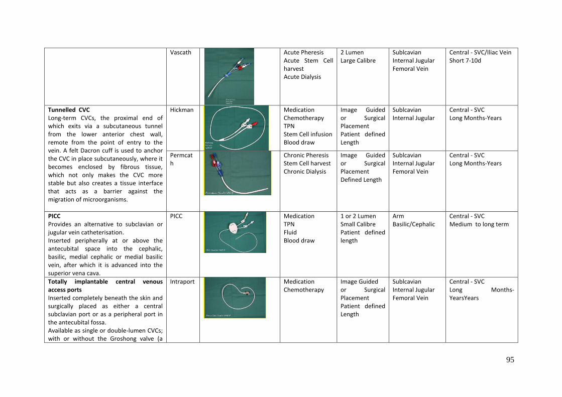
95
Vascath
Acute Pheresis Acute Stem Cell harvest Acute Dialysis
2 Lumen Large Calibre
Sublcavian Internal Jugular Femoral Vein
Central - SVC/Iliac Vein Short 7-10d
Tunnelled CVC Long-term CVCs, the proximal end of which exits via a subcutaneous tunnel from the lower anterior chest wall, remote from the point of entry to the vein. A felt Dacron cuff is used to anchor the CVC in place subcutaneously, where it becomes enclosed by fibrous tissue, which not only makes the CVC more stable but also creates a tissue interface that acts as a barrier against the migration of microorganisms.
Hickman
Medication Chemotherapy TPN Stem Cell infusion Blood draw
Image Guided or Surgical Placement Patient defined Length
Sublcavian Internal Jugular
Central - SVC Long Months-Years
Permcath
Chronic Pheresis Stem Cell harvest Chronic Dialysis
Image Guided or Surgical Placement Defined Length
Sublcavian Internal Jugular Femoral Vein
Central - SVC Long Months-Years
PICC Provides an alternative to subclavian or jugular vein catheterisation. Inserted peripherally at or above the antecubital space into the cephalic, basilic, medial cephalic or medial basilic vein, after which it is advanced into the superior vena cava.
PICC
Medication TPN Fluid Blood draw
1 or 2 Lumen Small Calibre Patient defined length
Arm Basilic/Cephalic
Central - SVC Medium to long term
Totally implantable central venous access ports Inserted completely beneath the skin and surgically placed as either a central subclavian port or as a peripheral port in the antecubital fossa. Available as single or double-lumen CVCs; with or without the Groshong valve (a
Intraport
Medication Chemotherapy
Image Guided or Surgical Placement Patient defined Length
Sublcavian Internal Jugular Femoral Vein
Central - SVC Long Months-YearsYears

96
two-slit valve that remains closed unless the CVC is in use).

97
Appendix 6: Aseptic (No touch) Technique
1. Hand hygiene Wash with an antimicrobial liquid soap and water, or If hands are physically clean, applying an alcohol based hand rub. Hands that are visibly soiled or contaminated with dirt or organic material must be washed with liquid soap and water before using an alcohol hand rub.
2. Prepare an aseptic surface Procedure trolleys/trays must be cleaned using a detergent and disinfectant.
3. Gather equipment for procedure
4. Hand hygiene and put on gloves a. Clean, non-sterile gloves: if the procedure can be completed without
touching key parts (intravenous drug administration, blood sampling or connecting or disconnecting intravenous fluids except TPN).
b. Sterile gloves if the procedure cannot be completed without touching key parts (e.g., line manipulation, insertion site dressing changes, connecting TPN and connecting or disconnecting catheters used for haemodialysis).
5. Identify ‘key parts’ e.g., cannula hub, port, infusion line, lumen etc.
6. Prepare equipment and patient ensuring that all key parts are protected Protect key part at all time using a non-touch technique. Non key parts can be touched with confidence.
7. Carry out procedure taking care to avoid contamination of sterile areas/items/key parts
8. Dispose of waste and sharps appropriately
9. Remove gloves
10. Hand hygiene

98
Appendix 7: Patient Information Leaflet Patient information leaflets are available for download on the HPSC website on the following link: http://www.hpsc.ie/A-Z/MicrobiologyAntimicrobialResistance/InfectionControlandHAI/IntravascularIVlines/Factsheets/
Appendix 8: CVC Insertion Procedure Guideline Before insertion Check patient's coagulation profile (PT, PTT, platelets) on day of procedure.
Do not insert CVC in patient receiving warfarin, clopidogrel unless in emergency. If non-emergency insertion, correct coagulopathy e.g., INR>1.4, platelets<50.
Balance indication (access, pressors, parenteral nutrition, antibiotics) against complication profile (neurovascular injury, haemorrhage, infection).
Site:
Use insertion site associated with least likelihood of injury (jugular, femoral, subclavian). Consider portable ultrasound imaging for selected patients at high risk of complications (e.g., known vascular anomaly) or where vascular access is likely to be difficult (e.g., children)
Remove hair at the insertion site using clippers if required. Physically clean the skin if necessary
Catheter type: Use single lumen or double lumen in preference to triple- or 4-lumen. If single-lumen access required, consider PICC. Preparation of sterile field
Only competent staff (or training staff supervised by competent staff) are to insert CVCs. (Section 1.2)
The CVC should be inserted in an area where asepsis can be maintained.
A trolley/cart that includes all supplies necessary for inserting a CVC including barrier precautions should be dedicated for CVC insertion. (Appendix 9)
The sterile field must be set up immediately prior to the procedure. Hand hygiene (Section 1.1)
Hands must be decontaminated by washing with an antimicrobial liquid soap and water, or if hands are physically clean, by an alcohol based hand rub. Hands that are visibly soiled or contaminated with dirt or organic material must be washed with liquid soap and water before using an alcohol hand rub
The use of gloves does not obviate the need for hand hygiene
Hand hygiene must be performed
Before and after inserting catheter CVC Maximal Barrier Precautions Before placing a CVC (including guidewire exchanges), the operator and any person who enters the sterile field to assist in the procedure, must don a mask, sterile long-sleeved gown, sterile gloves and protective eyewear. A surgical cap should be used to contain hair that may fall across the operator’s face during the procedure. The patient should be covered from head to toe with a sterile drape with an appropriate opening for the site of insertion.

99
Don protective cap, eyewear and surgical mask - The mask should cover the nose and mouth tightly
Perform hand hygiene and dry hands with a sterile towel
Aseptically don sterile gown
Aseptically don sterile gloves - Ensure gloves cover cuff of gown
Skin asepsis – Apply single patient use application of 2% chlorhexidine gluconate in 70% isopropyl alcohol (unless contraindicated – Section 1.2) in a circular motion beginning in the centre of the proposed site and moving outward, for at least 30 seconds. Repeat this step using a new swab for each application. Allow to air dry completely prior to inserting the catheter, do not wipe or blot.
Drape the entire body of the patient (while maintaining a sterile field) leaving only a small opening at the insertion site
Insertion Technique
Ensure skin and subcutaneous tissues are not infected locally
Consider some volume resuscitation to fill veins locally
Trendelenberg position (or reverse T for femoral veins) to promote venous filling
Local infiltration of local anaesthetic agent where necessary (lignocaine or bupivacaine) (if no allergy)
Use Seldinger technique to access internal jugular vein at apex of the triangle of the sternal and clavicular heads of the sternocleidomastoid muscle.
Landmark technique:
Palpate carotid artery in neck.
Insert 21G 'blue' seeker needle to locate vein
Where possible insert cannula into vein to observe for venous flow characteristics
Consider ultrasonic locating device as outlined above
Insert wire to 20cm
Incise skin locally to depth of 5mm to permit passage of introducer
Pass introducer with rotatory or swivelling action to prevent false passage
Advance catheter over wire while maintaining pincer grip on wire to prevent wire embolus.
Confirm intravenous placement of catheter by aspirating venous blood
Flush catheter lumens with normal saline. Catheter fixation
Secure the catheter with 2/0 silk sutures to minimise to-and-fro pistoning of the catheter and subsequent catheter tract invasion by cutaneous microorganisms
Do not apply antimicrobial ointments or creams to the insertion site
Apply a sterile, transparent, semipermeable, self-adhesive, polyurethane dressing Confirm CVC placement with Radiology (e.g., chest X-ray) If accidental insertion of wide-bore CVC into subclavian artery or femoral artery above inguinal ligament, leave catheter in situ. Consult Vascular Surgery/ Interventional Radiology on-call for possible endovascular repair with closure device. Documentation Date and time of insertion Type of CVC and gauge Anatomical/insertion site
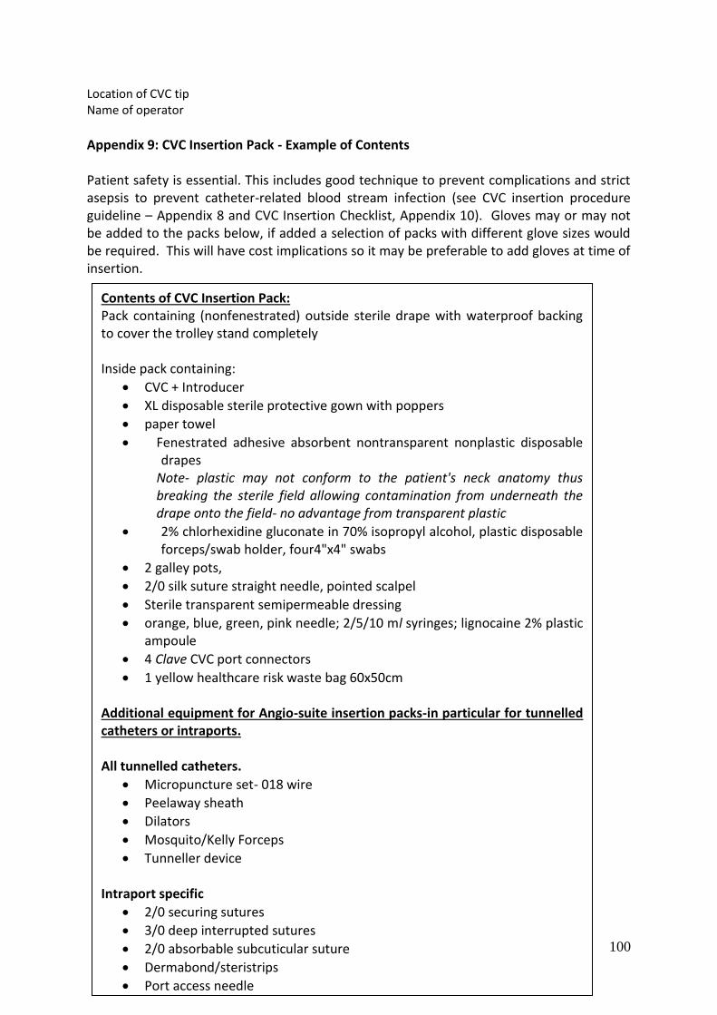
100
Location of CVC tip Name of operator
Appendix 9: CVC Insertion Pack - Example of Contents Patient safety is essential. This includes good technique to prevent complications and strict asepsis to prevent catheter-related blood stream infection (see CVC insertion procedure guideline – Appendix 8 and CVC Insertion Checklist, Appendix 10). Gloves may or may not be added to the packs below, if added a selection of packs with different glove sizes would be required. This will have cost implications so it may be preferable to add gloves at time of insertion.
Contents of CVC Insertion Pack: Pack containing (nonfenestrated) outside sterile drape with waterproof backing to cover the trolley stand completely Inside pack containing:
CVC + Introducer
XL disposable sterile protective gown with poppers
paper towel
Fenestrated adhesive absorbent nontransparent nonplastic disposable drapes
Note- plastic may not conform to the patient's neck anatomy thus breaking the sterile field allowing contamination from underneath the drape onto the field- no advantage from transparent plastic
2% chlorhexidine gluconate in 70% isopropyl alcohol, plastic disposable forceps/swab holder, four4"x4" swabs
2 galley pots,
2/0 silk suture straight needle, pointed scalpel
Sterile transparent semipermeable dressing
orange, blue, green, pink needle; 2/5/10 ml syringes; lignocaine 2% plastic ampoule
4 Clave CVC port connectors
1 yellow healthcare risk waste bag 60x50cm
Additional equipment for Angio-suite insertion packs-in particular for tunnelled catheters or intraports. All tunnelled catheters.
Micropuncture set- 018 wire
Peelaway sheath
Dilators
Mosquito/Kelly Forceps
Tunneller device
Intraport specific
2/0 securing sutures
3/0 deep interrupted sutures
2/0 absorbable subcuticular suture
Dermabond/steristrips
Port access needle

101
Appendix 10: CVC Insertion Checklist
An example of a CVC insertion checklist can be downloaded from the HPSC website at the following link: http://www.hpsc.ie/A-Z/MicrobiologyAntimicrobialResistance/CareBundles/CentralVascularCathetersCVCs/CVCinsertion/
Appendix 11: CVC Care Bundle
The CVC maintenance care bundle can be downloaded from the HPSC website at the following link: http://www.hpsc.ie/A-Z/MicrobiologyAntimicrobialResistance/CareBundles/CentralVascularCathetersCVCs/MaintenanceofCVCs/
Appendix 12: Form for Collection of Denominator Data for CRBSI Surveillance
An example of a form for collection of denominator data can be downloaded from the HPSC website at http://www.hpsc.ie/A-Z/MicrobiologyAntimicrobialResistance/InfectionControlandHAI/IntravascularIVlines/Surveillance/CRBSIsurveillance Appendix 13: Form 2: Form for Collection of CRBSI - Clinical and Laboratory Data An example of a form that can be used for collection of clinical and laboratory date for CRBSI surveillance can be downloaded on the HPSC websited at http://www.hpsc.ie/A-Z/MicrobiologyAntimicrobialResistance/InfectionControlandHAI/IntravascularIVlines/Surveillance/CRBSIsurveillance

102
Appendix 14: PIVC Insertion Procedure Guideline Asepsis, hand hygiene and appropriate technique must be adhered to. Either single-use tourniquets should be used or the tourniquet cleaned and disinfected between each patient use.
A dedicated trolley/cart that includes all supplies necessary should be available
Confirm identity of the patient and explain to the patient or parent/guardian the procedure and need for the PIVC
Only equipment required should be taken to the bedside and set up immediately prior to the procedure.
Perform hand hygiene (Sections 1.1 and 4.1.1)
Apply tourniquet (to the non dominant forearm if possible), select and palpate an appropriate vein for PIVC insertion
Release tourniquet and set up equipment on a clean trolley (sterile dressing/insertion pack)
If the skin is visibly dirty, wash prior to skin asepsis. In adults and children ≥ 2 months (assuming normal gestation at birth), use a single patient use application of alcoholic chlorhexidine gluconate solution (preferably 2% chlorhexidine gluconate in 70% isopropyl alcohol if compatible with the PIVC) for skin asepsis. (Section 4.1.1), Allow the site to dry and then reapply the tourniquet. Do not repalpate the area after skin asepsis
Perform hand hygiene
Apply clean (well-fitting) non-sterile gloves.
Apply tourniquet
Insert the PIVC using an aseptic (no-touch) technique.
If you have difficulty inserting the PIVC, do not attempt repeated insertions with the same cannulae. If after three attempts you are unsuccessful, request help from a colleague – do not continue to attempt to insert the PIVC.
The PIVC should be stabilised with a sterile transparent semipermeable dressing and sterile adhesive tape to prevent PIVC dislodgement. Do not apply non-sterile adhesive tape under the transparent semipermeable dressing. Do not obscure the ability to visualise the PIVC site and surrounding tissues with adhesive tape. Adhesive labels indicating insertion details, on dressings are recommended
Secure adhesive label to dressing or record insertion date on dressing
Dispose all sharps carefully into an approved sharps container
Discard waste and decontaminate trolley in a designated area away from the clean utility or where intravenous medications are prepared
Remove gloves and perform hand hygiene
Give the patient the IV information leaflet; and ask the patient to report symptoms of pain or discomfort at the PIVC site. It is important that the patient is educated regarding hand hygiene and the importance of keeping the PIVC site clean and dry
Accurate documentation and record keeping must be maintained to ensure patient safety, to allow for audits, and to track any outbreaks of infection. The documentation should include
o The date and time of PIVC insertion (so that HCW can clearly assess the duration the PIVC is in situ)
o Type of PIVC and gauge

103
o Anatomical site o Name of operator o When the PIVC is removed/replaced.

104
Appendix 15: Example of a Visual Infusion Phlebitis Score.75
Condition of site Score Degree of Phlebitis.
IV Site appears healthy 0
1
2
3
4
5
No signs of Phlebitis. - Observe cannula.
One of the following is evident: Slight discomfort at IV site Slight Swelling at IV site.
First signs of phlebitis. - Observe cannula.
Two of the following are evident: Pain at IV Site
Erythema Swelling
Early stages of phlebitis. - Resite cannula.
All of the following are present: Pain along path of cannula.
Erythema Induration
Medium stages of Phlebitis.
Resite cannula Consider treatment.
All of the following signs are evident: Pain along path of cannula
Erythema Induration
Palpable venous cord
Advanced stages of phlebitis, or the start of Thrombophlebitis.
Resite Cannula Consider treatment.
All of the following signs are evident and are extensive:
Pain along path of cannula. Erythema Induration
Palpable venous cord Pyrexia.
Advanced stages of Thrombophlebitis.
Initiate treatment Resite cannula.
Appendix 16: PIVC Care Bundle – Updated 2014 The updated PIVC care bundle can be downloaded from the HPSC website at the following link: http://www.hpsc.ie/A-Z/MicrobiologyAntimicrobialResistance/CareBundles/PeripheralVascularCatheterPIVC/

105
References
(1) Horan T, Andrus M, Dudeck M. CDC/NHSN surveillance definition of health care-associated infection and criteria for specific types of infections in the acute care setting. Am J Infect Control 2008; 36:309-332.
(2) SARI Infection Control Sub-committee. The Control and Prevention of MRSA in Hospitals and in the Community. 2005. National Disease Surveillance Centre, Ireland.
Ref Type: Report
(3) Health Information and Quality Authority I. National Standards for the Prevention and Control of Healthcare Associated Infections . 2009.
Ref Type: Report
(4) Mermel LA, Allon M, Bouza E, Craven DE, Flynn P, O'Grady NP et al. Clinical practice guidelines for the diagnosis and management of intravascular catheter-related infection: 2009 Update by the Infectious Diseases Society of America. Clin Infect Dis 2009; 49(1):1-45.
(5) Eggimann P. Prevention of intravascular catheter infection. Curr Opin Infect Dis 2007; 20:360-369.
(6) Dimick JB, Pelz RK, Consunji R, Swoboda SM, Hendrix CW, Lipsett PA. Increased resource use associated with catheter-related bloodstream infection in the surgical intensive care unit. Arch Surg 2001; 136(2):229-234.
(7) Warren DK, Quadir WW, Hollenbeak CS, Elward AM, Cox MJ, Fraser VJ. Attributable cost of catheter-associated bloodstream infections among intensive care patients in a nonteaching hospital. Crit Care Med 2006; 34(8):2084-2089.
(8) Renaud B, Brun-Buisson C. Outcomes of primary and catheter-related bacteremia. A cohort and case-control study in critically ill patients. Am J Respir Crit Care Med 2001; 163(7):1584-1590.
(9) Blot SI, Depuydt P, Annemans L, Benoit D, Hoste E, De Waele JJ et al. Clinical and economic outcomes in critically ill patients with nosocomial catheter-related bloodstream infections. Clin Infect Dis 2005; 41(11):1591-1598.
(10) O'Grady NP, Alexander M, Patchen Dellinger M, Gerberding JL, Stippel D, Heard SO et al. Guidelines for the Prevention of Intravascular Catheter-Related Infections. Clin Infect Dis 2002; 35:1281-1307.
(11) Rello J, Ochagavia A, Sabanes E, Roque M, Mariscal D, Reynaga E et al. Evaluation of outcome of intravenous catheter-related infections in critically ill patients. Am J Respir Crit Care Med 2000; 162(3 Pt 1):1027-1030.
(12) Fraher MH, Collins C, Bourke J, Phelan D, Lynch M. Cost-effectiveness of employing a total parenteral nutrition surveillance nurse for the prevention of catheter-related bloodstream infections. J Hosp Infect 2009; 73:129-134.
(13) Marschall J, Mermel LA, Classen D, Arias K.M, Podgorny K, Anderson DA et al. Strategies to Prevent Central Line-Associated Bloodstream Infections in Acute Care Hospitals. Infect Control Hosp Epidemiol 2008;S22-S30.
(14) Safdar N, Maki DG. Inflammation at the insertion site is not predictive of IVC-related bloodstream infection with short-term, noncuffed central venous catheters. Crit Care Med 2002; 30:2632-2635.
(15) Mermel LA, Farr BM, Sherertz RJ, Raad II, O'Grady NP, Harris JS et al. Guidelines for the Management of Intravascular Catheter-Related Infections. Clin Infect Dis 2001; 32:1249-1272.
(16) Costa SF, Miceli MH, Anaissie EJ. Mucosa or skin as source of coagulase-negative staphylococcal bacteraemia? Lancet Infect Dis 2004; 4(5):278-286.
(17) Department of Health and Children. North South Study of MRSA in Ireland. ISBN 07-0766-4950. 2000. Ref Type: Report
(18) Health Protection Surveillance Centre. EARSS Report for Quarter 1 2009. 2009. Ref Type: Report
(19) Smyth ET, McIlvenny G, Enstone JE, Emmerson AM, Humphreys H, Fitzpatrick F et al. Four country healthcare associated infection prevalence survey 2006: overview of the results. J Hosp Infect 2008; 69(3):230-248.

106
(20) Humphreys H, Newcombe RG, Enstone J, Smyth ET, McIlvenny G, Fitzpatrick F et al. Four country healthcare associated infection prevalence survey 2006: risk factor analysis. J Hosp Infect 2008; 69(3):249-257.
(21) Fitzpatrick F, McIlvenny G, Oza A, Newcombe RG, Humphreys H, Cunney R et al. Hospital infection society prevalence survey of Healthcare Associated Infection 2006: comparison of results between Northern Ireland and the Republic of Ireland. J Hosp Infect 2008; 69(3):265-273.
(22) Pratt RJ, Pellowe CM, Wilson JA, Lovedat HP, Harper PJ, Jones SRLJ et al. epic2: National Evidence-Based Guidelines for Preventing Healthcare-Associated Infections in NHS Hospitals in England. J Hosp Infect 2007; 65S:S1-S64.
(23) Boyce JM, Pittet D. Guideline for Hand Hygiene in Health-Care Settings: recommendations of the Healthcare Infection Control Practices Advisory Committee and the HICPAC/SHEA/APIC/IDSA Hand Hygiene Task Force. Infect Control Hosp Epidemiol 2002; 23(12 Suppl):S3-40.
(24) SARI Infection Control Sub-committee. Guidelines for Hand Hygiene in Irish Health Care Settings. 2005. Ref Type: Report
(25) Rowley S. Aseptic non-touch technique. Nursing Times 2001; 97(7):6.
(26) Rowley S, Sinclair S. Working towards an NHS standard for aseptic non-touch technique . Nursing Times 2004; 100(8):50.
(27) Vanherweghem JL, Dhaene M, Goldman M, Stolear JC, Sabot JP, Waterlot Y et al. Infections associated with subclavian dialysis catheters: the key role of nurse training. Nephron 1986; 42(2):116-119.
(28) Meier PA, Fredrickson M, Catney M, Nettleman MD. Impact of a dedicated intravenous therapy team on nosocomial bloodstream infection rates. Am J Infect Control 1998; 26(4):388-392.
(29) Kaplowitz LG, Comstock JA, Landwehr DM, Dalton HP, Mayhall CG. A prospective study of infections in hemodialysis patients: patient hygiene and other risk factors for infection. Infect Control Hosp Epidemiol 1988; 9(12):534-541.
(30) Penna TCV, Mazzola PG, Silva Martins AM. The efficacy of chemical agents in cleaning and disinfection programs. BMC Infectious Diseases 2001; 1(16 24th September).
(31) Garland JS, Alex CP, Mueller CD, Otten D, Shivpuri C, Harris MC et al. A randomized trial comparing povidone-iodine to a chlorhexidine gluconate-impregnated dressing for prevention of central venous catheter infections in neonates. Pediatrics 2001; 107(6):1431-1436.
(32) Wilson CM, Gray G, Read JS, Mwatha A, Lala S, Johnson S et al. Tolerance and safety of different concentrations of chlorhexidine for peripartum vaginal and infant washes: HIVNET 025. J Acquir Immune Defic Syndr 2004; 35(2):138-143.
(33) Datta MK, Clarke P. Current practices in skin antisepsis for central venous catheterisation in UK tertiary-level neonatal units. Archives of Disease in Childhood - Fetal and Neonatal Edition 2009; 93:F328.
(34) Mermel LA, McCormick RD, Springman SR, Maki DG. The pathogenesis and epidemiology of catheter-related infection with pulmonary artery Swan-Ganz catheters: a prospective study utilizing molecular subtyping. Am J Med 1991; 91(3B):197S-205S.
(35) Raad II, Hohn DC, Gilbreath BJ, Suleiman N, Hill LA, Bruso PA et al. Prevention of central venous catheter-related infections by using maximal sterile barrier precautions during insertion. Infect Control Hosp Epidemiol 1994; 15(4 Pt 1):231-238.
(36) Karakitsos D, Labropoulos N, De GE, Patrianakos AP, Kouraklis G, Poularas J et al. Real-time ultrasound-guided catheterisation of the internal jugular vein: a prospective comparison with the landmark technique in critical care patients. Crit Care 2006; 10(6):R162.
(37) McCarthy MC, Shives JK, Robison RJ, Broadie TA. Prospective evaluation of single and triple lumen catheters in total parenteral nutrition. J Parenter Enteral Nutr 1987; 11(3):259-262.

107
(38) Parienti JJ, Thirion M, Mégarbane B, Ouchikhe A, Polito A, Marqué S et al. Femoral vs Jugular Venous Catheterization and Risk of Nosocomial Events in Adults Requiring Acute Renal Replacement Therapy: A Randomized Controlled Trial. JAMA 2008; 229(20):2413-2422.
(39) Deshpande KS, Hatem C, Ulrich HL, Currie BP, Aldrich TK, Bryan-Brown CW et al. The incidence of infectious complications of central venous catheters at the subclavian, internal jugular, and femoral sites in an intensive care unit population. Crit Care Med 2005; 33(1):13-20.
(40) Gowardman JR, Montgomery C, Thirlwell S, Shewan J, Idema A, Larsen PD et al. Central venous catheter-related bloodstream infections: an analysis of incidence and risk factors in a cohort of 400 patients. Intensive Care Med 1998; 24(10):1034-1039.
(41) Gowardman JR, Robertson IK, Parkes S, Rickard CM. Influence of insertion site on central venous catheter colonization and bloodstream infection rates. Intensive Care Med 2008; 34(6):1038-1045.
(42) Safdar N, Maki DG. Use of vancomycin-containing lock or flush solutions for prevention of bloodstream infection associated with central venous access devices: a meta-analysis of prospective, randomized trials. Clin Infect Dis 2006; 43(4):474-484.
(43) Feely T, Copley A, Bleyer AJ. Catheter lock solutions to prevent bloodstream infections in high-risk hemodialysis patients. Am J Nephrol 2007; 27(1):24-29.
(44) Garland JS, Alex CP, Henrickson KJ, McAuliffe TL, Maki DG. A vancomycin-heparin lock solution for prevention of nosocomial bloodstream infection in critically ill neonates with peripherally inserted central venous catheters: a prospective, randomized trial. Pediatrics 2005; 116(2):e198-e205.
(45) Yahav D, Rozen-Zvi B, Gafter-Gvili A, Leibovici L, Gafter U, Paul M. Antimicrobial lock solutions for the prevention of infections associated with intravascular catheters in patients undergoing hemodialysis: systematic review and meta-analysis of randomized, controlled trials. Clin Infect Dis 2008; 47(1):83-93.
(46) Percival SL, Kite P, Eastwood K, Murga R, Carr J, Arduino MJ et al. Tetrasodium EDTA as a novel central venous catheter lock solution against biofilm. Infect Control Hosp Epidemiol 2005; 26(6):515-519.
(47) Simon A, Ammann RA, Wiszniewsky G, Bode U, Fleischhack G, Besuden MM. Taurolidine-citrate lock solution (TauroLock) significantly reduces CVAD-associated grampositive infections in pediatric cancer patients. BMC Infect Dis 2008; 8:102.
(48) Allon M. Prophylaxis against dialysis catheter-related bacteremia with a novel antimicrobial lock solution. Clin Infect Dis 2003; 36(12):1539-1544.
(49) Betjes MG, van AM. Prevention of dialysis catheter-related sepsis with a citrate-taurolidine-containing lock solution. Nephrol Dial Transplant 2004; 19(6):1546-1551.
(50) Onland W, Shin CE, Fustar S, Rushing T, Wong WY. Ethanol-lock technique for persistent bacteremia of long-term intravascular devices in pediatric patients. Arch Pediatr Adolesc Med 2006; 160(10):1049-1053.
(51) Opilla MT, Kirby DF, Edmond MB. Use of ethanol lock therapy to reduce the incidence of catheter-related bloodstream infections in home parenteral nutrition patients. J Parenter Enteral Nutr 2007; 31(4):302-305.
(52) van de Wetering MD, Woensel JBM. Prophylactic antibiotics for preventing early central venous catheter Gram positive infections in oncology patients. Issue 1. Art. No.: CD003295. DOI: 10.1002/14651858.CD003295.pub2. 2007. Cochrane Database of Systematic Reviews .
Ref Type: Report
(53) Jardine LA, Inglis GDT, Davies MW. Prophylactic systemic antibiotics to reduce morbidity and mortality in neonates with central venous catheters. Issue 1. Art. No.: CD006179. DOI: 10.1002/14651858.CD006179.pub2. 2008. Cochrane Database of Systematic Reviews.
Ref Type: Report
(54) Royal College of Nursing. Standards for Infusion therapy. 2007. Royal College of Nursing, London. Ref Type: Report
108
(55) Institute for Healthcare Improvement - Central Line Bundle. 2009. Ref Type: Internet Communication
(56) Hugonnet S, Sax H, Eggimann P, Chevrolet JC, Pittet D. Nosocomial bloodstream infection and clinical sepsis. Emerg Infect Dis 2004; 10(1):76-81.
(57) National Nosocomial Infections Surveillance (NNIS) System Report, data summary from January 1992 to June 2002, issued August 2002. Am J Infect Control 2002; 30(8):458-475.
(58) National Healthcare Safety Network (NHSN). CDC . 2009. Ref Type: Internet Communication
(59) Edwards JR, Peterson KD, Andrus ML, Dudeck MA, Pollock DA, Horan TC. National Healthcare Safety Network (NHSN) Report, data summary for 2006 through 2007, issued November 2008. Am J Infect Control 2008; 36(9):609-626.
(60) Raad II, Hanna H, Maki DG. Intravascular catheter-related infections: advances in diagnosis, prevention, and management. Lancet Infect Dis 2007; 7:645-657.
(61) Peacock SJ, Eddleston M, Emptage A, King A, Crook DW. Positive intravenous line tip cultures as predictors of bacteraemia. J Hosp Infect 1998; 40(1):35-38.
(62) Sherertz RJ, Heard SO, Raad II. Diagnosis of triple-lumen catheter infection: comparison of roll plate, sonication, and flushing methodologies. J Clin Microbiol 1997; 35(3):641-646.
(63) Ekkelenkamp MB, van der Bruggen T, van de Vijver DA, Wolfs TF, Bonten MJ. Bacteremic complications of intravascular catheters colonized with Staphylococcus aureus. Clin Infect Dis 2008; 46(1):114-118.
(64) Ruhe JJ, Menon A. Clinical significance of isolated Staphylococcus aureus central venous catheter tip cultures. Clin Microbiol Infect 2006; 12(9):933-936.
(65) US Food and Drug Administration. Information for Healthcare Professionals Linezolid (marketed as Zyvox). 16-3-2007.
Ref Type: Internet Communication
(66) Collins CJ, Fraher M, Bourke J, Phelan D, Lynch M. Epidemiology of Candida species Catheter Related-Bloodstream Infection in Patients on Total Parenteral Nurtition. Clin Infect Dis 2009; (in press).
(67) Health Protection Surveillance Centre. Enhanced EARSS surveillance - report for 2008. 2009. Ref Type: Report
(68) Raad I, Hanna H, Boktour M, Girgawy E, Danawi H, Mardani M et al. Management of central venous catheters in patients with cancer and candidemia. Clin Infect Dis 2004; 38(8):1119-1127.
(69) Korbila IP, Bliziotis I.A, Lawrence KR, Falagas ME. Antibiotic- lock therapy for long-term catheter-related bacteraemia: a review of the current evidence. Expert Rev Anti Infect Ther 2007; 5(4):639-652.
(70) Poole CV, Carlton D, Bimbo L, Allon M. Treatment of catheter-related bacteraemia with an antibiotic lock protocol: effect of bacterial pathogen. Nephrol Dial Transplant 2004; 19(5):1237-1244.
(71) Rijnders BJ, Van WE, Vandecasteele SJ, Stas M, Peetermans WE. Treatment of long-term intravascular catheter-related bacteraemia with antibiotic lock: randomized, placebo-controlled trial. J Antimicrob Chemother 2005; 55(1):90-94.
(72) Fortun J, Grill F, Martin-Davila P, Blazquez J, Tato M, Sanchez-Corral J et al. Treatment of long-term intravascular catheter-related bacteraemia with antibiotic-lock therapy. J Antimicrob Chemother 2006; 58(4):816-821.
(73) Eggimann P. Diagnosis of intravascular catheter infection. Curr Opin Infect Dis 2007; 20(4):353-359.
(74) Dougherty L, Lamb J. Intravenous Therapy in Nursing Practice. Churchill Livingstone. London.; 1999.

109
(75) Jackson A. A battle in vein- Infusion phlebitis. Nursing Times 1998; 94(4):68-70.
(76) Linares J. Diagnosis of catheter-related bloodstream infection: conservative techniques. Clin Infect Dis 2007; 44(6):827-829.
(77) Cleri DJ, Corrado ML, Seligman SJ. Quantitative culture of intravenous catheters and other intravascular inserts. J Infect Dis 1980; 141(6):781-786.
(78) Brun-Buisson C, Abrouk F, Legrand P, Huet Y, Larabi S, Rapin M. Diagnosis of central venous catheter-related sepsis. Critical level of quantitative tip cultures. Arch Intern Med 1987; 147(5):873-877.
(79) Sherertz RJ, Raad II, Belani A, Koo LC, Rand KH, Pickett DL et al. Three-year experience with sonicated vascular catheter cultures in a clinical microbiology laboratory. J Clin Microbiol 1990; 28(1):76-82.
(80) Maki DG, Weise CE, Sarafin HW. A semiquantitative culture method for identifying intravenous-catheter-related infection. N Engl J Med 1977; 296(23):1305-1309.
(81) Siegman-Igra Y, Anglim AM, Shapiro DE, Adal KA, Strain BA, Farr BM. Diagnosis of vascular catheter-related bloodstream infection: a meta-analysis. J Clin Microbiol 1997; 35(4):928-936.
(82) Blot F, Nitenberg G, Chachaty E, Raynard B, Germann N, Antoun S et al. Diagnosis of catheter-related bacteraemia: a prospective comparison of the time to positivity of hub-blood versus peripheral-blood cultures. Lancet 1999; 354(9184):1071-1077.
(83) Safdar N, Fine JP, Maki DG. Meta-analysis: methods for diagnosing intravascular device-related bloodstream infection. Ann Intern Med 2005; 142(6):451-466.
(84) Druskin MS, SIEGEL PD. BACTERIAL CONTAMINATION OF INDWELLING INTRAVENOUS POLYETHYLENE CATHETERS. JAMA 1963; 185:966-968.
(85) Cercenado E, Ena J, Rodriguez-Creixems M, Romero I, Bouza E. A conservative procedure for the diagnosis of catheter-related infections. Arch Intern Med 1990; 150(7):1417-1420.
(86) Atela I, Coll P, Rello J, Quintana E, Barrio J, March F et al. Serial surveillance cultures of skin and catheter hub specimens from critically ill patients with central venous catheters: molecular epidemiology of infection and implications for clinical management and research. J Clin Microbiol 1997; 35(7):1784-1790.
(87) Bouza E, Munoz P, Burillo A, Lopez-Rodriguez J, Fernandez-Perez C, Perez MJ et al. The challenge of anticipating catheter tip colonization in major heart surgery patients in the intensive care unit: are surface cultures useful? Crit Care Med 2005; 33(9):1953-1960.
(88) Kite P, Dobbins BM, Wilcox MH, Fawley WN, Kindon AJ, Thomas D et al. Evaluation of a novel endoluminal brush method for in situ diagnosis of catheter related sepsis. J Clin Pathol 1997; 50(4):278-282.
(89) Kite P, Dobbins BM, Wilcox MH, McMahon MJ. Rapid diagnosis of central-venous-catheter-related bloodstream infection without catheter removal. Lancet 1999; 354(9189):1504-1507.
(90) Tighe MJ, Kite P, Fawley WN, Thomas D, McMahon MJ. An endoluminal brush to detect the infected central venous catheter in situ: a pilot study. BMJ 1996; 313(7071):1528-1529.
(91) Rushforth JA, Hoy CM, Kite P, Puntis JW. Rapid diagnosis of central venous catheter sepsis. Lancet 1993; 342(8868):402-403.
(92) Martinez JA, DesJardin JA, Aronoff M, Supran S, Nasraway SA, Snydman DR. Clinical utility of blood cultures drawn from central venous or arterial catheters in critically ill surgical patients. Crit Care Med 2002; 30(1):7-13.
(93) Health Protection Agency U. Investigation of intravascular cannulae and associated specimens. HPA BSOP 20 . 2009.
Ref Type: Generic

110
(94) Raad II, Hanna HA, Darouiche RO. Diagnosis of catheter-related bloodstream infections: is it necessary to culture the subcutaneous catheter segment? Eur J Clin Microbiol Infect Dis 2001; 20(8):566-568.
(95) Longuet P, Douard MC, Arlet G, Molina JM, Benoit C, Leport C. Venous access port--related bacteremia in patients with acquired immunodeficiency syndrome or cancer: the reservoir as a diagnostic and therapeutic tool. Clin Infect Dis 2001; 32(12):1776-1783.
(96) Raad I, Costerton W, Sabharwal U, Sacilowski M, Anaissie E, Bodey GP. Ultrastructural analysis of indwelling vascular catheters: a quantitative relationship between luminal colonization and duration of placement. J Infect Dis 1993; 168(2):400-407.
(97) Bouza E, Alvarado N, Alcala L, Sanchez-Conde M, Perez MJ, Munoz P et al. A prospective, randomized, and comparative study of 3 different methods for the diagnosis of intravascular catheter colonization. Clin Infect Dis 2005; 40(8):1096-1100.
(98) Schmitt SK, Knapp C, Hall GS, Longworth DL, McMahon JT, Washington JA. Impact of chlorhexidine-silver sulfadiazine-impregnated central venous catheters on in vitro quantitation of catheter-associated bacteria. J Clin Microbiol 1996; 34(3):508-511.
(99) Schierholz JM, Bach A, Fleck C, Beuth J, Konig D, Pulverer G. Measurement of ultrasonic-induced chlorhexidine liberation: correlation of the activity of chlorhexidine-silver-sulfadiazine-impregnated catheters to agar roll technique and broth culture. J Hosp Infect 2000; 44(2):141-145.
(100) Catton JA, Dobbins BM, Kite P, Wood JM, Eastwood K, Sugden S et al. In situ diagnosis of intravascular catheter-related bloodstream infection: a comparison of quantitative culture, differential time to positivity, and endoluminal brushing. Crit Care Med 2005; 33(4):787-791.
(101) Bouza E, Alvarado N, Alcala L, Perez MJ, Rincon C, Munoz P. A randomized and prospective study of 3 procedures for the diagnosis of catheter-related bloodstream infection without catheter withdrawal. Clin Infect Dis 2007; 44(6):820-826.
(102) Warwick S, Wilks M, Hennessy E, Powell-Tuck J, Small M, Sharp J et al. Use of quantitative 16S ribosomal DNA detection for diagnosis of central vascular catheter-associated bacterial infection. J Clin Microbiol 2004; 42(4):1402-1408.
(103) Worthington T, Elliott TS. Diagnosis of central venous catheter related infection in adult patients. J Infect 2005; 51(4):267-280.
(104) Koh DB, Gowardman JR, Rickard CM, Robertson IK, Brown A. Prospective study of peripheral arterial catheter infection and comparison with concurrently sited central venous catheters. Crit Care Med 2008; 36(2):397-402.
(105) Pujol M, Hornero A, Saballs M, Argerich MJ, Verdaguer R, Cisnal M et al. Clinical epidemiology and outcomes of peripheral venous catheter-related bloodstream infections at a university-affiliated hospital. J Hosp Infect 2007; 67(1):22-29.
(106) Trick WE, Miranda J, Evans AT, Charles-Damte M, Reilly BM, Clarke P. Prospective cohort study of central venous catheters among internal medicine ward patients. Am J Infect Control 2006; 34(10):636-641.
(107) Eggimann P, Zanetti G. On the way towards eradication of catheter-related infections! Intensive Care Med 2008; 34(6):988-990.
(108) Lorente L, Henry C, Martin MM, Jimenez A, Mora ML. Central venous catheter-related infection in a prospective and observational study of 2,595 catheters. Crit Care 2005; 9(6):R631-R635.
(109) Smith E, Clarke L. Antimicrobial and antibiotic coated central venous catheters. 2004. Ref Type: Internet Communication
(110) Stevenson KB, Adcox MJ, Mallea MC, Narasimhan N, Wagnild JP. Standardized surveillance of hemodialysis vascular access infections: 18-month experience at an outpatient, multifacility hemodialysis center. Infect Control Hosp Epidemiol 2000; 21(3):200-203.

111
(111) Yu VL, Goetz A, Wagener M, Smith PB, Rihs JD, Hanchett J et al. Staphylococcus aureus nasal carriage and infection in patients on hemodialysis. Efficacy of antibiotic prophylaxis. N Engl J Med 1986; 315(2):91-96.
(112) James MT, Conley J, Tonelli M, Manns BJ, MacRae J, Hemmelgam BR. Meta-analysis: Antibiotics for Prophylaxis against Hemodialysis Catheter-Related Infections. Ann Intern Med 2008; 148(8):596-605.
(113) Rossney A, O'Connell B. Emerging Problem with High-level Mupirocin Resistance Among MRSA in Ireland. EpiInsight 2008; 9(3):4.
(114) Annigeri R, Conly J, Vas S, Dedier H, Prakashan KP, Bargman JM et al. Emergence of mupirocin-resistant Staphylococcus aureus in chronic peritoneal dialysis patients using mupirocin prophylaxis to prevent exit-site infection . Perit Dial Int 2001; 21(6):554-559.
(115) Health Protection Scotland. NHS Scotland MRSA Screening Pathfinder Programme - Interim report. 2009. Ref Type: Report
(116) European Renal Association. European Best Practice Guidelines for Haemodialysis (Part 1): VI.2 Prevention of infection: management of host
colonization by Staphylococcus aureus. Nephrol Dial Transplant 2002; 17(Suppl 7):74-75.
(117) Osono E, Takahashi M, Kurihara S, Ohwada K, Sakurai Y, Onoda N et al. Effects of "isolating hemodialysis" on prevention of methicillin-resistant Staphylococcus aureus cross-infection in a hemodialysis unit. Clin Nephrol 2000; 54(2):128-133.
(118) Mokrzycki MH, Zhang M, Cohen H, Golestaneh L, Laut JM, Rosenberg SO. Tunnelled haemodialysis catheter bacteraemia: risk factors for bacteraemia recurrence, infectious complications and mortality. Nephrol Dial Transplant 2006; 21(4):1024-1031.
(119) Garrouste-Orgeas M, Timsit JF, Soufir L, Tafflet M, Adrie C, Philippart F et al. Impact of adverse events on outcomes in intensive care unit patients. Crit Care Med 2008; 36(7):2041-2047.
(120) Garrouste-Org, Timsit JF, Tafflet M, Misset B, Zahar JR, Soufir L et al. Excess risk of death from intensive care unit-acquired nosocomial bloodstream infections: a reappraisal. Clin Infect Dis 2006; 42(8):1118-1126.
(121) Timsit JF. What is the best site for central venous catheter insertion in critically ill patients? Crit Care 2003; 7(6):397-399.
(122) Mermel LA. Prevention of intravascular catheter-related infections. Ann Intern Med 2000; 132(5):391-402.
(123) Eggimann P, Harbarth S, Constantin MN, Touveneau S, Chevrolet JC, Pittet D. Impact of a prevention strategy targeted at vascular-access care on incidence of infections acquired in intensive care. Lancet 2000; 355(9218):1864-1868.
(124) Pittet D. The Lowbury lecture: behaviour in infection control. J Hosp Infect 2004; 58(1):1-13.

112
References 2014
1. O’Grady NP, Pearson ML, Raad II, et al. Guidelines for the prevention of intravascular catheter-related infections.
Clin Infect Dis 2011;52:e162–e193.
2. Loveday, HP, Wilson JA, Pratt M. Golsorkhi A. Tingle A. et al epic3: National Evidence-Based Guidelines for
Preventing Healthcare-Associated Infections in NHS Hospitals in England. J Hosp Infect 2014: 86 S1-S70.
3. http://www.imb.ie/images/uploaded/swedocuments/LicenseSPC_PA1435-001-002_15122012111812.pdf
(Accessed 01.02.14)
4. Marschall J, Mermel LA, Classen D, Arias K.M, Podgorny K, Anderson DA et al. Strategies to Prevent Central Line-
Associated Bloodstream Infections in Acute Care Hospitals. Infect Control Hosp Epidemiol 2008; S22-S30.
5. Lai NM, Chaiyakunapruk N, Lai NA, O’Riordan E, Pau WS, Saint S. Catheter impregnation, coating or bonding for
reducing central venous catheter-related infections in adults. Cochrane Database Syst Rev 2013(6).
6. Timsit JF, Schwebel C, Bouadma L, et al. Chlorhexidine-impregnated sponges and less frequent dressing changes
for prevention of catheter-related infections in critically ill adults: a randomized controlled trial. JAMA 2009;
301:1231–41.
7. Ruschulte H, Franke M, Gastmeier P, et al. Prevention of central venous catheter related infections with
chlorhexidine gluconate impregnated wound dressings: a randomized controlled trial. Ann Hematol 2009;
88:267–72.
8. Ho KM, Litton E. Use of chlorhexidine-impregnated dressing to prevent vascular and epidural catheter
colonization and infection: a meta-analysis. J Antimicrob Chemother 2006; 58:281–7.
9. Chan R, NorthÀeld S, Alexander A, Rickard C. Using the collaborative evidence based practice model: a systematic
review and uptake of chlorhexidine-impregnated sponge dressings on central venous access devices in a tertiary
cancer centre. Aust J Cancer Nurs 2012;13:10–15.
10. Timsit JF, Mimoz O, Mourvillier B, et al. Randomized controlled trial of chlorhexidine dressing and highly adhesive
dressing for preventing catheter-related infections in critically ill adults. Am J Respir Crit Care Med
2012;186:1272–1278.
11. Schwebel C, Lucet JC, Vesin A, et al. Economic evaluation of chlorhexidine-impregnated sponges for preventing
catheter- related infections in critically ill adults in the Dressing Study. Crit Care Med 2012;40:11-17.
12. Timsit JF, Schwebel C, Bouadma L, et al. Chlorhexidine-impregnated sponges and less frequent dressing changes
for prevention of catheter-related infections in critically ill adults: a randomized controlled trial. JAMA 2009;
301:1231–41.
13. Garland JS, Alex CP, Mueller CD, et al. A randomized trial comparing povidone-iodine to a chlorhexidine
gluconate-impregnated dressing for prevention of central venous catheter infections in neonates. Pediatrics
2001; 107:1431–6.
14. levy I, Katz J, Solter E, et al. Chlorhexidine-impregnated dressing for prevention of colonization of central venous
catheters in infants and children: a randomized controlled study. Pediatr Infect Dis J 2005; 24:676– 9.
15. Bleasdale SC, Trick WE, Gonzalez IM, Lyles RD, Hayden MK, Weinstein RA. Effectiveness of chlorhexidine bathing
to reduce catheter-associated bloodstream infections in medical intensive care unit patients. Arch Intern Med
2007; 167:2073–9.
16. Munoz-Price LS, Hota B, Stemer A, Weinstein RA. Prevention of bloodstream infections by use of daily
chlorhexidine baths for patients at a long-term acute care hospital. Infect Control Hosp Epidemiol 2009; 30:1031–

113
17. Popovich KJ, Hota B, Hayes R, Weinstein RA, Hayden MK. Effectiveness of routine patient cleansing with
chlorhexidine gluconate for infection prevention in the medical intensive care unit. Infect Control Hosp Epidemiol
2009; 30:959–63.
18. O’Horo JC, Silva GL, Munoz-Price LS, Safdar N. The efÀcacy of daily bathing with chlorhexidine for reducing
healthcare- associated bloodstream infections: a meta-analysis. Infect Control Hosp Epidemiol 2012;33:257–267
19. Irish Blood Transfusion Service - National Blood Users Group. Guidelines for the Administration of Blood and
Blood Components. 2004.
20. National Institute for Health and Clinical Excellence. Infection control, prevention of healthcare-associated
infection in primary and community care (update) (clinical guideline 139). London: NICE; 2012.
21. Webster J, Osborne S, Rickard C, New K. Clinically-indicated replacement versus routine replacement of
peripheral venous catheters. Cochrane Database Syst Rev 2013;4(CD007798).
22. Barker P, Anderson ADG, Macfie J. Randomised clinical trial of elective re-siting of intravenous cannulae. Annals
of the Royal College of Surgeons of England 2004;86(4):281–3.
23. Nishanth S, Sivaram G, Kalayarasan R, Kate V, Ananthakrishnan N. Does elective re-siting of intravenous cannulae
decrease peripheral thrombophlebitis? A randomized controlled study. The International Medical Journal of India
2009;22(2):60–2.
24. Rickard CM, McCann D, Munnings J, McGrail M. Routine resite of peripheral intravenous devices every 3 days did
not reduce complications compared with clinically indicated resite: a randomised controlled trial. BMC Medicine
2010; 8:53.
25. Rickard CM, Zhang L, McClymont A, et al. Routine versus clinically indicated replacement of peripheral
intravenous catheters: a randomised controlled equivalence trial. Lancet 2012;380:1066–1074.
26. Van Donk P, Rickard CM, McGrail MR, Doolan G. Routine replacement versus clinical monitoring of peripheral
intravenous catheters in a regional hospital in the home program: A randomized controlled trial. Infection Control
and Hospital Epidemiology 2009;30(9):915–7.
27. Webster J, Lloyd S, Hopkins T, Osborne S, Yaxley M. Developing a research base for intravenous peripheral
cannula re-sites (DRIP trial). A randomised controlled trial of hospital in-patients. International Journal of Nursing
Studies 2007;44(5):664–71.
28. Webster J, Clarke S, Paterson D, Hutton A, van Dyke S, Gale C, et al.Routine care of peripheral intravenous
catheters versus clinically indicated replacement: randomised controlled trial. BMJ 2008;337:a339.
29. Medicines and Healthcare Products Regulatory Agency. Medical Device Alert: All medical devices and medicinal
products containing chlorhexidine. MDA/2012/075. London: Medicines and Healthcare Products Regulatory
Agency; 2012
30. Parienti JJ, du Cheyron D, Ramakers M, Malbruny B, Leclercq R, Le CX, et al. Alcoholic povidone-iodine to prevent
central venous catheter colonization: A randomized unit-crossover study. Critical Care Medicine. 2004;32(3):708–
713.