Embed Size (px)
Citation preview

MQP Report 1
UPCONVERSION NANOPARTICLES FOR
PHOTODYNAMIC THERAPY
A Major Qualifying Project Submitted to the Faculty of
Worcester Polytechnic Institute
in partial fulfillment of the requirements for the
Degree in Bachelor of Science in
Biomedical Engineering
By
__________________________________
Mahmoud El-Rifai
__________________________________
Hyungseok Lee
__________________________________
Amira Tokatli-Apollon
Date: 5/1/14
Sponsoring Organization:
University of Massachusetts Medical School:
Principal Investigator:
Dr. Gang Han
Project Advisors:
_______________________________
Dr. Marsha Rolle, Advisor
MQP Report 2
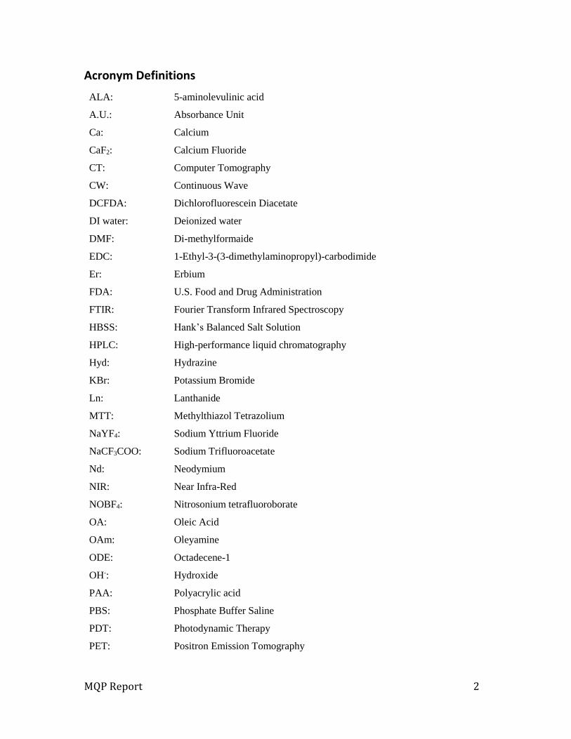
Acronym Definitions
ALA: 5-aminolevulinic acid
A.U.: Absorbance Unit
Ca: Calcium
CaF2: Calcium Fluoride
CT: Computer Tomography
CW: Continuous Wave
DCFDA: Dichlorofluorescein Diacetate
DI water: Deionized water
DMF: Di-methylformaide
EDC: 1-Ethyl-3-(3-dimethylaminopropyl)-carbodimide
Er: Erbium
FDA: U.S. Food and Drug Administration
FTIR: Fourier Transform Infrared Spectroscopy
HBSS: Hank’s Balanced Salt Solution
HPLC: High-performance liquid chromatography
Hyd: Hydrazine
KBr: Potassium Bromide
Ln: Lanthanide
MTT: Methylthiazol Tetrazolium
NaYF4: Sodium Yttrium Fluoride
NaCF3COO: Sodium Trifluoroacetate
Nd: Neodymium
NIR: Near Infra-Red
NOBF4: Nitrosonium tetrafluoroborate
OA: Oleic Acid
OAm: Oleyamine
ODE: Octadecene-1
OH-: Hydroxide
PAA: Polyacrylic acid
PBS: Phosphate Buffer Saline
PDT: Photodynamic Therapy
PET: Positron Emission Tomography

MQP Report 3
PpIX: Protoporphyrin IX
PS: Photosensitizer
ROS: Reactive Oxygen Species
TEM: Transmission Electronic Microscopy
TFA: Trifluoro-acetate
Tm: Thulium
UC: Upconversion
UCNP: Upconversion Nanoparticle
UV: Ultra Violet
Y: Yttrium
Yb3+: Ytterbium
1O2: Singlet oxygen

MQP Report 4
Table of Contents
Acronym Definitions .............................................................................................................................. 2
Authorship: ................................................................................................................................................ 6
Acknowledgements ................................................................................................................................ 6
Table of Figures ....................................................................................................................................... 7
Chapter 1: Introduction ........................................................................................................................ 9
Chapter 2: Literature Review .......................................................................................................... 11
Photodynamic Therapy (PDT) ................................................................................................... 11
Upconversion nanoparticles (UCNPs) .................................................................................... 12
Combination of PDT & UCNPs .................................................................................................... 15
5-Aminolevulinic acid .................................................................................................................... 16
ALA Advantages: ......................................................................................................................... 17
ALA disadvantages: ................................................................................................................... 18
Chapter 3: Project Strategy .............................................................................................................. 19
Initial Client statement.................................................................................................................. 19
Objectives ........................................................................................................................................... 19
Objective List ................................................................................................................................ 20
Design Constraints .......................................................................................................................... 22
Functions ............................................................................................................................................ 22
Revised Client Statement ............................................................................................................. 23
Project Approach ............................................................................................................................. 23
Upconversion Nanoparticle Size and its uniformity ..................................................... 24
Transducing near-Infrared into Visible light and Biocompatibility ....................... 25
Testing ............................................................................................................................................ 25
Chapter 4: Alternative Designs ....................................................................................................... 26
Requirements analysis .................................................................................................................. 26
Objectives ...................................................................................................................................... 26
Alternative designs .................................................................................................................... 26
ALA Coupling ................................................................................................................................ 32
Chapter 5: Design Verification ........................................................................................................ 36
Transmission Electron Microscopy (TEM): .......................................................................... 36
Fourier Transform Infrared Spectroscopy (FTIR) Spectral Analysis ......................... 37
High-Performance Liquid Chromatography (HPLC) Analysis: ...................................... 39

MQP Report 5
Methylthiazol Tetrazolium (MTT) Assay: .............................................................................. 39
Cell Viability Exposed to 20% and 80% (MTT Assay) ...................................................... 40
Singlet Oxygen Detection Cell Imaging ................................................................................... 41
Singlet Oxygen Detection Quantification ............................................................................... 42
MTT Assay with Porcine Tissue ................................................................................................ 43
Chapter 6: Discussion ......................................................................................................................... 45
Viability and Effectiveness of UCNPs-ALA for PDT as cancer treatment .................. 45
UCNPs for PDT as a cancer Treatment: A larger perspective ........................................ 48
Economics ..................................................................................................................................... 48
Societal Influence ....................................................................................................................... 48
Political Ramification ................................................................................................................ 49
Health and Safety ........................................................................................................................ 49
Ethical Concern ........................................................................................................................... 49
Chapter 7: Final Design and Validation ....................................................................................... 51
Chapter 8: Conclusions and Recommendations: ..................................................................... 54
Conclusions ........................................................................................................................................ 54
Recommendations .......................................................................................................................... 55
References .............................................................................................................................................. 57

MQP Report 6
Authorship:
Mahmoud, Hyungseok and Amira authored Chapters 1 and 2
Hyungseok authored Chapters 3, 4 and 8
Amira authored 5-Aminolevulinic acid section, Chapter 5, and Illustrations including
Figure 1, 2, 3, 11, 12, and 22
Mahmoud authored Chapters 6 and 7
Mahmoud, Hyungseok and Amira contributed to the editing and formatting of the
final report.
Acknowledgements
We would like to show appreciation to the following individuals for their unlimited
assistance, advice, and guidance throughout the course of this Major Qualifying Project:
Dr. Gang Han of UMass Medical School, for his support in coordinating and sponsoring
our research
Dr. Marsha Rolle of Worcester Polytechnic Institute, for her guidance, advice, and
encouragement throughout the entirety of the project
Amol Punjabi, Xiang Wu and Yuanwei Zhang of UMass Medical School for their
assistance and support for the completion of this project
MQP Report 7
Table of Figures Figure 1. General Schematic of PDT mechanism ..................................................................... 12
Figure 2. Upconversion Nanoparticle Diagram ........................................................................ 13
Figure 3. Schematic of the ALA-UCNPs Entering the Cell ..................................................... 17
Figure 4. Objective Tree ........................................................ Error! Bookmark not defined.
Figure 5. Pairwise Comparison Chart .............................. Error! Bookmark not defined.
Figure 6. Project Cost Breakdown ................................................................................................. 24
Figure 7. 99.5%Yb 0.5%Tm Core nanoparticle with laser and TEM imaging .............. 28
Figure 8. 99.5%Yb 0.5%Tm Core with NaYF4 shell nanoparticle with laser and TEM
imaging..................................................................................................................................................... 29
Figure 9. Alternative designs; CaF2 nanoparticles comparison with different ratio of
Yb; 20%, 40%, 60%, 80%, and 98% respectively ................................................................... 31
Figure 10. TEM image of CaF2 nanoparticle with 80%Yb 2%Er ....................................... 31
Figure 11. The Process of Conjugating 5-Aminolevulinic acid on
αNaYF4:Yb80%Yb,2%Er@CaF2 .................................................................................................... 34
Figure 12. Illustration demonstrating the Production of PpIX in the mitochondria . 35
Figure 13. Emission spectra (a) under CW 980 nm 1 W/cm2 excitation and
photographs (b) of α-NaYF4:Yb,Er@CaF2 UCNPs with different Yb-levels (c)
Integrated counts of red emission of α-NaYF4:Yb,Er@CaF2 UCNPs with different Yb-
levels. ........................................................................................................................................................ 36
Figure 14. Characterization of hydrophilic UCNPs: TEM images of PAA-UCNPs (a),
Hyd-UCNPs (b), ALA-UCNPs (c). Emission spectra of hydrophilic PAA-UCNPs in
distilled water and hydrophobic OA-UCNPs in hexane, both at 10mg/mL (e) and
integrated counts of their red emission indicating about 30% quenching. .................. 37
Figure 15. Images of the FTIR Process ........................................................................................ 38
Figure 16. Full FTIR spectra (A) and partial detailed spectra (B) of PAA-, Hyd-, and
ALA-UCNPs. ............................................................................................................................................ 39
Figure 17. HeLa cell viability exposed to ALA-UCNPs (100 µg/mL), Hyd-UCNPs (100
µg/mL), free ALA (100 µg/mL), and nothing (growth control) and irradiated with
CW 980 nm light at 1W/cm2 power density............................................................................. 40
MQP Report 8
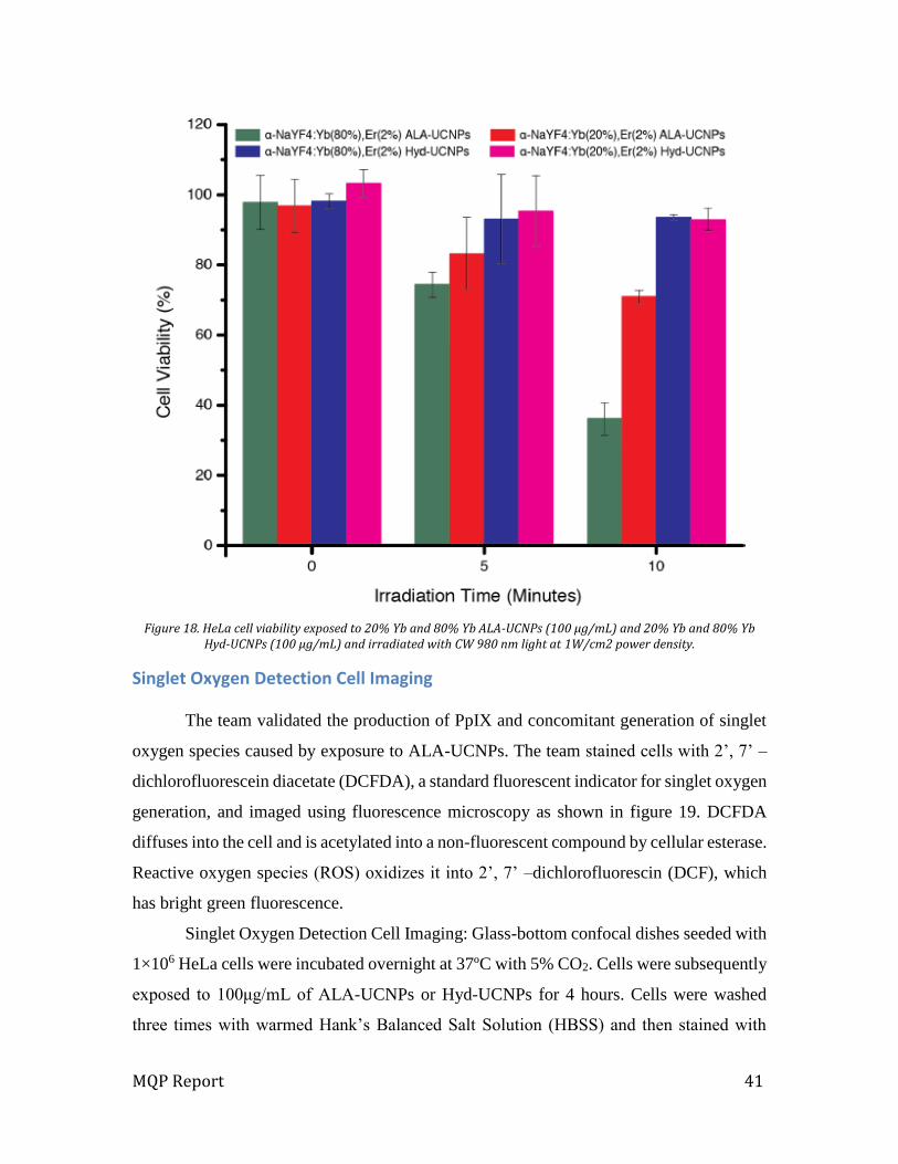
Figure 18. HeLa cell viability exposed to 20% Yb and 80% Yb ALA-UCNPs (100
μg/mL) and 20% Yb and 80% Yb Hyd-UCNPs (100 μg/mL) and irradiated with CW
980 nm light at 1W/cm2 power density. .................................................................................... 41
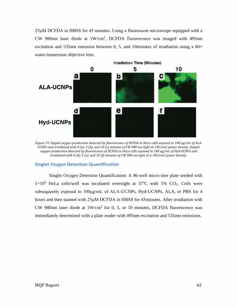
Figure 19. Singlet oxygen production detected by fluorescence of DCFDA in HeLa
cells exposed to 100 μg/mL of ALA-UCNPs and irradiated with 0 (a), 5 (b), and 10
(c) minutes of CW 980 nm light at 1W/cm2 power density. Singlet oxygen
production detected by fluorescence of DCFDA in HeLa cells exposed to 100 μg/mL
of Hyd-UCNPs and irradiated with 0 (d), 5 (e), and 10 (f) minutes of CW 980 nm
light at a 1W/cm2 power density. ................................................................................................. 42
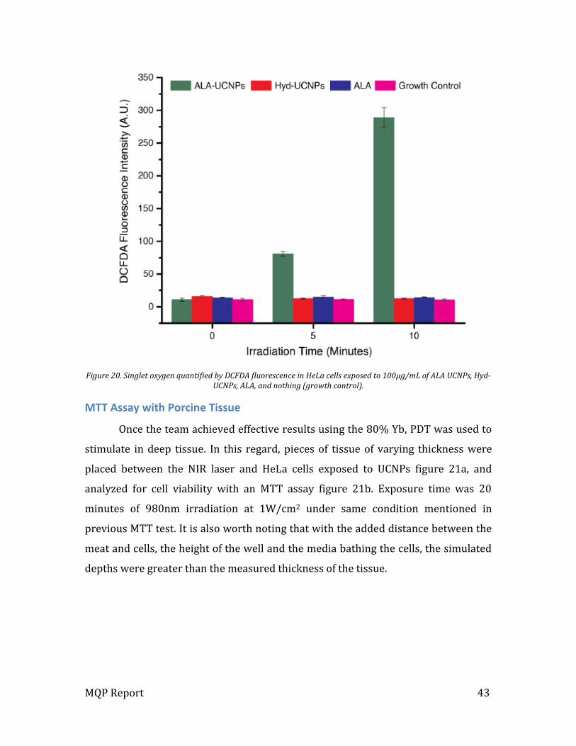
Figure 20. Singlet oxygen quantified by DCFDA fluorescence in HeLa cells exposed to
100μg/mL of ALA UCNPs, Hyd-UCNPs, ALA, and nothing (growth control). ............... 43
Figure 21. Photograph of the setup of simulated visceral tumor conditions in an in
vitro MTT assay (a). HeLa cell viability exposed to ALA-UCNPs (100 μg/mL) and
Hyd-UCNPs (100μg/mL) and irradiated with CW 980nm light at a 1W/cm2 power
density for 20 minutes ....................................................................................................................... 44
Figure 22. Schematic of the final design components and its work mechanisms....... 51
MQP Report 9
Chapter 1: Introduction
Cancer is an uncontrolled growth of cells in the human body. It is the second leading
cause of death in the US, with 1,660,290 new expected cases and about 580,350 expected
to die in 2013 (American Cancer Society, 2012a) . Cancer has a variety of treatments, and
the most effective ones are chemotherapy, radiotherapy and surgery, but all of these
treatments are not ideal in every cancer case, especially in the later stages when metastasis
occurs. Due to the side effects that cancer treatments have, scientists are always looking to
improve current treatments and find better alternatives. (American Cancer Society, 2012b)
Photodynamic Therapy (PDT) with Upconversion Nanoparticles (UCNPs) is a
treatment that shows significant potential in this field. PDT is a treatment that uses a
photosensitizing agent. When photosensitizers are exposed to a visible wavelength of light,
they produce singlet oxygen (1O2) that kills nearby cells; it has been used mostly for skin
diseases (Dougherty, 1998).
UCNPs, is a mixture of lanthanide-doped nano-crystals, which emit high energy
photons and visible light under excitation by near-infrared (NIR) light. Therefore UCNPs
will act as transducers, absorbing NIR light and converting it to visible light, which is
essential in PDT to activate the photosensitizer drug and release singlet oxygen to kill
cancer cells. Using both PDT and UCNPs for cancer treatment showed promising results,
especially in In-vivo testing (Wang, 2010).
The UCNPs that have been used with PDT for cancer treatment were excited at
980nm wavelength, which showed good results (Idris, 2012). Ytterbium (Yb3+) sensitizer
is the element commonly used for its unalterable excitation band centered at 980nm.
However, a longer wavelength from the UCNP with NIR causes the exciting light to
penetrate deeper into the biological tissue.
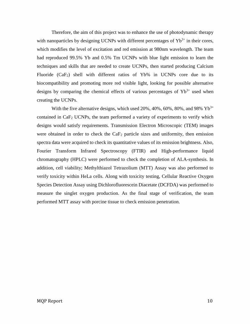
5-aminolevulinic acid (ALA) is currently used clinically in PDT as an FDA
approved photosensitizer drug. Through heme biosynthesis, ALA has a hydrophilic
characteristic and selectivity for photosensitizer protoporphyrin IX (PpIX) production in
cancer cells, and triggers and generates the red-absorbing photosensitizer PpIX. ALA was
found to be a suitable agent with the UCNPs designed by the team. It is important to note;
however, ALA usage in PDT requires the emission of red light, which here is converted
from NIR light by the UCNPs, in order to activate PpIX.
MQP Report 10
Therefore, the aim of this project was to enhance the use of photodynamic therapy
with nanoparticles by designing UCNPs with different percentages of Yb3+ in their cores,
which modifies the level of excitation and red emission at 980nm wavelength. The team
had reproduced 99.5% Yb and 0.5% Tm UCNPs with blue light emission to learn the
techniques and skills that are needed to create UCNPs, then started producing Calcium
Fluoride (CaF2) shell with different ratios of Yb% in UCNPs core due to its
biocompatibility and promoting more red visible light, looking for possible alternative
designs by comparing the chemical effects of various percentages of Yb3+ used when
creating the UCNPs.
With the five alternative designs, which used 20%, 40%, 60%, 80%, and 98% Yb3+
contained in CaF2 UCNPs, the team performed a variety of experiments to verify which
designs would satisfy requirements. Transmission Electron Microscopic (TEM) images
were obtained in order to check the CaF2 particle sizes and uniformity, then emission
spectra data were acquired to check its quantitative values of its emission brightness. Also,
Fourier Transform Infrared Spectroscopy (FTIR) and High-performance liquid
chromatography (HPLC) were performed to check the completion of ALA-synthesis. In
addition, cell viability; Methylthiazol Tetrazolium (MTT) Assay was also performed to
verify toxicity within HeLa cells. Along with toxicity testing, Cellular Reactive Oxygen
Species Detection Assay using Dichlorofluorescein Diacetate (DCFDA) was performed to
measure the singlet oxygen production. As the final stage of verification, the team
performed MTT assay with porcine tissue to check emission penetration.
MQP Report 11
Chapter 2: Literature Review
The aim of this chapter is to explain and describe the basic concepts of
Photodynamic therapy (PDT), Upconversion Nanoparticles (UCNPs), and the combination
use of PDT with UCNPs the benefits and drawbacks.
Photodynamic Therapy (PDT) Photodynamic therapy is a non-invasive treatment where involves three main
components. These components are; the laser with different wavelength, the
photosensitizer (PS) molecules, and the production of singlet oxygen (Wang, 2013).
Desired laser with a different wavelength used directly on targeted cells as a source of light,
this will activate the PS drug causing the production of singlet oxygen, leading to the
generation of reactive oxygen species (ROS), which is toxic product causing damage to
cellular components (Sharman, 1999).
PDT was used in many clinical trials, the first successful application was recorded
for curing acne by using ultra-violet (UV) light (Kalka, 2000). For many scientists and
researchers the achievement using PDT drew attention to the important use in different
medical fields such as cancer treatment since it was the biggest challenge this day. Cancer
studies using PDT showed positive results of substituting major cancer therapeutic
treatment from chemotherapy and radiotherapy with the PDT (Moan, 2003).
When the PS absorbs the UV light, the PS goes into a high energy state, transferring
this energy to the surrounding molecules causing the production of singlet oxygen (1O2)
or other reactive oxygen species. The ROS species are very toxic and has the ability to
destroy tumors by either direct cancer cell death by necrosis, apoptosis (Oleinick, 2002),
or by damaging tumor vasculatures as an anti-angiogenesis effect (Fingar, 1996).
The common procedures of PDT treatment starts with injecting drug with the PS in
a proper delivery system. Once the delivery system spread over a patient’s body, it causes
drugs to remain at the tumor tissue long period of time than healthy tissues. Then a certain
laser will be shed upon the area of tumor, which activates PS to release harmful chemicals
to kill tumor cells as shown in the Figure 1 below. Throughout the excretory system of the
patient’s body, the gradually clearance of the drug in the body, and the decrease of the
volume of the tumor will be done (Kalka, 2000).

MQP Report 12
Figure 1. General Schematic of PDT mechanism
Even though PDT had successful results, it had a poor tissue penetration due to the
lack of light transmission and severe photo-toxicity (Sharman, 1999). Most PDT PS
molecules currently used are excited by visible or UV light, which has limited penetration
depth in biological tissues, therefor this poor tissue penetration will limit the use of PDT
in treating large or internal tumors (Yang, 2012).
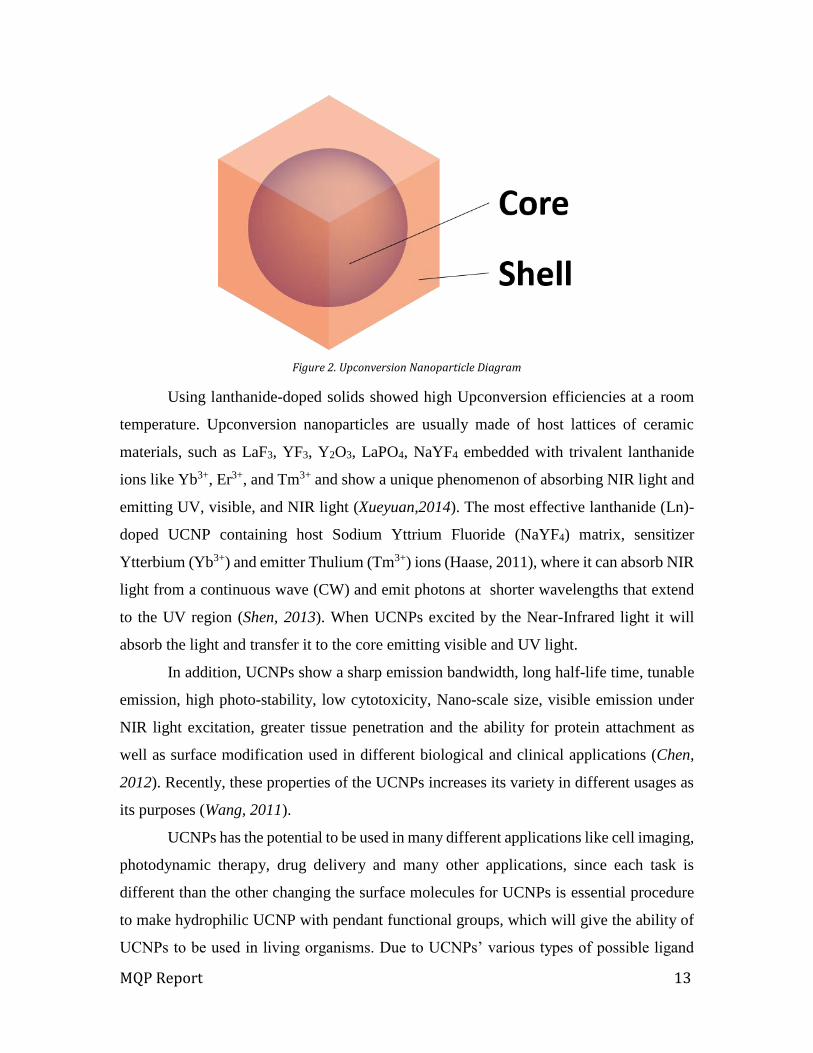
Upconversion nanoparticles (UCNPs) Upconversion Nanoparticles (UCNP) are usually composed with core and shell as
shown in figure 2 below. Core part of the UCNP, which mostly have a sphere shape, allows
the process in which the sequential absorption of two or more photons leads to the emission
of light at shorter wavelength than the excitation wavelength. By properties of the
composition of core structure, it would have different wavelength of near infra-red (NIR)
light convertibility and the color of visible light produced. UCNP shell functions to
affecting to have various intensity of emission and suitable material’s purposes from the
Core by having the photosensitizers loaded on host matrix of itself. The mechanisms of
UCNP are accomplished in solid-state materials doped with rare-earth ions, where it
convert long-wavelength radiation like infrared or near NIR, to a short-wavelength
radiation (Haase, 2011).
MQP Report 13
Figure 2. Upconversion Nanoparticle Diagram
Using lanthanide-doped solids showed high Upconversion efficiencies at a room
temperature. Upconversion nanoparticles are usually made of host lattices of ceramic
materials, such as LaF3, YF3, Y2O3, LaPO4, NaYF4 embedded with trivalent lanthanide
ions like Yb3+, Er3+, and Tm3+ and show a unique phenomenon of absorbing NIR light and
emitting UV, visible, and NIR light (Xueyuan,2014). The most effective lanthanide (Ln)-
doped UCNP containing host Sodium Yttrium Fluoride (NaYF4) matrix, sensitizer
Ytterbium (Yb3+) and emitter Thulium (Tm3+) ions (Haase, 2011), where it can absorb NIR
light from a continuous wave (CW) and emit photons at shorter wavelengths that extend
to the UV region (Shen, 2013). When UCNPs excited by the Near-Infrared light it will
absorb the light and transfer it to the core emitting visible and UV light.
In addition, UCNPs show a sharp emission bandwidth, long half-life time, tunable
emission, high photo-stability, low cytotoxicity, Nano-scale size, visible emission under
NIR light excitation, greater tissue penetration and the ability for protein attachment as
well as surface modification used in different biological and clinical applications (Chen,
2012). Recently, these properties of the UCNPs increases its variety in different usages as
its purposes (Wang, 2011).
UCNPs has the potential to be used in many different applications like cell imaging,
photodynamic therapy, drug delivery and many other applications, since each task is
different than the other changing the surface molecules for UCNPs is essential procedure
to make hydrophilic UCNP with pendant functional groups, which will give the ability of
UCNPs to be used in living organisms. Due to UCNPs’ various types of possible ligand
MQP Report 14
exchanges on the shell surface, ligand engineering involves a ligand exchange reaction
with hydrophilic bi-functional molecules or involves a direct oxidation of the terminal
group of native ligands to generate a pendant carboxylic functional group. Ligand attraction
involves absorption of an additional amphiphilic polymer onto the nanoparticle surface
through the hydrophobic Van der Waals attraction between the original ligand and
hydrocarbon chain of the polymer. Surface polymerization involves growing a dense cross-
linked shell on the nanoparticle core by condensation of small monomers. Last but not least
Layer-by-layer assembly involves electrostatic absorption of alternately charged poly-ions
on the nanoparticles surface. These entire synthetic procedures yield to hydrophilic
UCNPs, which could have additional functional groups and further bio-conjugation
capabilities (Wang, 2013).
As the first use of UCNP’s NIR-to-UV convertibility, Zhang and his team reported
NIR-to-UV β-NaYF4, 25%Yb3+, 0.3%Tm3+ UCNPs for photo-controllable gene expression
(Jayakumar, 2012). Photo-caging is any one of several molecular species that can be
activated by light; they are used especially in biochemistry to attach a molecule to a
biologically active compound and then study its behavior once activated. Photo-caging
involves the caging of molecules of interest by a light-sensitive molecule which can then
be destroyed by light irradiation to make it functional. Various molecules, like proteins,
peptides, nucleic acids, amino acids, and drugs, have been photo-caged and delivered to
animal’s cells and photolysis is done in the area of interest, enabling activation of these
molecules with very high spatial and temporal resolution. Due to the limitation of photo-
activating systems it can be only use in in-vitro applications, because UV light necessary
for the uncaging process, which is very harmful, besides having a poor tissue penetration
depth, which made it useless for in vivo and clinical use.
NIR light has the deepest tissue penetration compared to visible and UV light. It is
also safe and is expected to cause minimal photo-damage to the biological specimen
involved Zhang and his team chose to use UCNPs to convert NIR to UV light. Due to the
thin layers of mesoporous silica and caged DNA/siRNA in the mesopores, delivery of
higher payload and protection of the nucleic acids from the harsh environment was
possible. The method that Zhang and his team used has many advantages to improve photo-
caging by increasing the loading efficiency of nucleic acids when compared to chemical

MQP Report 15
crosslinking. The degree of activation reduces with increase in thickness of the tissue but
significant activation more than 50% was seen even with a tissue phantom thickness of 0.4
cm. Data provided by the team showed more efficient loading and delivery of the
DNA/siRNA cargo molecules.
The results support the team hypothesis, which showed the necessity of using
UCNPs for photo-controllable gene expression is that NIR to UV by using UCNPs, which
can be used for activating photo-caged nucleic acids in deeper tissues compared to
conventional systems. The results prove that this technique has enormous potential in a
variety of fields such as gene therapy and specific gene delivery or knockdown.
Combination of PDT & UCNPs
PDT based on UCNPs has a major advantage of targeting and treating such a deadly
disease as cancer. UCNP is the process of converting two or more low energy photons to
higher output photons. UCNPs have several properties useful in PDT due to its emission
ability upon a low level of excitation in the NIR of the spectral region including sharp
emission bandwidth, long lifetime, tunable, high photo-stability as well as lower
cytotoxicity. Upon UCNP excitation using NIR light converting longer wavelength into
emission at a shorter wavelength in UV, visible or, NIR light, which can activate loaded
photosensitizers molecules to produce singlet oxygen killing cancer cells that is more
specific to a nanometer regime with minimal photo damage and a significant enhancement
in light penetration depth.
Coated NaYF4 UCNP with mesoporous silica were irradiated using a 980nm laser
causing the loaded drug to release singlet oxygen (1O2). The 1O2 is known for its toxicity
to targeted cells such as cancer cells. After evaluating the efficiency of the UCNP as a PDT
agents in in vitro by minimizing the volume of cancer cells caused by the toxicity of 1O2,
Idris and his lab proceeded an In-vivo experiments (Idris, 2012). Melanoma cell coated
UCNP injection subcutaneously to mice in four different conditions; UCNP loaded with
drug then irradiated with a 980nm laser, UCNP loaded with drug, no drug with a laser
exposure of 980nm, and untreated. After two weeks, cells except UCNP loaded with drug
then irradiated with a 980nm laser condition formed a solid tumor while cells in group 1
has considerably been decreased. Current UCNP based PDT application achieved data
MQP Report 16
success, but this process is still limited due to insufficient upconverting efficiency of the
nanoparticles.
5-Aminolevulinic acid
As discussed in Chapter one, PDT is based on three important elements; light, 1O2
and a photosensitizer. Photosensitizer is a very important for photosensitization efficacy
and producing 1O2. Once light gets absorbed, it causes excitation of molecules, which
activates the photosensitizer (Tian, 2013). PDT can be a new approach of using an
endogenous photosensitizer protoporphyrin IX (PpIX). PpIX is known as the precursor of
prophyrins, which are naturally occurring organic compound. One of the most known
prophyrins is heme; the co-factor of the protein hemoglobin. PpIX is an excellent
photosensitizer due to its minimum to no photodynamic damage.
5-aminolevulinic acid (ALA) is one of the two FDA approved photosensitizers and
clinically used. When ALA is administrated, it acts as a prodrug. ALA synthesis occurs in
the mitochondria formed by the condensation of glycine and succinyl CoA, catalyzed by
ALA-synthase. ALA undergoes several synthetic steps after it is transported into the
cytosol. The transportation of cytosol causes the production of proto-prophyrins, then
precursor prophyrinogens, protoporphyrin IX and then heme. As the final stages of heme
biosynthesis, co-proporphyrinogen gets transported from the cytosol into the mitochondria
and there converted to prophyrin and PpIX with an iron inserted (Huang, 2005).
ALA is an appropriate solution for cancer therapy because of its specificity for
cancer cells. When ALA administrated exogenously, it acts as a prodrug generating
photosensitizer PpIX via heme biosynthesis pathway in the cancer cells. The ALA-PDT
depends on the generation of PpIX, which is the intermediate of heme biosynthesis and
does not accumulate in the pathway. Generating more PpIX quantities required; therefore,
ALA administrated exogenously with an active heme biosynthesis, the pathway becomes
temporarily overloaded. The control mechanism is bypassed, and downstream metabolites
are synthesized in excess. Next, the ferrochlelatase; catalyze in the biosynthesis of heme
where it is convert pro proporphyrin IX into heme with insertion of iron. PpIX accumulate
in the cells because of the low physiologic rate of iron insertion by ferrochelatase, which
is unable to compensate for the excess PpIX formed and render them photosensitive (Peng,
1997).
MQP Report 17
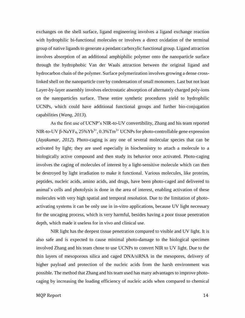
Following, the synthesis of 5-ALA induced in mitochondria, the PpIX selectively
accumulate in mitochondria causing the death of cells after PDT treatment. ALA has
excellent intestinal absorption when administrated orally. ALA uses the intestinal and renal
apical peptide transporters to enter epithelial cells and in some cells ALA is transported
using system Beta transporters.
There are various theories explaining why ALA has a tendency to accumulate in
tumors and other lesions. ALA tumor accumulation could be caused due to the low level
of ferrochelatase in comparison with normal cells where less PpIX covered to heme, plus
tumors have a low pH causing more PpIX retention leading to PpIX accumulation
(Collaud, 2004).
Figure 3. Schematic of the ALA-UCNPs Entering the Cell
ALA Advantages:
- Ability to penetrate the abnormal tumor under Stratum Corneum due to its small size
and solubility
- ALA-PpIX accumulate in cells and not the tumor vasculature
- Rapidly cleared from the system 1-2 days
MQP Report 18
- Easily synthesized
- ALA- PDT is noninvasive treatment, where it does not harm surrounding tissue
- ALA can be administrated topically
ALA disadvantages:
- ALA has low lipid solubility, therefore has a limited ability crossing biological barriers
- PpIX limited tissue penetration 1mm
- ALA stable is an acid environment, pH low as 2-2.5.
MQP Report 19
Chapter 3: Project Strategy In this chapter, each planned procedures prioritized by the various objectives and
constraints will be introduced. The project approach section will also explain the project
framework of necessary steps to complete the proposed Upconversion Nanoparticle
(UCNP) Synthesis.
Initial Client statement
The following initial client statement was given from the client, Dr. Gang Han from
University of Massachusetts Medical School.
“Photosensitizers are excited by visible or Ultra Violet (UV) light, which has
limited penetration depth due to the light absorption and scattering by biological
tissues, resulting in ineffective therapeutic effects to internal or large tumors.
UCNPs have the ability to convert Near Infra-Red (NIR) light to visible photons,
which can active photosensitizers adsorbed on nanoparticles via resonance energy
transfer to generate singlet oxygen (1O2) to kill cancer cells. It would provide an
alternative to overcome hurdles of current photodynamic therapies. The goal of this
project is to improve current UCNPs at a single excitation wavelength between
600nm and 1,200nm created by continuous-wave (CW) laser in order to perform
better at imaging and safer to use with photodynamic therapy in living organisms.”
Therefore, more research about photodynamic therapy (PDT) and UCNP has been
performed. Then, the team arranged meetings and client interviews to better understand
and clarify the initial client statement. Sets of objectives and constraints were listed to draft
a revised client statement, which stated the client’s problem and their desired solutions
more concisely and clearly.
Objectives Through research, the team discovered much information regarding upconversion
nanoparticles (UCNPs) and Photodynamic Therapy (PDT). With the information, various
objectives were brought up based on a compiled a list of current problems with UCNPs.
MQP Report 20
After analyzing the client statement, researching UCNP, and scheduling client meetings,
the following list of objectives were made.
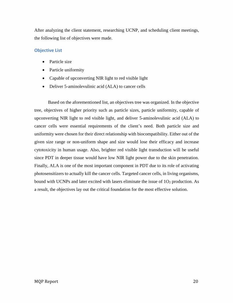
Objective List
Particle size
Particle uniformity
Capable of upconverting NIR light to red visible light
Deliver 5-aminolevulinic acid (ALA) to cancer cells
Based on the aforementioned list, an objectives tree was organized. In the objective
tree, objectives of higher priority such as particle sizes, particle uniformity, capable of
upconverting NIR light to red visible light, and deliver 5-aminolevulinic acid (ALA) to
cancer cells were essential requirements of the client’s need. Both particle size and
uniformity were chosen for their direct relationship with biocompatibility. Either out of the
given size range or non-uniform shape and size would lose their efficacy and increase
cytotoxicity in human usage. Also, brighter red visible light transduction will be useful
since PDT in deeper tissue would have low NIR light power due to the skin penetration.
Finally, ALA is one of the most important component in PDT due to its role of activating
photosensitizers to actually kill the cancer cells. Targeted cancer cells, in living organisms,
bound with UCNPs and later excited with lasers eliminate the issue of 1O2 production. As
a result, the objectives lay out the critical foundation for the most effective solution.
MQP Report 21
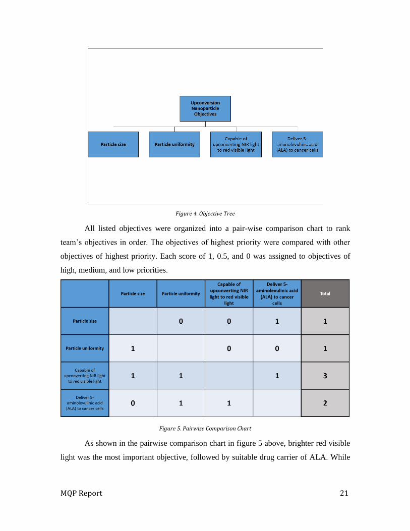
Figure 4. Objective Tree
All listed objectives were organized into a pair-wise comparison chart to rank
team’s objectives in order. The objectives of highest priority were compared with other
objectives of highest priority. Each score of 1, 0.5, and 0 was assigned to objectives of
high, medium, and low priorities.
Figure 5. Pairwise Comparison Chart
As shown in the pairwise comparison chart in figure 5 above, brighter red visible
light was the most important objective, followed by suitable drug carrier of ALA. While

MQP Report 22
both particle size and uniformity were deemed important, they were considered secondary
objectives.
Design Constraints Constraints served to determine the real-world effectiveness of designed ideas.
Failure to meet constraint criteria resulted in an automatic failure of a proposed solution.
In light of the prior analysis of project requirements and lab research of the boundaries of
UCNPs as a solution vector, the following list of constraints were made.
Must have particle size within a range of 20-40nm
Must have uniform particle diameter sizes with lower the standard deviation value
less than 5nm
Must transduce the wavelength between 700 to 1,100nm NIR light into visible red
light more than 1.00×10⁸ absorbance unit (A.U.)
Must be proven to be non-toxic through Methylthiazol Tetrazolium (MTT) assay
Must reduce cell viability with 12mm thickness of tissue penetration through MTT
assay
Must have stable ALA-conjugation
In order to have UCNPs fulfill solution scope requirements, the diameter size of the
particles must consistently be within a range of 20-40nm while responding to lasers emitted
at a wavelength of 700 to 1,100nm. The design must also demonstrate non-toxicity within
the body until exposure to controlled activation conditions.
Functions Based on the constraints the team came up with, there were some specific functions,
which the modified upconversion nanoparticle (UCNP) has to have. After analyzing the
client statement, researching UCNP, and having a client meeting, the following list of
functions can be listed.
In light of the aforementioned objectives and constraints, a formal list of functions
that the team's modified UCNPs must have was drafted.

MQP Report 23
Produce strong red visible light brighter than 1.00×10⁸ A.U.
Perform non-toxic activity through cell viability assay
Release 1O2 only when it shed with a certain NIR laser
In order to utilize UCNPs for the treatment of cancer under the tissue, it needed to
transduce NIR laser into strong red visible light in order to activate the photosensitizer the
controls the release of toxins to kill cancer cells.
Revised Client Statement Based on the results of all the mentioned objectives, constraints, and functions
above, a revised client statement was written. This statement helped the team successfully
propose a promising design as well as identify potential design alternatives.
“The goal of this project is to build upon current UCNP procedures so that it will
result in a stable and effective method of treatment for subcutaneous cancer through
increased light penetration and emission for ALA activation with designed
wavelength excitation between 600nm and 1,200nm.”
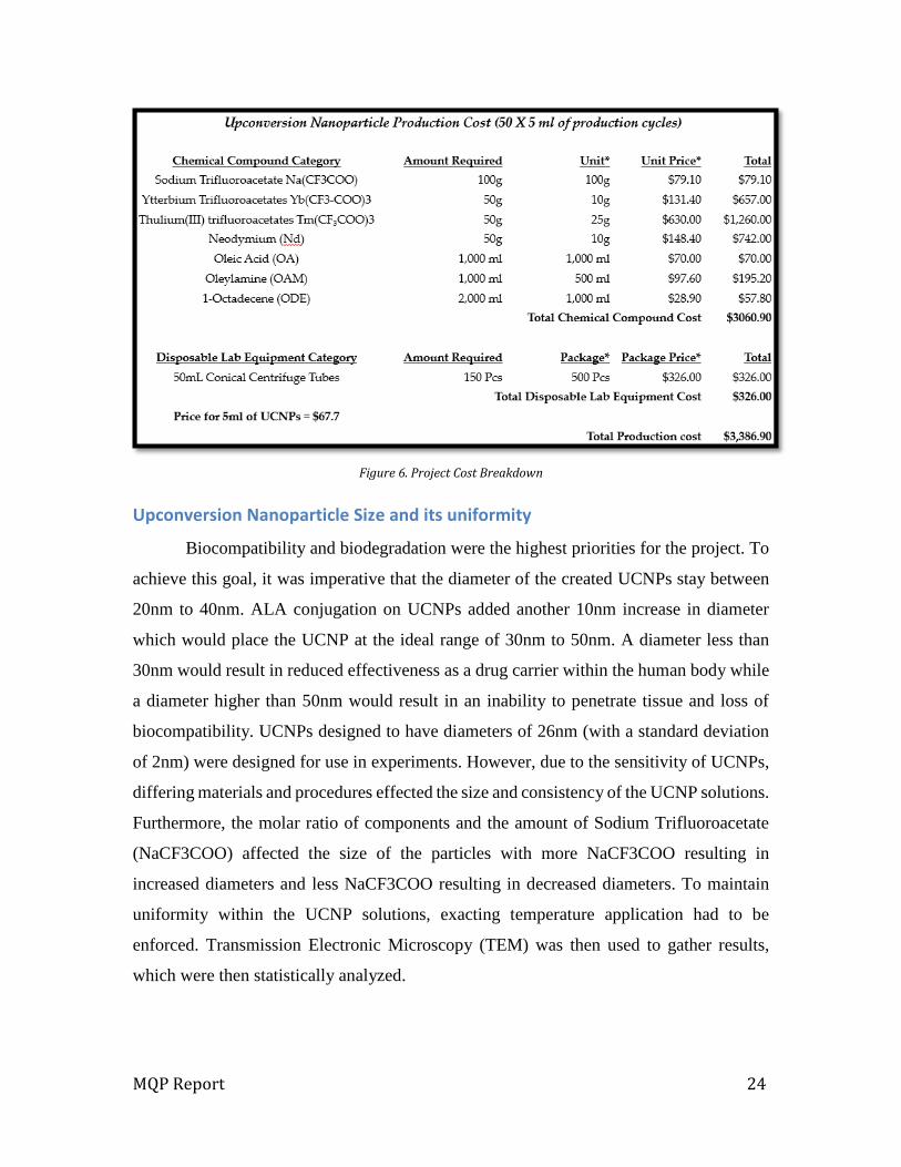
Project Approach
With the foundation of the project clearly defined, a project approach was then
drafted to guide the Upconversion Nanoparticle synthesis project. After establishing
objectives, constraints, and project goal, the next step was careful design. Due to the
complexity of synthesizing UCNPs, the team familiarized themselves with the production
procedures. Background research was conducted to fully understand the various chemical
compounds and their characteristics in chemical reactions. Due to financial limitations in
acquiring the necessary chemicals, as shown in figure 6 below, each compound candidate
was carefully and thoughtfully handled in order to minimize chemical utilization while
maximizing sample result usefulness. Lab equipment and space was provided by
University of Massachusetts Medical School.
MQP Report 24
Figure 6. Project Cost Breakdown
Upconversion Nanoparticle Size and its uniformity
Biocompatibility and biodegradation were the highest priorities for the project. To
achieve this goal, it was imperative that the diameter of the created UCNPs stay between
20nm to 40nm. ALA conjugation on UCNPs added another 10nm increase in diameter
which would place the UCNP at the ideal range of 30nm to 50nm. A diameter less than
30nm would result in reduced effectiveness as a drug carrier within the human body while
a diameter higher than 50nm would result in an inability to penetrate tissue and loss of
biocompatibility. UCNPs designed to have diameters of 26nm (with a standard deviation
of 2nm) were designed for use in experiments. However, due to the sensitivity of UCNPs,
differing materials and procedures effected the size and consistency of the UCNP solutions.
Furthermore, the molar ratio of components and the amount of Sodium Trifluoroacetate
(NaCF3COO) affected the size of the particles with more NaCF3COO resulting in
increased diameters and less NaCF3COO resulting in decreased diameters. To maintain
uniformity within the UCNP solutions, exacting temperature application had to be
enforced. Transmission Electronic Microscopy (TEM) was then used to gather results,
which were then statistically analyzed.
MQP Report 25
Transducing near-Infrared into Visible light and Biocompatibility Cancer cells are commonly located under the skin at a depth of roughly 12mm or
even deeper. In order to utilize UCNPs as treatment, a NIR laser of a 980nm wavelength
proved the best candidate. After successfully penetrating the skin with the NIR laser, the
UCNP compound must convert the energy into bright red visible light to trigger the
activation photosensitizer that controls the release of 1O2. The light emitted from the
UCNPs, upon activation, must be brighter than 1.00x10⁸A.U. Also, compound selection
requires biocompatibility, which promotes opsonization of reticuloendothelial system.
Testing Utilizing TEM imaging and Emission Spectra analysis, the team rooted out a
promising final candidate amongst all the proposed designs. In-vitro tests were then
performed followed by another round of TEM imaging to check for correct ALA
conjugation of UCNPs to determine the final size and uniformity. Once those two tests
were completed, a round of Fourier Transform Infrared Spectroscopy (FTIR) was
performed to verify the stability and conjugation status of the UCNPs. Next, to determine
whether or not the UNCPs were correctly releasing 1O2, Fluorescence Intensity of Cellular
Reactive Oxygen Species Detection Assay (DCFDA) was performed. After the
confirmation of the release of singlet oxygen, the final design with ALA conjugation was
then injected directly into a cell culture medium for Methylthiazol Tetrazolium (MTT)
Assay using HeLa cells. 12mm of porcine skin, which was obtained from a grocery market,
was then used to simulate human skin tissue and determine the effectiveness of the ability
of the 980nm wavelength laser to penetrate skin tissue. Despite passing through 12mm of
skin, the 980nm laser was still able to activate the photosensitizers. These experiments
were then repeated three times for statistical analysis.
MQP Report 26
Chapter 4: Alternative Designs
Requirements analysis
As mentioned in background chapter, Upconversion Nanoparticle (UCNP) for the
usage of photodynamic therapy (PDT) requires having certain functions as FDA-approved
drug. As the purpose of this project, primary functions such as particle sizes and uniformity,
emission brightness, and stability of ALA conjugation are fall within the scope.
Objectives
Particle size
o Diameter between 20 to 40nm
o With conjugation, diameter range between 30 to 50nm
Particle uniformity
o Uniformity in size and shape
o UCNP’s steady action in human body
Brighter red visible light
o More than 1.00×10⁸ absorbance unit (A.U.)
o Overcome reduced NIR light power after tissue penetration
Suitable drug carrier of ALA
o Delivery of ALA conjugation
o Stable ALA conjugation
Alternative designs
In order to properly evaluate the effects of different design components, it was
critical for the team to perform research on each and every step of the production process.
As a result, the team was able to identify three main core components that were essential
for the core of UCNPs. (Wilhelm, Hirsch, Schueucher, Mayr & Wolfbeis, 2012) They are
Thulium (Tm), Erbium (Er), and Hydroxide (OH-). As a first step, the team produced a
synthesis of β – 99.5%Yb 0.5%Tm for the core of the nanoparticle. Since Tm Nanoparticles
have significant blue emission, which is an ideal design to use as ultraviolet (UV) test.

MQP Report 27
99.5% Yb, 0.5% Tm (alpha phase)
In a 50ml flask, 0.1365g of Sodium Trifluoro-acetate (NaTFA), 1.3 mg of Thulium
Trifluoro-acetate (TmTFA), and 0.2547g of Ytterbium Trifluoro-acetate (YbTFA) are
added and mixed with 1.43g of Oleic Acid (OA), 2.53g of Octadecene-1 (ODE), and 1.34g
of Oleyamine (OAm). By using temperature controller and magnetic stirrer, the flask was
heat up to 110°C for degasing. With this degasing process, it would promote release of any
extra and unnecessary gas between molecules and helps its purity and reaction time. During
this process, bubbles occurred in the fluid and disappeared after couple of minutes. After
10 minutes of degasing process, the temperature was controlled to 300°C to initiate alpha
phase of the nanoparticle. This process lasted for 35minutes to fully transform the fluid
into alpha phase nanoparticles. Ytterbium Trifluoro-acetate (Yb3+TFA) functions as main
core, TmTFA as activators, and NaTFA as size controller. After the heating process, the
team let the fluid to be fully cool down as low as 70°C. Then it was centrifuged and all the
fluid residue were removed. With the centrifuged alpha phase nanoparticles were diluted
with 10ml of Hexane. Alpha phase nanoparticles can be checked with its particles sizes
and uniformity by having either Transmission Electron Microscopy (TEM) images, which
will be explained further below, taken or simple 980nm Laser test. For the 980nm Laser
test, increasing laser power more than 2 Ampere will show light emission to estimate its
uniformity and sizes.
Transmission Electron Microscopy
Transmission Electron Microscopy (TEM) uses a beam of electrons to transmit
through a thin nanoparticle specimen. Using the interaction of the electrons, it filters into
black and white images to show detailed images. Since the average sizes of UCNPs were
approximately between 15nm to 100nm, TEM was the only microscopy that allowed
accurate confirmation of particle size. Nanoparticle sample fluids were then placed in
circular shaped carbon mesh with a diameter of 2mm. The carbon mesh was then inserted
into the vacuum of the TEM, manipulating controllers to determine the best locations to
take images. For UCNPs, 160,000× zoom was recommended to get an image for the
particle comparison.
MQP Report 28
99.5% Yb, 0.5% Tm (beta phase)
Once the alpha phase of the nanoparticle production was complete, the team moved
onto the beta phase. Once all the hexane was dried, 0.1365g of NaTFA was added to the
flask. Then, a solvent of 2.83g of OA and 2.53g of ODE were added. An ultrasonic bath
diluted both the NaTFA and the dried alpha phase particles. The degassing process utilized
in the alpha stage was repeated for 10minutes. Afterwards, the temperature of the solution
was set to 325°C for 30minutes in order to properly transform the Beta cores. To obtain
the pure beta phase nanoparticles, the centrifugation and dilution with hexane steps from
alpha stage were repeated. TEM imagery was then repeated to verify uniformity and size
as well as the 980nm laser test with 0.5amperes. The 10ml of beta phase core nanoparticles
were then diluted in hexane. The team transferred half of the nanoparticles (5ml) into a
50ml flask. Once the solution was fully dried of all hexane, the pure core nanoparticles
were then mixed with 10% Yb, and 90%Y with 0.068g of NaTFA. 2.83g of OA and 2.53g
of ODE were also added to function as solvents. The flask was then heated first to 110°C
for 10min for degasing and then heated to 325°C for 30min, similar to the heating process
in the beta phase production of the cores. Both Centrifugation and dilution of hexane were
performed identically to the beta phase core production. After the transfer of the UCNPs
into hexane into a 20ml glass vial, TEM imagery and 800nm wavelength laser tests were
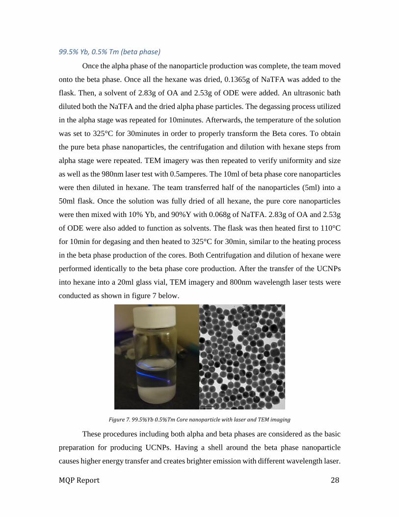
conducted as shown in figure 7 below.
Figure 7. 99.5%Yb 0.5%Tm Core nanoparticle with laser and TEM imaging
These procedures including both alpha and beta phases are considered as the basic
preparation for producing UCNPs. Having a shell around the beta phase nanoparticle
causes higher energy transfer and creates brighter emission with different wavelength laser.
MQP Report 29
For example, core nanoparticle already has an emission occurs at 980nm wavelength laser.
However, Addition of Nd in shell ingredient will make the nanoparticles to be emitted at
800nm wavelength laser with the same color of emission as the core had originally.
99.5% Yb, 0.5% Tm (Shell production)
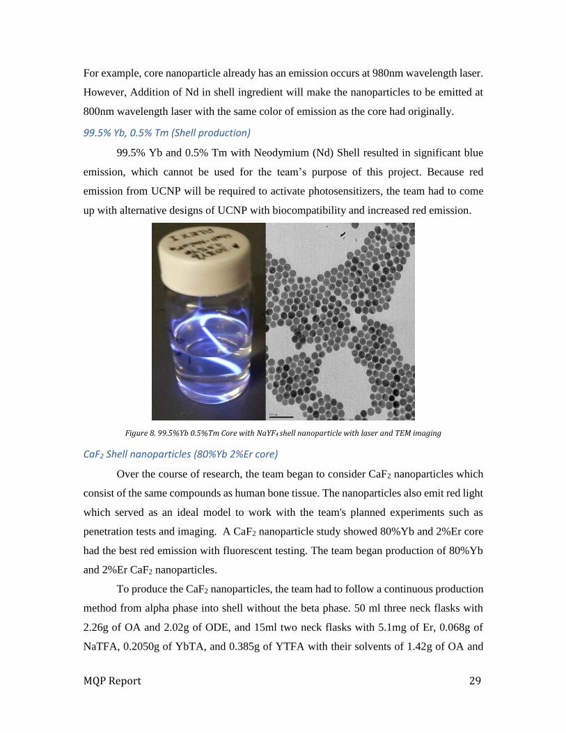
99.5% Yb and 0.5% Tm with Neodymium (Nd) Shell resulted in significant blue
emission, which cannot be used for the team’s purpose of this project. Because red
emission from UCNP will be required to activate photosensitizers, the team had to come
up with alternative designs of UCNP with biocompatibility and increased red emission.
Figure 8. 99.5%Yb 0.5%Tm Core with NaYF4 shell nanoparticle with laser and TEM imaging
CaF2 Shell nanoparticles (80%Yb 2%Er core)
Over the course of research, the team began to consider CaF2 nanoparticles which
consist of the same compounds as human bone tissue. The nanoparticles also emit red light
which served as an ideal model to work with the team's planned experiments such as
penetration tests and imaging. A CaF2 nanoparticle study showed 80%Yb and 2%Er core
had the best red emission with fluorescent testing. The team began production of 80%Yb
and 2%Er CaF2 nanoparticles.
To produce the CaF2 nanoparticles, the team had to follow a continuous production
method from alpha phase into shell without the beta phase. 50 ml three neck flasks with
2.26g of OA and 2.02g of ODE, and 15ml two neck flasks with 5.1mg of Er, 0.068g of
NaTFA, 0.2050g of YbTA, and 0.385g of YTFA with their solvents of 1.42g of OA and
MQP Report 30
1.27g of ODE were put in the heating plates at 110°C for the degas process. Prior to the
10 minutes of degasing process, the team performed ultrasonic bathing for the 15 ml flask
with solutes in order to fully dilute. After the degas, 50 ml three neck flasks were heated
up to 310°C, in order to activate the forming alpha phase process. The success of the
process was confirmed by the change of color from clear to yellow. Then with 5ml syringe,
the team transferred all of the degased fluid from the 15ml flask to 50ml flask with its
temperature of 310°C to start the alpha phase. During the transfer process, the fluid was
slowly and continuously injected into the 50ml flask over the duration of 5minutes. After
completing the transfer, the combination of two fluids was kept at a temperature of 310°C
for an hour. While they were forming alpha phase nanoparticles, the team prepared a
mixture of 0.5320g of Calcium Trifluoro-acetate (CaTFA) as solutes and 1.42g of OA and
1.27g of ODE as solvents for the CaF2 shell production. The fluid went through the
degasing process at 110°C twice with Argon injection in-between the two processes.
After one hour of alpha phase nanoparticle production, half of the CaF2 shell fluid,
approximately 1.5ml, was transferred into 50 ml three neck flask with its same method the
team took as previous fluid transfer with a syringe. Transferring 1.5ml of the fluid at a
continuous speed for 3minutes activated shell production around the alpha phase
nanoparticles. After 30minutes of shell production at a consistent temperature of 310°C,
the second half of the fluid from 15ml flask was transferred into 50ml three neck flask in
order to promote the stability of the shell. The purpose of separating shell fluid injection
into two different steps was to increase the chemical bonding structure of shell to be more
stable and evenly spread around all the alpha phase nanoparticles. 30minutes after the
second injection, the team removed the heating plate and fully cooled down the fluid to as
low as 70°C. After the cooling process was complete, the fluid was transferred into a 50
ml conical centrifuge tube and centrifuged at 7,000rev/min for 10minutes and all fluid
residues was removed. The centrifuged alpha phase nanoparticles were then diluted with
10ml of Hexane. TEM imagery and a 980nm laser test were conducted to determine particle
uniformity and size. The team confirmed the red emission of the UCNPs, which aligned
well with the planned experiments. In order to confirm with the CaF2 study of 80%Yb
2%Er, the team produced each different ratio of particles as shown in figure 9.

MQP Report 31
Figure 9. Alternative designs; CaF2 nanoparticles comparison with different ratio of Yb; 20%, 40%, 60%, 80%, and 98% respectively
As a result, TEM Images showed different molecule structure as Tm activated
cores. As previously showed in figure 7 and 8, Tm Cores and shells had a uniformed
circular shape of particles. However, CaF2 shell particles had very distinctive square shapes
in figure 10 below. Each particle sizes fell under the given size range of 20 to 40nm as
stated in objectives. 20%, 40%, 60%, 80%, and 98% Yb composed alternative designs had
average diameter size of 26, 25, 26, 26, and 28nm respectively.
Figure 10. TEM image of CaF2 nanoparticle with 80%Yb 2%Er
MQP Report 32
After confirming nanoparticles’ size and uniformity with TEM images, the team
proceeded to move on the experimental processes with CaF2 nanoparticle with 80%Yb
2%Er.
ALA Coupling
With the advantage of nanotechnology, the team combined ALA-PDT by
developing upconversion nanoparticles (UCNPs), which were excited by near infrared
light (NIR) and emitting multicolor to ultraviolet. In this case, the red right needed to
activate PpIX carried by the UCNP providing better deep tissue penetration.
In chapter 4 mentioned how the team developed α-NaYF4:Yb,Er with a
biocompatible CaF2 shell for imaging applications. Being a component of ossified tissues,
CaF2 is even more compatible than the conventional NaYF4 shell on UCNPs. The team
had amplified the red emission of NaYF4:Yb,Er@CaF2 by adjusting the ratio of the core
Yb3+.
Emission spectra under CW 980nm 1 W/cm3 excitation was obtained and
corresponding photographs of these UCNPs demonstrated chapter 4, it was discovered α-
NaYF4:80%Yb,2%Er@CaF2 had the optimal red emission with 980nm light excitation
required for the PpIX to produce singlet oxygen and induce tumor death, as seen in-vitro
with simulated deep-tissue conditions.
The team conjugated 5-ALA to the UCNPs by using covalent hydrazine linkage to
avoid pre-leakage of the ALA that would increase the success of the therapy by increasing
its bioavailability in figure19 demonstrate the process of conjugating ALA-on
αNaYF4:80%Yb,2%Er@CaF2.
Using a 4ml vial, 1ml of α-NaYF4:80%Yb,2%Er@CaF2, 4ml hexane, 5ml
dimethylformaide (DMF) and 2g of Nitrosonium tetrafluoroborate (NOBF4) were added
and mixed for 24h using a magnetic stirrer. After 24 hours took the vial contents are poured
into conical tube, then the vial was washed with a total of 10ml isopropanol ensuring no
particles are left behind. Using the counter top centrifuge the conical vial was balanced and
centrifuged for 10min at 7000rpm. After 10min supernatant decanted and small pellet
formed. The vial contained the pellet-dissolved in10ml of the DMF using sonicator, and
then added to 30ml single neck flask and 10mg/ml (100mg) of polyacrylic acid (PAA). The
MQP Report 33
flask was placed in an oil tube resting on a temperature/ stirrer controller, temperature set
on 80oC using a 1000rpm for another 24 hours.
The contents, after 24 hours, are poured into conical tube, washed the single neck
flask with a total of 30ml hexane and 30ml isopropanol and transferred into the same vial.
The vial was then centrifuged-using 11,000rpm for 30min at temperature of 20oC. The
pellet formed was dissolved in 10ml deionized water and centrifuged-using 11,000rpm for
30 minutes and 20oC, this step was repeated twice by the third centrifuge, the pellet re-
solubilized in 5ml phosphate buffer saline (PBS) and centrifuged using the same settings
previously, this step was repeated twice eliminating the pellet from any free PAA.
The solution was then transferred into glass vial, where 0.05g 1-Ethyl-3-(3-
dimethylaminopropyl)-carbodiimide (EDC) and 0.025g N-hydroxysulfosuccinimide
(Sulfo-NHS) is added to the glass vial and left on the stirrer for two hours. The contents
were transferred into the conical tube and centrifuged using the centrifuge settings above.
After completing the centrifuge time, the pellet dissolved in 5ml PBS using the sonicator
later transferred into glass vial and placed under the hood on a stirrer, carefully 5ml of
hydrazine transferred into the glass vial and closed immediately which left to react for 24
hours.
The final step before conjugating the ALA, using the contents from the previous
reaction and centrifuged using 11,000rpm, for 30minutes under 20°C temperature, the
pellet was then solubilized and centrifuged twice in 5ml deionized water using sonicator to
make absolutely sure there was no free hydrazine on the UCNPs. After the last wash 5ml
of methanol and acetic acid was added, repeated twice by following the earlier centrifuge
settings using the usual settings to make sure there was no water and only methanol and
acetic acid in the solution. The contents were transferred into glass vial covered with
aluminum with stir bar, added 4ml of ALA and placed on a medium-high speed stirrer for
48 hours.

MQP Report 34
Figure 11. The Process of Conjugating 5-Aminolevulinic acid on αNaYF4:Yb80%Yb,2%Er@CaF2
There are two reasons why the ALA-UCNPs are attracted to cancers cells. One, the
ALA is an amino acid, carbon based and naturally occurring; therefore the body will not
recognize it as a furan body and reject it. Second, the hydrazine is composed of two amino
groups and has a positive charge nitrogen. Since the UCNPs-ALA have a charge of -7mv,
which is less negative than the cells -40mv ALA-UCNPs they are phagocytized by the cell
and exposed to the low pH of the endosome. This caused the cleavage of the hydrazine
linkage and the ALA subsequently diffused to the mitochondria causing the overproduction
of PpIX. Meanwhile, the leftover Hyd-UCNP in the endosome would remain unharmed
and be activated by the NIR light, leading to the photosensitization of PpIX and death for
the cell shown in figure 12.

MQP Report 35
Figure 12. Illustration demonstrating the Production of PpIX in the mitochondria
Using the final chosen nanoparticle α-NaYF4:Yb(80%),Er(2%)@CaF2 conjugated
with 5-Aminolevulinic acid (ALA), several experiments were performed to determine the
consistency of the size using the TEM. A fluorometer was utilized to measure the red
emission of the ALA-UCNPs in both water and hexane, and Fourier Transform Infrared
Spectroscopy (FTIR) was used to confirm the conjugation of the ALA to the UCNPs via a
hydrazine linkage. Then Methylthiazol Tetrazolium Assay (MTT) was performed to
measure the cell viability and cell imaging demonstrating the generation of reactive oxygen
species (ROS)
MQP Report 36
Chapter 5: Design Verification
Transmission Electron Microscopy (TEM):
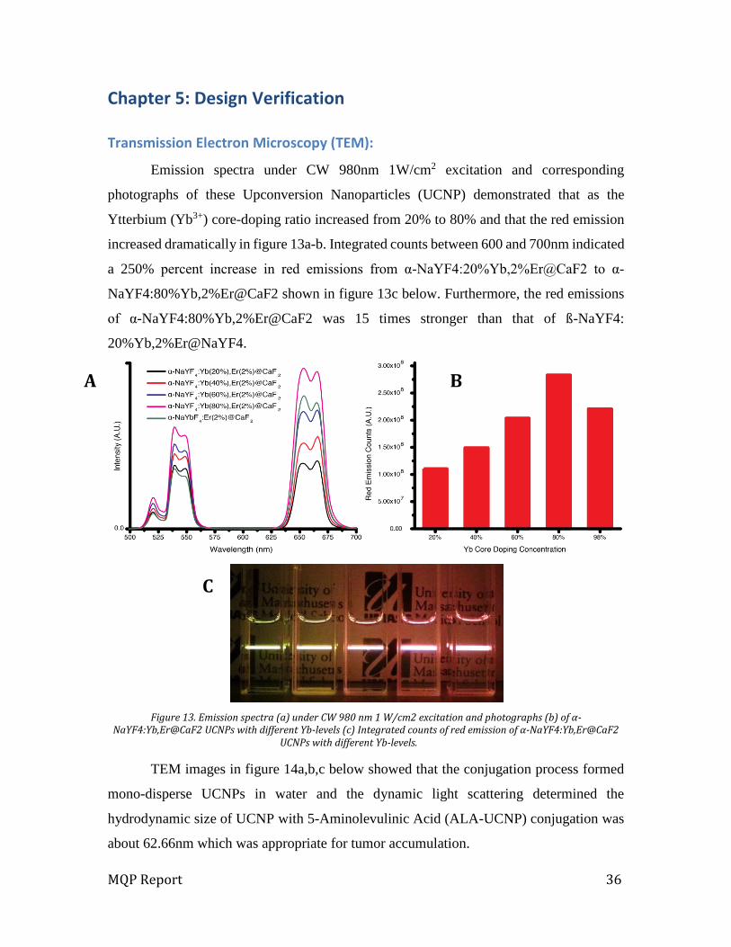
Emission spectra under CW 980nm 1W/cm2 excitation and corresponding
photographs of these Upconversion Nanoparticles (UCNP) demonstrated that as the
Ytterbium (Yb3+) core-doping ratio increased from 20% to 80% and that the red emission
increased dramatically in figure 13a-b. Integrated counts between 600 and 700nm indicated
a 250% percent increase in red emissions from α-NaYF4:20%Yb,2%Er@CaF2 to α-
NaYF4:80%Yb,2%Er@CaF2 shown in figure 13c below. Furthermore, the red emissions
of α-NaYF4:80%Yb,2%Er@CaF2 was 15 times stronger than that of ß-NaYF4:
20%Yb,2%Er@NaYF4.
Figure 13. Emission spectra (a) under CW 980 nm 1 W/cm2 excitation and photographs (b) of α-NaYF4:Yb,Er@CaF2 UCNPs with different Yb-levels (c) Integrated counts of red emission of α-NaYF4:Yb,Er@CaF2
UCNPs with different Yb-levels.
TEM images in figure 14a,b,c below showed that the conjugation process formed
mono-disperse UCNPs in water and the dynamic light scattering determined the
hydrodynamic size of UCNP with 5-Aminolevulinic Acid (ALA-UCNP) conjugation was
about 62.66nm which was appropriate for tumor accumulation.
A B
C

MQP Report 37
Additionally, PAA-UCNPs in water exhibited less than 25% quenching in red
emission from UCNPs in hexane, which indicated that the UCNPs would efficiently play
the role of NIR-to-Red visible light transducer for the endogenous protoporphyrin IX
(PpIX) shown in figure 14d,e below.
Figure 14. Characterization of hydrophilic UCNPs: TEM images of PAA-UCNPs (a), Hyd-UCNPs (b), ALA-UCNPs (c).
Emission spectra of hydrophilic PAA-UCNPs in distilled water and hydrophobic OA-UCNPs in hexane, both at 10mg/mL (e) and integrated counts of their red emission indicating about 30% quenching.
Fourier Transform Infrared Spectroscopy (FTIR) Spectral Analysis
FTIR spectral analysis was employed to confirm the conjugation of ALA to the
UCNPs via a hydrazine linkage. Using an agate mortar and pestle, a 3mg of each sample
MQP Report 38
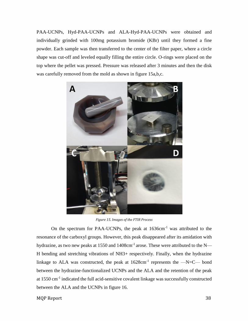
PAA-UCNPs, Hyd-PAA-UCNPs and ALA-Hyd-PAA-UCNPs were obtained and
individually grinded with 100mg potassium bromide (KBr) until they formed a fine
powder. Each sample was then transferred to the center of the filter paper, where a circle
shape was cut-off and leveled equally filling the entire circle. O-rings were placed on the
top where the pellet was pressed. Pressure was released after 3 minutes and then the disk
was carefully removed from the mold as shown in figure 15a,b,c.
Figure 15. Images of the FTIR Process
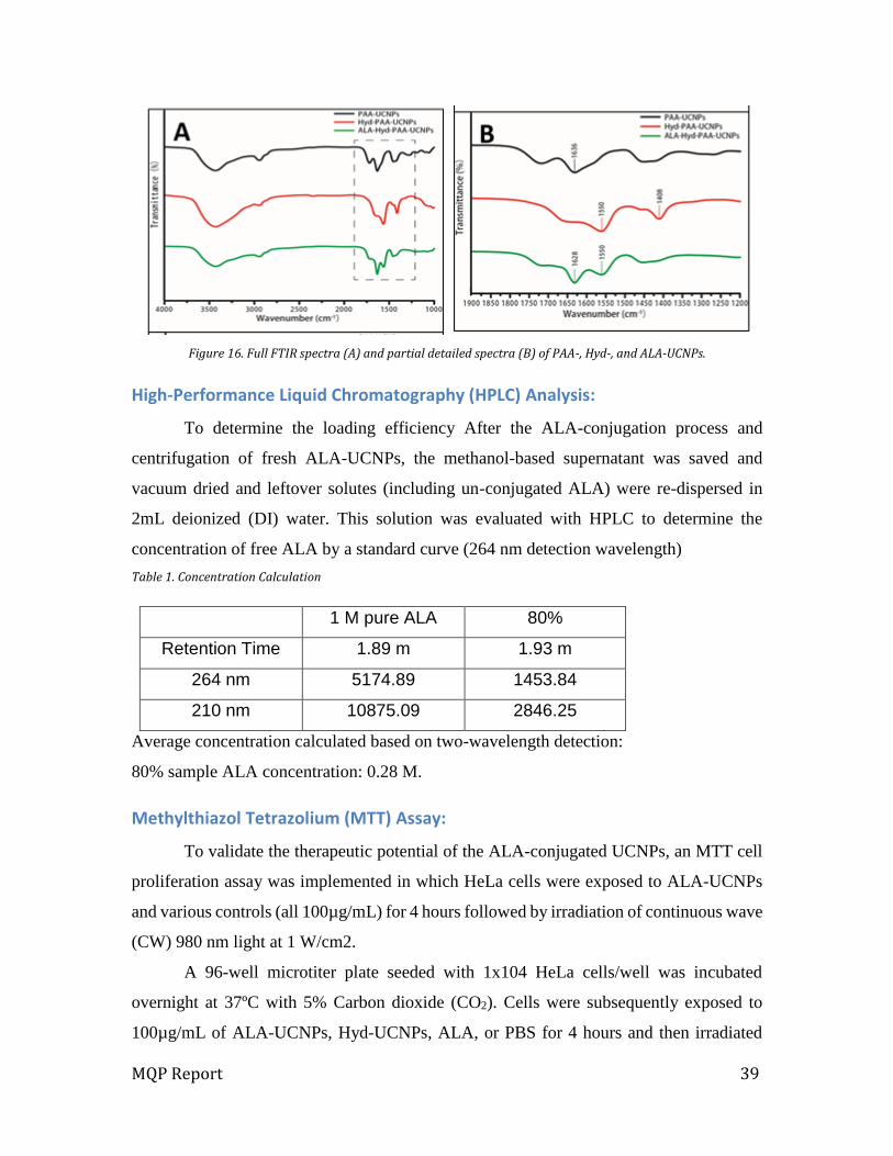
On the spectrum for PAA-UCNPs, the peak at 1636cm-1 was attributed to the
resonance of the carboxyl groups. However, this peak disappeared after its amidation with
hydrazine, as two new peaks at 1550 and 1408cm-1 arose. These were attributed to the N—
H bending and stretching vibrations of NH3+ respectively. Finally, when the hydrazine
linkage to ALA was constructed, the peak at 1628cm-1 represents the —N=C— bond
between the hydrazine-functionalized UCNPs and the ALA and the retention of the peak
at 1550 cm-1 indicated the full acid-sensitive covalent linkage was successfully constructed
between the ALA and the UCNPs in figure 16.
MQP Report 39
Figure 16. Full FTIR spectra (A) and partial detailed spectra (B) of PAA-, Hyd-, and ALA-UCNPs.
High-Performance Liquid Chromatography (HPLC) Analysis:
To determine the loading efficiency After the ALA-conjugation process and
centrifugation of fresh ALA-UCNPs, the methanol-based supernatant was saved and
vacuum dried and leftover solutes (including un-conjugated ALA) were re-dispersed in
2mL deionized (DI) water. This solution was evaluated with HPLC to determine the
concentration of free ALA by a standard curve (264 nm detection wavelength)
Table 1. Concentration Calculation
1 M pure ALA 80%
Retention Time 1.89 m 1.93 m
264 nm 5174.89 1453.84
210 nm 10875.09 2846.25
Average concentration calculated based on two-wavelength detection:
80% sample ALA concentration: 0.28 M.
Methylthiazol Tetrazolium (MTT) Assay:
To validate the therapeutic potential of the ALA-conjugated UCNPs, an MTT cell
proliferation assay was implemented in which HeLa cells were exposed to ALA-UCNPs
and various controls (all 100µg/mL) for 4 hours followed by irradiation of continuous wave
(CW) 980 nm light at 1 W/cm2.
A 96-well microtiter plate seeded with 1x10⁴ HeLa cells/well was incubated
overnight at 37ºC with 5% Carbon dioxide (CO2). Cells were subsequently exposed to
100µg/mL of ALA-UCNPs, Hyd-UCNPs, ALA, or PBS for 4 hours and then irradiated
MQP Report 40
with CW 980nm laser diode at 1W/cm2 for 0, 5, 10, or 20 minutes. After overnight
incubation, cells were labeled with 12mM solution of MTT in PBS for 4 hours. Finally, the
media was aspirated and replaced with 50µL DMSO and a plate reader at 540nm
determined the formazan absorption.
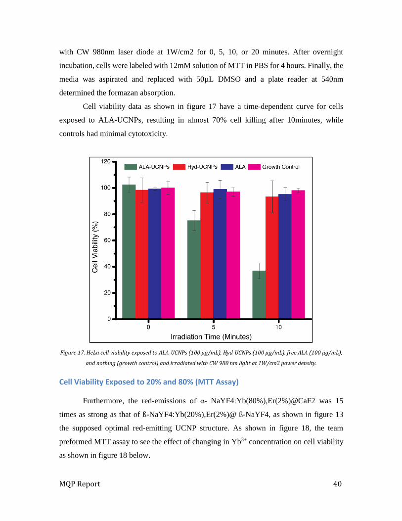
Cell viability data as shown in figure 17 have a time-dependent curve for cells
exposed to ALA-UCNPs, resulting in almost 70% cell killing after 10minutes, while
controls had minimal cytotoxicity.
Figure 17. HeLa cell viability exposed to ALA-UCNPs (100 µg/mL), Hyd-UCNPs (100 µg/mL), free ALA (100 µg/mL),
and nothing (growth control) and irradiated with CW 980 nm light at 1W/cm2 power density.
Cell Viability Exposed to 20% and 80% (MTT Assay)
Furthermore, the red-emissions of α- NaYF4:Yb(80%),Er(2%)@CaF2 was 15
times as strong as that of ß-NaYF4:Yb(20%),Er(2%)@ ß-NaYF4, as shown in figure 13
the supposed optimal red-emitting UCNP structure. As shown in figure 18, the team
preformed MTT assay to see the effect of changing in Yb3+ concentration on cell viability
as shown in figure 18 below.
MQP Report 41
Figure 18. HeLa cell viability exposed to 20% Yb and 80% Yb ALA-UCNPs (100 μg/mL) and 20% Yb and 80% Yb Hyd-UCNPs (100 μg/mL) and irradiated with CW 980 nm light at 1W/cm2 power density.
Singlet Oxygen Detection Cell Imaging
The team validated the production of PpIX and concomitant generation of singlet
oxygen species caused by exposure to ALA-UCNPs. The team stained cells with 2’, 7’ –
dichlorofluorescein diacetate (DCFDA), a standard fluorescent indicator for singlet oxygen
generation, and imaged using fluorescence microscopy as shown in figure 19. DCFDA
diffuses into the cell and is acetylated into a non-fluorescent compound by cellular esterase.
Reactive oxygen species (ROS) oxidizes it into 2’, 7’ –dichlorofluorescin (DCF), which
has bright green fluorescence.
Singlet Oxygen Detection Cell Imaging: Glass-bottom confocal dishes seeded with
1×10⁶ HeLa cells were incubated overnight at 37ºC with 5% CO2. Cells were subsequently
exposed to 100μg/mL of ALA-UCNPs or Hyd-UCNPs for 4 hours. Cells were washed
three times with warmed Hank’s Balanced Salt Solution (HBSS) and then stained with
MQP Report 42
25μM DCFDA in HBSS for 45 minutes. Using a fluorescent microscope equipped with a
CW 980nm laser diode at 1W/cm2, DCFDA fluorescence was imaged with 495nm
excitation and 535nm emission between 0, 5, and 10minutes of irradiation using a 60×
water-immersion objective lens.
Figure 19. Singlet oxygen production detected by fluorescence of DCFDA in HeLa cells exposed to 100 μg/mL of ALA-UCNPs and irradiated with 0 (a), 5 (b), and 10 (c) minutes of CW 980 nm light at 1W/cm2 power density. Singlet
oxygen production detected by fluorescence of DCFDA in HeLa cells exposed to 100 μg/mL of Hyd-UCNPs and irradiated with 0 (d), 5 (e), and 10 (f) minutes of CW 980 nm light at a 1W/cm2 power density.
Singlet Oxygen Detection Quantification
Singlet Oxygen Detection Quantification: A 96-well micro-titer plate seeded with
1×10⁴ HeLa cells/well was incubated overnight at 37ºC with 5% CO2. Cells were
subsequently exposed to 100μg/mL of ALA-UCNPs, Hyd-UCNPs, ALA, or PBS for 4
hours and then stained with 25μM DCFDA in HBSS for 45minutes. After irradiation with
CW 980nm laser diode at 1W/cm2 for 0, 5, or 10 minutes, DCFDA fluorescence was
immediately determined with a plate reader with 495nm excitation and 535nm emissions.
MQP Report 43
Figure 20. Singlet oxygen quantified by DCFDA fluorescence in HeLa cells exposed to 100μg/mL of ALA UCNPs, Hyd-UCNPs, ALA, and nothing (growth control).
MTT Assay with Porcine Tissue
Once the team achieved effective results using the 80% Yb, PDT was used to
stimulate in deep tissue. In this regard, pieces of tissue of varying thickness were
placed between the NIR laser and HeLa cells exposed to UCNPs figure 21a, and
analyzed for cell viability with an MTT assay figure 21b. Exposure time was 20
minutes of 980nm irradiation at 1W/cm2 under same condition mentioned in
previous MTT test. It is also worth noting that with the added distance between the
meat and cells, the height of the well and the media bathing the cells, the simulated
depths were greater than the measured thickness of the tissue.

MQP Report 44
Figure 21. Photograph of the setup of simulated visceral tumor conditions in an in vitro MTT assay (a). HeLa cell viability exposed to ALA-UCNPs (100 μg/mL) and Hyd-UCNPs (100μg/mL) and irradiated with CW 980nm light at a 1W/cm2 power density for 20 minutes
MQP Report 45
Chapter 6: Discussion
Photodynamic Therapy (PDT) with Upconversion Nanoparticles (UCNP) has been
used in many studies as a potential cancer treatment and showed promising results. As
such, it has been considered as an alternative treatment for current cancer treatments like
chemotherapy, radiotherapy and surgery. However, current techniques are limited by the
efficiency of the treatment with respect to tissue penetration. This project has demonstrated
a new technique being studied to increase tissue penetration by increasing red light
emission through the use of PDT with NaYF4:80%Yb,2%Er@CaF2 UCNPs, which are
responsible for the activation of the 5-Aminolevulinic Acid (ALA) drug photosensitizer
(PS). Ytterbium (Yb3+) plays an essential role in changing the amount of red light emission.
Previous studies showed that 20% Yb3+ produces the best red emission, while the team
found that 80% Yb3+ gives off the highest red light emission compared to different
percentages of Yb3+, as figure 21 illustrates. The team confirmed these results through a
variety of tests that showed the low toxicity of UCNPs, the best red emission intensity by
using NaYF4:80%Yb,2%Er@CaF2, and the successful and effective ALA conjugation
which prove that ALA-conjugated UCNPs have therapeutic potential.
Viability and Effectiveness of UCNPs-ALA for PDT as cancer treatment
In order to use NaYF4:80%Yb,2%Er@CaF2 UCNPs as an effective activator and
drug carrier for ALA, the team tested the size of UCNPs, red light emission, the amount of
ALA conjugation with UCNPs and tested the cell viability. Figure 14 shows results from
Transmission Electron Microscopy (TEM) imaging that portray the average size of UCNPs
37nm (Standard Deviation of 3nm) which is in the desired range for physiological testing
(Xueyuan, 2014). To determine the best red emission intensity that UCNPs are able to
produce, different percentages of Yb were used to make UCNPs. Figure 13a,b show
emission spectra under continuous wave (CW) 980nm 1W/cm2 excitation and Yb3+ core-
doping ratio increases 20%, 40, 60, 80, and 98% respectively. 80% Yb3+ produced the
highest red light emission compared to other percentages. Figure 13c is a photograph of
the UNCPs emitting. Figure 13 illustrates integrated counts between 600 and 700nm
indicating that there is a 250% percent increase in red emissions from α-
NaYF4:Yb20%Yb,2%Er@CaF2 to α-NaYF4:Yb80%Yb,2%Er@CaF2, which means that

MQP Report 46
the red light emissions of α- NaYF4:Yb80%Yb,2%Er@CaF2 are 15 times stronger than
that of ß NaYF4:Yb20%Yb,2%Er@NaYF4. The importance of red emission is that the PTD
photosensitizing drug is activated under red emission and in the actual experimental
process, the team was using the ALA as a photosensitizing drug.
After the team found that α-NaYF4:Yb80%Yb,2%Er@CaF2 had the optimal red
emissions, ALA-conjugated α-NaYF4:Yb80%Yb,2%Er@CaF2 were synthesized via the
process represented in figure 11. In cellular experimentation, the ALA-UCNPs were
expected to be phagocytized by the cell and exposed to the low pH of the endosome. This
would cause the cleavage of the hydrazine linkage causing the ALA to diffuse to the
mitochondria and cause an overproduction of Protoporphyrin IX (PpIX) as shown in figure
12. During this process the leftover Hyd-UCNP in the endosome would remain unharmed
within the cells and be activated by the NIR light, leading to the photosensitization of PpIX,
which will produce singlet oxygen (1O2) and cause the death of the tumor cell. Therefore,
high red light emission is an important factor in PDT with UCNPs treatment, because red
light is the excitation light that activates UCNPs which will trigger ALA and activate PpIX.
The stronger this light is, the deeper its effective distance will be, so it can be used for deep
cancer treatment.
After ALA conjugation with UCNPs the team performed various tests to check if
ALA successfully conjugated to the UCNPs and to verify the amount of ALA that was
conjugated. First the team tested if the ALA were successfully conjugated to UCNPs by a
Fourier transform infrared spectroscopy (FTIR) test and the results demonstrated the
existence of ALA on UCNP surfaces as shown in figure 16. When the hydrazine linkage
to ALA was constructed, the peak at 1628cm-1 represents the —N=C— bond between the
hydrazine-functionalized UCNPs and the ALA and the retention of the peak at 1550cm-1
indicates that the full acid-sensitive covalent linkage was successfully constructed between
the ALA and the UCNPs. The second test was High-Performance Liquid Chromatography
(HPLC), which was used to test the concentration of ALA after ALA conjugation with
HYD-UCNPs. The results in figure 17 showed that UCNPs 80%Yb-ALA with an ALA
concentration of 0.28M, exhibited a loading capacity of the ALA is 48.26µmol/10mg in
the HYD-UCNPs, approximately 50% of the initial amount.
MQP Report 47
After testing the conjugation of ALA with HYD-UCNPs by FTIR and calculating
the concentration of ALA by HPLC, the team determined that there is a sufficient amount
of ALA to be used as an effective cancer drug in in-vitro tests, and further, in-vivo testing.
The team performed Methylthiazol Tetrazolium (MTT) Assay which is used to
demonstrate multiple facts, firstly to test the toxicity of UCNPs because they will be used
for human testing and secondly to determine the effectiveness of ALA as a drug to kill
cancer cells as the team used Hela cells. Figure 17 shows Cell viability data which shows
that exposing cells to ALA-UCNPs resulted in almost 70% cell death after 10 minutes,
while UCNPs-HYD showed close similarity to control results, demonstrating that UCNPs
have low toxicity by themselves and proving that ALA works successfully with UCNPs as
a cancer treatment. UCNPs were exposed to cells for four hours before the tests were
performed under CW 980nm 1W/cm2 excitation.
The team tested the effect of using different Yb3+ concentration on the emission of
red light as figure 13 demonstrated using 80%Yb3+ increase red light emission 15 time
more than 20%Yb3+. The team performed MTT assay to see the affect in changing Yb3+
concentration on cell viability, as figure 18 shows 80%Yb3+ALA-UCNPs had 70%
decrease in cell viability while 20%Yb3+ALA-UCNPs had 30% decrease in cell viability,
the team’s results clearly show that increases in Yb3+ concentration has critical effect on
cell viability.
Throughout research, the team found that cells exposed to ALA-UCNPs show
intensifying DCFDA fluorescence over irradiation time, clearly indicating the production
of PpIX and then singlet oxygen shown in figure 19 a-c. This data was later corroborated
with a quantification of the singlet oxygen production by measuring the fluorescence
intensity of the Cellular Reactive Oxygen Species Detection (DCFDA) using 96-well
micro-titer plate reader in figure 20. Both DCFDA fluorescence imaging and micro-titer
plate measurement suggest effective prodrug delivery and subsequent PpIX activation by
the red emitting UCNPs.
Finally, once the team had established the optimal red-emission and PDT effect of
80% Yb3+ UCNPs, simulating PDT in deep-set tumor conditions was desired. After 20
minutes of irradiation at 1W/cm2, all tissue thicknesses had cell viabilities significant to
the control (Hyd-UCNPs). While no tissue (0mm) resulted in less than 20% cell viability,

MQP Report 48
6mm had less than 50% cell viability and 12mm had less than 80% cell viability. This
establishes and validates the feasibility of using UCNPs as a NIR-to-Red visible light
transducer for prodrug photodynamic therapy at a deep-tissue level (thickness greater than
1cm).
Even after ALA conjugation, UCNP size was around 62nm which is still in the
acceptable range to be used in human testing and appropriate for tumor accumulation, as
TEM images show in figures 14a,b,c, the conjugation process formed a uniform size and
distribution of ALA-UCNPs in water. Since ALA will form an extra layer on the UCNPs
the team tested emission spectra of UCNPs before and after ALA conjugation and the
results showed that PAA-UCNPs in water exhibit less than 25% quenching in red emission
from UCNPs in hexane, indicating the UCNPs are the main factor in NIR-to-Red visible
light transducer for the endogenous PpIX as shown in figure 14d,e.
UCNPs for PDT as a cancer Treatment: A larger perspective
Economics
It is well known that cancer treatment is one of the most expensive treatments used
in the medical field. Among the 12 drugs approved by the Food and Drug Administration
for various cancer conditions in 2012, 11 were priced above $100,000 for a year of
treatment, and since 2010 the median price has been around $10,000 (Levy, 2013). Despite
their extreme cost, current cancer drugs do not exhibit high efficiency in the majority of
cancer cases. If PDT with UCNPs were used as a cancer treatment the team believes that
it will be less expensive than existing drugs, because the materials that comprise UCNPs
and ALA are not expensive and there is no need for high a quantity of ALA-UCNPs due
to the highly tumor targeted nature of ALA.
Societal Influence
Environmental effects are very important factor in drug manufacturing, especially
with the complicated rules and laws that restrict any environmental or pollution effect. It
is important to understand future effects of the disposals from both the therapy itself and
also of the materials associated with its manufacturing. The production of UCNPs requires
the use of lanthanide salts. Yb3+ and Er, current compounds used for doping, show no
MQP Report 49
environmental threat to plants or animals. They are considered biologically lifeless. Water-
soluble compounds of Yttrium (Y) are considered mildly toxic, mostly affecting aquatic
animals. Therefore waste disposal in the manufacturing process would need to be
monitored but will not require special licensing or permits to be acquired. (Ytterbium
Oxide, 2010) (Erbium Oxide, 2008).
Political Ramification
Cancer is getting everybody's attention, because cancer is the second cause of death
in the US (American Cancer Society, 2012a). Any treatment for cancer will have big
influence on the society because it will change lives and give hope to millions of people
who are suffering from cancer so searching for affective and successful cancer treatment
is a need for every person, because there are a lot of factors are involved in casing cancer
like chemical radiation, genetic, tobacco, diet and physical activity and every single person
living these days are getting exposed to one of these factor which mean everyone is under
the risk of having cancer.
Health and Safety
Safety was a major objective of the design and has been addressed with toxicity
analyses in-vitro. As for any treatment, the health and safety of the patient is of the highest
importance. The ultimate goal of PDT with UCNP-ALA would be to kill cancer cells
without affecting other tissues. The drug is designed to specifically target tumor cells
without negatively affecting the health of any other organ system, and with minimal side
effects.
Ethical Concern
The main goal of this project is to find alternative cancer treatment for current
treatment by using PDT with UCNPs-ALA. This would directly improve the lives of all
patients receiving the treatment and minimal the side effects that occur due to current
cancer treatments. The ethical concerns that are usually raised are related to marketing,
administration, and availability of the treatment. During the marketing process it should be
carefully done so that the expectations of patients do not exceed the limitations of the
treatment. Patients need to be informed about potency and limitations of the drug, and what
the treatment does and how it works. Administration of the treatment has to be noninvasive
MQP Report 50
so patient does not have to suffer during treatment. To ensure the level of pain and the
risks, animal models should be used. Since cancer is a global problem, the treatment needs
to be made available on multiple continents and would have to be accessible for all
demographics. Therefore, efforts must be made to keep production costs affordable in the
international markets.

MQP Report 51
Chapter 7: Final Design and Validation
As mentioned above, cancer is fatal disease that does not yet have a viable cure.
There is a pressing need to find an effective preventative treatment. Heretofore, the team
proposed a design that utilizes Upconversion Nanoparticle conjugated with 5-
aminolevulinic acid (ALA-UCNP) with Photodynamic Therapy (PDT) to treat cancer by
killing cancer cells in human body. However, producing cancer treatment faces a lot of
challenges in regards to safety, scalability, efficacy, and specificity.
Figure 22. Schematic of the final design components and its work mechanisms.
The goal of this project was to find alternative treatment for cancer by enhancing
current PDT and making it more effective for cancer therapy, especially for deep cancer.
The main problem with the current PDT procedure is the penetration depth and, as figure
22 shows, that the current PDT use 650nm wavelength to activate ALA and produce singlet

MQP Report 52
oxygen. In order to enhance the light penetration and reach deeper tissue, the team used
UCNPs as transducer which will convert 980nm NIR to red visible light and activate ALA
to produce singlet oxygen as shown in figure 22. The team successfully synthesized ALA-
UCNPs and found the best red light emission by using 80%Ytterbium (Yb3+). Over the
course of this project, the team was able to demonstrate hypothetical PDT system with
various experiments, which showed effective and significant results which strongly make
PDT with UCNPs as real alternative cancer treatment with great potentials.
Thus, the specific objectives of this study were to assess the application of novel
UCNPs as ultraviolet (UV) light emitter to activate ALA is to increase tissue penetration
by increasing red light emission which was demonstrate by increasing the percentage ratio
of Yb3+ in UCNPs.
In order, their priority was determined to evaluate the success of the design:
1. Specificity
2. Scalability
3. Safety
4. Efficacy
To make sure that the team’s progress is directed by scientific criteria and as the
results of simple experiments would guide the parameters chosen for more complex
procedures. Below, experiments are described in the chronological order in which they
were performed, with the reasoning for experiment prioritization:
1. Safety was evaluated first because low cytotoxicity was a constraint for this project;
the cytotoxic effects of the UCNPs should not compromise cell viability below 90%.
Therefore, Methylthiazol Tetrazolium (MTT) assay tested the cytotoxicity and it was
first experiment conducted to ensure that future experiments fell within acceptable
particle dosages. Following cytotoxicity assays, team test the size of UCNPs to make
sure they fall in the range of 50nm to 60nm to be used in physiological testing.
2. Testing the red light emission that produces from UCNPs was next measure test that
the team conducted in this project, because the main goal of this project is to increase
the red emission to solve the limitation that current UCNPs have. In effect, data from
these tests enabled the team to scale the amount of Yb3+ that used in producing UCNPs
MQP Report 53
and at the end chose the best percentage Yb3+ ratio which will help the team maintaining
maximum design efficiency.
3. The FTIR and HPLC experiments were performed next, to gauge the efficacy of the
design by testing if ALA conjugated to UCNPs. Therefore these tests show that UCNPs
are capable to be used as drug carrier, which would indicate the specificity of the
delivery system in or design.
4. To demonstrate the efficacy of the team’s design, MTT assay was utilized if ALA
working effectively as cancer drug by increment cell death during 980nm wavelength
laser exposure.
The cell line chosen to conduct these studies was the HeLa human cervical cancer
line. All cultures were maintained and prepared using antibiotic-free, DMEM+10% FBS.
All assays were performed in standard tissue culture polystyrene and incubated at 37oC
21% Oxygen (O2) and 5% Carbon Dioxide (CO2). All cell counts were determined by
hemocytometer, without using trypan blue exclusion staining.
MQP Report 54
Chapter 8: Conclusions and Recommendations:
Conclusions
The experimental approach showed the possibilities of several features of Calcium
Fluoride (CaF2) shell Upconversion Nanoparticle’s (UCNP) in vitro activity. After
considering alternative designs for biocompatibility, one of the biomaterials; CaF2 was
chosen as the design component. Regarding UCNP’s design selection, Transmission
Electron Microscopic (TEM) images were used to measure particle size and uniformity for
each design samples with different percent of Ytterbium (Yb3+) component. Particle sizes
of 30 to 50nm diameter are allowed for biological usage and met the design objectives. All
of the samples passed particle size and uniformity requirements in the initials process of
the experiment series.
CaF2-shell UCNPs with 20%, 40%, 60%, 80%, and 98% component of Yb3+ were
measured their emission spectra through fluorometer. As a result, CaF2-shell with 80%
Yb3+ had the strongest red emission, which was approximately about 3.00 × 10⁸ A.U. Using
the selected alternative design, 5-Aminolevulinic Acid (ALA) and hydrazine conjugation
was performed for better water solubility and release of singlet oxygen (1O2) to kill target
cells. Two different conjugated samples were checked for usability through TEM and
fluorometer tests. The results showed hydrophilic PAA-UCNP were still emitting enough
brightness, at nearly 3.00×10⁸ A.U. With a uniform particle size of about 40nm.
In addition, Fourier Transform Infrared Spectroscopy (FTIR) confirmed the
conjugating condition of ALA and Hydrazine. Then Methylthiazol Tetrazolium (MTT)
Assay was preformed to demonstrate that UCNPs having low cytotoxicity, 98% cell
viability after 10minutes of exposure to 980nm wavelength laser with Hydrazine UCNPs.
On the other hand, ALA UCNP showed about 30% cell viability on the same MTT assay,
which proves theory of CaF2 UCNP with ALA Functionality are right. Along with MTT
Assay, fluorescence intensity images of cells proved about 7 times bigger size of the cells
were produced of singlet oxygen in the cells. After verified the major photodynamic
therapy with ALA conjugated UCNP, MTT assay of alternative designs showed CaF2-shell
UCNP with 80% Yb3+ had the most cell died during 10minutes of 980nm wavelength laser
exposure. As the last experiment, MTT assay with tissue penetration test resulted less than

MQP Report 55
60% cell viability in 6mm thickness of tissue and less than 80% cell viability with 12mm
thickness tissue after 10 minutes of exposure of 980nm wavelength laser.
This proves that this engineered biocompatible CaF2-shell showed 15-fold
increment in red emission compared with current one. Also, ALA-conjugation, which was
a prodrug for red-absorbing photosensitizer protoporphyrin IX (PpIX) to the UCNP, allows
great control of its release in the cell. In-vitro test of this photodynamic therapy system was
performed to prove its therapeutic potential such as cell viability of ALA conjugated
UCNPs and release of toxicity via photosensitizer.
Recommendations
Under the limitation of time scope such as UCNP production, conjugation, and
experimental process time, this study was limited into partial requirements to become an
outstanding product for actual pharmaceutical usage. The implementation of following
recommendations might expand and develop the current characterization of ALA-UCNP
for cancer treatment.
1. Repeat experiments in more alternative UCNPs. Due to the delicate process of
UCNP production, each UCNP sample had slight differences on its size and
emission. In order to get more accurate and detail data, more sample of UCNP need
to be tested.
2. Conduct experiments with in vivo tests. After concluding the project,
experiments showed promising results of ALA conjugation with CaF2-Shell UCNP
in PDT. In vivo testing will show great chance of proving its potential in its
pharmaceutical usage.
3. Repeat experiments with different laser power. During the course of
experimental process, 980nm wavelength laser approximately 1W/cm2 power was
used. Over-heating effects regarding using this laser power with UCNP was rarely
observed.
4. Repeat experiments with Zinc-PCA conjugation. Both ALA and Zinc-PCA have
great function of photosensitizing. Due to the limited research and experiment time,
the experimental processes were limited into ALA conjugation.
5. Possible over-heating concerns. Throughout these experiments, the team did not
discover any significant problem of over-heating effect during the experiments. As

MQP Report 56
the length of the Laser wave gets longer, it causes more over-heating effect due to
the hydrogen in human tissue. Since the team proved that 980nm wavelength was
strong enough to penetrate 12mm of tissue, further study needs to be proceeded in
order to find out the smaller wavelength laser such as 800nm or 750nm wavelength.
MQP Report 57
References o American Cancer Society, "Cancer Facts & Figures 2013." 4 Sept. 2012a. Web. 26 Sept.
2013. <http://www.cancer.org/research/cancerfactsfigures/cancerfactsfigures/cancer-facts-
figures-201>.
o American Cancer Society, "Learning About New Cancer Treatments." 4 Sept. 2012b. Web. 26
Sept. 2013.
<http://www.cancer.org/treatment/treatmentsandsideeffects/complementaryandalternativeme
dicine/learningaboutnewcancertreatments/learning-about-new-ways-to-treat-cancer-toc>.
o Auzel, F. (2004). Upconversion and anti-Stokes processes with f and d ions in solids.
Chemical reviews, 104(1), 139-174.
o Castano, A. P., Mroz, P., & Hamblin, M. R. (2006). Photodynamic therapy and anti-tumour
immunity. Nature Reviews Cancer, 6(7), 535-545.
o Chen, G., Shen, J., Ohulchanskyy, T. Y., Patel, N. J., Kutikov, A., Li, Z., ... & Han, G. (2012).
(α-NaYbF4: Tm3+)/CaF2 Core/Shell Nanoparticles with Efficient Near-Infrared to Near-
Infrared Upconversion for High-Contrast Deep Tissue Bioimaging. ACS nano, 6(9), 8280-
8287.
o Chen, Xueyuan, Yongsheng Liu, and Datao Tu. "Lanthanide-Doped Luminescent
Nanomaterials." (2014).
o Carling, C. J., Nourmohammadian, F., Boyer, J. C., & Branda, N. R. (2010). Remote‐Control
Photorelease of Caged Compounds Using Near‐Infrared Light and Upconverting
Nanoparticles. Angewandte Chemie International Edition,49(22), 3782-3785.
o Dougherty, T. J., Gomer, C. J., Henderson, B. W., Jori, G., Kessel, D., Korbelik, M., ... &
Peng, Q. (1998). Photodynamic therapy. Journal of the National Cancer Institute, 90(12),
889-905.
o Erbium Oxide; MSDS No. 25883 [Online]; Acros Organics N.V.: Fair Lawn, NJ,November
20, 2008, http://fscimage.fishersci.com/msds/25883.htm (accessed December 2, 2011).
o Fingar, V. H. (1996). Vascular effects of photodynamic therapy. Journal of clinical laser
medicine & surgery, 14(5), 323-328.
o Haase, M., & Schäfer, H. (2011). Upconverting nanoparticles. Angewandte Chemie
International Edition, 50(26), 5808-5829.
o Idris, N. M., Gnanasammandhan, M. K., Zhang, J., Ho, P. C., Mahendran, R., & Zhang, Y.
(2012). In vivo photodynamic therapy using upconversion nanoparticles as remote-controlled
nanotransducers. Nature Medicine.
MQP Report 58
o Jayakumar, Muthu Kumara Gnanasammandhan, Niagara Muhammad Idris, and Yong Zhang.
"Remote activation of biomolecules in deep tissues using near-infrared-to-UV upconversion
nanotransducers." Proceedings of the National Academy of Sciences 109.22 (2012): 8483-
8488.
o Kalka, K., Merk, H., & Mukhtar, H. (2000). Photodynamic therapy in dermatology. Journal of
the American Academy of Dermatology, 42(3), 389-413.
o Liu, Y., Tu, D., Zhu, H., Ma, E., & Chen, X. (2013). Lanthanide-doped luminescent nano-
bioprobes: from fundamentals to biodetection. Nanoscale, 5(4), 1369-1384.
o Levy, Alison Rose. "The Sky-High Price of Chemotherapy: Why Do Cancer Drugs Cost So
Much?" TakePart. N.p., 09 May 2013.
o Mader, H. S., Kele, P., Saleh, S. M., & Wolfbeis, O. S. (2010). Upconverting luminescent
nanoparticles for use in bioconjugation and bioimaging. Current opinion in chemical
biology, 14(5), 582-596.
o MOAN, J., & Peng, Q. (2003). An outline of the hundred-year history of PDT. Anticancer
research, 23(5A), 3591-3600.
o Oleinick, N. L., Morris, R. L., & Belichenko, I. (2002). The role of apoptosis in response to
photodynamic therapy: what, where, why, and how. Photochemical & Photobiological
Sciences, 1(1), 1-21.
o Sharman, W. M., Allen, C. M., & van Lier, J. E. (1999). Photodynamic therapeutics: basic
principles and clinical applications. Drug discovery today, 4(11), 507-517.
o Shen, J., Chen, G., Vu, A. M., Fan, W., Bilsel, O. S., Chang, C. C., & Han, G. (2013).
Engineering the Upconversion Nanoparticle Excitation Wavelength: Cascade Sensitization of
Tri‐doped Upconversion Colloidal Nanoparticles at 800 nm. Advanced Optical Materials.
o Wang, C., Cheng, L., & Liu, Z. (2013). Upconversion nanoparticles for photodynamic
therapy and other cancer therapeutics. Theranostics, 3(5), 317.
o Wang, F., Banerjee, D., Liu, Y., Chen, X., & Liu, X. (2010). Upconversion nanoparticles in
biological labeling, imaging, and therapy. Analyst, 135(8), 1839-1854.
o Wang, J., Wang, F., Wang, C., Liu, Z., & Liu, X. (2011). Single‐Band Upconversion Emission
in Lanthanide‐Doped KMnF3 Nanocrystals. Angewandte Chemie International Edition,
50(44), 10369-10372.
o Wilhelm, S., Hirsch, T., Schueucher, E., Mayr, T., & Wolfbeis, O. (2012). Magnetic core-shell
rare earth doped nanoparticles wih tunable upconversion luminescence for sensor
applications., Available from Analtyk Chemistry. (WO609-12-1)Retrieved from http://www-
analytik.chemie.uniregensburg.de/wolfbeis/hirsch/Poster/Poster_Europtrode_2012_Magnetic

MQP Report 59
_Core-Shell_UCLNPs.pdf Yang, Y., Shao, Q., Deng, R., Wang, C., Teng, X., Cheng, K., ... &
Xing, B. (2012). In vitro and in vivo uncaging and bioluminescence imaging by using
photocaged upconversion nanoparticles. Angewandte Chemie International Edition, 51(13),
3125 3129
o G. Tian, W. Ren, L. Yan, S. Jian, Z. Gu, L. Zhou, S. Jin, W. Yin, S. Li, Y. Zhao, Small 2013, 9,
1929.
o Ytterbium Oxide; MSDS No. 96772 [Online]; Acros Organics N.V.: Fair Lawn, NJ, July 20,
2009, http://www.jtbaker.com/msds/englishhtml/d7120.htm
o Z. Huang, Technology in cancer research & treatment 2005, 4, 283.
o Q. Peng, K. Berg, J. Moan, M. Kongshaug, J. M. Nesland, Photochemistry and Photobiology
1997, 65, 235.
o S. Collaud, A. Juzeniene, J. Moan, N. Lange, Current Medicinal Chemistry -Anti-Cancer
Agents 2004, 4, 301.