Embed Size (px)
Citation preview

5/23/2013
1
Unusual presentation of malignant tumor of the
eyelid
Ahmed Ali Ahmed 3rd year resident
Ophthalmology department
Sohag University Hospital

5/23/2013
2
A 45 years old lady presented to outpatient clinic with discharge and discomfort of her left eye for 2 years with redness and loss of lashes of the lower lid of the same eye
During these 2 years, she had many ophthalmological clinics visits and she was told that she has chronic blepharitis and in all visits , she was advised to use eye drops and ointments that all were of no value.

5/23/2013
3
We admit this lady in our department for full evaluation :-
History :- • No history of previous similar attacks before
2 years
• She has no history of any ocular operations.
• She isn,t known to be diabetic .
• She is hypertensive for 2 years and controlled on medications.
• She has no history suggesting other systemic diseases.
Ocular examination

5/23/2013
4
OS OD
CF 30 Cm 6\60 VA
Upper ….. Normal. Lower ……hyperemic, macerated margin , lost lashes with white patches of keratosis.
Upper and lower eyelids are normal
Eyelids
Hyperemic Normal Conjunctiva
More dense cataract Cataract Lens
Pale optic disc Normal Fundus
6 seconds 15 seconds Tear film break up time (BUT):
not clinically palpable not clinically palpable
Preauricluar and submandibular L.N

5/23/2013
5

5/23/2013
6
• No other body swellings or masses.
• No signs of seborrhea.
• RBS 128 mg\dl.
Systemic examination
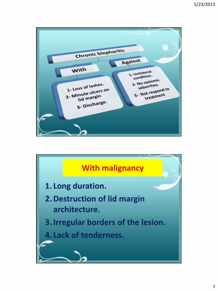
Differential diagnosis
1. Chronic blepharitis.
2. Basal cell carcinoma.
3. Squamous cell carcinoma.
4. Sebaceous gland carcinoma.
5/23/2013
7
With malignancy
1. Long duration.
2. Destruction of lid margin architecture.
3. Irregular borders of the lesion.
4. Lack of tenderness.

5/23/2013
8

5/23/2013
9
Preoperative biopsy
Preoperative biopsy taken from the hyperkeratotic area of the lesion and the histopathological examination reveals that the
specimen is of actinic keratosis.

5/23/2013
10

5/23/2013
11
Actinic keratosis
• Hyperkeratotic plaque with well defined borders, scaly surface that may be fissured.
• Palpable more than visible.
• It has a potential transformation into a squamous cell carcinoma.

5/23/2013
12
• Kaniski 2007
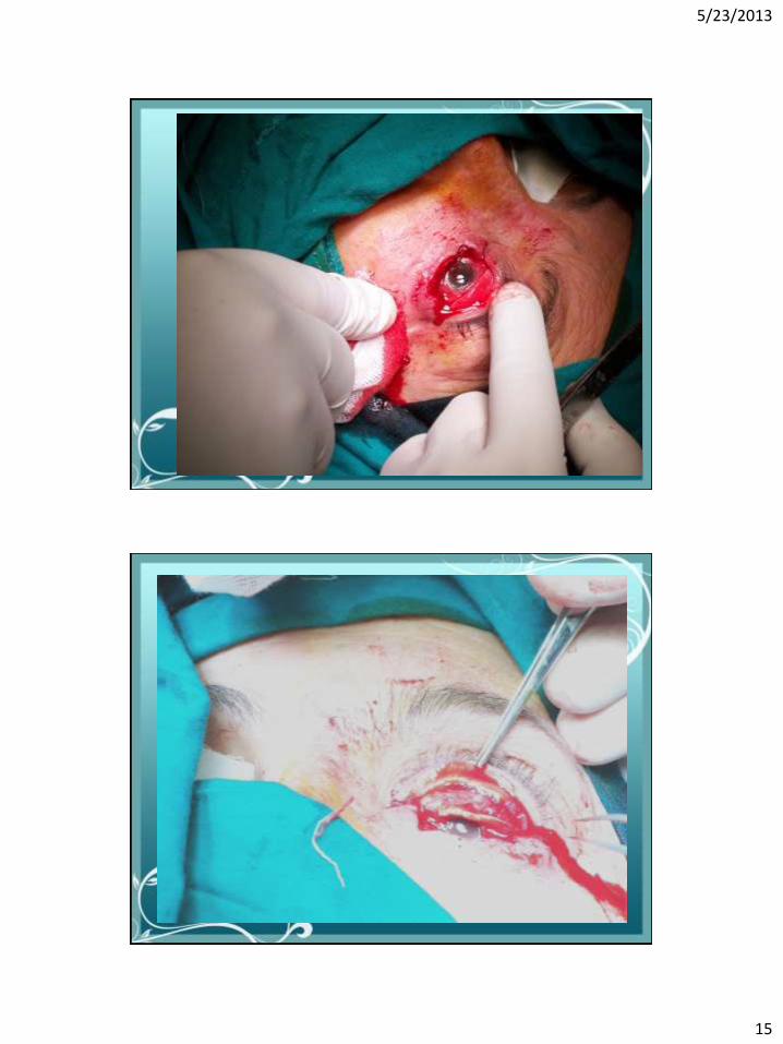
The decision was taken
To do surgery that involves total excision and reconstruction.
Reconstruction was done by Huges and Tenzel flaps.

5/23/2013
13
Surgery was done by
Ass. prof.
Ali Mahmoud Ismail

5/23/2013
14
5/23/2013
15

5/23/2013
16

5/23/2013
17

5/23/2013
18

5/23/2013
19

5/23/2013
20

5/23/2013
21
Two postoperative specimens
Were taken and sent to 2 different laboratories

5/23/2013
22
The results
The first specimen shows :-
Actinic keratosis with foci of severe dysplasia (carcinoma in situ)

5/23/2013
23
The results
The second specimen shows:-
• Intra-epithelial squamous cell carcinoma grade II.
• Negative stromal invasion.
• Ulcerated and inflamed.

5/23/2013
24
Take home message
• Cancer may presented with inflammation like condition.
• Malignant tumors appears not to respect the age.
• Any unilateral condition more than 4 weeks (chronic) should raise the suspicion of malignancy.
• Any excised tissue should be examined histopathologically.
• Follow up any patient with eyelid malignant tumor for at least 5 years for recurrence.
• To learn more, you should be adherent to a supervisor.
• My Prof: Gamal Abdellatief Radwan.
• My ASS. Prof: Ali M. Ismail
And
THANKS TO

5/23/2013
25