Embed Size (px)
Citation preview

Unravelling the role of a pain facilitatory area of the brain during chronic opioid exposure
Ana Rita Andrade Pereira da Costa Bioquímica Departamento de Química e Bioquímica
2014
Orientador Isabel Martins, Professora Auxiliar, FMUP


Todas as correções determinadas
pelo júri, e só essas, foram efetuadas.
O Presidente do Júri,
Porto, ______/______/_________


FCUP Unravelling the role of a pain facilitatory area of the brain during chronic opioid exposure
i
Abstract
Opiates represent the most commonly used drugs for the treatment of
moderate-to-severe postoperative and chronic pain. Chronic use of opioids can induce
paradoxical hyperalgesia (opioid-induced hyperalgesia; OIH). OIH is characterized
by hypersensitivity to innocuous or noxious stimuli during sustained opiate
administration, and is reported both in clinical and pre-clinical settings but its molecular
mechanisms are not fully understood. One of the mechanisms that contribute to OIH is
the activation of brain areas involved in pain facilitation. Here we studied the
involvement of an area located in the medulla oblongata, the dorsal reticular nucleus
(DRt), which plays a unique and exclusive pain facilitatory role. The studies included in
the present thesis aimed at i) determining whether chronic administration of morphine
induces OIH in acute pain and the spared nerve injury (SNI) model of chronic
neuropathic pain; ii) study the effects of chronic morphine on morphine reward and iii)
evaluate the involvement of the DRt in the mediation of OIH and morphine reward.
To determine the effects of chronic morphine administration the animals were
implanted with osmotic mini-pumps filled with morphine (45 μg.μl-1.h-1) or saline, which
released their content continuously for 7 days. The effects of chronic morphine
administration on pain behavior were tested before and at 2, 4 and 7 days after the
mini-pump implantation. In naïve animals pain behavior was tested by the von-Frey
and hotplate test which evaluate mechanical allodynia and thermal hyperalgesia
respectively. Pain assessment in SNI animals was performed by the von-Frey test, the
pin-prick test which evaluate changes in mechanical hyperalgesia and the acetone test
to assess cold allodynia. The continuous infusion of morphine induced OIH in naïve
animals and, for the first time, we show that chronic morphine administration induced
OIH in an animal model of neuropathic pain.
To study the effects of chronic morphine on the reward behavior we used the
conditioned place preference test (CPP). In animals chronically treated with morphine,
the acute administration of morphine failed to induce CPP, unlike in control animals,
which indicates a loss of the reward effect of morphine.
To study the involvement of the DRt in the mediation of OIH, we first
inactivated the DRt with lidocaine (0.5 µl; 4% w/v) in naïve animals. The injection of
lidocaine was performed on day 7 after implantation of the osmotic pumps containing
saline or morphine (45 μg.μl-1.h-1) and its effects were tested by behavioral tests
mentioned above. The administration of lidocaine at the DRt fully reversed mechanical
allodynia and thermal hyperalgesia in morphine-infused animals. Then we studied the

ii FCUP Unravelling the role of a pain facilitatory area of the brain during chronic opioid exposure
expression of the phosphorylated cAMP response element-binding protein (pCREB)
and µ-opioid receptor (MOR), at the DRt, by immunohistochemistry. We show that
chronic morphine treatment induces an increase of pCREB and MOR expression and
that MOR immunoreactive cells co-localized with pCREB. Finally, we performed a
lentiviral-mediated knock-down of the expression of MOR at the DRt. For that, the
animals were stereotaxically injected with lentiviral vectors at the DRt and implanted
with osmotic mini-pumps containing saline or morphine (45 μg.μl-1.h-1). The animals
were tested before and 7 days after the lentiviral injections and mini-pump implantation
by the behavioural tests mentioned above. The knock-down of MOR in control animals
showed an increase of pain behaviours. In animals chronically treated with morphine,
the knock-down of MOR prevented the development of OIH. We also evaluated the
effects of MOR knock-down to assess the involvement of the DRt in morphine reward
during chronic morphine exposure. Our preliminary results show that MOR knock-down
results in a reversion of the loss of morphine reward.
Our results indicate that chronic morphine exposure induces OIH in naïve and
neuropathic animals and, that the DRt is involved in the mediation of OIH, likely
through MOR activation whose effects appear switch from inhibitors to facilitation upon
chronic morphine. Our results also indicate that chronic morphine treatment induces a
loss of morphine reward and that the DRt might also be involved in the mediation of
such effects through MOR activation. Given the increase in the expression of pCREB
at the DRt, it would be interesting to explore the involvement of this transcription factor
in pain transmission from the DRt, during opioid-induced hyperalgesia. It would also be
interesting to explore the interactions between the DRt and brain areas involved both in
pain and reward on morphine reward.
Keywords: Opioid-induced hyperalgesia; Dorsal reticular nucleus; µ-opioid receptor;
Reward

FCUP Unravelling the role of a pain facilitatory area of the brain during chronic opioid exposure
iii
Resumo
Os opióides representam um dos tratamentos mais comuns tanto para o
tratamento da dor pós-operativa como para o da dor crónica. O uso crónico de
opióides promove um efeito paradoxal, a hiperalgesia induzida por opióides (HIO).
HIO é caracterizada por uma hipersensibilidade a estímulos inócuos ou nocivos
durante a administração crónica de opióides, relatada tanto em ambientes clínicos
como pré-clínicos mas os mecanismos moleculares subjacentes não estão ainda
totalmente elucidades. Um dos mecanismos que contribui para a HIO é a activação de
áreas supra-espinhais envolvidas na facilitação da dor. Neste trabalho estudou-se o
envolvimento de uma área localizada no bolbo raquidiano, o núcleo reticular dorsal
(DRt), que desempenha um papel exclusivo na facilitação da dor. Os estudos incluídos
na presente tese visam a i) determinar se a administração crónica de morfina induz
HIO em modelos de dor aguda ou de lesão do nervo ciático, (o modelo “spared nerve
injury”- SNI), um modelo de dor crónica neuropática; ii) estudar os efeitos da
exposição crónica a morfina sobre os efeitos de recompensa da morfina e iii) avaliar o
envolvimento do DRt na mediação da HIO e nos efeitos de recompensa da morfina.
Para determinar os efeitos da administração crónica de morfina, os animais
foram implantados com mini-bombas osmóticas contendo morfina (45 μg.μl-1.h-1) ou
soro, que permite a libertação contínua do seu conteúdo, durante 7 dias. Os efeitos da
administração crónica de morfina no comportamento nociceptivo foram testados antes
e aos 2, 4 e 7 dias após a implantação das mini-bombas. Em animais naïve o
comportamento nociceptivo foi testado pelo teste de von-Frey e pelo teste de Hotplate
que avaliam alodinia mecânica e hiperalgesia térmica, respectivamente. O
comportamento nociceptivo em animais SNI foi realizado através do teste de von Frey,
o teste de pin-prick que avalia hiperalgesia mecânica e pelo teste da acetona que
avalia alodinia ao frio. A infusão contínua de morfina induziu HIO em animais naïve e,
pela primeira vez, foi mostrado que a administração contínua de morfina induz HIO
num modelo animal de dor neuropática.
Para estudar os efeitos do tratamento crónico com morfina no comportamento
de recompensa foi utilizado o teste de “conditioning place preference” (CPP). Em
animais cronicamente tratados com morfina, a administração aguda de morfina não
induziu CPP, ao contrário dos animais controlo, o que indica uma perda do efeito de
recompensa da morfina.

iv FCUP Unravelling the role of a pain facilitatory area of the brain during chronic opioid exposure
Para estudar o envolvimento do DRt na mediação da HIO, primeiro inactivou-se
o DRt com lidocaína (0,5 mL; 4% m/v) em animais naïve. A injecção de lidocaína foi
realizada no sétimo dia após a implantação das bombas osmóticas contendo morfina
(45 μg.μl-1.h-1) ou soro e, os seus efeitos foram testados pelos testes comportamentais
já mencionados. A administração de lidocaína na DRt reverteu totalmente a alodinia
mecânica e hiperalgesia térmica nos animais tratados com morfina. Em seguida,
estudou-se a expressão da proteína de ligação ao elemento de resposta ao AMP
cíclico (pCREB) e do receptor μ-opióide (MOR), no DRt, por imuno-histoquímica.
Mostrou-se que o tratamento crónico com morfina induziu um aumento da expressão
de pCREB e MOR e as células marcadas com MOR apresentam uma percentagem
elevada de co-localização com pCREB. Por fim, foi realizada uma diminuição da
expressão, “knock-down”, de MOR através de vetores lentivíricos no DRt. Para isso,
os animais foram injetados com vectores no DRt e implantados com mini-bombas
osmóticas contendo morfina (45 μg.μl-1.h-1) ou soro. Os animais foram testados antes e
7 dias após a injecção com os lentivírus e implantação de mini-bombas osmóticas,
pelos testes comportamentais já mencionados. A diminuição de expressão de MOR
em animais de controlo, induziu um aumento da sensibilidade à dor. Em animais
tratados cronicamente com a morfina, o knock-down de MOR impediu o
desenvolvimento de OIH. Também foram avaliados os efeitos do knock-down de MOR
no DRt nos efeitos de recompensa da morfina durante a exposição crónica de morfina.
Os resultados preliminares demonstram que a diminuição de expressão de MOR
resulta numa pequena reversão da perda do efeito de recompensa da morfina.
Os resultados obtidos indicam que a exposição crónica à morfina induz HIO em
animais naïve e neuropáticos e, que o DRt está envolvido na mediação de HIO,
provavelmente através da ativação de MOR cujos efeitos parecem mudar de inibidores
para facilitadores após a exposição prolongada de morfina. Os resultados obtidos
também indicam que o tratamento crónico com morfina induz uma perda do efeito de
recompensa da morfina e que o DRt também pode estar envolvido na mediação
desses efeitos, através da ativação de MOR. Dado o aumento da expressão de
pCREB no DRt, seria interessante no futuro explorar o envolvimento deste factor de
transcrição na transmissão da dor a partir do DRt, durante a hiperalgesia induzida por
opióides. Também seria interessante explorar as interações entre o DRt e as áreas
cerebrais envolvidas tanto na dor como no efeito de recompensa da morfina.
Palavras-chave: Hiperalgesia induzida por opióides; Núcleo reticular dorsal; Recetor
-opióide; Efeito de recompensa;

FCUP Unravelling the role of a pain facilitatory area of the brain during chronic opioid exposure
v
Acknowledgments
Todo o trabalho desenvolvido nesta dissertação não teria sido possível sem o
apoio de inúmeras pessoas que ao longo desta tese estiveram presentes e que
contribuíram para a sua realização. A todas elas o meu sincero obrigado.
À minha orientadora, Professora Isabel Martins pela amabilidade, boa
disposição, paciência, conhecimentos transmitidos e ainda por toda a autonomia que
me incutiu demonstrando uma confiança imensa nas minhas capacidades. Obrigada
por todo o tempo despendido na realização desta dissertação.
À Professora Doutora Isaura Tavares, por me ter aceitado no seu grupo de
investigação bem como, pelos conhecimentos transmitidos e sugestões fulcrais para o
desenvolvimento deste projecto.
À Professora Carla Morgado e Marisa Oliveira por toda a disponibilidade e
sugestões no desenvolver desta dissertação.
À Amanda por ter sido a minha companheira de todas as horas. Obrigada por
toda a ajuda e todo o apoio durante estes meses. Foste mais do que fundamental para
o sucesso desta dissertação.
À Paulina pela amizade e por todo o tempo despendido a apoiar-me, com muita
paciência e muita dedicação.
À Marta Louçano, José Tiago Pereira, Marta Silva, Mariana Mesquita e Daniel
Martins por todos os momentos de boa disposição passados no laboratório. A todos, o
meu muito obrigada.
À Elisa por toda a simpatia e constante disponibilidade para me ajudar sempre
que necessitei.
Ao meu pai e ao meu irmão. Neste último ano crescemos muito como família e
o vosso apoio foi fundamental durante toda esta tese.
Ao Fábio pelo apoio incondicional e por estar presente em todos os momentos.
Sem ti tudo se ia tornar mais difícil.
Às minhas “TFDUPAS” obrigada pela compreensão e por todos os bons
momentos que passamos. Um dia tenho a certeza que todo o nosso esforço e força de
vontade vão ser recompensados.

vi FCUP Unravelling the role of a pain facilitatory area of the brain during chronic opioid exposure

FCUP Unravelling the role of a pain facilitatory area of the brain during chronic opioid exposure
vii
Table of contents
Abstract ........................................................................................................................ i
Resumo ...................................................................................................................... iii
Acknowledgments ...................................................................................................... v
Table of contents ...................................................................................................... vii
List of figures .............................................................................................................. x
List of tables............................................................................................................... xi
List of schemes .......................................................................................................... xi
Abbreviations ............................................................................................................ xii
Introduction ................................................................................................................. 1
1. Pain ....................................................................................................................... 1
1.1. Pain definition ................................................................................................. 1
1.2. Pain transmission ........................................................................................... 2
1.3. Descending pain modulation ........................................................................... 4
1.3.1 The endogenous pain control system ....................................................... 4
1.3.2 The dorsal reticular nuncleus .................................................................... 6
2. Opioids and pain ................................................................................................... 8
2.1. Opioid receptors ............................................................................................. 8
2.2. Effects of endogenous opioids in pain modulation ........................................ 10
3. Opioid-induced hyperalgesia ............................................................................... 11
3.1. Definition....................................................................................................... 11
3.2. Clinical and animal evidence ......................................................................... 12
3.3. Molecular mechanisms ................................................................................. 14
3.4. cAMP response element binding protein (CREB) in OIH ............................... 14
4. Opioids and reward ............................................................................................. 17
5. Genetic manipulation of the nociceptive system .................................................. 18
Aims and Methodology ............................................................................................ 21
Materials and methods ............................................................................................. 23
1. Animals ............................................................................................................... 23
2. Lentiviral vectors ................................................................................................. 23
3. Surgical procedures............................................................................................. 24
3.1. Osmotic mini-pumps ..................................................................................... 24

viii FCUP Unravelling the role of a pain facilitatory area of the brain during chronic opioid exposure
3.2. Neuropathic pain induction ........................................................................... 25
3.3. Stereotaxic surgeries .................................................................................... 27
3.3.1. Cannula implantation ............................................................................. 27
3.3.2. Vectors injection .................................................................................... 29
4. Behavioural analysis............................................................................................ 30
4.1. Nociceptive behavioural analysis .................................................................. 30
4.1.1. Pain assessment in naïve animals ......................................................... 30
4.1.2. Pain assessment in neuropathic animals .............................................. 34
4.2. Morphine reward behavioural analysis .......................................................... 36
4.2.1. Optimixation of visual cues .................................................................... 37
4.2.2. Optimization of tactile cues ................................................................... 37
4.2.3. Optimization of the number of conditioning trials/sessions .................... 38
4.2.4. Reward effects of morphine after chronic morphine administration ....... 39
4.2.5. Effects of MOR knock-down at the DRt on morphine reward after chronic morphine administration ................................................................................. 39
5. Material processing for histological and immunohistochemical analysis .............. 42
6. Histological verification of injection sites .............................................................. 42
7. Immunohistochemical analysis ............................................................................ 43
7.1. pCREB immunodetection .............................................................................. 43
7.2. MOR immunodetection ................................................................................. 44
7.3. Double immunodetection of MOR and pCREB............................................. 44
Results....................................................................................................................... 45
1. Effects of chronic morphine administration on pain behaviors ......................... 45
1.1. Effects on naïve animals ............................................................................... 45
1.2. Effects on neuropathic animals ..................................................................... 46
2. Effects of chronic morphine administration on the reward behavior ................. 47
2.1. Optimization of the conditioned place preference test ................................... 47
2.1.1. Visual cues ........................................................................................... 47
2.1.2. Tactile cues .......................................................................................... 48
2.1.3. The number of conditioning trials .......................................................... 49
2.2. Effects of chronic morphine administration on morphine reward .................. 51
3. Involvement of the DRt in chronic morphine effects ......................................... 52
3.1. Effects of DRt inactivation on nociceptive behavior ....................................... 52
3.2. Effects of chronic morphine on pCREB and MOR expression at the DRt ...... 55

FCUP Unravelling the role of a pain facilitatory area of the brain during chronic opioid exposure
ix
3.3. Effects of MOR Knock-down at the DRt ........................................................ 56
3.3.1. Pattern of lentiviral transduction ........................................................... 56
3.3.2. Effects of lentiviral transduction on MOR expression ............................ 57
3.3.3. Effects of lentiviral transduction on nociceptive behavior ...................... 59
3.3.3.1. Effects on naïve animals ........................................................ 59
3.3.3.1. Effects on SNI animals ........................................................... 60
3.3.4. Effects of lentiviral transduction on reward behavior ............................. 62
Discussion and Conclusions ................................................................................... 63
1. Effects of chronic administration of morphine .................................................. 63
2. Involvement of the DRt in OIH ......................................................................... 64
3. Reward effects of morphine ............................................................................. 65
4. Conclusions and future perspectives ............................................................... 66
References ................................................................................................................ 69
Appendix A: Composition of solutions ..................................................................... a

x FCUP Unravelling the role of a pain facilitatory area of the brain during chronic opioid exposure

FCUP Unravelling the role of a pain facilitatory area of the brain during chronic opioid exposure
xi
List of figures
Figure 1: Transmission of nociceptive information. ....................................................... 3
Figure 2: Schematic representation of the pain modularity circuitry. .............................. 5
Figure 3: Diagram of a coronal section of the caudal medulla oblongata....................... 6
Figure 4: DRt involvement in pain modulating circuitries. .............................................. 7
Figure 5: Seven transmembrane structure of opioid G-protein-coupled receptor.. ......... 9
Figure 6: Alterations in opioid dose-response relationship with chronic opioid administration. ............................................................................................................ 12
Figure 7: Cellular mechanisms of morphine hyperalgesia.. ......................................... 15
Figure 8: Schematic diagrams of the vectors. ............................................................ 24
Figure 9: Schematic representation of a osmotic mini-pumps.. ................................... 24
Figure 10: Implant of the mini-pumps in animals dorsum ........................................... 25
Figure 11: Exemplificative picture of the terminal branches of sciatic nerve.. .............. 26
Figure 12: The stereotaxic frame ................................................................................ 28
Figure 13: Dorsal view of the rat skull... ..................................................................... 28
Figure 14: Behavioural tests apparatus ....................................................................... 32
Figure 15: Behavioural tests apparatus... .................................................................... 35
Figure 16: Time course effects of morphine administration on mechanical allodynia tested and on thermal hyperalgesia ............................................................................ 46
Figure 17: Time course effects of morphine administration on mechanical allodynia, mechanical hyperalgesia and cold allodynia .............................................................. 47
Figure 18: Optimization of the visual cues of the CPP test .......................................... 48
Figure 19: Optimization of the tactile cues of the CPP test.... ...................................... 49
Figure 20: Optimization of the number of the conditioning trials. ................................ 49
Figure 21: Effects of chronic morphine administration on morphine reward.... ........... 52
Figure 22: Effects of the DRt inactivation on mechanical allodynia and on thermal hyperalgesia ............................................................................................................... 53
Figure 23: Effects of the VLM and Sol inactivation.... .................................................. 54
Figure 24: Effects of chronic administration of morphine on the expression of pCREB at the DRt. ................................................................................................... 55
Figure 25: Effects of chronic administration of morphine on the expression of MOR and pCREB ..... .................................................................................................. 56
Figure 26: Localization of the injection site in the DRt ................................................. 57

xii FCUP Unravelling the role of a pain facilitatory area of the brain during chronic opioid exposure
Figure 27: Evaluation of MOR expression by immunohistochemistry.... ...................... 58
Figure 28: Effects of MOR knock-down at the DRt on mechanical allodynia and on thermal hyperalgesia................................................................................................... 60
Figure 29: Effects of MOR knock-down at the DRt on mechanical allodynia, mechanical hyperalgesia and cold allodynia..... .......................................................... 61
Figure 30: Effects of MOR knock-down at the DRt on morphine reward .................... 62
Figure 31: Schematic representation of the relationship between descending facilitation and pain relief/reward ................................................................................................. 66
List of tables
Table 1: Endogenous opioid peptides and their receptors .......................................... 10
Table 2: Stereotaxic coordinates to target the left DRt, VLM and Sol ......................... 29
Table 3: Stereotaxic coordinates to target the left DRt ................................................ 30
Table 4: Effects of chronic morphine administration on locomotor activity. .................. 51
Table 5: Effects of chronic morphine administration on locomotor activity ............. 62
List of schemes
Scheme 1: Time course effects of chronic morphine administration in naïve animals. 32
Scheme 2: Effects of DRt inactivation by lidocaine on chronic morphine administration in naïve animals. ........................................................................................................ 33
Scheme 3: Effects of MOR knock down at the DRt on chronic morphine administration in naïve animals ......................................................................................................... 33
Scheme 4: Time course effects of chronic morphine administration in neuropathic (SNI) animals. ...................................................................................................................... 35
Scheme 5: Effects of MOR knock down at the DRt on chronic morphine administration in neuropathic animals. .............................................................................................. 36
Scheme 6: Evaluation of the reward effects of morphine after chronic morphine administration by the CPP test.. .................................................................................. 40
Scheme 7: Effects of MOR knock down at the DRt on morphine reward. .................... 41

FCUP Unravelling the role of a pain facilitatory area of the brain during chronic opioid exposure
xiii
Abbreviations
AC: Adenylate cyclase
ACC: Anterior cingulated cortex
AMY: Amygdala
AP: Anterior-posterior
Ca 2+-CaM: Calcium-calmodulin
CaMK: Ca2+-calmodulin-dependent
kinases
cAMP: Cyclic adenosine
monophosphate
cDNA: Complementary DNA
CNS: Central nervous system
COX: Cyclooxygenase
CPP: Conditioned place preference
CREB: cAMP response element-
binding protein
Cu: Nucleus cuneate
DAB: 3,3´-diaminobenzidine
tetrahydrochloride
DOR: δ-opioid receptor
DRt: Dorsal reticular nucleus
DV: Dorso-ventral
EGFP: Enhanced green fluorescent
protein
GAD: Glutamate decarboxylase
G-protein: Guanine-nucleotide binding
protein
GDP: guanosine diphosphate
GTP: guanosine triphosphate
H: Height
HSV-1: Herpes-simplex vírus-1
hSYN-1p: Human synapsin promoter
Hyp: Hypothalamus
i.p: Intraperitoneal injection
IASP: Internacional Association for the
Study of Pain
Ins: Insular cortex
IR: Immunoreactive
IRES: Internal ribosome entry site
KOR: κ-opioid receptor
L: length
LC: Locus coeruleus
LM: Latero-medial
LTR: Long terminal repeated
sequences
LV: Lentivirus
LV-Control: Lentiviral- Control
MOR: µ-opioid receptor
MOR-1: µ-opioid receptor 1

x FCUP Unravelling the role of a pain facilitatory area of the brain during chronic opioid exposure
MOR-2: µ-opioid receptor 2
MOR-R: μ-opioid receptor in reverse
orientation
Mot: Motor cortex
NAc: Nucleus accumbens
NMDA: N-methyl-D-aspartate
NO: Nitric oxide
OIH: Opioid-induced hyperalgesia
ORL-1: Opioid Receptor-Like -1
PAG: Periaqueductal grey matter
PB: Phosphate buffer
PBS: Phosphate buffer saline
PBS-T: Phosphate buffer saline with
Triton X-100
pCREB: phosphorylated cAMP
response element binding protein
PFC: Prefrontal cortex
PI3K: Phosphotidylinositol-3-kinase
PKA: Protein kinase A
PKC: Protein kinase C
PNS: Peripheral nervous system
RNAi: RNA interference
RVM: Rostral ventromedial medula
s.c: Subcutaneous injection
Sol: Nucleus tractus solitaries
SNI: Spared nerve injury
Som: Somatosensory cortex
Sp5C: Spinal trigeminal nucleus, pars
caudalis
TU: Transducing units
VLM: Caudal ventromedial medulla
VTA: Ventral tegmental area
W: Width
WAH: Withdrawl-associated
hyperalgesia
WPRE: Woodchuck hepatitis virus post-
transcriptional regulatory element
FCUP Unravelling the role of a pain facilitatory area of the brain during chronic opioid exposure
1
Introduction
1. Pain
1.1 Pain definition
According to the International Association for the Study of Pain (IASP), pain is
defined as an unpleasant sensory and emotional experience associated with actual or
potential tissue damage or described in terms of such damage [1]. Acute pain has a
protective role. The capacity to experience pain alert us of imminent or actual tissue
damage and leads to a behavioural response to minimize negative outcomes. On the
other hand, persistent pain syndromes offer no biological advantage. Chronic pain
persists beyond the expected normal time for healing, 3-6 months, and has no
physiological purpose [2].
Pain can be considered a high plasticity process that leads to several changes
in the neural structure and some of those changes are so drastic, specially chronic
pain, that pain cannot be considered just a symptom but, instead, it should be
considered as a pathological state [3].
Chronic pain is a major healthcare problem in Europe, it affects approximately
20% of the adult population, particularly women and elderly [4]. In Portugal it is
estimated that 30 % of the population suffers from chronic pain [5]. Chronic pain may
be inflammatory, neuropathic or functional and all forms share some common
characteristics [3]. Inflammatory pain is caused by tissue damage occurring mainly
after trauma, surgery or during chronic inflammatory diseases, having damaged and
inflammatory cells recruited to the damaged tissue that release activators of peripheral
nociceptors [3, 6]. Neuropathic pain is defined by IASP as a direct consequence of a
lesion or disease affecting the somatosensory system [1], in other words, it is
classified as an association of spontaneous pain and hypersensitivity with
pathological changes in the peripheral nervous system (PNS) or in the central
nervous system (CNS) [3]. Functional pain is a relatively new concept and is defined as
pain sensitivity caused by an abnormal processing or function of the CNS in response
to normal stimuli and may occur in fibromyalgia and irritable bowel syndrome [3].

2 FCUP Unravelling the role of a pain facilitatory area of the brain during chronic opioid exposure
1.2 Pain Transmission
Primary afferent neurons innervate cutaneous tissues, bone, muscle,
connective tissues, vessels and viscera and nociception occurs when these neurons
are activated by noxious stimuli [3, 7]. Primary afferent axons can be categorized by
their peripheral targets, conduction velocity, response properties and neurochemical
phenotype [8]. Aδ-fibers are characterized as medium cell bodies, thinly myelinated
fibers and conduct at intermediate velocities; A -fibers have larger cell bodies and are
heavily myelinated; C-fibers have small cell bodies, unmyelinated fibers and conduct
action potentials slowly [3, 5, 8, 9]. C- and Aδ-fibers are able to encode noxious
chemical, thermal and mechanical stimuli and, for this reason, are considered the main
nociceptive afferents signaling pain [9].
Primary afferent neurons convert noxious stimuli into electrical activity in
peripheral terminals, causing depolarization of the neuronal membrane. If the stimuli
are translated into a sufficiently intense electrical signal, voltage gated sodium
channels will be activated generating the transmission of stimuli to central terminals of
nociceptors in the spinal cord. The impulses generated in the dorsal horn travel through
second order neurons, which constitute the ascending pathways to thalamus and
brainstem where information is processed and pain is perceived, resulting in an
appropriate response, transported by descending pathways to the spinal dorsal horn
(Figure 1) [3, 10].
More recently, attention has focused on spinal cord projections to the
parabrachial region of the dorsolateral pons, because the output of this region provides
for a rapid connection with the amygdala, a region usually considered to process
information relevant to the aversive properties of pain experience [11]. From these
brainstem and thalamic loci, information reaches cortical structures [11, 12]. There is
no single brain area essential for pain. Rather, pain results from activation of a group of
structures, some of which are more related with the sensory-discriminative properties,
such as the somatosensory cortex, and others with the emotional aspects, such as the
anterior cingulate gyrus and insular cortex. Imaging studies demonstrated activation of
prefrontal cortical areas, as well as regions not generally associated with pain
processing such as the basal ganglia and cerebellum, but the contribution of the
activation of these areas to pain perception is not well understood [9].
FCUP Unravelling the role of a pain facilitatory area of the brain during chronic opioid exposure
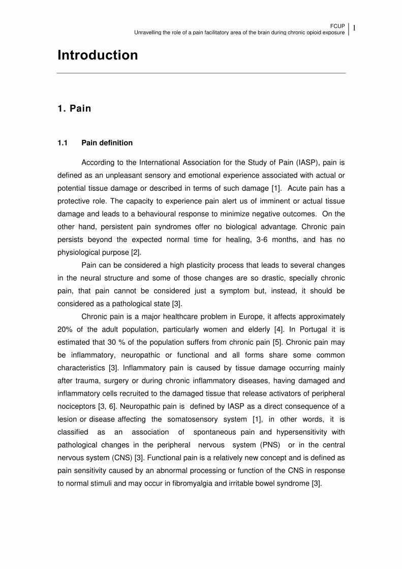
3
Figure 1: Transmission of nociceptive information. Nociceptive information is carried by primary afferent neurons from
the periphery to the spinal cord and then reaches the brainstem through ascending pathways constituted by second
order neurons. In the brain nociceptive information is evaluated and an appropriate response is generated and
conveyed by descending pathways to the dorsal horn of spinal cord. Adapted from Tavares & Martins [13].

4 FCUP Unravelling the role of a pain facilitatory area of the brain during chronic opioid exposure
1.3 Descending pain modulation
1.3.1 The endogenous pain control system
The endogenous pain control system is a complex web of brain areas
responsible for modulating pain transmission at the spinal cord. It is involved in pain
inhibition and, more recently was discovered to be also involved in pain facilitation [14,
15].
Several supraspinal sites play an important role in pain modulation but, the
most well characterized pain modulatory areas are the mesencephalic periaqueductal
grey (PAG) and the rostral ventromedial medulla (RVM) (Figure 2) [16].
The PAG is connected with the hypothalamus and limbic forebrain structures
including the amygdala, and also receives direct spinomesencephalic input. The PAG
is also connected with the RVM, which in turn sends its output to the spinal dorsal horn
[15]. The RVM is the final common relay in descending modulation of nociception from
the PAG, since the PAG does not project directly to the spinal cord, and other
supraspinal sites (Figure 2 and 4) [14, 15]. The RVM is constituted by the nucleus
raphe magnus and adjacent reticular formation and projects to superficial layers of
dorsal horn laminae and to deep dorsal horn [15]. In this area there are two distinct
types of cells classified as ON- and OFF- cells, which exert facilitatory and inhibitory
effects, respectively, on nociception. This two distinct populations of neurons project to
the dorsal horn and µ-agonists affect these two types of cells by direct inhibition of ON-
cells and by disinhibition of OFF-cells [17-19].

FCUP Unravelling the role of a pain facilitatory area of the brain during chronic opioid exposure
5
Figure 2: Schematic representation of the pain modularity circuitry. Primary afferent neurons convey nociceptive inputs
to the spinal dorsal horn. From the dorsal horn there are ascending projections (labelled in red) targeting the thalamus,
the DRt, the RVM and the PAG. The thalamus is connected to some cortical sites and to the amygdala. Descending
pain modulation is mediated through projections (labelled in green) from these cortical areas to the PAG, which
communicates with the RVM and the LC, and send descending projections to the spinal dorsal horn. Areas labelled “i–
iv” in the small diagram correspond to labelled details of the larger diagram. Abbreviations: DRt - dorsal reticular
nucleus; RVM- rostral ventromedial medulla ; PAG- mesencephalic periaqueductal grey; LC- locus coeruleus. Adapted
from Ossipov et al [14]

6 FCUP Unravelling the role of a pain facilitatory area of the brain during chronic opioid exposure
1.3.2 The dorsal reticular nucleus
The dorsal reticular nucleus (DRt) belongs to the endogenous pain control
system and deserves special attention since this area will be focus of the present
thesis.
The DRt is located in the most caudal portion of the medullary dorsolateral
reticular formation, more specifically, in the dorsolateral quadrant of the medulla
oblongata [20].This area is located medially to the spinal trigeminal nucleus, pars
caudalis (Sp5C), laterally to the nucleus tractus solitaries (Sol), ventral to the nucleus
cuneate (Cu) and dorsal to the ventral reticular nucleus (VRt) (Figure 3)[20].
DRt neurons are exclusively activated by cutaneous or visceral noxious
stimulation conveyed by Aδ- and C-fibers from the full body [20-23]. Glutamate
administration in the DRt induces a long-lasting increase in the responsiveness of
spinal nociceptive neurons [24], while lidocaine administration in the DRt results in the
suppression of responsiveness [20]. At the behavioral level, the DRt was shown to be
involved in pain facilitation both in acute and chronic pain models [25-27]. Recently, it
was found that the facilitatory effects of the DRt, in inflammatory and neuropathic pain
models, were mediated by noradrenaline release at the DRt [28, 29].
Figure 3: Diagram of a coronal section of the caudal medulla oblongata. Abbreviations: DRt-Dorsal reticular nucleus;
Cu-Nucleus cuneate; Sol-Nucleus tractus solitaries; Sp5C-Spinal trigeminal nucleus, pars caudalis; VLM-Caudal
ventromedial medulla; VRt-Ventral reticular nucleus. Adapted from Paxinos and Watson [30].

FCUP Unravelling the role of a pain facilitatory area of the brain during chronic opioid exposure
7
The DRt receives projections from the spinal cord laminae I, IV–VII and X, with
a clear ipsilateral predominance of those originated in the dorsal horn and the
connections between lamina I and the DRt are characterized by excitatory synaptic
contacts at both sites, which indicates that this reciprocal connection exerts excitatory
actions at both spinal and DRt levels functioning thus as a reverberating system that
leads to signal amplification[20].
The DRt has connections with several brainstem areas such the ventrolateral
medulla (VLM), PAG, RVM, locus coeruleus and the A5 and A7 noradrenergic cell
groups. The DRt also projects to medial thalamus and the limbic system, which
suggests an integration of DRt activity with emotional aspects of pain processing [16,
23]. Furthermore, the DRt is connected with the extrapyramidal and orofacial motor
system, which suggests an involvement of DRt in motor reactions associated with pain
[23].
Figure 4: DRt involvement in pain modulating circuitries. Ascending connections are represented in red, descending
projections are in blue and nociceptors are depicted in green. A–D are central nervous system sections and represent
the spinal dorsal horn (A), the medulla oblongata and pons (B), the mesencephalon (C) and the forebrain
(diencephalon and telencephalon, D). Abbreviations: ACC, anterior cingulate cortex; Ins, insular cortex; Mot, Motor
cortex; Som, somatosensory cortex; Hyp, hypothalamus. Adapted from Almeida et al[23].

8 FCUP Unravelling the role of a pain facilitatory area of the brain during chronic opioid exposure
2. Opioids and pain
The Sumerians in Mesopotamia were among the earliest civilizations identified
to have cultivated the poppy plant around 3400 BC [31]. Derived from opium poppy,
opioids have been used for millennia for the treatment of moderate to severe pain [32,
33]. Opioids play a unique role in society. They are widely feared compounds, which
are associated with addiction but they are also essential medications commonly used
to treat postoperative, cancerous, and, more recently, chronic nonmalignant pain [19,
34] as consequence, opioids are among the most often prescribed drugs to treat pain
[34].
2.1. Opioid receptors
Opioids activate peripheral, spinal and supraspinal opioid receptors. Currently
there are four well-established groups of opioid receptors: µ (MOR), δ (DOR), κ (KOR)
the and opioid receptor-like (ORL1) [18, 35, 36]. Subtypes of the receptors have been
proposed. MOR is divided into two subtypes: MOR 1 mediates the analgesic and
euphoric effects of opioids as well as the physical dependence and MOR 2 mediates
the bradycardic and respiratory depressant effects. DOR, with two subtypes identified
until now, DOR-1 and DOR-2, mediate spinal analgesic effects and have been
associated to modulation of tolerance. The three KOR subtypes, KOR-1, KOR-2 and
KOR-3, mediate spinal analgesia, miosis, sedation and diuresis [37].
The opioid receptors belong to the large family of seven-transmembrane G-
protein-coupled receptors. The binding of opioids to the receptor results in a
conformational change of the inhibitory Gi protein alternating from an inactive
guanosine diphosphate (GDP) to an active guanosine triphosphate (GTP) which results
in the activation of the α subunit of the G-protein. Once activated the α subunit
dissociate from the and subunits and binds to adenylate cyclase (AC) inhibiting it.
As a consequence of the inhibition of AC the intracellular concentrations of cyclic
adenosine monophosphate (cAMP) decrease, reducing phosphorylation and activation
of multiple proteins resulting in decreased excitatory activity (Figure 5) [18, 35, 36].

FCUP Unravelling the role of a pain facilitatory area of the brain during chronic opioid exposure
9
Figure 5: Seven transmembrane structure of opioid G-protein-coupled receptor. Receptor activation by opioid ligands
leads to initiation of intracellular transduction pathways that include stimulation of potassium efflux, decrease of
intracellular Ca2+ and inhibition of adenylyl cyclase resulting in decreased excitatory activity. Adapted from McDonald et
al [38].
MOR presents the widest distribution in the brain and spinal cord, while DOR
and KOR have a more restricted distribution. In peripheral tissues, opioid receptors are
responsible for the modulation of several physiological functions including alterations
during inflammation, analgesia and tolerance [36]. The ORL-1 has been detected in
the amygdala septum, the hypothalamus, the thalamus, in the DRG and the spinal
cord [39].
Investigations in pain have focused predominantly on MOR because its
activation is essential for the action of the most powerful analgesics as morphine,
oxycodone and hydrocodone [35]. The opioid receptors are expressed both on pre-
and post-synaptic neurons in the CNS and exert a major inhibitory influence in pain
transmission at the spinal level, exerting their actions via MOR expression in pre-
synaptic primary sensory neurons and in post-synaptic secondary neurons [18, 35,
36]. Furthermore, MOR is expressed in the main brain areas associated to pain
modulation, such as the insular cortex, amygdala, hypothalamus, PAG, RVM [35, 36],
DRt [40] and are abundantly expressed in the limbic system which is associated the
emotional perception of pain [36].

10 FCUP Unravelling the role of a pain facilitatory area of the brain during chronic opioid exposure
2.2. Effects of endogenous opioids in pain modulation
Opioid drugs act in peripheral, spinal, and supraspinal receptors which have
endogenous ligands, known as endogenous opioid peptides [36].
Endogenous opioids, which are naturally produced in the organism are involved
in the modulation of pain and also in other behavioural processes, such as reward,
dependency, sedation and stress response [41]. There are three families of
endogenous peptides that produce several active peptides: pro-opiomelanocortin that
produce -endorphin, proenkephalin that produce met- and leu-enkephalin peptides
and prodynorphin that produced dynorphins and neo-endorphins (Table 1) [36].
Table 1: Endogenous opioid peptides and their receptors.
Precursor Name Receptor
Proenkephalin Leu-enkephalin δ and µ
Proenkephalin Met-enkephalin δ and µ
Pro-opiomelanocortin -Endorphin µ and δ
Prodynorphin Dynorphins Κ
Unidentified Endomorphin-1 µ
Unidentified Endomorphin-2 µ
Pro-nociceptin/orphanin FQ Nociceptin/orphanin FQ ORL-1
Adapted from Ren and Dubner [42]
The opioid peptides -endorphin, dynorphins and enkephalins are widely
distributed throughout the brain, whereas in the spinal cord dynorphins are mainly
present in interneurons. Spinal enkephalins are found primarily in long descending
pathways from midbrain to the dorsal horn. Opioid peptides are also synthetized in
nonneuronal cells, such as endocrine cells and cells of the immune system [41].
The enkephalins, activate mainly the DOR, while the dynorphins activate mainly
the KOR (Table 1). The –endorphin peptide can produce a response through all three
receptors although this response is stronger when it acts through MOR and DOR
(Table 1) [41]. Two additional peptides endomorphin-1 and -2, with no precursor for
endogenous synthesis identified so far, bind with high affinity to MOR (Table 1) [36,
37, 41]. Also the endogenous opioid-like substance, nociceptin, is the product of a
novel gene distinct from the gene families from which the classical endogenous opioids
are derived (Table 1) [37]. In the CNS, opioids regulate nociceptive pathways both at
spinal and supraspinal levels. At the spinal level, opioids inhibit nociceptive
transmission conveyed by Aδ- and C-fibers [36].
FCUP Unravelling the role of a pain facilitatory area of the brain during chronic opioid exposure
11
Nonetheless, at the spinal cord, dynorphins have been associated with the
development of hyperalgesia and allodynia since it increases the release of
excitatory neurotransmitters, which contribute to intensify pain transmission [17, 36].
At the supraspinal level, opioid peptides inhibit ON-cells and disinhibit OFF-cells [35].
In the PAG, enkephalinergic neurons synapse with serotoninergic neurons in the RVM
that project to the spinal cord inducing the release of enkephalins that produce
inhibition of the activity of Aδ- and C- fibers entering the spinal cord [35, 36].
Noradrenergic cells from locus coeruleus projecting to the spinal dorsal horn are also
regulated by the opioidergic system [35].
The DRt is under opioidergic modulation since it expresses MOR and DOR [40,
43]. As to the effects of opiods at this area the overexpression of proenkephalin at the
DRt induced analgesia revealing thus that the effects of these opioid peptides inhibit
DRt descending facilitacion of pain [44].
3. Opioid-induced hyperalgesia
3.1. Definition
It is well know that the use of opioids may be a double-edged sword [34].They
provide straight analgesic and antihyperalgesic effects but, the knowledge that opioids
might have pronociceptive effects might have been suspected as early as the American
Civil War [45]. Opioids have several side effects such as the development of physical
dependence, tolerance and addiction [46, 47]. Nowadays there is an increased number
of evidences that opioids may cause another phenomenon often referred to as opioid-
induced hyperalgesia (OIH) [45, 46]. This phenomenon is characterized by increased
sensitivity to pain related to opioids exposure in the absence of disease progression or
opioid withdrawal [34, 45-47].
OIH definition is often mistaken with opioid tolerance and withdrawal-associated
hyperalgesia (WAH). These syndromes can manifest similar symptoms, but are
clinically differentiated from OIH due to differing effective interventions [48]. Tolerance
occurs when the patient seeks pain relief and increasing doses of opioids are
necessary to maintain appropriate analgesia (Figure 6 B) [48, 49]. This definition could
be confused with OIH, however, in opposition to tolerance, increasing doses of opioids
will only worsen pain (Figure 6 A) [46].

12 FCUP Unravelling the role of a pain facilitatory area of the brain during chronic opioid exposure
Figure 6: Alterations in opioid dose-response relationship with chronic opioid administration. It is a hypothetical
experience, where an acute opioid infusion is used to detect changes in the analgesic dose-experimental pain response
curve that occur as a result of chronic opioid exposure. The responses of opioid naïve patients are shown as a solid
line. A, In OIH, the dose-response curve of the chronic opioid user (dashed line) is shifted downward. B, In analgesic
tolerance, the slope of the dose-response curve of the chronic opioid user (dashed line) becomes attenuated and
rightward shifted, but, there is no significant change in pain sensitivity at baseline Adapted from Chu et al [50].
WAH is a time limited reaction, translated as a diffuse joint pain and body aches
taking place along with detoxification from chronic opioid use or if scheduled doses are
skipped [48].
3.2 Clinical and animal evidence
Several studies suggest that humans, as well as animals, treated with opioids
can develop OIH. In humans the development of OIH already showed important clinical
implications [34, 50]. Studies have been conducted using several distinct
methodologies namely: former opioid addicts on methadone maintenance therapy;
intraoperative exposure to opioids in patients undergoing surgery; healthy volunteers
after acute opioid exposure; and prospective observational studies in opioid-naïve pain
patients undergoing initiation of chronic opioid therapy [34, 50].
Diverse clinical studies have measured pain sensitivity in former opioid addicts, treated
with methadone, and this set of patients are compatible with the hypothesis that OIH,
when diagnosed, is caused by chronic opioid exposure [34, 51]. Evidences that
patients exposure to higher doses of intraoperative opioids increased postoperative
pain is also compatible with the view that OIH developed in these patients [34, 52].
There are also studies describing OIH in human volunteers after acute short-term

FCUP Unravelling the role of a pain facilitatory area of the brain during chronic opioid exposure
13
exposure to opioids and the results showed aggravation of induced hyperalgesic skin
lesions, expansion of the area of mechanical hyperalgesia induced by transdermal
electrical stimulation, aggravation of pressure-evoked pain or increased sensitivity to
cold pressor pain in healthy human volunteers following precipitated opioid withdrawal
after induction of acute physical opioid dependence [34, 50].
A few clinical studies also show evidences of the development of OIH. A small
prospective study in which OIH was notable in 6 patients with chronic back pain,
after one month of oral morphine treatment, when compared to baseline values
[49, 53]. Another research, with a larger sample population, showed a significant
negative correlation between experimental OIH and all clinical pain measures, in
a dose dependent manner [53]. One additional prospective study, with indirect
evidence of OIH, comes from patients with chronic pain receiving intermediate-term
opioid treatment who attended a pain rehabilitation program, which included the
cessation of opioid use. Heat pain thresholds were increased at the end of the program
compared to their levels prior to enrolment [53, 54].
The first time OIH was described in animals was 1971 [55] and now more than
a hundred publications are available describing this phenomenon in an extensive
diversity of animal models [50]. For more than three decades, it has been recognized
that systemic exposure of opioids to rodents can lead to a hyperalgesic response after
precipitating withdrawal with the administration of an opioid antagonist as well as
during spontaneous withdrawal after cessation of opioid administration [34].
Chronic administration of opioids also was shown to cause a sustained
pronociceptive response. In these experiments, OIH depended both on the dose of the
opioid and on the experimental pain model (i.e. thermal, mechanical, electrical or
chemical) [48]. Two fundamental patterns characterizing the onset and resolution of
OIH in animals can be distinguished. The first is observed after acute administration,
that is, the systemic administration of one to four relatively high opioid doses within one
hour, evoking a transient hyperalgesic response which lasts for hours or for days in a
dose dependent manner [34]. The second and most common pattern is observed after
animals are exposed to opioids on a chronic time course for three to twelve days via
repeated subcutaneous injections, implantation of subcutaneous opioid containing
pellets or pumps, or intermittent administration or continuous infusions through
indwelling intrathecal catheters. If animals were exposed to opioids by continuous
techniques, antinoceptive response is usually reported in the first day and then a loss
of this effect is observed or along with the induction of a hyperalgesic state during
ongoing drug administration. Alternatively, if animals receives repeated systemic or

14 FCUP Unravelling the role of a pain facilitatory area of the brain during chronic opioid exposure
intrathecal boluses of opioids for several days, they progressively develop hyperalgesia
to thermal or mechanical stimuli. When studied, it was also possible to directly correlate
the time course of resolution of OIH with the time course of its development [34].
3.3 Molecular mechanisms
The precise molecular mechanism of OIH are not yet well understood but, is
thought to result from neuroplastic changes in the PNS and in the CNS resulting in the
sensitization of pronociceptive pathways and it can been described based on the site of
the plasticity [19], where the relevant mechanisms are probably unique [50]. Spinal
cord plasticity underlying OIH has been demonstrated after both intraspinal and
systemic administration of opioids [19, 34, 50]. The consequence of spinal
sensitization is increased transmission of noxious inputs to supraspinal sites [17]. It is
well accepted that repeated excitation of spinal cord neurons, along with persistent
activation of the NMDA receptors, non-NMDA excitatory amino acid receptors, protein
kinase C (PKC) [19, 34, 50], spinal dynorphin, spinal prostaglandins [34] and spinal
cyclooxygenase (COX) [19] are involved in the sensitization of spinal neurons. The
spinal dorsal horn is vital to many mechanisms supporting OIH [19], as the correlation
between OIH and spinal cord plasticity is consonant with the emerging appreciation of
spinal inflammation as participating in many abnormal pain syndromes [19, 34, 50].
Regarding the molecular mechanisms underlying OIH, there are evidences
suggesting that after morphine binding to MOR, on a post-signaling neuron, there is
activation of G-protein mediated PKC translocation and the removal of the NMDA
receptor Mg2+ plug (Figure 7 – item 1). Glutamate is released from pre-synaptic cells
inducing the ionotropic NMDA receptor to allow Ca2+ influx, resulting in increased
intracellular Ca2+ which leads to several downstream effects, including activation
of calcium-calmodulin (Ca2+-CaM), changes in gene expression and further activation
of PKC (Figure 7 – items a-b-c). Ca2+-CaM in turn initiates the conversion of L-arginine
into nitric oxide (NO) by NO synthesis. NO may then act as a retrograde messenger to
enhance glutamate release from the pre-synaptic neuron. With continual activation of
these pathways, by opioid receptor occupation, PKC may uncouple the G-protein from
MOR preventing any downstream signalling upon ligand binding [56]. There are also
evidences suggesting that this process is not limited to neuronal cells and that glial
cells also play an important part in OIH. Chronic opioid administration may act through
MOR expressed on glial cells increasing the production and release of cytokines
and chemokines or act directly on glial and neuronal glutamate transporters to alter
synaptic glutamate levels (Figure 7 - items 2 and 3). Once released, cytokines may
FCUP Unravelling the role of a pain facilitatory area of the brain during chronic opioid exposure
15
then act on the pre- or post-synaptic neurons to induce hyperalgesia or on other glial
cells to promote further neuroimmune activation [56].
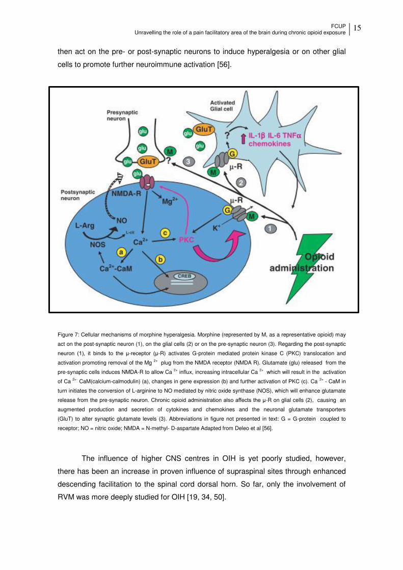
Figure 7: Cellular mechanisms of morphine hyperalgesia. Morphine (represented by M, as a representative opioid) may
act on the post-synaptic neuron (1), on the glial cells (2) or on the pre-synaptic neuron (3). Regarding the post-synaptic
neuron (1), it binds to the μ-receptor (μ-R) activates G-protein mediated protein kinase C (PKC) translocation and
activation promoting removal of the Mg 2+ plug from the NMDA receptor (NMDA R). Glutamate (glu) released from the
pre-synaptic cells induces NMDA-R to allow Ca 2+ influx, increasing intracellular Ca 2+ which will result in the activation
of Ca 2+- CaM(calcium-calmodulin) (a), changes in gene expression (b) and further activation of PKC (c). Ca 2+ - CaM in
turn initiates the conversion of L-arginine to NO mediated by nitric oxide synthase (NOS), which will enhance glutamate
release from the pre-synaptic neuron. Chronic opioid administration also affects the μ-R on glial cells (2), causing an
augmented production and secretion of cytokines and chemokines and the neuronal glutamate transporters
(GluT) to alter synaptic glutamate levels (3). Abbreviations in figure not presented in text: G = G-protein coupled to
receptor; NO = nitric oxide; NMDA = N-methyl- D-aspartate Adapted from Deleo et al [56].
The influence of higher CNS centres in OIH is yet poorly studied, however,
there has been an increase in proven influence of supraspinal sites through enhanced
descending facilitation to the spinal cord dorsal horn. So far, only the involvement of
RVM was more deeply studied for OIH [19, 34, 50].

16 FCUP Unravelling the role of a pain facilitatory area of the brain during chronic opioid exposure
3.4 cAMP response element binding protein (CREB) in OIH
CREB is a member of a superfamily of proteins that function as transcription
factors and is expressed in all cells in the brain [57, 58]. Phosphorylation and
successive activation of CREB is a site of convergence for several signal transduction
cascades, including the cAMP pathway via protein kinase A (PKA), intracellular Ca2+
via Ca2+-calmodulin-dependent kinases (CaMK), the Ras/ extracellular signal regulated
kinase (ERK) protein kinase pathway, the phosphotidylinositol-3-kinase (PI3K)/ Akt
kinase pathway, PKC pathways and stress-induced signaling cascades [58-60].
CREB is of particular interest since its activation is downstream of the cAMP
signaling pathway, whose upregulation has been widely characterized as an adaptation
to opioid chronic exposure [58]. Generally when morphine binds to MOR, adenylyl
cyclase (AC) is inhibited and in consequence cAMP decrease and reduces the
phosphorylation of CREB (pCREB) however, chronic opiate administration, increases
levels of AC and pCREB implying a homeostatic or compensatory regulatory
mechanism. This increased CREB activity appears to play an important role in physical
opiate dependence and withdrawal [58, 61-63].
Several experimental evidences suggests that pCREB is involved in the opioid-
induced effects in vitro and in vivo, beginning in cultured neuronal cell lines and
extending to several brain areas [64]. In vitro it was shown that chronic administration
of morphine increase pCREB in the coeruleus-like cell line [65]. In vivo it was shown
that chronic administration of morphine increase pCREB in the locus coeruleus (LC)
[58, 61, 66] or in the nucleus accumbens (NAc) [58, 67, 68]. Also mice containing
targeted mutations on α and Δ isoforms of CREB gene showed attenuated physical
symptoms of morphine withdrawal [58, 69]. The knockdown of CREB levels in the LC,
using antisense oligonucleotides, blocks the capability of chronic opiates to upregulate
some components of the cAMP pathway and consequently blocked some of the effects
of morphine like physical dependence and withdrawal [62]. Similar results were
observed when increasing or decreasing CREB levels in the LC using viral vectors [61].
A remaining mystery, however, is the exact mechanism which opiate exposure
switches from acute inhibition of the cAMP pathway and CREB to chronic upregulation
[61].

FCUP Unravelling the role of a pain facilitatory area of the brain during chronic opioid exposure
17
4. Opioids and reward
Reward is defined as a stimulus that brain interprets as intrinsically positive or
as something to be approached [70]. Usually, rewards are conditionally learned based
upon their positive influence on survival or reproduction [71, 72].
Pain and reward are opposite processes that can interact and influence each
other. Some rewarding stimuli decrease pain sensitivity but on the other hand it has
been proved that pain affect reward processing [73]. For example, chronic pain is
associated with anhedonia, i.e. the incapacity to feel pleasure. Several brain systems
are implicated in pain and reward processing such as the amydala, anterior and
posterior insula, anterior cingulate cortex, dorsal and ventral striatum and the
orbitofrontal cortex [73, 74].
Dopamine is a key neurotransmitter of reward [73, 75]. This catecholamine is
released from the ventral tegmental area (VTA) that project mainly to the NAc and the
prefrontal cortex (PFC) as a result of natural rewarding experiences, such as eating
[71, 76] or in response to administration of drugs of abuse such as cocaine,
amphetamine, opiates, nicotine and alcohol [71, 77, 78]. Although the dopaminergic
system represents the cornerstone of the reward system other neurotransmitters, such
as endogenous opioids, affect this mesolimbic dopaminergic system [72].
The opioid system represents an important substrate for the prejudicial effects
of drugs of abuse. As a matter of fact activation of MOR reinforces the properties of
countless abused drugs, which may be a potential molecular gateway to drug addiction
[79, 80]. Van Ree et al [81] in 1980 was the first to demonstrate that rats self-
administer an opioid receptor agonist into the VTA. Since then, several studies using
intracerebral self-administration or conditioning place preference (CPP) in rats have
confirmed the contribution of the VTA in opiate reinforcement [79, 82, 83]. More
recently, genetic approaches using knockout animals have confirmed the role of opioid
system in drug reinforcement and dependence [79]. Constitutive knockout mice for
MOR, DOR and KOR have been used to study reward processes [70]. Several
researches demonstrated an essential role of MOR in facilitating reward. Opiate reward
studies in MOR knockout mice showed a loss of morphine reward on CPP as well as
morphine self-administration test [70, 84, 85]. The DOR influence on reward is less
evident. The analysis of DOR knockout animals showed a decreased morphine place
preference but morphine self-administration was maintained, suggesting that this opioid
receptor contribute to contextual learning rather than opioid reward [70, 79].
18 FCUP Unravelling the role of a pain facilitatory area of the brain during chronic opioid exposure
In addiction a large number of studies using KOR knockout mice confirm that this
receptor negatively modulates reward, although this is not shown for all drugs of abuse,
for example deletion of the KOR gene did not modify morphine CPP but in contrast
reduced alcohol place preference [70, 79].
Chronic exposure to opioids, or another drugs of abuse enhance the activity of
the cAMP–PKA pathway in NAc. Activation of PKA and subsequent phosphorylation of
CREB within Nac reduces the rewarding effects of stimulant drugs, whereas PKA
inhibition has the opposite effect [86]. Indeed, elevation of CREB levels within the rat
NAc using viral vectors reduce the rewarding effects of morphine and cocaine and
make low doses aversive, suggesting that CREB activity in this region can control
reward qualities of drugs of abuse [57].
5. Genetic manipulation of the nociceptive system
Conventional drug treatment for pain has numerous limitations, such as drug
dependence, tolerance, respiratory depression, and other systemic side effects [87].
The development of gene transfer has a possibility of using nonviral or viral vectors to
transduce genes encoding antinociceptive substances to treat chronic pain and study
the nociceptive system [87, 88].
An ideal delivery system would transduce cells with high efficiency, mediate
high level and long-term expression, cause limited cytotoxicity, produce a small
immune response in vivo and incorporate sufficient DNA so that transgenes of interest
can be accommodated and enable regulated expression. These characteristics are
difficult to achieve in a single vector system consequently, a variety of viral gene
delivery systems have been developed, each with its own advantages and
disadvantages [89]. Nonviral systems, like naked DNA or RNA, liposomes and
nanoparticles, compared with viral vectors are less efficient since viral vectors are more
capable of delivering exogenous genes to target cells and inducing long-term gene
expression [87, 88].
In recent years, the development of selective genetic manipulation has largely
enriched the understanding of molecular mechanisms of the descending pain
modulatory system [13, 90]. Pre-clinical trials of gene therapy for pain control reporting
promising results, related to safety and efficacy, along with an early clinical trial with
exciting outcomes show the potential of the genetic manipulation of the nociceptive
system [44, 91, 92]. All knowledge acquired on the mechanisms of pain, allowed to
develop vectors carrying transgenes with specific promoters directed to targets of the

FCUP Unravelling the role of a pain facilitatory area of the brain during chronic opioid exposure
19
CNS and of the PNS deeply involved in facilitation of pain and somatosensory
system areas [13]. Thereby, gene transfer allows the delivery or manipulation of
genes with high specificity, avoiding side effects and off- target toxicity, mediating gene
expression for a controlled and prolonged period of time [93]. The greater advantage of
gene therapy is that this system is readily controllable. There are three main
components that can be manipulated: the vector, the transgene and the promoter [13].
The vector is the carrier of the transcriptional cassette and its main function is to deliver
its content to specific cell targets. Some of the viral vectors have the ability to be
transported retrogradely, which allows the vector to be uptaken at the nerve terminal
and then migrate to the nucleus, often located in remote areas, surgically difficult to
access [13]. The most commonly used viral vectors for gene therapy for chronic pain
are derived from the herpes-simplex virus (HSV-1), adeno-associated virus, adenovirus
and lentivirus due to some characteristics, such as their low immunogenicity, natural
integration ability and whether they can infect both dividing and nondividing cells [87,
91].
Lentiviral vectors belong to a subclass of retroviruses capable of inserting DNA
into the host cell genome. They are interesting vectors due to their natural integration
ability and tropism for non-dividing cells such as neurons. They have also been used
for gene delivery in neural stem cells and progenitor cells [87].
The transgene is a coding sequence of a gene which can be fused with small
unrelated sequences or even expressed under the same promoter with fluorescent
proteins, so cells transfected with the transgene can be easily detected [93].
These coding sequences generally express antisense sequences or RNAi molecules in
order to down-regulate gene expression [93], or neurotransmitters and receptors
involved in pain transmission, neurotrophic factors and anti-inflammatory substances
[13].
As for the promoter, cell-type specific promoters are preferred in order to restrict
gene expression to a specific cell type. Synapsin I, calcium/calmodulin-dependent
protein kinase II, tubulin alpha I and neuron-specific enolase are some examples of the
promoters specifically targeting neurons [13].
Targeting brain circuits of pain is definitely challenging mainly because the
access to brainstem areas is a great obstacle and the complex neuronal circuits are
also difficult to manipulate. Gene transfer in the endogenous pain control system has
been mainly achieved with HSV-1 vectors to express opioid peptides [92]
glutamate decarboxylase (GAD) [94] and tyrosine hydroxylase [28] inducing analgesia
in several pain models [13, 40, 44, 92].

20 FCUP Unravelling the role of a pain facilitatory area of the brain during chronic opioid exposure

FCUP Unravelling the role of a pain facilitatory area of the brain during chronic opioid exposure
21
Aims and methodology
The analgesic role of opioids for the treatment of chronic pain is of extreme
importance, since chronic pain afflicts a large amount of people worldwide.
Nonetheless, chronic opioid administration may lead to several side effects, including a
paradoxical hyperalgesic effect, also known as opioid-induced hyperalgesia (OIH).
Several evidences suggest that descending facilitatory pathways are involved in the
modulation of OIH. The dorsal reticular nucleus (DRt) exerts a unique role in
descending pain facilitation and its activity is modulated by opioids.
The first goal of the present thesis was to determine the behavioural effects of
chronic morphine administration in naïve animals and in a chronic pain model, the
spared nerve injury (SNI), which is a model that presents substantial and prolonged
changes in mechanical sensitivity and thermal responsiveness that mimic several
features of clinical neuropathic pain [95]. First we assessed the effects of chronic
administration of morphine on pain behaviors then on the reward effects of morphine.
We used the von-Frey test by to evaluate mechanical allodynia and the hot plate test to
verify changes in thermal hyperalgesia in naïve animals. Pain assessment in SNI
animals was performed using the von-Frey test to assess mechanical allodynia, the
pin-prick test to verify changes in mechanical hyperalgesia and the acetone test to
study cold allodynia. To study reward behavior we used the conditioned place
preference (CPP) test which is an established rodent paradigm of drug reward [96].
First, we performed the optimization of the different experimental conditions involved in
the test. Then, we performed the CPP test to evaluate the effects of chronic morphine
treatment in morphine reward.
The second aim of this thesis consisted on studying the involvement of the DRt
in chronic morphine effects. For that, first we evaluated the effects of DRt inactivation
by local injection of lidocaine on chronic morphine pain behavior, by the behavioural
tests described above in naïve animals. Then we studied the effect of chronic morphine
on the expression of the phosphorylated cAMP response element-binding protein
(pCREB) and opioid receptor (MOR) by immunohistochemical detection, at the DRt.
Additionally, we evaluated the effects of MOR knock-down at the DRt in naïve and SNI
animals using a lentiviral vector. This vector was chosen since it does not undergo
retrograde transport and , unlike other viral vectors, only transduce on local neurons
[97]. We determined the effects of MOR knock-down on pain behaviour during chronic
morphine administration. We also evaluated the effects of MOR knock-down to assess

22 FCUP Unravelling the role of a pain facilitatory area of the brain during chronic opioid exposure
the involvement of the DRt, in reward during chronic morphine administration. MOR
knock-down was confirmed by immunohistochemical analysis.

FCUP Unravelling the role of a pain facilitatory area of the brain during chronic opioid exposure
23
Materials and Methods
1. Animals
Pathogen-free adult male Wistar rats (Charles River colony, France) were pair-
housed in standard Plexiglas cages with free access to food and water. After
stereotaxic injections, the animals were housed individually. The colony room was
maintained at 22 ± 2°C on a standard 12/12h light/dark cycle. All experiments were
conducted during the light phase. Upon arrival, rats were allowed five days of
acclimation before any procedure. All procedures were performed in accordance with
the European Community Council Directive (2010/63/EU) and the ethical guidelines for
pain investigation (Zimmermann, 1983).
2. Lentiviral vectors
The lentiviral vectors used in this study were kindly provided by Professor
Steven Wilson from the University of South Carolina (Dpt. of Pharmacology, Physiology
and Neurosciences).
We used a lentiviral vector that knocks down MOR expression (MOR-R, Figure
8) carrying the human synapsin promoter (hSYN-p), MOR cDNA in reverse orientation
and the enhanced green fluorescent protein (EGFP) cDNA. The control vector (LV-
Cont; Figure 8) only carries the EGFP transgene. The vectors further carry the
encephalomyocarditis virus internal ribosome entry site (IRES) and transcriptional
regulatory element (WPRE).
The lentiviral particles were produced by co-transfection of human embryonic
kidney β9γT cells with the lentiviral vectors, a packaging plasmid (pCMVΔR8.9β), a
plasmid encodingthe rev protein (pRSV-Rev) and a plasmid encoding the vesicular
stomatitis virus G glycoprotein (pMD.G). The vectors were titrated by quantitative real-
time PCR and stored in 10% sucrose in PBS (Appendix A).

24 FCUP Unravelling the role of a pain facilitatory area of the brain during chronic opioid exposure
Figure 8: Schematic diagrams of the vectors. The lentiviral vectors contain the human synapsin promoter (hSYN-1p),
the coding region of EFGP vector (LV-Cont) or MOR cDNA in reverse orientation (MOR-R vector). The vectors further
carry the encephalomyocarditis virus internal ribosome entry site (IRES) and the transcriptional regulatory
element (WPRE). Abreviations: LTR- long terminal repeat.
3. Surgical procedures
3.1. Osmotic mini-pump implantation
Mini-osmotic pumps (ALZET-model 2001- 200L; figure 9) were filled with
saline or morphine (45 g-1. L-1.h-1), and immersed in a 0,9% saline solution at 37ºC
for at least 4 hours before implantation, for stabilization purposes.
Figure 9: Schematic representation of a osmotic mini-pumps. These mini pumps act by osmotic pressure differences
between their interior side (osmotic layer) and the tissue where the mini-pump is implanted. The higher concentration of
the osmotic layer constituents triggers the entrance of water to the mini-pump through a semi-permeable membrane
covering the surface of the mini-pumps. As water comes in, the osmotic layer compresses the flexible compartment
releasing the agent previously packaged inside the reservoir. The flow is determined by the exterior membrane
permeability, by the temperature (which should be around 37ºC) and by the osmolatily.
LV-Control
MOR-R
LTR hSYN p WPRE EGFP LTR’
LTR hSYN p IRES WPRE EGFP LTR’ MOR

FCUP Unravelling the role of a pain facilitatory area of the brain during chronic opioid exposure
25
Animals were anesthetized with isofluorane (IsoFlo®) and their dorsum was
shaved and cleaned with Betadine® solution. A midline incision was made in the skin
and with a blunt-pointed scissors the skin was separated from the fascia and pumps
were implanted subcutaneously (Figure 10). The incision was closed with surgery
staples (Stoelting®, U.S.A.) and animals returned to their home cage.
Figure 10: Implant of the mini-pumps in animals dorsum.
The animals were monitored daily to evaluate body weight and to detect
withdrawal signs caused by incorrect functioning of the mini-pumps such as teeth
chattering, diarrhea, rhinorrhea, ptosis, irritability, lacrimation, escaping, penile erection
and abnormal posture [98].
3.2. Neuropathic pain induction
The neuropathic pain model Spared Nerve Injury (SNI) was induced as
described by Decosterd and Woolf [95]. First, rats were administered intraperitoneally
(i.p.) with a mixture of ketamine hydrochloride (Imalgene 1000®- 0,06 g/Kg) and
medetomidine (Medetor®- 0,25 g/Kg). Then, the left thigh of the animals was shaved
and disinfected with Betadine®, the skin was incised vertically approximately 3,5 cm

26 FCUP Unravelling the role of a pain facilitatory area of the brain during chronic opioid exposure
and, using a blunt-pointed scissors, a section was made directly through the biceps
femoris muscle exposing the sciatic nerve and its three terminal branches: the common
peroneal (Fig. 11, item 1), tibial (Fig. 11, item 2) and sural (Fig. 11, item 3) nerves.
Figure 11: Exemplificative picture of the terminal branches of sciatic nerve. (1) common peroneal, (2) tibial (3) sural
nerves.
The SNI procedure comprised the axotomy and ligation of the tibial and
common peroneal nerves leaving the sural nerve intact. The common peroneal and the
tibial nerves were isolated from the sciatic nerve, tight-ligated with 4.0 silk and
sectioned distal to the ligation, removing 2 ± 4 mm of the distal nerve stump, preventing
nerve regeneration. During the procedure, the sural nerve was carefully maintained
intact. After the procedure, the muscle was sutured with 4.0 silk and the skin was
sutured with surgery staples (Stoelting®, U.S.A.). All animals were rehydrated by
subcutaneous (s.c.) administration of saline solution 0,9% and the anaesthesia was
reverted with a s.c injection of atipamezole hydrochloride (Revertor®- 0,5 g/Kg). The
staples suturing the skin were removed approximately 7 days after surgery.
Mini-pump implantation or mini-pump implantation in simultaneous with
stereotaxic surgeries were performed between 2 to 3 weeks after SNI induction. This
timing was chosen because after that period pain behaviours were shown to be robust
and stable [28, 95].

FCUP Unravelling the role of a pain facilitatory area of the brain during chronic opioid exposure
27
3.3. Stereotaxic surgeries
Stereotaxic surgeries were performed for cannula implantation and lentiviral
vectors injection into the left DRt of animals weighing 285-315g.
3.3.1 Cannula implantation
Naïve animals were deeply anesthetized by an i.p injection of a mixture of
ketamine hydrochloride (0.06 g/Kg) and medetomidine (0.25 g/Kg). The animals were
placed on a stereotaxic frame (David Kopf Instruments, U.S.A.) by positioning their
head in the incisor bar (figure 12 B) and insertion of earbars into each ear canal(figure
12 C). Once each earbar was inserted, verified by a blink reflex usually induced by the
contact of the earbar with the ear canal, the rat was placed into the holder and fixed.
The head of the animal was cleaned with Betadine® solution and using a scalpel, a
midline incision was made to separate the muscle and fascia, to expose the bone.
Then four small burr holes were drilled, one over the left DRt and the other three in the
surrounding area in order to implant screws to support the guide cannula (Plastics
One® C315G Guide 266A 38172 11MM). The coordinates to target the left DRt, shown
in Table 1, were determined according to the rat brain atlas relative to the interaural
line. The interaural line was used as a reference to calculate the coordinates (Figure
13). The guide cannula was lowered (figure 12 A) until its tip was 3 mm above the DRt
and was immobilized using a self-polymerizing acrylic (Vertex™ Self-Curing). A dummy
Cannula (Plastics One®-Fit 11 MM C 315G W-O) was placed in the guide cannula to
prevent its obstruction. At the completion of the stereotaxic procedure, the animals
were implanted with osmotic mini-pumps filed with morphine (45µg-1.µL-1.h-1) (n = 6) or
saline (n= 7). The animals were rehydrated by subcutaneous (s.c.) administration of a
saline solution 0,9% and the anesthesia was reverted with an s.c. injection of
atipamezole hydrochloride (Revertor®-0,5 g/Kg). The animals were individually housed
and monitored daily to evaluated body weight, and visible motor deficit and sedation.
One week after cannula and mini-pumps implantation, a volume of 0.5μl of lidocaine at
4% was injected through the guide cannula and the behavioral effects of lidocaine were
tested before and 30 min after injection (Scheme 2).
A guide cannula was also implanted into the left VLM (n = 3) or left Sol (n = 3) in
order to verify that the effect of lidocaine was not the result of lidocaine spreading to
surrounding areas of the DRt. The coordinates to target the left VLM and left Sol are
shown in Table 2. At the completion of the stereotaxic procedure, the animals were

28 FCUP Unravelling the role of a pain facilitatory area of the brain during chronic opioid exposure
implanted with osmotic mini-pumps filed with morphine and 0.5μl of lidocaine at 4%
was injected through the guide cannula as described above.
Figure 12: The stereotaxic frame. (A) cannula support, (B) Incisor bar and (C) earbars.
Figure 13: Dorsal view of the rat skull. Position of the interaural line used as a reference to calculate the
coordinates to target the DRt. The positions of bregma and lambda points are also shown in the diagram. Bregma and
lambda are used as references to calculate coordinates to target rostral brain regions. Adapted from Paxinos and
Watson [30].
A
C B

FCUP Unravelling the role of a pain facilitatory area of the brain during chronic opioid exposure
29
Table 2: Stereotaxic coordinates to target the left dorsal reticular nucleus (DRt), caudal ventromedial medulla (VLM)
and Solitary nucleus (Sol) for implantation of a guide cannula.
Cordinates (mm)
Area
↓
AP (Anterior-
posterior)
LM (Latero-
medial)
DV (Dorso-
ventral)
Left DRt -6,0 +1,4 -1,5+3
Left VLM −5.8 −2.2 −2.6+3
Left Sol −6.0 mm −0.8 mm −1.2+3
3.3.2 Vectors injection
Stereotactic injections were performed to inject lentiviral vectors into the left DRt
of naïve and neuropathic animals (2-3 weeks after SNI induction). Rats were deeply
anesthetized by an i.p injection of a mixture of ketamine hydrochloride (0.06 g/Kg) and
medetomidine (0.25 g/Kg) and placed on a stereotaxic frame as explained above. The
head of the animal was cleaned with Betadine® solution and using a scalpel, a midline
incision was made to separate the muscle and fascia, to expose the bone. Then a
small burr hole was drilled over a targeted area and, with a blunt needle, the dura was
carefully pierced. Using a Hamilton syringe the rats received two injections of the
control vector (LV-Cont; naïve animals n = 11; SNI animals n = 11) and MOR-R (naïve
animals n = 11; SNI animals n = 12) at a volume of 0.6 l each injection at 5.106 TU1/l
in two different rostrocaudal locations of the DRt following the stereotaxic coordinates
shown in Table 3. The lentiviral suspensions were injected at the slow rate (0.2 l/2
min) and, at the completion of each injection, the needle was left in place for 10 min to
avoid reflow, before being slowly removed. At the completion of the stereotaxic
procedure, the animals were implanted with osmotic mini-pumps filled with morphine
(45 µg-1.µL-1.h-1) or saline. The animals were rehydrated by s.c. administration of a
saline solution 0.9% and the anesthesia was reverted with a s.c. injection of
atipamezole hydrochloride (Revertor®-0.5 g/Kg). The animals were then individually
housed and monitored daily to evaluated body weight, and any visible motor deficit and
sedation. The effects of the lentiviral vectors were behaviorally tested one week after
stereotaxic surgery and mini-pump implantation (Schemes 3, 7).
1 TU- Transducing units
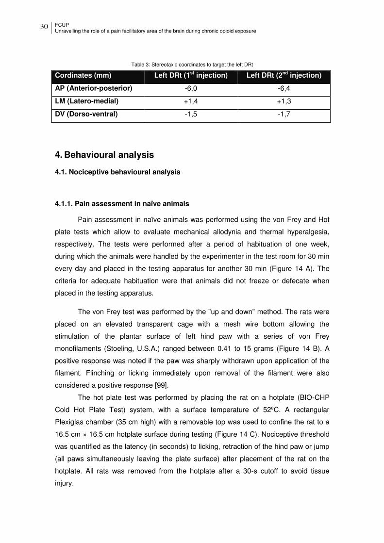
30 FCUP Unravelling the role of a pain facilitatory area of the brain during chronic opioid exposure
Table 3: Stereotaxic coordinates to target the left DRt
Cordinates (mm) Left DRt (1st injection) Left DRt (2nd injection)
AP (Anterior-posterior) -6,0 -6,4
LM (Latero-medial) +1,4 +1,3
DV (Dorso-ventral) -1,5 -1,7
4. Behavioural analysis
4.1. Nociceptive behavioural analysis
4.1.1. Pain assessment in naïve animals
Pain assessment in naïve animals was performed using the von Frey and Hot
plate tests which allow to evaluate mechanical allodynia and thermal hyperalgesia,
respectively. The tests were performed after a period of habituation of one week,
during which the animals were handled by the experimenter in the test room for 30 min
every day and placed in the testing apparatus for another 30 min (Figure 14 A). The
criteria for adequate habituation were that animals did not freeze or defecate when
placed in the testing apparatus.
The von Frey test was performed by the "up and down" method. The rats were
placed on an elevated transparent cage with a mesh wire bottom allowing the
stimulation of the plantar surface of left hind paw with a series of von Frey
monofilaments (Stoeling, U.S.A.) ranged between 0.41 to 15 grams (Figure 14 B). A
positive response was noted if the paw was sharply withdrawn upon application of the
filament. Flinching or licking immediately upon removal of the filament were also
considered a positive response [99].
The hot plate test was performed by placing the rat on a hotplate (BIO-CHP
Cold Hot Plate Test) system, with a surface temperature of 52ºC. A rectangular
Plexiglas chamber (35 cm high) with a removable top was used to confine the rat to a
16.5 cm × 16.5 cm hotplate surface during testing (Figure 14 C). Nociceptive threshold
was quantified as the latency (in seconds) to licking, retraction of the hind paw or jump
(all paws simultaneously leaving the plate surface) after placement of the rat on the
hotplate. All rats was removed from the hotplate after a 30-s cutoff to avoid tissue
injury.

FCUP Unravelling the role of a pain facilitatory area of the brain during chronic opioid exposure
31
These behavioral tests were used to evaluate:
i) the time course effects of chronic morphine administration. For this, the animals
were implanted with a mini-pump filled with morphine (45 µg-1.µL-1.h-1) or saline
(n=6 each group; Scheme 1). Von Frey and Hot plate tests were performed before
and at 2, 4 and 7 days after mini-pump implantation (Scheme 1);
ii) the effects of DRt inactivation by lidocaine on chronic morphine administration. For
this, two subsets of animals were used, as described above. One subset of
animals was implanted with a guide cannula into DRt and with osmotic mini-pumps
for the s.c. delivery of morphine (45 µg-1.µL-1.h-1) (n=7) or saline (n=6). The second
subset of animals was implanted with off target cannulas and with osmotic mini-
pumps for the s.c. delivery of morphine (45 µg-1.µL-1.h-1; n=6). The behavioural
tests were performed 7 days after guide cannula and mini-pump implantation
before and 30 min after lidocaine injection through the guide cannula (Scheme 2);
iii) the effects of MOR knock down at the DRt on chronic morphine administration.
For this, the animals were injected with lentiviral vectors at the DRt, as explained
above, and implanted with mini-pumps for the s.c. delivery of morphine (45 µg-1.µL-
1.h-1;LV-Cont n = 6; MOR-R n= 5) or saline (LV-Cont n = 5; MOR-R n= 6) as
described above. The behavioural tests were performed before and at 2, 4 and 7
days after lentiviral vectors injection and mini-pump implantation (Schema 3).
The time course effects of chronic morphine administration was analysed by
two-way repeated measures of ANOVA followed by Tukey post-hoc test for multiple
comparisons. The effects of lidocaine or lentiviral vectors injection into the DRt were
analysed by two-way ANOVA followed by Tukey post-hoc test for multiple
comparisons. The effects of lidocaine injection into the off-target sites were analysed
by paired t tests.

32 FCUP Unravelling the role of a pain facilitatory area of the brain during chronic opioid exposure
Figure 14: Behavioural tests apparatus. (A) Animal placed in the individual Plexiglas container in the wire grid; (B) von-
Frey test; (C) Hotplate test.
Scheme 1: Time course effects of chronic morphine administration in naïve animals. Mini-pump implantation for the s.c.
delivery of morphine (45 µg-1.µL-1.h-1) or saline was performed at Day 0. The animals were tested at Day 0 (Baseline;
BL), before mini-pump implantation and at Days 2, 4 and 7 after mini-pump implantation.
A C
B
Day 0 Day 7 Day - 5
Mini-pump
implantation
Baseline (BL)
Habituation
Day 2 Day 4

FCUP Unravelling the role of a pain facilitatory area of the brain during chronic opioid exposure
33
Scheme 2: Effects of DRt inactivation by lidocaine on chronic morphine admnistration in naïve animals. Stereotaxic surgery for the implantation of a guide cannula and osmotic mini-pumps implantation for the s.c. delivery of morphine (45 µg-1.µL-1.h-1) or saline were performed at Day 0. The animals were tested at Day 7 before and after lidocaine injection through the guide cannula.
Scheme 3: Effects of MOR knock down at the DRt on chronic morphine admnistration in naïve animals. The stereotaxic
injection of the lentiviral vectors LV-Control and MOR-R and the implantation of osmotic mini-pumps for the s.c. delivery
of morphine (45 µg-1.µL-1.h-1) or saline was performed at Day 0. The animals were tested before the surgical procedures
at Day 0 and at Day7.
4.1.2. Pain assessment in neuropathic animals
Pain assessment in neuropathic animals was performed by the von Frey, pin
prick and acetone tests which allow to evaluate mechanical allodynia, mechanical
hyperalgesia and cold allodynia, respectively. The tests were performed after a period
of habituation of one week, during which the animals were handled by the experimenter
in the test room for 30 min every day and placed in the testing apparatus for another 30
Da Day 7 Day - 5
Mini-pump implantation
Baseline (BL)
Ha ituation
Stereotaxic inje tion of lentiviral vectors
Da Day 7
Day 7
Mini-pump implantation
Baseline (BL)
Stereotaxic implantation of a guide Stereotaxic implantation of a guide cannula
Injection of Lidocaine through the guide cannula
34 FCUP Unravelling the role of a pain facilitatory area of the brain during chronic opioid exposure
min. The criteria for adequate habituation were that animals did not freeze or defecate
when placed in the test apparatus.
To perform the tests, the rats were placed on an elevated transparent cage with
a mesh wire bottom allowing the stimulation of the lateral surface of the left hind paw
(Figure 15). The von-Frey test was performed by applying von Frey monofilaments
(Stoeling, U.S.A.) in a sequence of increasing stiffness for 5 seconds [100]. The
threshold was taken as the lowest force that evoked a brisk withdrawal response to one
of five repetitive applications (Figure 15 A). The pin prick test was performed by the
application of a brief stimulation with a safety pin (Figure 15 B) at an intensity sufficient
to produce a reflex withdrawal response but not penetrate the skin and the duration of
paw withdrawal was clocked [101]. The acetone test was performed by application of
40 µL of acetone using a micropipette tip connected to a micropipette without touching
the skin (Figure 15 C) and the duration of the withdrawal was timed [102].
These behavioral tests were used to evaluate:
i) the time course effects of chronic morphine administration. For this, 2-3 weeks
after SNI induction, the animals were implanted with a mini-pump filled with
morphine (45 µg-1.µL-1.h-1) or saline (n=6 each group; Scheme 4). Von-Frey, pin-
prick and acetone tests were performed before and at 2, 4 and 7 days after mini-
pump implantation (Scheme 4);
ii) the effects of MOR knock down at the DRt on chronic morphine administration. For
this, 2-3 weeks after SNI induction, the animals were injected with lentiviral vectors
at the DRt and implanted with mini-pumps for the s.c. delivery of morphine (45 µg-
1.µL-1.h-1;LV-Cont n = 6; MOR-R n= 6) or saline (LV-Cont n = 5; MOR-R n= 6) as
described above. The behavioural tests were performed before and at 2, 4 and 7
days after lentiviral vectors injection and mini-pump implantation (Scheme 5).
The time course effects of chronic morphine administration and the effects of
lentiviral vectors injection into the DRt were analysed by two-way ANOVA followed by
Tukey post-hoc test for multiple comparisons.
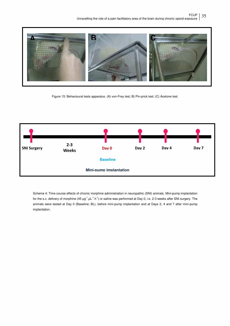
FCUP Unravelling the role of a pain facilitatory area of the brain during chronic opioid exposure
35
Figure 15: Behavioural tests apparatus. (A) von-Frey test; B) Pin-prick test; (C) Acetone test.
Scheme 4: Time course effects of chronic morphine administration in neuropathic (SNI) animals. Mini-pump implantation
for the s.c. delivery of morphine (45 µg-1.µL-1.h-1) or saline was performed at Day 0, i.e. 2-3 weeks after SNI surgery. The
animals were tested at Day 0 (Baseline; BL), before mini-pump implantation and at Days 2, 4 and 7 after mini-pump
implantation.
A B C
Day 0 Day 7 SNI Surgery
Mini-pump implantation
Baseline
- Weeks
Day 2 Day 4

36 FCUP Unravelling the role of a pain facilitatory area of the brain during chronic opioid exposure
Scheme 5: Effects of MOR knock down at the DRt on chronic morphine administration in neuropathic (SNI) animals.
The stereotaxic injection of the lentiviral vectors LV-Control and MOR-R and the implantation of osmotic mini-pumps for
the s.c. delivery of morphine (45 µg-1.µL-1.h-1) or saline was performed Day 0, i.e. 2-3 weeks after SNI surgery. The
animals were tested before the surgical procedures at Day 0 and at Day7.
4.2. Morphine reward behavioural analysis
The analysis of the reward behaviour was performed using the Conditioned
Place Preference test (CPP). The CPP paradigm is a standard behavioural test used to
study the rewarding and aversive effects of drugs. The test involves the association of
a particular environment, through visual and tactile cues, with a drug treatment. This is
known as the conditioning phase of the test. The conditioning phase is followed by a
post-conditioning phase, during which the animals associate a different environment
with a drug but in the absence of the drug [103]. A common variation of this design
consists of a three-compartment chambers with two different conditioning
chambers/contexts, distinguished by visual and sensory cues, to which the animal is
exposed once or several times, that are connected by a neutral chamber, with no
special characteristics and is not paired with a drug. Before the conditioning phase (i.e.
during the pre-conditioning phase), the animal should not present a previous
preference for any chamber/context [103, 104].
The CPP test was used for the first time in the laboratory therefore we first
optimized the test. We tested different visual and tactile cues/contexts in order to obtain
contexts towards which the animals present no preference before the conditioning
sessions (i.e. during the pre-conditioning phase). We also tested the number of
conditioning sessions, which is the number of conditioning trials/sessions the animal is
Da Da SNI Surgery
Mini-pump
implantation
Baseline
2-3 Weeks
Habituation
Stereota i inje tion of lentiviral vector

FCUP Unravelling the role of a pain facilitatory area of the brain during chronic opioid exposure
37
exposed to one of the different chambers/contexts during the conditioning phase. All
experiments were performed using neuropathic animals 2 to 3 weeks after SNI
induction.
4.2.1. Optimization of visual cues
The optimization of the visual cues was performed in a Plexiglas apparatus
measuring 100 [length (L)] X 40 [width (W)] X 40 [height (H)] cm and comprising two
distinct conditioning chambers separated by a neutral chamber. The neutral chamber
measured 20 X 40 X 40 cm with black Plexiglas walls. Each conditioning chamber
measured 40 (L) X 40 (W) X 40 (H) cm and contained different visual clues. One
chamber had walls with alternating 3 cm wide black and white horizontal stripes. The
second chamber had alternating 3 cm wide black and white vertical stripes. The tactile
cues were the same in the three chambers, which is all chambers, had black Plexiglas
floors.
Rats (n = 5 were placed in the neutral chamber with full access to all chambers
for 15 min for 2 consecutive days. Day 1 was used for habituation purposes. On day 2
(pre-conditioning phase), rats behaviour was recorded for 15 minutes using a video
camera. The time spend in each chamber was analysed by Ethlog 2.2.
4.2.2. Optimization of tactile cues
The optimization of tactile cues was performed in a Plexiglas apparatus with the
characteristics described above. The visual clues in the conditioning chambers were
the ones tested above. The following tactile textures in the floor of each conditioning
chamber were tested:
1- Mesh wire floor in one chamber / Black Plexiglas floor in the other chamber;
2- Mesh wire floor in one chamber / Corn cob in the floor of the other chamber;
3- Mesh wire floor in one chamber / Metallic floor with 0.5 cm diameter circles spaced
1 cm in the other chamber;
4- Mesh wire floor in one chamber / 0.5 cm diameter metal rods spaced 2 cm in the
other chamber.
Rats (n = 15) were placed in the neutral chamber with full access to all
chambers for 15 min for 2 consecutive days. Day 1 was used for habituation purposes.
On day 2 (pre-conditioning phase), rats behaviour was recorded for 15 minutes using
a video camera. The time spend in each chamber was analysed by Ethlog 2.2.

38 FCUP Unravelling the role of a pain facilitatory area of the brain during chronic opioid exposure
4.2.3. Optimization of the number of conditioning trials/sessions.
The optimization of the number of conditioning sessions was performed in a
Plexiglas apparatus with the dimensions described above and with visual and tactile
cues that showed unbiased pre-conditioning results. One chamber of the CPP
apparatus had a floor with 0.5 cm diameter metal rods spaced 2 cm and walls with
alternating 3 cm wide black and white horizontal stripes. The floor of the second
chamber was a mesh wire and the walls had with alternating wide black and white
vertical stripes.
We tested the effect of morphine (10 mg/Kg) on a single and a multi-trial
conditioning protocol.
Rats were placed in the neutral chamber with full access to all chambers for 15
min for 2 consecutive days, Day 1 was used for habituation purposes and on day 2
(pre-conditioning phase), rats behaviour was recorded for 15 minutes, as explained
above. Rats spending less than 20% or more than 80% of the entire time in one of the
chambers were excluded. Each rat was then randomly assigned to a treatment group
and a conditioning chamber/environment in a counterbalanced fashion.
During the conditioning phase, Plexiglas partitions matching their respective
environments were inserted to restrict the rats to a specific designated
chamber/environment. In the single-trial conditioning protocol (n = 3), on Day 3 the
animals, were subcutaneously injected with saline and randomly placed in a
conditioning chamber for 1 hour. Four hours later, the animals were subcutaneously
injected with morphine (10 mg/Kg) and placed in the other chamber for 1 hour. In the
multi-trial conditioning protocol (n = 3), the animals were conditioned on Days 3-8. The
animals were subcutaneously injected with saline or morphine (10 mg/Kg) once a day
on alternate days. The animals were placed into the designated conditioning chamber
for 1 hour immediately after the injection.
On post-conditioning day (Day 4 in the single single-trial conditioning protocol;
Day 9 in the multi-trial conditioning protocol), rats were placed in the neutral chamber in
a drug-free state, with access to all chambers, their behaviour was recorded for 15 min.
The time spend in each chamber was analysed by Ethlog 2.2., as above, and the
difference between post- and pre-conditioning time spent in each chamber was
calculated to determine the preference score for each chamber.

FCUP Unravelling the role of a pain facilitatory area of the brain during chronic opioid exposure
39
4.2.4. Reward effects of morphine after chronic morphine administration
The evaluation of the reward effects of morphine after chronic morphine
treatment was performed in a Plexiglas apparatus with the dimensions described
above. The visual and tactile cues in the conditioning chambers, optimized above, were
as follows: one chamber with a floor constituted by 0.5 cm diameter metal rods spaced
2 cm and walls with alternating 3 cm wide black and white horizontal stripes; the
second chamber with a mesh wire floor and walls with alternating wide black and white
vertical stripes. The multi-trial conditioning protocol was chosen to perform the CPP
test.
Rats were placed in the neutral chamber with full access to all chambers for 15
min for 2 consecutive days, Day 1 was used for habituation purposes and on day 2
(pre-conditioning phase; Scheme 6), rats behaviour was recorded for 15 minutes, as
explained above. Rats spending less than 20% or more than 80% of the entire time in
one of the chambers were excluded. Each rat was then randomly assigned to a
treatment group and a conditioning chamber/environment in a counterbalanced
fashion. Immediately after recording their behavior, the rats were implanted with
osmotic mini-pumps filled with morphine (45 µg-1.µL-1.h-1) or saline for the chronic
treatment (Scheme 6). On days 3-8 (conditioning phase; Scheme 6) one group of
animals was subcutaneously injected with saline or morphine (10 mg/Kg) once a day
on alternate days (saline mini-pumps group n = 7; morphine mini-pumps group n = 8).
The animals were placed into the designated conditioning chamber for 1 hour.
immediately after injection (Scheme 6 – item 2). The animals from the control group
(saline mini-pumps group n = 4; morphine mini-pumps group n = 3) were
subcutaneously injected with saline every day and placed into the designated
conditioning chamber for 1 hour immediately after injection (Scheme 6 – item 1). On
day 9 (post- conditioning phase; Scheme 6) rats were placed in the neutral chamber in
a drug-free state, with access to all chambers, their behaviour was recorded for 15 min
and the videos were analyzed by Ethlog 2.2 to determine the difference between post-
and pre-conditioning time spent in each chamber. The number of chamber crossings
was also analysed as it is considered a good indicator of locomotor activity [105, 106].
The statistical analysis was performed by two-way ANOVA followed by Tukey
post-hoc test for multiple comparisons.

40 FCUP Unravelling the role of a pain facilitatory area of the brain during chronic opioid exposure
Scheme 6: Evaluation of the reward effects of morphine after chronic morphine administration by the CPP test. Animals
were allowed a 15 min period of habituation at Day 1 and were recorded on Day 2 (pre-conditioning phase) to ascertain
preferences. Mini-pumps filled with morphine (45 µg-1.µL-1.h-1) or saline for chronic treatment were implanted on Day 2
immediately after behavioural recording. Days 3-8 (conditioning phase): one group (1) received saline (S) s.c every day
and the other group (2) received morphine (M, 10 mg/Kg) or saline on alternate days. Immediately after injection the
animals were placed in the assigned chamber/environment for 1 hour each day. On day 9 (post-conditioning phase)
animals were allowed to explore the apparatus in a drug free state for 15 min and were recorded to ascertain
preferences.
4.2.5. Effects of MOR knock-down at the DRt on morphine reward after chronic
morphine administration
The evaluation of the effects of MOR knock-down at the DRt on morphine
reward was performed in a Plexiglas apparatus with the dimensions and characteristics
described above.
Rats were placed in the neutral chamber with full access to all chambers for 15
min for 2 consecutive days, Day 1 was used for habituation purposes and on day 2
(pre-conditioning phase; Scheme 6), rats behaviour was recorded for 15 minutes, as
explained above. Rats spending less than 20% or more than 80% of the entire time in
one of the chambers were excluded. Each rat was then randomly assigned to a
treatment group and a conditioning chamber/environment in a counterbalanced
fashion. Immediately after recording their behavior, the rats were stereotaxically
injected MOR-R into the left DRt (n = 3), as decribed above, and implanted with
osmotic mini-pumps filled with morphine (45 µg-1.µL-1.h-1) for the chronic treatment
(Scheme 7). On days 3-8 (conditioning phase; Scheme 7) the animals were
subcutaneously injected with saline or morphine (10 mg/Kg) once a day on alternate
days. The animals were placed into the designated conditioning chamber for 1 hour
immediately after injection (Scheme 7). On day 9 (post- conditioning phase; Scheme 7)
rats were placed in the neutral chamber in a drug-free state, with access to all
chambers, their behaviour was recorded for 15 min and the videos were analyzed by
Day
SNI Surgery
-
Habituation Pré-
conditioning
Mini-pump
implantation
MOR R
S S S S S S
M M M S S S
Post-
conditioning
(1)
(2)

FCUP Unravelling the role of a pain facilitatory area of the brain during chronic opioid exposure
41
Ethlog 2.2 to determine the difference between post- and pre-conditioning time spent in
each chamber. The number of chamber crossings was also analysed.
The statistical analysis was performed by was one-way ANOVA followed by
Tukey post-hoc test for multiple comparisons.
Scheme 7: Effects of MOR knock down at the DRt on morphine reward. Animals were allowed a 15 min period of habituation at Day 1 and were recorded on Day 2 (pre-conditioning phase) to ascertain preferences. MOR-R was injected at the DRt and mini-pumps filled with morphine (45 µg-1.µL-1.h-1) for chronic treatment were implanted on Day 2 immediately after behavioural recording. Days 3-8 (conditioning phase): the animals received morphine (M, 10 mg/Kg) or saline on alternate days. Immediately after injection the animals were placed in the assigned chamber/environment for 1 hour each day. On day 9 (post-conditioning phase) animals were allowed to explore the apparatus in a drug free state for 15 min and were recorded to ascertain preferences.
5. Material processing for histological and
immunohistochemical analysis
After the last behavioral evaluation, the animals that received lidocaine
injections were deeply anaesthetized by an overdose of sodium pentobarbital (65
mg/Kg i.p.), injected with 0.5 µl of 0.6% Chicago sky blue dye (Sigma, St. Louis, USA)
through the guide cannula and sacrificed by decapitation. After decapitation, the brain
was removed and immersed in a fixative solution containing 4% paraformaldehyde in
0.1 M PB, pH 7.2 (Appendix A) for 4h followed by 30% sucrose in 0.1M PB, pH 7.2
overnight at 4ºC.
At the completion of the experiments performed with naïve animals used to
study the time course effects of chronic morphine administration and all animals
injected with the lentiviral vectors, the animals were anaesthetized by an overdose of
Day
SNI Surgery
-
Habituation Pré-
conditioning
Mini-pump
implantation
MOR R
Injection
M M M S S S Post-
conditioning

42 FCUP Unravelling the role of a pain facilitatory area of the brain during chronic opioid exposure
sodium pentobarbital (65 mg/Kg i.p.). The animals were then placed in the supine
position, the abdomen and the thorax were opened to expose the heart and 0,2 mL of
heparine (Braun Medical, Portugal) were injected into the left ventricle. A catheter was
then introduced into the ascending aorta for perfusion with 200 mL of calcium-free
Tyrode’s solution (Appendix A), followed by 800 mL of fixative solution containing 4%
paraformaldehyde in 0.1 M PB, pH 7.2 (Appendix A). The brains were removed,
immersed in fixative for 4 hours followed by 30% sucrose in 0.1M phosphate-buffered
saline (PBS) overnight, at 4ºC.
The brainstems of all animals were serially cut in a freezing microtome at 40
µm, collected in 4 sets and stored in a cryoprotector solution (Appendix A) at -20ºC.
Brainstem sections from naïve animals were used for the immunohistochemical
analysis of MOR and pCREB. Brainstem sections from all animals injected with the
lentiviral vectors were used for the immunohistochemical analysis of MOR.
6. Histological verification of injection sites
The location of the injection site of animals injected with lidocaine was
determined by the location of the Chicago sky blue dye after counterstaining medullary
sections encompassing the DRt with thionin. Only animals with cannula correctly
targeting the DRt were included in data analysis.
To analyze the transduction patterns of the lentiviral vectors, one set of
brainstem sections from animals injected with LV-Control were mounted on gelatine-
coated slides, cover slipped with a solution of glycerol diluted in PB (1vol/3vol) and
analyzed in an ApoTome Slider (Zeiss®) fluorescence microscope with an excitation
length of 488 nm. EFGP positive cells were plotted on diagrams of medulla oblongata
sections. MOR-R vectors also carry the EFGP transgene (Figure 8) but its expression
levels are undetectable, likely due to the RNA interference reaction induced by
antisense RNA of MOR which degrades EGFP RNA placed in the second position of
the bicistronic construct. Therefore the location of the injection sites of MOR-R-
injected-animals was determined by checking the position of the needle tract after
counterstaining medullary sections encompassing the DRt with thionin (Appendix A)
[107]. Only animals with vector injections centred in the DRt were included in data
analysis.

FCUP Unravelling the role of a pain facilitatory area of the brain during chronic opioid exposure
43
7. Immunohistochemical analysis.
7.1. pCREB immunodetection.
One in every fourth brainstem section, after washing with PBS, was incubated
with a 30% Hydrogen peroxide solution to block endogenous peroxidase activity,
followed by an incubation with a blocking solution (Glycine + 10% PBS-T) to prevent
the nonspecific binding of the antibodies. After blocking, the sections were incubated
with a rabbit polyclonal anti-pCREB antibody (Neuromics; U.S.A.) in PBS-T at
1:10000 for 48 hours at 4°C. After washing with PBS-T the sections were
incubated for 1h with a swine biotinylated anti-rabbit serum (Dako, Denmark) diluted
in PBS-T containing 2% normal swine serum. Sections were washed again and the
detection of the immunoreaction was performed using the ABC solution (1:200; ABC;
Vector Laboratories, U.S.A) as above. The sections were mounted on gelatin-coated
slides, cleared in xylol and coverslipped with Eukitt. Five sections encompassing the
rostro-caudal extent of the DRt were taken from each animal and photomicrographs of
the DRt were taken using a Zeiss® light microscope with a high-resolution digital
camera. The number of pCREB positive nucleus/particles was calculated using an
automated cell counting in the ImageJ® software. Briefly, all images were transformed
into a 8-bit greyscale followed by an adjustment of the threshold, to highlight the
structures to be counted. Then the images were converted to a binary watershed
image to separate overlapped particles. The statistical analysis was performed using
an unpaired t-test.
7.2. MOR immunodetection
One in every fourth brainstem section, after washing with PBS, was incubated
with a 30% Hydrogen peroxide solution to block endogenous peroxidase activity,
followed by an incubation with a blocking solution (Glycine + 10% PBS-T) to prevent
the nonspecific binding of the antibodies. After blocking, the sections were incubated
with rabbit polyclonal anti-MOR antibody (Neuromics; U.S.A.) in PBS-T at 1:1000 for
48 hours at 4°C. After washing with PBS-T the sections were incubated for 1h
with a swine biotinylated anti-rabbit serum (Dako, Denmark) diluted in PBS-T
containing 2% normal swine serum. Sections were washed again and the detection
of the immunoreaction was performed using the ABC solution (1:200; ABC; Vector
Laboratories, U.S.A) as above. The sections were mounted on gelatin-coated slides,
cleared in xylol and coverslipped with Eukitt. Five sections encompassing the rostro-
caudal extent of the DRt were taken from each animal and the numbers of MOR

44 FCUP Unravelling the role of a pain facilitatory area of the brain during chronic opioid exposure
neurons into the DRt were counted using an Olympos® light microscope with a high-
resolution digital camera. The statistical analysis was performed by was two-way
ANOVA followed by Tukey post-hoc test for multiple comparisons.
7.3. Double immunodectetion of MOR and pCREB
One in every fourth brainstem section, after washing with PBS, was incubated
with a 1% sodium borohydride solution to reduce the tissue autofluorescence followed
by an incubation with a blocking solution (Glycine + 10% PBS-T) to prevent the
nonspecific binding of the antibodies. After blocking, the sections were incubated with a
guinea-pig polyclonal anti-MOR antibody (Neuromics; U.S.A.) in PBS-T at 1:500 for
72 hours at 4°C. After washing with PBS-T the sections were incubated for 1h with
a goat biotinylated anti-guinea-pig serum (Dako, Denmark) diluted in PBS-T
containing 2% normal goat serum. Sections were washed again with PBS-t and
were incubated with streptavidin Alexa 488 (Molecular Probes®) at 1:500 for 1h. After
that, sections were again incubated with a blocking solution as above, followed by the
incubation with rabbit polyclonal anti-pCREB antibody (Neuromics; U.S.A.) in PBS-T
at 1:1000 for 48 hours at 4°C. After washing with PBS-T the sections were
incubated for 1h with a donkey anti-rabbit Alexa 594 (Molecular Probes®). The
sections were mounted on gelatin-coated slides, coverslipped with glycerol–phosphate
buffer (3:1, vol/vol) and stored at 4°C. Five sections encompassing the rostro-caudal
extent of the DRt were taken from each animal and the total number of MOR-IR
neurons and double-IR for MOR and pCREB were counted using ApoTome Slider
(Zeiss®) fluorescence microscope. The statistical analysis was performed using the
unpaired t-test.

FCUP Unravelling the role of a pain facilitatory area of the brain during chronic opioid exposure
45
Results
1. Effects of chronic morphine administration on pain behaviors
1.1. Effects on naïve animals
The effects of chronic morphine administration on naïve animals were tested
before and at 2, 4 and 7 days after mini-pumps implantation (Scheme 1) by the von
Frey and hot plate tests. In the von-Frey test, animals chronically administered with
morphine showed a significant decrease of withdrawal thresholds at day 4 (p=0.0128;
Figure 16 A) and day 7 (p=0.0001; Figure 16 A) compared to baseline. At day 7, the
withdrawal threshold of the morphine group was also significantly lower than in the
saline group (p=0.0012; Figure 16 A). No significant differences were observed in the
saline group compared to baseline.
In the hot plate test, animals treated with morphine showed a significant
decrease of the withdrawal latency at day 4 (p= 0.0094; Figure 16 B) and day 7
(p=0.0001; Figure 16 B) compared to baseline. At day 7, the withdrawal latency of the
morphine group was also significantly lower than in the saline group (p=0.0129; Figure
16 B). No significant differences were observed in the saline group compared to
baseline.

46 FCUP Unravelling the role of a pain facilitatory area of the brain during chronic opioid exposure
Figure 16: Time course effects of morphine administration on mechanical allodynia tested by the von Frey test (A) and
on thermal hyperalgesia tested by the hot plate test (B). Data are presented as mean ± SEM. (saline – blue line n=6;
morphine – red line n=6 # p ˂ 0. 05; ## p ˂ 0. 01; ### p ˂ 0.001 vs. BL; * p ˂ 0. 05; ** p ˂ 0.01 saline vs treated group.)
1.2. Effects on neuropathic animals
The effects of the chronic morphine administration were tested before and at 2,
4 and 7 days after mini-pumps implantation (Scheme 4) by the von Frey, pin prick and
acetone tests. In the von-Frey test, no significant differences were observed within
each group and between both groups (Figure 17 A).In the pin-prick test, the animals
chronically administered with morphine showed a significant increase of the withdrawal
duration at day 4 (p=0.0027; Figure 17 B) and day 7 (p=0.0001; Figure 17 B)
compared to baseline. At day 7, the withdrawal duration in the morphine group was
also significantly higher than in the saline group (p=0.0159; Figure 17 B). No significant
differences were observed in the saline group compared baseline.
A
B

FCUP Unravelling the role of a pain facilitatory area of the brain during chronic opioid exposure
47
In the acetone test, animals treated with morphine showed a significant
increase of the withdrawal duration at day 7 compared both to BL (p=0.0287; Figure
17 C) and the saline group (p=0.0289; Figure 17 C). No significant differences were
observed in the saline group compared to baseline.
Figure 17: Time course effects of morphine administration on mechanical allodynia tested by the von Frey test (A),
mechanical hyperalgesia tested by the pin prick test (B) and cold allodynia tested by the acetone test (C). Data are
presented as mean ± SEM. (saline – blue line n=6; morphine – red line (n=6) # p ˂ 0. 05; ## p ˂ 0. 01; ### p ˂ 0.001 vs.
BL; * p ˂ 0. 05 saline vs treated group).
2. Effects of chronic morphine administration on the reward behavior
2.1. Optimization of the conditioned place preference test
2.1.1 Visual cues
Two different visual contexts were tested in the CPP apparatus. One
conditioning chamber had walls with alternating 3 cm wide black and white horizontal
stripes. The second conditioning chamber had alternating 3 cm wide black and white
vertical stripes. The chambers were separated by a neutral chamber with black
Plexiglas walls.
A B
C

48 FCUP Unravelling the role of a pain facilitatory area of the brain during chronic opioid exposure
The animals did not show a preference for any context (Figure 18). Given the
unbiased results obtained, these two visual contexts were elected to carry on the CPP
test.
Figure 18: Optimization of the visual cues of the CPP test. The CPP apparatus contained one chamber with walls
containing alternating 3 cm wide black and white horizontal stripes and a second chamber containing alternating 3 cm
wide black and white vertical stripes. The chambers were separated by a neutral chamber with black Plexiglas walls.
Data are presented as mean ± SEM (n=5).
2.1.2. Tactile cues
To optimize the tactile cues, four different combinations of floors in the
conditioning chambers were tested: i) “Mesh wire floor vs black Plexiglas floor”; ii)
“Mesh wire floor vs Corn cob floor”; iii) “Mesh wire floor vs metallic floor with circles”
and iv) “Mesh wire floor vs metal bars floor”.
The animals presented a clear preference for the compartment presenting the
mesh wire floor when it was combined with the black Plexiglas (Fig. 19 A), corn cob
(Fig. 19 B) and metallic floor with circles (Fig. 19 C). When the mesh wire floor was
combined with the metal bars floor, the animals no longer presented any preference
(Figure 19 D). Given the unbiased results obtained with the “Mesh wire floor” and the
“Metal bars floor” combination, these two tactile contexts were elected to carry on the
CPP test.
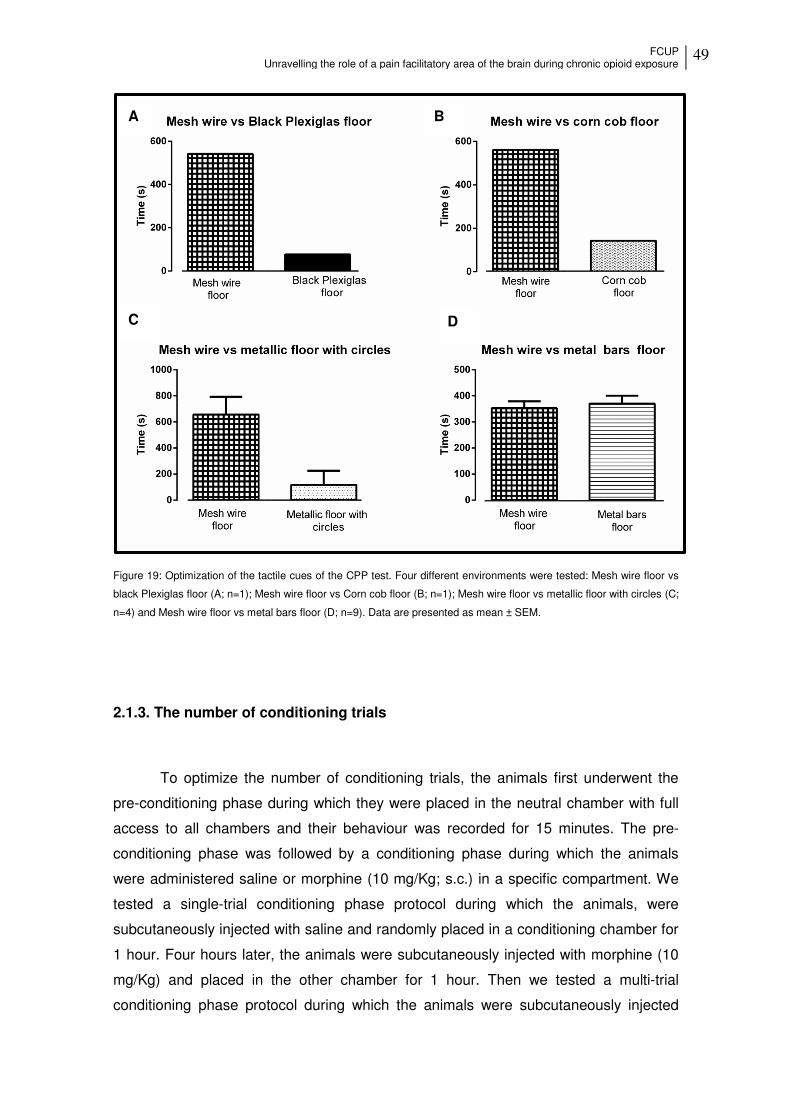
FCUP Unravelling the role of a pain facilitatory area of the brain during chronic opioid exposure
49
Figure 19: Optimization of the tactile cues of the CPP test. Four different environments were tested: Mesh wire floor vs
black Plexiglas floor (A; n=1); Mesh wire floor vs Corn cob floor (B; n=1); Mesh wire floor vs metallic floor with circles (C;
n=4) and Mesh wire floor vs metal bars floor (D; n=9). Data are presented as mean ± SEM.
2.1.3. The number of conditioning trials
To optimize the number of conditioning trials, the animals first underwent the
pre-conditioning phase during which they were placed in the neutral chamber with full
access to all chambers and their behaviour was recorded for 15 minutes. The pre-
conditioning phase was followed by a conditioning phase during which the animals
were administered saline or morphine (10 mg/Kg; s.c.) in a specific compartment. We
tested a single-trial conditioning phase protocol during which the animals, were
subcutaneously injected with saline and randomly placed in a conditioning chamber for
1 hour. Four hours later, the animals were subcutaneously injected with morphine (10
mg/Kg) and placed in the other chamber for 1 hour. Then we tested a multi-trial
conditioning phase protocol during which the animals were subcutaneously injected
A B
C D

50 FCUP Unravelling the role of a pain facilitatory area of the brain during chronic opioid exposure
with saline or morphine (10 mg/Kg) once a day on alternate days for 6 days. The
animals were placed into the designated conditioning chamber for 1 hour immediately
after the injection. The conditioning phase was then followed by the post-conditioning
phase during which the rats were placed in the neutral chamber in a drug-free state,
with access to all chambers, their behaviour was recorded for 15 min and the
difference between pre- and post-conditioning time spent in the morphine-paired
chamber was determined.
The animals that underwent the multi-trial conditioning protocol, showed higher
preference scores in the morphine-paired chamber compared to the animals that
underwent the single-trial conditioning protocol (Figure 20). Given the higher scores of
the multi-trial conditioning protocol, this conditioning protocol was elected to carry on
the CPP test.
Figure 20: Optimization of the number of the conditioning trials. During the conditioning phase, the animals were
administered saline or morphine (10 mg/kg; s.c.) and then placed on a specific compartment. In the single-trial protocol
(n = 3) the animals, were subcutaneously injected with saline and randomly placed on a conditioning chamber, 4 hours
later, they were subcutaneously injected with morphine (10 mg/Kg) and placed in the other chamber. In the multi-trial
conditioning protocol (n = 3) the animals were subcutaneously injected with saline or morphine (10 mg/Kg) once a day
on alternate days for 6 days. On the post-conditioning day, rats behavior was recorded for 15 min and the videos were
analyzed to determine the difference between pre - and post -conditioning time spent in the morphine-paired chamber.
Data are presented as mean ± SEM.

FCUP Unravelling the role of a pain facilitatory area of the brain during chronic opioid exposure
51
2.2. Effects of chronic morphine administration on morphine reward
The CPP test was performed on animals chronically administered morphine (45
µg-1.µL-1.h-1; n=11) or saline (n=11). Animals chronically treated with saline and
conditioned with morphine (10 mg/kg; s.c.) on alternate days (n=7) spent longer time in
the morphine-paired chamber compared to control animals conditioned with saline
(n=4) every day (p= 0.0087;Figure 21). Animals chronically treated with morphine and
conditioned with morphine in alternate days (n=8) spent significantly less time in the
morphine paired chamber compared to animals chronically treated with saline and
conditioned with morphine in alternate days (p=0.0438; Figure 21).
We also analyzed the effect of chronic morphine treatment in the locomotion by
analyzing the number of compartment crossings (Table 4). The statistical analysis
reveals that in general chronic morphine treatment induces a higher number of
crossings compared to chronic saline treatment (F1,19=5.26; p = 0.03). Within the
chronic morphine treated group the number of compartment crossing was statistically
higher when conditioning was performed with saline (p = 0.0002). Nonetheless, when
conditioning was performed with morphine, the number of crossings was not
statistically different between animals chronically treated with saline and morphine (p =
0.98). Within the chronic saline treated group no differences were observed between
conditioning with saline and morphine (p = 0.25).
Table 4: Effects of chronic morphine administration on locomotor activity. Locomotion was evaluated by the number of
compartment crossings. Data are presented as mean ± SEM. *** p ˂ 0. 001 vs. CPP-Saline/Saline of the morphine
treated group.
Saline mini-pumps Morphine mini-pumps
Saline/Saline Morphine/Saline Saline/Saline Morphine/Saline
73.25± 14.1
53.5± 5.72
111±12.2
50.875± 2.01***

52 FCUP Unravelling the role of a pain facilitatory area of the brain during chronic opioid exposure
Figure 21: Effects of chronic morphine administration on morphine reward. During conditioning morphine (orange bars -
10 mg/kg, s.c.) was injected in alternate days in animals chronically administered with saline (n=7) or morphine (n=8).
Control animals were injected saline (white bars) every day in animals chronically administered with morphine (n=3) or
saline (n=4). Data are presented as mean ± SEM. ( *p ˂ 0. 01 vs. CPP-Saline/Saline; # p ˂ 0. 05 vs. CPP-
Morphine/Saline of the morphine treated group).
3. Involvement of the DRt in chronic morphine effects
3.1. Effects of DRt inactivation on nociceptive behavior
DRt inactivation was achieved by local injection of lidocaine. The effects of
lidocaine on nociceptive behavior were 7 days after mini-pump implantation before
(Figure 22; -Lido) and 30 min after (Figure 22; +Lido) lidocaine injection (Scheme 2).
In the von-Frey test, before lidocaine injection (i.e. at day 7 after mini-pump
implantation) animals chronically administered with morphine showed a significant
decrease of the withdrawal threshold compared with animals chronically administered
with saline (p=0.0001;Figure 22 A). Lidocaine injection completely reversed the effects
of chronic morphine (p=0.0001; Figure 22 A). No significant differences were observed
after lidocaine injection on animals chronically administered with saline (Figure 22 A).
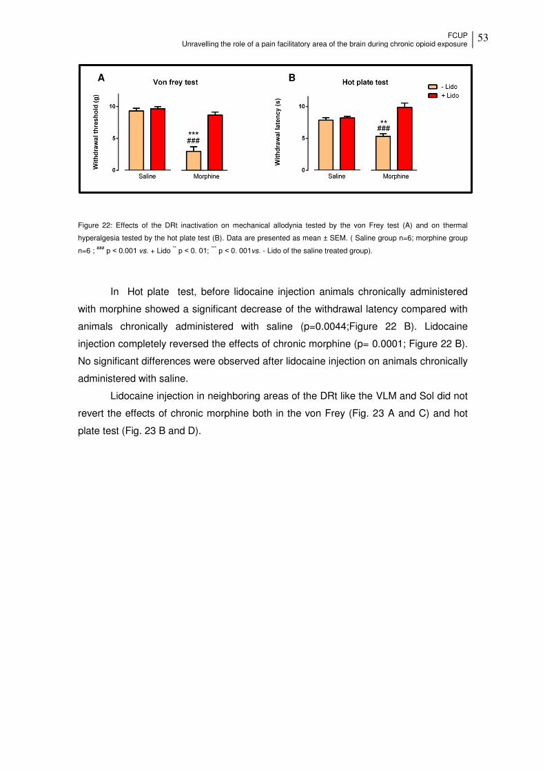
FCUP Unravelling the role of a pain facilitatory area of the brain during chronic opioid exposure
53
Figure 22: Effects of the DRt inactivation on mechanical allodynia tested by the von Frey test (A) and on thermal
hyperalgesia tested by the hot plate test (B). Data are presented as mean ± SEM. ( Saline group n=6; morphine group
n=6 ; ### p ˂ 0.001 vs. + Lido ** p ˂ 0. 01; *** p ˂ 0. 001vs. - Lido of the saline treated group).
In Hot plate test, before lidocaine injection animals chronically administered
with morphine showed a significant decrease of the withdrawal latency compared with
animals chronically administered with saline (p=0.0044;Figure 22 B). Lidocaine
injection completely reversed the effects of chronic morphine (p= 0.0001; Figure 22 B).
No significant differences were observed after lidocaine injection on animals chronically
administered with saline.
Lidocaine injection in neighboring areas of the DRt like the VLM and Sol did not
revert the effects of chronic morphine both in the von Frey (Fig. 23 A and C) and hot
plate test (Fig. 23 B and D).
A B

54 FCUP Unravelling the role of a pain facilitatory area of the brain during chronic opioid exposure
Figure 23: Effects of the VLM and Sol inactivation on mechanical allodynia tested by the von Frey test (A and C) and on
thermal hyperalgesia tested by the hot plate test (B and D). Data are presented as mean ± SEM (n=3).
A B
C D

FCUP Unravelling the role of a pain facilitatory area of the brain during chronic opioid exposure
55
3.2. Effects of chronic morphine on pCREB and MOR expression at the DRt
Chronic morphine administration in naïve animals induced a significant increase
of pCREB at the DRt compared with animals chronically treated with saline (p= 0.0304;
Figure 24).
Figure 24: Effects of chronic administration of morphine on the expression of pCREB at the DRt.
Representative photomicrographs of pCREB labeling at the DRt of animals chronically administered with morphine (A)
and saline (B). Scale bar in A: 100m (B is at the same magnification). Data in C are presented as mean ± SEM
(n=6/group *p<0,05).
Chronic morphine administration in naïve animals induced a significant increase
in the number of MOR cells at the DRt compared with animals chronically treated with
saline (p= 0.0022; Figure 25 A). The percentage of double labelled cells for MOR and
pCREB (Figure 25 C) was high both in saline and morphine treated animals and did not
differ berween the groups (p= 0.2098 ; Figure 25 B); Saline treated animals showed an
average of 92.6 ± 1.73% of co-localization (201.0 ± 11.0 double labelled cells; Figure
25 A and B). Morphine treated animals showed an average of 95.4 ± 0,85% co-
localization (Figure 25 B).

56 FCUP Unravelling the role of a pain facilitatory area of the brain during chronic opioid exposure
Figure 25: Effects of chronic administration of morphine on the expression of MOR and pCREB . Total number of
neurons single labelled for MOR and double labelled for MOR and pCREB (A); Percentage of neurons that co-localize
with CREB (B) in animals chronically treated with saline and morphine. Representative photomicrograph of double-
labeled neurons for MOR and pCREB (yellowish; double arrows) and pCREB labeling (sigle arrow) at the DRt (C). Data
in A and B presented as mean ± SEM (saline n=6; morphine n=5)
3.3. Effects of MOR Knock-down at the DRt
3.3.1. Pattern of lentiviral transduction
The pattern of lentiviral transduction was analyzed in animals injected with LV-
Control and with the injection site centered at the DRt (Figure 26 A, B). In those
animals, the injection site was constituted by a central dark zone corresponding to the
needle tract with numerous EGFP+ neurons (Figure 26 B) around this central region. All
EGFP + neurons were located within the boundaries of the DRt which indicates that
injections correctly placed at the DRt show a pattern of lentiviral transduction
restricted to the DRt.
The injection site of MOR-R-injected animals was analyzed in medullary
sections stained with thionin, because EGFP expression from these constructs (Figure
8) was almost undetectable. The injection site was identified by the presence of the
needle tract in the DRt. In those animals the injection site was constituted by a central
dark core corresponding to the needle tract surrounded by a peripheral zone lightly
stained by thionin (Figure 26 C). Only animals with the injection site placed at the DRt
were included in data analysis.

FCUP Unravelling the role of a pain facilitatory area of the brain during chronic opioid exposure
57
Figure 26: Localization of the injection site in the DRt. (A) Diagram depicting the location of the DRt, at 5.60 mm caudal
to the interaural line adapted from the Paxinos and Watson (2007 Paxinos) (B) Fluorescence photomicrograph of the
injection site at the DRt showing EGFP neurons better depicted in the insert. (C) Representative photomicrograph of a
thionin-stained section illustrating a correct vector injection at the DRt. Scale bar in B: β00 μm (C is at the same
magnification).
3.3.2. Effects of lentiviral transduction on MOR expression
MOR expression at the DRt was evaluated in naïve and SNI animals chronically
treated with saline or morphine and injected with LV-cont or MOR-R. LV-cont-injected
animals treated with morphine showed increased numbers of MOR-IR cells compared
to LV-cont-injected animals treated with saline both in naïve (p=0.0001; Figure 27 C)
and SNI animals (p=0.0001; Figure 27 C). In naïve rats, MOR-IR-injected animals
showed a significant reduction of MOR-IR cells compared LV-cont-injected rats both
after chronic treatment with saline (p=0.0001; Figure 27 C) and morphine (p=0.0001;
Figure 27 C). In SNI animals MOR-IR also induced a significant reduction of MOR-IR
cells compared to LV-cont-injected rats both after chronic treatment with saline
(p=0.0040 Figure 27 C) and morphine (p=0.0001; Figure 27 C).

58 FCUP Unravelling the role of a pain facilitatory area of the brain during chronic opioid exposure
Figure 27: Evaluation of MOR expression by immunohistochemistry after injection of a lentiviral vector that knocks down
MOR (MOR-R vector) and a control vector (LV-cont) at the DRt. Representative photomicrographs of MOR-IR cells in
the DRt of naïve rats chronically treated with morphine and injected with LV-cont (A) and MOR-R (B). Typical MOR
immunolabeling is marked by arrows. Scale bar in B: 50m (A is at the same magnification). Data in C are represented
as mean ±SEM (naïve animals treated with saline LV-Cont n = 7; MOR-R n = 6; or morphine: LV-Cont n= 6; MOR-R
n=6 and SNI animals treated with saline: LV-Cont n = 6; MOR-R n = 5; or morphine: LV-Cont n= 5; MOR-R n=6; ###p ˂
0. 001 vs. LV-Cont of saline group; *p ˂ 0. 05; *** p ˂ 0. 001 vs. Lv-Cont).

FCUP Unravelling the role of a pain facilitatory area of the brain during chronic opioid exposure
59
3.3.3. Effects of lentiviral transduction on nociceptive behavior
3.3.3.1. Effects on naïve animals
The effect of the MOR knock-down at the DRt on naïve animals during
chronic treatment with morphine was evaluated before and at 7 days after the
mini-pumps implantation and stereotaxic injections of the lentiviral vectors (Scheme 3).
In von-Frey test, animals chronically treated with morphine and injected with the
control vector showed a significant decrease of the withdrawal threshold compared to
baseline (p=0.0001; Figure 28 A). Animals injected with MOR-R and chronically
exposed to morphine also showed a significant decrease of the withdrawal threshold
compared to baseline (p=0.0001; Figure 28 A). Nonetheless, this decrease was less
important than that observed in animals injected with the control vector, indeed the
withdrawal thresholds of MOR-R-animals were significantly higher than in the LV-
Control group (p= 0.0149; Figure 28 A). In animals chronically treated with saline, the
injection of LV-Control did not induce significant alterations compared to baseline.
MOR-R caused a significant decrease of the withdrawal threshold compared to the
baseline (p= 0.0001; Figure 28 A) and the LV-Control group (p= 0.0105; Figure 28 A).
In the Hot plate test, animals chronically treated with morphine and injected with
LV-Control showed a significant decrease in the withdrawal latency compared to
baseline (p= 0.0001; Figure 28 B). Animals injected with MOR-R showed higher
withdrawals latencies compared to the LV-Control group (p= 0.0032; Figure 28 B) with
values similar to the baseline (Figure 28 B). In animals chronically treated with saline,
the injection of LV-Control did not induce significant alterations compared to baseline
(Figure 28 B). MOR-R induced a significant decrease of the withdraw latency
compared to baseline (p = 0.0129; Figure 28 B).

60 FCUP Unravelling the role of a pain facilitatory area of the brain during chronic opioid exposure
Figure 28: Effects of MOR knock-down at the DRt on mechanical allodynia tested by the von Frey test (A) and on
thermal hyperalgesia tested by the hot plate test (B) during chronic saline or morphine administration. Data are
presented as mean ± SEM. (chronic morphine treatment: LV-Control-injected n=6, MOR-R-injected n=5; chronic saline
treatment: LV-Control-injected n=5; MOR-R-injected n=6; * p ˂ 0. 05; **p˂ 0. 01; *** p ˂ 0. 001).
3.3.3.2. Effects on SNI animals
The effect of MOR knock-down at the DRt on SNI animals during chronic
treatment with morphine was evaluated before and 7 days after the mini-pumps
implantation and stereotaxic injections (i.e 3 to 4 weeks after SNI induction; Sheme 5).
In von-Frey test no significant differences were observed (Figure 29 A).
In the pin-prick test, animals chronically treated with morphine and injected with
the control vector showed a significant augmented withdrawal duration compared to
baseline (p=0.0045; Figure 29 B). Animals injected with MOR-R showed a significantly
lower withdrawal duration compared to the LV-Control group (p= 0.0014: Figure 29 B)
with values similar to the baseline (Figure 29 B). In animals chronically treated with
saline, the injection of LV-Control did not induce significant alterations compared to
baseline (Figure 29 B). MOR-R induced a significant increase of the withdrawal
duration compared both to the LV-Control group (p= 0.0017; Figure 29 B) and baseline
(p= 0.0001; Figure 29 B).
In the acetone test, animals chronically treated with morphine and injected with
LV-Control showed a significant increase of the withdrawal duration compared to
baseline (p= 0.0041; Figure 29 C). Animals injected with MOR-R showed a significantly
lower withdrawal duration compared to the LV-Control group (p= 0.0001; Figure 29 C)
with values similar to the baseline (Figure 29 C). In animals chronically administered
with saline the injection of LV-Control did not induce significant alterations compared to
baseline (Figure 29 C). MOR-R induced a significant increase of the withdraw duration
A B

FCUP Unravelling the role of a pain facilitatory area of the brain during chronic opioid exposure
61
compared both to the LV-Control group (p= 0.0001; Figure 28 C) and baseline (p=
0.0001; Figure 29 C).
Figure 29: Effects of MOR knock-down at the DRt on mechanical allodynia tested by the von Frey test (A), mechanical
hyperalgesia tested by the pin prick test (B) and cold allodynia tested by the acetone test (C) during chronic saline or
morphine administration. Data are presented as mean ± SEM. (chronic morphine treatment: LV-Control-injected n=6,
MOR-R-injected n=6; chronic saline treatment: LV-Control-injected n=5; MOR-R-injected n=6 ** p ˂ 0. 01; *** p ˂ 0.
001)
A
B
C

62 FCUP Unravelling the role of a pain facilitatory area of the brain during chronic opioid exposure
3.3.4. Effects of lentiviral transduction on reward behavior
The CPP test was performed on animals chronically administered morphine (45
µg-1.µL-1.h-1) and injected with MOR-R at the DRt. Animals injected with MOR-R and
chronically treated with morphine and conditioned with morphine (10 mg/Kg; s.c.) (n=3)
spent more time in the morphine-paired chamber compared to animals only chronically
treated with morphine and conditioned with morphine (n=8) although this comparison
did not reach statistical significance (Figure 30).
The statistical analysis showed no differences in the number of crossings between the groups (Table 5).
Figure 30: Effects of MOR knock-down at the DRt on morphine reward. During conditioning morphine (10 mg/kg, s.c. -
orange bars) was injected in alternate days in animals chronically administered with saline (n=7) or morphine (n=8). In
MOR-R-injected animals morphine (red bar) was injected in alternate days in animals chronically administered with
morphine (n=3). Data are presented as mean ± SEM.
Table 5: Effects of chronic morphine administration on locomotor activity. Locomotion was shown as the number of
compartment crossings. Data are means ± SEM.
Saline mini-pumps Morphine mini-pumps
Morphine/Saline Morphine/Saline Morphine/Saline+ MOR-R
53.5± 5.72
50.875± 2.01
63.75± 3.42

FCUP Unravelling the role of a pain facilitatory area of the brain during chronic opioid exposure
63
Discussion and Conclusions The results gathered in the present thesis show that chronic administration of
morphine induces mechanical and thermal sensibility in naïve animals and, an
aggravation of pre-existing pain in animals with chronic neuropathic pain. In the CPP
test, neuropathic animals chronically treated with morphine failed to show preference
for the morphine-paired chamber which indicates a loss of the reward effect of
morphine. Inactivation of the DRt with lidocaine fully reversed mechanical allodynia and
thermal hyperalgesia of morphine-infused animals. Chronic morphine treatment
induces an increase of pCREB and MOR expression at the DRt. Lentiviral-mediated
MOR knock-down at the DRt prevented the development of opioid-induced
hyperalgesia and in the CPP test, the preliminary results obtained indicate a small
reversion of the loss of the reward effect of morphine.
1. Effects of chronic administration of morphine
Our results show that chronic morphine administration in naïve animals induce
enhanced pain responses during acute pain induction, which is in agreement with
previous reports [108-110]. Moreover, we show for the first time that chronic morphine
administration induces an aggravation of pain in a chronic neuropathic pain model.
These results are clinically relevant since opioid drugs are used in patients struggling
with moderate and severe chronic pain [17, 34, 50].
Unlike naïve animals, neuropathic animals did not show an aggravation of
mechanical thresholds upon chronic morphine administration. This is probably due to
technical issues. Indeed, due to neuropathic pain, animals respond to the lowest
microfilament available (0.008 g), therefore it was impossible to observe further
decreases of the mechanical threshold. However, since in naïve animals chronic
morphine induced a decrease of mechanical thresholds it is likely that in SNI animals
this pain modality might be affected.

64 FCUP Unravelling the role of a pain facilitatory area of the brain during chronic opioid exposure
2. Involvement of the DRt in OIH
The injection of lidocaine directly into the DRt fully reversed mechanical
allodynia and thermal hyperalgesia after chronic morphine exposure. These results
show that descending facilitation from the DRt is involved in OIH. Furthermore, our
results indicate that MOR is likely involved in increased DRt facilitation during chronic
administration of morphine. Usually MOR exerts a inhibitory action by inhibiting AC
activity, causing activation of Kir3 K+ channels and inhibition of the voltage-dependent
Ca2+ channels, leading to hyperpolarization of the cell [111, 112]. This inhibitory effect
would decrease DRt facilitation and MOR knock-down at the DRt would then lead to
disinhibition of the DRt and thus hyperalgesia. The hyperalgesic effects observed in
naïve animals or the aggravation of pain behaviors in SNI animals upon saline
treatment are consistent with the inhibitory effects of MOR. Previous reports also
confirm the inhibitory effects of opioids at the DRt in acute pain models [40, 44]. The
opposite effects of MOR knock-down during chronic morphine, i.e. MOR knock-down
prevented the development of OIH both in naïve and SNI animals, are more consistent
with MOR mediating facilitatory effects. This switch of MOR signalling have been also
reported during chronic morphine in vitro [111, 113-115], and at the locus coeruleus
[112, 116] and at the nucleus acumbens [112].
Accelerated desensitization, increased constitutive activity or endocytosis of the
receptor are suggested as cellular adaptations to chronic morphine exposure [117].
The most likely explanation might lie on a switch on the G-protein coupled to MOR,
from Gi/o to a Gs-protein, leading to an increase in AC, which causes augmented levels
of cAMP, altering the hyperpolarized state of the neuron by changing the intracellular
concentrations of Ca2+ and K+ [118, 119]. Futher corroborating this, chronic morphine
exposure in naïve animals also increased pCREB levels at the DRt and MOR-IR cells
colocalized with pCREB. The increased expression of pCREB during chronic morphine
administration has been previously described in other areas like the nucleus of the
solitary tract [59] and locus coeruleus [62]. The evaluation of the co-localization of
MOR and pCREB in neurophatic animals was not performed but will be held in the
future.
It is worth noting that chronic morphine increased MOR expression more
significantly in naïve animals than in neuropathic animals. This may be the due to the
neuropathy. Indeed, several studies reveal that chronic pain induces down-regulation
of MOR signaling and expression [120, 121]. This decrease of the MOR expression
likely contributes to increased descending facilitation during chronic pain [11, 122]. In

FCUP Unravelling the role of a pain facilitatory area of the brain during chronic opioid exposure
65
spite of this chronic treatment with morphine induces an increase of MOR expression,
leading to an increase of pain sensibility characteristic of the OIH.
3. Reward effects of morphine
Pain and reward are opponent and interacting processes with a large overlap of
the brain systems that control them. Opioids are particularly important for processing
the aspects of reward as well as the processes of pain modulation [73]. Relief from an
unpleasant state is usually associated with rewarding qualities. Pain relief is an
important type of reward, in particular in chronic pain [73, 74], and has been proven in
humans [123] and rats [124]. In fact, in the past years, the CPP test, has also been
used for the assessment of ongoing pain both in inflammatory [125, 126] and in
neuropathic pain models [127, 128].
Our results show that chronic morphine exposure reversed the preference of
the animals for the morphine-paired chamber. This loss morphine reward in
neuropathic animals is not associated with sedative effects of morphine since the
number of the compartments crossing is similar to the animals chronically treated with
saline. On the contrary, the chronic administration of morphine in animals increased
locomotor activity [105, 129] which was also demonstrated in our work in animals
chronically administrated with morphine and conditioned with saline.
In the reward system, morphine binds to MOR expressed on GABAergic
neurons which normally inhibit dopaminergic neurons in the VTA. MOR activation on
GABAergic neurons leads to disinhibition of dopaminergic neurons witch induces an
increase of dopamine release in the NAc [120]. Therefore, the loss of morphine reward
should be the result of decreased inhibition of GABAergic neurons, also consistent with
the switch of MOR activity, resulting thus in a reduction of dopamine release in areas
involved in the reward system.
Given the overlap of these two systems, to assess the influence of pain
modulatory areas in the reward system, we studied the effect of the MOR knock-down
at the DRt on the effect of morphine reward. Our hypothesis is that during chronic pain,
increased descending facilitation [11, 122] along with the aggravation of DRt
descending facilitation, due to chronic morphine exposure shown by our results, likely
lead to a less pain relief/reward (Figure 31). Therefore, if we reduce MOR expression

66 FCUP Unravelling the role of a pain facilitatory area of the brain during chronic opioid exposure
at the DRt, this should lead to more pain relief and consequently more morphine
reward.
Figure 31: Schematic representation of the relationship between descending facilitation and pain relief/reward.
Although these experiments were performed with a small number of animals,
they are indicative of a reversion of the loss of the reward effect of morphine.
Increasing the number of animals should reinforce these results. Our results suggest
that the DRt is connected to the reward system. These connections are probably
relayed by other areas connected to the DRt [130] that are involved both in pain and
reward such as the amygdala, the dorsal and ventral striatum and insular cortex. In
future studies it would be interesting to evaluate the effect of these connections on the
effects of pain relief and reward mediated by morphine.
4. Conclusions and future perspectives
Opioids are a common therapy to treat moderate and severe pain. Unravelling
the molecular mechanisms involved in OIH is fundamental since, instead of relieving
pain, these drugs may be responsible for hyperalgesia, in some patients.
We show in this thesis that prolonged exposure to opioids likely induces plastic
changes in opioidergic circuits causing an exacerbation of DRt descending facilitation
that contributes to OIH. In continuity of this thesis it would be interesting to further
Descending facilitation during chronic pain + Aggravation of DRt descending facilitation by chronic
morphine
Pain relief /reward
Hig
h H
igh
Low
Low

FCUP Unravelling the role of a pain facilitatory area of the brain during chronic opioid exposure
67
extend the study of pCREB and MOR in neuropathic animals mainly by inhibiting some
kinases responsible for the activation of CREB at the DRt.
Unveiling the molecular mechanisms behind OIH is the key to overcome its side
effects, namely the loss of the pain relief observed after morphine chronic
administration at the CPP test. In the future, it would be interesting to continue the
studies of this thesis by evaluating the connections between the DRt and areas
involved in pain and reward on the effects of pain relief mediated by morphine.

68 FCUP Unravelling the role of a pain facilitatory area of the brain during chronic opioid exposure

FCUP Unravelling the role of a pain facilitatory area of the brain during chronic opioid exposure
69
References 1. Loeser, J.D. and R.D. Treede, The Kyoto protocol of IASP Basic Pain
Terminology. Pain, 2008. 137(3): p. 473-7.
2. Woolf, C.J. and R.J. Mannion, Neuropathic pain: aetiology, symptoms,
mechanisms, and management. Lancet, 1999. 353(9168): p. 1959-64.
3. Fornasari, D., Pain mechanisms in patients with chronic pain. Clin Drug
Investig, 2012. 32 Suppl 1: p. 45-52.
4. Breivik, H., et al., Survey of chronic pain in Europe: prevalence, impact on daily
life, and treatment. Eur J Pain, 2006. 10(4): p. 287-333.
5. Azevedo, L.F., et al., Epidemiology of chronic pain: a population-based
nationwide study on its prevalence, characteristics and associated disability in
Portugal. J Pain, 2012. 13(8): p. 773-83.
6. Pace, M.C., et al., Neurobiology of pain. J Cell Physiol, 2006. 209(1): p. 8-12.
7. Lawson, S.N., Phenotype and function of somatic primary afferent nociceptive
neurones with C-, Adelta- or Aalpha/beta-fibres. Exp Physiol, 2002. 87(2): p.
239-44.
8. Todd, A.J., Neuronal circuitry for pain processing in the dorsal horn. Nat Rev
Neurosci, 2010. 11(12): p. 823-36.
9. Basbaum, A.I., et al., Cellular and molecular mechanisms of pain. Cell, 2009.
139(2): p. 267-84.
10. Argoff, C., Mechanisms of pain transmission and pharmacologic management.
Curr Med Res Opin, 2011. 27(10): p. 2019-31.
11. Tracey, I. and P.W. Mantyh, The cerebral signature for pain perception and its
modulation. Neuron, 2007. 55(3): p. 377-91.
12. Millan, M.J., The induction of pain: an integrative review. Prog Neurobiol,
1999. 57(1): p. 1-164.
13. Tavares, I. and I. Martins, Gene Therapy for Chronic Pain Management. In
Gene Therapy - Tools and Potential Applications. Vol. Francisco Martin Molina
(Ed.), ISBN: 978-953-51-1014-9, InTech. DOI: 10.5772/55050. 2013.
14. Ossipov, M.H., G.O. Dussor, and F. Porreca, Central modulation of pain. J Clin
Invest, 2010. 120(11): p. 3779-87.
15. Heinricher, M.M., et al., Descending control of nociception: Specificity,
recruitment and plasticity. Brain Res Rev, 2009. 60(1): p. 214-25.
16. Almeida, A., H. Leite-Almeida, and I. Tavares, Medullary control of nociceptive
transmission: reciprocal dual communication with the spinal cord. Drug
Discovery Today: Disease Mechanisms, 2006. 3(3): p. 305-312.
17. Ossipov, M.H., et al., Underlying mechanisms of pronociceptive consequences
of prolonged morphine exposure. Biopolymers, 2005. 80(2-3): p. 319-24.
18. Koppert, W., [Opioid-induced hyperalgesia. Pathophysiology and clinical
relevance]. Anaesthesist, 2004. 53(5): p. 455-66.
19. Lee, M., et al., A comprehensive review of opioid-induced hyperalgesia. Pain
Physician, 2011. 14(2): p. 145-61.
20. Lima, D. and A. Almeida, The medullary dorsal reticular nucleus as a
pronociceptive centre of the pain control system. Prog Neurobiol, 2002. 66(2): p.
81-108.
21. Villanueva, L., et al., Encoding of electrical, thermal, and mechanical noxious
stimuli by subnucleus reticularis dorsalis neurons in the rat medulla. J
Neurophysiol, 1989. 61(2): p. 391-402.

70 FCUP Unravelling the role of a pain facilitatory area of the brain during chronic opioid exposure
22. Villanueva, L., et al., Convergence of heterotopic nociceptive information onto
subnucleus reticularis dorsalis neurons in the rat medulla. J Neurophysiol,
1988. 60(3): p. 980-1009.
23. Leite-Almeida, H., A. Valle-Fernandes, and A. Almeida, Brain projections from
the medullary dorsal reticular nucleus: an anterograde and retrograde tracing
study in the rat. Neuroscience, 2006. 140(2): p. 577-595.
24. Dugast, C., A. Almeida, and D. Lima, The medullary dorsal reticular nucleus
enhances the responsiveness of spinal nociceptive neurons to peripheral
stimulation in the rat. Eur J Neurosci, 2003. 18(3): p. 580-8.
25. Almeida, A., et al., The medullary dorsal reticular nucleus facilitates acute
nociception in the rat. Brain Res Bull, 1996. 39(1): p. 7-15.
26. Almeida, A., et al., The medullary dorsal reticular nucleus facilitates pain
behaviour induced by formalin in the rat. Eur J Neurosci, 1999. 11(1): p. 110-
22.
27. Sotgiu, M.L., et al., Contribution by DRt descending facilitatory pathways to
maintenance of spinal neuron sensitization in rats. Brain Res, 2008. 1188: p. 69-
75.
28. Martins, I., et al., Reversal of neuropathic pain by HSV-1-mediated decrease of
noradrenaline in a pain facilitatory area of the brain. Pain, 2010. 151(1): p.
137-45.
29. Martins, I., et al., Noradrenaline increases pain facilitation from the brain
during inflammatory pain. Neuropharmacology, 2013. 71: p. 299-307.
30. Paxinos, G., and Watson, C., The Rat Brain in Stereotaxic Coordinates
(Academic Press). 2007.
31. Rosenblum, A., et al., Opioids and the treatment of chronic pain: controversies,
current status, and future directions. Experimental and clinical
psychopharmacology, 2008. 16(5): p. 405.
32. Savage, S.R., Long-term opioid therapy: assessment of consequences and risks.
J Pain Symptom Manage, 1996. 11(5): p. 274-86.
33. Katz, N., Methodological issues in clinical trials of opioids for chronic pain.
Neurology, 2005. 65(12 Suppl 4): p. S32-49.
34. Angst, M.S. and J.D. Clark, Opioid-induced hyperalgesia: a qualitative
systematic review. Anesthesiology, 2006. 104(3): p. 570-87.
35. Fields, H., State-dependent opioid control of pain. Nature Reviews
Neuroscience, 2004. 5(7): p. 565-575.
36. Nadal, X., et al., Involvement of the opioid and cannabinoid systems in pain
control: New insights from knockout studies. European journal of pharmacology,
2013. 716(1): p. 142-157.
37. Welch, S.P., Interaction of the cannabinoid and opioid systems in the
modulation of nociception. International Review of Psychiatry, 2009. 21(2): p.
143-151.
38. McDonald, J. and D. Lambert, Opioid receptors. Continuing Education in
Anaesthesia, Critical Care & Pain, 2005. 5(1): p. 22-25.
39. Przewlocki, R. and B. Przewlocka, Opioids in chronic pain. Eur J Pharmacol,
2001. 429(1-3): p. 79-91.
40. Pinto, M., et al., Opioids modulate pain facilitation from the dorsal reticular
nucleus. Mol Cell Neurosci, 2008. 39(4): p. 508-18.
41. Hallberg, M., Neuropeptides: Metabolism to Bioactive Fragments and the
Pharmacology of Their Receptors. Medicinal Research Reviews, 2014.

FCUP Unravelling the role of a pain facilitatory area of the brain during chronic opioid exposure
71
42. Ren, K., and Dubner, R., Descending control mechanisms. San Diego:
Academic Press, 2008.
43. Neto, F.L., et al., Delta opioid receptor mRNA expression is changed in the
thalamus and brainstem of monoarthritic rats. Journal of chemical
neuroanatomy, 2008. 36(2): p. 122-127.
44. Martins, I., et al., Dynamic of migration of HSV-1 from a medullary
pronociceptive centre: antinociception by overexpression of the
preproenkephalin transgene. Eur J Neurosci, 2008. 28(10): p. 2075-83.
45. Raffa, R.B. and J.V. Pergolizzi, Jr., Opioid-induced hyperalgesia: is it clinically
relevant for the treatment of pain patients? Pain Manag Nurs, 2013. 14(3): p.
e67-83.
46. Low, Y., C.F. Clarke, and B.K. Huh, Opioid-induced hyperalgesia: a review of
epidemiology, mechanisms and management. Singapore Med J, 2012. 53(5): p.
357-60.
47. Lee, S.H., et al., Tramadol induced paradoxical hyperalgesia. Pain Physician,
2013. 16(1): p. 41-4.
48. Tompkins, D.A. and C.M. Campbell, Opioid-induced hyperalgesia: clinically
relevant or extraneous research phenomenon? Curr Pain Headache Rep, 2011.
15(2): p. 129-36.
49. Chu, L.F., D.J. Clark, and M.S. Angst, Opioid tolerance and hyperalgesia in
chronic pain patients after one month of oral morphine therapy: a preliminary
prospective study. J Pain, 2006. 7(1): p. 43-8.
50. Chu, L.F., M.S. Angst, and D. Clark, Opioid-induced hyperalgesia in humans:
molecular mechanisms and clinical considerations. Clin J Pain, 2008. 24(6): p.
479-96.
51. Compton, P., V. Charuvastra, and W. Ling, Pain intolerance in opioid-
maintained former opiate addicts: effect of long-acting maintenance agent. Drug
Alcohol Depend, 2001. 63(2): p. 139-146.
52. Guignard, B., et al., Acute opioid tolerance: intraoperative remifentanil
increases postoperative pain and morphine requirement. Anesthesiology, 2000.
93(2): p. 409-17.
53. Suzan, E., et al., A negative correlation between hyperalgesia and analgesia in
patients with chronic radicular pain: is hydromorphone therapy a double-edged
sword? Pain Physician, 2013. 16(1): p. 65-76.
54. Hooten, W.M., et al., Associations between heat pain perception and opioid
dose among patients with chronic pain undergoing opioid tapering. Pain
Medicine, 2010. 11(11): p. 1587-1598.
55. Kayan, S., L.A. Woods, and C.L. Mitchell, Morphine-induced hyperalgesia in
rats tested on the hot plate. J Pharmacol Exp Ther, 1971. 177(3): p. 509-13.
56. DeLeo, J.A., F.Y. Tanga, and V.L. Tawfik, Neuroimmune activation and
neuroinflammation in chronic pain and opioid tolerance/hyperalgesia.
Neuroscientist, 2004. 10(1): p. 40-52.
57. Carlezon Jr, W.A., R.S. Duman, and E.J. Nestler, The many faces of CREB.
Trends in neurosciences, 2005. 28(8): p. 436-445.
58. Chao, J. and E.J. Nestler, Molecular neurobiology of drug addiction. Annu Rev
Med, 2004. 55: p. 113-32.
59. Martin, F., M.L. Laorden, and M.V. Milanes, Morphine withdrawal regulates
phosphorylation of cAMP response element binding protein (CREB) through
PKC in the nucleus tractus solitarius-A2 catecholaminergic neurons. J
Neurochem, 2009. 110(5): p. 1422-32.
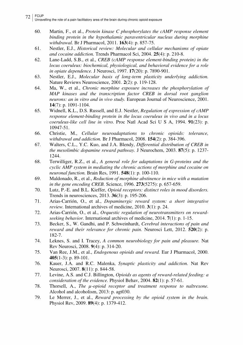
72 FCUP Unravelling the role of a pain facilitatory area of the brain during chronic opioid exposure
60. Martin, F., et al., Protein kinase C phosphorylates the cAMP response element
binding protein in the hypothalamic paraventricular nucleus during morphine
withdrawal. Br J Pharmacol, 2011. 163(4): p. 857-75.
61. Nestler, E.J., Historical review: Molecular and cellular mechanisms of opiate
and cocaine addiction. Trends Pharmacol Sci, 2004. 25(4): p. 210-8.
62. Lane-Ladd, S.B., et al., CREB (cAMP response element-binding protein) in the
locus coeruleus: biochemical, physiological, and behavioral evidence for a role
in opiate dependence. J Neurosci, 1997. 17(20): p. 7890-901.
63. Nestler, E.J., Molecular basis of long-term plasticity underlying addiction.
Nature Reviews Neuroscience, 2001. 2(2): p. 119-128.
64. Ma, W., et al., Chronic morphine exposure increases the phosphorylation of
MAP kinases and the transcription factor CREB in dorsal root ganglion
neurons: an in vitro and in vivo study. European Journal of Neuroscience, 2001.
14(7): p. 1091-1104.
65. Widnell, K.L., D.S. Russell, and E.J. Nestler, Regulation of expression of cAMP
response element-binding protein in the locus coeruleus in vivo and in a locus
coeruleus-like cell line in vitro. Proc Natl Acad Sci U S A, 1994. 91(23): p.
10947-51.
66. Christie, M., Cellular neuroadaptations to chronic opioids: tolerance,
withdrawal and addiction. Br J Pharmacol, 2008. 154(2): p. 384-396.
67. Walters, C.L., Y.C. Kuo, and J.A. Blendy, Differential distribution of CREB in
the mesolimbic dopamine reward pathway. J Neurochem, 2003. 87(5): p. 1237-
1244.
68. Terwilliger, R.Z., et al., A general role for adaptations in G-proteins and the
cyclic AMP system in mediating the chronic actions of morphine and cocaine on
neuronal function. Brain Res, 1991. 548(1): p. 100-110.
69. Maldonado, R., et al., Reduction of morphine abstinence in mice with a mutation
in the gene encoding CREB. Science, 1996. 273(5275): p. 657-659.
70. Lutz, P.-E. and B.L. Kieffer, Opioid receptors: distinct roles in mood disorders.
Trends in neurosciences, 2013. 36(3): p. 195-206.
71. Arias-Carrión, O., et al., Dopaminergic reward system: a short integrative
review. International archives of medicine, 2010. 3(1): p. 24.
72. Arias-Carrión, O., et al., Orquestic regulation of neurotransmitters on reward-
seeking behavior. International archives of medicine, 2014. 7(1): p. 1-15.
73. Becker, S., W. Gandhi, and P. Schweinhardt, Cerebral interactions of pain and
reward and their relevance for chronic pain. Neurosci Lett, 2012. 520(2): p.
182-7.
74. Leknes, S. and I. Tracey, A common neurobiology for pain and pleasure. Nat
Rev Neurosci, 2008. 9(4): p. 314-20.
75. Van Ree, J.M., et al., Endogenous opioids and reward. Eur J Pharmacol, 2000.
405(1-3): p. 89-101.
76. Kauer, J.A. and R.C. Malenka, Synaptic plasticity and addiction. Nat Rev
Neurosci, 2007. 8(11): p. 844-58.
77. Levine, A.S. and C.J. Billington, Opioids as agents of reward-related feeding: a
consideration of the evidence. Physiol Behav, 2004. 82(1): p. 57-61.
78. Thorsell, A., The µ-opioid receptor and treatment response to naltrexone.
Alcohol and alcoholism, 2013: p. agt030.
79. Le Merrer, J., et al., Reward processing by the opioid system in the brain.
Physiol Rev, 2009. 89(4): p. 1379-412.

FCUP Unravelling the role of a pain facilitatory area of the brain during chronic opioid exposure
73
80. Contet, C., B.L. Kieffer, and K. Befort, Mu opioid receptor: a gateway to drug
addiction. Curr Opin Neurobiol, 2004. 14(3): p. 370-8.
81. van Ree, J.M. and D. de Wied, Involvement of neurohypophyseal peptides in
drug-mediated adaptive responses. Pharmacol Biochem Behav, 1980. 13 Suppl
1: p. 257-63.
82. McBride, W.J., J.M. Murphy, and S. Ikemoto, Localization of brain
reinforcement mechanisms: intracranial self-administration and intracranial
place-conditioning studies. Behav Brain Res, 1999. 101(2): p. 129-52.
83. Van Ree, J.M., M.A. Gerrits, and L.J. Vanderschuren, Opioids, reward and
addiction: an encounter of biology, psychology, and medicine. Pharmacological
reviews, 1999. 51(2): p. 341-396.
84. Matthes, H.W., et al., Loss of morphine-induced analgesia, reward effect and
withdrawal symptoms in mice lacking the mu-opioid-receptor gene. Nature,
1996. 383(6603): p. 819-23.
85. Becker, A., et al., Morphine self-administration in µ-opioid receptor-deficient
mice. Naunyn-Schmiedeberg's archives of pharmacology, 2000. 361(6): p. 584-
589.
86. Self, D.W., et al., Involvement of cAMP-dependent protein kinase in the nucleus
accumbens in cocaine self-administration and relapse of cocaine-seeking
behavior. The Journal of neuroscience, 1998. 18(5): p. 1848-1859.
87. Huang, Y., et al., Development of viral vectors for gene therapy for chronic
pain. Pain Res Treat, 2011. 2011: p. 968218.
88. Goins, W.F., et al., Herpes simplex virus vector-mediated gene delivery for the
treatment of lower urinary tract pain. Gene Ther, 2009. 16(4): p. 558-69.
89. Howarth, J.L., Y.B. Lee, and J.B. Uney, Using viral vectors as gene transfer
tools (Cell Biology and Toxicology Special Issue: ETCS-UK 1 day meeting on
genetic manipulation of cells). Cell biology and toxicology, 2010. 26(1): p. 1-20.
90. Tavares, I. and D. Lima, From neuroanatomy to gene therapy: searching for
new ways to manipulate the supraspinal endogenous pain modulatory system. J
Anat, 2007. 211(2): p. 261-8.
91. Simonato, M., et al., Progress in gene therapy for neurological disorders. Nat
Rev Neurol, 2013. 9(5): p. 277-91.
92. Martins, I., et al., Reversal of inflammatory pain by HSV-1-mediated
overexpression of enkephalin in the caudal ventrolateral medulla. Eur J Pain,
2011. 15(10): p. 1008-14.
93. Molet, J. and M. Pohl, Gene-based approaches in pain research and exploration
of new therapeutic targets and strategies. European journal of pharmacology,
2013. 716(1): p. 129-141.
94. Jasmin, L., et al., Analgesia and hyperalgesia from GABA-mediated modulation
of the cerebral cortex. Nature, 2003. 424(6946): p. 316-320.
95. Decosterd, I. and C.J. Woolf, Spared nerve injury: an animal model of persistent
peripheral neuropathic pain. Pain, 2000. 87(2): p. 149-58.
96. Tzschentke, T.M., Measuring reward with the conditioned place preference
(CPP) paradigm: update of the last decade. Addict Biol, 2007. 12(3-4): p. 227-
462.
97. Snyder, B.R., N.M. Boulis, and T. Federici, Viral vector-mediated gene transfer
for CNS disease. Expert Opin Biol Ther, 2010. 10(3): p. 381-94.
98. Hao, S., et al., The role of TNFalpha in the periaqueductal gray during
naloxone-precipitated morphine withdrawal in rats. Neuropsychopharmacology,
2011. 36(3): p. 664-76.

74 FCUP Unravelling the role of a pain facilitatory area of the brain during chronic opioid exposure
99. Bonin, R.P., C. Bories, and Y. De Koninck, A simplified up-down method
(SUDO) for measuring mechanical nociception in rodents using von Frey
filaments. Mol Pain, 2014. 10: p. 26.
100. Tal, M. and G.J. Bennett, Extra-territorial pain in rats with a peripheral
mononeuropathy: mechano-hyperalgesia and mechano-allodynia in the territory
of an uninjured nerve. Pain, 1994. 57(3): p. 375-82.
101. Decosterd, I., et al., Intrathecal implants of bovine chromaffin cells alleviate
mechanical allodynia in a rat model of neuropathic pain. Pain, 1998. 76(1-2): p.
159-66.
102. Choi, Y., et al., Behavioral signs of ongoing pain and cold allodynia in a rat
model of neuropathic pain. Pain, 1994. 59(3): p. 369-76.
103. Prus, A.J., J.R. James, and J.A. Rosecrans, Conditioned Place Preference, in
Methods of Behavior Analysis in Neuroscience, J.J. Buccafusco, Editor 2009:
Boca Raton (FL).
104. Navratilova, E., et al., Evaluation of reward from pain relief. Ann N Y Acad Sci,
2013. 1282: p. 1-11.
105. Esmaeili, M.-H., et al., Transient inactivation of the nucleus accumbens reduces
both the expression and acquisition of morphine-induced conditioned place
preference in rats. Pharmacology Biochemistry and Behavior, 2012. 102(2): p.
249-256.
106. Sahraei, H., et al., GABAB receptors within the ventral tegmental area are
involved in the expression and acquisition of morphine-induced place preference
in morphine-sensitized rats. Pharmacol Biochem Behav, 2009. 91(3): p. 409-16.
107. Donovick, P.J., A metachromatic stain for neural tissue. Stain Technol, 1974.
49(1): p. 49-51.
108. Grilly, D.M., et al., Morphine dependence in rats assessed in a shock
discrimination task. Psychopharmacology (Berl), 1981. 74(3): p. 250-5.
109. Li, X., M.S. Angst, and J.D. Clark, A murine model of opioid-induced
hyperalgesia. Brain Res Mol Brain Res, 2001. 86(1-2): p. 56-62.
110. Mao, J., Opioid-induced abnormal pain sensitivity: implications in clinical
opioid therapy. Pain, 2002. 100(3): p. 213-7.
111. Harrison, C., D. Smart, and D.G. Lambert, Stimulatory effects of opioids. Br J
Anaesth, 1998. 81(1): p. 20-8.
112. Bilecki, W. and R. Przewlocki, Effect of opioids on Ca2+/cAMP responsive
element binding protein. Acta Neurobiol Exp (Wars), 2000. 60(4): p. 557-67.
113. Chakrabarti, S., A. Regec, and A.R. Gintzler, Chronic morphine acts via a
protein kinase Cgamma-G(beta)-adenylyl cyclase complex to augment
phosphorylation of G(beta) and G(betagamma) stimulatory adenylyl cyclase
signaling. Brain Res Mol Brain Res, 2005. 138(1): p. 94-103.
114. Shy, M., S. Chakrabarti, and A.R. Gintzler, Plasticity of adenylyl cyclase-related
signaling sequelae after long-term morphine treatment. Mol Pharmacol, 2008.
73(3): p. 868-79.
115. Chakrabarti, S., A. Chang, and A.R. Gintzler, Subcellular localization of mu-
opioid receptor G(s) signaling. J Pharmacol Exp Ther, 2010. 333(1): p. 193-200.
116. Cao, J.L., et al., Essential role of the cAMP-cAMP response-element binding
protein pathway in opiate-induced homeostatic adaptations of locus coeruleus
neurons. Proc Natl Acad Sci U S A, 2010. 107(39): p. 17011-6.
117. Williams, J.T., et al., Regulation of mu-opioid receptors: desensitization,
phosphorylation, internalization, and tolerance. Pharmacol Rev, 2013. 65(1): p.
223-54.

FCUP Unravelling the role of a pain facilitatory area of the brain during chronic opioid exposure
75
118. Bianchi, E., et al., Supraspinal Gbetagamma-dependent stimulation of PLCbeta
originating from G inhibitory protein-mu opioid receptor-coupling is necessary
for morphine induced acute hyperalgesia. J Neurochem, 2009. 111(1): p. 171-
80.
119. Wang, H.Y. and L.H. Burns, G that interacts with adenylyl cyclase in opioid tolerance originates from a Gs protein. Journal of neurobiology, 2006. 66(12):
p. 1302-1310.
120. Niikura, K., et al., Neuropathic and chronic pain stimuli downregulate central
mu-opioid and dopaminergic transmission. Trends Pharmacol Sci, 2010. 31(7):
p. 299-305.
121. Smith, H.S., Opioids and neuropathic pain. Pain Physician, 2012. 15(3 Suppl):
p. ES93-110.
122. Porreca, F., M.H. Ossipov, and G.F. Gebhart, Chronic pain and medullary
descending facilitation. Trends Neurosci, 2002. 25(6): p. 319-25.
123. Baliki, M.N., et al., Predicting value of pain and analgesia: nucleus accumbens
response to noxious stimuli changes in the presence of chronic pain. Neuron,
2010. 66(1): p. 149-60.
124. Edelmayer, R.M., et al., Medullary pain facilitating neurons mediate allodynia
in headache-related pain. Ann Neurol, 2009. 65(2): p. 184-93.
125. Okun, A., et al., Transient inflammation-induced ongoing pain is driven by
TRPV1 sensitive afferents. Mol Pain, 2011. 7(1): p. 4.
126. De Felice, M., et al., Capturing the aversive state of cephalic pain preclinically.
Ann Neurol, 2013. 74(2): p. 257-65.
127. Qu, C., et al., Lesion of the rostral anterior cingulate cortex eliminates the
aversiveness of spontaneous neuropathic pain following partial or complete
axotomy. Pain, 2011. 152(7): p. 1641-8.
128. Wang, R., et al., Descending facilitation maintains long-term spontaneous
neuropathic pain. J Pain, 2013. 14(8): p. 845-53.
129. Rozisky, J.R., et al., Morphine treatment in neonate rats increases exploratory
activities: reversal by antagonist D2 receptor. British Journal of Medicine and
Medical Research, 2014. 4(1): p. 351-367.
130. Almeida, A., et al., Brain afferents to the medullary dorsal reticular nucleus: a
retrograde and anterograde tracing study in the rat. Eur J Neurosci, 2002.
16(1): p. 81-95.


FCUP Unravelling the role of a pain facilitatory area of the brain during chronic opioid exposure
a
Appendix A: Composition of solutions
1. PHOSPHATE BUFFER SALINE (PBS) (1L)
Phosphate buffer (PB) 0,1M pH=7,2:
Na2 H2PO4H2O – 15,60g
K2HPO4 – 17,4g
H2O up to 1L
PBS:
PB 250 ml
H2O up to 1L
NaCl - 9g
2. PHOSPHATE BUFFER SALINE WITH TRITON X-100 (PBS-T)
PBS - 996ml
Triton X-100 - 4ml
3. TYRODE’S SOLUTION (1L)
NaCl – 6,8 g
KCl – 0,40g
MgCl 2 6 H2O – 0,32 g
MgSO 4 7 H2O – 0,1 g
NaH 2 PO4 H2O – 0,17 g
Glucose 1 g
NaHCO 3 – 2,2 g
H2O up to 1L
4. CRYOPROTECTOR SOLUTION (1L)
PB 0,1M pH=7,2 - 125 ml

b FCUP Unravelling the role of a pain facilitatory area of the brain during chronic opioid exposure
H2O - 375 ml
Sucrose - 300g
Ethylene glycol - 300ml
PB 0,1M pH=7,2 up to 1L
5. THIONIN STAINING
1. Solutions :
Acid acetone: acetone / acetic acid (4 vol / 1 vol)
0,1% thionin in 10% formalin
2. Protocol :
Incubate the slides in acid acetone for 5 min;
Rinse with distilled water;
Stain in formol-thionin for 1 minute; Rinse with distilled water;
Dry at 37 ° C;
Dehydrate in xylene for 5 min;
Mount with Eukitt.