Embed Size (px)
Citation preview

UNIVERSITY OF KWAZULU-NATAL
Hypoglycaemic And Renal Effects Of A Bioactive Plant Extract In Streptozotocin
Induced Diabetic Rats.
RUDO FIONA MAPANGA
2008

Hypoglycaemic and renal effects of a bioactive plant
extract in streptozotocin induced diabetic rats
BY
Rudo Fiona Mapanga (207521118)
Submitted in fulfilment of the requirements for the degree of Master of Medical Science in
Human Physiology in the Discipline of Human Physiology, School of Medical Sciences,
Faculty of Health Sciences
Supervisor: Professor C.T. Musabayane Discipline of Human Physiology Faculty of Health Sciences
t • J UNIVERSITY OF ?* KWAZULU-NATAl
DECLARATION
I, Rudo Fiona Mapanga, hereby declare that the dissertation entitled
"Hypoglycaemic and renal effects of a bioactive plant extract in streptozotocin-induced
diabetic rats"
is the result of my own investigation and research and that it has not been submitted in part or
in full for any other degree or to any other university. Where use was made of the work of
others, it is duly acknowledged in the text.
Student: Rudo F. Mapanga Signature
Supervisor: Professor C.T. Musabayane
G96837
Su
11

ACKNOWLEDGEMENTS
First and foremost I thank God Almighty for granting me such an opportunity in the
advancement of my academic foundation. I also want to thank the following people whose
input made this work achievable:
I owe much to my supervisor and mentor, Professor C.T. Musabayane. It is a great
blessing having him as a supervisor, for 1 have received constant and committed
guidance, constructive criticisms and financial support for all the requirements of this
research all the way through. I truly appreciate all that I learnt from him;
I am very grateful to Professor F.O. Shode, for the supervision, advice and
encouragement during the phytochemical studies in his laboratory and Professor H.
Baijnath, for identifying and supplying the plant material used in this study;
Mark Tufts and Dr M Zungu, I thank you for the guidance, mentorship and positive
criticisms throughout the course of this study.
I wish to acknowledge the technical assistance rendered to me by staff members of
the Biomedical Resource Unit, especially David, Linda, Dennis and Dr Singh;
I pay my sincere thanks to my family, especially my mother, and also my colleagues
for the encouragement, love, co-operation and moral support throughout the course of
this project;
Last but not least I would like to thank the National Research Foundation (NRF) for
granting me the funds to pursue my studies.
iii

LIST OF ABBREVIATIONS
a
AGE
AMPK
ANOVA
ATP
P Ca2+
cAMP
DAG
DCCT
DEPT
DMSO
EDHF
eNOS
ESRD
ET-1
ETRA
GADPH
GBM
GFR
GFAT
GLP
GLUT-1
GLUT-2
GLUT-3
GLUT-4
GLUT-6
GLUT-8
GLUT-10
Alpha
Advanced glycation end-product
Adenosine monophosphate protein kinase
One way analysis of variance
Adenosine triphosphate
Beta
Calcium
Cyclic adenosine monophosphate
Diacylglycerol
Diabetes Control and Complications Trial
Distortion Enhancement Proton Testing
Dimethyl sulphoxide
Endothelium derived hyperpolarizing factor
endothelium nitric oxide synthase
End-stage renal disease
Endothelin-1
Endothelin receptor A
Glyceraldehyde-3 -phosphate dehydrogenase
Glomerular basement membrane
Glomerular filtration rate
Glutamine:fructose-6-phosphate amidotransferase
Glucagon like peptide
Glucose transporter-1
Glucose transporter-2
Glucose transporter-3
Glucose transporter-4
Glucose transporter-6
Glucose transporter-8
Glucose transporter-10
iv
GLUT-12
GOx
GSH
h
HbAlc
HBP
HDL
HC1
HMQC
H2S04
ICAM-1
IR
K+
KATP
Kg
LTD
MAP
1
LDL
M
MAPK
MHz
mg
mmHg
mmol
NaCl
NAD+
NADH
Glucose transporter-12
Glucose oxidase
Glutathione
Hour
Glycosylated haemoglobin
Hexosamine biosynthetic pathway
High density lipoprotein
Hydrochloric acid
Heteronuclear multiple quantum coherence
Sulphuric acid
Intracellular cell adhesion molecule-1
Insulin receptor
Potassium
Adenosine-5'- triphosphate sensitive potassium channels
kilogram
limited
Mean arterial pressure
litre
Low density lipoprotein
Molar
Mitogen activated protein kinase
millihertz
micro
microgram
microlitre
milligram
Millimetres of mercury
millimole
Sodium chloride
Oxidised nicotinamide dinucleotide
Nicotinamide adenine dinucleotide hydrogen
v

NADPH
NEI
NF-Kp
NMR
NO
NPDR
OA
OGTT
oxLDL
PAI-1
PI-3-K
PKC
PKC-a
PKC-p
PKC-5
PKC-s
PKC-y
PKC-^
pmol
p.o.
PPAR-a
PPAR-Y
ppm
RAGE
sc
SEM
SGLT
SGLT-1
SGLT-2
STZ
SU
Nicotinamide adenine dinucleotide phosphate hydrogen
National Eye Institute
Nuclear factor-kappa beta
Nuclear Magnetic Resonance
Nitric oxide
Non-proliferative diabetic retinopathy
oleanolic acid
Oral glucose tolerance test
Oxidized low density lipoprotein
Plasminogen activator inhibitor-1
Phosphatidyl inositol-3-kinase
Protein kinase C
Protein kinase C-alpha
Protein kinase C-beta
Protein kinase C-delta
Protein kinase C-epsilon
Protein kinase C-gamma
Protein kinase-xi
picomole
per os (by mouth, orally)
Peroxisome proliferator alpha
Peroxisome proliferator gamma
parts per million
Receptor for advanced glycation end product
subcutaneous
Standard error of means
Sodium glucose transporter
Sodium glucose transporter-1
Sodium glucose transporter-2
Streptozotocin
Sulphonylurea
VI

SUR-1
SUR-2
TGF-p
TMB
TNF-a
TZD
UA
UDP
UDP-GlycNac
UK
UKPDS
USA
UKZN
VCAM-1
VEGF
VLDL
WHO
Sulphonylurea receptor-1
Sulphonylurea receptor-2
Transforming growth factor beta
3,3',5,5' Tetramethylbenzidine
Tumour necrosis factor alpha
Thiazolidinediones
Ursolic acid
Uridine diphosphate
Uridine diphosphate acetylglucosamine
United Kingdom
United Kingdom Prospective Diabetes Study
United States of America
University of KwaZulu-Natal
Vascular cell adhesion molecule-1
Vascular endothelial growth factor
Very low density lipoprotein
World Health Organisation
vii

TABLE OF CONTENTS
Page no.
Declaration ii
Acknowledgements iii
Abbreviations iv
Table of contents viii
List of Tables xii
List of Figures xiii
List of Appendices xv
Abstract xvii
CHAPTER 1- Introduction/ Literature review 1
1.1. Background 1
1.2. Diabetes mellitus 2
1.2.1. Glucose transport 2
1.3. Aetiology of diabetic complications 3
1.3.1. Polyol pathway 4
1.3.2. Advanced glycosylation end products (AGE's) 5
1.3.3. Protein kinase C 6
1.3.4. The hexosamine pathway 7
1.4. Diabetic complications 8
1.4.1. Macro vascular complications 8
1.4.1.1. Arterial diseases 9
1.4.1.2. Atherosclerosis 9
1.4.2. Microvascular complications 11
1.4.2.1. Diabetic neuropathy 11
1.4.2.2. Diabetic retinopathy 13
1.4.2.3. Diabetic nephropathy 14
1.5. Diabetes management 17
1.5.1. Insulin 18
1.5.1.1. Insulin like medicinal plants 19
viii

1.5.2. Insulin sensitizers 19
1.5.2.1. Biguanides 19
1.5.2.2. Plant insulin sensitizers 21
1.5.2.3. Thiazolidinediones 21
1.5.2.4. Plant PPAR agonists 22
1.5.3. Insulin secretagogues 22
1.5.3.1. Sulphonylureas 22
1.5.3.2. Meglitinides 23
1.5.3.3. Plant insulin secretagogues 23
1.5.4. a-Glucosidase inhibitors 23
1.5.4.1. a-Glucosidase inhibiting plants 24
1.6. Traditional (indigenous/folk) medicine 24
1.6.1. Plant based management of diabetes mellitus 25
1.6.2. Syzygium cordatum (Hochst.) [Myrtaceae] 26
1.6.3. Triterpenoids 27
1.7. Basis of the present study 29
1.7.1. Hypothesis 29
1.7.2. Aims 29
CHAPTER 2- Materials and methods 30
2.1. Ethical consideration 30
2.2. Materials 30
2.2.1 Drugs and chemicals 30
2.3. Plant material 30
2.4. Bio-assay directed isolation of OA 31
2.4.1. Thin layer chromatography 31
2.4.2. Isolation of OA 31
2.4.3. OA structure elucidation 32
2.5. Animals 32
2.5.1. Induction of diabetes mellitus 32
2.6. Experimental design 33
ix

2.7. Acute studies
2.7.1. OGTT protocol
2.7.1.1. Effects of acute OA treatment on insulin
2.7.2. Acute effects of OA on renal function
2.7.2.1. Acute effects of OA on blood pressure
2.8. Short-term studies
2.8.1. Physico-metabolic changes
2.8.2. Short-term OA effects on MAP
2.8.3. Terminal studies
2.9. Laboratory analyses
2.9.1. Biochemical measurements
2.9.2. Insulin assay
2.9.3. Glycogen measurements
2.9.4. Data presentation
CHAPTER 3- Results
3.1. Structural elucidation of OA
3.2. OGTT responses
3.2.1. OGTT responses to standard anti-diabetic drugs
3.2.1.1. Non-diabetic rats
3.2.1.2. STZ-induced diabetic rats
3.2.2. OGTT responses to OA
3.2.2.1. Non-diabetic rats
3.2.2.2. STZ-induced diabetic rats
3.2.3. OA effects on insulin secretion
3.2.3.1. Non-diabetic rats
3.2.3.2. STZ-induced diabetic rats
3.3. Acute renal effects of OA
3.3.1. Non-diabetic rats
3.3.2. STZ-induced diabetic rats
x

3.4. Short-term effects of OA on body weight, food and
water intake 60
3.5. Short-term renal effects of OA 63
3.5.1. Non-diabetic rats 63
3.5.2. STZ-induced diabetic rats 63
3.5.3. Haemodynamic changes 64
3.5.4. Summary of the Short-term effects of OA 64
3.5.5. Effects of OA on terminal blood glucose and
plasma concentrations 69
3.5.5.1. Effects of OA on hepatic and
muscle glycogen concentrations 69
3.6. Overall summary of OA effects on blood glucose and kidney function 70
CHAPTER 4- Discussion 74
4.1. General 74
4.1. Structural elucidation of OA 74
4.3. Hypoglycaemic effects 75
4.4. Renal effects 78
CHAPTER 5- Conclusions 82
5.1. Shortfalls of the study 82
5.2. Recommendations for future studies 83
CHAPTER 6- References 84
APPENDICES 113
XI

LIST OF TABLES
Table number
Page no.
Table 1: 13C NMR spectral data of plant derived OA and reported OA 47
Table 2: Comparison of total amounts of urine volume, Na+, K+ and CI"
excreted during the treatment period by anaesthetized OA-treated
non-diabetic and STZ-induced diabetic rats 60
Table 3: Comparison of the effects of 5-week treatment with OA and/or
standard anti-diabetic drugs of non-diabetic (ND) and STZ-induced
diabetic animals on food intake, water intake and body weight changes 62
Table 4: Plasma biochemical parameters of control non-diabetic and STZ-diabetic
rats administered OA 67
Table 5: Comparison of the effects of 5-week treatment with OA alone and/or
standard anti-diabetic drugs on hepatic and muscle glycogen
concentrations in non-diabetic and STZ-induced diabetic rats 73
xn

LIST OF FIGURES
Page no.
Figure 1: 5". cordatum tree and leaves 28
Figure 2: Layout of the Harvard Apparatus infusion pump and Power lab
transducer systems 37
Figure 3: Layout of the IITC Model 303sc Animal Test Chamber apparatus 40
Figure 4: Syzygium cordatum leaf derived OA *H and 13C- NMR
spectra (ID and 2D) 46
Figure 5: Structure and numbering of oleanolic acid (IUPAC) 48
Figure 6: OGTT responses to standard anti-diabetic drugs of non-diabetic rats
and STZ-induced diabetic rats 52 Figure 7: OGTT responses to various OA doses of non-diabetic rats and
STZ-induced diabetic rats 53
Figure 8: OGTT responses to OA (40 mg/kg) alone and in combination with
standard anti-diabetic drugs of non-diabetic rats and STZ-induced
diabetic rats 54
Figure 9: OGTT responses to OA (80 mg/kg,) alone and in combination with
various standard anti-diabetic drugs of non-diabetic rats and
STZ-induced diabetic rats 55
Figure 10: Effects of OA and standard anti-diabetic drugs on plasma insulin and
glucose concentrations of non-diabetic and STZ-induced diabetic rats 56
xiii

Figure 11: Acute effects of 1V* h OA infusion on Na+, K+ and CI" excretion and
urine flow rates in anaesthetized non-diabetic rats 58
Figure 12: Acute effects of 1V2 h OA infusion on Na+, K+ and CI" excretion and
urine flow rates in anaesthetized STZ-induced diabetic rats 59
Figure 13: Short-term OA effects on 24 h Na+, K+, CI" excretion and urine flow
rates in non-diabetic rats 65
Figure 14: Short-term OA effects on 24 h Na+, K+ and CI"excretion and urine flow
rates in STZ-induced diabetic rats 66
Figure 15: Acute effects of OA infusion on MAP in non-diabetic and
STZ-induced diabetic rats and short-term administration in non-diabetic
and diabetic rats 68
Figure 16: Short-term effects of OA and standard hypoglycaemic drugs on
blood glucose and plasma insulin concentrations in non-diabetic and
STZ-induced diabetic rats 71
Figure 17: Short-term effects of OA on 24 h urinary glucose outputs of
STZ-induced diabetic rats 72
xiv

LIST OF APPENDICES
Page No.
APPENDIX I: Ethical clearance A 113
APPENDIX II: Ethical clearance B 114
APPENDIX III: Paper submitted for publication 115
Mapanga RF, Tufts MA, Shode FO, Musabayane CT. Renal effects of plant-derived
oleanolic acid in streptozotocin-induced diabetic rats. (Submitted to Renal Failure, USA
MS08168)
APPENDIX IV: Conference presentations
(a) RF Mapanga, MA Tufts, 0 0 Oyedeji, FO Shode, CT Musabayane. Plant derived
oleanolic acid augments the hypoglycaemic effects of exogenous insulin in
streptozotocin-induced diabetic rats. Physiological Society of Southern Africa, 36th
Congress, 16-19 September 2008. 139
(b) RF Mapanga, MA Tufts, 0 0 Oyedeji, FO Shode, CT Musabayane. Renal effects of
plant derived oleanolic acid in Sprague Dawley rats. University of Zimbabwe, Annual
Medical Research Day, 25 October, 2008. 140
(c) CT Musabayane, RF Mapanga, MA Tufts, OO Oyedeji, FO Shode. Plant derived
oleanolic acid augments the hypoglycaemic effects of exogenous insulin in
streptozotocin-induced diabetic rats. Submitted to Society of Endocrinology BES
2009, Harrogate Meeting. (Abstract number 0009). 141
(d) RF Mapanga, MA Tufts, OO Oyedeji, FO Shode, CT Musabayane. Renal effects of
plant derived oleanolic acid in Sprague Dawley rats. Submitted to Society of
Endocrinology BES, Harrogate Meeting 2009. (Abstract number 0050). 142
xv

ABSTRACT
Background
Evidence from our laboratories indicates that triterpene constituents of Syzygium cordatum
(Hochst.) [Myrtaceae] crude leaf extracts can be used to treat diabetes mellitus. For the plant
derived triterpenes to have further potential in diabetes management, they should, however,
additionally alleviate or prevent some of the complications of diabetes mellitus such as
impaired kidney function and cardiovascular disorders. Accordingly, this study was designed
to isolate the triterpene, oleanolic acid (OA) from S. cordatum leaves and evaluate its effects
on blood glucose, renal function and blood pressure in streptozotocin (STZ)-induced diabetic
rats. OA was studied because it is the major constituent of many African plant species used
in traditional medicine.
Materials and Methods
S. cordatum crude leaf ethyl acetate solubles (EAS) were obtained after defatting the leaves
with hexane followed by dichloromethane before maceration with ethyl acetate. Preliminary
experiments indicated that EAS contained triterpenes with hypoglycaemic properties. Solvent
extraction and fractionation of EAS yielded mixtures of oleanolic acid/ursolic acid (OA/UA)
and methyl maslinate/methyl corosolate. Recrystallisation of the OA/UA mixture using
ethanol yielded OA, the structure of which was confirmed by NMR spectroscopy ('H & 13C).
Oral glucose tolerance test (OGTT) responses to various doses of OA (40, 80 and 120 mg/kg)
were monitored in separate groups of non-diabetic and STZ-induced diabetic rats given a
glucose load (0.86 g/kg, p.o.) after an 18-h fast. Rats treated with deionized water (3 ml/kg
p.o.), or standard drugs, (insulin, 200 Hg/kg, s.c; metformin, 500 mg/kg, p.o. and
glibenclamide, 500 f̂ g/kg, p.o.) acted as untreated and treated positive controls, respectively.
To investigate the possible interaction between OA and standard drugs in lowering blood
glucose, OGTT responses were studied in separate groups of animals simultaneously treated
xvi

with OA at either 40 or 80 mg/kg and insulin (100 or 200 ug/kg, s.c), metformin, (250 or
500 mg/kg, p.o.) or glibenclamide (250 or 500 mg/kg, p.o.). Blood glucose was monitored at
15-min intervals for the first hour, and hourly thereafter for 3 h. Plasma insulin
concentrations were measured in separate parallel groups of rats prepared as for OGTT
studies to examine whether there was an association between OA treatment and pancreatic
insulin secretion. Acute effects of OA on kidney function and mean arterial blood pressure
(MAP) were investigated in anaesthetized rats challenged with hypotonic saline after a 3'/2-h
equilibration for 4 h consisting of 1 h control, 1XA h treatment and 1 Vi h recovery periods. OA
was added to the infusate during the treatment period. Short-term effects of OA were studied
in individually-caged rats treated twice daily with OA (80 mg/kg, p.o.) for 5 weeks.
Results
OA decreased blood glucose concentrations of both non-diabetic and diabetic rats, as did
some standard drugs except glibenclamide which did not exhibit any effects in STZ-induced
diabetic animals. The blood glucose lowering effects were most potent in STZ-induced rats
treated with combined OA and insulin by comparison with all other treatments. Short-term
treatment of non-diabetic and STZ-induced diabetic rats with OA alone for 5 weeks
decreased blood glucose concentrations, but the reduction in non-diabetic rats was to values
that did not achieve statistical significance. Except for non-diabetic rats treated with insulin
alone or in combination with OA, plasma insulin concentrations were not altered by
treatment in non-diabetic and STZ-induced diabetic animals. Hepatic glycogen
concentrations of non-diabetic and STZ-induced diabetic rats were significantly increased by
all treatments at the end of 5 weeks.
Acute intravenous infusion of OA in anaesthetized rats significantly increased Na+ excretion
outputs of non-diabetic and STZ-induced diabetic rats without affecting urine flow, K+ or CI"
excretion rates. Similarly, daily OA treatment (80 mg/kg, p.o.) significantly increased Na+
excretion rates of non-diabetic and STZ-induced diabetic rats throughout the 5 week
experimental period without affecting urine flow, K+ or CI" excretion rates. By comparison
with respective control animals, Short-term administrations of OA significantly (p<0.05)
xvii

increased GFR of non-diabetic (2.88±0.14 vs 3.71±0.30 ml/min) and STZ-diabetic rats
(1.81± 0.32 vs 3.07±0.16 ml/min, n=6 in all groups) with concomitant reduction of plasma
creatinine concentrations. Acute and Short-term administrations of OA non-diabetic and STZ-
induced diabetic rats reduced mean arterial blood pressure by comparison with respective
control animals.
Discussion
The results suggest that S. cordatum leaf derived OA not only has the potential to lower
blood glucose in diabetes, but also has beneficial effects on kidney function and blood
pressure. We suggest that the hypoglycaemic effects of OA mimic those of metformin as
evidenced by the fact that neither of these treatments altered plasma insulin concentration of
non-diabetic rats. OA-evoked increases in urinary Na+ outputs of STZ-diabetic rats and
elevation of GFR suggest up-regulation of renal function by the triterpene. The findings are
of considerable importance because they suggest the hypoglycaemic, renal and hypotensive
effects of OA in the management of diabetes mellitus.
Conclusion
The results demonstrated that the oleanolic acid extracted from S. cordatum leaf has blood
glucose-lowering effects comparable to standard anti-diabetic drugs in STZ-induced diabetic
rats. Furthermore, OA augmented the hypoglycaemic effects of insulin in STZ-induced
diabetic rats. These findings suggest that OA may have beneficial effects on some of the
processes that are associated with renal derangement in STZ-induced diabetic rats. The
results introduce the first in vivo evidence that OA ameliorates kidney function in STZ-
induced diabetic rats.
Keywords: Renal function; diabetes mellitus; triterpenoids; oleanolic acid, hypoglycaemia
xviii

CHAPTER 1
1.0. INTRODUCTION/ LITERATURE REVIEW
1.1. Background
Diabetes mellitus is characterized by hyperglycaemia because glucose transport across the
cells is impaired. The sustained hyperglycaemia leads to compromised kidney function and
the development of microvascular and macrovascular complications through different
mechanisms. Hence the goal in diabetes management is not only to maintain optimal blood
glucose control, but also to avert these adverse outcomes. Current conventional diabetes
therapy using blood glucose-lowering medications such as insulin or oral hypoglycaemic
agents has limitations. For instance insulin therapy does not achieve glycaemic control in
patients with insulin resistance, and oral hypoglycaemic agents may lose their efficacy after
prolonged use. Alternative methods of lowering blood glucose are, therefore, needed. Herbal
medicines have been used for many years by different cultures around the world, both for the
prevention and management of diabetes (Kim, Kang, Park, Jung, Choi and Ku, 2007b).
Previously the crude leaf extract of Syzygium cordatum (Hochst.) [Myrtaceae] has been
shown to reduce blood glucose levels in streptozotocin (STZ)-induced diabetic rats perhaps
due to its triterpene constituents. The study described in this dissertation was designed to
isolate and investigate the blood glucose lowering and renal function effects of one of S.
cordatum leaf derived triterpene constituents, oleanolic acid (3|3-hydroxy-olea-12-en-28-oic
acid, OA) in STZ-induced diabetic rats. The effects of OA were investigated because it is the
main triterpene that has been isolated from many medicinal plants, such as Momordica
charantia (Linnaeus) [Curcubitaceae] (Kumar, Shetty and Salimath, 2008), Olea europaea
(Linnaeus) [Oleaceae] (Chen, Liu, Zhang, Wu, Hua, Wu and Sun, 2006). The effects of OA
on kidney function were also investigated to establish whether OA has beneficial effects on
renal function in diabetes mellitus management.
1

1.2. Diabetes mellitus
Diabetes mellitus is a chronic metabolic disorder due to acquired or inherited causes leading
to absolute or relative deficiency of insulin or due to insulin resistance (Evcimen and King,
2007). Diabetes can be classified into three main types: type 1, type 2 and gestational
diabetes. Type 1 is due to autoimmune destructive lesions of P-cells of the pancreas leading
to an absolute lack of insulin secretion. In type 2 which accounts for 90-95% of diabetic
patients, there is a combination of decreased insulin secretion and insulin sensitivity (The
Committee of the Japan Diabetes Society on the diagnostic criteria of diabetes mellitus,
2002; Jung, Park, Lee, Kang, Kang and Kim, 2006; World Health Organisation (WHO),
2006). The third type, gestational diabetes is defined as the first onset of diabetes mellitus in
women during pregnancy precipitated by an excess production of glucocorticoids (American
Diabetes Association, 2004).
According to WHO (2006), 180 million people have diabetes worldwide. Estimates are that
by 2030 the number will more than double to about 366 million, making it an epidemic
(Stovring, Andersen, Beck-Nielsen, Green and Vach, 2007). Chronic hyperglycaemia due to
carbohydrate, lipid and protein metabolism disorders in diabetes mellitus leads to the
development of macrovascular and microvascular complications (Rahimi, Nikfar, Larijani,
Abdollahi, 2005). The hyperglycaemia occurs from alterations in the glucose homeostatic
systems, resulting from impaired glucose transport across cells. The various ways through
which glucose is transported are briefly described below.
1.2.1. Glucose transport
Glucose is transported across cell membranes through two main carrier proteins, sodium-
glucose co-transporters (SGLT's) and glucose transporters (GLUT). The two main isoforms
of SGLT, SGLT-1 and SGLT-2 are mostly expressed in the intestine and kidney, respectively
(Barnard and Youngren, 1992; Kakuda and MacLeod, 1994; Bouche, Serdy, Kahn and
Goldfine, 2004). They utilize the electrochemical sodium gradient to transport glucose
against a concentration gradient. Inhibition of glucose uptake by the SGLT's using phlorizin
2

is one of the strategies used in glycaemic control (Panayotova-Heiermann, Loo, Kong, Lever
and Wright, 1996; Link and Sorensen, 2000). On the other hand, GLUT transporters are a
family of 13 hexose transporters that passively transport monosaccharides, including glucose,
down a concentration gradient. According to Bouche, Serdy, Kahn and Goldfine, (2004),
these can be classified as high affinity (GLUT-1, 3, 4, 6, 8, 10 and 12) and low affinity
(GLUT-2). GLUT-2 is expressed in pancreatic P-cells, and tissues with high glucose fluxes
like intestine, liver and kidney. GLUT-1 is expressed mostly in erythrocytes, kidney, colon
and cells of blood-tissue barriers like the blood brain barrier and at low levels in liver,
adipose tissues and muscles (Bouche, Serdy, Kahn and Goldfine, 2004; Wiernsperger, 2005).
GLUT-1 and GLUT-3 are the primary GLUT transporters in foetal and neuronal tissues,
respectively (Bouche, Serdy, Kahn and Goldfine, 2004). GLUT-4 mediates insulin-
stimulated glucose uptake by skeletal muscles, heart, and white and brown adipose tissues.
Insulin and exercise stimulate translocation of the GLUT-4 to the cell membrane from an
intracellular pool (Barnard and Youngren, 1992; Bouche, Serdy, Kahn and Goldfine, 2004;
Wiernsperger, 2005). In contrast, the GLUT-1 is expressed on the cell membrane in the
absence of insulin. The activity of the GLUT transporters can, therefore, be insulin or non-
insulin mediated, with GLUT-1 contributing to about 75% of the glucose transport. In type 2
diabetes mellitus, there are defects in GLUT-4 translocation due to insulin resistance and
GLUT-1 is over-expressed (Miura, Itoh, Kaneko, Ueda, Ishida, Fukushima, Matsuyama and
Seino, 2004; Wiernsperger, 2005). The high glucose influx in diabetes mellitus mediated via
GLUT-1 transporters increases intracellular glucose metabolism leading to the development
of complications.
1.3. Aetiology of diabetic complications
The metabolic pathways involved in the aetiology of diabetic complications are discussed in
the following sections.
3

1.3.1. Polyol pathway
Aldose reductase, the first enzyme in the polyol pathway, catalyzes the nicotinamide adenine
dinucleotide phosphate (NADPH)-dependent reduction of glucose to sorbitol which is
oxidized to fructose by sorbitol dehydrogenase, an enzyme that uses NAD+ as a cofactor
(Brownlee, 2001; Sheetz and King, 2002; Lorenzi, 2007). This pathway is activated in
diabetes mellitus in non-insulin sensitive tissues including the lense, peripheral nerve and the
glomerulus (Brownlee, 2001; Sheetz and King, 2002; Lorenzi, 2007). Studies suggest that
sorbitol evokes osmotic vascular damage and cataracts in the eyes (Sheetz and King, 2002;
Lorenzi, 2007). The involvement of sorbitol in osmotic vascular damage, however, is often
difficult to rationalize (Brownlee, 2001; Sheetz and King, 2002). Additionally, diabetic
complications arise from advanced glycosylating agents formed by the breakdown of
fructose (Brownlee, 2001; Lorenzi, 2007). Development of diabetic complications also
involves decreasing the activity of enzymes that use NADPH/NADH as a cofactor. The
enzymes include glutathione reductase which would cause redox imbalances due to depletion
of reduced glutathione levels (Brownlee, 2001; Sheetz and King, 2002; Vincent, Russel, Low
and Feldman, 2004; Lorenzi, 2007). Endothelial generation of nitric oxide is also impaired by
the decreased NADPH levels. In addition there is inhibition of glyceraldehyde-3-phosphate
dehydrogenase (GADPH), leading to an increase in triose phosphate. This increases the
formation of methylglyoxal, a precursor of advanced glycation end products (AGE's) and
diacylglycerol (DAG), which activates protein kinase C (PKC) (Brownlee, 2001). Despite the
osmotic damage, studies suggest that oxidative stress is the main cause of cellular damage by
the polyol pathway (Lorenzi, 2007).
Inhibition of aldose reductase by various synthetic drugs such as zenarestat and sorbinil and
anti-diabetic plants has been effective in preventing and alleviating complications associated
with diabetes mellitus (Brownlee, 2001; Lorenzi, 2007). Plant extracts of Stelechocarpus
cauliflorous [Annonaceae] and its glycoside derivatives engeletin and astilbin have been
reported to inhibit aldose reductase (Wirasathiena, Pengsuparpa, Suttisria, Uedab, Moriyasub
and Kawanishib, 2007). Reno-protective effects were seen with berberine, one of the main
constituents of Coptis chinensis (Franch) [Ranunculaceae] and Phellodendron amurense
4
(Ruprecht) [Rutaceae], and this was associated with a concomitant inhibition of oxidative
stress (Wei-ha, Zi-qing, Hong, Fu-tian, He-qing, Xue-juan, Yan-hui, Shao-rui, Fen-fen, Wen-
ge, Feng-ying and Pei-qing, 2008). Other isolated bioactive compounds from plants that
inhibit aldose reductase activity include quercetin, silymarin, fiavonoids and puerarin
(Adaramoye and Adeyemi, 2005; Li, Dai, Yu, Li, Wu, Luan, Meng, Zhang and Deng, 2007).
Reduced activity of aldose reductase prevents production of sorbitol and its downstream
stimulation of the formation of AGE's and PKC pathways which are described in the next
sections.
1.3.2. Advanced glycosylation end products (AGE's)
The formation and accumulation AGE's from dicarbonyl compounds is accelerated in
diabetes. Major sources of the carbonyl group in the glycation reaction include glucose and
carbonyl compounds, such as glyceraldehyde (Sakurai, Yonekura, Yamamoto, Watanabe,
Tanaka, Li, Rahman, Myint, Kim and Yamamoto, 2003). The dicarbonyl compounds react
irreversibly with amino groups of intracellular and extracellular proteins in the Maillard
reaction to form AGE's (Brownlee, 2001; Sheetz and King, 2002; Sakurai et al., 2003;
Yonekura, Yamamoto, Sakurai, Watanabe, and Yamamoto, 2005; Thallas-Bonke, Thorpe,
Coughlan, Fukami, Yap, Sourris, Penfold, Bach, Cooper and Forbes 2008). The structure and
function of proteins, such as serum albumin, lens crystallin, intracellular proteins and
collagen in the extra cellular matrix is altered by advanced glycation end products (AGE's)
(Brownlee, 2001; Sheetz and King, 2002; Sakurai et al, 2003; DeGroot, 2004). Altered cell
function by AGE's occurs through the receptor for AGE's (RAGE) on endothelial cells,
mesangial cells and macrophages (Brownlee, 2001; Sheetz and King, 2002; DeGroot, 2004;
Yonekura, Yamamoto, Sakurai, Watanabe, and Yamamoto, 2005). Activation of the receptor
has been reported to induce production of reactive oxygen species through activation of the
nuclear transcription factor-KP (Brownlee, 2001; Yonekura, Yamamoto, Sakurai, Watanabe,
and Yamamoto, 2005). This occurs via a cascade of cellular signalling events, such as
activation of mitogen-activated protein kinase (MAPK) or PKC, which can lead to cellular
dysfunction (Sheetz and King, 2002). Additionally AGE's have been shown to reduce matrix
protein flexibility through modification of extra-cellular matrix proteins leading to an
5

abnormal interaction with other matrix components and the receptors for matrix components
on cells (Brownlee, 2001; Hartog, Voors, Bakker, Smit and van Veldhuisen, 2007). The
activation of these events leads to development of diabetic complications like retinopathy,
nephropathy and atherosclerosis. There are various strategies that can be used to prevent the
effects of AGE's accumulation. These may involve glycaemic control which prevents the
glucose-dependent first step in the Maillard reaction or use of antioxidants like flavonoid that
inhibit the final Maillard step catalyzed by oxidative stress. In diabetic atherogenesis,
blocking or genetically deleting the receptor for advanced glycation end product (RAGE) in
experimental animals reverses atherosclerosis (Ihara, Egashira, Nakano, Ohtani, Kubo, Koga,
Iwai, Horiuchi, Gang, Yamagishi and Sunagawa, 2007). Amino guanidine and pyridoxamine,
AGE's formation inhibitors, have reno-protective effects in diabetic animals (Lassila, Seah,
Allen, Thallas, Thomas, Candido, Burns, Forbes, Calkin, Cooper and Jandeleit-Dahm, 2004;
Hartog, Voors, Bakker, Smit and van Veldhuisen, 2007). Furthermore, inhibition of AGE's
effects can be achieved through breaking of the AGE's cross links by drugs such as
alagebrium and also by inhibiting AGE signal transduction (incadronate disodium and
cerivastatin) (Hartog, Voors, Bakker, Smit and van Veldhuisen, 2007). Additionally various
medicinal plants and their derived compounds have been shown to prevent the complications
in diabetes mellitus through inhibition of the AGE's or RAGE formation. The extracts of
Panax quinquefolium (Linnaeus) [Araliaceae] and plant-derived bioactive compounds
resveratrol, a phytoestrogen from Vitis vinifera (Linnaeus) [Vitaceae]; curcumin from
Curcuma longa (Linnaeus) [Zingiberaceae] and glycosides from Stelechocarpus cauliflorus
(R.E. Fr) [Annonaceae] have been reported to inhibit formation of AGE's or RAGE (Sheetz
and King, 2002 Rahbar and Figarola, 2003; Kim, Kang, Yamabe, Nagai, Yokozawa, 2007a;
Wirasathiena et ah, 2007).
1.3.3. Protein kinase C
PKC is a family of at least twelve enzyme isoforms which are involved in the development of
diabetic complications (Brownlee, 2001; Sheetz and King, 2002; Evcimen and King, 2007).
Nine of the PKC isoforms (including PKC-a, pi, pil, y, 8, 8 and £,) are activated by the
second messenger diacyl-glycerol (DAG), a critical signalling molecule that regulates many
6

vascular functions such as permeability, growth factor signalling, vasodilator release and
endothelial activation (Brownlee, 2001; Aiello, 2002; Sheetz and King, 2002; Khan and
Chakrabarti, 2007). DAG levels are increased both in vascular tissues, including those of the
aorta, heart and renal glomeruli and also in non vascular tissues such as liver and skeletal
muscles in diabetes mellitus (Brownlee, 2001; Sheetz and King, 2002; Evcimen and King,
2007). Multiple pathways have been shown to increase DAG levels in the diabetic state and
one such is hydrolysis of phosphatidylcholine by phospholipase C (Brownlee, 2001; Sheetz
and King, 2002; Evcimen and King, 2007). The generation of the reactive oxygen species,
like hydrogen peroxide in the polyol pathway also activates PKC (Brownlee, 2001; Aiello,
2002; Sheetz and King, 2002; Vincent, Russel, Low and Feldman, 2004; Evcimen and King,
2007).
The activation of PKC is associated with changes in blood flow, basement membrane
thickening, extra cellular matrix expansion, vascular permeability, angiogenesis, cell growth
and enzymatic activity alterations (Evcimen and King, 2002). In early diabetes, activation of
PKC has been implicated to impair retinal and renal blood flow possibly by increasing
endothelin-1 levels (Brownlee, 2001). The effects of PKC activation on nitric oxide are
unclear though there is evidence of reduced production of nitric oxide (Brownlee, 2001;
Evcimen and King, 2007). PKC activation also increases directly the permeability of
macromolecules across endothelial or epithelial barriers by phosphorylating cytoskeletal
proteins or indirectly by regulating expression of various growth factors such as vascular
endothelial growth factor (VEGF) (Brownlee, 2001; Aiello, 2002; Evcimen and King, 2007).
Most of the manifestations due to PKC activation in diabetes mellitus are reversed with the
use of PKC inhibitors (Evcimen and King, 2007). One such is ruboxistaurin, a PKC-P
inhibitor which has been shown to reverse haemodynamic changes in retinopathy,
nephropathy and neuropathy (Sheetz and King, 2002; Evcimen and King, 2007).
1.3.4. The hexosamine pathway
The hexosamine biosynthesis pathway (HBP) is a relatively minor branch of glycolysis. It
involves conversion of fructose-6-phosphate to glucosamine-6-phosphate by the enzyme
7

glutamine: fructose-6-phosphate amidotransferase (GFAT). The major end-product is uridine
diphosphate-iV-acetylglucosamine (UDP-GlcNAc) which along with other amino-sugars
generated by hexosamine biosynthetic pathway (HBP) provides essential building blocks for
glycosyl side chains, of proteins and lipids (Buse, 2006). Shunting of excess intracellular
glucose into the HBP may account for several manifestations of diabetic renal and vascular
complications (Buse, 2006). This implies that under hyperglycaemic conditions there are
increased amounts of fructose-6-phosphate diverted from glycolysis that provide substrates
for reactions which require UDP-vV-acetylglucosamine, such as proteoglycan synthesis and
the formation of O-linked glycoproteins (Brownlee, 2001). The altered protein function due
to O-linked GlcNAcylation results in diminished expression of sarcoplasmic reticulum Ca2+-
ATPase in cardiomyocytes and induction of TGF-P and plasminogen activator inhibitor-1 in
vascular smooth muscle cells, mesangial cells and aortic endothelial cells (Buse, 2006). A
study by Kolm-Litty, Sauer, Nerlich, Lehmann and Schleicher, (1998) showed that at high
levels, D-glucosamine was more potent than D-glucose in increasing the matrix of mesangial
cells. This is associated with an increase in TGF-P production which is converted to its active
form TGF-P 1 and subsequent production of matrix components heparan sulphate
proteoglycan and fibronectin. Activation of the HBP is associated with reduced insulin
mediated translocation of GLUT-4 transporters (Marshall, Garvey, Traxinger, 1991). Some
of the effects of the HBP in diabetes include inhibition of endothelium nitric oxide synthase
(eNOS) through hyperglycaemia-induced O-acetyl-glucosaminylation (Brownlee, 2001).
1.4. Diabetic complications
1.4.1. Macrovascular complications
Diabetes mellitus is associated with coronary, cerebral and peripheral arterial disease (Sobel
and Schneider, 2005). Coronary and cerebral arterial diseases can result in myocardial
infarction (MI) and stroke, respectively. These disorders, with peripheral arterial disease are
defined as macrovascular diseases (Brownlee, 2001; Vinik and Flemmer, 2002).
8

1.4.1.1. Arterial diseases
Arterial disease is more associated with type 2 than type 1 diabetes mellitus, because in type
2 there is a metabolic syndrome characterised by hypertension, dyslipidaemia (increased
triglycerides, decreased high density lipoproteins (HDL), and increased low density
lipoproteins (LDL) and cholesterol) resulting in inflammation and impaired fibrinolysis. All
these factors precipitate changes in the vasculature and create an environment conducive for
accelerated atherosclerosis (Beckman, Creager and Libby, 2002; Vinik and Flemmer, 2002;
Reusch, 2003). A greater risk to develop cardiovascular diseases, as well as increased
morbidity and mortality is thus observed in diabetic patients due to the toxic metabolic milieu
in the circulatory system (Beckman, Creager and Libby, 2002; Vinik and Flemmer, 2002;
Reusch, 2003; Vinik and Vinik, 2003). The chief cause of cardiovascular disease in diabetes
is atherosclerosis which is described below.
1.4.1.2. Atherosclerosis
Diabetes is associated with impaired function of the endothelium which contributes to
atherosclerosis (Creager and Luscher, 2003). The endothelial dysfunction is due to a
disruption of the homeostatic factors. Homeostasis is maintained through integrity of the
endothelium barrier and a balance of the vasodilators and vasoconstrictors (Vinik and
Flemmer, 2002; Sadeghi, 2006). The vasodilators include nitric oxide (NO), prostacyclin and
endothelium-derived hyperpolarizing factor (EDHF) whereas the vasoconstrictors are
endothelin-1 and angiotensin II (Brandes, Behra, Lebherz, Boger, Bode-Boger and Miigge,
1999; Vinik and Flemmer, 2002; Creager and Luscher, 2003; Sadeghi, 2006). NO, however,
represents a key marker in vascular health (Creager and Luscher, 2003).
In diabetes mellitus the endothelium barrier is disrupted by the oxidative stress and increased
activity of the metalloproteinases resulting in the entrapment of some of the excess
atherogenic lipoproteins like very low density lipoproteins (VLDL), oxidized lipoprotein
(oxLDL) and lipoprotein (a) (Vinik and Flemmer, 2002; Esposito, Francesco, Marfella,
Giugliano, Giugliano, Ciotola and Quagliaro, 2002; Fan and Watanabe, 2003). This
9

infiltration triggers an inflammatory response thereby attracting monocytes and T-cells
(Hansson, 2005; Sadeghi, 2006). The atherogenic lipoproteins also increase expression of the
following adhesion molecules on the endothelium; the vascular cell adhesion molecule-1
(VCAM-1) and intracellular cell adhesion molecule-1 (ICAM-1), P-selectin and E-selectin
(Vinik and Flemmer, 2002; Esposito et ah, 2002; Fan and Watanabe, 2003; Natarajan and
Nadler, 2004). These trigger adhesion of the attracted monocytes and T-cells to the
endothelium. After binding to the arterial wall the monocytes and T-cells then migrate into
the sub-endothelial space where they differentiate into macrophages and foam cells. This
migration is attributed to chemo-attractants like oxLDL, interleukin-1 and tumor necrosis
factor alpha (TNF-a) which are produced due to activation of the transcription factors nuclear
factor-Kp (NF-KP) and activator protein 1 (Beckman, Creager and Libby, 2002; Esposito et
al., 2002; Fan and Watanabe, 2003).
The inflammatory reactions together with reduced amounts of NO precipitate atherosclerosis
in diabetes. Reduced NO may be due to the fact that, the deformed endothelium exposes
eNOS to uncoupling by the peroxynitrite formed from superoxide anion and nitric oxide
making eNOS produce super oxide free radicals instead of NO (Beckman, Creager and
Libby, 2002; Pacher, Obrosova, Mabley and Szabo, 2005). NO production may be inhibited
by excessive liberation of free fatty acids from adipose tissue due to activation of the
signalling enzyme protein kinase C which inhibits an eNOS agonist pathway
phosphatidylinositol-3 (PI-3) kinase (Beckman, Creager and Libby, 2002). Thus, as NO
bioavailability progressively decreases, concomitant increases in peroxynitrite further impair
production of subsidiary vasodilators like the antiplatelet prostanoid prostacyclin (Beckman,
Creager and Libby, 2002; Vinik and Flemmer, 2002; Creager and Luscher, 2003). In the
disordered endothelium there will be, therefore, an imbalance between the vasodilators and
vasoconstrictors with an increase in the vasoconstrictors endothelin-1 and angiotensin II.
Vasoconstriction exacerbated by paradoxical vasoconstriction due to the effect of
acetylcholine on smooth muscle muscarinic receptors is observed in atherosclerosis
(Beckman, Creager and Libby, 2002; Vinik and Flemmer, 2002; Vinik and Vinik, 2003). The
processes discussed above together with hypertension and the impaired fibrinolytic capacity
10

in the prothrombotic milieu leads to atherosclerosis in the metabolic syndrome (Beckman,
Creager and Libby, 2002; Fan and Watanabe, 2003).
Some of the anti-atherosclerotic drugs such as statins have been shown to lower the
atherogenic lipoproteins and possess anti-inflammatory effects (Brandes et ah, 1999). These
effects have also been observed with some oral anti-diabetic drugs (see pages 19-23).
Atherosclerosis suppression also occurs with anti-diabetic plant extracts and their derivatives
which possess anti-inflammatory and anti-hyperlipidaemic effects e.g. Tamarindus indica
(Linnaeus) [Fabaceae], Cortex Lycii radicis (Miller) [Solanaceae] and Annona squamosa
(Linnaeus) [Annonaceae] (Gupta, Kesari, Murthy, Chandra, Tandon and Watal, 2005; Maiti,
Das and Ghosh, 2005; Gao, Li, Liu, Li, Liu, Fan, Li, Han and Li, 2007). Other plants contain
derivatives with antioxidant effects, for example the dietary polyphenols and flavonoids such
as quercetin which reduce atherogenic lipoproteins and ameliorate the oxidative stress in
diabetes mellitus (Stoclet, Chataigneau, Ndiaye, Oak, El Bedoui, Chataigneau and Schini-
Kerth, 2004; Machha, Achike, Mustafa and Mustafa, 2007). Prevention of atherosclerosis in
diabetes mellitus is associated with a reduction in the development of macrovascular and
microvascular complications.
1.4.2. Microvascular complications
The pathogenesis of microvascular complications is similar in type 1 and type 2 diabetes
(Sheetz and King, 2002). Microvascular complications most common in type 1 diabetes
mellitus include retinopathy, neuropathy, and nephropathy (Gerich, 2003; Pacher, Obrosova,
Mabley and Szabo, 2005; Waisundara, Hsu, Huang and Tan, 2008).
1.4.2.1. Diabetic neuropathy
Diabetic neuropathy is defined as signs and symptoms of peripheral nerve dysfunction in a
diabetic patient where other causes of peripheral nerve dysfunction have been excluded
(Bansal, Kalita and Misra, 2006). It is one of the commonest complications of diabetes with
about half of the patients having some degree of the disease as polyneuropathy or
11
mononeuropathy (Sheetz and King, 2002; Tesfaye, 2003). The disease can develop in all
types of diabetes mellitus; more in type 1 than in type 2 (Little, Edwards and Feldman,
2007). Diabetic neuropathy leads to increased incidences of ulceration and limb amputations
due to the irreversible progressive development of the disease, (Brown, Bird, Watling,
Kaleta, Hayes, Eckert, Foyt, 2004; Bansal, Kalita and Misra, 2006). It accounts for silent
myocardial infarction and shortens the lifespan of diabetic patients. The prevalence of
diabetic neuropathy increases with the duration of the diabetic state (Bansal, Kalita and
Misra, 2006; Little, Edwards and Feldman, 2007).
The cause(s) of diabetic neuropathy may include any of the following; oxidative stress,
ischaemia and inflammation leading to dysfunction and loss of axons (Little, Edwards and
Feldman, 2007). Oxidative stress can be due to increased activity of protein kinase C. The
blood supply to neurones may be impaired by vascular damage and endoneural hypoxia due
to oxidative stress (Sheetz and King, 2002; Vincent, Russell, Low and Feldman, 2004).
Hypoxia further leads to capillary damage aggravating disturbances in axonal metabolism
and nerve conduction (Bansal, Kalita and Misra, 2006). Distal symmetrical sensorimotor
polyneuropathy characterized by thickening of axons of small myelinated and non
myelinated C-fibers is the most common type of diabetic neuropathy (Sheetz and King, 2002;
Tesfaye, 2003; Bansal, Kalita and Misra, 2006; Little, Edwards and Feldman, 2007). Distal
symmetrical sensorimotor polyneuropathy is manifested by paraesthesia, dysaesthesia, pain,
impaired reflexes and decreased vibratory sensation (Sheetz and King, 2002; Bansal, Kalita
and Misra, 2006).
Inhibition of the pathways involved in the aetiology of diabetic complications in addition to
glycaemic control, antidepressants and analgesics may be used to manage diabetic
neuropathy (Vincent, Russell, Low and Feldman, 2004; Wong, Chung and Wong, 2007).
Studies indicate that plant derivatives such as alpha-lipoic acid, primrose oil and capsaicin
have potential in the management of diabetic neuropathy (Halat and Denneby, 2003). With
respect to the focus of this study, diabetic neuropathy influences renal function via changes
in sympathetic input to various parts of the nephron, thereby modulating renin secretion and
12
the renal tubular reabsorption of sodium and water (Di Bona, 1985). Such a relationship may
be a potential target for drugs or medicinal plants that alleviate diabetic nephropathy.
1.4.2.2. Diabetic retinopathy
Diabetic retinopathy due to damage of the blood vessels of the retina, is the most common
cause of blindness in diabetic patients (Khan and Chakrabarti, 2007; Kowluru and Chan,
2007). Nearly all people with type 1 and more than half with type 2 diabetes develop
retinopathy 15-20 years after diagnosis (Williams, Airey, Baxter, Forrester, Kennedy-Martin
and Girach, 2004). Retinal complications in chronic diabetes may culminate from
microvascular dysfunction, neuroglial abnormalities, and the toxic metabolic environment
(Khan and Chakrabarti, 2007). Diabetic retinopathy is a duration-dependent disease that
develops in stages and which may not be detected in the first few years of diabetes (Kowluru
and Chan, 2007).
Diabetic retinopathy can be described as non-proliferative diabetic retinopathy (NPDR) and
proliferative diabetic retinopathy (Khan and Chakrabarti, 2007; Williams et al., 2004).
Additionally NPDR has further been divided in progressive stages: mild, moderate and
severe (Khan and Chakrabarti, 2007; Kowluru and Chan, 2007). NPDR is characterized by
capillary basement membrane thickening, pericyte loss, micro-aneurysms, increased
permeability, exudates deposits, and retinal micro-infarcts. The earliest sign of retinal
damage during NDPR results from abnormal permeability and non-perfusion of capillaries,
leading to the formation of micro-aneurysms. Visual acuity is impaired by macular oedema,
following the leakage of fluid and solutes into the surrounding retinal tissue (Williams et al.,
2004). The later stages, sometimes called pre-proliferative retinopathy, show increased
retinal damage as evidenced by increased retinal vascular blockage and infarcts. Proliferative
retinopathy develops if the pre-proliferative retinopathy is not treated. This is characterized
by abnormal proliferation of blood vessels on the retina. These vessels, however, are fragile
and haemorrhage easily. The resulting accumulation of blood in the vitreous humour from
these haemorrhaging vessels impairs vision; this impairment can be permanent due to
13

complications such as retinal detachment (Khan and Chakrabarti, 2007; Williams et al.,
2004).
The pathways previously described (see section 1.3) lead to the structural and functional
changes that occur in diabetes mellitus, particularly the aldose reductase pathway (Khan and
Chakrabarti, 2007). Diabetic retinopathy, like most other complications of diabetes mellitus,
does not usually occur in isolation in the diabetic state. Therefore, drugs and medicinal plants
that are used to inhibit the pathways involved in the development of diabetic complications
may also prevent development of diabetic retinopathy (see section 1.3).
1.4.2.3. Diabetic nephropathy
Diabetic nephropathy a leading cause of end-stage renal disorder (ESRD), accounts for
significant morbidity and mortality in diabetes (Mogensen, 2003; Schena and Gesualdo,
2005; Sarafidis, 2007; Bloomgarden, 2008). The pathophysiology of diabetic nephropathy
involves interactions between the metabolic and haemodynamic factors. Some of the
metabolic factors include increased formation of AGE's, polyols and activation of protein
kinase C (Cooper, Gilbert and Epstein, 1998; Rao and Nammi, 2006). The involvement of
these pathways in the development of diabetic nephropathy has been described above in
section 1.3. Haemodynamic factors include systemic hypertension and the tone of both
afferent and efferent arterioles (Cooper, Gilbert and Epstein, 1998). Diabetic nephropathy
progresses from microalbuminuria to overt proteinuria and then renal failure. In the initial
stages of diabetes, there is enlargement of the kidneys and increased glomerular filtration rate
(GFR), then progressively the GFR decreases (Thomson, Vallon and Blantz, 2004; Rao and
Nammi, 2006). The two main hypotheses that describe the initial events of diabetic
nephropathy are the 'vascular hypothesis' and the 'tubular hypothesis' (Zerbini, Bonfanti,
Meschi, Bognetti, Paesano, Gianolli, Querques, Maestroni, Calori, Maschio, Fazio, Luzi and
Chiumello, 2006). The vascular hypothesis states that the initial hyperfiltration is due to an
excessive production of vasodilator products like nitric oxide and prostaglandins and the
increased osmolar load (Vinik and Vinik, 2003). Additionally, there is increased glomerular
hydrostatic pressure associated with microalbuminuria (Mason and Wahab, 2003; Kumar,
14

Shetty and Salimath, 2008). These features result in basement membrane thickening,
mesangial proliferation, and glomerulosclerosis as a compensatory mechanism to prevent
electrolyte loss. In contrast, the tubular hypothesis contends that hyperglycaemia induces
increased production of growth factors and cytokines which cause hyperplasia and
hypertrophy of the nephron, particularly the proximal tubule (Zerbini et al., 2006). As a
result increased reabsorption of sodium occurs in the proximal tubule, consequently reducing
the sodium load to the macula densa (Zerbini et al., 2006). In experimental animals, it is,
suggested that the vascular hypothesis is more applicable considering that the hyperfiltration
is observed within 24 hours of diabetes induction.
The hypertrophy of the nephron in diabetic nephropathy occurs because of excessive
deposition of extra cellular matrix proteins involved in the architecture of glomerular
basement membrane (GBM). These include multifunctional glycoproteins, laminin,
fibronectin and type IV collagen. At the same time, there is a decrease in production and
under-sulphation of heparan sulphate proteoglycan. This enhances the permeability to
macromolecules since the glycoproteins and heparan sulphate proteoglycan normally interact
to form a barrier to charged molecules (Kumar, Shetty and Salimath, 2008). Some anti
diabetic plants, such as Momordica charantia (Linnaeus) [Curcubitaceae], have been shown
to improve renal function and to delay the onset of diabetic nephropathy by preventing the
decrease in heparan sulphate and its under-sulphation (Kumar, Shetty and Salimath, 2008).
Endothelin-1 (ET-1), which increases five-fold in diabetic animal models has been
implicated in causing glomerular hypertrophy mediated by TGF-pV Indeed, experimental
evidence indicates that inhibition of ET-1 and TGF- Pi and the endothelin-1 receptor A
(ETRA) with plant extracts improves renal function and ameliorates glomerular injury
(Khan, Farhangkhoee, Mahon, Bere, Gonder, Chan, Uniyal and Chakrabarti, 1999; Hargrove,
Dufresne, Whiteside, Muruve, and Wong, 2000; Sorokin and Kohan, 2003; Nakagawa, Goto,
Hikiami, Yokozawa, Shibahara and Shimada, 2007). The production of ET-1 is augmented
by vasoconstrictor, profibrotic and inflammatory substances, all of which are increased in
hyperglycaemic conditions. Additionally the activation of PKC favours ET-1 production, as
this is the signalling pathway leading to up regulation of this hormone (Sorokin and Kohan,
2003). The effects of ET-1 are mainly directed at mesangial cells, and the proliferation of
15

these cells is due to direct or indirect stimulation of their mitogenesis (Sorokin and Kohan,
2003).
Irrespective of all the other structural and functional changes, the mesangial alterations
appear to be the main cause of declining renal function in experimental diabetic animal
models (Gnudi, Viberti, Raij, Rodriguez, Burt, Cortes, Hartley, Thomas, Maestrini and
Gruden, 2003; Mason and Wahab, 2003; Kumar, Shetty and Salimath, 2008). Hyperfiltration
can be attributed to increased production of the vascular permeability factor in response to
stretching in the mesangium (Gruden, Thomas, Burt, Lane, Chusney, Sacks and Viberti,
1997). The decline in glomerular filtration rate is due to the expanded extracellular mesangial
matrix which compresses the glomerular capillaries thereby reducing the filtration surface
area (Gnudi et al, 2003; Mason and Wahab, 2003; Kumar, Shetty and Salimath, 2008). The
mesangial cells have also been shown to increase glucose uptake through the increased
expression of GLUT-1 transporters (Schena and Gesualdo, 2005; Gnudi, Thomas and
Viberti, 2007). Increased glucose uptake exacerbates intracellular hyperglycaemia and
increased activity in the previously described pathways (see section 1.3.).
The increased levels of endothelin-1 and angiotensin II in diabetes mellitus lead to
development of systemic and intrarenal hypertension (Vinik and Vinik, 2003). An enhanced
tubuloglomerular feedback characterized by reduced sodium excretion in the kidney is the
compensatory response to the elevated blood pressure (Zerbini et al., 2006; Oh, Joo, Lee,
Jeon, Lim, Han, Knepper and Na, 2007). Against this background are observations that mean
arterial pressure (MAP) is not altered in experimental diabetic animals despite showing
impaired fluid and electrolyte handling (Musabayane, Ndhlovu and Balment, 1995).
Several plants which include Sclerocarya birrea [(A. Rich) Hochst.] [Anachardiaceae],
Persea americana (Miller) [Lauraceae] and Ficus thonningii (Blume) [Moraceae] have been
shown to possess reno-protective effects (Musabayane, Gondwe, Kamadyaapa, Churtugoon
and Ojewole, 2007; Gondwe, Kamadyaapa, Tufts, Chuturgoon and Musabayane, 2008 and
Gondwe, Kamadyaapa, Tufts, Chuturgoon, Ojewole and Musabayane, 2008). For instance
studies indicate that Cornus officinalis [Sieb et. Zucc] inhibits AGE formation and decreases
16

levels of RAGE (Yokozawa, Yamabe, Kim, Kang, Hur, Park, Tanaka, 2008). Polyphenols
isolated from Vigna angularis [(Willdenow Ohwi & H. Ohashi)] [Fabaceae] were shown to
attenuate glomerular expansion in STZ-induced diabetic rats, as well as suppressing the
number of infiltrating macrophages (Sato, Yamate, Hori, Hatai, Nozawa and Sagai, 2005).
Some plant extracts may, however, further compromise kidney function in diabetes mellitus
by impairing renal fluid and electrolyte handling (Musabayane, Xozwa and Ojewole, 2005).
1.5. Diabetes management
Discussions in the preceding paragraphs indicate that hyperglycaemia is associated with
impaired kidney and cardiovascular functions. The goal in the management of diabetes
mellitus should, therefore, be to achieve near normal or improved glycaemia control to
diminish the risk of long-term diabetic complications (DeFronzo, 1999; Robertson, Drexler
and Vernillo, 2003; Krentz and Bailey, 2005; Yurgin, Secnik and Lage, 2008). The
importance of blood glucose control in preventing microvascular complications of diabetes
mellitus is now well recognized and the treatment regimen incorporates a controlled-energy
diet, regular aerobic exercises and weight loss (Inzucchi, 2002; The Diabetes Control and
Complications Trial Research Group (DCCT), 1993; Robertson, Drexler and Vernillo, 2003;
Krentz And Bailey, 2005; Paterson, Rutledge, Cleary, Lachin and Crow, 2007; Holman,
Paul, Bethel, Matthews and Neil, 2008). This, however, should include pharmacotherapy,
since most patients fail to achieve adequate blood glucose control with lifestyle interventions
alone (Inzucchi, 2002; Fowler, 2007) (see pages 18-24 below on pharmacotherapy). There
are many standard anti-diabetic drugs used in the management of diabetes mellitus. These
include various formulations of insulin and oral anti-diabetic agents which can be used as
monotherapy or in combination to achieve better glycaemic regulation (Robertson, Drexler
and Vernillo, 2003; Jung et al., 2006). The oral anti-diabetic agents such as metformin with
insulin, sulphonylureas and thiazolidinediones have been found to be more effective when
used in combination than when singly administered. Indeed, single therapy has been found to
be ineffective in maintaining normoglycaemia particularly as diabetes progresses
(Kirpichnikov, McFarlane, Sowers, 2002; Krentz and Bailey, 2005). Recently, a 'polypill'
treatment was suggested as a potential overall remedy for diabetes and its complications
17
(Gershell, 2005; Grundy, 2006). This combination pill includes an anti-hyperglycaemic, anti
inflammatory, anti-hypertensive, and anti-angiogenic agent. According to Krentz and Bailey,
(2005), the anti-diabetic drugs can be classified as insulin secretagogues, insulin sensitizers
and those that delay carbohydrate absorption. In addition to the use of synthetic drugs
medicinal plants and their derivatives are also used in the management of diabetes mellitus.
The following sections discuss the anti-diabetic mechanisms of the synthetic drugs and some
of the medicinal plants.
1.5.1. Insulin
Insulin, discovered in 1921, is the major current hypoglycaemic agent used in the
management of type 1 diabetes and late stage type 2 diabetes (Emilien, Maloteaux and
Ponchon, 1999). Patients who do not achieve effective glycaemic control with oral agents or
for whom other oral agents are contraindicated are also treated with insulin (Laube, 2002;
Krentz and Bailey, 2005). Robertson, Drexler and Vernillo, (2003) classify insulin as human
insulin and insulin analogues. Another form of classification based on the duration of action
gives four main types; rapid-acting, short-acting, intermediate-acting and long-acting
(Vazquez-Carrera and Silvestre, 2004; Bethel and Feinglos, 2005).
The short acting types have been designed to mimic bolus insulin secretion, while
intermediate or long acting insulin analogues are designed to mimic basal glycaemic control
(Vazquez-Carrera and Silvestre, 2004; Fonseca, 2006). Insulin is administered
subcutaneously using multiple daily injections, or an external pump for continuous delivery
(Hirsch, Bode, Garg, Lane, Sussman, Hu, Santiago and Kolaczynski, Insulin Aspart
CSII/MDI Comparison Study Group, 2005). Other delivery routes include oral, inhaled,
nasal, rectal, ocular, intravaginal and transdermal (Grover, Vats and Rathi, 2000; Takei and
Kasatani, 2004). The disadvantages of insulin use include short shelf life, severe
hypoglycaemia with overdosage, pain with subcutaneous injection and weight gain. A
combination of insulin with oral anti-diabetic drugs has been shown to reduce some of these
disadvantages, for example weight gain (DeFronzo, 1999; Robertson, Drexler and Vernillo,
2003; Takei and Kasatani, 2004; Laube, 2002; Massi-Benedetti and Orsini-Federici, 2008).
18

1.5.1.1. Insulin-like medicinal plants
Olea europaea (Linnaeus) [Oleaceae] fruit has been shown to increase glycogen
concentration by inhibiting glycogen phosphorylase activity (Chen, Liu, Zhang, Wu, Hua,
Wu and Sun, 2006). Puerarin, an isoflavone derived from the roots of Pueraria lobata
(Willdenow Ohwi) [Fabaceae] has been shown to increase translocation of GLUT-4
transporters in the absence of insulin in isolated soleus muscles (Hsu, Liu, Kuo, Chen, Su and
Cheng, 2003). Extracts of Bauhinia candicans (Benthamantha) [Caesalpiniaceae] and
procyanidins from Vitis vinifera (Linnaeus) [Vitaceae] seeds have been shown to have
insulin-mimetic effects on glucose uptake in insulin sensitive cell lines thereby increasing
peripheral glucose utilization (Pinent, Blay, Blade, Salvado, Arola and Arde, 2004; Fuentes
and Alarcon, 2006). Maiti, Das and Ghosh, (2005) showed that the aqueous extract of the
seed of Tamarindus indica (Linnaeus) [Fabaceae] reduced blood glucose levels by increasing
hepatic glycogen concentrations.
1.5.2. Insulin sensitizers
1.5.2.1. Biguanides
Phenformin (phenethylbiguanide) and metformin (1,1 dimethylbiguanide hydrochloride) are
examples of biguanides. Metformin was derived from Galega officinalis (Linnaeus)
[Fabaceae] (French lilac), a plant rich in biguanides (Grover, Yadav and Vats, 2002; Krentz
and Bailey, 2005; Modak, Dixit, Londhe, Ghaskadbi and Devasagayam, 2007). Metformin
can be used alone or in combination with other drugs like sulphonylureas in diabetes
management (Bailey, Path and Turner, 1996). Despite the mechanisms of the hypoglycaemic
effects of biguanides being unclear, the end result is that they increase insulin sensitivity in
type 2 diabetes (Bailey, Path and Turner, 1996) perhaps by enhancing insulin effects. It is
known that the lowering of blood glucose by metformin not only involves suppression of
gluconeogenesis and glycogenolysis, but also enhancement of insulin-stimulated glucose
uptake in skeletal muscles (Rendell, 2004; Krentz and Bailey, 2005; Ajjan and Grant, 2006).
Studies indicate that metformin activates 5AMP-activated protein kinase (AMPK), a
19

heterotrimeric enzyme composed of a catalytic subunit (a) and two regulatory subunits (P
and y) (Ubl, Chen and Stucki, 1994; Krentz and Bailey, 2005). There are two isoforms of the
catalytic subunit: AMPK al, which is widely distributed, and AMPK a2, which is expressed
in skeletal muscle, heart, and liver (Fryer, Parbu-Patel and Carling, 2002; Musi, Hirshman,
Nygren, Svanfeldt, Bavenholm, Rooyackers, Zhou, Williamson, Ljunqvist, Efendic, Moller,
Thorell, and Goodyear, 2002). Previous studies have shown that AMPK is activated
following depletion of cellular ATP resulting in phosphorylation of AMPK to prevent
breakdown of carbohydrates (Fryer, Parbu-Patel and Carling, 2002). The increase in AMPK
activity results in the stimulation of glucose uptake in cells and the inhibition of hepatic
glucose production, cholesterol and triglyceride synthesis, and lipogenesis (Musi et ah, 2002;
Krentz and Bailey, 2005). Metformin also lowers blood glucose concentrations by decreasing
intestinal absorption of glucose (Kirpichnikov, McFarlane and Sowers, 2002). Novel
antihyperglycaemic mechanisms of metformin have been reported to involve enhancement of
beta P-endorphin secretion from adrenal glands and stimulating opioid u-receptors located in
peripheral tissues. The stimulation of the opioid u-receptors increases GLUT-4 transporters
(Cheng, Huang, Liu, Tzeng and Chang, 2006).
The efficacy of glycaemic control achieved with metformin is similar to that achieved with
sulphonylureas, although their modes of action differ. Metformin has beneficial effects on
several cardiovascular risk factors (Krentz and Bailey, 2005). Furthermore, beneficial effects
of metformin in type 2 diabetes, include weight reduction, improved lipid profiles, and
enhanced endothelial function (Cheng, Huang, Liu, Tzeng and Chang, 2006; Skaer, Sclar,
Robinson, 2006). The use of metformin, however, is contraindicated in conditions such as
hypoxia, reduced perfusion of the heart in respiratory insufficiency and impaired renal
function (Krentz and Bailey, 2005).
20

1.5.2.2. Plant insulin sensitizers
Some bioactive compounds have been shown to improve insulin sensitivity by primarily
facilitating GLUT-4 translocation. Examples are flavonoids from Cephalotaxus sinensis
[(Rehder & E.H. Wilson) H. L. Li] [Cephalotaxaceae] (Li, Dai, Yu, Li, Wu, Luan, Meng,
Zhang and Deng, 2007). Punica granatum (Linnaeus) [Lythraceae] has been shown to
enhance insulin secretion by increasing the number of pancreatic P-cells (Katz, Newman and
Lansky, 2007). Jung, Ha, Shim, Choi, Yun-Choi and Lee, (2007) showed that Campsis
grandiflora (K. Schumann) [Bignoniaceae] enhanced the effects of insulin on the signalling
of the insulin receptor (IR). This was associated with an increase in insulin-mediated tyrosine
autophosphorylation of the IR-|3 subunit as well as GLUT-4 translocation.
1.5.2.3. Thiazolidinediones
Thiazolidinediones (TZDs), troglitazone, pioglitazone and rosiglitazone are selective and
potent agonists of peroxisome proliferator-activated receptor (PPAR)-y (Chiarelli and Di
Marzio, 2008). PPAR-y and its isoforms PPAR-a and PPAR-5, are members of the nuclear
hormone receptors (Beckman, Creager and Libby, 2002; Kudzma, 2002; Chiarelli and Di
Marzio, 2008). PPAR-y receptors are found on insulin-sensitive tissues where they act as
lipid sensors and regulate carbohydrate and lipid metabolism. Upon activation of PPAR-y
there is an improvement in insulin sensitivity thereby increasing glucose uptake in the
skeletal muscles and reducing hepatic glucose output (Beckman, Creager and Libby, 2002;
Kudzma, 2002; Chiarelli and Di Marzio, 2008). This effect has been attributed to be the
cause of TZDs' ability to protect pancreatic P-cell function (Kudzma, 2002; Ajjan and Grant,
2006; Chiarelli and Di Marzio, 2008). TZDs have also been shown to modulate most risk
factors for cardiovascular diseases (Kudzma, 2002; Krentz and Bailey, 2005). They modify
lipid profiles, lowering blood pressure and prevent inflammation and atherosclerosis in
vascular tissues (Kudzma, 2002).
21
1.5.2.4. Plant PPAR agonists
A study by Han, Choi, Lee, Song, Joe, Jung, and Hwang, (2008), showed that macelignan
derived from Myristica fragrans (Gronov.) [Myrsticaceae] exerted anti-diabetic effects
through dual activation of peroxisome proliferator activated receptors-a/y in obese diabetic
mice. In vivo and in vitro studies have shown that dietary soy isoflavones improve lipid
metabolism and reduce blood glucose levels through activation of the PPAR receptors
(Mezei, Banz, Steger, Peluso, Winters and Shay, 2003).
1.5.3. Insulin secretagogues
1.5.3.1. Sulphonylureas
The drugs in the sulphonylurea group include glibenclamide, chlopropamide, tolbutamide
and gliclazide. Sulphonylureas (SUs) augment glucose-induced insulin release from the [3-
cells of the pancreas (Dhindsa, Davis and Donnelly, 2002; Krentz and Bailey, 2005). They
block the opening of potassium channels (KATP) by binding to the pancreatic P-cell
sulphonylurea receptors (SUR)l and depolarise the membrane leading to calcium influx
through opened voltage gated calcium channels (Rendell, 2004; Krentz and Bailey, 2005).
Increased intracellular calcium levels mobilise calcium dependant insulin vesicles to fuse
with the membrane and release insulin (Krentz and Bailey, 2005; Ajjan and Grant, 2006).
SUR1 is the regulatory subunit on P-cell KATP channels whereas variants of SUR2 are on
KATP channels of cardiac (SUR2A) and vascular smooth muscles (SUR2B) (Dhindsa, Davis
and Donnelly, 2002). Tolbutamide and gliclazide block the SUR1 subunit, whereas
glibenclamide and glimepiride have affinity for both SUR1 and SUR2 isoforms (Dhindsa,
Davis and Donnelly, 2002). The binding of SUs to KATP channels of coronary vessels causes
dilatation to enable the heart to adapt in ischaemic conditions (Ajjan and Grant, 2006).
Sulphonylureas can cause hypoglycaemia for the release of insulin is initiated even when
glucose concentrations are below the normal threshold for the release of the peptide (Krentz
and Bailey, 2005).
22
1.5.3.2. Meglitinides
Meglitinides which include repaglinide and nateglinide enhance the initial surge of insulin
release in response to meals by binding to specific SUR1 sites on pancreatic p-cell
membranes (Gerich, 2003; Krentz and Bailey, 2005). Activity of meglitinides depends on the
concentration of the blood glucose and the dose of the drug used (Gerich, 2003). The drugs
are taken prior to meals to prevent postprandial hyperglycaemia because they have a short
half life (Krentz and Bailey, 2005).
1.5.3.3. Plant insulin secretagogues
The aqueous extract of Scoparia dulcis (Linnaeus) [Scrophulariaceae] (Sweet broomweed)
has been shown to increase insulin secretion in isolated pancreatic islet P-cells (Latha, Pari,
Sitasawad and Bhonde, 2004). Similar effects were reported with various medicinal plants
like Viscum album (Linnaeus) [Lorantaceae], Sambucus nigra (Linnaeus) [Adoxaceae]
(elder) and Smallanthus sonchifolius [(Poepigg and Andlicher) H. Robinson] [Asteraceae]
(yacon) (Gray and Flatt, 1999; Gray, Abdel-Wahab and Flatt, 2000; Aybar, Riera, Grau and
Sanchez, 2001). Some bioactive compounds isolated from anti-diabetic plant extracts have
also been shown to stimulate insulin secretion in vitro (Jayaprakasam, Vareed, Olson and
Nair, 2005). An example is phanoside, a gypenoside isolated from Gynostemma
pentaphyllum (Thunb. Makino) [Cucurbitaceae] which acts distal to the KATP and L-type
calcium channels on the exocytotic machinery (Hoa, Norberg, Sillard, Van Phan, Thuan,
Dzung, Jornvall and Ostenson, 2004; 2007). Asparagus racemosus (Willdenow)
[Asparagaceae] root extracts have been shown to increase insulin secretion in isolated P-cells
and perfused pancreas via the cyclic adenosine monophosphate (cAMP) pathway (Hannan,
Marenah, Ali, Rokeya, Flatt and Abdel-Wahab, 2007).
1. 5.4. a-Glucosidase inhibitors
a-Glucosidase inhibitor drugs such as acarbose, miglitol and voglibose inhibit intestinal
glucose absorption (Krentz and Bailey, 2005; Ajjan and Grant, 2006; Modak, Dixit, Londhe,
23

Ghaskadbi and Devasagayam, 2007). a-Glucosidase a membrane-bound enzyme on the
epithelium of the small intestine hydrolyses disaccharides and oligosaccharides (AH,
Jahangir, Hussan and Choudhary, 2002). Inhibition of a-glucosidase prevents a postprandial
increase in blood glucose concentration due to delayed carbohydrate absorption (Ajjan and
Grant, 2006). Acarbose has also been shown to increase the secretion of glucagon-like
peptide (GLP)-l, although the relative contribution of this effect to the reduction in
postprandial hyperglycaemia is unknown (Gerich, 2003).
1.5.4.1. a-Glucosidase inhibiting plants
Various medicinal plant extracts have been shown to lower blood glucose concentrations
through inhibition of the a-glucosidase enzyme. The extracts include those from Pinus
densiflora (Siebold and Zuccarini) [Pinaceae], Syzygium cumini (Linnaeus Skeels)
[Myrtaceae], Actinidia deliciosa (C.F. Liang & A.R. Ferguson) [Actinidiaceae], Malmea
depressa (Baillon) R.E. Fries [Annonaceae] and Punica granatum (Linnaeus) [Lythraceae].
Most of the plants that inhibit a-glucosidase have also been shown to inhibit a-amylase
(Kim, Wang and Rhee, 2004; Li, Wen, Kota, Peng, Li, Yamahara and Roufogalis, 2005;
Andrade-Cetto, Becerra-Jimenez and Cardenas-Vazquez, 2008; Shinde, Taldone, Barletta,
Kunaparaju, Hu, Kumar, Placido and Zito, 2008; Shirosaki, Koyama and Yazawa, 2008;
Subramanian, Asmawi and Sadikun, 2008). The discussion in the preceding sections
highlights blood glucose lowering effects of some synthetic anti-diabetic agents and plant
derived extracts/agents. On this basis this study investigated the potential of a bioactive plant
extract in diabetes mellitus management.
1.6. Traditional (indigenous/folk) medicine
Traditional medicine predominant in developing countries is established knowledge systems
incorporating plant, animal and mineral-based medicines (Gilani and Rahman, 2005;
Stangeland, Dhillion and Reksten, 2008). Examples of such systems include traditional
African medicine, Chinese medicine, Indian ayurveda and Arabic unani medicine. In the sub-
Saharan African region the majority of the population rely on traditional medicine (WHO
24

traditional medicine (WHO Report, 2002-2005). This is attributed to limited resources and
poor access to modern health care systems, in contrast to traditional medicine which is easily
available and affordable (WHO Expert Committee on Diabetes, 1980; WHO Report, 2002-
2005). Plants have always been a source of drugs and many of the currently available drugs
have been derived directly or indirectly from plant extracts (Gurib-Fakim, 2006). Drugs like
aspirin, digoxin, ephedrine, reserpine and tubocurarine, in use today, were derived from
plants (Stangeland, Dhillion and Reksten, 2008). The use of medicinal plants is also based on
the belief that they have fewer side effects as compared to allopathic drugs (Maries and
Farnsworth, 1994; Kim, Kang, Park, Jung, Choi and Ku, 2007b). Various disorders managed
with medicinal plant extracts include diabetes mellitus, ulcers, cancers, cardiac diseases,
renal diseases and some infectious diseases (Liu, 1995; Jouad, Haloui, Rhiouani, Hilaly and
Eddouks, 2001).
1.6.1. Plant based management of diabetes mellitus
Diabetes mellitus is expected to increase the burden on health systems in the future, not only
because of the absence of a known cure, but also due to its high prevalence and associated
complications (Sobngwi, Mauvais-Jarvis, Vexiau, Mbanya and Gautier, 2001; Vinik and
Vinik, 2003). Current conventional diabetes therapy using blood glucose-lowering
medications such as insulin or oral hypoglycaemic agents has limitations. Alternative
methods of lowering blood glucose are, therefore, needed. Herbal medicines have been used
for many years by different cultures around the world, both for the prevention and
management of diabetes mellitus (Jung et ah, 2006; Kim et al., 2007b). Scientific
investigations have established the potential of plant extracts in the management of diabetes
mellitus and alleviation of associated complications (Maries and Farnsworth, 1994; Wang
and Ng, 1999; Sharma, Nasir, Prabhu, Murthy and Dev, 2003; Li et al, 2007). Indeed,
extracts of Trichosanthis radix (Maximowicz) [Curcubitaceae], Garcinia kola (Heckel)
[Clusiaceae], Opunthia megacantha (Miller) [Cactaceae], Allium sativum (Linnaeus)
[Alliaceae], Punica granatum (Linnaeus) [Lythraceae] and Syzygium aromaticum
[(Linnaeus) Merrill & Perry] [Myrtaceae] possess anti-diabetic properties (Bwititi and
Musabayane, 1997; Bwititi, Musabayane and Nhachi, 2000; Adaramoye and Adeyemi, 2006;
25
Wei-ha et al., 2008). The multiple beneficial activities like manipulating carbohydrate
metabolism, and preventing and restoring integrity and function of P-cells suggest that
medicinal plant extracts offer a useful source of hypoglycaemic drugs (Tiwari and Rao,
2002). Metformin was derived from the traditional approach of using the guanidine-rich
Galega officinalis (Linnaeus) [Fabaceae] (French lilac) to treat diabetes mellitus (Parturier,
1957; Grover, Yadav and Vats, 2002; Krentz and Bailey, 2005; Modak, Dixit, Londhe,
Ghaskadbi and Devasagayam, 2007). Bioactive compounds with hypoglycaemic effects
isolated from plants include polysaccharides, flavonoids, xanthones, peptides and
triterpenoids (Wang and Ng, 1999; Grover, Yadav and Vats, 2007).
Most of the work in our laboratory thus far has investigated the blood glucose lowering
effects of crude plant extracts. The World Health Organisation recommends scientific
evaluation of anti-diabetic plants and their bioactive compounds. Despite this, the
establishment of the mechanisms through which medicinal plants exert their hypoglycaemic
effects and proper scientific screening has been done for only a few ethnomedicinal plants.
Scientific evaluation of ethnomedicinal plants may pave way for the discovery of natural
products for developing new anti-diabetic drugs (Pushparaj, Tan and Tan, 2001; Sharma,
Nasir, Prabhu, Murthy and Dev, 2003; Jung et al., 2006). The bioactive compound of interest
in this study was from Syzygium cordatum (Hochst.) [Myrtaceae]. Preliminary studies
indicated that the crude extracts lowered blood glucose concentrations (Musabayane,
Mahlalela, Shode and Ojewole, 2005). Compounds isolated from S. cordatum plant parts
include pentacyclic triterpenoids (Candy, McGarry and Pegel, 1968; Bruneton, 1995)
1.6.2. Syzygium cordatum (Hochst.) [Myrtaceae]
S. cordatum an evergreen tree, grows to a height of 8-15meters along stream banks, on forest
margins or in swampy spots from KwaZulu-Natal northwards to Zimbabwe and Mozambique
(Van Wky, Van Oudtdhoorn and Gericke, 1997). The leaves are elliptic to circular, bluish
green on top and a paler green below. The fruits (Figure 1) are oval berries, red to dark-
purple when ripe with a slightly acidic taste. S. cordatum tree is known for its many uses.
The fleshy fruit is used sometimes to make an alcoholic drink. The powdered bark is used as
26

a fish poison. In central Africa, the tree is used to treat stomach aches, diarrhoea, respiratory
ailments and tuberculosis (Watt and Breyer-Brandwijk, 1962; Iwu, 1993). The crude leaf
extract of S. cordatum has been shown to possess hypoglycaemic properties and increase
muscle and hepatic glycogen concentrations (Musabayane, Mahlalela, Shode and Ojewole,
2005). The antihyperglycaemic effect of the leaf extract was attributed to its triterpenoid
constituents which are discussed in the next section.
1.6.3. Triterpenoids
The triterpenoids are molecular structures with four or five rings composed of 30 carbon
atoms formed by cyclization of squalene (Stangeland, Dhillion and Reksten, 2008) (see
Figure 5 on page 49). Triterpenoids include oleanolic acid, maslinic acid, gallic acid,
corosolic acid and ursolic acid (Liu, 1995; Shishodia, Majumdar, Banerjee and Aggarwal,
2003; Dzubak, Hajduch, Vydra, Hustova, Kvasnica, Biedermann, Markova, Urban and
Sarek, 2006). Triterpenoids exist in food and medicinal herbs as free acids (aglycone or
genin) or attached to carbohydrate residues, saponins or glycosides (Liu, 1995; Choi, You
and Jeong, 2001). The triterpenoids can be classified in relation to the triterpene class
(oleanane group), herbs that contain them (platycodon) and by therapeutic action (antitumor)
(Connolly and Hill, 2002). The occurrences of the acids vary within the same plant and in
different plants (Dzubak et ah, 2006).
The triterpenoids have a range of unique and potentially usable biological effects like anti
inflammatory, antitumor, anti-diabetic, antihypertensive and antimicrobial activity with
varying potencies (Ghosh, Thejomoorthy and Veluchamy, 1981; Liu, 1995; Somova, Nadar,
Ramnanan and Shode, 2003; Dzubak et ah, 2006; Lee, Nam, Kim and Lee, 2006; Jung et ah,
2007). The variations in the potencies are attributed to the different structures, such as the
number of the hydroxyl groups. Thus, the pharmacokinetic and pharmacodynamic properties
of the acids and ultimately the bioavailability and the biological effects are influenced by the
structure of the acids (Liu, 2005; Jung et ah, 2007). Triterpenoids have been identified as the
major components of many traditional medicinal plants and, therefore, a lot of interest has
grown in elucidation of their biological roles (Choi, You and Jeong, 2001).
27

B
Figure 1 S. cordatum tree (A) and the fruit and leaves (B)
28

1.7. Basis of the present study
The previously reported antihyperglycaemic effect of crude S. cordatum leaf extract has been
attributed to its triterpenoid constituents. Oleanolic acid (OA) is the most abundant triterpene
from most medicinal plants. This study was, therefore, designed to isolate and investigate the
acute and short-term term effects of oleanolic acid derived from S. cordatum on blood
glucose, renal function and mean arterial blood pressure in streptozotocin (STZ)-induced
diabetic rats.
1.7.1. Hypothesis
S. cordatum derived oleanolic acid possesses anti-hyperglycaemic and reno-protective effects
in STZ-induced diabetic rats.
1.7.2. Aims
The overall aim was to study the anti-hyperglycaemic and renal effects of S. cordatum
derived oleanolic acid in STZ-diabetic rats.
The specific research objectives were to:
a) isolate the oleanolic acid from S. cordatum leaves.
b) investigate the acute {VA h) and short-term (5-weeks) effects of the oleanolic acid on
blood glucose concentration in STZ-induced diabetic rats and compare them to those
of allopathic anti-diabetic drugs.
c) investigate the combined effects of the oleanolic acid with allopathic anti-diabetic
drugs on blood glucose concentration in STZ-induced diabetic rats.
d) investigate the acute (1XA h) effects of the oleanolic acid on renal fluid and electrolyte
handling and blood pressure in STZ-induced diabetic rats.
29

CHAPTER 2
2.0. MATERIALS AND METHODS
2.1. Ethical consideration
Ethical clearance was obtained from the University of KwaZulu-NataPs Ethics
committee (reference 037/07 Animal and 048/08 Animal, see Appendices I and II).
2.2. Materials
2.2.1. Drugs and chemicals
Drugs and chemicals were sourced as indicated: glibenclamide (glyburide; N-p-[2-(5-
chloro-2-methoxybenzamido] ethylbenzene-sulfonyl-jV'-cyclohexylurea), metformin (1,1-
dimethylbiguanide hydrochloride), streptozotocin, dimethyl sulphoxide (DMSO), inactin
(5-ethyl-5-(l'-methylpropyl)-2-thiobarbiturate) (Sigma St Louis, MO, USA), D-glucose,
citric acid, sodium citrate, sodium chloride, potassium hydroxide, hydrochloric acid,
sulphuric acid, silica gel, ethyl acetate, dichloromethane, ethanol, methanol and
chloroform (Merck, Waderville, RSA), porcine insulin (Intervet SA (PTY) Ltd, Isando,
South Africa), heparin Novo (Novo Nordisk (Pty) Ltd, Johannesburg, RSA), halothane
(Fluothane®, AstraZeneca Pharmaceuticals, Johannesburg, RSA). All chemical reagents
were of analytical grade.
2.3. Plant material
Syzygium cordatum (Hochst.) [Myrtaceae] was identified and authenticated by Professor
H. Baijnath, the former Chief Taxonomist/Curator of the University of KwaZulu-Natal's
Department of Botany. A voucher specimen (collector number MH/05) of the plant has
been deposited in the University's Botany Discipline Herbarium. Fresh leaves of S.
cordatum were collected in and around Durban, South Africa, between February and
30

June, 2006 to eliminate seasonal variations of chemical constituents. The leaves were air
dried at room temperature (±24°C) for approximately four weeks.
2.4. Bioassay-directed isolation of OA
Air-dried leaves of S. cordatum (1.65 kg) were milled using a Waring commercial
blender. The powder was extracted thrice at room temperature for 24 h intervals
sequentially in hexane, dichlomethane, ethyl acetate and methanol on each occasion with
3 litres, to give residues of hexane-solubles (HES), dichloromethane-solubles (DCMS),
ethyl acetate-solubles (EAS), and methanol-solubles (MES), respectively. Filtering was
done using 30 cm filter papers (Whatman, England). The filtrates were concentrated in
vacuo at 60±1°C using a laboratory 4000 efficient rotary evaporator (supplied by
Laboratory Consumables and Chemical Supplies, South Africa).
2.4.1. Thin layer chromatography
The crude extracts were analyzed by thin layer chromatography (TLC) on pre-coated
aluminium plates using Merck Si gel F254 to reveal the type of chemical constituents
they contained. This was done by spotting a diluted portion of the crude extract on a TLC
plate with authentic oleanolic acid. The TLC plate was developed with ethyl
acetate/hexane (8:2) in a TLC tank. The developed TLC plate was visualised initially by
exposure to ultraviolet light at 254 to 366 nm wavelength and then sprayed with
anisaldehyde/ sulphuric acid/ alcohol solution and heated at 110°C. Appearance of a blue
or violet-blue colouration indicated the presence of triterpenoids (Hostettmann and
Marston, 1995).
2.4.2. Isolation of OA
Since preliminary studies indicated that the EAS of S. cordatum contained triterpenoids,
the solubles were, therefore, subjected to further purification processes. A portion of EAS
(2 g) was fractionated on silica gel (70-230 mesh, 3.5 x 45 cm) by open column
31

chromatography using a step gradient of n-hexane-ethyl acetate 90:10 (250 ml), 80:20
(250 ml), and 70:30 (800 ml). Data from collected fractions analyzed by TLC were
compared with authentic OA values. Fractions were pooled according to similar TLC
profiles and concentrated in vacuo using a rotary evaporator at 55°C. The concentrates
were reconstituted using minimal amounts of chloroform and allowed to air dry in pre-
weighed vials. Measured eluates were collected and combined into fractions on the basis
of their thin layer chromatography (TLC) similarities. The eluates with similar TLC
profiles to oleanolic acid were combined and subjected to further chromatographic
purification.
2.4.3. OA structure elucidation
Pure OA was obtained by recrystallization with ethanol and its structure (Figure 4)
confirmed by spectroscopic analysis using ID and 2D, 'H and 13C Nuclear Magnetic
Resonance (NMR) techniques.
2.5. Animals
Male Sprague Dawley rats (250-300 g) bred and housed in the Biomedical Resource Unit
of the University of KwaZulu-Natal, Westville campus were used in the study. The
animals were maintained under standard laboratory conditions of constant temperature
(22±2 °C), CO2 content of <5000 p.p.m, relative humidity of 55±5%, and illumination
(12 h light/dark cycles) and the noise levels of <65 decibels. The animals had free access
to standard rat chow (Meadows, Pietermaritzburg, South Africa) and water.
2.5.1. Induction of diabetes mellitus
Diabetes mellitus was induced in male Sprague Dawley rats with a single intraperitoneal
injection of streptozotocin (STZ), at a dosage of 60 mg/kg dissolved in freshly prepared
0.1M citrate buffer (pH 6.3). Control animals were injected with the vehicle. Animals
that exhibited glucosuria after 24 h, tested by urine strips (Rapidmed Diagnostics,
32
Sandton, South Africa) were considered diabetic. Blood glucose concentration of 20
mmol/1 or above measured after one week was considered as a stable diabetic state before
experimental procedures.
2.6. Experimental design
Oral glucose tolerance test (OGTT) responses were carried out in conscious non-diabetic
and STZ-induced diabetic rats. The effects of short-(acute) administration of OA on renal
fluid and electrolyte handling were evaluated in anesthetized non-diabetic and STZ-
induced diabetic rats while the long-term (subchronic) effects of OA on renal parameters
and blood glucose were assessed in conscious animals.
2.7. Acute studies
2.7.1. OGTT protocol
OGTT responses were carried out as previously described (Musabayane, Gondwe,
Kamadyaapa, Chuturgoon and Ojewole, 2007) with slight modifications. The rats were
divided into the following groups: non-diabetic control, non-diabetic treated, STZ-
induced diabetic rats control and STZ-induced diabetic treated rats (n=6 in each group).
An 18 h fast was allowed for all animals, followed by measuring blood glucose (time 0)
and then loading glucose (0.86 g/kg, body weight, p.o.). OGTT responses were also
monitored in separate groups of non-diabetic and STZ-induced diabetic rats OA was
administered at various doses (40, 80 and 120 mg/kg, p.o.). OA was freshly dissolved in
dimethyl sulphoxide (DMSO, 2 ml) and normal saline (19 ml) before use in each case
(Musabayane, Mahlalela, Shode and Ojewole, 2005). Rats treated with deionized water
(3 ml/kg, p. o.) served as control animals.
Separate groups of non-diabetic and STZ-induced diabetic rats administered insulin (200
ug/kg, s.c.) (Intervet, SA Pty Ltd, Isando, South Africa), metformin (1,1-
dimethylbiguanide hydrochloride) (500 mg/kg, p.o.) (Sigma Aldrich, St. Louis, Missouri,
33
USA, or glibenclamide (glyburide; Af-p-[2-(5-chloro-2-methoxybenzamido)
ethylbenzene-sulfonyl-JV'-cyclohexylurea] (500 ug/kg, p.o) (Sigma Aldrich, St. Louis,
Missouri, USA), acted as positive control animals. This was to establish whether OA
possesses hypoglycaemic effects comparable to standard anti-diabetic drugs.
The influence of standard drugs on OA-induced OGTT responses was studied in separate
groups of animals that were similarly treated with OA at either 40 or 80 mg/kg followed
by insulin (100 or 200 ug/kg, s.c), metformin, (250 or 500 mg/kg, p.o.) and
glibenclamide (250 or 500 mg/kg, p.o.). Blood samples were collected from the tail veins
of the animals at 15 min intervals for the first hour, and hourly thereafter for the
subsequent 3 h for glucose measurements, using Bayer's Glucometer Elite® (Elite (Pty)
Ltd, Health Care Division, Isando, South Africa).
2.7.1.1. Effects of acute OA treatment on insulin secretion
Plasma insulin concentrations were determined in blood samples of separate groups of
non-diabetic and STZ-induced diabetic rats (n=6 in each group) following separate
administration of OA (80 mg/kg, p.o.), metformin (500 mg/kg, p.o.), insulin (200 (xg/kg,
s.c.) or OA combined with the standard anti-diabetic drugs for plasma insulin
measurement. This allowed comparison of incremental changes in plasma insulin
concentration following OA administration with levels observed in control rats and
animals treated with insulin. In preliminary experiments, the plasma lowering effect was
found to reach plateau within 30 min and maintained for 60 min in fasted rats after OA or
insulin administration. Thus, plasma insulin concentration was determined using blood
samples collected after 60 min.
The rats were anaesthetized in an anaesthetic chamber with halothane (Fluorothane®,
Astra Zeneca pharmaceuticals (Pty) LTD) and blood was collected by cardiac puncture
into pre-cooled heparinized tubes for insulin determination. The blood was centrifuged
(Eppendorf centrifuge 5403, Germany) at 4 °C, 3000rpm for 15 minutes and separated
plasma was stored at -70 °C in a Bio Ultra freezer (Snijers Scientific, Holland) until
34

insulin assay.
2.7.2. Acute effects of OA on renal function
Acute effects of OA on renal function and electrolyte handling were assessed in separate
groups of anaesthetized non-diabetic and STZ-induced diabetic rats (n=6 in each group)
using a modified procedure that has been previously described (Musabayane, Gondwe,
Kamadyaapa, Chuturgoon and Ojewole, 2007) (see Figure 2 page 38). The animals were
anaesthetized by an intraperitoneal injection of inactin (5-ethyl-5-(l'-methylpropyl)-2-
thiobarbiturate, Sigma-Aldrich, St Louis, MO, USA) at 0.11 g/kg body weight.
Tracheostomy was performed to maintain clear airway. The right jugular vein was
cannulated with polyethylene tubing (internal diameter, (i.d.) 0.86 mm; external diameter,
o.d. 1.27 mm; Portex, Hythe, Kent, UK) to allow intravenous infusion of 0.077M NaCl at
9 ml/h using Harvard Apparatus Syringe Infusion Pump 22 (Harvard Apparatus,
Holliston, Massachusetts, USA). The urinary bladder of each rat was exposed after an
abdominal incision and cannulated with similar calibre polythene tubing to facilitate
timed collection of urine samples. The body temperature of each animal was maintained
at 37±1 °C with a heated table. Non-diabetic and STZ-induced diabetic control groups
were placed on a continuous infusion of 0.077M NaCl at 9 ml/h. Following an initial
equilibration period of 3'/2 h, eight consecutive urine collections were made into pre-
weighed plastic vials at 30 min intervals over the subsequent 4 h for measurements of
urine flow, and Na+, CF and K+ excretion rates. The control group was designed to check
the stability of renal function.
In those animals in which the effects of OA were studied, OA was added to the infusate
at 90 ng/ h for 1 Vi h (treatment period), resulting in a total dose of 0.45 mg/ kg (for a 300
g rat), before the animals were returned to infusate alone for the last 1/4 h (recovery
period). Depth of anaesthesia was monitored throughout the experiments, and additional
i.v. bolus doses of inactin (0.05 g/kg body weight) were administered when necessary.
35

2.7.2.1. Acute effects of OA on blood pressure
The acute effects of OA on mean arterial blood pressure (MAP) were assessed in parallel
groups of rats (n=6 in each group) prepared as for renal studies, except that a heparinized
cannula (Portex i.d. 0.86 mm; o.d. 1.27 mm, Hythe, Kent, UK) was inserted into the left
common carotid artery and attached to a blood pressure transducer (Stratham MLT
0380/D, AD Instruments Inc., Bella Vista, NSW, Australia) compatible with the
PowerLab System ML410/W, (AD Instruments Inc., Bella Vista, NSW, Australia) (see
Figure 2). This permitted recording of mean arterial blood pressure at 30 minute intervals
following equilibration for the subsequent 4 h experimental period.
36

Urine collected into
r v „ . . - , . - » - / • * •
Infusion pump • • • . • • . •
J*::
Figure 2: Layout of the Harvard Apparatus infusion pump and Power lab transducer
systems used for investigations of acute effects of OA on renal function and mean arterial
blood pressure.
37

2.8. Short-term studies
2.8.1. Physico-metabolic changes
Groups of treated and control non-diabetic and STZ-induced diabetic male Sprague-
Dawley rats (250-300g body weight) were housed individually in Makrolon
polycarbonate metabolic cages (Techniplats, Labotec, South Africa) for five weeks at the
Biomedical Resource Unit, University of KwaZulu Natal, (n=6 in each group). The
animals were maintained on a 12 h dark/light cycle with free access to water and standard
rat chow (Meadows, Pietermaritzburg, South Africa). Effects of OA were investigated in
rats orally administered OA (80 mg/kg) twice every third day at 09h00 and 15h00 by
means of a bulbed steel tube for 5 weeks. Rats similarly treated with deionized water (3
ml/kg, p.o.) and standard anti-diabetic drugs (metformin, 500 mg/kg, p.o., insulin, 200
(j.g/kg, s.c) acted as untreated and treated positive controls, respectively. The
hypoglycaemic effects of the administration of OA (80 mg/kg, p.o.) followed by insulin
(200 |ig/ kg, s.c.) were also monitored in separate groups of animals.
24 h urine volume and urinary outputs of glucose, Na+, K+ and CI" were determined for
all groups every third day. The amounts of food and water consumed were recorded daily
at 09h00. The weights of the animals were assessed every third day at 09h00.
2.8.2. Short-term effects of OA on MAP
Mean arterial blood pressure (MAP) was monitored once every week at 09h00 in
conscious Sprague Dawley rats for five weeks using the non invasive tail cuff method
(IITC Model 31 Computerised Blood Pressure Monitor, Life Sciences, Woodland Hills,
CA) as previously described (Musabayane, Gondwe, Kamadyaapa, Chuturgoon and
Ojewole, 2007). The unit utilizes IITC hardware system employing an automatic scanner
pump, sensing cuff and amplifier to measure blood pressure in the animals' tail and the
results are displayed on the computer screen (see Figure 3). The equipment was
calibrated before each day of mean arterial blood pressure measurements. The animals
38


were warmed in an enclosed chamber (IITC Model 303sc Animal Test Chamber, IITC
Life Sciences, Woodland Hills, California, USA) for 30 minutes at ±30°C before taking
three blood pressure recordings of each animal (Gondwe, Kamadyaapa, Tufts,
Chuturgoon, Ojewole and Musabayane, 2008).
39


Figure 3: Layout of the IITC Model 303sc Animal Test Chamber apparatus used for
investigations of short-term measurement of mean arterial blood pressure in non-diabetic
and STZ-induced diabetic rats.
40

2.8.3. Terminal studies
Blood samples were collected by cardiac puncture 24 h after the last treatment from all
non fasted groups of animals for glucose, electrolytes, urea, creatinine, osmolality and
insulin assay at the end of the 5-week treatment period. The animals were initially
anaesthetized by placing them in an anaesthetic chamber with 100 mg/kg of halothane
(Fluorothane®, Astra Zeneca 2002) pharmaceuticals (Pty) LTD) for 3 minutes. Blood
was centrifuged in an Eppendorf centrifuge 5403, Germany for 15 minutes at 3500 rpm g
x 100 at 4 °C to separate the plasma. Separated plasma samples for insulin measurements
were stored in a Bio Ultra freezer (Snijers Scientific, Holland) at -70 °C until assayed.
Kidneys were removed, weighed and snap frozen in liquid nitrogen. The liver and
gastrocnemius muscles were removed and weighed gravimetrically (Mettler balance PC
180-insruments, Protea Laboratory Services, South Africa) and then for glycogen
measurement placed immediately on ice (Ong and Khoo, 2000). The plasma, liver and
muscle samples were stored in a BioUltra freezer (Snijers Scientific, Holland) at -70 °C
until assayed.
2.9. Laboratory analyses
2.9.1. Biochemical measurements
Urine volume was determined gravimetrically using a balance (Mettler balance PC 180-
instruments, Protea Laboratory Services, South Africa). Na+, K+, CI", urea and creatinine
concentrations were determined by ion activity using the Beckman Coulter (Synchron
LX20 Clinical Systems, USA). Creatinine estimation employed the reaction of creatinine
and sodium picrate to form creatinine picrate. Urea estimation employed the hydrolytic
degradation of urea in the presence of urease. The methods used reagent kits from
Beckman Coulter, Dublin, Ireland. Osmolalities of the plasma and urine were measured
on 50 ul samples by freezing point depression using an osmometer (Osmomat 030, Lasec
Laboratory and Scientific Company, Berlin, Germany). Glomerular filtration rate (GFR),
as assessed by creatinine clearance (Co) was calculated using the standard formulae from
41

measurements of the plasma and urinary concentrations of creatinine and urine flow rate
in the 5 th week.
2.9.2. Insulin assay
Plasma insulin was measured using ultra sensitive rat insulin ELISA kit (DRG
diagnostics EIA-2746 GmbH, Marburg, Germany). The kit contained a 96 well plate
coated with mouse monoclonal anti-insulin, standards, enzyme conjugate, enzyme
conjugate buffer, wash buffer, substrate 3,3',5,5'-tetramethylbenzidine (TMB) and a stop
solution. The principle of the assay is a solid phase two-site enzyme immunoassay based
on the direct sandwich technique in which two monoclonal antibodies are directed
towards separate antigenic determinants on the insulin molecule. During the incubation
period insulin in the sample reacts with peroxidase-conjugated anti-insulin antibodies and
anti-insulin antibodies bound to the microtitration well. The washing step removes
unbound enzyme labeled antibody, leaving the bound conjugate which reacts with TMB.
This reaction is stopped by adding acid to give a colorimetric endpoint that is read
spectrophotometrically.
Each determination was performed in duplicate for standards and samples. The insulin
assay procedure was as follows: 50 u.1 of each insulin standard (0.02, 0.05, 0.15, 0.4 and 1
(j.g/1) were added to anti-insulin wells. 50 u.1 of the plasma samples were then added to
the remaining wells followed by 50 \i\ enzyme conjugate to all wells of standards and
plasma samples. The plates were incubated at room temperature for 2 hours on a plate
shaker (Velp® Scintifica, Milano, Italy) followed by aspirating the reaction volume,
adding wash buffer (350 ul) and aspirating 6 times. After the final wash the plates were
inverted firmly against absorbent paper to absorb all the liquid in the plates. 200 (xl
substrate TMB was added to all the wells and incubated for 30 minutes. The reaction was
stopped by adding 50 ul of stop solution to all wells and mixing on a shaker for 5
minutes. The absorbance was measured at 450 nm using a Biotek microplate reader
(Winooski, Vermont, USA). A standard curve was constructed from the absorbance
values of the known concentrations of the standards using the spline regression curve in
42

Graph Pad Instat software (version 4.00). The respective insulin concentrations of the
unknown samples were then extrapolated from the standard curve. The lower and upper
limits of detection were 1.39 pmol/1 and 960 pmol/1, respectively. The intra-assay
analytical coefficient of variation ranged from 4.4 to 5.5% and the inter-assay coefficient
variation from 4.7 to 8.9%.
2.9.3. Glycogen measurements
Hepatic and muscle glycogen concentrations were determined as described by Ong and
Khoo, (2000). Briefly, the tissue samples were homogenized in 2 ml of 30% potassium
hydroxide (300 g/1) and boiled at 100 °C for 30 min, and then cooled in ice saturated
sodium sulphate.
2 u.1 of each sample were added to 2 ul of 95% ethanol mixed with 1.2 ml of distilled
water. Blanks were made by adding 2 u,l of distilled water followed by ethanol mixed
with 1.2 ml of distilled water. 4 ml of anthrone reagent (0.5 g of anthrone powder was
dissolved in 250 ml of sulphuric acid (H2SO4) to make the reagent) was pipetted into
samples and blanks. Glycogen powder from oyster, type II was used to prepare standards
(10-2000 mg/1). The samples together with the blanks were boiled for 10 minutes after
vortexing. The glycogen was precipitated with ethanol, pelleted and resolubilized in
deionized water. The glycogen content determined by treatment with anthrone reagent
was measured at 620 nm using the Cary dual spectrophotometer (Biochrom Ltd,
Cambridge, England) using Cary Win UV Simple Reads Application, software version
3.00(182).
3.0. Data presentation
All data were expressed as means ± standard error of means (SEM). Data for untreated
non-diabetic and STZ-induced diabetic rats were used as baseline. The data were treated
and presented separately for the non-diabetic and STZ-diabetic rats. OGTT responses to
oleanolic acid alone and/or standard anti-diabetic drugs were presented separately for the
43

non-diabetic and STZ-induced diabetic rats. This was to establish whether OA possesses
pharmacological activities comparable to standard antidiabetic drugs already in use or
influenced standard drugs' evoked OGTT responses. Renal excretion data for acute
studies were presented graphically showing 30-minute collections over the 4-h post-
equilibration period. The total amounts of fluid voided and electrolytes excreted during
the VA h administration of the OA were calculated and compared to values in controls for
the corresponding time. For chronic studies, calculating mean daily fluid voided and
urinary amounts of electrolytes excreted assessed renal function. GFR was evaluated by
creatinine clearance as assessed by 24-hour urinary excretion rates of creatinine in
relation to plasma concentration. Statistical analyses were done with GraphPad InStat
Software (version 4.00, GraphPad Software, San Diego, California, USA). Statistical
comparison between groups was done using one way one-way analysis of variance
(ANOVA) followed by Turkey-Kramer multiple comparison test. Values of p <0.05 were
taken to imply statistical significance.
44

CHAPTER 3
3.0. RESULTS
This chapter describes the following:
i) phytochemical and spectroscopic data elucidating the structure of OA;
ii) OGTT responses and acute and sub-chronic renal effects of various treatments and.
iii) sub-chronic effects on body weight, food and water intake
3.1. Structural elucidation of OA
The S. cordatum crude leaf extract was sequentially extracted with ethyl acetate to give ethyl
acetate-solubles (EAS). The percentage yield of OA from EAS varied from 0.79% to 1.72%.
Spectroscopic analyses of the white powder obtained after recrystallization with ethanol carried
out using ]H- and 13C-NMR (ID and 2D) spectroscopy are shown in Figure 4. Figure 4A is the ID
*H NMR spectra showing all the hydrogens in the molecule. Figure 4B shows the ID 13C NMR
spectra of all carbons in the molecule with two carbon signals at 143.6 and 122.7 ppm
corresponding to the carbon-carbon double bond at carbon 12 and 13. Figure 4C and 4D are the
two dimensional 'H and 13C NMR spectra of OA. In Figure 4C the bottom line shows all the
carbons attached to hydrogen atoms; the middle line shows carbons that are attached to one
hydrogen atom (CH groups) and in the uppermost line, signals pointing downwards are CH2 and
upwards are CH and CH3. Table 1 compares the relative resonation frequencies of all the carbon
atoms in the S. cordatum-derived OA with literature data (Mahato and Kundu 1994). Figure 5
shows OA structure as elucidated by *H- and 13C-NMR.
45

B
mmmmimmm I •part
J..jJvA
*.v»* £
F l Ippm]
Figure 4: Syzygium cordatum leaf derived OA one dimensional 'H and 13C- NMR spectra (A-B) and two dimensional 'H and 13C- NMR spectra by Distortion Enhancement Proton Testing (DEPT) (C) and Heteronuclear multiple quantum coherence
(HMQC) (D).
46

Table 1: 13C (100.64 MHz) Bruker Avance III NMR spectral data of plant derived OA and
reported OA (Mahato and Kundu 1994)
Carbon Position
1 2 3 4 5 6 7 8 9 10 11 12 13 14 15 16 17 18 19 20 21 22 23 24 25 26 27 28 29 30
Plant Derived OA
g_
38.4 27.2 79.0 38.8 55.2 18.3 32.6 39.3 47.6 37.1 23.0 122.7 143.6 41.6 27.7 23.4 46.5 41.0 45.9 30.7 33.8 32.4 28.1 15.5 15.3 17.1 25.9 182.2 33.07 23.6
Reported OA [ Mahato and Kundu
1994] ~*C~
38.5 27.4 78.7 38.7 55.2 18.3 32.6 39.3 47.5 37.1 22.9 122.5 143.5 41.6 27.7 23.4 46.5 40.9 45.9 30.6 33.8 32.4 28.1 15.5 15.3 17.1 25.9 183.5 33.1 23.6
47

A
C0 2H
B
HO
30 \
29
11 25 | T . 9 10 I 5
6 23 '24
.12 ^ 1 3
26 J 8 ^
I .14
I 27
19 I
.18
15
,20 ^21
I
1 7 ^ l 6 X0 2 H
28
Figure 5: Structure (A) and numbering (B) of oleanolic acid (International Union of Pure and
Applied Chemistry, IUPAC).
48

3.2. OGTT responses
Oral glucose tolerance (OGTT) responses to various doses of OA (40, 80 and 120 mg/kg) were
monitored in separate groups of non-diabetic and STZ-induced diabetic rats following a glucose
load (0.86 g/kg, p.o.) after 18-h fast. Rats treated with deionized water or standard anti-diabetic
drugs (insulin, metformin and glibenclamide) acted as untreated and treated positive controls,
respectively.
3.2.1. OGTT responses to standard anti-diabetic drugs
Figure 6 compares OGTT responses of separate groups of non-diabetic and STZ-induced diabetic
rats to anti-diabetic drugs with respective control animals.
3.2.1.1. Non-diabetic rats
The blood glucose concentrations of control non-diabetic animals increased to peak levels (7
mmol/1) by 15 minutes following glucose load from preloading values of 4.2±0.1 mmol/1, but
declined at the end of the 4 h experimental period to values slightly higher than the preloading
values (5.0±0.1 mmol/1), though not statistically significant (Figure 6). All standard anti-diabetic
drugs significantly (p<0.05) reduced blood glucose concentrations at all sampling time points by
comparison with the control groups. The blood glucose lowering effects of insulin were most
potent throughout the 4-h experimental period when compared with the effects of metformin and
glibenclamide. Blood glucose concentrations of standard drug-treated animals, however, returned
to pre-loading values at the end of the experiment.
3.2.1.2. STZ-induced diabetic rats
The mean fasting blood glucose concentration of STZ-induced diabetic rats was five-fold more
than that of non-diabetic animals (Figure 6). Blood glucose concentrations of control STZ-induced
diabetic rats increased to a peak value of 24.8±0.5 mmol/1 after 15 minutes of glucose loading and
declined to values below the pre-loading values by the end of the 4-h experimental period (Figure
6). OGTT responses of separate groups of STZ-induced diabetic rats to insulin and metformin
49

were lower at all the time points that blood was sampled when compared with control groups at the
corresponding time periods. Glibenclamide, however, did not have any effects on blood glucose of
these STZ-induced diabetic rats.
3.2.2. OGTT responses to OA
OGTT responses to various doses of OA (40, 80 and 120 mg/kg, p.o.) were monitored in non-
diabetic and STZ-induced diabetic rats to determine the blood glucose lowering effects of the
triterpene.
3.2.2.1. Non-diabetic rats
Figure 7 shows that OA decreased blood glucose concentrations of non-diabetic rats throughout
the experimental period by comparison with control animals. The OGTT responses were, however,
not dose-dependant although the highest dose (120 mg/kg) showed the most potent effects. The
blood glucose concentrations of OA-treated non-diabetic rats returned to pre-loading values at the
end of the 4-h experimental period.
The influence of standard drugs (insulin, metformin, and glibenclamide) on OA-induced OGTT
responses was studied in separate groups of animals that were similarly treated with the OA
followed by the standard drugs. OGTT responses to combined treatments were, however, not
significantly different to responses evoked by each respective drug alone (Figures 8 and 9).
3.2.2.2. STZ-induced diabetic rats
Figure 7 compares OGTT responses to various doses of OA of STZ-induced diabetic rats with
control diabetic animals. All doses of OA significantly decreased blood glucose concentrations
after 30 minutes with the highest dose showing most potent effects. The blood glucose
concentrations were significantly low throughout the 4 h experimental period by comparison with
control animals at corresponding times. Administration of OA in combination with standard anti
diabetic drugs (metformin, glibenclamide and insulin) reduced blood glucose of STZ-induced
diabetic rats (Figures 8 and 9). The blood glucose concentrations, however, decreased to
50
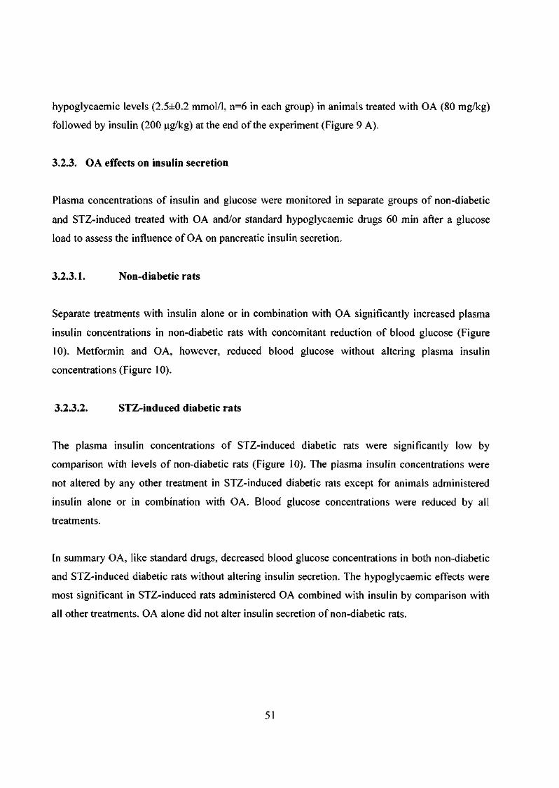
hypoglycaemic levels (2.5±0.2 mmol/1, n=6 in each group) in animals treated with OA (80 mg/kg)
followed by insulin (200 ug/kg) at the end of the experiment (Figure 9 A).
3.2.3. OA effects on insulin secretion
Plasma concentrations of insulin and glucose were monitored in separate groups of non-diabetic
and STZ-induced treated with OA and/or standard hypoglycaemic drugs 60 min after a glucose
load to assess the influence of OA on pancreatic insulin secretion.
3.2.3.1. Non-diabetic rats
Separate treatments with insulin alone or in combination with OA significantly increased plasma
insulin concentrations in non-diabetic rats with concomitant reduction of blood glucose (Figure
10). Metformin and OA, however, reduced blood glucose without altering plasma insulin
concentrations (Figure 10).
3.2.3.2. STZ-induced diabetic rats
The plasma insulin concentrations of STZ-induced diabetic rats were significantly low by
comparison with levels of non-diabetic rats (Figure 10). The plasma insulin concentrations were
not altered by any other treatment in STZ-induced diabetic rats except for animals administered
insulin alone or in combination with OA. Blood glucose concentrations were reduced by all
treatments.
In summary OA, like standard drugs, decreased blood glucose concentrations in both non-diabetic
and STZ-induced diabetic rats without altering insulin secretion. The hypoglycaemic effects were
most significant in STZ-induced rats administered OA combined with insulin by comparison with
all other treatments. OA alone did not alter insulin secretion of non-diabetic rats.
51

I A N o n - d i a b e t i c | - C o n t - G l i b • - M e t - I n s
•©
7 . 5
6 . 0
4 . 5
3 . 0 —
1 . 5
S T Z - d i a b e t i c C o n t G l i b M e t I n s
• »
2 4 0
T i m e ( m i n )
Figure 6: Comparison of OGTT responses to standard anti-diabetic drugs in separate groups of
non-diabetic rats (A) and STZ-induced diabetic (B) rats with respective control animals. Values
are presented as means, and vertical bars indicate SEM (n=6 in each group).
* p<0.05 by comparison with respective control animals
52

A Non-d iabet ic
7.5 —|
• Cont ro l - -A- - O A 40 - «*- - OA 80 - -*- - OA 120
B STZ-diabe tic | Cont ro l O A 4 0 O A 8 0 O A 1 2 0
1 8 0 2 4 0
Time (min)
Figure 7: Comparison of OGTT responses to various 0A doses in separate groups of non-diabetic
rats (A) and STZ-induced diabetic (B) rats with respective control animals. Values are presented as
means, and vertical bars indicate SEM (n=6 in each group).
* p<0.05 by comparison with respective control animals
53

O 6 0 1 2 0 1 8 0 2 4 0
T i m e ( m i n )
Figure 8: Comparison of OGTT responses to OA (40 mg/kg) alone and in combination with
standard anti-diabetic drugs in separate groups of non-diabetic rats (A) and STZ-induced diabetic
(B) rats with respective control animals. Values are presented as means, and vertical bars indicate
SEM (n=6 in each group). * p<0.05 by comparison with respective control animals
54
A N o n - d i a b e t i c
7 .5 —i
6 .0 —
4 . 5 —
3 . 0
1 . 5
•0)
1
B S T Z - d i a b e t i c C o n t r o l - O A 8 0
- -sir — + G l i b - • » - + M e t - ^ - + ins
Figure 9: Comparison of OGTT responses to OA (80 mg/kg,) alone and in combination with
various standard anti-diabetic drugs in separate groups of non-diabetic rats (A) and STZ-induced
diabetic (B) rats with respective control animals. Values are presented as means, and vertical bars
indicate SEM (n=6 in each group). * p<0.05 by comparison with respective control animals
55

Non-diabetic
STZ-diabetic
1 D
Control
5 0 0 -
4 0 0 -
I 30°" | 2 0 0 -
" 100«
0«
# # # *
Control OA Met Ins
Treatment
J S 1
OA + Ins
Figure 10: Comparison of the effects of OA and standard anti-diabetic drugs on plasma insulin
and glucose concentrations in non-diabetic and STZ-induced diabetic rats with respective control
groups. Blood samples were collected 60 min after glucose load. Values are presented as means,
and vertical bars indicate SEM (n=6 in each group).
* p<0.05 by comparison with respective control animals;
p<0.05 by comparison with respective non-diabetic animals
56

3.3. Acute renal effects of OA
Acute effects of OA on kidney function were investigated in anaesthetized non-diabetic and STZ-
induced diabetic rats challenged with hypotonic saline after a 3/4 h equilibration for 4 h of 1 h
control, 11/2 h treatment and 1 V% h recovery periods.
3.3.1. Non-diabetic rats
Figure 11 shows stable urine flow and Na+, K+ and CI" excretion rates of control non-diabetic rats
during the 4 h post equilibration period. Acute intravenous infusion of OA during the V/2 h
treatment period increased Na+ excretion rate from a pre-treatment mean value of 718±25 umol/h
to 880±25 umol/h (n=6 in each group) without affecting K+, CI" and urine flow rates (Figure 11).
The increase in urinary Na+ output was reflected as the increased mean total amount of Na+
excreted during the treatment period (Table 2).
3.3.2. STZ-induced diabetic rats
The mean urine flow, Na+, K+ and CI" excretion rates of anaesthetized, control and OA-treated STZ-
induced diabetic rats during the 4 h post-equilibration period are compared in Figure 12. STZ-induced
diabetic control rats exhibited lower urine flow and electrolyte excretion rates by comparison with
non-diabetic rats. OA infusion increased the mean Na+ excretion rate from a pre-treatment value of
297±29 umol/h to 420±38 umol/h by the cessation of 1V2 h treatment. The increase in Na+ excretion
was maintained during the recovery period. OA, however, did not influence K+, CI" and urine
excretion rates by comparison with control animals (see Figure 12). The mean total amounts of Na+
excreted during the 11/2 h OA treatment was significantly more than that of control animals at the
corresponding time. The total amounts of fluid voided and K+ and CI" excreted were, however, not
altered by OA treatment.
In summary acute OA treatment increased Na+ excretion in non-diabetic and STZ-induced diabetic
rats without altering urine volume or urinary outputs of K+ and CI".
57

-io
8 —
6 —
4 —
2
O '
H T r o a t m e n t
r.^. A
1 I C o n t r o l
1 0 0 0
7 6 0 —
e o o
2 6 0 —
3 0 0 —n
2 0 0 —
1 0 0 0 ^
7 S O —
eoo —
« 2 6 0 —
• • -I
Ix S I
1 2 3
P
2 3
T i m e ( r i )
Figure 11: Comparison of the effects of 1!4 h OA infusion on Na+ (A), K+ (B) and Cf (C) excretion and urine flow (D) rates with control animals in anaesthetized non-diabetic rats (n=6 in each group). Values are presented as means, and vertical bars indicate SEM (n=6 in each group). * p<0.05 by comparison with control animals at each corresponding time
58
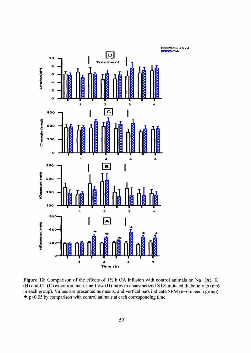
1 C o n t r o l O A
1 0 0
9 0 0 -
6 0 0 -
3 0 0 —
I H I
Figure 12: Comparison of the effects of IV2 h OA infusion with control animals on Na+ (A), K+
(B) and Cl" (C) excretion and urine flow (D) rates in anaesthetized STZ-induced diabetic rats (n=6 in each group). Values are presented as means, and vertical bars indicate SEM (n=6 in each group). * p<0.05 by comparison with control animals at each corresponding time
59

Table 2: Comparison of total mounts of urine volume, Na+, K+ and CI" excreted during the
treatment period by anaesthetized OA-treated non-diabetic and STZ-induced diabetic rats with
respective control animals (n = 6 in all groups). Data are expressed as means ± SEM (n = 6 in each
group). * p<0.05 by comparison with respective control animals; p<0.05 by comparison with
respective non-diabetic animals
Non diabetic
Diabetic
Control
Treated
Control
Treated
Urine (ml)
13.1±0.6
12.7±0.8
10.4±0.5
10.1±0.6
Na+ (umol)
1056±25
1394±15*
425±23#
622±41*#
K+(u.mol)
322±7
344±15
260±42
243±16
Cr(^mol)
1264±14
1388±43
1031±92
1011±70
3.4. Short-term effects on body weight, food and water intake
Table 3 compares the effects of OA and/or standard anti-diabetic drugs on mean weight changes,
food and water intake in separate groups of non-diabetic and STZ-induced diabetic rats over a 5-
week period with respective control animals. Non-diabetic control rats progressively gained weight
while STZ-induced diabetic rats exhibited severe wasting throughout the 5-week period. OA
administration reduced the body weight loss of STZ-induced diabetic rats in the 5th week, whereas
treatment with metformin, insulin or combined insulin and OA stabilized body weight of STZ-
induced diabetic rats from the 3rd to the 5th week.
The total amounts of food taken per day by control non-diabetic and STZ-induced diabetic rats and
respective treated groups did not significantly differ, except for metformin which significantly
decreased food intake of STZ-induced diabetic rats in the 5th week.
Control STZ-induced diabetic rats exhibited polydipsia by comparison with the respective non-
diabetic animals as evidenced by the fact that STZ-induced diabetic rats drank four to five times
more water than non-diabetic rats. OA administration caused significant reduction of water intake
60
by STZ-induced diabetic rats in week 5 by comparison with respective control animals at the
corresponding period. On the other hand, separate treatment of STZ-induced diabetic rats with
metformin, insulin alone or in combination with OA significantly (p<0.05) reduced water intake
from the first week up to the end of the five weeks.
In summary, treatment with OA alone or in combination with insulin stabilized body weight of
STZ-induced diabetic rats without altering food intake.
61

Table 3: Comparison of the effects of 5-week treatment with OA and/or standard anti-diabetic
drugs of non-diabetic (ND) and STZ-induced diabetic animals on food intake, water intake and
body weight changes with the respective control animals (n=6 in each group). Values are presented
as means ± SEM. * p<0.05 by comparison with control animals
Parameter
Food intake (g/day)
Water intake (ml/day)
%b.wt change/week
Treatment
Control
OA
Metformin
Insulin
OA4 insulin
ND
STZ
ND
STZ
ND
STZ
ND
STZ
ND
STZ
week 1
30 ±1
35 ±1
28 ±1
34 ±1
29 ±2
33 ±1
30 ±1 34 ±1
32 ±1
38 ±1
week 2
32 ±1
33 ±1 29 ±1
32 ±1
30 ±1
28 ±1
26 ±2
33 ±1
27 ±1 35 ±1
week 3
31±1
32±2 2941
3143
29±2
27±1 23±1
3241
28±1
33±2
week 4
3041
2842
2942 3241
2541
2841
3041
3041
3042 3241
week 5
26 4 1
34 4 2 28 4 1
33 4 2 28 4 1
2 7 4 1 *
26 4 1
36 4 1
30 4 1
33 4 2
Control
OA
Metformin
Insulin
OA4 insulin
ND
STZ
ND
STZ
ND
STZ
ND
STZ
ND
STZ
33 ±2
123±4
33 ±2
125±4
34 ±2
107±3* 33 ±3
115±2* 37 ±1
10944*
36±3
13149
34±1
141±7 40±1
107412*
30±2
11146*
31±2
11043*
33±1
136±9
36±1
132±3
3342
10648*
3142
11345*
3842 109411*
3442
133410 3341
12748
2942
10046*
3542
102410*
3543
10446*
3141
135413
29 4 2
10747* 3141
94 48*
36 42 101411*
33 41
10246*
Control
OA
Metformin
Insulin
0 A 4 insulin
ND
STZ
ND
STZ
ND
STZ
ND
STZ
ND
STZ
8±1
-6±2
5±1 -5±2 6±1
-8±1
7±1 -7±1
8±1
-4±2
11 ±2
-4±2 12 ±1
-2 ±2 1141
-7 ±2 15 ±1
-3±1
14 ±1
-3±1
1841
-5±2
17±1
-243
1 8 4 2 -241*
2141
-241*
2043
-141*
2241
-442
2141
-343 2 1 4 1
-242* 2341
-142*
2443
142*
23 4 2
-5 4 2 24 4 1
- 1 4 3* 24 4 1
1 4 2* 27 4 1*
- 1 4 2* 2541
2 4 2*
62

3.5. Short-term renal effects of OA
The effects of OA on renal function were studied in individually-caged non-diabetic and STZ-
induced diabetic rats that were treated with OA (80 mg/kg, p.o.) twice every third day for five
weeks.
3.5.1. Non-diabetic rats
The mean daily Na+ excretion rate of control non-diabetic rats ranged from 547±21 mmol/day to
678±68 mmol/day throughout the 5 week experimental period (Figure 13). OA administration
significantly increased the mean daily Na+ output from 1st week value of 645±28 mmol/day to
837±49 mmol/day by the end of the 5th week, but the volume of urine voided and urinary outputs
of K+ and CT were not altered. The OA-elicited natriuresis was not reflected in plasma collected
after 5 weeks treatment as the plasma Na+ concentrations compared with those of control animals
at the corresponding time (Table 4). OA administration for 5 weeks significantly reduced plasma
creatinine concentrations of non-diabetic rats with concomitant elevation of GFR. In contrast,
plasma urea concentrations of non-diabetic animals were not altered by OA treatment. OA
administration caused slight, but statistically insignificant decreases of plasma and increases of
urine osmolalities of non-diabetic rats (Table 4). Administration of OA did not affect kidney
masses of non-diabetic rats (Table 4).
3.5.2. STZ-induced diabetic rats
The mean daily urine volume of untreated STZ-induced diabetic rats was greatly elevated throughout
the 5-week compared to non-diabetic control animals, although the urinary Na+, K+ and CI" outputs
were enormously reduced (Figure 14). Administration of OA increased urinary Na+ outputs of STZ-
induced diabetic rats from 35±3 mmol/day in the first week to 48±2 mmol/day in the 5th week. The
mean daily urine volume, urinary K+ and CI" outputs were, however, not altered by OA
administration.
63

As described for non-diabetic rats, Table 4 shows that OA administration in STZ-induced diabetic
rats did not affect plasma Na+, K+ and CI" by the end of the 5-week experimental period. In
contrast with non-diabetic rats, OA administration significantly reduced (p<0.05) plasma
osmolality of STZ-treated diabetic rats with concomitant increase in urine osmolality. Plasma
creatinine and urea concentrations of STZ-induced diabetic rats were elevated when compared
with non-diabetic rats. OA treatment did not alter plasma urea concentrations unlike plasma
creatinine levels which were significantly reduced with a concomitant elevation of GFR (Table 4).
Kidney mass which was significantly increased in STZ-induced diabetic rats by comparison with
non-diabetic animals was not altered by sub-chronic OA treatment (Table 4).
3.5.3. Haemodynamic changes
Acute infusion of hypotonic saline to control non-diabetic rats did not cause any significant
variations in the mean arterial blood pressure (MAP) throughout the 4-h post-equilibration period
(Figure 15). Acute intravenous infusion of OA at 90 ug/h for 1 Vz h, however, reduced the mean
arterial blood pressure from a pre-treatment value of 115±1 mmHg (n=6) to 105±1 mmHg (n=6) by
the end of 1 !4 h treatment period. The hypotensive effect of OA persisted during the post-treatment
period to a mean value of 108±2 mmHg at the end of the experiment (Figure 15A). Similar trends
were observed in STZ-induced diabetic rats. The MAP changes due to long-term (sub-chronic) OA
treatments are shown in Figure 15. Treatment of the rats with OA (80 mg/kg daily for 5 weeks,
p.o.) caused significant decreases (p<0.05) in MAP in non-diabetic and STZ-treated diabetic rats
throughout the study period by comparison with control rats at the corresponding time periods
(Figure 15).
3.5.4. Summary of the sub-chronic effects of OA
Sub-chronic OA treatment significantly increased Na+ excretion rates of non-diabetic and STZ-
induced diabetic rats without affecting the volumes of urine voided and urinary concentrations of K+
and CI". OA treatment decreased plasma concentrations of creatinine with concomitant elevation of
creatinine clearance Ccr. Significant hypotensive effects of OA were also observed in non-diabetic
and STZ-induced diabetic rats.
64

20 -n
if -15 H
5 H
I Cont ro l • OA
1200 ^
900 H
H 5
600 H
3 0 0
m
T 5
800 -n
600 —I
4 0 0 —|
200 H
B| J .
9 0 0
600 H
300 <H
0,
2 3 4 Time ( w e e k s )
Figure 13: Comparison of 24h Na+(A), K+ (B), CI" (C) excretion and urine flow (D) rates in non-diabetic rats treated with OA twice every third day with control animals. Values are presented as means, and vertical bars indicate SEM (n=6 in each group). * p<0.05 by comparison with control animals at each corresponding time
65
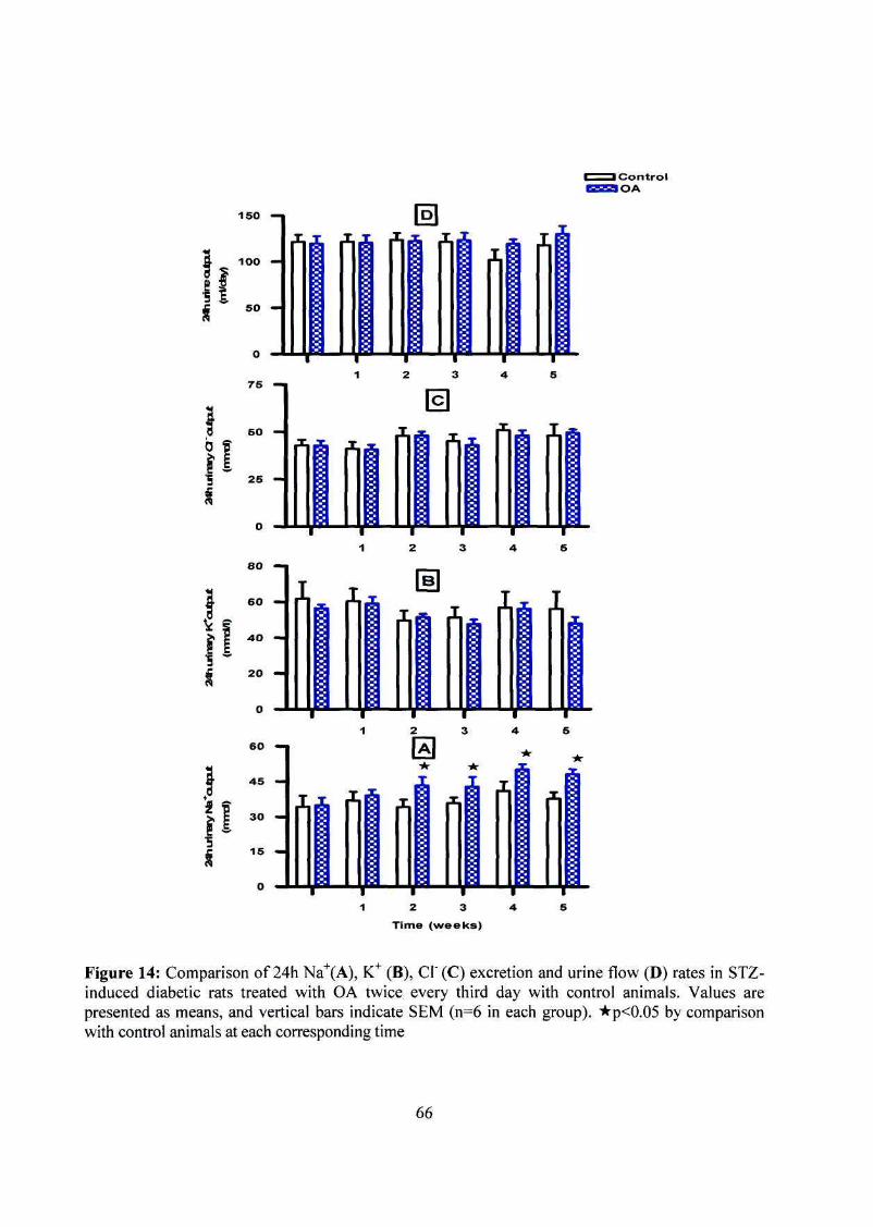
C o n t r o l OA
1 5 0 — 1
if s
1 0 0
I '
u
If
80 -n
60 —
4 0 —
20 —
6 0
4 5
3 0
15 —
2 3 Time ( w e e k s )
Figure 14: Comparison of 24h Na+(A), K+ (B), CI" (C) excretion and urine flow (D) rates in STZ-induced diabetic rats treated with OA twice every third day with control animals. Values are presented as means, and vertical bars indicate SEM (n=6 in each group). *p<0.05 by comparison with control animals at each corresponding time
66

Table 4: Plasma biochemical parameters of non-diabetic and STZ-diabetic control and rats
administered OA twice every third consecutive day for 5 weeks (n = 6 in all groups).
* p<0.05 by comparison with respective control animals
p<0.05 by comparison with respective non diabetic animals
Parameter
Na+ (mmol/1)
K+(mmol/l)
CI" (mmol/1)
Urea (mmol/1)
Creatinine(umol/l)
Plasma osmolality (mOsm/kgH20) GFR (ml/min)
Kidney mass (g/lOOgb.wt) Urine osmolality (mOsm/kgH20)
Non diabetic
Control
142±1
3.68±0.25
101±1
7.3±0.9
25±2
263±27
2.88±0.14
0.46±0.02
2512±99
Treated
142±1
3.59±0.17
103±1
8.3±0.7
19±1*
247±15
3.71±0.30*
0.44±0.02
2661±78
STZ-diabetic
Control
129±6
3.76±0.21
102±2
10.7±1.7#
31±3#
382±10#
1.81±0.32#
0.78±0.07#
991±92#
Treated
124±3
3.53±0.20
96±4
12.1±1.8#
23±2*#
316±16#
3.07±0.16*#
0.73±0.04#
1115±56*#
67

A Non-diabetic
120 - i
115 i
§. 110 -a. < S
105 -
100
Treatment
* \
• •
B STZ-diabetic
T r T 60 120 180
Time (min) 240
C Non-diabetic
D STZ-diabetic
Treatment
• Control
•OA
I ?-•-.,
T 1 T" 1 2 3
Time (weeks)
1 •
Figure 15: Comparison of acute effects of OA infusion on MAP in non-diabetic and STZ-induced
diabetic rats (A-B) and sub-chronic short-term administration in non-diabetic and diabetic rats (C-
D) with respective control animals (n=6 in each group). Values are presented as means, and
vertical bars indicate SEM (n=6 in each group). * p<0.05 by comparison with respective control
animals at each corresponding time
68

3.5.5. Effects of OA on terminal blood glucose and plasma insulin concentrations
Figure 16 shows the short-term effects of treatment with OA and standard anti-diabetic drugs on
blood glucose concentrations and plasma insulin concentrations in non-diabetic and STZ-induced
diabetic rats measured 24 h after the last treatment. Treatment of non-diabetic rats with OA alone
or standard anti-diabetic drugs reduced blood glucose concentrations by amounts that did not
achieve statistical significance. Combined OA and insulin treatment, however, significantly
decreased blood glucose concentrations of non-diabetic animals by comparison with respective
control animals. The administrations of OA alone and/or standard anti-diabetic drugs significantly
(p<0.05) reduced blood glucose concentrations of STZ-induced diabetic rats by comparison with
the respective control animals. Figure 16 shows that the circulating plasma insulin concentrations
of control STZ-induced rats were significantly low by comparison with non-diabetic animals. OA
treatment did not alter plasma insulin concentrations of non-diabetic or STZ-induced diabetic rats.
Significant increases in plasma insulin concentrations were observed only in non-diabetic and
STZ-induced diabetic rats treated with combined insulin and OA or insulin alone. Figure 17 shows
that treatment with OA alone significantly (p<0.05) decreased the excretion of glucose of STZ-
induced diabetic rats throughout the 5-week period by comparison with the respective control rats.
3.5.5.1. Effects of OA on hepatic and muscle glycogen concentrations
Hepatic and muscle glycogen concentrations were measured in separate groups of non-diabetic
and STZ-induced diabetic rats after 5 weeks of treatment with OA alone and/or standard anti
diabetic rats in an effort to establish in part the mechanism(s) of the hypoglycaemic effects of OA.
All treatments increased hepatic glycogen concentrations of non-diabetic and STZ-induced
diabetic rats (Table 5) whereas muscle glycogen concentrations were elevated by these treatments
only in non-diabetic rats.
In summary, sub-chronic OA treatment like standard anti-diabetic drugs decreased blood glucose
concentrations in rats without influencing pancreatic insulin secretion. The hypoglycaemic effects
were most significant in animals co-administered OA and insulin. Muscle glycogen concentrations
of STZ-induced diabetic rats were increased by combined insulin and OA treatment.
69

Overall summary of the results of this study
The absolute stereostructure of S. cordatum derived OA was elucidated using ID and 2D 1 13
H NMR and C NMR techniques on the basis of chemical and physicochemical evidence.
OGTT responses elicited by S. cordatum leaf derived OA in non-diabetic and STZ-induced
diabetic rats were comparable to those of standard anti-diabetic drugs.
OA augments the insulin-elicited OGTT responses in STZ-induced diabetic rats.
OA treatment improved renal function of STZ-induced diabetic rats as shown by increased
urinary Na+ outputs, reduced plasma creatinine with concomitant increase in GFR.
OA evoked potent hypotensive effects in STZ-induced diabetic rats.
70

Non-diabetic STZ-diabetic
to o u _3 O)
• a o _o m
32 ^
i 16 E
8 -
4 -
l r Control OA
r Met
T Ins
T OA + ins
400 n
=> 300 H "o E a ^ 200 _c "5 (0
=. 100
s
1
Control
1 ^
OA
# #
I
Met
Treatment
Ins
ft
#
i
OA + Ins
Figure 16: Comparison of the sub-chronic effects of OA and standard hypoglycaemic drugs on
blood glucose (A) and plasma insulin (B) concentrations in non-diabetic and STZ-induced diabetic
rats with respective control groups. Blood samples were collected 24 h after the last treatment.
Values are presented as means, and vertical bars indicate SEM (n=6 in each group).
* p<0.05 by comparison with respective control animals;
p<0.05 by comparison with respective non-diabetic animals
71

STZ-diabetic Control OA
Figure 17: Comparison of mean daily urinary glucose outputs of control STZ-induced diabetic rats
with STZ-induced diabetic rats treated with OA twice every third day for 5 weeks (n=6 in each
group).
* p<0.05 by comparison with respective control animals
72

Table 5: Comparison of the effects of 5-week treatment with OA alone and/or standard anti
diabetic drugs on hepatic and muscle glycogen concentrations in non-diabetic and STZ-induced
diabetic rats with respective control groups. Data are expressed as mean ± SEM (n=6 in each
group).
* p<0.05 by comparison with respective control group
Treatment
Non-diabetic rats (n-6 in each group)
STZ-induced diabetic rats (n=6 in each group)
Control
OA
Metformin
Insulin
OA + insulin
Control
OA
Metformin
Insulin
OA + insulin
[Hepatic glycogen] (mg/100mg tissue/ 300g b.wt) 28.4±1.9
37.9±3.7*
39.5±2.0*
66.3±2.7*
74.0±6.1*
17.1 ±0.4
30.3 ± 0.6*
36.0 ±0.7*
42.3 ± 0.7*
56.4 ±0.5*
[Muscle glycogen] (mg/lOOmg tissue) 2.6±0.3
3.5±0.3*
5.2±0.7*
5.2±0.4*
5.0±0.3*
1.1 ±0.1
2.0 ±0.4
2.1 ±0.4
2.4 ±0.3
3.1 ±0.3*
73

CHAPTER 4
4.0. DISCUSSION
4.1. General
The aim of the present study was to investigate the effects of S. cordatum leaf-derived
OA on blood glucose concentrations and renal function of STZ-induced diabetic rats. The
results suggest that OA not only has the potential to lower blood glucose and blood
pressure in diabetes, but may also have beneficial effects on kidney function. The
findings are of considerable importance because they contextualize the hypoglycaemic,
renal and hypotensive effects of OA in the management of diabetes. This is clinically
relevant considering the prevalence of microvascular and macrovascular complications in
diabetic patients (The Diabetes Control and Complications Trial Research Group, 1993;
Vinicor, 1994). Management of diabetes mellitus with OA may, therefore, be associated
not only with the regulation of blood glucose, but also the prevention or alleviation of
these complications.
4.2. Structural elucidation of OA
The absolute stereostructure of OA from the crude ethyl acetate extract of S. cordatum
leaves was established on the basis of chemical and physicochemical evidence. The
spectra of the plant-derived OA elucidated using H- and C-NMR was compared with
previously reported data (Mahato and Kundu 1994). The spectral data of OA, particularly
the presence of the olefenic bond between carbon 12 and 13 which is peculiar to
triterpenoids, was identical with that of previously reported OA (Mahato and Kundu,
1994). The identification of OA from S. cordatum leaves confirms previous reports that
the S. cordatum leaf crude ethyl acetate extract contains triterpenoid mixtures
(Musabayane, Mahlalela, Shode and Ojewole, 2005). Other investigators have also
isolated the triterpenes OA and ursolic acid from various medicinal plants following
74

sequential extraction with methanol (Jung et ah, 2007; Begum, Zehra and Siddiqui,
2008).
4.3. Hypoglycaemic effects
The experimental evidence presented in this study extends previous observations in our
laboratories that the S. cordatum crude leaf extract lowers blood glucose of STZ-induced
diabetic rats (Musabayane, Mahlalela, Shode and Ojewole, 2005) by indicating that the
antihypeglycaemic effect is elicited by triterpene constituent, OA.
The fasting blood glucose concentrations of STZ-induced diabetic rats were significantly
higher by comparison with the non-diabetic animals. The rise in blood glucose
concentrations in untreated STZ-induced diabetic rats can be attributed to the absence of
insulin, since streptozotocin induces permanent diabetes mellitus by direct pancreatic B-
cell cytotoxicity (Junod, Lambert, Stauffacher and Renold, 1969; Pinent, Blay, Blade,
Salvado, Arola and Arde, 2004). Streptozotocin first isolated from Streptomyces
achromogenes is an antibiotic that selectively destroys pancreatic B-cells thereby
inducing type 1 diabetes mellitus (Ruiz, Alegria, Barbera, Farre and Lagarda, 1994;
Coskun, Kanter, Korkmaz and Oter, 2005; Li et al., 2007; Srinivasan and Ramarao,
2007). Indeed, plasma insulin concentrations were significantly lower in STZ-induced
diabetic rats compared with non-diabetic rats, thereby contributing to high glucose
concentrations. This is consistent with the mechanism of action of streptozotocin on the
beta-cells of the pancreas (Junod, Lambert, Stauffacher and Renold, 1969; Pinent, Blay,
Blade, Salvado, Arola and Arde, 2004). The hypothesis is further supported by the
observation of a steady, gradual fall in blood glucose concentrations of control non-
diabetic rats following glucose load, presumably due to increased pancreatic insulin
secretion. In addition, glibenclamide was associated with blood glucose reduction only in
non-diabetic rats and not STZ-induced animals. The hypoglycaemic effect of
glibenclamide is seen only when pancreatic beta-cell activity is present, as glibenclamide
action involves augmentation of insulin secretion (Luzi and Pozza, 1997). The blood
glucose-lowering effects of OA at all doses (40, 80 and 120 mg/kg) in non-diabetic rats
75

could not be separated at any time. OGTT responses of STZ-induced diabetic rats to OA
at 40 and 80 mg/kg were similar but, significantly lower than those of OA at 120 mg/kg
throughout (Figure 7). The reasons for the discrepancy of OGTT responses to three doses
of OA in non-diabetic and STZ-induced diabetic rats remain to be determined. We
suggest that endogenous insulin masked the differences in hypoglycaemic effects evoked
by the small range of OA used in non-diabetic rats.
OA alone and/or insulin and metformin decreased plasma glucose concentrations in non-
diabetic and STZ-diabetic rats following sub-chronic treatment for 5 weeks. Blood
glucose concentrations of STZ-induced diabetic rats following long-term treatment were
significantly reduced by all treatments to levels that did not achieve euglycaemia. The
reduction of blood glucose concentrations of non-diabetic rats did not reach statistical
significance. The lack of pronounced OA blood glucose-lowering effects observed may
be attributed to experimental design and data presentation. The blood glucose
concentrations were measured 24 h after the last dosing and by that time the administered
OA or standard anti-diabetic drugs may have been metabolized. The peak hypoglycaemic
effect of triterpene containing herbal extracts occurs 2-4 hours after administration, with
blood sugar returning to pre-treatment levels in 6-10 hours (Kakuda, Sakane, Takihara,
Ozaki, Takeuchi and Kuroyanagi, 1996).
Plasma insulin concentrations were significantly elevated in non-diabetic rats
administered insulin alone or in combination with OA by comparison with STZ-induced
diabetic rats similarly treated. Therefore, insulin involvement in the hypoglycaemic
effects of OA is excluded, since plasma insulin concentrations of non-diabetic and STZ-
induced diabetic rats were not altered by treatment with OA alone. OA, however, would
not be expected to significantly affect insulin levels in STZ-induced diabetic rats as the
pancreatic 6-cells would have been selectively destroyed by streptozotocin administration
(Junod, Lambert, Stauffacher and Renold, 1969; Pinent et ah, 2004). It is possible that
OA alters glucose metabolism in the liver of diabetic animals, thus reducing blood
glucose concentrations by increasing hepatic glycogenesis (Table 5). This has been
previously reported for other plant extracts (Gondwe et al., 2008). It is suggested that the
76
hypoglycaemic effects of OA mimic those of metformin as evidenced by the fact that
both these treatments did not alter plasma insulin concentrations (Figure 10 and 16).
Metformin improves insulin sensitivity in peripheral target tissues and suppresses hepatic
glucose output to reduce blood glucose (Lee and Morley, 1998; Krentz and Bailey, 2005).
The possibility of lowering blood glucose concentrations through other mechanisms
cannot be totally excluded.
Hypoglycaemic effects were most pronounced in STZ-induced diabetic rats treated with
combined OA and insulin (Figures 8 and 9). Since the current data suggest that OA
possesses metformin-like blood glucose lowering properties, it is possible that OA
augmented the hypoglycaemic effects of insulin by increasing the sensitivity to insulin.
This is in agreement with previously reported effects of other triterpenoids, ursolic acid
and corosolic acid (Miura et al., 2004; Jung et ah, 2007). It is also possible that OA might
have interfered with gastric emptying and glucose absorption, as been previously reported
for metformin (Lee and Morley, 1998). Indeed some triterpenoids have been shown to
lower blood glucose concentrations in a similar way (Matsuda, Li, Yamahara and
Yoshikawa, 1998; Matsuda, Li, Mukarami, Yamahara and Yoshikawa, 1999; Katz,
Newman and Lansky, 2007). The ability of OA to prevent hyperglycaemia indicates that
OA may be useful in preventing some of the complications associated with diabetes
mellitus.
OA like metformin and insulin increased hepatic glycogen concentrations of non-diabetic
and STZ-induced diabetic rats by comparison with respective controls (Table 4). Hepatic
glycogen concentrations were, however, significantly lower in STZ-induced diabetic rats
as compared to the non-diabetic rats. Glycogen is the primary storable form of glucose
and diabetes mellitus is associated with reduced capacity to store glycogen
(Chandramohan, Ignacimuthu and Pugalendi, 2008). This is in agreement with previous
observations that the crude leaf extract of S. cordatum increases hepatic glycogen
concentrations, thereby reducing blood glucose levels (Musabayane, Mahlalela, Shode
and Ojewole, 2005). On the contrary, muscle glycogen concentration of STZ-induced
diabetic rats was not altered by treatment with OA alone, presumably due to the lack of
77

insulin. The observed difference between non-diabetic and STZ-induced diabetic rats
muscle glycogen concentration may be due to the fact that the conversion of glucose into
glycogen in skeletal muscle is dependent on insulin (Saltiel and Kahn, 2001; Hsu, Liu,
Kuo, Chen, Su and Cheng, 2003; Jensen, Jebens, Brennesvik, Ruzzin, Soos, Engebretsen,
O'Rahilly and Whitehead, 2006). In contrast, hepatic glycogen concentrations are
determined by the extracellular glucose concentrations and glycogenic enzyme activities
(Tana, Tanb and Pushparaj, 2005). It is possible; therefore, that OA-induced hepatic
glycogen synthesis in STZ-induced diabetic rats was due to increased activities of GLUT-
2 and glycogen synthase (Buschiazzo, Exton and Park, 1970; Libal-Weksler, Gotlibovitz,
Stark and Madar, 2001; Tana, Tanb and Pushparaj, 2005). The activity of the GLUT-2
transporters is non-insulin dependent. Glycogen synthesis also involves inhibition of
glycogen phosphorylase, decreased glucose output, enhancement of GLUT-4
translocation, insulin receptor autophosphorylation and inhibition of protein tyrosine
phosphatase B as been reported for other triterpenoids (Chen et al., 2006; Jung et ah,
2007). It is possible to speculate that the effects of the combined treatment could be due
to OA sensitizing responses to exogenous insulin.
The non-diabetic rats showed weight gain throughout the study, whilst untreated STZ-
induced diabetic rats showed progressive weight losses. The discrepancy between the
groups may be attributed to the anabolic effects of insulin (Chandramohan, Ignacimuthu
and Pugalendi, 2008). Treatment with OA alone or in combination with insulin, however,
stabilized body weight of STZ-induced diabetic rats without altering food intake. Weight
gain may be attributed to increase in the incorporation of glucose into tissues, as reported
earlier in STZ-treated diabetic mice (Naik, Barbosa-Filho, Dhuley and Deshmukh, 1991;
Okine, Nyarko, Osei-Kwabena, Oppong Barnes and Ofosuhene, 2005). Interestingly,
polydipsia and increased water intake as a result of enhanced proteolysis associated with
hyperosmotic dehydration has been shown to be associated with weight losses in STZ-
induced diabetic rats (Rebsomen, Pitel, Boubred, Buffat, Feuerstein, Raccah, Vague and
Tsimaratos, 2006; Narendhirakannan, Subramanian and Kandaswamy, 2006).
78

4.4. Renal effects
Renal function studies were designed to investigate whether S. cordatum derived OA can
reverse the previously reported inability of the kidney to excrete Na+ in STZ-induced
diabetic rats (Musabayane, Ndhlovu and Balment, 1995). The hypotonic saline used has
been shown to suppress endogenous arginine vasopressin secretion in rats (Balment,
Brimble, Forsling and Musabayane, 1984). Long-term diabetes mellitus complications
often include compromised kidney function and elevated blood pressure (Haller, Drab
and Luft, 1996; Stengel, Billon, van Dijk, Jager, Dekker, Simpson, Briggs, 2003). The
results indicate that OA not only has the potential to lower blood pressure in diabetes, but
may also have beneficial effects on kidney function. The results introduce the first in vivo
evidence that oleanolic acid extracted from S. cordatum leaves ameliorates kidney
function in STZ-induced diabetic rats.
Kidney weights of STZ-induced diabetic rats were significantly increased by comparison
with non-diabetic animals possibly due to accumulation of extra cellular matrix proteins
(Aybar, Riera, Grau and Sanchez, 2001; Kang, Kim, Yamabe, Nagai and Yokozawa,
2006). The extra-cellular matrix protein induced kidney hypertrophy causes structural
changes of glomerular basement membrane thereby impairing kidney function (Hargrove,
Dufresne, Whiteside, Muruve, Wong 2000; Mason and Wahab, 2003; Kumar, Shetty,
Salimath. 2008). This perhaps explains why STZ-induced rats exhibited marked daily
decreases in urinary Na+ excretion and elevated plasma creatinine concentration with
concomitant reduction in Ccr at the end of 5 weeks by comparison with non-diabetics.
These changes were attenuated in the OA-treated groups indicating that oleanolic acid
might have beneficial effects on renal function in diabetes. OA-evoked increases urinary
Na+ outputs of STZ-diabetic rats and elevation of GFR as assessed by creatinine
clearance suggest up-regulation of renal function by the triterpene. GFR is an important
marker of kidney function and treatment-related increases in creatinine clearance indicate
an improvement of kidney function in experimental animals and man rather than looking
at plasma concentrations of urea because the latter varies with dietary protein (Satirapoj,
Supasyndh, Patumanond and Choovichian, 2006; Yang and Bankir, 2006; Almond,
79

Siddiqui, Robertson, Norrie and Isles, 2008; van Hoek, Lefebvre, Kooistra, Croubels,
Binst, Peremans and Daminet, 2008). Our laboratory and several other authors have
previously used creatinine clearance (Ccr) in rats as a fundamental parameter of
evaluating renal tubular function (Rebsomen et ah, 2006; Kumar, Shetty and Salimath,
2008). Low GFR is associated with fluid and electrolyte imbalance in nephrotic
syndrome and acute renal failure (Fernandez-Llama, Andrews, Nielsen, Ecelbarger and
Knepper, 1998; Kwon, Frokiaer, Fernandez-Llama, Knepper and Nielsen, 1999; Kim,
Jeon, Lee, Kang, Kook, Ahn, Kim, Cho, Kim, Han and Choi, 2000). A correlation
between increased urinary Na+ excretion and elevated GFR has been reported in the rat
(Marin-Grez, Fleming and Steinhausen, 1986). It is speculated that the effects of OA on
renal Na+ excretion can be dissociated from its influence on fluid handling. Indeed, some
studies dissociate the arginine vasopressin evoked increase in Na+ excretion via Vi
receptors (Musabayane, Forsling and Balment, 1997) from its V2 mediated antidiuretic
effects. It is speculated that OA administration significantly increased urinary Na+
excretion perhaps in part mediated via elevation of GFR possibly due to increases of the
filtered load of Na+ in the tubular filtrate. In addition, OA may have inhibited increased
proximal reabsorption of Na+ which occurs in diabetes mellitus (Palm, Liss, Fasching,
Hansell and Carlsson, 2001; Vallon, 2003; Rebsomen et ah, 2006; Kanetsuna, Hirano,
Nagata, Gannon, Takahashi, Harris, Breyer and Takahashi, 2006). Hence, OA improved
kidney function in diabetic rats despite the failure to reduce elevated plasma urea
concentrations. Kidneys maintain optimum chemical composition of body fluids by
removal of urea and creatinine and the concentrations of these metabolites increase in the
blood during renal disease. The low plasma creatinine concentrations probably explain
the observed increase of GFR of both non-diabetic and STZ-induced diabetic rats treated
with OA (Table 4). The influence of kidney mass on GFR in non-diabetic and diabetic
rats is unclear as evidenced in the current study. Indeed, there were no apparent
differences in kidney mass in all animals though there were apparent differences in GFR
between the control and OA-treated animals. It is, therefore, hypothesized that the
differences observed in GFR between control and treated rats are probably due to OA
treatment.
80
OA treatment had no effect on urine flow in non-diabetic and STZ-induced diabetic rats,
though it reduced water intake by the STZ-induced diabetic rats. The mechanisms by
which OA caused these effects could not be elicited in this study. The increased water
intake by STZ-induced diabetic rats was perhaps due to polydipsia arising from polyuria.
This hypothesis is supported by marked increases in plasma osmolality of STZ-induced
diabetic rats compared with the non-diabetic rats. Treatment with OA reduced water
intake and plasma osmolality of STZ-induced diabetic rats at the end of the 5-week period
perhaps due to increased Na+ excretion and improved glycaemic control. Indeed OA
decreased urine glucose outputs of STZ-induced diabetic rats, suggestive of improved
glycaemic control (Rebsomen et al., 2006). The mechanisms involved in the reduction of
urine glucose could not be elicited in this study.
OA reduced MAP of non-diabetic and STZ-induced diabetic rats. The potent hypotensive
properties of OA confirm previous observations in Dahl salt sensitive rats (Somova,
Shode, Ramnanan and Nadar, 2003). The mechanisms of this effect were, however, not
elucidated in this study. We speculate this effect may possibly be modulation of the
parasympathetic nervous system. The results are significant considering that diabetes is
characterized by hypertension, and risks of cardiovascular morbidity and mortality (Palm,
Liss, Fasching, Hansell and Carlsson, 2001; Vallon, 2003; Kanetsuna et al., 2006).
81

CHAPTER 5
5.0. CONCLUSIONS
The results described in this study demonstrate that the S. cordatum leaf extract oleanolic
acid (OA) has blood glucose lowering effects comparable to standard anti-diabetic drugs.
Furthermore, OA augmented the hypoglycaemic effects of insulin in STZ-induced
diabetic rats. The results also show that OA-elicited blood glucose-lowering effects in
STZ-induced diabetic rats are in part mediated via increased hepatic glycogen synthesis.
OA may, therefore, be useful as adjuvant treatment to enhance insulin effects in diabetes
mellitus management. The administration of OA improved renal function of STZ-induced
diabetic rats as shown by increased urinary Na+ outputs, reduction of plasma creatinine
with concomitant increase in GFR. OA evoked potent hypotensive effects in STZ-
induced diabetic rats. The results introduce the first in vivo evidence that OA ameliorates
kidney function in STZ-induced diabetic rats. This suggests that in addition to
ameliorating hyperglycaemia and kidney function, OA has the potential to manage
hypertension, a cardiovascular complication that frequently occurs as a result of diabetes
mellitus. The findings described in this thesis are of considerable importance because
they contextualize the hypoglycaemic, renal and hypotensive effects of OA in the
management of diabetes mellitus.
5.1. Shortfalls of the study
The present study had some limitations to elucidate some of the mechanisms of the
biological effects of OA. For instance the study did not investigate the effects of OA on
reactive oxygen species generation in tissues, glycosylated haemoglobin, lipidaemia and
glycogenic enzymes. Moreover, the effects of OA on renally active hormones such as
aldosterone and vasopressin should have been established so as to relate to renal function
parameters.
82

5.2. Recommendations for future studies
This study has established increased glycogen synthesis as one of the blood glucose-
lowering mechanism of OA. Future studies should, therefore, investigate the effects of
OA on glucose transporters as well as the enzymes involved in the glycogenic pathway.
To establish the renal function effects of OA, hormones related to sodium homeostasis
should be measured, such as aldosterone and vasopressin. Previous studies have indicated
increased proximal reabsorption of sodium occurring in diabetes mellitus, with
implications for the Na+-glucose co-transporters, therefore effects of OA on sodium
transporters may explain the mechanism of the natriuretic effects observed in this study.
83

CHAPTER 6
6.0. REFERENCES
1) Adaramoye, O.A., Adeyemi, E.O. (2006). Hypoglycaemic and hypolipidaemic
effects of fractions from kolaviron, a biflavonoid complex from Garcinia kola in
streptozotocin-induced diabetes mellitus rats. Journal of Pharmacy and
Pharmacology; 58: 121-128.
2) Aiello, L.P. (2002). The potential role of PKC in diabetic retinopathy and macular
oedema. Survey of Ophthalmology; 47: Supplement 2.
3) Ajjan, R.A., Grant, P.J. (2006). Cardiovascular disease prevention in patients with
type 2 diabetes: the role of oral anti-diabetic agents. Diabetes and Vascular Disease
Research; 3:147-58.
4) AH, M.S., Jahangir, M., Hussan, S.S., Choudhary, M.I. (2002). Inhibition of a-
glucosidase by oleanolic acid and its synthetic derivatives. Phytochemistry; 60: 295-
299.
5) Almond, A., Siddiqui, S., Robertson, S., Norrie, J., Isles, C. (2008). Comparison of
combined urea and creatinine clearance and prediction equations as measures of
residual renal function when GFR is low. Monthly Journal of the Association of
Physicians; 101(8): 619-624.
6) American Diabetes Association (2004). Gestational diabetes mellitus. Diabetes
Care; 27: S88-S90.
7) Andrade-Cetto, A., Becerra-Jimenez, J., Cardenas-Vazquez, R. (2008). Alfa-
glucosidase-inhibiting activity of some Mexican plants used in the treatment of type
2 diabetes. Journal of Ethnopharmacology; 116: 27-32.
84

8) Aybar, M.J., Riera, A.N.S., Grau, A., Sanchez, S.S. (2001). Hypoglycaemic effect
of the water extract of Smallantus sonchifolius (yacon) leaves in normal and diabetic
rats. Journal of Ethnopharmacology; 74: 125-132.
9) Bailey, C.J., Turner, R.C. (1996). Metformin. The New England Journal of
Medicine; 334(9): 574-579.
10) Bansal, V., Kalita, J., Misra, U.K. (2006). Diabetic neuropathy. Postgraduate
Medical Journal; 82: 95-100.
11) Barnard, R.J., Youngren, J.F. (1992). Regulation of glucose transport in skeletal
muscle. Federation of American Societies for Experimental Biology Journal; 6:
3238-3244.
12) Beckman, J.A., Creager, M.A., Libby, P. (2002). Diabetes and atherosclerosis:
Epidemiology, pathophysiology and management. The Journal of American
Medical Association; 28 7(19).
13) Begum, S., Zehra, S.Q., Siddiqui, B.S. (2008). Two New Pentacyclic Triterpenoids
from Lantana camara LINN. Chemical and Pharmaceutical Bulletin; 55(9): 1317-
1320.
14) Bethel, M.A., Feinglos, M.N. (2002). Insulin analogues: new therapies for type 2
diabetes mellitus. Current diabetes reports; 2(5): 403-408.
15) Bloomgarden, Z.T. (2008). Diabetic nephropathy. Diabetes Care; 31(4).
16) Bouche, C, Serdy, S., Kahn, C.R., Goldfine, A.B. (2004). The cellular fate of
glucose and its relevance in type 2 diabetes. Endocrine Reviews; 25(5): 807-830.
85

17) Brandes, R.P., Behra, A., Lebherz, C, Boger, R.H., Bode-Boger, S.M., Miigge, A.
(1999). Lovastatin maintains nitric oxide-but not EDHF-mediated endothelium-
dependent relaxation in the hyper-cholesterolaemic rabbit carotid artery.
Atherosclerosis; 142: 97-104.
18) Brown, M.J., Bird, S.J., Watling, S., Kaleta, H., Hayes, L., Eckert, S., Foyt, H.L.
(2004). Natural progression of diabetic peripheral neuropathy in the Zenarestat
study population. Diabetes Care; 27: 1153-1159.
19) Brownlee, M. (2001). Biochemistry and molecular cell biology of diabetic
complications. Nature; 414.
20) Bruneton, J. (1995). Pharmacolognosy, Phytochemistry, Medicinal plants.
Intercept, Hampshire.
21) Buschiazzo, H., Exton, J.H., Park, C.R. (1970). Effects of glucose on glycogen
sythetase, phosphorylase and glycogen deposition in the perfused rat liver.
Proceedings of the National Academy of Sciences of the United States of America;
65(2): 383-387.
22) Buse, M.G. (2006). Hexosamines, insulin resistance and the complications of
diabetes: current status. American Journal of Physiology. Endocrinology and
Metabolism; 290(1): E1-E8.
23) Bwititi, P., Musabayane, C.T. (1997). The effect of plant extracts on plasma
glucose levels in rats. Acta Medica et Biologica (Japan); 45(4): 167-169.
24) Bwititi, P., Musabayane, C.T., Nhachi, C.F.B (2000). Effects of Opuntia
megacantha on blood glucose and kidney function in streptozotocin diabetic rats.
Journal of Ethnopharmacology; 69(3): 247-52.
86

25) Candy, H.A., McGarry, E.J., Pegel, K.H. (1968). Constituents from Syzygium
cordatum. Phytochemistry; 7: 889-890.
26) Chandramohan, G., Ignacimuthu, S., Pugalendi, K.V. (2008). A novel compound
from Casearia esculenta (Roxb.) root and its effect on carbohydrate metabolism in
streptozotocin-diabetic rats. European Journal of Pharmacology; 590: 437-443.
27) Chen, J., Liu, J., Zhang, L., Wu, G., Hua, W., Wu, X., Sun, H. (2006). Pentacyclic
triterpenes. Part 3: Synthesis and biological evaluation of oleanolic acid derivatives
as novel inhibitors of glycogen phosphorylase. Bio-organic & Medicinal Chemistry
Letters; 2915-2919.
28) Cheng, J.T., Huang, C.C., Liu, I.M., Tzeng, T.F., Chang, C.J. (2006). Novel
mechanism for plasma glucose-lowering action of metformin in streptozotocin-
induced diabetic rats. Diabetes; 55: 819-825.
29) Chiarelli, F., Di Marzio, D. (2008). Peroxisome proliferator-activated receptor-y
agonists and diabetes: Current evidence and future perspectives. Vascular Health
and Risk Management; 4(2): 297-304.
30) Choi, C.Y., You, H.J., Jeong, H.G. (2001). Nitric oxide and tumor necrosis factor-a
production by oleanolic acid via nuclear factor-Kp activation in macrophages.
Biochemical and Biophysical Research Communications; 288: 49-55.
31) Connolly, J.D., Hill, R.A. (2002). Triterpenoids. Natural Products Report; 19(4):
494-513.
32) Cooper, M.E., Gilbert, R.E., Epstein, M. (1998). Pathophysiology of diabetic
nephropathy. Metabolism; 47(12), Suppl 1: 3-6.
87

33) Coskun, O., Kanter, M., Korkmaz, A., Oter, S. (2005). Quercetin, a flavonoid
antioxidant, prevents and protects streptozotocin-induced oxidative stress and |3-cell
damage in rat pancreas. Pharmacological Research; 51: 117-123.
34) Creager, M.A., Luscher, T.M. (2003). Diabetes and Vascular disease:
Pathophysiology, clinical consequences, and Medical Therapy: Part I. Circulation;
108:1527-1532.
35) DeFronzo, R.A. (1999). Pharmacologic therapy for type 2 diabetes mellitus. Annals
of Internal Medicine; 131: 281-303.
36) DeGroot, J. (2004). The AGE of the matrix: chemistry, consequence and cure.
Current Opinion in Pharmacology; 4:301-305.
37) Dhindsa, P., Davis, K.R., Donnelly, R. (2002). Comparison of the micro- and
macro-vascular effects of glimepiride and gliclazide in metformin-treated patients
with Type 2 diabetes: a double-blind, crossover study. British Journal of Clinical
Pharmacology; 55: 616-619.
38) Di Bona, G.F. (1985). Neural control of renal function: role of renal alpha
receptors. Journal of Cardiovascular Pharmacology; 7 (Suppl 8): S18-S23.
39) Dzubak, P., Hajduch, M, Vydra, D., Hustova, A., Kvasnica, M., Biedermann, D.,
Markova, L., Urban, M., Sarek, J. (2006). Pharmacological activities of natural
triterpenoids and their therapeutic implications. Natural Products Report; 23(3):
394-411.
40) Emilien, G., Maloteaux, J.M., Ponchon, M. (1999). Pharmacological management
of diabetes: recent progress and future perspective in daily drug treatment.
Pharmacology & Therapeutics; 81(1): 37-51.
88
41) Esposito, K., Francesco, N., Marfella, R., Giugliano, G., Giugliano, F., Ciotola, M.,
Quagliaro, L. (2002). Inflammatory cytokine concentrations are acutely increased
by hyperglycaemia in humans: Role of oxidative stress. Circulation; 106: 2067-
2072.
42) Evcimen, N.D., King, G.L. (2007). The role of protein kinase C activation and the
vascular complications of diabetes. Pharmacological Research; 55: 408-510.
43) Fan, J., Watanabe, T. (2003). Inflammatory reactions in the pathogenesis of
atherosclerosis. Journal of Atherosclerosis and Thrombosis; 10(2): 63-71.
44) Fernandez-Llama, P., Andrews, P., Ecelbarger, C.A., Nielsen, S., Knepper, M.
(1998). Concentrating defect in experimental nephritic syndrome: altered expression
of aquaporins and thick ascending limb Na+ transporters. Kidney International;
54(1): 170-9.
45) Fonseca, V. (2006). Management of diabetes in the hospital. Reviews in
Cardiovascular Medicine; 7 (Suppl 2): SI0-7.
46) Fowler, M.J. (2007). Diabetes treatment, Part 2: Oral agents for glycaemic
management. Clinical Diabetes; 25(4).
47) Fryer, L.G., Parbu-Patel, A., Carling, D. (2002). The anti-diabetic drugs
rosiglitazone and metformin stimulate AMP-activated protein kinase through
distinct signalling pathways. The Journal of Biological Chemistry; 277(28): 25226-
25232.
48) Fuentes, O., Alarcon, J. (2006). Bauhinia candicans stimulation of glucose uptake
in isolated gastric glands of normal and diabetic rabbits. Fitoterapia; 77: 271-275.
89

49) Gao, D., Li, Q., Liu, Z., Li, Y., Liu, Z., Fan, Y., Li, K., Han, Z., Li, J. (2007).
Hypoglycaemic effects and mechanisms of action of Cortex Lycii radicis on
alloxan-induced diabetic mice. The Pharmaceutical Society of Japan; 127(10):
1715-1721.
50) Gerich, J.E. (2003). Clinical significance, pathogenesis, and management of
postprandial hyperglycaemia. Archives of Internal Medicine; 163:1306-1316.
51) Gershell, L. (2005). Type 2 diabetes market. Nature reviews. Drug Discovery; 4(5):
367-8.
52) Ghosh, D., Thejomoorthy, P., Veluchamy. (1981). Anti-inflammatory and
analgesic activities of oleanolic acid 3-/3-glucoside (RDG-1) from Randia
dumetorum (Rubiaceae). Indian Journal of Pharmacology; 15(4): 331-342.
53) Gilani, A.H., Rahman, A.U. (2005). Trends in ethnopharmacology. Journal of
Ethnopharmacology; 100(1-2): 43-49.
54) Gnudi, L., Thomas, S.M., Viberti G. (2007). Mechanical forces in diabetic kidney
disease: a trigger for impaired glucose metabolism. Journal of the American Society
of Nephrology; 18: 2226-2232.
55) Gnudi, L., Viberti, G., Raij, L., Rodriguez, V., Burt, D., Cortes, P., Hartley, B.,
Thomas, S., Maestrini, S., Gruden, G. (2003). Glut-1 over-expression Link between
haemodynamic and metabolic factors in glomerular injury. Hypertension; 42: 19-
24.
56) Gondwe, ML, Kamadyaapa, D.R., Tufts, M.A., Chuturgoon, A.A., Ojewole, J.A.O.,
Musabayane, C.T. (2008). Effects of Persea americana (Mill) [Lauraceae]
["Avocado"] ethanolic extract on blood glucose and kidney function in
streptozotocin-induced diabetic rats and on kidney cell lines of the proximal (LLC-
90

PK1) and distal tubules (MDBK). Methods and Findings in Experimental and
Clinical Pharmacology; 30(1): 25-35.
57) Gray, A.M., Abdel-Wahab, Y.H.A., Flatt, P.R. (2000). The traditional plant
treatment, Sambucus nigra (elder), exhibits insulin-like and insulin-releasing actions
in vitro. Journal of Nutrition; 130: 15-20.
58) Gray, A.M., Flatt, P.R. (1999). Insulin secreting activity of the traditional
antidiabetic plant Viscum album (mistletoe). Journal of Endocrinology; 160: 409-
414.
59) Grover, J.K., Vats, V., Rathi, S.S. (2000). Anti-hyperglycaemic effect of Eugenia
jambolana and Tinospora cordifolia in experimental diabetes and their effects on
key metabolic enzymes involved in carbohydrate metabolism. Journal of
Ethnopharmacology; 73(3): 461-470.
60) Grover, J.K., Yadav, S., Vats,V. (2002). Medicinal plants of India with antidiabetic
potential. Journal of Ethnopharmacology; 81: 81-100.
61) Gruden, G., Thomas, S., Burt, D., Lane, S., Chusney, G., Sacks, S., Viberti, G.
(1997). Mechanical stretch induces vascular permeability factor in human mesangial
cells: Mechanisms of signal transduction. Proceedings National Academy of
Sciences (USA); 94: 12112-12116.
62) Grundy, S.M. (2006). Does the metabolic syndrome exist? Diabetes Care; 29(7):
1689-1692.
63) Gupta, R.K., Kesari, A.N., Murthy, P.S., Chandra, R., Tandon, V., Watal, G.
(2005). Hypoglycaemic and antidiabetic effect of ethanolic extract of leaves of
Annona squamosa L. in experimental animals. Journal of Ethnopharmacology; 99:
75-81.
91

64) Gurib-Fakem, A. (2006). Medicinal plants: Traditions of yesterday and drugs of
tomorrow. Molecular aspects of Medicine; 27: 1-93.
65) Halat, K.M., Denneby, C.E. (2003). Botanicals and dietary supplements in diabetic
peripheral neuropathy. The Journal of the American Board of Family Practice; 16:
47-57.
66) Haller, H., Drabm, M., Luft, F.C. (1996). The role of hyperglycaemia and
hyperinsulinaemia in the pathogenesis of diabetic angiopathy. Clinical Nephrology;
46(4): 246-55.
67) Han, K.L., Choi, J.S., Lee, J.Y., Song, J., Joe, M.K., Jung, M.H., Hwang, J. (2008).
Therapeutic potential of peroxisome proliferators-activated receptor-a/y dual
agonist with alleviation of endoplasmic reticulum stress for the treatment of
diabetes. Diabetes; 57: 737-745.
68) Hannan, J.M.A., Marenah, L., AH, L., Rokeya, B., Flatt, P.R., Abdel-Wahab, Y.H.
(2007). Insulin secretory actions of extracts of Asparagus racemosus root in
perfused pancreas, isolated islets and clonal P-cells. Journal of Endocrinology; 192:
159-168.
69) Hansson, G.K. (2005). Mechanisms of disease: Inflammation, atherosclerosis, and
coronary artery disease. New England Journal of Medicine; 352:1685-1695.
70) Hargrove, G.M., Dufresne, J., Whiteside, C, Muruve, D.A., Wong, N.C.W. (2000).
Diabetes mellitus increases endothelin-1 gene transcription in rat kidney. Kidney
International; 58: 1534-1545.
71) Hartog, J.W.L., Voors, A.A., Bakker, S.J.L., Smit, A.J., van Veldhuisen, D.J.
(2007). Advanced glycation end-products (AGE's) and heart failure:
Pathophysiology and clinical implications. European Journal of Heart Failure; 9:
92

1146-1155.
72) Hirsch, I.B., Bode, B.W., Garg, S., Lane, W.S., Sussman, A., Hu, P., Santiago,
O.M., Kolaczynski, J.W., Insulin Aspart CSII/MDI Comparison Study Group.
(2005). Continuous subcutaneous insulin infusion (CSII) of insulin aspart versus
multiple daily injection of insulin aspart/insulin glargine in type 1 diabetic patients
previously treated with CSII. Diabetes Care; 28(3): 533-538.
73) Hoa, N.K., Norberg, A., Sillard, R., Van Phan, D., Thuan, N.D., Dzung, D.T.,
Jornvall, H., Ostenson, C.G. (2007). The possible mechanisms by which phanoside
stimulates insulin secretion from rat islets. Journal of Endocrinology; 192(2): 389-
394.
74) Holman, R.R., Paul, S.K., Bethel, M.A., Matthews, D.R., Neil, H.A.W. (2008). 10-
year follow-up of intensive control in type 2 diabetes. New England Journal of
Medicine; 359.
75) Hostettmann, K., Marston, A. (1995). Chemistry and Pharmacology of Natural
Products: Saponins. Cambridge University Press, UK.
76) Hsu, F.L., Liu, I.M., Kuo, D.H., Chen, W.C, Su, H.C., Cheng, J.T. (2003).
Antihyperglycaemic effect of puerarin in streptozotocin-induced diabetic rats.
Journal of Natural Products; 66: 788-792.
77) Ihara, Y., Egashira, K., Nakano, K., Ohtani, K., Kubo, M., Koga, J., Iwai, M.,
Horiuchi, M., Gang, Z., Yamagishi, S., Sunagawa, K. (2007). Upregulation of the
ligand-RAGE pathway via the angiotensin II type I receptor is essential in the
pathogenesis of diabetic atherosclerosis. Journal of Molecular and Cellular
Cardiology; 43: 455-464.
93

78) Inzucchi, S.E. (2002). Oral antihyperglycaemic therapy for type 2 diabetes. Journal
of the American Medical Association; 287: 360-372.
79) Iwu, M.M. (1993). Handbook of African Medicinal Plants. CRC Press, Florida
80) Jayaprakasam, B., Vareed, S.K., Olson, L.K., Nair, M.G. (2005). Insulin secretion
by bioactive anthocyanins and anthocyanidins present in fruits. Journal of
Agricultural and Food Chemistry; 53: 28-31.
81) Jensen, J., Jebens, E., Brennesvik, E.O., Ruzzin, J., Soos, M.A., Engebretsen,
E.M.L., O'Rahilly, S., Whitehead, J.P. (2006). Muscle glycogen inharmoniously
regulates glycogen synthase activity, glucose uptake, and proximal insulin signaling.
American Journal of Physiology Endocrinology and Metabolism; 290: E154-E162.
82) Jouad, H., Haloui, M., Rhiouani, H., Hilaly, J.E., Eddouks, M. (2001).
Ethnobotanical survey of medicinal plants used for the treatment of diabetes, cardiac
and renal diseases in the North centre region of Morocco (Fez-Boulemane). Journal
of Ethnopharmacology; 77: 175-182.
83) Jung, S.H., Ha, Y.J., Shim, E.K., Choi, S.Y., Jin, J.L., Yun-Choi, H.S., Lee, J.R.
(2007). Insulin-mimetic and insulin-sensitizing activities of a pentacyclic
triterpenoid insulin receptor activator. Biochemical Journal; 403: 243-250.
84) Jung, M., Park, M., Lee, H.C., Kang, Y.H., Kang, E.S., Kim, S.K. (2006).
Antidiabetic agents from medicinal plants. Current Medicinal Chemistry 13: 1203-
1218.
85) Junod, A., Lambert, A.E., Stauffacher, W., Renold, A.E. (1969). Diabetogenic
action of streptozotocin: relationship of dose to metabolic response. Journal of
Clinical Investigation; 48: 866-872.
94

86) Kakuda, D.K., MacLeod, C.L. (1994). Na(+)-independent transport (uniport) of
amino acids and glucose in mammalian cells. Journal of Experimental Biology;
196: 93-108.
87) Kakuda, T., Sakane, I., Takihara, T., Ozaki, Y., Takeuchi, H., Kuroyanagi, M.
(1996). Hypoglycemic effect of extracts from Lagerstroemia speciosa L. leaves in
genetically diabetic KK-AY mice. Bioscience Biotechnology and Biochemistry;
60(2): 204-8.
88) Kanetsuna, Y., Hirano, K., Nagata, M., Gannon, M.A., Takahashi, K., Harris, R.C.,
Breyer, M.D., Takahashi, T. (2006). Characterization of diabetic nephropathy in a
transgenic model of hypoinsulinemic diabetes. American Journal of Physiology,
Renal Physiology; 291: F1315-F1322.
89) Kang, K.S., Kim, H.Y., Yamabe, N., Nagai, R., Yokozawa, T. (2006). Protective
Effect of Sun Ginseng against Diabetic Renal Damage. Biological and
Pharmaceutical Bulletin; 29(8): 1678-1684.
90) Katz, S.R., Newman, R.A., Lansky, E.P. (2007). Mini Review Punica granatum:
Heuristic treatment for Diabetes Mellitus. Journal of Medicinal Food 10(2): 213-
217.
91) Khan, Z.A., Chakrabarti, S. (2007). Cellular signalling and potential new treatment
targets in diabetic retinopathy. Experimental Diabetes Research Volume.
92) Khan, Z.A., Farhangkhoee, H., Mahon, J.L., Bere, L., Gonder, J.R., Chan, B.M.,
Uniyal, S., Chakrabarti, S. (1999). Endothelins: Regulators of extracellular matrix
protein production in Diabetes. Experimental Biology and Medicine; 231: 1022-
1029.
95

93) Kim, H.Y., Kang, K.S., Yamabe, N., Nagai, R., Yokozawa, T. (2007a). Protective
effect of heat-processed American ginseng against diabetic renal damage in rats.
Journal of Agricultural and Food Chemistry; 55: 8491-8497.
94) Kim, J.D., Kang, S.M., Park, M.Y., Jung, T.Y., Choi, H.Y., Ku, S.W. (2007b).
Ameliorative anti-diabetic activity of Dangnyosoko, a Chinese herbal medicine in
diabetic rats. Bioscience, Biotechnology and Biochemistry; 71 (6): 1527-1534.
95) Kim, Y., Wang, M., Rhee, H. (2004). A novel a-glucosidase inhibitor from pine
bark. Carbohydrate Research; 339: 715-717.
96) Kim, S.W., Jeon, Y.S., Lee, J.U., Kang, D.G., Kook, H., Ahn, K.Y., Kim, S.Z.,
Cho, K.W., Kim, N.H., Han, J.S., Choi, K.C. (2000). Diminished adenylate cyclase
activity and aquaporin 2 expression in acute renal failure rats. Kidney International;
57(4): 1239-1417.
97) Kirpichnokov, D., McFarlane, S.I., Sowers, J.R. (2002). Metformin: an update.
Annals of Internal Medicine; 137(1): 150.
98) Kolm-Litty, V., Sauer, U., Nerlich, A., Lehmann, R., Schleicher, E.D. (1998). High
glucose-induced transforming growth factor pi production is mediated by the
hexosamine pathway in porcine glomerular mesangial cells. Journal of Clinical
Investigations; 101: 160-169.
99) Kowluru, R.A., Chan, P.S. (2007). Oxidative stress and diabetic retinopathy.
Experimental Diabetes Research Volume; 2007: 43603.
100) Krentz, A.J., Bailey, C.J. (2005). Oral antidiabetic agents: Current role in type 2
diabetes mellitus. Drugs; 65(3): 385-411.
96

101) Kudzma, D.J. (2002). Effects of thiazolidinediones for early treatment of type 2
diabetes mellitus. American Journal of Managed Care; 8: S472-S482.
102) Kumar, G.S., Shetty, A.K., Salimath, P.V. (2008). Modulatory effect of bitter
gourd (Momordica charantia LINN.) on alterations in kidney heparan sulfate in
streptozotocin-induced diabetic rats. Journal of Ethnopharmacology 115: 276-283.
103) Kwon, T.H., Frekiaer, J., Fernandez-Llama, P., Knepper, M, A., Nielsen, S
(1999). Reduced abundance of aquaporins in rats with bilateral ischemia-induced
acute renal failure: prevention by alpha-MSH. The American Journal of Physiology;
277(3 Pt 2): FVI3-427.
104) Lassila, M., Seah, K.K., Allen, T.J., Thallas, V., Thomas, M.C., Candido, R.,
Burns, W.C., Forbs, J.M., Calkin, A.C., Cooper, M.E., Jandeleit-Dahm, K.A.M.
(2004). Accelerated nephropathy in diabetic apolipoprotein E-knockout mouse:
Role of advanced glycation. Journal of the American Society of Nephrology; 15:
2125-2138.
105) Latha, M., Pari, L., Sitasawad, S., Bhonde, R. (2004). Insulin-secretagogue
activity and cytoprotective role of the traditional antidiabetic plant Scoparia dulcis
(Sweet Broomweed). Life Sciences; 75: 2003-2014.
106) Laube, B.L. (2002). Advances in treating diabetes with aerosolized insulin.
Diabetes Technology & Therapeutics; 49(4): 515-518.
107) Lee, A., Morley, J.E. (1998). Metformin decreases food consumption and induces
weight loss in subjects with obesity with type II non-insulin dependent diabetes.
Obesity Research; 6(1): 47-53.
108) Lee, H.K., Nam, G.W., Kim, S.H., Lee, S.H. (2006). Phytocomponents of
triterpenoids, oleanolic acid and ursolic acid, regulated differently the processing of
97

epidermal keratinocytes via the PPAR-a pathway. Experimental Dermatology: 66-
73.
109) Li, W., Dai, R.J., Yu, Y.H., Li, L., Wu, CM., Luan, W.W., Meng, W.W., Zhang,
X.S., Deng, Y.L. (2007). Antihyperglycaemic effect of Cephalotaxus sinensis leaves
and GLUT-4 translocation facilitating activity of its flavonoid constituents.
Biological and Pharmaceutical Bulletin; 30(6): 1123-1129.
110) Li, Y., Wen, S., Kota, B.P., Peng, G., Li, G.Q., Yamahara, J., Roufogalis, B.D.
(2005). Punica granatum flower extract, a potent a-glucosidase inhibitor, improves
postprandial hyperglycemia in Zucker diabetic fatty rats. Journal of
Ethnopharmacology; 99: 239-244.
111) Libal-Weksler, Y., Gotlibovitz, O., Stark, A.H., Madar, Z. (2001). Diet and
diabetic state modify glycogen synthase activity and expression in rat hepatocytes.
Journal of Nutritional Biochemistry; 12: 458-464.
112) Link, J.T., Sorensen, B.K. (2000). A method for preparing C-glycosides related to
phlorizin. Tetrahedron Letters; 41: 9213-9217.
113) Little, A.A., Edwards, J.L., Feldman, E.L. (2007). Diabetic neuropathies.
Practical Neurology; 7:82-92.
114) Liu, J. (1995). Pharmacology of oleanolic acid and ursolic acid. Journal of
Ethnopharmacology; 49(2): 57-68.
115) Liu, J. (2005). Oleanolic acid and ursolic acid: research perspectives. Journal of
Ethnopharmacology; 100(1-2): 92-94.
116) Lorenzi, M. (2007). The polyol pathway as a mechanism for diabetic retinopathy:
attractive, elusive, and resilient. Experimental Diabetes Research; 2007: 61038.
98

117) Luzi, L., Pozza, G. (1997). Glibenclamide: an old drug with a novel mechanism
of action? Acta Diabetologica; 34: 239-244.
118) Machha, A., Achike, F.I., Mustafa, A.M., Mustafa, M.R. (2007). Quercetin, a
flavonoid antioxidant, modulates endothelium-derived nitric oxide bioavailability in
diabetic rat aortas. Nitric Oxide; 16: 442-447.
119) Mahato, S.B., Kundu, A.P. (1994). 13C NMR Spectra of pentacyclic triterpenoids-
a complication and some salient features. Phytochemistry; 37:1517-1573.
120) Maiti, R., Das, U.K., Ghosh, D. (2005). Attenuation of hyperglycaemia and
hyperlipidaemia in streptozotocin-induced diabetic rats by aqueous extract of seed
Tamarindus indica. Biological and Pharmacology Bulletin; 28(7): 1172-1176.
121) Marin-Grez, M., Fleming, J.T., Steinhausen, M. (1986). Atrial natriuretic peptide
causes pre-glomerular vasodilation and post-glomerular vasoconstriction in rat
kidney. Nature; 324: 473-476.
122) Maries, R., Farnsworth, N. (1994). Plants as sources of antidiabetic agents. In:
Wagner, H., Farnsworth, N.R. (Eds.), Economic and Medicinal Plant Research, vol.
6. Academic Press Ltd., UK, pp. 149-187.
123) Marshall, S., Garvey, W.T., Traxinger, R.R. (1991). New insights into the
metabolic regulation of insulin action and insulin resistance: role of glucose and
amino acids. Federation of American Societies for Experimental Biology Journal;
5: 3031-3036.
124) Mason, R.M., Wahab, N.A. (2003). Extra-cellular matrix metabolism in diabetic
nephropathy. Journal of American Society of Nephrology; 14: 1358-1373.
99

125) Massi-Benedetti., M., Orsini-Federici, M. (2008). Treatment of type 2 diabetes
with combined therapy. Diabetes Care; 31(S2): S131-S135.
126) Matsuda, H., Li, Y., Mukarami, T., Yamahara, J., Yoshikawa, M. (1999).
Structure-related inhibitory activity of oleanolic acid glycosides on gastric emptying
in mice. Bio-organic & Medicinal Chemistry; 7(2): 323-327.
127) Matsuda, H., Mukarami, T., Li, Y., Yamahara, J., Yoshikawa, M. (1998). Mode
of action of ecsins la and II a and E, Z-senegin II on glucose absorption in
gastrointestinal tract. Bio-organic & Medicinal Chemistry; 6(7): 1019-1023.
128) Mezei, O., Banz, W.J., Steger, R.W., Peluso, M.R., Winters, T.A., Shay, N.
(2003). Biochemical and molecular actions of nutrients soy isoflavones exert
antidiabetic and hypolipidemic effects through the PPAR pathways in obese Zucker
rats and murine raw 264.7 cells 1. Journal of Nutrition; 133: 1238-1243.
129) Miura, T., Itoh, Y., Kaneko, Y., Ueda, N., Ishida, T., Fukushima, M., Matsuyama,
F., Seino, Y. (2004). Corosolic acid induces GLUT-4 translocation in genetically
type 2 diabetic mice. Biological and Pharmaceutical Bulletin; 27(7): 1103-1105.
130) Modak, M., Dixit, P., Londhe, J., Ghaskadbi, S., Devasagayam, P.A.T. (2007).
Indian herbs and herbal drugs used for the treatment of diabetes. Journal of Clinical
Biochemistry and Nutrition; 40(3): 163-173.
131) Mogensen, C.E. (2003). Genetics and diabetic renal disease: still a black hole.
Diabetics Care; 26(5): 1631-1632.
132) Musabayane, C.T., Forsling, M.L., Balment, R.J. (1997). Arginine vasopressin
increases renal sodium excretion in the anaesthetized rat through vl receptors. Renal
Failure; 19(1): 23-32.
100

133) Musabayane, C.T., Gondwe, M., Kamadyaapa, D.R., Chuturgoon, A.A., Ojewole,
J.A.O. (2007). Effects of ficus thonningii (Blume)[Moraceae] stem-bark ethanolic
extract on blood glucose, cardiovascular and kidney functions of rats, and on kidney
cell lines of the proximal (LLC-PK1) and distal tubules (MDBK). Renal Failure;
29: 389-397.
134) Musabayane, C.T., Mahlalela, N., Shode, F.O., Ojewole, J.A.O. (2005). Effects of
Syzygium cordatum (Hochst.)[Myrtaceae] leaf extract on plasma glucose and
hepatic glycogen in streptozotocin-induced diabetic rats. Journal of
Ethnopharmacology; 97: 485-490.
135) Musabayane, C.T., Ndhlovu, C.E., Balment, R.J. (1995). Renal fluid and
electrolyte handling in streptozotocin-diabetic rats. Renal Failure; 17(2):107-116.
136) Musabayane, C.T., Xozwa, K., Ojewole, J.A.O. (2005). Effects of Hypoxis
hemerocallidea (Fisch. & C.A. Mey) [Hypoxidaceae] corm (African Potato)
aqueous extract on renal electrolyte and fluid handling in the rat. Renal Failure;
27(5): 763-770.
137) Musi, N., Hirshman, M.F., Nygren, J., Svanfeldt, M., Bavenholm, P., Rooyackers,
O., Zhou, G., Williamson, J.M., Ljunqvist, O., Efendic, S., Moller, D.E., Thorell,
A., Goodyear, L.J. (2002). Metformin increases AMP-activated protein kinase
activity in skeletal muscle of subjects with type 2 diabetes. Diabetes; 51(7): 2074-
2081.
138) Naik, S.R., Barbosa Filho, J.M., Dhuley, J.N., Deshmukh, V. (1991). Probable
mechanism of hypoglycaemic activity of basic acid, a natural product isolated from
Bumelia sartorum. Journal of Ethnopharmacology; 33(1-2): 37-44.
139) Nakagawa, T., Goto, H., Hikiami, H., Yokozawa, T., Shibahara, N., Shimada, Y.
(2007). Protective effects of keishibukuryogan on the kidney of spontaneously
diabetic WBN/Kob rats. Journal of Ethnopharmacology; 110: 311-317.
140) Narendhirakannan, R.T., Subramanian, S., Kandaswamy, M. (2006). Biochemical
evaluation of anti-diabetogenic properties of some commonly used Indian plants on
streptozotocin-induced diabetes in experimental animals Clinical and Experimental
Pharmacology and Physiology; 33(12): 1150-1157.
141) Natarajan, R., Nadler, J.L. (2004). Lipid inflammatory mediators in diabetic
vascular disease. Arteriosclerosis, Thrombosis and Vascular Biology; 24: 1542-
1548.
142) Oh, Y.K., Joo, K.W., Lee, J.W., Jeon, U.S., Lim, C.S., Han, J.S., Knepper, M.A.,
Na, K.Y. (2007). Altered renal sodium transporter expression in an animal model of
type 2 diabetes mellitus. Journal of Korean Medical Science; 22: 1034-1041.
143) Okine, L.K., Nyarko, A.K., Osei-Kwabena, N., Oppong, I.V., Barnes, F.,
Ofosuhene, M. (2005). The antidiabetic activity of the herbal preparation ADD-199
in mice: a comparative study with two oral hypoglycaemic drugs. Journal of
Ethnopharmacology; 97(1): 31-38.
144) Ong, K.C., Khoo, H.E. (2000). Effects of myricetin on glycaemia and glycogen
metabolism in diabetic rats. Life Sciences; 67: 1695-1705.
145) Pacher, P., Obrosova, I.G., Mabley, J.G., Szabo (2005). Role of nitrosative stress
and peroxynitrite in the pathogenesis of diabetic complications: Emerging new
therapeutic strategies. Current Medicinal Chemistry; 12(3): 267-275.
146) Palm, F., Liss, P., Fasching, A., Hansell, P., Carlsson, P.O. (2001). Transient
glomerular hyperfiltration in the streptozotocin-diabetic Wistar Furth rat. Upsala
Journal of Medical Sciences; 106: 175-182.
102
147) Panayotova-Heiermann, M., Loo, D.D.F., Kong, C, Lever, J.E., Wright, E.M.
(1996). Sugar binding to Na+/glucose co-transporters is determined by the carboxyl-
terminal half of the protein. The Journal of Biological Chemistry; 271(17): 10029-
10034.
148) Parturier, G. (1957). Galega, the liver and diabetes. Revue Medico-Chirurgicale
Des Maladies Du Foie; 26(12): 5-8.
149) Paterson, A.D., Rutledge, B.N., Lachin, J.M., Crow, R.S. (2007). The effect of
intensive diabetes treatment on resting heart rate in type 1 diabetes; The Diabetes
Control and Complications Trial/epidemiology of diabetes interventions and
complications study. Diabetes Care; 30: 2107-2112.
150) Pinent, M., Blay, M., Blade, M.C., Salvado, M.J., Arola, L., Arde, A. (2004).
Grape seed-derived procyanidins have an anti-hyperglycaemic effect in
streptozotocin-induced diabetic rats and insulino-mimetic activity in insulin-
sensitive cell lines. Endocrinology; 145: 4985-4990.
151) Pushparaj, P.N., Tan, B.K.H., Tan, C.H. (2001). The mechanism of
hypoglycaemic action of the semi-purified fractions of Averrhoa bilimbi in
streptozotocin-diabetic rats. Life Sciences; 70: 535-547.
152) Rahbar, S., Figarola, J.L. (2003). Novel inhibitors of advanced glycation end-
products. Archives of Biochemistry and Biophysics; 419: 63-79.
153) Rahimi, R., Nikfar, S., Larijani, B., Abdollahi, M. (2005). Dossier: A review on
the role of antioxidants in the management of diabetes and its complications.
Biomedicine & Pharmacology; 59: 365-373.
154) Rao, N.K., Nammi, S. (2006). Antidiabetic and renoprotective effects of the
chloroform extract of Terminalia chebula Retz. Seeds in streptozotocin-induced
103

diabetic rats. BMC Complementary and Alternative Medicine; 6:17.
155) Rebsomen, L., Pitel, S., Boubred, F., Buffat, C, Feuerstein, J.M., Raccah, D.,
Vague, P., Tsimaratos, M. (2005). C-peptide replacement improves weight gain and
renal function in diabetic rats. Diabetes Metabolism; 32: 223-228.
156) Rendell, M. (2004). Advances in diabetes for the millennium: drug therapy of
type 2 diabetes. Medscape General Medicine; 6( 3 Suppl): 9.
157) Reusch, J.E.B. (2003). Diabetes, microvascular complications, and cardiovascular
complications: What is it about glucose? Journal of Clinical Investigations; 112:
986-988.
158) Robertson, C, Drexler, A.J., Vernillo, A.T. (2003). Update on diabetes diagnosis
and management. Journal of American Dental Association; 134: 16S-23S.
159) Ruiz, C, Alegria, A., Barbera, R., Farre, R., Lagarda, M. J. (1994). Lipid
peroxidation and antioxidant enzyme activities in patient with type-I diabetes
mellitus. Scandinavian Journal of Clinical and Laboratory Investigation; 59: 99-
105.
160) Sadeghi, M.M. (2006). The pathobiology of the vessel wall: Implications for
imaging. Journal of Nuclear Cardiology; 13: 402-414.
161) Sakurai, S., Yonekura, H., Yamamoto, Y., Watanabe, T., Tanaka, N., Li, H.,
Rahman, A.K.M.A., Myint, K.M., Kim, C.H., Yamamoto, H. (2003). The AGE-
RAGE system and diabetic nephropathy. Journal of American Society of
Nephrologists; 14: S259-S263.
162) Saltiel, A.R., Kahn, C.R. (2001). Insulin signalling and the regulation of glucose
and lipid metabolism. Nature; 414(6865): 799-806.
104

163) Sarafidis, P.A. (2007). Thiazolidinediones and diabetic nephropathy: Need for a
closer examination? Journal of Cardiometabolic Syndrome; 2:297-301.
164) Sato, S., Yamate, J., Hori, Y., Hatai, A., Nozawa, M, Sagai, M. (2005).
Protective effect of polyphenol-containing azuki bean (Vigna angularis) seed coats
on the renal cortex in streptozotocin-induced diabetic rats. Journal of Nutritional
Biochemistry; 16: 547-553.
165) Satirapoj, B., Supasyndh, O., Patumanond, J., Choovichian, P. (2006). Estimating
glomerular filtration rate in Asian patients with chronic kidney diseases from
bioelectrical impedance analysis. Journal of the Medical Associaion of Thailand; 89
(10):1584-1591.
166) Schena, F.P., Gesualdo, L. (2005). Pathogenic mechanisms of diabetic
nephropathy. Journal of American Society ofNephrologists; 16: S30-S33.
167) Sharma, S.B., Nasir, A., Prabhu, K.M., Murthy, P.S., Dev, G. (2003).
Hypoglycaemic and hypolipidaemic effect of ethanolic extract of seeds of Eugenia
jambolana in alloxan-induced diabetic rabbits. Journal of Ethnopharmacology; 85:
201-206.
168) Sheetz, M.J., King, G.L. (2002). Molecular understanding of hyperglycaemia's
adverse effects for diabetes complications. The Journal of the American Medical
Association; 288: 2579-2588.
169) Shinde, J., Taldone, T., Barletta, M., Kunaparaju, N., Hu, B., Kumar, S., Placido,
J., Zito, S.W. (2008). a-Glucosidase inhibitory activity of Syzygium cumini (Linn.)
Skeels seed kernel in vitro and in Goto-Kakizaki (GK) rats. Carbohydrate
Research; 343: 1278-1281.
105

170) Shirosaki, M., Koyama, T., Yazawa, K. (2008). Anti-hyperglycemic activity of
kiwifruit leaf (Actinidia deliciosa) in mice. Bioscience, Biotechnology and
Biochemistry; 72(4): 1099-1102.
171) Shishodia, S., Majumdar, S., Banerjee, S., Aggarwal, B.B. (2003). Ursolic acid
inhibits nuclear factor-kappa B activation induced by carcinogenic agents through
suppression of IkappaBalpha kinase and p65 phosphorylation: correlation with
down-regulation of cyclooxygenase 2, matrix metalloproteinase 9, and cyclin Dl.
Cancer Research; 63(15): 4375-4383.
172) Skaer, T.L., Sclar, D.A., Robinson, L.M. (2006). Trends in the prescribing of oral
agents for the management of type 2 diabetes mellitus in the United States, 1990-
2001: Does type of insurance influence access to innovation? The Diabetes
Educator; 32(6) November/December.
173) Sobel, B.E., Schneider, D.J. (2005). Cardiovascular complications in diabetes
mellitus. Current Opinion in Pharmacology; 5: 143-148
174) Sobngwi, E., Mauvais-Jarvis, F., Vexiau, P., Mbanya, J.C., Gautier, J.F. (2001).
Diabetes in Africans part 1: epidemiology and clinical specificities. Diabetes
Metabolism (Paris); 27: 628-634.
175) Somova, L.I., Shode, F.O., Ramnanan, P., Nadar, A. (2003). Antihypertensive,
antiathersoclerotic and antioxidant activity of triterpenoids isolated from Olea
europaea, subspecies africana leaves. Journal of Ethnopharmacology 84: 299-305.
176) Somova, L.O., Nadar, A., Rammanan, P., Shode, F.O. (2003). Cardiovascular,
antihyperlipidemic and antioxidant effects of oleanolic and ursolic acids in
experimental hypertension. Phytomedicine 10:115-121.
106

177) Sorokin, A., Kohan, D.E. (2003). Physiology and pathology of endothelin-1 in
renal mesangium. American Journal of Physiology. Renal Physiology; 285: F579-
F589.
178) Srinivasan, K., Ramarao, P. (2007). Animal models in type 2 diabetes research:
An overview. Indian Journal of Medical Research; 125: 451-472.
179) Stangeland, T., Dhillion, S.S., Reksten, H. (2008). Recognition and development
of traditional medicine in Tanzania. Journal of Ethnopharmacology; 117: 290-299.
180) Stengel, B., Billon, S., van Dijk, P.C.W., Jager, K.J., Dekker, F.W., Simpson, K.,
Briggs, J.D. (2003). Trends in the incidence of renal replacement therapy for end-
stage renal disease in Europe, 1990-1999. Nephrology Dialysis Transplantation; 18
(9): 1824-1833.
181) Stoclet, J., Chataigneau, T., Ndiaye, M., Oak, M, El Bedoui, J., Chataigneau, M,
Schini-Kerth, V.B. (2004). Vascular protection by dietary polyphenols. European
Journal of Pharmacology; 500: 299-313.
182) Stevring, H., Andersen, M., Beck-Nielsen, H., Green, A., Vach, W. (2007).
Counting drugs to understand the disease: The case of measuring the diabetes
epidemic. Population Health Metrics, 5:2.
183) Subramanian, R., Asmawi, M.Z., Sadikun, A. (2008). In vitro a-glucosidase and
a-amylase enzyme inhibitory effects of Andrographis paniculata extract and
andrographolide. Acta Biochimica Polonica; 55(2): 391-398.
184) Takei, I., Kasatani, T. (2004). Future therapy of diabetes mellitus. Biomedicine &
Pharmacotherapy; 58(10): 578-81.
107

185) Tana, B.K.H., Tanb, C.H., Pushparaj, P.N. (2005). Anti-diabetic activity of the
semi-purified fractions of Averrhoa bilimbi in high fat diet fed-streptozotocin-
induced diabetic rats. Life Sciences; 76: 2827-2839.
186) Tesfaye, S. (2003). Diabetic neuropathy: achieving best practice. British Journal
of Diabetes & Vascular Disease; 3 (2): 112-117.
187) Thallas-Bonke, V., Thorpe, S.R., Coughlan, M.T., Fukami, K., Yap, F.Y.T.,
Sourris, K.C., Penfold, S.A., Bach, L.A., Cooper, M.E., Forbes, J.M. (2008).
Inhibition of NADPH oxidase prevents advanced glycation end product-mediated
damage through a protein kinase C-a-dependent pathway. Diabetes; 57:460-469.
188) The Committee of the Japan Diabetes Society on the diagnostic criteria of
diabetes mellitus (2002). Report of the Committee on the classification and
diagnostic criteria of diabetes mellitus. Diabetes Research & Clinical Practice 55;
65-85.
189) The Diabetes Control and Complications Trial Research Group (1993). The effect
of intensive treatment of diabetes on the development and progression of long-term
complications in insulin-dependent diabetes mellitus. New England Journal of
Medicine; 329: 977-986.
190) Thomson, S.C., Vallon, V., Blantz, R.C. (2004). Kidney function in early
diabetes: the tubular hypothesis of glomerular filtration. American Journal of
Physiology. Renal Physiology; 286: F8-F15.
191) Tiwari, A.K., Rao, J.M. (2002). Diabetes mellitus and multiple therapeutic
approaches of phytochemicals: Present status and future prospects. Current Science;
83(1).
108

192) Ubl, J.J., Chen, S., Stucki, J.W. (1994). Anti-diabetic biguanides inhibit hormone-
induced intracellular Ca2+ concentration oscillations in rat hepatocytes. The
Biochemical Journal; 304: 561-567.
193) Vallon, V. (2003). Tubuloglomerular feedback and the control of glomerular
filtration rate. News of Physiological Sciences; 18: 169-174.
194) van Hoek, I., Lefebvre, H.P., Kooistra, H.S., Croubels, S., Binst, D., Peremans,
K., Daminet, S. (2008). Plasma clearance of exogenous creatinine, exo-iohexol, and
endo-iohexol in hyperthyroid cats before and after treatment with radioiodine.
Journal of Veterinary Internal Medicine; 22(4): 879-885.
195) Van Wyk, B.E., Van Oudtshoom, B., Gericke, N. (1997). Medicinal plants of
South Africa. 1st edition. Briza Publications, South Africa.
196) Vazquez-Carrera, M., Silvestre, J.S. (2004). Insulin analogues in the management
of diabetes. Methods and Findings in Experimental and Clinical Pharmacology;
26(6): 445-461.
197) Vincent, A.M., Russell, J.W., Low, P., Feldman, E.L. (2004). Oxidative stress in
the pathogenesis of diabetic neuropathy. Endocrine Reviews; 25: 612-628.
198) Vinicor, F. (1994). Is diabetes a public-health disorder? Diabetes Care; 17(SI):
22-27.
199) Vinik, A., Flemmer, M. (2002). Diabetes and macrovascular disease. Journal of
Diabetes and Its complications; 16: 235-245.
200) Vinik, A.I., Vinik, E. (2003). Prevention of the complications of diabetes.
American Journal of Managed Care; 9: S63-S80.
109

201) Waisundara, V.Y., Hsu, A., Huang, D., Tan, B.K. (2008). Scutellaria baicalensis
enhances the anti-diabetic activity of metformin in streptozotocin-induced diabetic
Wistar rats. American Journal of Chinese Medicine; 36(3): 517-540.
202) Wang, T.G., Ng, T.B. (1999). Natural Products with hypoglycemic, hypotensive,
hypocholestrolemic, antiatherosclerotic and antithrombotic activities. Life Sciences;
65(25): 2663-2677.
203) Watt, J.M., Breyer-Brandwijk, M.G. (1962). The medicinal and poisonous plants
of Southern and Eastern Africa. 2n edition, Livingstone, London.
204) Wei-ha, L., Zi-qing, H., Hong, N., Fu-tian, T., He-qing, H., Xue-juan, L., Yan-
hui, C, Shao-rui, C, Fen-fen, G., Wen-ge, H., Feng-ying C., Pei-qing, L. (2008).
Berberine ameliorates renal injury in streptozotocin-induced diabetic rats by
suppression of both oxidative stress and aldose reductase. Chinese Medical Journal;
121(8): 706-712.
205) WHO Expert Committee on Diabetes Mellitus: second report (1980). World
Health Organisation technical Report Series; 646: 1-80.
206) Wiemsperger, N.F. (2005). Is non-insulin dependent glucose uptake a therapeutic
alternative? Part 1: physiology, mechanisms and role of non-insulin dependent
glucose uptake in type 2 diabetes. Diabetes Metabolism; 31: 415-426
207) Williams, R., Airey, M., Baxter, H., Forrester, J., Kennedy-Martin, T., Girach, A.
(2004). Epidemiology of diabetic retinopathy and macular oedema: a systematic
review. Eye; 18: 963-983.
208) Wirasathiena, L., Pengsuparpa, T., Suttisria, R., Uedab, H., Moriyasub, M.,
Kawanishib, K. (2007) Inhibitors of aldose reductase and advanced glycation end-
products formation from the leaves of Stelechocarpus cauliflorus R.E. Fr.
110

Phytomedicine; 14: 546-550.
209) Wong, M, Chung, J.W.Y., Wong, T.K.S. (2007). Effects of treatments for
symptoms of painful diabetic neuropathy: systematic review. British Medical
Journal; 335 (7610): 87.
210) World Health Organisation (WHO), (2006).The World Health Report 2006:
Working together for health.
211) Yang, B., Bankir, L. (2005). Urea and urine concentrating ability: new insights
from studies in mice. American Journal of Physiology, Renal Physiology; 288:
F881-F896.
212) Yokozawa, T., Yamabe, N., Kim, H.Y., Kang, K.S., Hur, J.M., Park, C.H.,
Tanaka, T. (2008). Protective effects of morroniside isolated from Corni fructus
against renal damage in streptozotocin-induced diabetic rats. Biological and
Pharmaceutical Bulletin; 31(7): 1422-1428.
213) Yonekura, H., Yamamoto, Y., Sakurai, S., Watanabe, T., and Yamamoto, H.
(2005). Roles of the receptor for advanced glycation end products in diabetes-
induced vascular injury. Journal of Pharmacological Sciences; 97(3): 305- 311.
214) Yurgin, N., Secnik, K., Lage, M.J. (2008). Obesity and the use of insulin: a study
of patients with type 2 diabetes in the UK. Journal of Diabetes and its
Complications; 22(4): 235-240.
215) Zerbini, G., Bonfanti, R., Meschi, F., Bognetti, E., Paesano, P.L., Gianolli, L.,
Querques, M., Maestroni, A., Calori, G., Maschio, A.D., Fazio, F., Luzi, L.,
Chiumello, G. (2006). Persistent renal hypertrophy and faster decline of glomerular
filtration rate precede the development of microalbuminuria in type 1 diabetes.
Diabetes; 55: 2620-2625.
I l l

216) Zheng, S., Noonan, W.T., Metreveli, N.S., Coventry, S., Kralik, P.M., Carlson,
E.C., Epstein, P.N. (2004) Development of late-stage diabetic nephropathy in
OVE26 diabetic mice. Diabetes; 53: 3248-3257.
112

APPENDIX I
1 1 UNIVERS i "
^ ' T - KV/A2ULU-NATAL
ru.iiiils
i rpcrs jcs ^u?>cons?nii
113

APPENDIX II
|
4
J» UNIVERSITY OF ^ KWAZULU-NATAL
RESEARCH office HOWARD COLLEGE (A Mfl S
E-Mail: ff<QQdHwMRHi3.-.M.-M
23 JaH 200x
Rererence: CMS OS^Aiiimai
Ms R Mapanea School of Medical Sciences (Physiology! University of Kov a Zulu-Natal vVFSTVlU.E CAMPUS
Dear Ms Mapartga
Ethical Approval of Research Project using Animals
I have pleasure in informing you that on recontmerKladoii oi 'he review panef. the Animas Fthtcs Suh-cominittee of the University FUrics Committee has granted ethical approval, tor 2008 on the following project:
"Hypogheaennc and renai effects of some binae'dve plant extracts in STZ -induced diabetic rats".
Yours sincere!)
Professor Theresa HT ("oef/er Chairperson: Animai Ethics Sub-committee
Co Registrar
Research Office HeadofSchoo.
114

APPENDIX III
RENAL HKFECTS OF PLANT-DERIVED OLEANCHJC ACID IN
STREPTOZOTOCIN-ENDUCF.D DIABETIC RAJ'S
By
*MapanRa RE, Tufts MA, "Shotlc FO & *Musabayane CT
Disciplines at''*Human Physiuiogy and "Chemistry. *facuit/c.x til"Health Science* and'
' Science and Agriculture. University of KwaZittu-Natai. Wntvilic (. 'umpus. Private Bag
X5-W01. Durban 4/JUO. South Africa
Correspondence : Professor CT. Vlusabayane
Department of Human Physiology.
School oi"Medical Sciences
I iniversity of Kwa/ulu-Natal
Private Rag X54O0]
Durban 4000
South Africa
Plume : (27) (31 1260 7975
Fax : (27) (31)260 7132
E-mail ; mus.iihauinecw'uk/.!i.ac.?3
i'i irui ab.iyaiiL i(_"i>;U"i ill i>>m
Running title: Phytomedicine
115

ABSTRACT
Previous studies front our laboratories indicate thai the ami-diabetic effects of Syzvxiiim
tonlutum (Hoc-list.) [Myrtacreie] leaf extract in strepioxolocin-inditced diabolic rats may
be attributed in part lo mixtures of triterpenes, oleanolie acid f}(.Miytiroxy-olca-l2-en-
28-oie, acid. OA) and ursolic acid (3f5 -hydro.\yl-urs-12-cn-28-oic acid, iJA). for the
bioaetivc compounds to have potential in diabetes management. the\ should alleviate or
prevent complications of diabetes mellitus: kidney function and cardiovascular
disorders. This study was. therefore, designed to assess whether ,S'. conlulum leal'
derived OA influenced renal function evaluated by die ability to increase urinary Na"
outputs parameters and creatinine clearance (C'crJ of streptuauocin (Sr/.l-irtdueed
diabetic rats. Extraction and fractionation of S. coriiuium powdered leaf ethyl acetate-
solubles (b'AS) yielded mixtures of O A/It A and methyl nmslinate'Vncthyl corosolatc.
Rccrystallisation of OAT'A mixiure using ethanoi afforded OA whose structure was
con firmed by NMR spectroscopy ('fl & "C), .Acute effects of OA on kidney function
and mean arterial blood pressure (.MAP) were investigated in anesthetized rats
challenged with hypotonic saline after a 3.5-h equilibration for 4h of 1 h control, 1.5 h
treatment and 1.5 h recover)' periods. OA was added to lite isrfnsate during the treatment
period. Chronic effects of OA were studied in individually-caged rats treated twice daily
with OA (60 ni|>/ki'„ p,o,) for 5 weeks, By eotnpari.son with respective control animals
administrations OA significantly increased Na" excretion rates of non-diabetic and SI'Z-
mduced diabetic rats without affecting urine flow. K' ami Cf rates. At the end of 5 weeks,
OA treatment significantly (p<0.()5) increased Ccr in non-diabetic (2.88±0.i4 vs
3,71+0.30 ml/niin) and STZ-diabefie rats (i.81± 0.32 vs 3.07-TU6 ml/min) with
concomitant reduction ol" plasma creatinine concentration (n--6 in all groups). OA also
caused significant decreases in MAP in non-diabetic and STX-uxhiced diabetic rats.
These findings suggest that OA may have* beneficial effects on some processes
associated with renal derangement of SfZ-induced diabetic rais.
Keywords: Renal function: diabetes inellitus; Iriterpcnes; oleanolie acid; Syzygium cuniatum.
116

INTRODUCTION
i'lams containing triterpene constituents, oleanotie acid (38-lndroxy-olea-l 2-cn-28-oic
acid, OA) and ursolic acid (3fi -b.ycSroxyl-urs-12-eti-28~oic acid, UA) have several
traditional uses in folk medicines in terms of nirti-intianimatory, hepatoproteciive.
analgesic, cardiotonic, sedative and tonic effects. ""' We have also recently reported
that Sysygiuni asrdaium (Hoehst.) [Myrtaeeae| leaf extract with constituent mixtures of
OA and UA possesses blood glucose lowering properties in strepfo/otoein-imiueed
diabetic rats- Hyperglycemia is a major risk factor for the development and
progression end stage renal failure: thus preventing the progression of kidney
damage based on specific, alterations associated with nephropathy represents a serious
medical challenge '"''. Studies, however, indicate that there is no effective treatment lor
diabetic nephropathy. Current biomedical evidence indicates that some crude plant
products and plant derived triterpenes ameliorate- the deterioration of kidney function in
experimental animals. ' ' " '' In the present study, we thus tested the hypothesis that X
coniatiim derived triterpene OA improves renal fluid and electrolyte handling in
experimental diabetic animals. The main objective of the present study was. therefore, to
assess whether S. cordaum leaf derived OA inlluenced renal functional parameters such
as urinary Na outputs and creatinine clearance as a measure oi'OFR in the rats, >l>"<,i
Since glomerular filtration rate (GFR.i determines the degree of electrolyte retention. | ! ' '
intervention trials in diabetes mcllitus should adopt reversing a decline in Cf'R as an
outcome measure. We investigated the effects of OA on rcna! function because it is the
main triferpeite that has been isolated front many medicinal plants. ! '8!
117
MATERIALS AND METHODS
lirttit material
Sszygium coniutum (Hoehsl.) IVfyitaeeae] leaves were collected around Durban, South
Africa, between January and June 2006. The plant was identified by Professor II.
Baijriath, the former Chief 1 axonomisOCurator of the University of KwaZulu-Nalal's
Department of Botany. A voucher specimen (collector number MH/05) of the plant was
deposited in the University's Botany Discipline Herbarium.
Extraction
Milled air-dried leaves of S cordalum (1,65 kg) were sequentially extracted thrice at
24ii intervals with 3 litres on each occasion of hexanc. dicliloromethane. ethyl acetate
and methanol. The solvents were removed under reduced pressure to give corresponding
residues of bexane-softtbles (HKS) (10. Ig). dicliloromethane-solubles (DCMS) (29,3g>.
ethyl acetate-solubles (TAS) (27.3gj, and methanol- solubles (MHS) (145.?u),
respectively. Ihe crude ethyl acetate extract was subjected to further purification since
previous studies indicated that CAS contained bioaclive compounds. ''''
Isolation of oleanolie acid (OA)
A portion of LAS (2g) was subjected to open column chromatography on silica gel (70-
230 mesh, 3.5 K 45cm) using a step gradient of D-hexane-ethyl acetate 90:10 (250mL).
80:20 (250inl). and 70:30 iSOOinl). l-'raclions (Ci5) of 20ml... each were collected and
monitored by TIX (,Si(K n-hexane/ ethyl acetate 7:3; anisaldeiiydeiUSO.i spray
reagent}. The semi-polar column fractions (fractions 28-60. -640 ml,} enriched in
irilerpenes were combined and submitted for further silica gel chromatography. OA
(lOOiiig) was obtained as white powder and its structure was determined by *H- and '"'('-
MVfR (ID and 2D) spectroscopy (Figures 1),
118

Animals
Male SpraguedAnvk:) rats {25!} ?t)0 g body weight) maintained under laboratory
conditions of temperature, humidity and 12 h HgblM2 h dark regime <il She Biomedical
Resource i.hiil, University oi'KwaZulu-Natai. were used. The rats were exposed to both
food iF.pol-diel 4700, Bpol, South Africa) and water ad libitum. All experiments were
performed in accordance with tSie University of KwaZulu-Naia! Research Fifties
Committee guidelines for experimental animals.
Induction of experimental diabetes
Diabetes meliiius was induced with 60 mg/'kg of streptozotoein (Sigma Aldrieh. St.
I.ouis. Missouri. USA) which was administered onee by inlraperitoneal injection,
dissolved in citrate buffer, pi! 6,3 us previously described.''' Vehicle (curate buffer) -
injected animals acted as controls. Animals that exhibited gliteosuna after 24 hours,
tested by urine test strips (Rapiclmcd Diagnostics. Sandton. South Africa), were
considered diabetic. Blood glucose concentration of 2.0 mmoll measured after one week
was considered as a stable diabetic state before our experimental procedures.
Experimental design
The effects of short-(acutc) administration of OA on renal fluid and electrolyte handling
were evaluated in anesthetized male Sprague-Dawley non-diabetic and S'i'/-induced
diabetic rats while the long-term (sribehronie) effects of OA on renal parameters and
blood glucose were assessed in conscious animals.
Acute studies
Non-diabetic and STX-iridueed diabetic rats divided into groups of untreated control arid
treated rats (n-6 in each group) were anesthetized by intraperitoneal injection of inactin
(5-ethyl-5~(l'-mcthydpropyh-2-lhiobarbiiui'ate, 0.11 gy'kg body weight (Sigma Aldrich,
St. Louis. Missouri. USA) and placed on a thermally-controlled beating tabic (37+IT ).
After tracheotomy, a catheter was inserted into the jugular vein for intravenous infusion
of 0.077M NaCl at 9 rnl/li (Harvard syringe infusion Pump 22. Harvard .Apparatus,
IfoUiston, Massachusetts, USA). An additional heparinized catheter was also inserted
119
into she left carotid artery for blood pressure measurement at 30 min intervals via a
pressure transducer (Statham Ml.'['0380. Ad Instruments. Bella Vista NSW. Australia),
compatible with I'owcrfab System M1..4I0/W (Bella Visui NSW. Australia), following
a V: ;• h equilibration period, measurements were recorded over die 4h posi-equilibration
period oi' 1 h control. 1 h JO min treatment and 1 h 30 min recovery periods.
In control groups, eight consecutive urine collections were made into pre-weighed
plastic vials at 30 min intervals over the subsequent 4 h for measurements of urine How,
Nad K.' and CI' excretion rates. In those animals in which the effects of OA were
studied. t")-\ was added to the ittftisate at TO ug.; h for 1.5 h (treatment periods, resulting
in a total dose of 0.45 irt;,y kg, respectively (for a 300 g rat) betore the animals were
returned to infusaic alone for she last 1.5 h (recovery period). Depth of anesthesia was
monitored throughout the experiments, and additional i.v. bolus doses of irtactin !.().!)>
g/kg body weight) were administered when necessary.
Chronic studies
Non-diabetic and ST/.-indueed diabetic male Sprague-Uawley rats divided into groups
of untreated and treated were used. The rats were given both food thpol-dief 4700, F.pol,
South Africa! and water mi libilum. Separate groups of non-diabetic and SI /.-induced
diabetic rats were treated with OA (60 nie/'kg, p.o.j twice every third day at 09h00 and
1 5h(HJ for 5 weeks by means of a bulbed steel tube. fiaoh rat was housed individually in
Makrolon polycarbonate metabolic cages (Teeniplast. Labotee, South Africa) kepi at the
Biomedical Resource Unit, University of KwaAilu-Nutai to allow quantitative
measurements every thiol day at OOhOO for 5 weeks of urine volume and analysis of Na'.
K and CI". Rats treated with deionized water (3 uiL'kg. p.o.) acted as umreated
controls.
Mean arterial blood pressure (MAP) was monitored every third consecutive day for 5
weeks at t)9h00 using non-invasive tail cuff method with photoelectric sensors (IITC1
Model 31 Computerized Blood Pressure Monitor, Idle Sciences. Woodland Hills, CAj
in all groups of animals. At the end of the 5-week experimental period, the kidneys
were removed weights determined. Blood was collected from all groups of animals by
cardiac puncture 24h after the last treatment. Blood glucose was measured using
0
120

Bayer's glucomcter Mile" (Flite (Pry) Ltd, Health Care Division, South Al'ricaV i'he
plasma concentrations, creatinine and urea were determined rising reagents reaecm kit.-;
from Reekman Coulter'. Ireland, inc., and measured rising Beckman Coulter (Synehron
LX.20 Clinical Systems. USA), <il;R values were calculated through the creatinine
clearance tC u ) using the standard formulae following measurements of urine arid
plasma concentrations of creatinine. Osmolalities of the plasma and urine were measured
on 50 al samples by Iree/.ing point depression using, an osmometer (Osmomat 030. 1, asec
i ahoraiory and Scientific Company, Berlin. Germany).
Data analysis
All data are expressed as mean = standard error of the mean iS.h.M.y The renal
excretion data are presented graphically showing timed collections over the acute and
subehronie experimental periods. The total amounts of fluid voided and electrolytes
excreted durintr. the lh sfhnin administration of (DA were calculated and compared to
values in controls for the corresponding tune. Al the end of 5 weeks, renal function was
evaluated with 24 h Cer, plasma electrolytes, and urea and creatinine concentrations, do
determine the effects of OA. the data were treated and presented separately for the non-
diabetic and SI/.-diabetic rats. All statistical calculations were performed with
Graph Pad InStat Software (version 4.00, UraphPad Software, Inc.. San Diego.
California. USA). Comparison between groups was performed using one-way analysis
of variance tANOVA), followed by Tukey-Knrmer multiple comparison test. A value of
p<(l,05 was considered significant.
121

RESULTS
Structural elucidation of compounds
Olcaiiolic acid spectral data identified by 'll- and ''Y'-NtMR (1U and 2D) compared
with literature data, ' I lie purity (about 99%) til' OA was elucidated on the basis of
chemical and physicocitemicai evidence f i'able 1).
Acute renal effects
I he mean Na" excretion rates of anaesthetized non-diabetic and S IZ-iuduced diabetic rats
significantly increased during the 1 Ob OA administration by comparison with respective
control animals (Figure J). Urine How. K.' and CI" rates, however, varied little during OA
treatment period as evidenced by the tact that mean total volume of. fluid voided and the
amounts of K" or CI' excreted did not significantly differ from those of respective control
animals ('fable 2). OA, however, significantly elevated urinary Na' in both non-diabetic
and Si /-induced diabetic rats.
Subchronic renal OA effects
1'he. moan weekly urine volume was elevated in untreated conscious STZ-indueed diabetic
rats throughout the 5-week although the urinary electrolyte concentrations were
significantly reduced compared to non-diabetic control animals, Subchronic
administrations of OA (60 mg/kg, p.o.. twice every third day) to non-diabetic .and S'l'Z-
indtteed diabetic rats caused significant increases in urinary Na* outputs by comparison
with respective control animals at (he corresponding time periods. The weekly urine
volume, urinary K and CI" were, however, not altered by OA administration. By the
end of the 5Bl week experimental period, plasma Na1'. K'and Cf were similar in
untreated and OA treated STZ-indueed diabetic rats {Table 3), Conversely, plasma
creatinine and urea concentrations were elevated in ST /.-induced diabetic rats in
comparison with non-diabetic fats. OA treatment, however, significantly reduced
(p- 0.05) plasma creatinine concentration of S'i'Z-indueed diabetic rats with a concomitant
elevation in (SIR (fable si.
122

Effects of OA on blood and urinary glucose concentrations
OA treatment CMS m».'kg. p.o.I twice every third day significantly fp* 0.05) decreased
blood glucose and urinary glucose output oi' STX-induced diabetic rats Ibicure 5 and
tabic 3). By the end oi' the 5-week study period. OA administration significantly
reduced (p-'O.Oli plasma osmolality in STZ-trcated diabetic rats with concomitant
increase in urine osmolality. The increase in plasma osmolality of non-diabetic rats did
not achieve statistical significance level (fable ,yi. Kidney mass which was significantly
increased in S'fZ-imiueed diabetic rats by comparison with non-diabetic animals was
not altered by subchronic OA treatment (Table 3).
Effects of OA on blood pressure
Acute infusion of hypotonic saline to control non-diabetic ami STZ-induced diabetic
animals did not show any significant variations in the mean arterial blood pressure
throughout the 4-h post-equilibration period (Fisrure 4), OA administration, however,
elicited immediate reduction of blood pressure which persisted during the post treatment
period. Subchronic. treatment of the rats with OA (60 trtgdcg daily for 5 weeks, p.o.) also
caused significant decreases in MAP in non-diabetic and STZ-treated diabetic rats
throughout the study period by comparison with control rats at the corresponding period
(Figure 4),
DISCUSSION
The experimental evidence presented in this study extends otir previous observations
that S.cordatum leaf extract lowers blood glucose of STZ-tndnced diabetic rats, b'i jfe
results indicate that the X ccruatiim leaf extracted trrterpene. oleanolie acid (OAt not
only has the potential to lower blood pressure in diabetes, but may also have beneficial
effects on the kidney function. Long-term diabetes mellitus complications often include
compromised kidney function ' " ; and elevated blood pressure '*'. l'hc spectra of
S.cordalum leaf extracted OA elucidated using 'id- and "C-NMR compared with
previously reported data of oleanolie acid.;"'!
In this study. SI /-induced rats exhibited marked weekly decreases in urinary Na'
excretion and elevated plasma creatinine concentration with concomitant reduction in
o
123

Ccr at the end of 5 weeks. These changes were attenuated in (he OA-ireated "roups
indicalini: that oleanolic acid might have beneficial effects on renal function in diabetes.
The OA-evoked increases urinary Na' outputs of S fZ-diabctic rats and elevated GFR as
assessed h> creatinine clearanee suggest up-regulation of renal function by the
triterpene We and several other authors have previously used Ccr in rats as a
fundamental parameter of evaluating renal tubular function to monitor GFR. l v r ' '"''""'
Low GFR is; associated with fluid and electrolyte imbalance in nephrotic syndrome ^ "
and acute renal failure.i"'>"'"i It is, therefore, speculated that OA administration
si an its cattily improved renal function of SI/"-induced diabetic rats perhaps in part
mediated via elevation of GFR. We suggest that elevated GTR possibly increased the
filtered load of Na" in the tubular iilirate thereby facilitating urinary Na* excretion. A
correlation between increased urinary Na* excretion arid elevated GFR has been
reported its the rat. ' ' ' ' The increased Na' excretion perhaps accounted for decreased
plasma osmolality and increased 24-b urine osmolality at the end of the 5-vveck period.
The beneficial effects of OA on kidney function in the ST7.-induced diabetic cannot be
explained on gtycaeniic control us OA-treatmenl was unable ro achieve an etuiiyeaemic
state and yet decreased urinary glucose output.
We suggest that the triterpene ameliorates kidney function in diabetes as marked
increased in creatinine clearanee despite the failure of OA to reduce elevated plasma
urea concentrations of ST/.-induccJ diabetic rats. Treatment-related increases in
creatinine clearance indicate improvement of kidney function in experimental animals
''"''' and man !'~' rather urea because the latter varies with dietary protein. f""~'d Plasma
creatinine concentration can, therefore, be used as a clinical chemistry end point to
detect OA treatment-related effects on the kidney. Kidneys maintain optimum chemical
composition of body fluids by removal of urea and creatinine and the concentrations of
those metabolites increase in the blood during renal disease.
Kidney weights of SI /'-induced diabetic rats were significantly increased by comparison
with non-diahehc animals. Kidney hypertrophy due to accumulation of extra cellular
matrix proteins is directly related to a decline in kidney function perhaps due to
structural changes of glomerular basement membrane. '*"""'*' Typically, elevation of GFR
accompanies increased kidney size as a result of increased glomerular volume and
capillary surlacc area"'''. We hypothesize that the differences observed in GFR between
124

control and treated rats are probably due to OA tieatmem. This is supported bv the
observation that kidney weights of treated non-diabetic and Si'/-induced diabetic rats did
not differ from (host- of respective control animals.
The potent hypotensive properties and amelioration of kidney function by OA of STZ-
•nduted diabetic rats confirms previous observations in Dalil salt sensitive rats. |!"! [n
conclusion, tlie results of the present study introduce the first in vivo evidence that
oleanolic acid extracted from ,S'. corjatiwi leaves ameliorates kidney function in STZ-
indttced diabetic rais. The possible mechanisms behind the OA-evoked amelioration of
kidney function are yet to be studied. The results are significant considering that;
impaired kidney function in diabetes is characterized by a progressive decline in (jFR.
hypertension, and a high risk of cardiovascular morbidity and mortality.
ACKNOWLEDGEMENTS
fills study was partly funded by the t'niversisy of Kvva/ulu-Natal. Research. Division.
Die authors are grateful to the following: Prof. H. Baijnath for the identification of X
amlittum: Dr S Singh and Ms I. Bester of She Biomedical Research I nit and Mr 0 0
Oyedeji. School of Chemistry. University of K.waZulu-Natal for assistance in
plmocUcnncal studies.
RF.FEKIiNCKS
1. Gan KM, bin CN. Studies on the constituents of 1 ormossan gentinnaceous
plants. XI. Constituentsot (jenthma flavor-maculata and Iripterrospermum
kiiwaiii'iisa and the antihepatotoxic activity of ursolic acid derivatives. Chin.
Phanmictui ./, 1988: 40:77-84.
2. Singh GB. Singh S. Bani S, Gupta BL). Barierjee SK. Anti-inflammatory activity
of oleanolic acid in rats and mice.,/ Pharm. I'harmucol. 1992; -14:456-458
T Niikawa M. Hayashi II, Sato i. N aga.se H. Kilo II. Isolation of substances from
glossy privet (Ugustrwn luddimi Ait) inhibiting the mutauenicity of
beir/ofaipyrenc in bacteria. Munition Res. 1093; 31^:1-9.
1 !
125

Huang M 1'. Ho C.'Y. Wang 7.Y., Ferrate T. et ai. Inhibition oi skin lumoriye-nesi.s
by rosemary arid its constituents eamosol and ursolic acid, i'anccr !h->. 1<W4;
54:705-708,
Liu J. Pharmacology oi' olcanoiic acid and ursolic acid. J. Etlwophurmucol.
1W5:49:57-(„S.
Muxabayaue CT, Mahlaicia N. Sliodc Ft). Ojewole JAO (2005) Hfiects (if
Syzygium conhtum leaf extract on plasma glucose and hepatic glycogen in
strepto/.otocin-induced diabetic rats.,/. litlmopharmucol. 2005: 07 Or: 485-440.
Selhy JV. FitzSimmons SC. Newman JM. Katt PP. Sepe 8, Showstack J. The
structural history and epidemiology of diabetic nephropathy. Implications tor
prevention and control./. Am. Me,!. Assoc. 1 WO; 263:1954-1960.
Held PJ. Port FK, Webb RL, ct al. I he United Slates renal data system's 1991
annual data report: an introduction./!*?! ,/. kidney Dis. 1^91: 18:1-16.
Cooper Ml:. Pathogenesis, prevention and treatment of diabetic nephropathy.
Lancet 1WS; 40:213-2IV.
The Diabetes and Complications Trial Research Group. The effect of intensive
treatment of diabetes on the development and progression of long-term
complications in insulin-dependent diabetes meiliius, .Voir LnaL J. Med. 1093;
52'T977-<:)S(S.
(irover .IK, Vats V. Rathi SS, Davvar R. Traditional Indian anti-diabetic plants
attenuate progression of renal damage in streplozotoein induced diabetic mice../,
atmopharmticol. 2001; 76:253-238.
Li H. Zhang YY, Huang XV, Sun YN, .lia YT. 1 i D. Beneficial effect of
ti'iterpene on systemic lupus erythematosus induced b\ chromatin in BAI.B'c
mice. Eur. .1. Pharmacol. 2005; 512:231-237.
126
Ahdef-Zalter AO. Abdei-Rah-rian MM. Hafez MM. Omram PM Role ol' nitric
oxide and reduced glutathione in the protective effects of aminoguanidhic.
gadolinium chloride raid oleaitolic acid attains! acetaminophen-indueed hepatic
and renal dama.Lv. Toxicohxy 2007; 234 (1-2): 124-1 34.
I.i Y, liigashiura K. Ura \r, Torii, !. Agata J. Warn: I.., loyaslii N, Shiinarsioto K.
Isffecis ill' tlif Chinese medicine, TSJ'N on insulin resistance and hypertension in
fructose-fed rats. Hypericin. Res. 2000: 23 (2): i 01-107.
Bursxtyn M. Ben-lshay D, Meklcr J. Rax I. Insulin-induced renal dysfunction in
regular Sabra rats. Clin. F.xp. f'harmucvi. 1995: 22 (1 ):S32-S33.
Travlos OS. Morris RVV. f (well MR. Duke A. Rosenbium S. Thompson MB.
frequency and relationships of clinical chemistry and licet* and kidney
histopadioiogy findings in 1.3-week toxicity studies in rats. Toxicology 1996: 107
(1): 17-20.
O'Harc JA. Ferriss Hi, Brady D. Twomey 13, O'Sullivao DJ. fixchungeable
sodium and renin in hypertensive diabetic patients with and without
nephropathy, Hypertension 1085: 7 (Suppl 7): i! - 43 - il - 48.
Wane B. Jianp 7.11. Studies on oleanoiic acid. Chin. Pharmoceut, J. 1992:
27:393-397.
Somova LI, Shode FO. Nadar A, Ramnanan P. Antihypertensive.
at'tiattieroscleritic and anhoxidant activity of triterpenoids isolated from ("Ass:/
europtica, subspecies ofricunu leaves../ F.thtiopharmucol. 2003; 84:299-303.
Musabayane CT. Gondwe M Kamadyaapa DR. Chiilurgoon A A. Ojewole JAO.
Effects of Fiats thonningii (Blume) [Morarccac| stem-bark ethanolic extract on
blood glucose, cardiovascular and kidney functions of rats, and on kidney ceil
lines of the proximal (LLC-PK.1) and distal tubules (Ml)BK). Renal Failure
(USA) 2007: 29(4):3S9-3')7.
13
127
Mahuto SB. Kundu Al'. '"(.' NMR Spectra ol' pentacycltc triicrpeinncis-a
compilation and sisme salient features, i'fnlnchemi.siry 1994; 37:1517-1575
Stengel B. Billion S. \an Dijk PCW, el al. Trends in the incidence ol' renal
replacement therapy for end-stage renal disease in Europe. 1990-1999. Sephrot.
Dial Transplant 2003: 18 03):1R24-<S33.
Ilaller H. Drab M Lull F(.'. The role" of hyperglycemia and hyperinsuiinerrna in
ihe pathogenesis ofdiabetie angiopathy. Clin. Nephrol 1996; 46 (4): 246-255.
Girciiev R, Markova P. Mikhov IJ. Natcheff N. Renal excretory function in
conscious Long Fivuris and vasopressin deficient (Braulehoro) rats after
cndoihelin-A receptor inhibition. Acta Physio!. Pharmacol. Bulg. 199S; 23 (3-
4): 73-77.
Beilu/yi, ML. Bensi N. Mayer N. Niebylski A, Armario A. Gamut HI-'. Renal
mechanisms involved in stress-induced aiuinatriiiresi.s ami ant id i tires is in rats.
Arch. 1'hy.tiot. Ihochcm. 2003: S11 (3): 259-264.
Gondwe M. Karnadyaapa DR. Tufts, MA, Chuturgoots, AA. Musabayane CI'.
Scierocaryu hirrea |(A. Rich.) Hochst.] jAnacardiaeeae) stem-bark elhanoiic
extract (SBL) modulates blood glucose, glomerular filtration rate (GFR) and
mean arsenal blood pressure (MAP) ol STZ-imhieed diabetic rat, I'l'.yiomcdicine
2SH1S; 15:699-709.
fernaiKkv-Llania P. Andrews P. Nielsen S, licelharster CA. Kneppev MA,
Impaired aqtiaporin and urea transporter expression in rats with adrianiycin-
indueed nephrotic syndrome. Kidney hit. 1998: 53 I.5l:i41 7-1418.
Rwon 111. Iwkiacr 3, Fernandez-Llama P, Knepper MA, Nielsen S. Reduced
abundance of aquaporins in rats with bilateral ischemia-induced acute renal
failure: prevention byalpha-MSH. Am. ./. Phy.siyl 19')'); 277 (3P12): F4I3-F427,
14
128

Kim SW, J eon YS. Lee Jl : et al. Diminished adenylate cyclase activity and
aqtiaporin 2 expression in acute renal failure cats. Kidney hit. 2000; 57 (4): 1643-
1650,
Lee J. Kartg D C Kim Y. increased expression and shuttling of aipiaporirt-2
water channels in the kicine> in DOCA-salt hypertensive rats. Clin. Exp.
Ihperlens. 2000:. 22 (5):531-541,
Marirt-Grez M. Meriting JT. Steinhansen M, Atrial natriuretic, peptide causes
pre-glomemlar vasodilation and post-glomerate vasoconstriction in rat kidney.
Kaiitrc 1086; 324:472-476,
van Hock i. Lefcbvre HP. Kooistra US, et al. Plasma clearance of exogenous
creatinine, exo-ioitexol. and cndo-iohcxol in hyperthyroid cats before and after
treatment wiih radioiodine.,/ i'ct Intern Med. 2008; 22 (4):S70-885.
Almond A. Siddiijui S, Robertson S, Norrie J. Isles C. Comparison of combined
area and creatinine clearance and prediction equations as measures of residua!
renal function when GFR is low. OA//2008; 101 :( X) 610-624,
Milch \VF.. Conservative management of chronic renal failure. In: Brenner B \ l
Stein J (edsj; Contemporary Issues of'Sephrology. Chrome Roto' Failure. New
York. Churchill Livingstone. 1981. pp 116-152.
Satirapoj B, Supasyndh O, Patiiinanond J, Chooviehian P. Estimating glomerular
nitration rate in Asian patients with chronic kidney diseases from bioeleetrieal
impedance analysis,./. KM. Assoc Thai. 2006; .SO (10): 1584-1591.
Hargrove GM. Dufrcsne J. Whiteside C. Miiruve DA. Wong NOW. Diabetes
meliil'us increases endothclin-1 gene transcription in rat kidney, Kidmy Int.
2000;58:1534-1545.
Kumar OS, Shelly AK, Salimath f'V. Modulatory effect of bitter yourd
iMomordiaj clujrunlit) LiNN.j on alterations in kidney heparan sulfate in
strcptozotiicin-induced diabetic rats, J. Ethnophwrrtdcoi. 2008: I 15:276-28"!
129

38. Mason RM. Wahab NA. bxti'a cellular matrix rneSalxdism in diabetic
nephropathy ./. Am. Soe Nephrol 2003: 14:1338-1373.
39. Anderson S. Vora JP. Current concepts of renal hemodynamics in diabetes,,./.
Diabetes Complication* 1995: 9:304-307,
40. Cmudi I., Viberti G. Rati 1... ct al, Cilut-! overexpression: Link between
haemoctynamic and metabolic factors in idonientlac injury. Hypertension 2003;
42:14-24.
T >CO,H
.1. ?.k f 44 kCP? HO' >."
HO'
Oleanolic acid SUPAC numbering of oleanolic acid
Figure 1. CSiemieaS structure of oleanolic aeid
130
131

Figure 2. KMR spectra 'll- (A). ''(?- (B), Distortion Enhancement Proton
I esting (DKP'n [€) and IIMQC (D) of Syzyium cordautm leaf derived OA.
132

KCTGA
B-riOfi-diafcetis
XI
jt^SYi^UB&etJcJ
Al5
Tioie !'*eexs]
Figure 3. Comparison of the- effects of OA treatment on Na" excretion rates non-
diabetic (A-B) and ST/.-indueed diabetic rats (C-I)) with respective control animals.
Values arc presented as means and vertical bars indicate SI- of means (ivf> in each
group).
* p<0.05 by comparison with respective control animals
133

i
k-1 * *
1 r
1\ *
l i
V *
1
« l m
Y *
, - i"
*
i ISI>
* •1
1 241
>• TreaJii^fti *—
• f "
* --*-. * > -
* i
1 1 1 : 3 *
l i i iK^t-eM)
Figure 4, Comparison of the effects of OA treatment on mean arterial blood pressure
(MAP) of non-diabetic (A-B) and SfZ-indueed diabetic rais (C-O) with the respective
control, animals. Values are presented as means and vertical bars indicate SI: of means
(n~(> in each group).
* p'• 11.1)5 by comparison with respective control animals
134
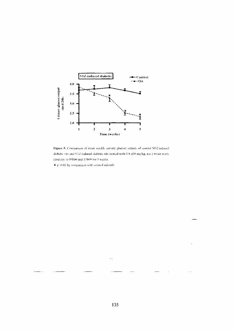
Figure 5. Comparison of mean weekly urinary glucose outputs of control SI/.-induced
diabetic rats and S I'/.-induced diiibetit rats treated wish OA (60 mg/kg, p.a.) twite every
third day at WliOO and 1 ShOO lor 5 weeks.
* p<tU)5 by comparison with control animals
135

Table I. "CUOO.fvl VlH/i Bruker Avance HI NMK spectral data of plant derived OA
and reported OA,1''1
Carbon Position
1
Pinnt Dtrivei l OA
Reported OA | Msihnronw! Kuiidu I W 4 p
8
»> 10
11
\l
13
14
IS
16
18
l<i
20
21
:a 23
.;.:/.> .19. J
47.6
j7.l
23.1)
CA?
143 6
41.6 27,7
33,4 • 1 A *
41.i1
4531
30,7
3 3.S
32,4
2S.I
143.5
11.6
463
!(>3.
45
j0,(
2S
29
JO
136

Table 2. Total amounts of urine voided and NV. K", and (."!' excreted by non-diabetie
and ST/.-diabetie control mud rats during lOh OA treatment period (n = 6 in all groups,].
Group Treatment Urine volume ; Ma" K* CI" |
(mi) (jtimii) (fini(il) \ (funol) I
Nun diabetic Control 13.1±1 I ll^i > _^ O ' " ; 12M±t4 !
" I V.i-1 n-;"*" Tu~\l [~13K8--i43 | _ _ ^ _ 2Mh^i, I ' j Q j ^ J ^ r - ]
(OJ 41"' I d v l T , " lu 11 170" i
Rats
ST/.-inducc«l
diabetic rats
OA
Control
12.7+1
in 4 0
OA It). 0 1
*p<0.05 by comparison with respeelive control animals
p*'0.05 by comparison with non-diabetie animals
137

Tabic ?, Effects of OA on solute excretion ami plasma biochemical parameters in non-
diaheiie and STZ-diabctic control and rats administered OA twice every third
consecutive day for 5 weeks. (11 ~~ 6 in all groups*.
Measure
Glucose (mrnofl)
.Na'inmiol/f)
K'fmniol/l)
CRrttmofl)
1. !rea('nimoI/l)
<_ -fealmint'Cumol 1)
Plasma osmolality
<mOsm/kg H30)
Urine osmolality
(mOsm/kg H2<>)
Gf'R (ml.-'min)
Kidney weights
(g/lOOy b.wr)
j Non-diabetic
j Conlrol
1 4,s>±().l
| 142i1 \ j 3.68x0.25
| 10! i l
! 7.3.-t(j.9
1 25-2 j 1 263i27
' M M u «
• KXHl.JJ
I (f46.'t0i)2
O A-treated
4.2+0.1
142 i 1
>.5*tO.!7
!03:J:1
8 "5=0.7
247 i: 15
266!±78
3.71 ±0.30*
0.44) 0.02
STZ-induced tl
Control
2 6 U "
140=2
3 76.0 M
102=2
16,7 • 1.7"
3! i 3*
382=10'
v u i . y 2
1.81=0. >2:'
0,78=0.07''
iabctic
OA-t rented
2i+r* 137.;3
3.\3±0.20
0614
12.1 = 1,8"
23=2*
316i 16+"
U15*56**
3.07=0.16*4
0.73=0.04'-
*P'0.05 by comparison with respective control animals
' p'0.05 by comparison with non-diabetic animals
138

APPENDIX IV (a)
19. -COMPETITION
turn mmm-&mMM&:m® OF EXOGSHOUS amm m. wemooacmiic'si.
DIABETIC RMS
139

APPENDIX IV (b)
-; V . RD/04/08
ANT-DERIVED OLEANOLIC ACID IN SPRAGUE DAWLEY RATS
'Tuf ts , MA, ^Oyedeji, OO, +Shode FO & *Musabayane, CT rysiology and *Chemistry, University of KwaZulu-Natal, Westville Campus, an 4000, South Africa.
the ethanolic crude Syzygium cordatum (Hochst.) [Myrtaceae] leaf extract etes mellitus management, perhaps due to triterpenoid constituents. .ontrol of diabetes mellitus should include prevention or alleviation of renal tes is associated with impaired kidney function.
was to investigate the renal effects of S. cordatum leaf-derived oleanolic ats.
6kg) (milled) of S. cordatum was sequentially extracted with hexane, Detate, and methanol to give hexane —solubles (HS), dichloromethane-tate-solubles (EAS), and methanol-solubles (MS). Preliminary experiments ned bioactive compounds. A portion of the EAS was subjected to column :a'gel. Gradient elution with n-hexane-ethyl acetate (9:1 to 6:4) produced re combined on the basis of their TLC similarities into three major sub-i-polar fraction (F1) was discarded and semi polar/polar fractions (F2 and 1 to silica gel column chromatography. F2 gave a mixture of oleanolic acid >. F3 gave a mixture of methyl maslinate and methyl corosolate. )A/UA mixture afforded OA (mp > 300°C [Lit. 306-308°C] using ethanol rture was confirmed by NMR spectroscopy (1H & 13C) (Seo ef a/., 1975).
rlriey function and mean arterial blood pressure (MAP) were investigated in iged with hypotonic saline after a 3.5-h equilibration for 4h of 1 h control, 1.5 :pvery periods. OA was added to the infusate during the treatment period." Liced significant (p<0.01) decrease in Na+, K+ and C!" excretion rates without Furthermore, OA significantly (p<0.05) reduced the MAP from a mean pre-rnmHg to reach 105±1 mmHg after the cessation of treatment. The blood ased by the end of the 4-h experimental period to values that did not tt of control animals at the corresponding period.
jat.OA at the dose used may influence kidney function. Further studies are DA leads to impairment of renal electrolyte excretion.
vnimal
140

APPENDIX IV (c)
Dear author, Thank you for submitting your abstract entitled "RENAL EFFECTS OF PLANT-DERIVED OLEANOLIC ACID IN STREPTOZOTOCIN-INDUCED DIABETIC RATS". Your reference number for this submission is 0009 A copy of your submission is attached to this email. The abstracts will be reviewed shortly and you will be informed by January 2009 whether or not your abstract has been accepted for presentation. Please remember to quote the conference name and the reference number in any communication with us. With best wishes, Conference Administrator Society for Endocrinology BES, HARROGATE 2009 THE EFFECTS OF COMBINED PLANT DERIVED OLEANOLIC ACID AND INSULIN ON BLOOD GLUCOSE CONCENTRATION IN STREPTOZOTOCIN-INDUCED DIABETIC RATS
* Cephas T Musabayane. *Rudo F Mapanga, *Mark A Tufts, # 0 0 Oyedeji, & #Francis O. Shode ^Disciplines of Human Physiology and Chemistry, University of KwaZulu-Natal, Westville Campus, Private Bag X54001, Durban 4000, South Africa.
We have previously attributed the hypoglycaemic effects of Syzygium cordatum leaf extract to crude ethyl acetate extract solubles (EAS) containing triterpenoid compounds. The present study was designed to isolate and characterize triterpene components EAS and investigate the antihyperglycaemic effects alone or in combination with insulin in non-diabetic and streptozotocin (STZ)-induced diabetic rats. EAS soluble extracts were obtained after defatting the S. cordatum leaves with hexane followed by dichloromethane before maceration with ethyl acetate. Column chromatography on silica gel using n-hexane-ethyl acetate yielded mixtures of oleanolic acid, ursolic acid, methyl maslinate and methyl corosolate. Oleonolic acid (OA) was obtained by recrystallisation and its structure was determined by !H- and 13C-NMR spectroscopy. Oral glucose tolerance test (OGTT) responses were monitored in separate groups of non-diabetic and STZ-induced diabetic rats given glucose load after 18-h fast, followed by various OA doses (40, 80 and 120 mg/kg, p.o.). Rats treated with deionized water (3 ml/kg, p.o.), or insulin (200 \xg/ kg, s.c) acted as untreated and treated positive controls, respectively. The influence of OA on insulin-induced OGTT responses were studied in separate groups of animals that were treated with combined OA (40 or 80 mg/kg) and insulin (100 or 200 ng/kg, s.c). Blood glucose was monitored at 15-min intervals for the first hour, and hourly thereafter for 3h. All doses of OA as well as insulin significantly (p<0.05) decreased blood glucose concentrations of non-diabetic and STZ-induced diabetic rats. More notable, however, were the dramatic hypoglycaemic effects produced by combined OA and insulin administration. Compared with rats separately administered with insulin or OA, blood glucose concentrations were significantly (p<0.05) lower at all the time points that blood was sampled in animals treated with combined OA and insulin. We suggest that OA may be useful as adjuvant treatment to enhance insulin effects in diabetes mellitus management.
Key words: Diabetes mellitus; hypoglycaemic plants; Syzygium cordatum, triterpernoids; oleanolic acid.
141

APPENDIX IV (d)
Dear author, Thank you for submitting your abstract entitled "RENAL EFFECTS OF PLANT-DERIVED OLEANOLIC ACID IN STREPTOZOTOCIN-INDUCED DIABETIC RATS". Your reference number for this submission is 0050. A copy of your submission is attached to this email. The abstracts will be reviewed shortly and you will be informed by January 2009 whether or not your abstract has been accepted for presentation. Please remember to quote the conference name and the reference number in any communication with us. With best wishes, Conference Administrator Society for Endocrinology BES, HARROGATE 2009 RENAL EFFECTS OF PLANT-DERIVED OLEANOLIC ACID IN STREPTOZOTOCIN-INDUCED DIABETIC RATS
*Rudo F Mapanga,*Cephas T Musabayane, *Mark A Tufts, # 0 0 Oyedeji, & #Francis O. Shode *Disciplines of Human Physiology and Chemistry, University of KwaZulu-Natal, Westville Campus, Private Bag X54001, Durban 4000, South Africa.
We have previously attributed the hypoglycaemic effects of Syzygium cordatum leaf extract to crude ethyl acetate extract solubles (EAS) containing triterpenoid compounds. The present study was designed to isolate and characterize triterpene components EAS and investigate the antihyperglycaemic effects alone or in combination with insulin in non-diabetic and streptozotocin (STZ)-induced diabetic rats. EAS soluble extracts were obtained after defatting the S. cordatum leaves with hexane followed by dichloromethane before maceration with ethyl acetate. Column chromatography on silica gel using n-hexane-ethyl acetate yielded mixtures of oleanolic acid, ursolic acid, methyl maslinate and methyl corosolate. Oleonolic acid (OA) was obtained by recrystallisation and its structure was determined by 'H- and 1JC-NMR spectroscopy. Oral glucose tolerance test (OGTT) responses were monitored in separate groups of non-diabetic and STZ-induced diabetic rats given glucose load after 18-h fast, followed by various OA doses (40, 80 and 120 mg/kg, p.o.). Rats treated with deionized water (3 ml/kg, p.o.), or insulin (200 ug/ kg, s.c) acted as untreated and treated positive controls, respectively. The influence of OA on insulin-induced OGTT responses were studied in separate groups of animals that were treated with combined OA (40 or 80 mg/kg) and insulin (100 or 200 ng/kg, s.c). Blood glucose was monitored at 15-min intervals for the first hour, and hourly thereafter for 3h. All doses of OA as well as insulin significantly (p<0.05) decreased blood glucose concentrations of non-diabetic and STZ-induced diabetic rats. More notable, however, were the dramatic hypoglycaemic effects produced by combined OA and insulin administration. Compared with rats separately administered with insulin or OA, blood glucose concentrations were significantly (p<0.05) lower at all the time points that blood was sampled in animals treated with combined OA and insulin. We suggest that OA may be useful as adjuvant treatment to enhance insulin effects in diabetes mellitus management.
Key words: Diabetes mellitus; hypoglycaemic plants; Syzygium cordatum, triterpernoids; oleanolic acid.
142