Embed Size (px)
Citation preview

UNIVERSITY OF HAWAI'I LIBRARY
FUNCTIONAL VOLLEYBALL SI)IKE-JUMP LANDING BIOMECHANICS AND INJURY INCIDE CE OF
ADOLESCENT FEMALE CLUB VOLLEYBALL ATHLETES
A THESIS SUBMITTED TO THE GRADUATE DIVISION OF THE UNIVERSITY OF HAWAI'1 [N PARTIAL FULFILLMENT OF
THE REQUIREMENTS FOR THE DEGREE OF
MASTER OF SCIENCE IN
KINESIOLOGY AND LEISURE SCIENCE
AUGUST 2008
By
Rumi Isogai Bumbera
Thesis Committee:
Iri s F. Kimura, Chairperson
Ronald Hetzler
Andrew Nichols

ii
We certifY that we have read this thesis and that, in our opinion, it is satisfactory in scope
and quality as a thesis for the degree of Master of Science in Kinesiology and Leisure
Science.
THESIS COMMITTEE
. Chairperson

111
Table of Contents
LIST OF TABLES ......................................................................................................................... v
LIST OF FIGURES ....................................................................................................................... vi
ACKNOWLEDGEMENTS ......................................................................................................... vii
ABSTRACT ................................................................................................................................ viii
INTRODUCTION .......................................................................................................................... 1
Research Questions ............................................................................................................ 3
METHODS ..................................................................................................................................... 4
Research Design ................................................................................................................. 4
Subjects ............................................................................................................................... 4
Instrumentation .................................................................................................................. 5
Procedures .......................................................................................................................... 6
Statistical Analysis ............................................................................................................. 8
RESULTS ....................................................................................................................................... 8
DISCUSSION '" ............................................................................................................................ 11
REFERENCES ............................................................................................................................. 17
APPENDICES .............................................................................................................................. 19
Appendix A The Problem .......................................................................................................... 19
A 1. Problem statement ................................................................................................. 19
A.2. Independent variable(s) .......................................................................................... 19
A3. Dependent variable(s) ............................................................................................. 19
A4. Dependent measure(s) ............................................................................................ 19
A.5. Research question(s) ............................................................................................... 20

IV
A6. Operational definitions ........................................................................................... 20
A.7. Experimental hypotheses ....................................................................................... 21
A.S. Assumptions ............................................................................................................ 21
A9. Delimitations ........................................................................................................... 21
AI0. Limitations ............................................................................................................ 21
All. Significance of the study ...................................................................................... 22
Appendix B. Review of Literature ............................................................................................. 23
Appendix C. Additional Methods .............................................................................................. 36
C.l. Institutional Review Board Fonn .......................................................................... 36
C.2. Institutional Review Board ProposaL .................................................................. 40
C.3. Infonned consent fonn ........................................................................................... 42
C.4. Questionnaire(s) ...................................................................................................... 46
C.5. Data collection fonns .............................................................................................. 50
C.6. Specific testing protocols ....................................................................................... 51
C.7. Standard instructions .............................................................................................. 51
Appendix D. Additional Results ................................................................................................ 52
D.l. Raw data tables ....................................................................................................... 52
D.2. Statistical tables ...................................................................................................... 54
Appendix F. Bibliography .......................................................................................................... 56

v
LIST OF TABLES
Table 1. Subject Demographic Means and Standard Deviations, and Club VolIeybail
Experience ....................................................................................................................... 5
Table 2. Knee Injury History Status by Leg Dominance, Hitting Arm Preference, and
Landing Pattern ............................................................................................................... 9
Table 3. Dependent Variable Means and Standard Deviations by Injury Status ....................... 9
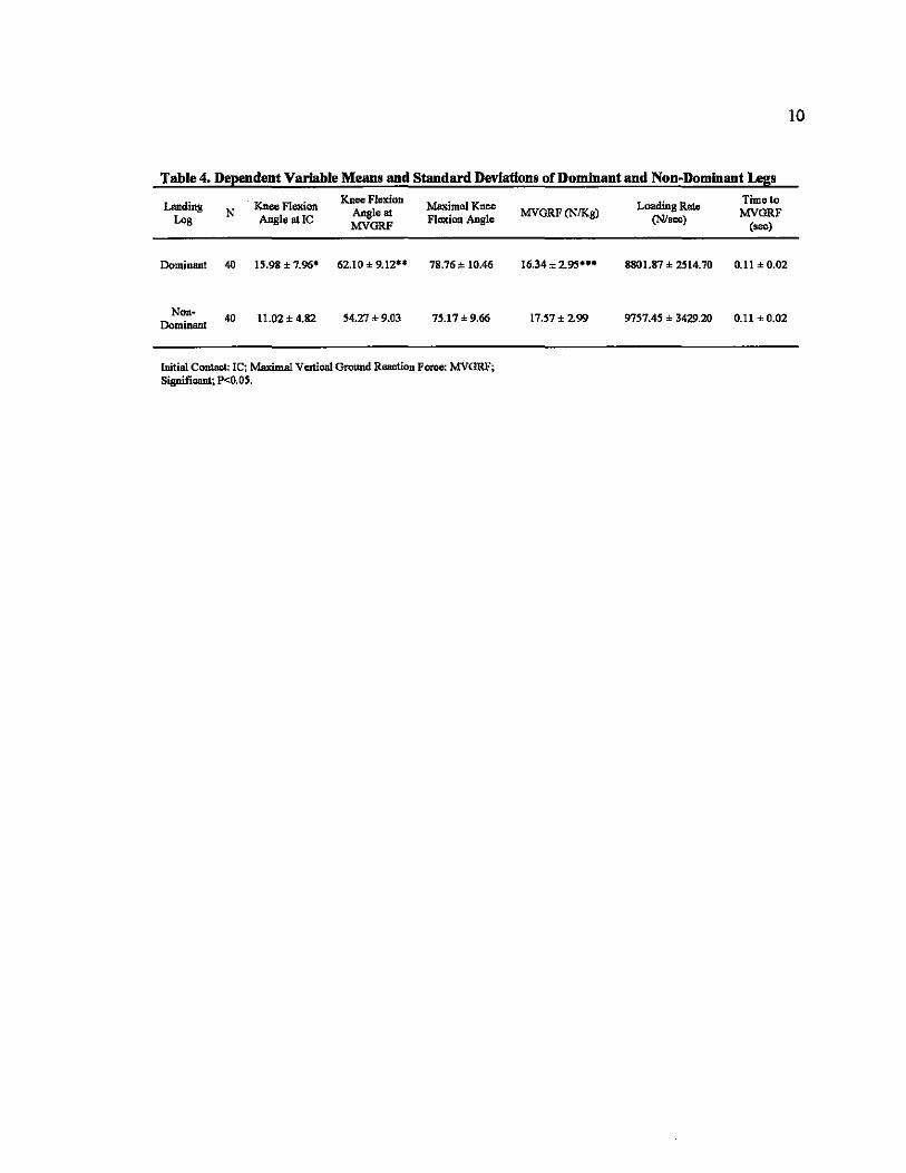
Table 4. Dependent Variable Means and Standard Deviations of Dominant and Non-
Dominant Legs .............................................................................................................. 10

VI
LIST OF FIGURES
Figure 1. Mean Knee Flexion Angle Difference between PIK and NIK .................................. 12
Figure 2. Mean MVGRF Difference between PIK and NIK ..................................................... 13
Figure 3. Mean Loading Rate Differences between PIK and NIK ........................................... 13
Figure 4. Mean Knee Flexion Angle Difference between Dominant and Non-dominant Leg 15
Figure 5. Mean MVGRF Difference between Dominant and Non-dominant Leg .................. 15

ACKNOWLEDGEMENTS
To those who offered substantial support to complete this manuscript, I would like to express my deepest sincere appreciation.
Dr. Iris F. Kimura, Dr. Hetzler, and Dr. Nichols Thank you for giving me an opportunity for my first scientific research in
biomechanics and helping me throughout this project. Without your professional advices, I could not complete this big task
eris, Kaori, and Jenn
Thank you for spending a lot of time with me to finish this project. Without your support, I could not come this far.
Your point of views made me think a lot and I learned a lot from it.
Rie
We did it!
VII
Rachel, Alexis, Rachele, Melissa, and Danny
Thank you for your support throughout whole research process, especially during the data collection. Your idea and opinions made my research more interesting and
the hard processes got more to be fun.
Sheila
Always, you smiled and encouraged me. Thank you very much.
My mother, Sachiko and My sister, Miyuki
Thank you for understanding me and giving me unlimited support from beginning of this study abroad to complete the Master's degree in the United States.
There were some difficult times, but I could come here because of your help.
My husband, John
Thank you for your everlasting patience and a lot of encoumgements. I cannot find the word how much I appreciated your presence.
Mahalo and a lot of Aloha!

V111
ABSTRACI'
Purpose: To functionally investigate volleyball spike-jump landing biomechanics in
adolescent female club volleyball athletes to detennine injury risk factors, and injury
incidence. The independent variables were knee injury history (previously injured knee, PIK,
and Non-injured knee, NIK) and landing leg (Dominant and Non-dominant legs). Dependent
variables were lower extremity kinematics and kinetics.
Methods: We used a causal-comparative retrospective research design to identify associated
knee injury risk factors. Subjects were 40 highly trained adolescent female club volleyball
athletes who completed a retrospective injury questionnaire. Six high-speed three
dimensional motion capture cameras and two force plates were used to collect kinematic and
kinetic data. Two way analyses of variance (ANOVA) linear model (P < 0.05) were used to
analyze two independent (injury status) and six dependent (knee flexion angle at initial
ground contact (IC), knee flexion angle at maximal vertical ground reaction force (MVGRF),
maximal knee flexion angle, MVGRF (NlKg), time from IC to MVGRF (sec), and loading
rate from IC to MVGRF (N/sec) variables.
Results: ANOV A findings indicated significant main effects between PIK and NIK in three
of the six dependent variables. Subjects with PIKs revealed significantly larger knee flexion
angles at initial contact (p = 0.03), exerted significantly greater maximal vertical ground
reaction forces (p = 0.029), and significantly greater loading rates (p = 0.0212) compared
with NIKs. Results also revealed significant main effects between dominant and non
dominant legs in three of the dependent variables. Dominant leg data results revealed larger
knee flexion angles at initial contact (p = 0.0007), larger knee flexion angles at MVGRF (p <
0.0001), and larger MVGRFs (p = 0.0035) than in the non-dominant leg. No interaction
effects were indicated in injury status (PIK vs. NIK) or in landing legs (dominant vs. non
dominant).
Key Words: LOWER EXTREMITY INJURY, KINEMATICS, KINETICS, MOTION ANALYSIS, GROUND REACTION FORCE

I
INTRODUCTION
Sports-related adolescent injury increases in the United States have been paralleled by
increases in youth sports participation. 1 An estimated 2.6 miIlion sports-related emergency
room visits occur each year primarily to individuals between the ages of five and 14 years. 1
The majority of these sports-related injuries involve the lower extremities (LE)I, and are
commonly seen in activities such as volleyball, basketball, and soccer where cutting and
repetitive jumping-landing sequences are fundamental requirements of the sport 2-4.
Consequently, in order to prevent and decrease the consistent rise in sports-related
injuries, gender,5.9 developmental stage (i.e.Tanner and musculoskeletal),7Io injury status,1l12
and sport-specific skill activities13 have been biomechanically analyzed to identify LE injury
risk factors. Gender related findings indicate that females are at greater risk for injuries than
their male counterparts, especially with regard to LE injuries.5•9 These results may be
attributed to fuctors such as larger Q-angle, genu recurvatum, and lower muscle strength
values in female athletes.5 6 9 Additionally, females demonstrate smaller hip and knee flexion
angles, and larger ground reaction forces (GRF) during jumping and landing activities than
males.569
Jumping and landing studies on pre- and post-pubescent subjects are limited and
converse.7lO One study 7 concluded that pre-pubescent subjects were at greater risk for LE
injuries than post-pubescent subjects because they demonstrated smaller knee and hip flexion
angles which were associated with larger ground reaction forces (GRF) during vertical jump
landings sequences.7 Conversely, another biomechanical study which compared pre- and
post-pubescent groupslO concluded that post-pubescent subjects were at greater risk for
Anterior Cruciate Ligament (ACL) injury than pre-pubescent subjects because they

2
demonstrated smaller knee flexion angles during jump landings. \0 Additionally, rapid
musculoskeletal growth confounded by sport activity stress has been shown to cause growth
plate apophysis and joint surface injuries in adolescent athletes. 14 Moreover, repetitive stress
and rapid increases in training at consistently high intensities have been associated with
over-use and chronic injuries on the immature musculoskeletal system of adolescent
athletes. 14
Prior injury history has also been identified as a potential lower extremity risk factor
in biomechanicallanding studies. IH3 15 Athletes with prior ACL injury histories revealed
smaller hip and knee flexion angles upon landing than uninjured athletes.l112 This finding is
converse to the results of one study that examined functional volleyball specific tasks and
identified the relationship between patellar tendonitis and volleyball landing biomechanics. 13
Results of this study indicated that larger knee flexion angles demonstrated during landings
were one of the predictors of patellar tendonitis in elite adult male volleyball players. \3
Volleyball is the third most popular sport in the United States for adolescent female
athletes. 16 Additionally, most volleyball injuries occur in the right, middle, and left front
positions,4 and the majority of these injuries occur to the LE, at the knee and ankle in both
acute and overuse conditions2-4. Patellar tendonitis, has been identified as the most common
overuse injury among volleyball athletes as a result of repetitive high-intensity jumping.2-4
While acute knee injuries are not as common as ankle injuries, ACL sprains, have been
associated with greater sports participation time losses than other injuries.2 3
Several studies have been conducted to examine landing biomechanics by using
different jump techniques.5-13 IS Despite having provided useful information about landing
biomechanics,5-13 IS all but the aforementioned volleyball study 13 involved non-functional,

non- sport specific activities. Therefore, the purpose of this study was to functionally
investigate volleyball spike-jump landing biomechanics to determine injury risk factors and
injury incidence in adolescent female club volleyball athletes.
Research Questions
(I) What were the spike-jump landing kinetic differences between previous knee injury
and non-injury groups?
(2) What were the spike-jump landing kinematic differences between previous knee
injury and non-injury groups?
(3) What were the spike-jump landing kinetic differences between dominant and non
dominant landing legs?
3
(4) What were the spike-jump landing kinematic differences between dominant and non
dominant landing legs?

4
METIIODS
Research Design
We used a causal-comparative retrospective research design. The goal of this study
was to simulate a functional volleyball spike-:iump landing that would occur in an actual
sports setting. A retrospective injury questionnaire was administered prior to biomechanical
data collection and used to identify knee injury history. (Appendix C4) The volleyball
spike-jump landings of adolescent female club volleyball athletes of different knee injury
histories (previously injured knee; PIK or non-injured knee; NIK), and landing legs
(dominant or non-dominant) were compared to examine differences in lower extremity
kinematics and kinetics. In this study, the "dominant leg" was defined as the contralateral
leg of the spiking arm.
Subjects
Forty highly trained female adolescent club volleyball athletes aged 12 to 18 years
volnnteered to participate in this study. Subject qualifications included participation in at
least three practices per week for between five and 11 consecutive months. Highly
competitive club team classification was based on historic and consistent USA Volleyball
Junior Olympics showings and win-loss ranking in the upper 25 percent nationally. Prior to
study participation, all subjects and their parentsllegal guardians read and signed written
informed assent and consent forms approved by the University Committee on Human Studies
(Appendix C3). General medical and injury questionnaires (Appendix C4) were reviewed by
the university team physician to screen for pathologies or physical contraindications to study
participation. All subjects were healthy and asymptomatic at the time of data collection and

able to properly peIfonn volleyball spike jumps. Subjects' physical characteristics are
presented in Table 1.
Table 1. Subject Demographic Mean. and Standard Deviatlou., and Club Voneyban Experience
Skill Club VB
Level N Age (years) Experience Height (em) Weight (Kg) (years)
Total 40 14.40 ,1,2.21 3.84,1,2.14 165.80,1,821 60.57,1, 10.87
-High 21 16.52,1,0.60 5.16,1, 2.22 17023%6.91 67.80,1,9.55
-Low 19 12.32,1,0.48 2.53,1, 0.96 160.89,1, 6.82 52.58,1,5.43
Volleyball: VB; Injury Status: Previously Injured Knee: PIK; Non-Previously Injured Knee:PIK
Instrumentation
Injury history questiounaires were used to identify knee injury status of subjects as
PIK or NIK. The definition of knee injury was any injury that resulted in volleyball practice
or game participation time loss and/or medical attention. The questionnaire consisted of 14
closed and five open ended questions and anterior and posterior pictorial injury location
identification. (AppendixC4)
Three-dimensional (3D) infrared motion capture system Vicon Motion Capture
System (Vicon MX, Centennial, Colorado) and Peak Motus software (version 8.0, Vicon,
Inc., Centennial, Colorado) were used to capture, reduce, and analyze kinematic spike-jump
5
landing data. Six 3D cameras were placed on each side of the testing area so that at least two
of the six cameras captured the position of the reflective markers (1.4 cm in diameter) during
spike jump landings. Three-dimensional kinematic data were time synchronized and
collected at 240 (Hz). Both kinematic and kinetic data were smoothed using the Butterworth

filter optimized by Peak Motus software. Knee flexion angles were calculated with Peak
Motus software using projected segmental angles.
6
Full body reflective marker set described previouslyll was used for placement of24
reflective bilateral markers. Bilateral reflective marker placements included:
acromioclavicular (AC) joint, anterior superior iliac spine (ASIS), posterior superior iliac
spine (PSIS), greater trochanter (GT), anterior aspect of thigh (10 cm above the superior pole
of patella), lateml epicondyle, tibial tubelosity, anterior aspect of distal tibia, lateral malleolus,
calcaneus, and distal head of the second and fiflh metatarsal bones.
Two force plates (Advanced Mechanical Technology Incorporated, Boston,
Massachusetts) embedded parallel to each other and anterior to the volleyball net
(representation), and flush with the floor surface were used to collect kinetic data during
spike-jump landings. Ground reaction force (GRF) data were time synchronized and
collected at 480 (Hz) measured in Newtons (N), and normalized to body mass.
VoUeyball net fabricated by the investigator and "Spike It" (Komey Board Aid, Inc.
Roxton, Texas) hall holder were used to simulate functional volleyball spiking. The
simulated volleyball net was positioned anterior and parallel to the force plates and set at
regulation heights. The ball holder height was adjustable to one inch increments relative to
the appropriate volleyball spike-jump.
Procedures
All data were collected in the University Human Performance Labomtory by National
Athletic Trainers' Association (NATA) Board of Certification (BOC) certified athletic
trainers (ATC). Potential study subjects were provided with team and individual information
sessions that included: introduction of study procedures via power point presentation, data
collection demonstration, administration of consent and assent forms, and general medical

7
and injury history questionnaire form completion. Following medical health clearance and
injury status establishment, data collection session appointments were selected. All subjects
reported for data collection rested (no organized pmctice or game participation) wearing
sports bra, spandex volleyball tights, and volleyball shoes regularly worn during pmctices
and games. Immediately prior to biomechanical data collection, the same female SOC ATC
collected anthropometric data consisting of height, weight, body composition (3 sites), and
Q-angle. All subjects were given a I O-minute warm up session on a stationary bike and
self-addressed stretching session. Siomechanical test mmiliarization included identification
of appropriate ball holder height and standard three-step approach spiking practice. Subjects
were instructed to pmctice until they could perform three successful spike jumps. Spike
jump success and consequent data collection acceptance consisted of: a proper three-step
approach; appropriate ball contact (spike); and, landing with entire right and left foot
placement on adjacent force plates. Upon successful and consistent spike-jump landing bout
acceptance, adhesive reflective markers were attached to aforementioned marker set
anatomical placement sites directly on the skin or spandex tights (ASIS, PSIS, GT) by the
same female HOC ATC.
Three classifications were used to identifY spike-jump landing patterns of subjects.
The double leg pattern involved simultaneous ground contact with both feet and/or ground
contact by individual feet in less than or equal to 33 rns. The single leg pattern involved
unilatemI ground contact by individual feet in greater than 33 ms. 17 Subjects were identified
as exhibiting a double leg pattern or a single leg pattem when all three data collection landing
trials could be classified as either double or single leg landings. When data collection

8
landing trials involved double and single leg landings the subject was classified as exhibiting
an inconsistent landing pattern.
Statistical Analysis
Two-way analyses of variance (ANOVA) linear model were used to analyze six
dependent variables. Independent variables consisted of injury status (PIK or NIK) and
landing leg (dominant or non-dominant). Dependent variables consisted of knee flexion
angle at initial ground contact (IC~ knee flexion angle at maximal vertical ground reaction
force (MVGRF), maximal knee flexion angle, MVGRF (N1Kg), time from IC to MVGRF
(soo), and loading rate from IC to MVGRF (N/soo). Statistical Analysis Software (SAS)
version 9.1 (SAS Institute, Inc., North Carolina) was used to analyze the biomechanical data.
Significance level was established at P < 0.05.
RESULTS
Subject descriptive data are presented in Table 1. Knee injury history status, hitting
arm, and landing pattern are presented in Table 2. Dependent variable means and standard
deviations of previous injury (PIK) and non-injury (NIK) history knees are presented in
Table 3. Dependent variable means and standard deviations of each landing leg are
presented in Table 4.
Analysis of Variance findings indicated significant main effects between PIK and
NIK in three of the six dependent variables. Subjects with PIK revealed significantly larger
knee flexion angles at initial contact (p = 0.03), exerted significantly greater MVGRF (p =
0.029), as well as significantly greater loading rate (p = 0.0212) than in the NIK.
Results also revealed significant main effects between dominant and non -dominant
legs in three of the six dependent variables. Dominant leg data ofsubjects revealed larger

9
knee flexion angles at initial contact (p = 0.0007). larger knee flexion angles at MVGRF (p <
0.0001). and larger MVGRF (p = 0.0035) than in the non-dominant leg. A tendency for
larger maximal knee flexion angles (p = 0.065). was found in dominant legs. No interaction
effects were indicated in injury status (PIK vs. NIK) or in landing legs (dominant vs. non-
dominant).
Table 2. Knee Inju!}: Histo!}: Status b;r .Lea Domlnauce, HittiuS Arm Preference, and Landini! Pattern
Skill PIK PIK Right Left Single Leg Double Lag Inconsistent
Level N Dominant Non- Ann Ann Landing Landing Landing
dominant Hitter Hitter Pattern Pattern Pattern
Total 40 4 5 37 3 0 35 5
-High 21 3 5 20 1 0 19 2
-Low 19 0 17 2 0 16 3
Table 3. Dependent Variable Means and Standard Deviations b;r InJu!}: Status
ugury Knel: Flexion Knee Flexion Maximal Knee Timeta N Angle at MVGRF (N1Kg) Loading Rate (NIsec) MVGRF
Hx Angle at IC MVGRF Flexion AnsIe (,eo)
NIC 71 1294 ± 7.00. '7.87 ± 9.96 76.80 ±9.67 16.71 ± 296" 9006.71 ± 2818.45." 0.11 ±O.O2
PIC 9 17.91± 5.'2 60.67± 8.95 7826± 14.10 18.93 ± 292 11432.91± 3879.95 0.12± 0.03
Initial Conlala: IC, Maximal Vertioal Ground Reaotion Foroe; MVGRF: Previously Iqjurcd Knee: PIC. Non-Injured Knee: NI~
10
Table 4. Dependent Variable Means and Standard Deviations of Dominant and Non-Dornmant Legs
Landing Knee Flexion Knee Flexion Maximal Knee Loading Rate Time to
N Anglo aI MVGRF CN/Kg) MVGRF Log Anglo aile
MVGRF FlClCion Angle (NIB .. )
(so.)
Dominant 40 13.98 ± 7.96· 62.10 ± 9.12" 78.76 ± 10.46 16.34± 295 ••• 8801.87± 2j14.70 0.11 ± 0.02
Non-40 11.02± 4.82 54.27 ± 9.03 75.17 ±9.66 17.57 ± 2.99 9757.45 ± 3429.20 0.11 ±0.02
Dominant
Initial Con1af)l: IC; Maximal V crtieal Ground Reaction Force: MVGRF; Signifi .... ; P<O.05.

11
DISCUSSION
The most important finding of our study was that the landing kinematics and kinetics
ofsubjects who presented with a history of knee injuries (PIK) were significantly different
than subjects who had not previously injured their knees (NIK) in volleyball. Subjects with
previous knee injury histories initially contacted the ground with larger knee flexion angles
(17.91° ± 5.52° > 12.94° ± 7.00"~ greater maximal vertical ground reaction forces (18.93 ±
2.92 Nlkg> 16.71 ± 2.96 Nlkg) and higher loading rates (11432.91 ± 3879.95 N/s>
9006.71± 2818.45 N/s) during the total landing sequence, than NIK subjects. Interestingly,
maximal knee flexion angles were similar between PIK (78.26° ± 14.10°) and NIK (76.80° ±
9.67"), suggesting that PIK subjects had less available knee range of motion. Since subjects
with NIK initially contacted the ground with straighter knees than PIK, but both PIK and
NIK end range of motion were similar, it appears that PIK had less available knee flexion
range of motion to dissipate force resulting in significantly greater MVGRF than NIK.
(18.93 ± 2.92 Nlkg> 16.71 ± 2.96 Nlkg). (Figures 1-3) This landing characteristic ofPIK is
similar to the previous study which compared single leg landing biomechanics of males and
females. 6 Study findings demonstrated that females had smaller (less available) total knee
and hip range of motion and greater peak GRF s than males.6 The previous results further
substantiated our retrospective injury data that this type oflanding strategy may increase the
risk of knee injury due to less of the available range of motion at the knee and hip joints to
dissipate energy re~ulting in greater ground reaction forces6 Since the current study is
retrospective in nature, it is unclear whether our subjects' injury characteristics were related
to injury development or whether the injury precipitated the development of our
aforementioned injury characteristics.

Knee Flexion Angle at Ie Maximal Knee Flexion Angle
Figure 1. Mean Knee Flexion Angle Difference between PIK and NIK DNIK (n = 71)
IiIIPIK (n =9)
12

225
22
215
21
20.5
20
195
~ 19 ~ ~ 185
16
175
17
165
16
155
15
16000
14000
12000
MVGRF (N/Kg)
Figure 2. Mean MVGRF Difference between PIK and NIK
~
ONIK (n = 71)
" PIK (n=9)
Loading Rate (N/sec) ONIK (n = 71)
Figure 3. Mean Loading Rate Difference between PIK and NIK III PIK (n = 9)
13

14
Findings of our study also indicated biomechanical differences between dominant and
non-dominant landing legs during functional volleyball spike jumps. Dominant leg data of
our subjects revealed significantly: larger knee flexion angles at initial contact (i 5.98° ±
7.96° > 11 .02° ± 4.82°); larger knee flexion angles at MVGRF (62.10 ° ± 9.12 ° > 54.27 ° ±
9.03°); and , smaller MVGRF (1634 ± 2.95 Ikg < 17.57 ± 2.99 N/kg) Although not
significant (p = 0065), dominant leg maximal knee flexion ang le data appeared larger than
non-dominant leg data (Figure 5 and 6). Our findings are supported by previous
biomechanical studies,l H 9 II that indicated an inverse relationship between knee flexion angle
and ground reaction force . Consequently, non-dominant leg data of our subjects revealed
significantly smaller knee flexion angles during the landing sequence whi le exhibiting
greater MVGRF, often referred to as "stiff landings".ll l 18 "Stiff landing" has been
identified as a risk factor for ACL injury secondary to the anterior dislocating force of
quadriceps musclelll 19 Conversely, large knee flexion angles during landing were identified
as patellar tendonitis risk factors secondary to eccentric loading of the patellar tendon 13
Based on the previous findings, there may be increased risk of ACL injury on the
non-dominant leg and increased risk of patellar tendonitis on the dominant leg offemale
adolescent volleyball athletes .

0 ~
~ '" ~ 0
~ 0 "-
100
00
00
70
60
eo
40
'" 20
10
a
21
205
20
19.5
19
18.5
18
175
17
16.5
16
155
15
Knee Flexion Angle at Ie Knee Flexion Angle at MVGRF
Maxirral Knee Flexion Angle
Figure 4. Mean Knee Flexion Angle Difference between Dominant and Non-dominant Leg
[ilDominant (n =40)
I. Non-Dominant (n = 40)
MVGRF (N/Kg) rJ Doninan! (n .40)
Figure 5. Mean MVGRF Difference between Dominant and Non-dominant eg ~n-Dominant (n = 40)
IS

16
Single leg landing has also been identified as an injUIY risk fuctor due to the fuct that
a single limb must dissipate and absorb the forces created by the entire body.20 A previous
study involved investigation of volleyball spike and block jump landing patterns of collegiate
female athletes.17 Results indicated that almost half of all landings from spike jumps were
single leg landings, and more than two-thirds of those single leg landings were performed on
the left leg. 17 The results also suggested that the occurrence of single leg landings might be
related to the sequence of the spiking technique.17 When a right-handed player spikes a ball,
the trunk is laterally flexed to the left. This lateral flexion raises the right side of the body
and can precipitate left foot loading (dominant leg) contact upon landing.17
ContraIY to the previous landing pattern study,17 the majority of our subjects
demonstrated "double leg" landings with subsequent higher MVGRF on the non-dominant
leg than the dominant leg. Differences in landing patterns may be attributed to the difference
in subject group characteristics. The subjects of the previous study were elite college female
volleyball players17, while our subjects were adolescent female club volleyball athletes under
the age of 18. Age and skill level may have influenced resulting landing patterns, as study
results indicate that landing patterns val)' with increased skill and experience.7 17 In addition,
it should be noted that our subjects were trained to land on both legs simultaneously to
prevent injuries. These findings reflect the importance of early instruction in proper
volleyball jump landing techniques by individuals associated with the development of youth
volleyball players in order to prevent future knee injuries. Our study supports the importance
of the sports specific functional research.
In conclusion, subject injUIY status and landing leg dominance influenced the
kinematic and kinetic results of our study.

REFERENCES
1. Burt CW, Overpeck MD. Emergency visits for sports-related injuries. Ann Emerg Med 2001;37(3):301-8.
17
2. Powell JW, Barber-Foss KD. Injury Patterns in Selected High School Sports: A Review of the 1995-1997 Seasons. J Athl Train 1999;34(3):277-284.
3. Agel J, Palmieri-Smith RM, Dick R, Wojtys EM, Marshall Sw. Descriptive epidemiology of collegiate women's volleyball injuries: National Collegiate Athletic Association Injury Surveillance System, 1988-1989 through 2003-2004. J Athl Train 2007;42(2):295-302.
4. Augustsson SR, Augustsson J, Thomee R, Svantesson U. Injuries and preventive actions in elite Swedish volleyball. Scand J Med Sci Sports 2006; 16(6):433-40.
5. Salci Y, Kentel BB, Heycan C, Akin S, Korkusuz F. Comparison of landing maneuvers between male and female college volleyball players. Clin Biomech (Bristol, Avon) 2004;19(6):622-8.
6. Schmitz RJ, Kulas AS, Perrin DH, Riemann BL, Shultz SJ. Sex differences in lower extremity biomechanics during single leg landings. Clin Btomech (Bristol, Avon) 2007;22(6):681-8.
7. Swartz EE, Decoster LC, Russell PJ, Croce RV. Effects of Developmental Stage and Sex on Lower Extremity Kinematics and Vertical Ground Reaction Forces During Landing. J Athl Train 2005;40(1):9-14.
8. Russell KA, Palmieri RM, Zinder SM, Ingersoll CD. Sex differences in valgus knee angle during a single-leg drop jump. J Athl Train 2006;41 (2): 166-71.
9. Lephart SM, Ferris CM, Riemann BL, Myers ill, Fu FH. Gender differences in strength and lower extremity kinematics during landing. Clin Orthop Relat Res 2002(401):162-9.
10. Hass CJ, Schick EA, Tillman MD, Chow JW, Brunt D, Cauraugh JH. Knee biomechanics during landings: comparison of pre- and postpubescent females. Med Sci Sports &erc 2005;37(1):100-7.
11. Hewett TE, Myer GD, Ford KR, Heidt RS, Jr., Colosimo AJ, McLean SG, et al. Biomechanical measures of neuromuscular control and valgus loading of the knee predict anterior cruciate ligament injury risk in female athletes: a prospective study. Am J Sports Med2005;33(4):492-501.
12. Louw Q, Grimmer K, Vaughan C. Knee movement patterns of injured and uninjured adolescent baskethall players when landing from a jump: a case-control study. BMC Musculoskelet Disord 2006;7:22.
13. Richards DP, Ajemian SV, Wiley JP, Zemicke RF. Knee joint dynamics predict patellar tendinitis in elite volleyball players. Am J Sports Med 1996;24(5):676-83.
14. Soprano JP, Fuchs SM Common Overuse Injuries in the Pediatric and Adolescent Athlete. Clinical Pediatric Emetgency Medicine 2007;8(1 ):7-14.
15. Bisseling RW, Hof AL, Bredeweg SW, Zwerver J, Mulder T. Relationship between landing strategy and patellar tendinopathy in volleyball. Br j Sports Med 2007;41 (7):e8.
16. Associations NFoSHS. NFHS Participation Figures Search, Year of2004-2005, 2007. 17. Tillman MD, Hass CJ, Brunt D, Bennett GR. Jumping and landing techniques in elite
women's volleyball. Journal of Sports Science andMedicine 2004;3:30-36.

18
18. Sell TC, Ferris CM, AbtJP, Tsai YS, Myers JB, Fu FH, etal. The effect of direction and reaction on the neuromuscular and biomechanical characteristics of the knee during tasks that simulate the noncontact anterior cruciate ligament injury mechanism. Am J Sports Med 2006;34(1 ):43-54.
19. Li G, Rudy TW, Sakane M, Kanamori A, Ma CB, Woo SL. The importance of quadriceps and hamstring muscle loading on knee kinematics and in-situ forces in the ACL. J Biomech 1999;32(4):395-400.
20. Schafle MD. Common injuries in volleyball. Treatment, prevention and rehabilitation. Sports Moo 1993;16(2):126-9.
21. Smith R, Ford KR, Myer GD, Holleran A, Treadway E, Hewett TE. Biomechanical and performance differences between female soccer athletes in National Collegiate Athletic Association Divisions I and ill. J Athl Train 2007;42(4):470-6.

AI. Problem statement.
APPENDICES
AppendixA The Problem
The purpose of this study was to functionally investigate volleyball spike-jump
landing biomechanics to detennine injury risk factors, and injury incidence in adolescent
female club volleyball athletes.
A2. Inde,pendent variable(s).
The independent variables were previous knee injury history (previous injury knee;
PIK or non-injured knee; NIK), and landing leg (dominant or non-dominant leg).
A3. Dependent variable(s).
The dependent variables were lower extremity kinematics and kinetics.
A.4. De,pendent measure(s).
19
The dependent measures were knee flexion angle at IC, knee flexion angle at
MVGRF, maximal knee flexion angle, MVGRF, Time from IC to MVGRF, and loading
rate from IC to MVGRF.

20
A.5. Research Question(s).
1. What were the spike-jump landing kinetic differences between previous knee injury
and non-injury groups?
2. What were the spike-jump landing kinematic differences between previous knee
injury and non-injury groups?
3. What were the spike-jump landing kinetic differences between dominant and non
dominant landing legs?
4. What were the spike-jump landing kinematic differences between dominant and non
dominant landing legs?
A.6. Operational definitions.
1. Dominant leg was defined as the contraiateralleg of the spiking arm. For instance, a
subject's hitting hand was right, left leg was her dominant landing leg and right leg
was her non-dominant leg.
2. Successful spike jump criteria were: a proper three or four step approach; contact
(spike) with the volleyball; landing on the dominant leg where the whole dominant
foot must be on one of the force plates; the non-dominant leg landed on the other
force plate.
3. The criteria of lower extremity injury were subject saw medical personnel (i,g, an
athletic trainer, physical therapist, or doctor) for the specific condition and had to
miss participation to the practice or game due to the condition.
21
A. 7. Experimental hypotheses.
I. There will be kinematic differences between previously injured knees and non-injured
knees.
2. There will be kinetic differences between previously injured knees and non-injured
knees.
3. There will be kinematic differences between dominant legs and non-dominant legs.
4. There will be kinetic differences between dominant legs and non-dominant legs.
A.S. Assumptions.
The assumptions for this study were: (I) The subjects were able to understand the
directions and their tasks, (2) The subjects answered the medical history and injury
questionnaire honestly and correctly, and (3) The subjects were able to perform proper spike
jumps.
A.9. Delimitations.
The delimitations of this study were: (I) Subjects with current injury, and (2) The
subjects who played other sports except volleyball.
A.IO. Limitations.
The limitations of this study were (I) Different maturation level among
subjects, and (2) Small sample size.

22
A.II. Significance of the study.
This study focused on simulating functional movement that would occur in a natural
sports setting. Only one study, conducted by Richards et aI,13 studied functional volleybalI
landing biomechanics in adult male athletes. This study was focused on volIeybalI spike
jump landings in adolescent female club volIeyball athletes, the population with a greater risk
of lower extremity injuries due to their physical immaturity. Investigating biomechanical
characteristics associated with lower extremity injury may help to reduce lower extremity
injuries and ultimately lengthen the span of the individual's athletic life.

23
Appendix B. Review of Literature
Lower extremity injuries are often seen in sports which require repetitive jump-land
sequences such as volleyball.4 The sports-related injuries in volleyball are commonly seen at
the lower extremities3 4, and the number of those injuries have increased over the past twenty
years due to a higher number of participants in sports activities1. Injury historyl1-131S 19,
gender difference6 8 9 21, and different developmental stages 710 have drawn attention to the
importsnce of understanding the mechanisms of injuries to prevent sports-related injuries.
Volleyball Landing and Injurv Incidence
Augustsson et al.4 conducted a survey of 225 elite Swedish volleyball players (10
men's teams and nine women's teams) to describe type, location, and severity of injury, as
well as player position relative to injury incidence. The operational definition of injury was
an injury that occurred as a result of participation in volleyball that caused the athlete to leave
the court at the time of injury or to reduce their level of training. Injury severity was
categorized by the length of time they were absent from participation. Acute and chronic
injuries were not distinguished in their study. The majority ofinjuries were located in the
athletes' lower extremities in the following percentages: ankle (23%); knee (17%); and back
(16%). Most injuries were minor (absence for less than one week), or moderate (absence for
two to four weeks). Major injuries (absence for more than four weeks) accounted for 19 out
of 121 injuries, or 6%. Seventy-three percent of the injuries were related to the three front
players (attackers and blockers) and occurred during spiking and blocking.
Agel et al. 3 reviewed 16 years of National Collegiate Athletic Association injury
surveillance data specifically for women's volleyball players from the academic year of
1988-1989 to 2003-2004. They found that more than 55% of all game and practice injuries

were to the lower extremities. In both game and practice situations, injuries to the ankle
(44.1 % from game situations and 29.4% from practice situations) and knee (14.1 % from
game situations and 7.8% from practice situations) were the most common locations for
injuries in college female volleyball players. Menisci, collateral and cruciate ligament
injuries comprised the highest proportion of the internal knee injuries. Further, more than
25% of knee injuries were caused by "no apparent contact", 21.1 % involved "a player
landing on another player" and 20.6% were the result of "contact with the floor". The
majority of injuries (67.3%) occurred in athletes in the front three positions.
24
Tillman et al.17 investigated spike and block jump take-off and landing patterns in
elite female collegiate athletes. Each movement was categorized by jump type (spike or
block) and phase (taking-off or landing). The jump type was further subcategorized as
bilateral or unilateral (right or left) landingltake-offpattern. If one foot left the ground one
frame (33rns) ahead of the other, the jump was categorized as unilateral (right or left), and a
similar convention was used to categorize landing. The majority (84%) of spike jump take
off were performed using both legs and 16% of spike jumps take-off were performed using
one leg (right - 2%, left -14%). Bilateral spike jump landing percentages decreased to 55%,
while unilateral leg usage increased to 45% (right -10%,left - 35%) compared to the
corresponding take-off. The authors concluded that the increased frequency of single leg
landings in landing might be related to the sequence of spiking technique. When right
handed players spike balls, their trunks are laterally flexed to the left. This lateral flexion
raises the right side of the body and can lead to a left foot first contact upon landing. They
noted that unilateral landings could lead to a loss of balance and subsequent injury.

25
In summary, the majority of injuries in volleyball occur in the lower extremities,
especially at the ankle and knee.3 4 This is the result of repetitive spike and block jump-land
sequences which cause tremendous forces on the lower extremities during landing.17
Unilatemllanding might increase the overall risk of lower extremity injuries.17
Knee Injurv Risk Factors Associated with Landing
Richards et a!. 13 investigated the biomechanics of volleyball spike and block jump
take-offs and landings, and incidences of patellar tendonitis (or Jumper's knee) in elite male
volleyball players. Three-dimensional high-speed cameras and one force plate were used to
collect all biomechanical data oflower extremities. In the study, six out of ten subjects had a
history of patellar tendonitis, and all subjects were right-handed. Sport specific simulation
using a portable net setat regulation height (2.43 meters) where subjects hit and blocked
spiked and set volleyballs, respectively during trials. SUbjects repeatedly performed three
different trials for each leg landing on a force plate (right and left), each task (spike and
block), and each phase (take-off and landing). The logistic regression revealed that maximal
left knee flexion angle during spike jump landing, peak external tibial torsional moment for
the right knee during the spike jump take-off and the left knee during block jump take-off.
Peak vertical ground reaction force for the right limb during both spike and block jump take
offs were revealed a predictors of patellar tendonitis.
Hewett et aL 11 investigated the relationship between lower extremity landing
biomechanics and ACL injuries in female adolescent athletes. The study involved a
prospective study design; prior to the season the authors collected the drop vertical jump
landing data of205 female adolescent athletes who participated in soccer, baskethall, and
volleyball. During the season nine ACL ruptures were reported. The landing biomechanical

26
data of nine ACL ruptured limbs versus 390 non-injured limbs was compared to determine if
any differences pre-existed. The injured limbs demonstrated significantly different knee
posture and loading compared to non-injured limbs. The ACL injured group demonstrated
greater knee abduction (valgus) angle (8.4 degrees greater than the non-injured) at initial
contact, and at maximum knee flexion (7.6 degrees greater than the non-injured). hi the ACL
injured group, there was a strong correlation between knee abduction (valgus) angle and peak.
vertical ground reaction force, whereas no correlation was found in the non-injured group.
Even though differences in knee flexion angles did not reach the level of statistical
significance, the maximum knee flexion angle at landing was 10.5 degrees less in the ACL
injured group than in the non-injured group. Significant knee loading was also observed in
the ACL injured group but not in the non-injured group. The logistic regression analysis
revealed that the knee abduction moment and angles (lC and peak. values) were significant
predictors of ACL injury status.
Louw et al. 12 compared landing biomechanics of subjects with previous knee injury
and those without. The subjects of this study consisted of22 adolescent male and female
basketball players 14 to 16 years of age. During the landing biomechanical trials, subjects
performed ten "jump-shots" landing where each foot landed five times on each force plate.
They found that peak. knee flexion angles were negatively correlated with peak. ground
reaction forces. The group with no prior knee injuries demonstrated significantly deeper
knee flexion angles on landing (66.4degrees) than the group who had a history of knee
injuries (57 .1 degrees). The high correlation between knee angle and maximum ground
reaction force suggested knee flexion angle ( degree) could possibly be one of the most

important factors relative to impact reduction after landing from a jump which may reduce
the chance ofLE injuries.
27
Bisseling et al. IS compared landing biomechanics in elite adult male volleyball
players in relation to patellar tendonitis Gumpers ' knee). The subjects were divided into
three groups based on an injury questionnaire: the control group, the previous jumper's knee
(pJK) group, and the recent jumpers' knee (RJK.) group. Subjects with a history of recent
injury or surgery at the LE or the back in the past 3 months were excluded from the study. If
bilateral patellar tendinopathy was reported, the more symptomatic knee was selected for
analysis. The Victorian Institute of Sport Assessment (VISA) Scale was used to record pain,
function, and athletic activities. More points in the assessment indicated better knee
condition. The subjects in the control group had no history of patella tendonitis, no pain
during a single leg decline squat, and no palpation tenderness. They had a score of over 80
points on the VISA Scale. The subjects in the PJK group were classified as having
asymptomatic patellar tendonitis, a history of pain located at the proximal patellar tendon or
insertion of the quadriceps tendon, and patellar tenderness. They had no pain during single
leg decline squat and were pain free in the last five months, and the VISA score was over 80
points. The subjects in the RGK group had pain during single leg decline squat, palpation
tenderness, and less than 80 VISA points. The subjects performed drop jumps from 30, 50,
and 70 cm high platforms. They were required to land facing forward with both feet on the
ground with one foot on the force plate. They found that the knee flexion angle at time to
peak vertical ground reaction force (pVGRF) was negatively correlated with PVGRF as well
as with loading rate VGRF (peak VGRF value divided by time from touch down to peak
value) among all three groups and heights, except for the RJK group at 70cm. The PJK

group showed higher knee angular velocities and higher ankle planter flexion moment
loading rates. Furthermore, the PJK group tended to have higher loading rates compared
with the control group. It was concluded that the subjects in the PJK group might have a
higher risk of developing patellar tendonitis due to a higher PVGRF and loading VGRF.
28
Aside from the kinetic and kinematic characteristics during landing task, Li et al.19
investigated the relationship between force produced by muscles and force applied on ACL
at different knee angles. They investigated the role of isolated quadriceps and combined
quadriceps and hamstrings load on knee kinematics and the in-situ forces in the ACL. They
used a robotic/universal force-moment sensor (UFS) to measure the in-situ forces in the ACL
and knee kinematics in response to isolated quadriceps load and combined quadriceps and
hamstrings loads in cadaveric knee specimens during simulated isometric extension of the
knee. Ten fresh-frozen human cadaveric knees were used, and the age of the specimens
ranged between 42 and 72 years old. No ligamentous injury or sign of degenerative joint
disease were found in these specimens. The tibia and femur were cut to a length of20 cm
from the joint line, and the fibula was fixed to the tibia using a cortical screw. The tests were
repeated at knee flexion angles of 0, 15,30,60,90, and 120 degrees. The knee underwent
anterior and latenIl tibial translation as well as internal tibial rotation relative to a 200 N
quadriceps load on the femur. Translation and rotation increased when the knee was
extended to a 30 degree knee flexion angle and these motions decreased with further knee
flexion. Adding 80 N of antagonistic hamstrings load, represented by applying 40 N to both
medical and latenIl hamstrings, decreased anterior and lateral tibial translation as well as
intemal tibial rotation at knee flexion angles tested, except at full knee extension. At 30
degrees of knee flexion, tibial translation was significantly reduced. The in-situ forces in the
29
ACL under the quadriceps load were increased with fuIl knee extension, but the force in the
ACL decreased with more knee flexion. Adding hamstrings load helped to reduce the force
on the ACL which may reduce the risk of ACL injuries.
In summary, landing biomechanics might be influenced by injury history.1112 "Stiff
leg" landing techniques, which represent less hip and/or knee flexion angle, and greater
MVGRF are related to the risk of ACL injuries. 11 12 Also, greater knee flexion angles may
increase the risk of pateIlar tendonitis. 13 15 The load on the ACL was different at different
knee angles associated with force produced by quadriceps, or quadriceps and hamstrings
muscles, which may be related to ACL injuries.19
Landing Biomechanics in Female Athletes
Schmitz et al.6 specifically investigated single-leg landing biomechanics among
recreationally active healthy young adults and compared the results between genders. They
utilized single leg landings where the subjects jumped down from a 0.3 m high platform and
landed on a force plate with their dominant limb. The dominant limb was defined as the
preferred limb used to kick a ball. They found significant biomechanical differences between
genders. The female group demonstrated 60% less hip flexion and 36% less knee flexion
during landing compared with the male group. The female group also demonstrated 52%
shorter hip times to peak flexion and 22% shorter knee times to peak flexion, 9"10 greater
peak normalized vertical ground reaction force than the male group. The male group
exhibited 24% significantly greater amount of energy absorption per unit of body weight at
hip, knee, and ankle joints compared to the female group. They discussed that females are
more prone to potential LE injuries due to the biomechanica1 differences.

30
Russell et al. 8 investigated the relationship between landing biomechanics and gluteus
medius muscle strength in genders during single leg drop-jump landing. The subjects were
16 males and 16 females with ages ranging from 18 to 30 years old. The subjects' physical
activity levels were unknown. In order to simulate the deceleration phase during athletic
activities, the subjects performed drop landing tasks from a 60 cm high platform, and landed
on their dominant limb. The dominant limb was defined as the limb on which the subjects
preferred to land. The frontal-plane knee angle and the gluteus medius muscle activation
were measured during the landing trials by using motion capture cameras, a force plate, and
electromyography (EMG). At initial contact, females landed with valgus knee and males
landed with varus knee. At maximal knee flexion, both males and females were in a position
of knee varus, but the magnitude of varus was less in females than in males. The females
demonstrated "relatively" greater knee valgus at the time of maximum knee flexion. There
was no significant difference found in gluteus medius muscle strength between the genders.
They concluded that limiting the valgus position of the knee during a single-leg landing
could reduce strain on the ACL and in turn reduce the number of no-contact ACL injuries.
Since single-leg landings involving forceful valgus has been identified as a common
mechanism of ACL injury, and in their findings, they also suggested that the females are
higher risk of ACL injury than males.
Salci et al ~ investigated landing biomechanics and muscle strength in the lower
extremities to identify gender difference. Motion capture cameras and force plates were used
for landing biomechanical data collection. For the quantitative muscle strength measurement
(also known as, "Biodex), angular peak torque in knee flexion and extension was determined
at an angular speed of 600/s with five repetitions in the dominant leg. The dominant leg was

31
detennined by pushing the subjects from behind and observing the foot that moved forward
first The subjects of their study were 16 elite collegiate volleyball players (eight for each
gender) who had no previous history of severe lower leg injuries. In the study, researchers
attempted to simulate volleyball spike and block jumps by using different jumping heights
and platform distance. For spike landings, the platform was placed ata distance oflO cm
from the force plate, and for block landings the platform was placed at a distance of 15 cm
from the force plate. The height of the platforms from which the subjects stepped off was 40
and 60 em. They stepped off the platforms without jumping or lowering their body, and they
landed as vertically as possible on the force plates. They found that there were
biomechanical differences between genders during the landing trials. The male group
demonstrated greater hip and knee flexion and less vertical ground reaction force than the
female group. The male group also demonstrated significantly higher quadriceps and
hamstring peak torque than the female group. Quadriceps muscle strength and knee flexion
angles were positively correlated in the male group, but not in the female group.
Lephart et aI.9 investigated LE biomechanics and strength in healthy collegiate female
basketball, volleyball, and soccer players compared with similar male subjects. Subjects
jumped off a 20 em platfonn that was placed 11 em from the back edge of the force plate.
The subjects started at a distance of 45% of their height away from the X marked on the force
plate and were told to land on dominant leg. The dominant leg was defined as the leg with
which subjects preferred to kick a ball. Isokinetic strength data were collected with a Biodex
System to assess peak torque of the quadriceps and hamstrings. In both tasks, female
subjects had significantly less knee flexion and lower leg internal rotation maximum angular
displacement, and less knee flexion time to maximum angular displacement than male

32
subjects. Female subjects demonstrated lower quadriceps and hamstring strength compared
with male subjects, and this finding may playa fundamental role in the landing position
observed in the female subjects during landing. The role of the quadriceps during landing
seems to be critical to the distribution and absorption of the impact of forces resulting from
landing. Though no significance was found in the value of the ground reaction force between
genders in the study, the relative lack of knee flexion subsequent to impact in females has
significant implications for the manner in which force transmission up the kinetic chain
occurs.
As previous studies revealed increased knee injury risk in female, Smith et al.21
investigated LE biomechanics between different skill levels in female athletes. The subjects
of this study were NCAA Division I and Division ill collegiate female soccer athletes. They
examined drop vertical jumps landing biomechanics to determine the ACL injury risk factors
are related to the skill levels. Sagitta1- and coronal-plane movements at LE were investigated
during drop vertical jump landing. Subjects showed similar physical characteristics, however,
subjects mean age and previous injury histories were not reported. Leg dominance was
determined as the leg preferred to use to kick a ball. They found that Division I athletes
landed with a smaller knee flexion angle compared with Division m athletes. However,
motion of the coronal-plane showed similar landing biomechanical characteristics in both
groups. They concluded that different landing biomechanics were seen in different skill
groups.
In summary, females tend to land with less knee flexion, greater reaction force, and
greater knee valgus during various landing tasks compared with males. S 6 8 9 Females also

demonstrated lower muscle strength in LE.' 8 9 These factors may increase risk of knee
injury, especially in the ACL.' 8921
Physical Development Stages and Landing Biomechanics
33
Hass et al. lo conducted an LE landing biomechanical study in pre- and post
pubescent subjects using functional jumping and landing tasks. A total of32 subjects were
divided into two groups: 16 pre-pubescent girls and 16 post-pubescent women. Pre
pubescent subjects were pre-onset of menarche. Menarche is defined as an increase in height
of more than 5 cm or an increase in body weight of 10% or more during the preceding 3
months. Post-pubescent subjects were defined as at least 6 years past the onset of menarche
with a normal menstrual cycle. All subjects were free from any orthopedic or neurological
conditions. The subjects' dominant leg was defined as the leg the subject preferred to use to
perform single-leg landing. The height of the box from which the subjects performed drop
jumps was decided based on each subjects maximal vertical jump. The pre-pubescent group
demonstrated greater knee flexion angle and peak vertical ground reaction force, whereas the
post-pubescent group demonstrated less knee flexion angle and peak vertical ground reaction
force during landing. The results contradicted those of previous studies conducted by
Richards et al. 13 and Swartz et al. 7 where greater knee flexion angle caused less ground
reaction force during landing. They noted that a possible reason for the differences in the
two groups were due to different muscle activation patterns. From the findings, they
concluded that post-pubescent groups have a greater risk of knee injury.
Swartz et al.7 examined vertical jump landings between pre- and post-pubescent
groups and compared the results between genders. The definitions of pre-and post-pubescent
were based on the guidelines established by Tanner, in which the onset of puberty is
34
correlated with a growth spurt. All pre-pubescent subjects were either current or recent past
participants of a youth sports program in which the athletes were required to perform
jumping and landing activities (i.e. basketball, volleyball, and gymnastics). Adult subjects
were recreationally active, but were excluded if they had participated in National Collegiate
Athletic Association Division I jumping sports. All subjects had no severe lower back or LE
injury histories. The subjects jumped from the height of 50% of their maximal vertical jump,
and landed with both feet. There were differences in landing techniques between pre- and
post-pubescent groups, but no difference was found between genders. The pre-pubescent
group demonstrated smaller flexion angles of the knee and hip associated with greater ground
reaction force during landings from vertical jumps compared with the post-pubescent group.
The researchers discussed that physical maturation, skill development, and experience were
factors that would influence landing biomechanical characteristics. They concluded that the
pre-pubescent group had a greater knee injury risk according to their findings.
In summary, subjects in different developmental stages demonstrated different
landing biomechanics, however, some of those outcomes and related injury risk factors were
remain inconclusive.7lO
The sports-related acute and chronic injuries are often seen in the sports requires
repetitive jump-land sequences, such as volleyball, and these injuries were common in lower
extremities.34 To understand the risk factors of lower extremity injuries, landing
biomechanical studies have been conducted. The stiff leg landing techniques, which
characterized by less hip andlor knee flexion angle, and greater MVGRF are closely related
to ACL injuries. 5 91112 Individual(s) who has a history ofLE injury and female have
tendency to have stiff leg landing. II 12 Different landing strategies were used in different

35
developmental stages, but the results related to risk of injuries have been inconclusive.7 !O
Only one study examined volleyball spike and block jump landing in male athletes, and some
patellar tendonitis injury predictors were found.!3

Appendix C. Additional Methods
C.l. Institutional Review Board Form
CHS 04/04 CHS #15023 Application for New Approval of a Study Involving Human SUbjects
University ofHawai'i, Committee on Human Studies (CHS) Spalding Hall 253, 2540 Maile Way, Honolulu, Hawai'i 96822
Telephone: (808) 956-5007 Date: March 7. 2007 PI (name & title): Iris F. Kimura. PhD. ATC. PT, Professor: Rumi Bumbem. ATC: Rie Harada.ATC; Kaori Tamum. MS. ATC: Christopher Stickley MA, ATC: Email: [email protected]:[email protected]: [email protected] Phone: 956-516213797 Department: Kinesiology and Leisure Science [ x 1 Faculty or Staff [x 1 Student - name of supervising professor: Iris F. Kimura. PhD. ATC. PT
36
Tmining in Human Subject Protection: When, where, & what? Sentember 2005. October 2006. September 2006. Januruy 2007. University of Hawaii Manoa. Human Subject Tmining Project Title: Jumping Biomechanics as Predictors of Injury in Adolescent Female Volleyball Athletes Proposed Sponsoring Agency: ",N",I A!A.... ______________ _ Start Date: April 1.2007 Complete Agency address: ...,N,,-,I A-"----______________ _
1. Summarize your proposed research. Outline objectives and methods.
Upper and lower extremity injuries are common among athletes who participate in jumping activities, particularly skeletally immature athletes tmining at high intensities for long periods of time. Currently, the research involving the relationship between injuries and jumping biomechanics are limited and primarily involves adult athletes who participate in jumping activities. The purpose of this study is to investigate spike and block "jump LANDING" kinematics and kinetics of adolescent female volleyball players to determine the relationship to upper and lower extremity injuries.
Subjects will be 100 highly trained and well-conditioned female volleyball players 10 to 18 years of age_recruited from local volleyball "club" teams from the greater Honolulu community. Club teams and coaches will be contacted through public club web sites and contact information. Interested club teams will be asked to volunteer to participate in the study following a power point presentation to players, parents/legal guardians, and coaches (attachment #1).
All data will be collected in one 45-minute session in the University of Hawaii, Manoa, Kinesiology and Leisure Science Human Performance Laboratory. Testing order will commence with the older competitive levels (i.e. :517s, :516s, :515s, :514s,:513s, :512s) and continue to the youngest competitive level. Demogmphic data will be collected (e.g. age, competition level, height, weight, vertical jump, Q-angle, and two skinfolds (triceps and calf) prior to biomechanical assessment. Biomechanic data

37
collection will involve bilateral reflective markers placement on the following anatomical land marks: head, shoulders, elbows, wrists, hands, lower back, hips, thighs, knees, shins, ankles, and feet The samefemale National Athletic Trainers' Association (NATA), Board of Certification (BOC) certified athletic trainer will collect all demographic data and apply all reflective markers. All subjects will undergo a familiarization and instructional session prior to testing. Subjects will be asked to perform three to five spike jumps and three to five block jumps (total jumps = 10). Kinematic data will be captured via Vicon Optical Capture System. Kinetic data will be collected through ground reaction forces measured via two Advanced Mechanical Technology Incorporated (AMTI) force plates.
2. Summarize all involvement of humans in this project (who, how many, age, sex, length ofinvolvement, frequency, etc.) and the procedures they will be exposed to. Attach survey instrument, if applicable.
All data will be collected in one 45-minute session by National Athletic Trainers' Association (NATA), Board of Certification (BOC) certified athletic trainers. Subjects will be 100 highly trained and well-conditioned female volleyball players 10 to18 years of age recruited from local volleyball "club" teams from the greater Honolulu community. Club teams and coaches will be contacted through public club web sites and contact information. Interested club teams will be asked to volunteer to participate in the study following a power point presentation to players, parentsllegal guardians, and coaches (attachment #1).
Volunteers will complete injury and health history questionnaires (attachment #2 & 3), which will be reviewed by a medical doctor to screen for pathologies or contraindications to subject inclusion. Only non-pregnant subjects free of injuries within the last six months will be included in the study. Signed informed both consents and the assent forms approved by the University of Hawaii Committee on Human Studies will be obtained prior to participation in the study (attachment #4, 5 & 6).
Check whether any subject of your research will be selected from the following categories:
[Xl Minors [1 PregnantWomen [1 MentallyDisabled [1 Fetuses [ 1 Abortuses [1 Physically Disabled [ 1 Prisoners
3. Research involving humans often exposes the subjects to risks. For the purpose of this application, "risk" is defined as exposure of any person to the possibility of injury, including physical, psychological, or social injury, as a consequence of participation as a subject in any research, development, or related activity which departs from the application of those established and accepted methods necessary to meet his needs, or which increases the ordinary risks of daily life, including the recognized risks inherent in a chosen occupation or field or service.
a. Check all the risks to human subjects that apply to your project: [Xl Physical trauma or pain [ 1 Deception [ ] Experimental diagnostic
procedures

38
[ I Side effects of medications [Xl Loss of privacy [ I Experimental treatment procedures
[ I Contraction of disease [ I Worsening of illness [I Other - explain [ I Psychological pain [ I Loss ofiegal rights
b. Check procedures that will be used to protect human participants from risks: [Xl MD. or other appropriately trained individuals in attendance [ I Sterile equipment [ I Precautions in use of stressor or emotional material (explain below) [ I When deception used, subjects fully informed as to nature of research at
feasible time (explain below) [ I Procedures to minimize changes in self-concept (explain below) [Xl Confideniality of subjects maintained via code numbers and protected files [ I Anonymity - no personally identifiable information collected [ I Others-- explain
c. Has provision been made to assure that Human Subjects will be indemnified for expenses incurred as a direct or indirect result of participating in this research? [ I Not applicable
[Xl No - The following language should appear in the written consent form: I understand that if I am injured in the course of this research procedure, I alone moy be responsible for the costs of treating "U' injuries.
[ I YES, explain: d. Are there non-therapeutic tests that the research subjects may be required to pay
for? [ I Not applicable [Xl No [ I Yes - explain below. The following language should appear in the written
consent form: I understand that I moy be responsible for the costs of procedures that are solely part of the research project.
4. Describe mechanism for safety monitoring: How will you detect if greater harm is accruing to your subjects than you anticipated? What will you do if such increased risk is detected?
Due to the level of physical activity involved, there is risk of muscle strains, soreness, and pain. A very remote possibility of cardiac arrest and death also exists. Subjects may also experience discomfort, muscle cramping or shortness of breath while testing. The investigators are National Athletic Trainers' Association, Board of Certification certified Athletic Trainers, First Aid/CPR certified and trained to use the portable automated external defibrillator (AED) on site. In the event of any physical injury from the research procedure, only immediate and essential medical treatment is available. First Aid/CPR and referral to a medical emergency room will be provided.
S. Briefly describe the benefits that will accrue to each human subject or to mankind in general, as a result of the individual's participation in this project, so that the committee can access the risk benefit/ratio.
39
Subjects may not receive direct/immediate benefits. However, subjects will receive information regarding jumping kinematics and kinetics while playing volleyball and learn about how it may affect upper and lower extremity injury incidence. Also, results of this study may assist athletic trainers, coaches and sport biornechanists in preventing volleyball related adolescent injuries.
6. Participation must be voluntary: the participants canuot waive legal Rights, and must be able to withdraw at any time without prejudice. Indicate how you will obtain informed consent: [Xl Subject (or Parent/Guardian) reads complete consent form & signs ('written' form) [ 1 Oral briefings by PI or project personnel, with simple consent form ('oral' form).
Explain below the reason(s) why a written consent form is not used [ 1 Other- explain
7. Are there any other local JRBls reviewing this proposal? [Xl No [ 1 Yes, Location:.
I affirm: (i) that the above and any attachments are a tnle and accurate statement of the proposed
research and of any and all risks to human subjects.
Signed: Date: Principal Investigator
Signed: Date: Principal Investigator
Signed: Date: Principal Investigator
Signed: Date: Principal Investigato

C.2. Institutional Review Board Pr!J!losal
Jumping Biomechanics as Predictors of Injury in Adolescent Female Volleyball Athletes
40
Principal Investigators: Iris F. Kimura, PhD, ATC, PT; Rumi Bumbera, ATC, RieHarada, ATC; CSCS, Kaori Tamura, ATC, MS; Christopher Stickley, MA, ATe
Department of Kinesiology and Leisure Science 1337 Lower Campus Road, University of Hawaii, Manoa, Honolulu, H196822
Introduction
Upper and lower extremity injuries are common among athletes who participate in jumping activities, particularly those with high training volume or skeletal immaturity. Currently, the research examining the relationship between injuries and jumping kinematics and kinetics are limited and primarily involves adult athletes who participate in jumping activities. The purpose of this study is to investigate the jumping kinematics and kinetics of adolescent female volleyball players and the relationship to upper and lower extremity injuries.
Methodology
Subjec15 Subjects will be 100 highly trained and well-conditioned female volleyball players 10
to18 years of age_recruited from local volleyball "club" teams from the greater Honolulu community. Club teams and coaches will be contacted through public club web sites and contact information. Interested club teams will be asked to volunteer to participate in the study following a power point presentation to players, parents/legal guardians, and coaches (attachment #1).
Volunteers will complete health history and injury questionnaires (attachment #2 & 3), which will be reviewed by a medical doctor to screen for pathologies or physical contraindications to subject inclusion. Only those subjects free of injuries within the last six months will be allowed to volunteer to participate in the study. Signed informed consent and assent forms approved by the University of Hawaii Committee on Human Studies will be obtained prior to participation in the study (attachment #4 & 5).
Procedures All data will be collected in one 30-minute session by the samefemale National
Athletic Trainers' Association (NATA), Board of Certification (BOC) certified athletic trainer in the University of Hawaii, Manoa, Kinesiology and Leisure Science Human Performance Laboratory. On the assigned test/data collection day, demographic and physical characteristics will be collected prior to biomechanical assessment (e.g. age, competition level, height, weight, vertical jump, Q-angle, and two skinfold (triceps and calf)).

Reflective markers will be placed on the following anatomical land marks: head, shoulders, elbows, wrists, hands, lower back, hips, thighs, knees, shins, ankles, and feet by the aforementioned female NAT ABOe certified athletic trainer (attachment #6 & 7). All subjects will undergo a familiarization and instructional session prior to data collection. Subjects will be asked to perform three to five spike jumps and three to five block jumps (total jumps = 10). Kinematic data will be captured via Vicon Optical Capture System. Kinetic data will be collected through ground reaction forces measured via two Advanced Mechanical Technology Incorporated (AMTI) force plates.
Research Design:
41
Multiple 2 X 7 ANOV A's with repeated measures and interclass correlation analyses will be performed on the kinematic and kinetic data. Independent variables will include subject data grouped according to competition level (i.e. < 10, 11, 12, 13, 14, 15, 16, 17, 18), injury history and skill type (i.e. spike or block jump). Dependent variables will include lower extremity, upper extremity and trunk rotation, angles, velocities, and ground reaction forces. All data will be analyzed using Statistical Analysis Software (SAS) version 9.0 and statistical significance will be established at the < 0.05 probability level.

C.3. Informed consent fonn
INFORMED ASSENT To Participate in a Research Study
Department of Kinesiology and Leisure Science, University of Hawaii at Manoa 1337 Lower Campus Road, PEl A Complex Rm. 231, Honolulu, ill 96822 Phone: 808-956-7606
L INVESTIGATORS
42
Principle Investigators: Iris F. Kimura, PhD, ATC, PT; Rumi I. Bumbera, ATC; Rie Harada, ATC, CSCS; Christopher D. Stickley, MA, ATC; Kaori Tamura, MS, ATC
n. TITLE Jumping Biomechanics as Predictors of Injury in Adolescent Female Volleyball Athletes
llL INTRODUcnON The following information is being provided to help you decide if you would like to participate in this study. This form may have words that you do not understand. If you have questions, please do not hesitate to ask us. The purpose of this study is to see what body movements during spike and block jump landings affect injuries. You are being asked to participate in this study because you are a highly competitive, well trained young female volleyball player.
IV. DESCRIPTION OF PROCEDURES You and your parents/legal guardians will be asked to fill out injury and health history questionnaires before the study begins to see if it is safe for you to be in this study. You will then report to the University of Hawaii, Manoa, Kinesiology and Leisure Science Human Perfonnance Laboratory for testing (report to KLUM Gym). Your height, weight, knee angle, 2 skinfolds (arm & leg), and vertical jump will be measured. Next, reflective markers will be applied to your head and both sides of your body on your shoulders, elbows, wrists, hands, lower back, hips, thighs, knees, shins, ankles, and feet). All measurements and reflective marker applications will be done by a female athletic trainer. You will be asked to perform 3 to 5 spike and 3 to 5 block jumps that will be recorded, however only the reflective markers will be visible on the computer. You will be given instructions and practice time until you are comfortable with the testing procedure. The entire procedure will take about 45 minutes.
V. RISKS Due to the level of physical activity involved, there is a risk of injury. You may have
muscle soreness and/or pain after testing. You may also have some discomfort, muscle cramping or shortness of breath while testing. There is also a very slim (small) chance of cardiac arrest and death. The investigators are National Athletic Trainers' Association, Board of Certification certified athletic trainers and First Aid/CPR trained. In the event of any physical injury from the research, only immediate and essential medical treatment is available. First Aid/CPR and a referral to a medical emergency room will be provided. Please note that if you are pregnant, you are not eligible to participate in this study.

43
VI. BENEFITS You may not receive direct/immediate benefits. However, you will learn how your body
moves during spiking and blocking landings. Results of this study may help athletic trainers, coaches and sport biomechanists prevent future injuries to young female volleyball players.
VII. CONFIDENTIALITY Your research records will be confidential (private) to the extent permitted by law.
Agencies with research oversight, such as The University of Hawaii Committee on Human Studies, have the right to review (look at) research records.
A code number (ID #) will be used instead of your name and that code # will be known only to you and the researchers. All research data and subject (identity) information will be kept under lock and key in the Department of Kinesiology and Leisure Science at the University of Hawaii at Manoa. These materials will be permanently destroyed in a period not longer than 5 years. You will not be personally identified in any publication resulting from this study. Personal information about your test results will not be given to anyone without your written permission.
vm. CERTIFICATION I certify that I have read and I understand the above information, that I have been given
satisfactory answers to my questions concerning the study and that I am free to withdraw (quit) participation in the study at any time without prejudice or negative consequences.
I give my assent (agree) to be in this study with the understanding that my assent (agreeing) does not waive (eliminate) any of my legal rights, and it does not release the investigators or institution or any employee or agent (involved persons) thereof from liability for negligence.
I understand that a parent or legal guardian must also sign the consent form for me to participate in this study.
If you have any questions related to this study, please contact any of the principle investigators: Rumi Burnbers at 956-9455, Rie Harada at 956-8793, Christopher Stickley at 956-3798, Kaori Tamura at 956-3801, or Dr. Iris F. Kimura at 956-3797 at any time.
ID# __ _ Signature of Participant -'-'Date
If you cannot obtain satisfactory answers to your questions, nor have complaints about your treatment in this study, please contact: Committee on Hwnan Subjects, University of Hawai'i at Manoa, 2540 Maile Way, Honolulu, Hawaii 96822, Phone (808) 956-5007.

INFORMEll CONSENT To Participate in a Research Study
Department of Kinesiology and Leisure Science, University of Hawaii at Manoa 1337 Lower Campus Road, PEl A Complex Rm. 231, Honolulu, ill 96822 Phone: 808-956-7606
L INVESTIGATORS
44
Principle Investigators: Iris F. Kimura, PhD, ATC, PT; Rumi I. Bumbem, ATC; Rie Harada, ATC, CSCS; Christopher Stickley, MA, ATC; Kaori Tamura, MS, ATC
n. TITLE Jumping Biomechanics as Predictors of Injury in Adolescent Female Volleyball Athletes
llL INTRODUCTION The following information is being provided to help you decide if you would like to
participate in this study. This form may have words that you do not understand. If you have questions, please ask us. The purpose of this study is to determine how body movements (joint angles) and forces sustained during spike and block jumps affect injuries. You are being asked to participate in this study because you are a highly competitive well trained female volleyball player.
IV. DESCRIPTION OF PROCEDURES You will be asked to fill out health history and injury questionnaires prior to testing to
determine if it is safe for you to participate in this study. If there are no contraindications to study participation you will be asked to report to the University of Hawaii at Manoa, Kinesiology and Leisure Science Laboratory for testing. Your height, weight, knee angle, 2 skinfolds (arm & leg), and vertical jump will be measured by a female National Athletic Trainers' Association (NATA), Board of Certification (BOC) certified athletic trainer. Next, reflective markers will be app lied by a female N AT A, BOC certified athletic trainer to the following landmarks on both sides of your body (ex. Head, shoulders, elbows, wrists, hands, lower back, hips, thighs, knees, shins, ankles, and feet). You will be asked to perform 3 to 5 spike and 3 to 5 block jumps, which will be recorded, however only the reflective markers will be visible (no human images). Instructions and practice time will be provided to you until you are comfortable with the procedure. The entire procedure will take about 45 minutes.
V. RISKS Due to the level of physical activity involved, there is a risk of injury. You may have
muscle soreness and/or pain after testing. You may also have some discomfort, muscle cramping or shortness of breath while testing. There is also a very remote chance of cardiac arrest and death. The investigators are NATA, BOC certified athletic trainers and First Aid/CPR trained. In the event of any physical injury from the research, only immediate and essential medical treatment is available. First Aid/CPR and a referral to a medical emergency room will be provided. Please note that if you are pregnant, you are not eligible to participate in this study.

45
You should understand that if you are injured in the course of this research process that you alone will be responsible for the costs of treating your injuries.
VI. BENEFITS You may not receive direct or immediate benefits. However, you will obtain information
regarding your physical characteristics and how your body moves duriog volleyball spike and block jump landings. Results of this study may assist athletic trainers, coaches and sport biomechanists in preventing future volleyball injuries to female athletes.
VIT. CONFIDENTIALITY Your research records will be confidential to the extent permitted by law. Agencies with
research oversight, such as The University of Hawaii Committee on Human Studies, have the right to review research records. You will be assigned a subject identification number (10) that will be used instead of your name and will be known only to you and the study personnel. In addition, all data and subject (identity) information will be kept under lock and key in the Department of Kinesiology and Leisure Science at the University of Hawaii at Manoa. These materials will be permanently disposed of in a period not longer than 5 years. You will not be personally identified in any publication arising from this study. Personal information about your test results will not be given to anyone without your written permission.
VIII. CERTIFICATION I certify that I have read and I understand the foregoing, that I have been given
satisfactory answers to my inquiries concerning the project procedures and other matters and that I have been advised that I am free to withdraw my consent to participate and to discontinue my participation in the study or activity at any time without prejudice.
I herewith give my consent to participate in this project with the understanding that such consent does not waive any of my legal rights, nor does it release the principle investigators or institution or any employee or agent thereof from liability for negligence.
If you have any questions related to this study, please contact any of the principle investigators: Dr. Iris F. Kimura at 956-3797, Rumi Bumbera at 956-9455, Rie Harada at 956-8793, Christopher Stickley at 956-3798, or Kaori Tamura at 956-3801 at any time.
ID# __ _ Signature of Participant
_1_1-Date
If you CIIlll10t obtain satisfactory answers to your questions, or have complaints about your treatment in this study, please contact: Committee on Human Subjects, University of Hawai'i at Manoa, 2540 Maile Way, Honolulu, Hawaii 96822, Phone (808) 956-5007.

C.4. Questionnaire(s)
UNIVERSITY OF HAWAII AT MANOA DEPARTMENT OF KINESIOLOGY AND LEISURE SCIENCE
MEDICAL HISTORY FORM
Today's date, ____ Subject ID # ___ Date of Birth _1-----.l_Age, __
Home Address
Home Phone _____ Work Phone ______ ,Cell Phone. ____ _
Emergency Contact Person
ParentI Guardian Information Relationship _______ _
Home Phone _____ Work Phone _____ Cell Phone'--___ _
Hospital Preference _____________________ _
DoctorPreference __________ _ Phone _________ _
46
Please identify any condition that you have or had that might restrict your participation in physical activity. If you answer yes to any of the following. please describe the proper aid requirements on the
neJdpage.
A. Geheral Cbnditions S.lnjwies Fainting Spells Yes No Pest Present Toes Yes No Pest Present Headaches Yes No Pest Present Feet Yes No Pest Present ConwIsionslepllepsy Yes No Pest Present Ankles Yes No Pest Present As1hrna Yes No Pest Present Lower Legs Yes No Pest Present High Blood Pressure Yes No Pest Present Knees Yes No Pest Present Kidney Problems Yes No Pest Present Thighs Yes No Pest Present IrrtsstinaI Disorder Yes No Pest Present Hips Yes No Pest Present Hernia Yes No Pest Present Lower Back Yes No Pest Present Diabetes Yes No Pest Present Upper Back Yes No Pest Present Heart Disease/Disa'der Yes No Pest Present Ribs Yes No Pest Present Den1aJ plals Yes No Pest Present Abdomen Yes No Pest Present Poa VISion Yes No Pest Present Chest Yes No Pest Present Poa Heartng Yes No Pest Present Neck Yes No Pest Present Skin Disorder Yes No Pest Present Fingers Yes No Pest Present Allergies Yes No Pest Present Hands Yes No Pest Present SpeciIIc Pest Present WrisJs Yes No Pest Present
F IX'8aTlIIS Yes No Pest Present Joint Dislocation Elbows Yes No Pest Present Or sepsrations Yes No Upper Arms Yes No Pest Present Specify Pest Present Shoulders Yes No Pest Present
Pest Present Head Yes No Pest Present Specify
Allergies Yes No Pest Present Specific Pest Present
others other Pest Present
Pest Present
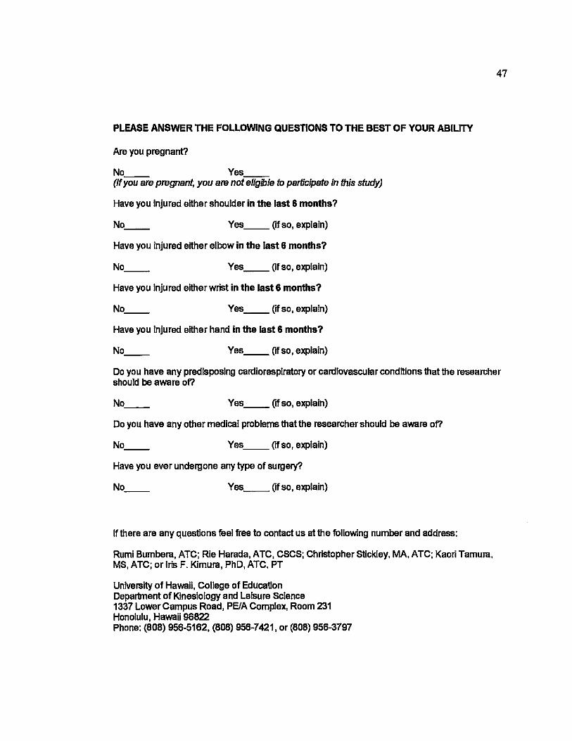
47
PLEASE ANSWER THE FOLLOWING QUESTIONS TO THE BEST OF YOUR ABILITY
Are you pregnant?
No__ Yes __ (If you are pregnant, you are not eligible to participate in this study)
Have you Injured either shoulder in the last 6 months?
No __ Yes __ (If so, explain)
Have you injured either elbow in the last 6 months?
No __ Yes __ (If so, explain)
Have you Injured either wrist in the last 6 months?
No __ Yes __ [If so, explain)
Have you Injured either hand in the last 6 months?
No __ Yes __ [If so, explain)
Do you have any predisposing cardiorespiratory or cardiovascular conditions that the researcher should be aware of?
No __ Yes __ [If so, explain)
Do you have any other medical problems that the researcher should be aware of?
No __ Yes __ [If so, explain)
Have you ever undergone any type of surgery?
No __ Yes __ [If so, explain)
If there are any questions feel free to contact us at the following number and address:
Rumi Bumbera, ATC; Rie Harada, ATC, CSCS; Christopher stickley, MA, ATC; Kaori Tamura, MS, ATC; or Iris F. Kimura, PhD, ATC, PT
University of HawaII, College of EdUcation Department of Kinesiology and Leisure Science 1337 Lower Campus Road, PEiA Complex, Room 231 Honolulu, Hawaii 96822 Phone: (808) 956-5162, (808) 956-7421, or (808) 956-3797
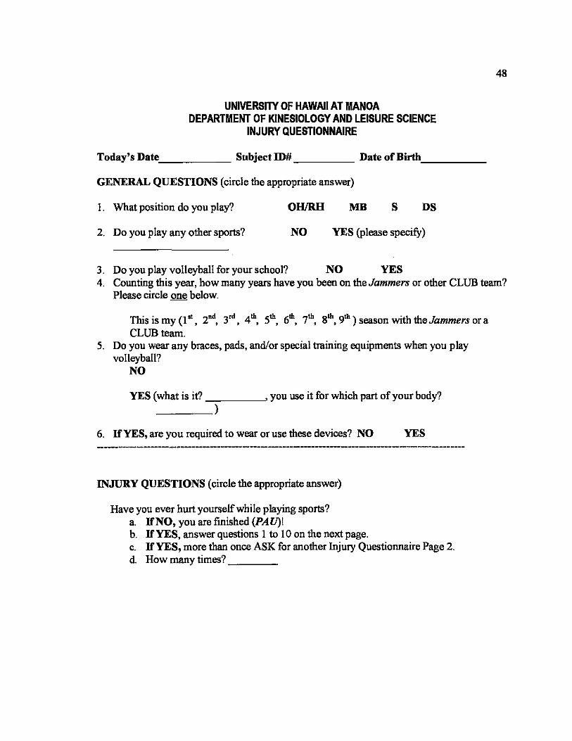
UNIVERSITY OF HAWAII AT MANOA DEPARTMENT OF KINESIOLOGY AND LEISURE SCIENCE
INJURY QUESTIONNAIRE
Today's Date'--_____ Subject ID# _____ Date of Birth. ____ _
GENERAL QUESTIONS (circle the appropriate answer)
1. What position do you play? OHIRH MB S OS
2. Do you play any other sports? NO YES (please specify)
3. Do you play volleyball for your school? NO YES
48
4. Counting this year, how many years have you been on the Jammers or other CLUB team? Please circle one below.
This is my (1 at, 2nd, 3rd, 4th, 5th, 6th, 7th, 8th, 9th ) season with the Jammers or a
CLUB team. S. Do you wear any braces, pads, and/or special training equipments when you play
volleyball? NO
YES (what is it? -,. ___ --" you use it for which part of your body?
---)
6. IT YES, are you required to wear or use these devices? NO YES
INJURY QUESTIONS (circle the appropriate answer)
Have you ever hurt yourself while playing sports? a. ITNO, you are finished (PAll)! b. IT YES, answer questions 1 to lOon the next page. c. ITYES, more than once ASK for another Injury Questionnaire Page 2. d. How many times? ___ _

49
INJURY QUESTIONNAIRE (page 2) SubJectlD# ___ _ ON THE PICTURE BELOW, circle where (location) you have or had the injury or pain
Please answer I to 10 below that describes the injwy location you circled
I. What sport were you injured in? 2. How old were you when you got the injwy? 3. Approximate date of the injwy (month/year) 4. Write the name of injwy (if you know what it is called) 5 Have or did you miss practices or games because of this injury? NO YES 6. Did you see a doctor for this injury/pain? NO YES 7. Did the dcctor: (circle all that apply)
a Give you or prescribe medicine(s) b. Give you exercise(s) c. Send you to an athletic trainer d. Send you to a physioai therapist e. Recommended surgery f. Recommended you stay out of practices/games g. OTHER
8. Did you have surgery for this injury? NO YES a When was the surgery?
9. Is this injwy still painful? NO YES a Does it hurt when you walk? NO YES b. Does it hurt when you run? NO YES c. Does it hurt when you jump? NO YES d. Does is hurt at rest? NO YES
10. Are you able to participate in volleyball now? NO YES

C.S. Data collection forms
Jump Study Data Collection Check List University of Hawaii at Manoa
Kinesiology and Leisure Science
SUbjects I.D. # Name ______ _
Age Date __ _
D D D D D
D
Check in / documentation check
Stationary bike / Stretch
Landing trial standard instruction / practice
Reflective marker placement / shoes coverage
Landing trials
Block right 12345678910 II 1213 14 15
Block left 1 2 3 4 5 6 7 8 9 10 II 12 13 14 15
Spike 123456789 10 II 12 13 14 15
Check out
50

C.6. Specific testing protocols
1. Anthropometric Measurements a. Height, Weight, Q-angle, Body composition
2. Warm-up a. Stationary bike riding b. Self stretching
3. Standard instruction 4. Practice session 5. Reflective marker placement 6. Landing trials
a. 3 successful trials
51
C.7. Standard instructions
General instruction: Hello, my name is Name. I am a certified Athletic Trainer and a graduate student
here at the University of Hawaii Manoa. Thank you for your participation in our research. If you have any questions, please ask at any time. First aid care will be provided in case of emergency by certified athletic trainers here.
Today, we will complete a landing trial. First, we will measure your height, weight, skinfold, and knee angle. Then, you will have a warm-up session which including stationary bike riding and self-stretching. After the warm-up, we will measure your vertical jump. All measurements will be recorded. Finally, a certified athletic trainer will put markers on your body for volleyball jump trial. Before the actual volleyball jump trial, you will have another instruction session. If you have any questions, please feel free to ask us at any time.
Landing instruction: Hello, my name is Name. I am a certified athletic trainer and a graduate student here
at the University of Hawaii Manoa. Thank you for your participation in ourresearch. If you have any questions, please ask at any time.
Today, we will be measuring your spike jumps. You are going to perform spike jumps until we have 3 successful trials. You can practice until you feel comfortable with your tasks. First, you will perform spike jumps in which you will take an approach from your preferred side (right or left) to hit this suspended volleyball (placed here in this holder).
Please do the following things for this process; first, use an approach of 3 or 4 steps. Second, make sure to hit this volleyball across the net. We may ask you change the approach starting position to adjust your landing, and you will land as close as your normal spike landing. You can practice as much as you want, and we will adjust the height of the holder. Please feel free to ask questions, and tell us what the best setting is for you to do this task.

52
Appendix D. Additional Results
D.1. Raw data tables
KInematic and KInetic Data on Dominant Leg
I.D.#: Knee Rexlon Knee Flexion Maximum MVGRF llmeto Loading Rate Angle@ Knee Flexion DomInant Leg Angle@IC MVGRF Angle (NlKg) MVGRF(sec) (N"""')
D1Ul307Tl :33.54 85.85 105.23 16.78 0..12 8831.87 02Ul307T1 34.08 73.63 79.94 19.21 0..12 7581.02
O3U1307T1 18.49 60.60 73.93 14.24 0.12 6639.78
05Ul307Tl 40.00 63.97 75.22 14.39 0..11 6970.00
OBUl307Tl 13.42 '5l.27 68.27 17.32 0..10. 8108.71
D7U13D7Tl 16.98 82.31 70..27 22.93 0..10. 11761.93 0BU13D7Tl 14.22 59.47 n.1D 20.82 0..11 11385.06
09U1307T1 13.86 60.79 63.94 17.72 0.11 8251.74
lOU1307T1 6.35 64.98 87.64 14.46 0..14 5211.00
llUl307T1 15.19 50.70 61.16 14.32 0..10. 8218.41 12Ul307Tl 9.44 64.52 n.'5l 7.44 0..14 :3329.59
13U1307Tl 0.20 39.08 49.02 19.26 0..10. 9050.31
14U1307Tl 17.27 68.22 94.00 14.78 0..16 5093.90
15U1307Tl 14.52 82.74 76.86 20..34 0..09 1210.1.65
16U1307Tl 2.58 63.63 71.46 20..02 0..11 8505.67
17U1307Tl 11.64 59.34 63.63 14.76 0..12 6402.08
16U13D7Tl 9.75 '5l.86 68.16 18.23 0..12 6517.47
19U1307Tl 17.11 63.24 82.'5l 19.46 0..11 9460.11
21Ul307T1 7.50 60.76 79.94 16.54 0..12 6319.n
C1U17D7Tl 19.02 65.42 64.20 15.55 0..11 8223.22
D1Ul607Tl 16.32 63.59 89.94 15.n 0..11 9720.37
02U17C7Tl 8.85 58.52 87.82 14.64 0..11 9046.13
02Ul607Tl 13.61 65.76 91.31 15.46 0..11 8551.86
03U17C7Tl 12.38 51.30 74.72 10..91 0..11 6764.06
03Ul607Tl 25.30 51.73 n.n 12.57 0..10. 7501.15
04U17C7Tl 18.n 58.64 55.58 17.03 0..12 10645.12
04U18C7Tl 11.76 61.67 90.26 15.82 0..10. 91:33.12
05U17D7Tl 2288 68.14 63.92 20.68 0..10. 14475.06
05U1807T1 17.98 59.73 74.04 14.21 0.21 6195.44
06U17D7Tl 14.67 73.27 91.69 14.60 0..12 65:33.74
06Ul607Tl 18.54 71.06 81.54 16.23 0..12 1048026
D7U17C7Tl 17.13 68.92 n.43 19.23 0..0.9 14689.10.
D7Ul607Tl 18.15 67.05 79.42 17.64 0..11 1096926
06U1707T1 16.73 58.59 74.43 15.82 0..11 7513.30
06Ul607Tl 11.61 60.81 76.97 13.68 0..11 10608.0.7
09U17C7Tl 8.55 50.67 n.63 18.96 0..10. 12968.40
09U18C7Tl 19.42 59.65 68.68 12.86 0..12 7192.06
lOU 17D7Tl 9.68 58.97 67.38 16.63 0..10. 12694.63
lOU1607T1 13.45 75.60 68.60 14.68 0..12 6904.55
l1Ul607T1 26.14 51.60 74.92 17.59 0..12 9547.55
Mean 15.96 82.10. 78.76 16.34 0..11 880.1.87
SO 7.96 9.12 10..46 2.95 0..0.2 2514.70

53
KInematic and Kinetic Data on Non-domlnant Lea 1.0.#: Knee Fiexion
Knee Flexion Maximum MVGRF Time to loading Rate Non-domlnant Angle@ Knee Flexion
Leg Angle@IC
MVGRF Angle (NlKg) MVGRF(B) (NIB)
01Ul307Tl 11.45 50.73 82.01 13.71 0.10 8417.83 02Ul307Tl 14.27 41.43 65.54 14.53 0.12 5734.37
O3U1307T1 16.65 84.36 77.71 18.02 0.11 8728.87
05Ul307Tl 21.38 53.88 74.33 14.54 0.11 6956.48
08Ul307Tl 11.88 53.38 83.48 18.59 0.10 8352.38
07U1307Tl 10.22 58.17 67.58 15.72 0.12 8824.91
08Ul307Tl 13.74 78.61 81.26 9.55 0.15 3801.72
O9U1307T1 8.78 53.94 77.08 17.22 0.10 9085.83
lOU1307T1 10.65 64.72 65.52 13.74 0.13 5054.67 llUl307Tl 16.14 88.05 83.03 19.83 0.12 9182.88
12Ul307Tl 7.26 48.44 77.70 22.50 0.11 12486.13
13U1307Tl 5.64 40.43 52.88 20.88 0.10 9269.26
14U1307Tl 17.31 .59.87 82.54 17.23 0.12 8065.25
15U1307T1 4.44 37.74 83.54 14.83 0.09 8827.45
16U1307Tl 4.71 49.52 73.47 16.16 0.10 7829.51 17U1307Tl 9.88 54.78 80.73 16.51 0.12 8809.35 16U1307T1 12.41 83.60 78.31 2004 0.10 8309.48
19U1307Tl 14.12 61.93 78.64 14.04 '0.12 6503.60
21U1307Tl 6.57 60.28 75.44 17.79 0.11 9646.26
O1U1707T1 8.72 58.60 95.33 17.74 0.12 8941.82
01Ul807T1 21.22 88.53 88.57 18.18 6.10 12503.95
02U1707Tl 5.73 53.88 83.92 14.14 0.12 7889.83
02U1807Tl 10.32 58.74 88.78 15.81 0.10 9207.98
O3U1707T1 8.70 47.15 76.88 19.79 0.11 13078.45
03U1807T1 8.73 32.73 72.52 18.83 0.17 8891.50
04U1707Tl 15.72 47.31 49.54 21.83 0.11 14745.53
04Ul607Tl 10.83 62.83 91.26 1611 021 449674
05U1707T1 8.42 48.25 83.40 14.27 0.11 9495.20
O5U1807T1 12.11 54.42 88.54 21.97 0.10 19284.73
06U1707Tl 8.31 49.83 60.26 17.39 0.10 8803.24
06Ul607Tl 5.65 45.65 65.38 17.88 0.10 13088.18
07U1707Tl 4.22 48.08 65.88 18.53 0.09 13304.94
07U1807Tl 13.88 58.81 72.70 15.60 0.11 9583.61
06U1707T1 16.02 64.04 76.20 18.93 0.13 6028.74
08U1807Tl 8.03 65.31 73.76 20.52 0.11 15525.99
09U1707Tl 1.78 44.10 78.43 17.12 0.10 12103.17
09Ul607Tl 9.88 55.00 77.82 2016 0.10 14057.95
lOU1707Tl 11.31 48.73 61.61 24.38 0.11 17682.79
lOU1807T1 15.03 57.08 78.12 22.23 0.11 11650.18
l1Ul607Tl 21.00 57.77 78.39 16.04 0.10 1028322
Mean 11.02 54.27 75.17 17.57 0.11 9757.45
SO 4.82 9.03 9.66 2.99 0.02 3426.20

54
D.2. Statistical tables
ANOVA Table for Spike-Jump Landing Kinematic and Kinetic Variables
ANOVA Table for Mean Knee Flexion Anole at Ie
Source DF TypelSS Mean Square F-Value P-Value
Injury Status 1 197.22 197.22 4.79 0.0318
landing Leg 1 518.16 518.16 12.57 0.0007
Injury Status·Landing 1 21.60 21.60 0.52 0.4714 Lea
ANOVA Table for Mean Knee Flexion Anole et MVGRF
Source DF TypelSS Mean Square F-Value P-Value
Injury Status 1 62.71 62.71 0.81 0.3721
Landing Leg 1 1672.74 1672.74 21.5 <.0001
Injury Status·Landing 1 3.13 3.13 0.04 0.8416 Lea
ANOVA Table for Mean Maximal Knee Flexion Anole
Source DF Type ISS Mean Square F-Value P-Value
Injury Status 1 17.17 17.17 0.17 0.6818
Landing Lag 1 355.1 355.1 3.5 0.0651
Injury Status·Landing 1 81.81 81.81 0.81 0.3718 Lao
ANOVA Table for Mean MVGRF
Source DF Type ISS Mean Square F-Value P-Value
Injury Status 1 39.55 39.55 4.95 0.0291
landing Leg 1 72.43 72.43 9.07 0.0035
Injury Status·Landlng 1 0.05 0.05 0.01 0.9372 Lao

55
ANOVA Table for Mean TIme from IC to MVGRF
Source OF Type ISS Mean Square F-Value P-Value
Injury Status 1 O.OOOS 0.0005 1.36 0.2479
Landing Lag 1 0.0001 0.0001 0.22 0.6371
Injury Status"Landing 1 0.0010 0.0010 2.33 0.1309 Leg
ANOVA Table for Mean TIme from IC to Maximal Knee Flexion
Source OF TypeiSS Mean Square F-Value P-Value
injury Status 1 0.0013 0.0013 1.16 0.2859
Landing Lag 1 0.0022 0.0022 2.00 0.1614
Injury Status"Landing 1 0.0003 0.0003 0.28 0.5997 Leg
ANOVA Table for Mean Loading Rate
Source OF Type ISS Mean Square F-Value P-Value
injury Status 1 47017695.87 47017695.87 5.54 0.0212
Landing Lag 1 27523269.36 27523269.36 3.24 0.0757
injury Status"Landing 1 4116378.48 4116378.48 0.49 0.4882 Leg

Appendix F. Bibliography
1. Burt CW, Overpeck MD. Emergency visits for sports-related injuries. Ann Emerg Med 2001;37(3}:301-8.
56
2. Powell JW, Barber-Foss KD. Injury Patterns in Selected High School Sports: A Review of the 1995-1997 Seasons. J Athl Train 1999;34(3}:277-284.
3. Agel J, Palmieri-Smith RM, Dick&, Wojtys EM, Marshall SW. Descriptive epidemiology of collegiate women's volleyball injuries: National Collegiate Athletic Association Injury Surveillance System, 1988-1989 through 2003-2004. J Athl Train 2007;42(2}:295-302.
4. Augustsson SR, Augustsson J, Thomee R, Svantesson U. Injuries and preventive actions in elite Swedish volleyball. ScandJ Med Sci Sports 2006;16(6}:433-40.
5. Salci Y, Kentel BB, Heycan C, Akin S, Korkusuz F. Comparison of landing maneuvers between male and female college volleyball players. Clin Biomech (Bristol, Avon) 2004; 19( 6}:622-8.
6. Schmitz RJ, Kulas AS, Perrin DH, Riemann BL, Shultz SJ. Sex differences in lower extremity biomechanics during single leg landings. Clin Biomech (Bristol, Avon) 2oo7;22(6}:681-8.
7. Swartz EE, Decoster LC, Russell PJ, Croce RV. Effects of Developmental Stage and Sex on Lower Extremity Kinematics and Vertical Ground Reaction Forces During Landing. J Athl Train 2005;40(1}:9-14.
8. Russell KA, Palmieri RM, Zinder SM, Ingersoll CD. Sex differences in valgus knee angle during a single-leg drop jump. J Athl Train 2006;41 (2): 166-71.
9. Lephart SM, Ferris CM, Riemann BL, Myers JB, Fu FH. Gender differences in strength and lower extremity kinematics during landing. Clin Orthop Relat Res 2002(401 }: 162-9.
10. Hass CJ, Schick EA, Tillman MD, Chow JW, Brunt D, Cauraugh JH. Knee biomechanics during landings: comparison of pre- and postpubescent females. Med Sci Sports Exerc 2oo5;37(1}:I00-7.
11. Hewett TE, Myer GD, Ford KR, Heidt RS, Jr., Colosimo AJ, McLean SG, et al. Biomechanica1 measures of neuromuscular control and valgus loading of the knee predict anterior cruciate ligament injury risk in female athletes: a prospective study. Am J Sports Med 2005;33(4}:492-501.
12. Louw Q, Grimmer K, Vaughan C. Knee movement patterns of injured and uninjured adolescent basketball players when landing from a jump: a case-control study. BMC Musculoskelet Disord 2006;7:22.
13. Richards DP, Ajemian SV, Wiley JP, Zernicke RF. Knee joint dynamics predict patellar tendinitis in elite volleyball players. Am J Sports Med 1996;24(5}:676-83.
14. Soprano JP, Fuchs SM Common Overuse Injuries in the Pediatric and Adolescent Athlete. Clinical Pediatric Emetgency Medicine 2007;8(1 }:7-14.
15. Bisseling RW, Hof AL, Bredeweg SW, Zwerver J, Mulder T. Relationship between landing strategy and patellar tendinopathy in volleyball. Br J Sports Med 2007;41 (7}:e8.
16. Associations NFoSHS. NFHS Participation Figures Search, Year of2004-2005, 2007. 17. Tillman MD, Hass CJ, Brunt D, Bennett GR Jumping and landing techniques in elite
women's volleyball. Journal of Sports Science and Medicine 2004;3:30-36.

57
18. Sell TC, Ferris CM, Abt JP, Tsai YS, Myers JB, Fu PH, et al. The effect of direction and reaction on the neuromuscular and biomechanical characteristics of the knee during tasks that simulate the noncontact anterior cruciate ligament injury mechanism. Am J Sports Med 2006;34(1 ):43-54.
19. Li G, Rudy TW, Sakane M, Kanamori A, MaCB, Woo SL. The importance of quadriceps and hamstring muscle loading on knee kinematics and in-situ forces in the ACL. J Biomech 1999;32(4):395-400.
20. Schafle MD. Cornmon injuries in volleyball. Treatment, prevention and rehabilitation. Sports Med 1993;16(2):126-9.
21. Smith R, Ford KR, Myer GD, Holleran A, Treadway E, Hewett TE. Biomechanical and performance differences between female soccer athletes in National Collegiate Athletic Association Divisions I and m. J Alhl Train 2007;42(4):470-6.
22. Dufek JS, Bates BT. Biomechanical factors associated with injury during landing in jump sports. Sports Med 1991 ;12(5):326-37.
23. Earl JE, Monteiro SK, Snyder KR. Differences in lower extremity kinematics between a bilateral drop-vertical jump and a single-leg step-<iown. J Orthop Sports Phys Ther 2007;37(5):245-52.
24. Ferretti A, Papandrea P, Conteduca F. Knee injuries in volleyball. Sports Med 1990; 1 0(2): 132-8.
25. Ferretti A, Papandrea P, Conteduca F, Mariani PP. Knee ligament injuries in volleyball players. Am J Sports Med 1992;20(2):203-7.
26. Kadaba MP, Ramakrishnan HK, Wootten ME. Measurement oflower extremity kinematics during level walking. J Orthop Res 1990;8(3):383-92.
27. Verhagen EA, Van der Beek AJ, Bouter LM, Bahr RM, Van Mechelen W. A one season prospective cohort study of volleyball injuries. Br J Sports Med 2004;3 8(4):477-81.
28. Zhang SN, Bates BT, Dufek JS. Contributions of lower extremity joints to energy dissipation during landings. Med Sci Sports Exerc 2000;32(4):812-9.