Embed Size (px)
Citation preview
University of Groningen
Stress relaxation analysis facilitates a quantitative approach towards antimicrobial penetrationinto biofilmsHe, Yan; Peterson, Brandon W; Jongsma, Marije A; Ren, Yijin; Sharma, Prashant K;Busscher, Henk J; van der Mei, Henny CPublished in:PLoS ONE
DOI:10.1371/journal.pone.0063750
IMPORTANT NOTE: You are advised to consult the publisher's version (publisher's PDF) if you wish to cite fromit. Please check the document version below.
Document VersionPublisher's PDF, also known as Version of record
Publication date:2013
Link to publication in University of Groningen/UMCG research database
Citation for published version (APA):He, Y., Peterson, B. W., Jongsma, M. A., Ren, Y., Sharma, P. K., Busscher, H. J., & van der Mei, H. C.(2013). Stress relaxation analysis facilitates a quantitative approach towards antimicrobial penetration intobiofilms. PLoS ONE, 8(5), [e63750]. https://doi.org/10.1371/journal.pone.0063750
CopyrightOther than for strictly personal use, it is not permitted to download or to forward/distribute the text or part of it without the consent of theauthor(s) and/or copyright holder(s), unless the work is under an open content license (like Creative Commons).
The publication may also be distributed here under the terms of Article 25fa of the Dutch Copyright Act, indicated by the “Taverne” license.More information can be found on the University of Groningen website: https://www.rug.nl/library/open-access/self-archiving-pure/taverne-amendment.
Take-down policyIf you believe that this document breaches copyright please contact us providing details, and we will remove access to the work immediatelyand investigate your claim.
Downloaded from the University of Groningen/UMCG research database (Pure): http://www.rug.nl/research/portal. For technical reasons thenumber of authors shown on this cover page is limited to 10 maximum.
Stress Relaxation Analysis Facilitates a QuantitativeApproach towards Antimicrobial Penetration intoBiofilmsYan He1, Brandon W. Peterson1, Marije A. Jongsma2, Yijin Ren2, Prashant K. Sharma1, Henk J. Busscher1,
Henny C. van der Mei1*
1 Department of Biomedical Engineering, W.J. Kolff Institute, University Medical Center Groningen and University of Groningen, Groningen, The Netherlands,
2 Department of Orthodontics, University Medical Center Groningen, University of Groningen, Groningen, The Netherlands
Abstract
Biofilm-related infections can develop everywhere in the human body and are rarely cleared by the host immune system.Moreover, biofilms are often tolerant to antimicrobials, due to a combination of inherent properties of bacteria in theiradhering, biofilm mode of growth and poor physical penetration of antimicrobials through biofilms. Current understandingof biofilm recalcitrance toward antimicrobial penetration is based on qualitative descriptions of biofilms. Here wehypothesize that stress relaxation of biofilms will relate with antimicrobial penetration. Stress relaxation analysis of single-species oral biofilms grown in vitro identified a fast, intermediate and slow response to an induced deformation,corresponding with outflow of water and extracellular polymeric substances, and bacterial re-arrangement, respectively.Penetration of chlorhexidine into these biofilms increased with increasing relative importance of the slow and decreasingimportance of the fast relaxation element. Involvement of slow relaxation elements suggests that biofilm structuresallowing extensive bacterial re-arrangement after deformation are more open, allowing better antimicrobial penetration.Involvement of fast relaxation elements suggests that water dilutes the antimicrobial upon penetration to an ineffectiveconcentration in deeper layers of the biofilm. Next, we collected biofilms formed in intra-oral collection devices bonded tothe buccal surfaces of the maxillary first molars of human volunteers. Ex situ chlorhexidine penetration into two weeks oldin vivo formed biofilms followed a similar dependence on the importance of the fast and slow relaxation elements asobserved for in vitro formed biofilms. This study demonstrates that biofilm properties can be derived that quantitativelyexplain antimicrobial penetration into a biofilm.
Citation: He Y, Peterson BW, Jongsma MA, Ren Y, Sharma PK, et al. (2013) Stress Relaxation Analysis Facilitates a Quantitative Approach towards AntimicrobialPenetration into Biofilms. PLoS ONE 8(5): e63750. doi:10.1371/journal.pone.0063750
Editor: Eshel Ben-Jacob, Tel Aviv University, Israel
Received January 30, 2013; Accepted March 26, 2013; Published May 27, 2013
Copyright: � 2013 He et al. This is an open-access article distributed under the terms of the Creative Commons Attribution License, which permits unrestricteduse, distribution, and reproduction in any medium, provided the original author and source are credited.
Funding: The China Scholarship Council and W.J. Kolff Institute, University Medical Center Groningen are gratefully acknowledged for scholarships, enabling thisstudy. The funders had no role in study design, data collection and analysis, decision to publish, or preparation of the manuscript.
Competing Interests: The authors declare that no competing interes exist
* E-mail: [email protected]
Introduction
In the 17th century the Dutch fabric merchant Antonie van
Leeuwenhoek started to construct his own microscopes in order to
be able to better examine the quality of the fabrics he bought and
sold. He examined more than just his fabrics and after utilizing
one of his own microscopes in 1684 to look at the accumulation of
matter on his teeth, he remarked in a report to the Royal Society
of London: "The number of these animalcules in the scurf of a
man’s teeth are so many that I believe they exceed the number of
men in a kingdom". This was not enough however, to satisfy the
curiosity of the fabric merchant, who would become one of the
most famous microbiologists of all times, and he furthermore
discovered ‘‘that the vinegar with which I washt my Teeth, kill’d
only those Animals which were on the outside of the scurf, but did
not pass thro the whole substance of it’’.
Translated to one of the important topics in modern microbi-
ology, Van Leeuwenhoek was referring to the biofilm mode of
growth of bacteria adhering on a surface [1], embedding
themselves in a matrix of extracellular polymeric substances
(EPS) [2] that not only offers physical protection against
antimicrobial penetration but can also yield bacterial properties
that are different from their planktonic counterparts. Bacteria in
their adhering, biofilm mode of growth can become inherently
resistant to antimicrobials through mutation [3], formation of
antibiotic degrading enzymes [4], endogenous oxidative stress [5],
phenotypic changes [6], and low metabolic activities [7]. Despite
extensive studies over many centuries, prevention of biofilm
formation remains a prime challenge in many industrial and
biomedical applications. In industrial applications, biofilms inflict
major damage when formed on processing equipment or in pipes
used to transport resources [8]. In the biomedical field, biofilm-
related infections can develop everywhere in the human body from
head (oral biofilms [9]) to toe (infected diabetic foot ulcers [10]).
Biofilm-related infections are rarely cleared by the host immune
system and especially infections that arise after implantation of
biomaterial implants (e.g. prosthetic hips and knees) or devices
(e.g. pace makers) are known to be persistent and difficult to treat,
since the antimicrobial tolerance of bacteria in their biofilm mode
of growth extends to many antibiotics used in modern medicine
PLOS ONE | www.plosone.org 1 May 2013 | Volume 8 | Issue 5 | e63750
ts .
[11]. Moreover, dental caries and periodontal diseases, the most
wide-spread infectious diseases in the world, are due to biofilms
that Van Leeuwenhoek tried to eliminate by using vinegar as an
antimicrobial mouthrinse [12].
Although the microscopes used nowadays are more sophisticat-
ed than the ones Van Leeuwenhoek employed, our understanding
of the recalcitrance of biofilms toward antimicrobial penetration is
still based on qualitative description of biofilms [13], using
expressions as ‘‘water channels’’, ‘‘mushroom structures’’, ‘‘whis-
kers’’ and ‘‘streamers’’ [14,15]. This raises the question whether
quantifiable properties of biofilms exist that would relate with
antimicrobial penetration into a biofilm. As for polymeric
materials, structural and compositional properties of biofilms,
should be reflected in their viscoelastic properties. Visco-elastic
properties of oral biofilms depend on the degree of compaction
during formation, the absence or presence of flow during growth,
their architecture and microbial composition [16,17]. The
viscoelastic properties of oral biofilms can be determined by
evaluating their relaxation after deformation during external
loading. Stress relaxation during external loading is a time-
dependent process and can be separated into a number of
responses, each with a characteristic time-constant [18]. Although
Maxwell analysis of stress-relaxation to derive the characteristic
time-constants of the various relaxation processes that occur in a
biofilm under external loading has been done before [19], results
have been regarded mainly from a mathematical perspective and
the details of the relaxation-structure-composition relation in
biofilms and the physical processes associated with the different
time-constants, are mostly neglected. Stress relaxation may involve
a number of processes, like the outflow of water and EPS from the
biofilm and re-arrangement of the bacteria in the biofilm [20].
Since penetration of an antimicrobial into a biofilm depends on
diffusion [21] and therewith on its structural and compositional
features, like the presence of water-filled channels in the biofilm or
EPS-containing spaces, we here hypothesize that the penetration
of an antimicrobial into a biofilm may relate with stress relaxation
and its underlying processes.
The aim of this study is to gain evidence in support of this
hypothesis. To this end, single-species biofilms of two oral bacterial
strains, Streptococcus oralis and Actinomyces naeslundii were grown in a
parallel plate flow chamber (PPFC) [22] and in a constant depth
film fermenter (CDFF) [23]. Subsequently, we measured their
visco-elastic properties using a low load compression tester, as well
as the penetration of chlorhexidine into the biofilms. Following
Van Leeuwenhoek, we chose to collect support for our hypothesis
based on oral biofilms, because the human oral cavity is highly
accessible and also allows for sampling of in vivo formed biofilm.
Therefore, in order to not only gain in vitro evidence in support of
our hypothesis, an intra-oral biofilm collection device was
developed to grow oral biofilms in situ, in absence of mechanical
perturbation. In vivo formed biofilms in the devices worn by human
volunteers were examined ex situ with respect to their visco-elastic
properties and chlorhexidine penetration and results and conclu-
sions compared with those obtained for in vitro formed oral
biofilms. Chlorhexidine is known to be the most effective oral
antimicrobial to date [24] and surprisingly, despite its extensive
use, inherent bacterial resistance against chlorhexidine has hardly
or never been reported as compared to antibiotic resistance of
many bacterial pathogens. This makes chlorhexidine an ideal
antimicrobial to separate a possible inherent tolerance of biofilm
bacteria for the antimicrobial from the physical protection offered
by the biofilm mode of growth and study its penetration through a
biofilm.
Results
Biofilms of coccal-shaped S. oralis J22 and rod-shaped A.
naeslundii T14V-J1 grown in the PPFC reached a thickness of
131615 mm and 109626 mm, respectively (Table 1). The biofilm
thickness in the CDFF for S. oralis J22 was 11966 mm and
12569 mm for A. naeslundii T14V-J1. There were no significant
differences (p.0.05, Student t-test) in thickness between biofilms
grown under flow and in the CDFF. Also differences in biofilms
thickness across strains were not statistically significant (p.0.05,
Student t-test).
The penetration of chlorhexidine in biofilms grown in the PPFC
was significantly different (p,0.05, Mann-Whitney Rank Sum
test) for S. oralis J22 and A. naeslundii T14V-J1, and the penetration
ratio amounted 0.3360.09 and 0.5660.08, respectively (see also
Table 1 and Fig. S1). On the other hand, there were no significant
strain-dependent differences in penetration of chlorhexidine into
biofilms grown in the CDFF, showing penetration ratios of
0.4860.04 and 0.3960.06 in biofilms of S. oralis J22 and A.
naeslundii T14V-J1, respectively (p.0.05, Mann-Whitney Rank
Sum test). Interestingly, whereas biofilms offered a clear physical
protection against chlorhexidine, bacteria dispersed from biofilms
grown either in the PPFC or in the CDFF were highly susceptible
to chlorhexidine (Fig. S2), confirming that the absence of bacterial
killing in the deeper layers of the biofilms are not due to changes in
inherent properties of the bacteria in their biofilm mode of growth,
but solely to difficulties encountered by the antimicrobial in
penetrating to the deeper layers. Note that a similar conclusion has
been drawn for three days old in vivo grown oral biofilms, after
dispersal and exposure to chlorhexidine [25].
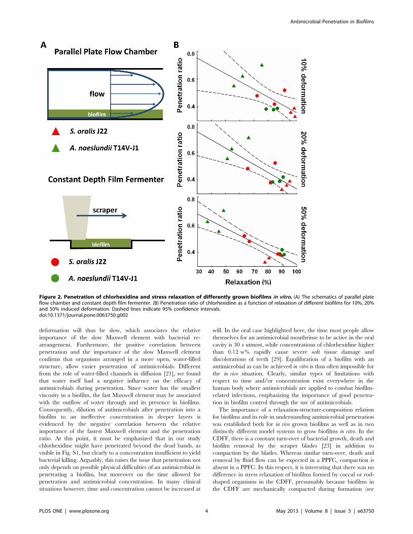
Total stress relaxation (Fig. 1A) of biofilms grown in the PPFC
were different for both strains and S. oralis J22 biofilms showed
significantly (p,0.05, Mann-Whitney Rank Sum test) more stress
relaxation than biofilms of A. naeslundii T14V-J1, especially after
10% and 20% induced deformation (Table 1). There were no
significant differences (p.0.05, Mann-Whitney Rank Sum test) in
stress relaxation between biofilms of the coccal and rod-shaped
organisms when grown in the CDFF. Interestingly, the penetration
ratio of chlorhexidine decreased with increasing stress relaxation of
the biofilms, regardless of the induced deformation (Fig. 2).
Total stress relaxation was subsequently resolved in a fast,
intermediate and slow component (Fig. 1B). Since bacteria in a
biofilm constitute the heaviest masses, their re-arrangement upon
an induced deformation will be slow, and we associate the relative
importance of the slow Maxwell element with bacterial re-
arrangement in a biofilm. On the other hand, water has the
smallest viscosity in a biofilm, and therefore the fast Maxwell
element is associated with the flow of water through a biofilm,
which leaves an association between the behavior of EPS with the
intermediate Maxwell element. Analysis of the stress relaxation
according to a three element Maxwell model revealed that
penetration increased with increasing relative importance of the
slow relaxation component and decreasing importance of the fast
component (Fig. 3). This confirms the existence of a relaxation-
structure-composition relation that may facilitate a quantitative
approach towards antimicrobial penetration in biofilms.
In order to confirm that a relaxation-structure-composition
relation facilitates understanding of antimicrobial penetration also
for in vivo grown biofilms, we first developed an intra-oral biofilm
collection device (Fig. S3). The average thickness of the oral
biofilms formed in vivo over a time period of two weeks was
121686 mm, comparable to the thickness of in vitro biofilms
(p.0.05, Mann-Whitney Rank Sum test), as can be seen in
Table 1.
Antimicrobial Penetration in Biofilms
PLOS ONE | www.plosone.org 2 May 2013 | Volume 8 | Issue 5 | e63750
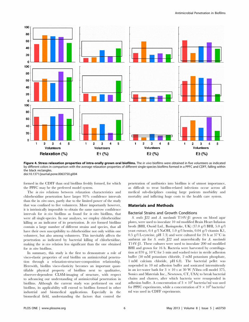
Total stress relaxation of in vivo biofilms upon 10% and 20%
deformation were more comparable to the stress relaxation
observed for in vitro biofilms grown in the PPFC than in the
CDFF, as averaged over both bacterial strains (Table 1). On the
other hand, upon inducing a deformation of 50%, stress relaxation
of in vivo biofilms became more comparable to the one of in vitro
biofilms grown in the CDFF. On average, in vitro biofilms showed
higher total stress relaxation than in vivo formed biofilms, although
this difference was only significant (p,0.05, Student t-test) for 10%
and 20% induced deformations (Fig. 4).
In vivo formed biofilms furthermore distinguished themselves
significantly from in vitro averages by a smaller importance of the
fast component (E1) and larger importance of the slow component
(E3) (p,0.05, Student t-test; Table 1) for induced deformations of
10% and 20%. At 50% induced deformation however, differences
in the importance of the different relaxation parameters had
disappeared (see also Fig. 4). The importance of the intermediate
component (E2) was relatively similar across the different biofilms
(Table 1).
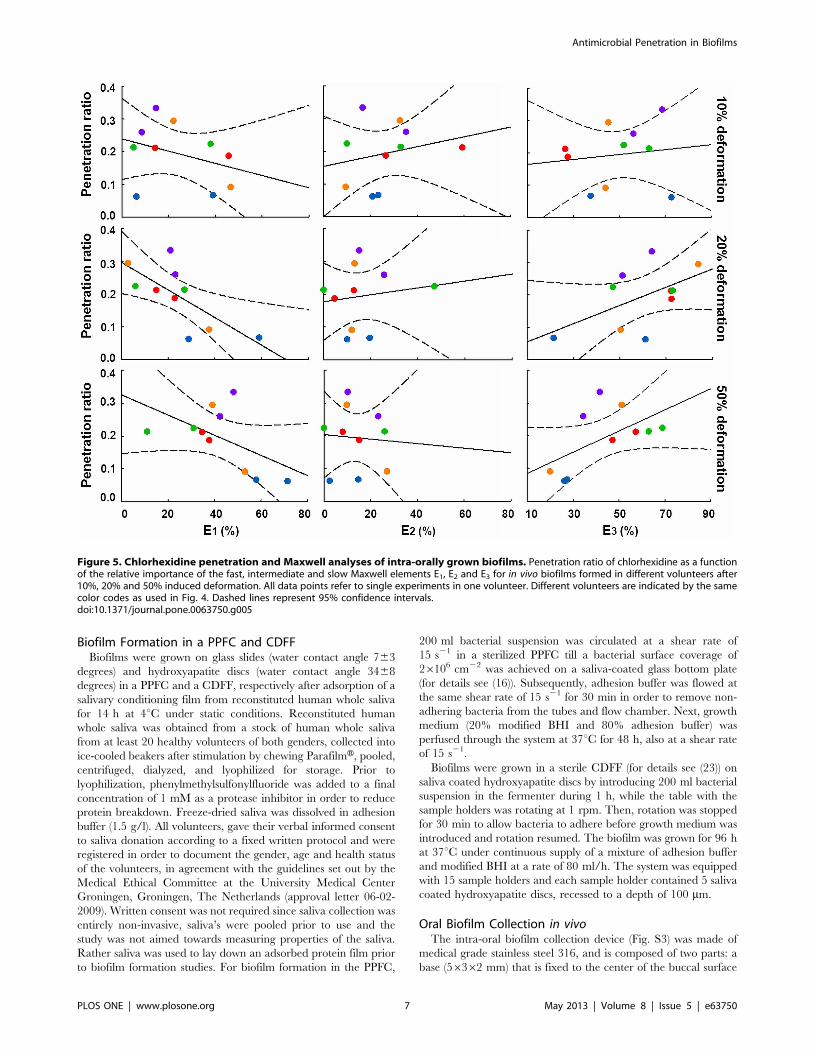
The chlorhexidine penetration ratio for in vivo formed biofilms
was smaller than the average penetration into in vitro biofilms
(p,0.05, Student t-test; Table 1). Similarly as observed for in vitro
biofilms, penetration decreased with increasing importance of the
fast (E1) component and increased with the importance of the slow
component (E3) (Fig. 5). No relation was observed with the
importance of the intermediate component (E2), as was also
lacking for in vitro biofilms.
Discussion
The recalcitrance of oral biofilm toward penetration of
antimicrobials is known ever since Van Leeuwenhoek wrote in
the 17th century that ‘‘the vinegar with which I washed my teeth killed only
those animals which were on the outside of the scurf, but did not pass through
the whole substance of it’’. Over recent years, the limited penetration
of antimicrobials into a biofilm has been attributed to reduced
solute diffusion in water, the presence of bacterial cells, EPS,
abiotic particles or gas bubbles trapped in a biofilm [21].
Interestingly, whereas the influence of the chemistry and biology
of biofilms on diffusion have been amply described and reviewed
[21,26,27], antimicrobial penetration has never been related with
quantifiable, physical properties of a biofilm. This study demon-
strates for the first time since Van Leeuwenhoek his observation of
the poor penetration of vinegar into an oral biofilm, that through a
relaxation-structure-composition relation, biofilm properties can
be derived that facilitate explanation of antimicrobial penetration
into a biofilm on basis of quantitative biofilm properties.
Incidentally, not only antimicrobials have difficulty penetrating a
biofilm, but also nutrients may have difficulty penetrating a
biofilm, causing reduced viability of organisms residing in deeper
layers of biofilms [28].
The bacteria in a biofilm constitute the heaviest masses, and
their re-arrangement during stress relaxation upon an induced
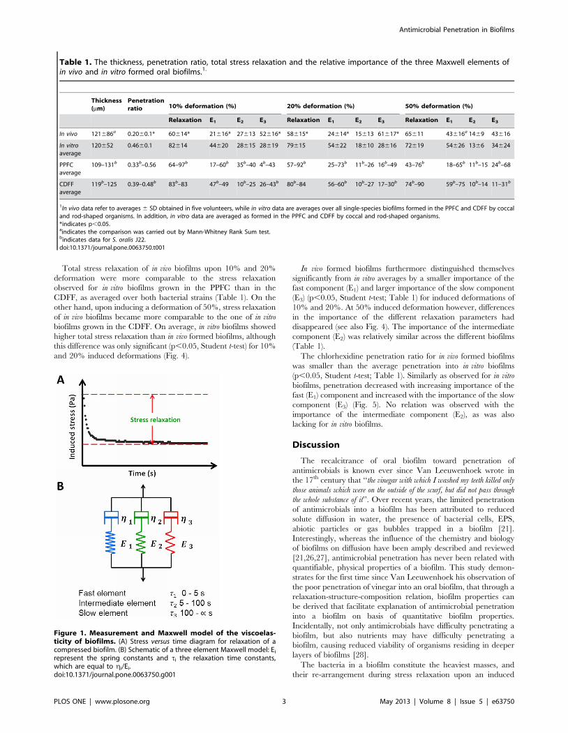
Table 1. The thickness, penetration ratio, total stress relaxation and the relative importance of the three Maxwell elements ofin vivo and in vitro formed oral biofilms.1.
Thickness(mm)
Penetrationratio 10% deformation (%) 20% deformation (%) 50% deformation (%)
Relaxation E1 E2 E3 Relaxation E1 E2 E3 Relaxation E1 E2 E3
In vivo 121686a 0.2060.1* 60614* 21616* 27613 52616* 58615* 24614* 15613 61617* 65611 43616a 1469 43616
In vitroaverage
120652 0.4660.1 82614 44620 28615 28619 79615 54622 18610 28616 72619 54626 1366 34624
PPFCaverage
109–131b 0.33b–0.56 64–97b 17–60b 35b–40 4b–43 57–92b 25–73b 11b–26 16b–49 43–76b 18–65b 11b–15 24b–68
CDFFaverage
119b–125 0.39–0.48b 83b–83 47b–49 10b–25 26–43b 80b–84 56–60b 10b–27 17–30b 74b–90 59b–75 10b–14 11–31b
1In vivo data refer to averages 6 SD obtained in five volunteers, while in vitro data are averages over all single-species biofilms formed in the PPFC and CDFF by coccaland rod-shaped organisms. In addition, in vitro data are averaged as formed in the PPFC and CDFF by coccal and rod-shaped organisms.*indicates p,0.05.aindicates the comparison was carried out by Mann-Whitney Rank Sum test.bindicates data for S. oralis J22.doi:10.1371/journal.pone.0063750.t001
Figure 1. Measurement and Maxwell model of the viscoelas-ticity of biofilms. (A) Stress versus time diagram for relaxation of acompressed biofilm. (B) Schematic of a three element Maxwell model: Ei
represent the spring constants and ti the relaxation time constants,which are equal to gi/Ei.doi:10.1371/journal.pone.0063750.g001
Antimicrobial Penetration in Biofilms
PLOS ONE | www.plosone.org 3 May 2013 | Volume 8 | Issue 5 | e63750
deformation will thus be slow, which associates the relative
importance of the slow Maxwell element with bacterial re-
arrangement. Furthermore, the positive correlation between
penetration and the importance of the slow Maxwell element
confirms that organisms arranged in a more open, water-filled
structure, allow easier penetration of antimicrobials. Different
from the role of water-filled channels in diffusion [21], we found
that water itself had a negative influence on the efficacy of
antimicrobials during penetration. Since water has the smallest
viscosity in a biofilm, the fast Maxwell element may be associated
with the outflow of water through and its presence in biofilms.
Consequently, dilution of antimicrobials after penetration into a
biofilm to an ineffective concentration in deeper layers is
evidenced by the negative correlation between the relative
importance of the fastest Maxwell element and the penetration
ratio. At this point, it must be emphasized that in our study
chlorhexidine might have penetrated beyond the dead bands, as
visible in Fig. S1, but clearly to a concentration insufficient to yield
bacterial killing. Arguably, this raises the issue that penetration not
only depends on possible physical difficulties of an antimicrobial in
penetrating a biofilm, but moreover on the time allowed for
penetration and antimicrobial concentration. In many clinical
situations however, time and concentration cannot be increased at
will. In the oral case highlighted here, the time most people allow
themselves for an antimicrobial mouthrinse to be active in the oral
cavity is 30 s utmost, while concentrations of chlorhexidine higher
than 0.12 w% rapidly cause severe soft tissue damage and
discolorations of teeth [29]. Equilibration of a biofilm with an
antimicrobial as can be achieved in vitro is thus often impossible for
the in vivo situation. Clearly, similar types of limitations with
respect to time and/or concentration exist everywhere in the
human body where antimicrobials are applied to combat biofilm-
related infections, emphasizing the importance of good penetra-
tion in biofilm control through the use of antimicrobials.
The importance of a relaxation-structure-composition relation
for biofilms and its role in understanding antimicrobial penetration
was established both for in vivo grown biofilms as well as in two
distinctly different model systems to grow biofilms in vitro. In the
CDFF, there is a constant turn-over of bacterial growth, death and
biofilm removal by the scraper blades [23] in addition to
compaction by the blades. Whereas similar turn-over, death and
removal by fluid flow can be expected in a PPFC, compaction is
absent in a PPFC. In this respect, it is interesting that there was no
difference in stress relaxation of biofilms formed by coccal or rod-
shaped organisms in the CDFF, presumably because biofilms in
the CDFF are mechanically compacted during formation (see
Figure 2. Penetration of chlorhexidine and stress relaxation of differently grown biofilms in vitro. (A) The schematics of parallel plateflow chamber and constant depth film fermenter. (B) Penetration ratio of chlorhexidine as a function of relaxation of different biofilms for 10%, 20%and 50% induced deformation. Dashed lines indicate 95% confidence intervals.doi:10.1371/journal.pone.0063750.g002
Antimicrobial Penetration in Biofilms
PLOS ONE | www.plosone.org 4 May 2013 | Volume 8 | Issue 5 | e63750
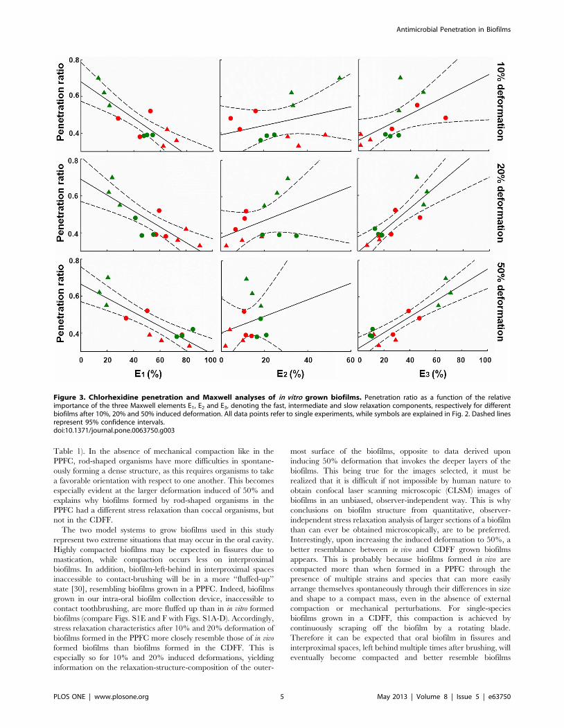
Table 1). In the absence of mechanical compaction like in the
PPFC, rod-shaped organisms have more difficulties in spontane-
ously forming a dense structure, as this requires organisms to take
a favorable orientation with respect to one another. This becomes
especially evident at the larger deformation induced of 50% and
explains why biofilms formed by rod-shaped organisms in the
PPFC had a different stress relaxation than coccal organisms, but
not in the CDFF.
The two model systems to grow biofilms used in this study
represent two extreme situations that may occur in the oral cavity.
Highly compacted biofilms may be expected in fissures due to
mastication, while compaction occurs less on interproximal
biofilms. In addition, biofilm-left-behind in interproximal spaces
inaccessible to contact-brushing will be in a more ‘‘fluffed-up’’
state [30], resembling biofilms grown in a PPFC. Indeed, biofilms
grown in our intra-oral biofilm collection device, inaccessible to
contact toothbrushing, are more fluffed up than in in vitro formed
biofilms (compare Figs. S1E and F with Figs. S1A-D). Accordingly,
stress relaxation characteristics after 10% and 20% deformation of
biofilms formed in the PPFC more closely resemble those of in vivo
formed biofilms than biofilms formed in the CDFF. This is
especially so for 10% and 20% induced deformations, yielding
information on the relaxation-structure-composition of the outer-
most surface of the biofilms, opposite to data derived upon
inducing 50% deformation that invokes the deeper layers of the
biofilms. This being true for the images selected, it must be
realized that it is difficult if not impossible by human nature to
obtain confocal laser scanning microscopic (CLSM) images of
biofilms in an unbiased, observer-independent way. This is why
conclusions on biofilm structure from quantitative, observer-
independent stress relaxation analysis of larger sections of a biofilm
than can ever be obtained microscopically, are to be preferred.
Interestingly, upon increasing the induced deformation to 50%, a
better resemblance between in vivo and CDFF grown biofilms
appears. This is probably because biofilms formed in vivo are
compacted more than when formed in a PPFC through the
presence of multiple strains and species that can more easily
arrange themselves spontaneously through their differences in size
and shape to a compact mass, even in the absence of external
compaction or mechanical perturbations. For single-species
biofilms grown in a CDFF, this compaction is achieved by
continuously scraping off the biofilm by a rotating blade.
Therefore it can be expected that oral biofilm in fissures and
interproximal spaces, left behind multiple times after brushing, will
eventually become compacted and better resemble biofilms
Figure 3. Chlorhexidine penetration and Maxwell analyses of in vitro grown biofilms. Penetration ratio as a function of the relativeimportance of the three Maxwell elements E1, E2 and E3, denoting the fast, intermediate and slow relaxation components, respectively for differentbiofilms after 10%, 20% and 50% induced deformation. All data points refer to single experiments, while symbols are explained in Fig. 2. Dashed linesrepresent 95% confidence intervals.doi:10.1371/journal.pone.0063750.g003
Antimicrobial Penetration in Biofilms
PLOS ONE | www.plosone.org 5 May 2013 | Volume 8 | Issue 5 | e63750
formed in the CDFF than oral biofilms freshly formed, for which
the PPFC may be the preferred model system.
The in vivo relations between relaxation characteristics and
chlorhexidine penetration have larger 95% confidence intervals
than the in vitro ones, partly due to the limited power of the study
that was confined to five volunteers. More importantly however,
it is intrinsically impossible to obtain the same narrow confidence
intervals for in vivo biofilms as found for in vitro biofilms, that
were all single-species. In our analyses, we employ chlorhexidine
killing as an indicator of its penetration. In vivo formed biofilms
contain a large number of different strains and species, that all
have their own susceptibility to chlorhexidine not only within one
volunteer, but also among volunteers. This inevitably affects the
penetration as indicated by bacterial killing of chlorhexidine,
making the in vivo relation less significant than the one obtained
for in vitro biofilms.
In summary, this study is the first to demonstrate a role of
visco-elastic properties of oral biofilm on antimicrobial penetra-
tion through a relaxation-structure-composition relationship.
Herewith, biofilm visco-elasticity becomes an important quan-
tifiable physical property of biofilms next to qualitative,
observer-dependent CLSM-imaging of structure, with respect
to advancing our understanding of antimicrobial penetration in
biofilms. Although the current study was performed on oral
biofilms, its applicability will extend to biofilms formed in other
industrial and biomedical applications. Especially in the
biomedical field, understanding the factors that control the
penetration of antibiotics into biofilms is of utmost importance,
as difficult to treat biofilm-related infections occur across all
medical sub-disciplines causing large patients morbidity and
mortality and inflicting huge costs to the health care system.
Materials and Methods
Bacterial Strains and Growth ConditionsS. oralis J22 and A. naeslundii T14V-J1 grown on blood agar
plates, were used to inoculate 10 ml modified Brain Heart Infusion
broth (BHI, Oxoid Ltd., Basingstoke, UK) (37.0 g/l BHI, 5.0 g/l
yeast extract, 0.4 g/l NaOH, 1.0 g/l hemin, 0.04 g/l vitamin K1,
0.5 g/l L-cysteine, pH 7.3) and were cultured for 24 h at 37uC in
ambient air for S. oralis J22 and anaerobically for A. naeslundii
T14V-J1. These cultures were used to inoculate 200 ml modified
BHI and grown for 16 h. Bacteria were harvested by centrifuga-
tion at 870 g, 10uC for 5 min and washed twice in sterile adhesion
buffer (50 mM potassium chloride, 2 mM potassium phosphate,
1 mM calcium chloride, pH 6.8). The bacterial pellet was
suspended in 10 ml adhesion buffer and sonicated intermittently
in an ice-water bath for 3 6 10 s at 30 W (Vibra cell model 375,
Sonics and Materials Inc., Newtown, CT, USA) to break bacterial
chains and clusters, after which bacteria were resuspended in
adhesion buffer. A concentration of 3 6108 bacteria/ml was used
for PPFC experiments, while a concentration of 9 6108 bacteria/
ml was used in CDFF experiments.
Figure 4. Stress relaxation properties of intra-orally grown oral biofilms. The in vivo biofilms were obtained in five volunteers as indicatedby different colors in comparison with the average relaxation properties of different single-species biofilms formed in a PPFC and CDFF, falling withinthe black rectangles.doi:10.1371/journal.pone.0063750.g004
Antimicrobial Penetration in Biofilms
PLOS ONE | www.plosone.org 6 May 2013 | Volume 8 | Issue 5 | e63750
Biofilm Formation in a PPFC and CDFFBiofilms were grown on glass slides (water contact angle 763
degrees) and hydroxyapatite discs (water contact angle 3468
degrees) in a PPFC and a CDFF, respectively after adsorption of a
salivary conditioning film from reconstituted human whole saliva
for 14 h at 4uC under static conditions. Reconstituted human
whole saliva was obtained from a stock of human whole saliva
from at least 20 healthy volunteers of both genders, collected into
ice-cooled beakers after stimulation by chewing ParafilmH, pooled,
centrifuged, dialyzed, and lyophilized for storage. Prior to
lyophilization, phenylmethylsulfonylfluoride was added to a final
concentration of 1 mM as a protease inhibitor in order to reduce
protein breakdown. Freeze-dried saliva was dissolved in adhesion
buffer (1.5 g/l). All volunteers, gave their verbal informed consent
to saliva donation according to a fixed written protocol and were
registered in order to document the gender, age and health status
of the volunteers, in agreement with the guidelines set out by the
Medical Ethical Committee at the University Medical Center
Groningen, Groningen, The Netherlands (approval letter 06-02-
2009). Written consent was not required since saliva collection was
entirely non-invasive, saliva’s were pooled prior to use and the
study was not aimed towards measuring properties of the saliva.
Rather saliva was used to lay down an adsorbed protein film prior
to biofilm formation studies. For biofilm formation in the PPFC,
200 ml bacterial suspension was circulated at a shear rate of
15 s21 in a sterilized PPFC till a bacterial surface coverage of
26106 cm22 was achieved on a saliva-coated glass bottom plate
(for details see (16)). Subsequently, adhesion buffer was flowed at
the same shear rate of 15 s21 for 30 min in order to remove non-
adhering bacteria from the tubes and flow chamber. Next, growth
medium (20% modified BHI and 80% adhesion buffer) was
perfused through the system at 37uC for 48 h, also at a shear rate
of 15 s21.
Biofilms were grown in a sterile CDFF (for details see (23)) on
saliva coated hydroxyapatite discs by introducing 200 ml bacterial
suspension in the fermenter during 1 h, while the table with the
sample holders was rotating at 1 rpm. Then, rotation was stopped
for 30 min to allow bacteria to adhere before growth medium was
introduced and rotation resumed. The biofilm was grown for 96 h
at 37uC under continuous supply of a mixture of adhesion buffer
and modified BHI at a rate of 80 ml/h. The system was equipped
with 15 sample holders and each sample holder contained 5 saliva
coated hydroxyapatite discs, recessed to a depth of 100 mm.
Oral Biofilm Collection in vivoThe intra-oral biofilm collection device (Fig. S3) was made of
medical grade stainless steel 316, and is composed of two parts: a
base (56362 mm) that is fixed to the center of the buccal surface
Figure 5. Chlorhexidine penetration and Maxwell analyses of intra-orally grown biofilms. Penetration ratio of chlorhexidine as a functionof the relative importance of the fast, intermediate and slow Maxwell elements E1, E2 and E3 for in vivo biofilms formed in different volunteers after10%, 20% and 50% induced deformation. All data points refer to single experiments in one volunteer. Different volunteers are indicated by the samecolor codes as used in Fig. 4. Dashed lines represent 95% confidence intervals.doi:10.1371/journal.pone.0063750.g005
Antimicrobial Penetration in Biofilms
PLOS ONE | www.plosone.org 7 May 2013 | Volume 8 | Issue 5 | e63750
of the upper first molars and a replaceable cover plate
(46360.2 mm). Biofilms formed on the inner side of the
replaceable cover plate in the absence of mechanical perturba-
tions, were considered for this study.
Five volunteers (aged 26 to 29 years) were included in this study.
Volunteers all had a complete dentition with maximally one
restoration, no bleeding upon probing and were not using any
medication. Each volunteer was assigned a random number
between 1 and 5 used for later data processing. The study was
approved according to the guidelines of the Medical Ethics
Committee of the University Medical Center Groningen, Gronin-
gen, The Netherlands (letter 28-9-2011), including the written
informed consent by the volunteers and the tenets of the
Declaration of Helsinki.
A base device was fixed to buccal surfaces of the upper first
molars of the volunteers (see also Fig. S3) after mild etching of the
tooth surface using light cure adhesive paste (TransbondTM XT,
3M Unitek, USA), a procedure similar to the one used for the
bonding of orthodontic brackets. Prior to bonding, the base and
cover plate of the device were brushed using a rubber cup and
cleaner paste (ZircateH Prophy Paste, Densply, Caulk, USA) at low
speed (less than 2,500 rpm/min) and autoclaved. Subsequently,
the base surface was coated with a thin layer of primer and
bonding agent (CLEARFIL SE BOND, Kurary Medical Inc.,
Japan). The stainless steel cover plate was inserted using a pair of
tweezers and kept in place using Light Cure Adhesive Paste
(TransbondTM XT, 3M Unitek, USA). Volunteers were asked to
wear the device for a total of eight weeks during which they were
requested to perform manual brushing with a standard fluoridated
toothpaste (Prodent SoftmintH, Sara Lee Household & Bodycare,
Exton, USA) according to their habitual oral hygiene but to refrain
from the use of an additional mouthrinse.
The cover plates could be removed with a dental explorer, after
which cover plates with biofilm were placed in a moisturized petri
dish for transport from the dental clinic to the laboratory. In a
separate pilot study, it was established that two weeks of intra-oral
biofilm formation in the device yielded biofilm thicknesses that
were similar to the ones obtained in vitro. Therewith, in vivo
biofilms could be collected four times from each volunteer. After
each experiment, cover plates were sanded to remove biofilm and
other residuals, prior to autoclaving.
After the experiments, the base of the device was removed from
the tooth surface with a debracketing plier and residual adhesive
was grinded off the tooth surface with a low speed hand piece. A
base device was only used once in each volunteer. The tooth
surface was polished and cleaned with rubber cup and cleaner
paste. No signs of gingival inflammation were observed in any
volunteer after removal of the base device.
Low Load Compression TestingThe thickness and stress relaxation of the biofilms were
measured with a low load compression tester, described before
(16). Stress relaxation was monitored after inducing 10, 20, and
50% deformation of the biofilms within 1 s and held constant for
100 s, while monitoring the stress relaxation (see Fig. 1A). Each
deformation was induced three times at different locations on the
same biofilm.
Stress relaxation as a function of time was analyzed using a
generalized Maxwell model containing three elements (see Fig. 1B)
according to
E(t)~E1e{t=t1zE2e
{t=t2zE3e{t=t3 ð1Þ
in which E(t) is the total stress exerted by the biofilm divided by the
strain imposed, expressed as the sum of three Maxwell elements
with a spring constant Ei, and characteristic decay time, ti (see also
Fig. 1B). For calculating E(t), deformation was expressed in terms
of strain, e, according to the large strain model using
e~ ln (1zDh
h) ð2Þ
where Dh is the decrease in height and h is the un-deformed height
of the biofilm. The model fitting for Ei and ti values of the three
elements was done by minimizing the chi-squared value using the
Solver tool in Microsoft Excel 2010. Fitting to three Maxwell
elements yielded the lowest chi-squared values and increasing the
number of Maxwell elements only yielded minor decreases in chi-
squared values of less than 3%. The elements derived were rather
arbitrarily named fast, intermediate or slow based on their tvalues, i.e. t1,5 s, 5 s,t2,100 s and t3.100 s, respectively (see
also Fig. 1B). Relative importance of each element, based on the
value of its spring constant Ei, was expressed as the percentage of
its spring constant to the sum of all elements’ spring constants at
t = 0.
Penetration of Chlorhexidine into BiofilmsIn vitro and in vivo formed biofilms were all exposed in vitro to a
0.2 wt% chlorhexidine-containing mouthrinse (CorsodylH,
SmithKline Beecham Consumer Brands B.V., Rijswijk, The
Netherlands) for 30 s and subsequently immersed in adhesion
buffer for 5 min. After exposure to chlorhexidine, biofilms were
stained for 30 min with live/dead stain (BacLightTM, Invitrogen,
Breda, The Netherlands) and CLSM (Leica TCS-SP2, Leica
Microsystems Heidelberg GmbH, Heidelberg, Germany) was used
to record a stack of images of the biofilms with a 406 water
objective lens. Images were analyzed with Leica confocal software
to visualize live and dead bacteria in the biofilms. The ratio of the
intensity of red (dead bacteria) to green (live bacteria), R/G, was
plotted versus the biofilm thickness (see Fig. S1). The biofilm
thickness where the ratio R/G became less than 1.5 was taken as
the thickness of the dead band. Next, a penetration ratio was
calculated according to
Penetration ratio~dead band thickness
total biofilm thicknesð3Þ
Penetration ratios were calculated for three different, randomly
chosen locations on the biofilms and presented as averaged over
the different locations.
Statistical AnalysisStatistical analysis was performed with SigmaPlot software
(version 11.0, systat software, Inc., California, USA). Differences in
biofilm thickness and visco-elasticity were evaluated after testing
for normal distribution and equal variance of the data. If data
failed one of these tests, a Mann-Whitney Rank Sum test was used
to determine statistical significance, otherwise a Student t-test was
applied. Pearson Product Moment Correlation test was used to
disclose relations between the penetration of chlorhexidine into
and the relaxation of biofilms.
Supporting Information
Figure S1 Chlorhexidine penetration into in vitro andin vivo biofilms and calculation of the penetration ratio.
Antimicrobial Penetration in Biofilms
PLOS ONE | www.plosone.org 8 May 2013 | Volume 8 | Issue 5 | e63750
(I) Representative CLSM-images (cross sectional view) of the
penetration of chlorhexidine (0.2 wt%) during 30 s into oral
biofilms grown in vitro and in vivo (exposure to chlorhexidine was
done in vitro). (A) S. oralis J22 biofilm grown under flow in a PPFC.
(B) S. oralis J22 biofilm grown under compaction in a CDFF. (C) A.
naeslundii T14V-J1 biofilm grown under flow in a PPFC. (D) A.
naeslundii T14V-J1 biofilm grown under compaction in a CDFF. (E
and F) two weeks old, in vivo formed oral biofilm. Scale bar
represents 75 mm. (II) Red to green intensity ratio (R/G), denoting
the ratio of dead to live organisms in a biofilm versus the thickness
of the biofilm. a is the dead band thickness and b is the total biofilm
thickness. R/G = 1.5 was taken as the cut-off for the thickness of
the dead band.
(TIF)
Figure S2 Tolerance and intolerance of biofilm organ-isms to chlorhexidine prior to and after their dispersal.Fluorescence images of dispersed S. oralis J22 and A. naeslundii
T14V-J1, treated with chlorhexidine for 30 s in their biofilm mode
of growth prior to dispersal and treated immediately after
dispersal. Live (green)–dead (red) staining was used to show the
viability of bacteria. (A) S. oralis J22 grown in the PPFC and treated
in its biofilm mode of growth. (B) S. oralis J22 grown in the PPFC
and treated in its dispersed state. (C) A. naeslundii T14V-J1 grown
in the CDFF and treated in its biofilm mode of growth. (D) A.
naeslundii T14V-J1 grown in the CDFF and treated in its dispersed
state. Scale bar represents 10 mm.
(TIF)
Figure S3 Intra-oral biofilm collection device. (A) The
stainless steel base and cover plate of the device. (B) The base of
the intra-oral biofilm collection device fixed to the center of the
buccal surface of a maxillary first molar. (C) Side view of the intra-
oral biofilm collection device, showing the open spacing in which
undisturbed biofilm growth to the cover plate occurred. (D) Top
view of the closed intra-oral biofilm collection device in situ,
showing the hole in the cover plate used for its removal with a
dental explorer.
(TIF)
Acknowledgments
We thank Mr. Yun Chen for his help in data processing and Mrs. Jelly
Atema-Smit for the help with CLSM.
Author Contributions
Conceived and designed the experiments: YH BWP YR HJB HCM.
Performed the experiments: YH MAJ. Analyzed the data: YH BWP MAJ
YR PKS HJB HCM. Contributed reagents/materials/analysis tools: YR
HJB HCM. Wrote the paper: YH BWP MAJ YR PKS HJB HCM.
References
1. Hall-Stoodley L, Costerton JW, Stoodley P (2004) Bacterial biofilms: From the
natural environment to infectious diseases. Nat Rev Microbiol 2: 95–108.2. Flemming HC, Wingender J (2010) The biofilm matrix. Nat Rev Microbiol 8:
623–633.3. Driffield K, Miller K, Bostock JM, O’Neill AJ, Chopra I (2008) Increased
mutability of Pseudomonas aeruginosa in biofilms. J Antimicrob Chemother 61:
1053–1056.4. Hoiby N, Bjarnsholt T, Givskov M, Molin S, Ciofu O (2010) Antibiotic
resistance of bacterial biofilms. Int J Antimicrob Agents 35: 322–332.5. Boles BR, Singh PK (2008) Endogenous oxidative stress produces diversity and
adaptability in biofilm communities. Proc Natl Acad Sci U S A 105: 12503–12508.
6. Post JC, Stoodley P, Hall-Stoodley L, Ehrlich GD (2004) The role of biofilms in
otolaryngologic infections. Curr Opin Otolaryngol Head Neck Surg 12: 185–190.
7. Brown MR, Allison DG, Gilbert P (1988) Resistance of bacterial biofilms toantibiotics: A growth-rate related effect? J Antimicrob Chemother 22: 777–780.
8. Tiirola M, Lahtinen T, Vuento M, Oker-Blom C (2009) Early succession of
bacterial biofilms in paper machines. J Ind Microbiol Biotechnol 36: 929–937.9. Wolff D, Frese C, Maier-Kraus T, Krueger T, Wolff B (2012) Bacterial biofilm
composition in caries and caries-free subjects. Caries Res 47: 69–77.10. Davis SC, Martinez L, Kirsner R (2006) The diabetic foot: The importance of
biofilms and wound bed preparation. Curr Diab Rep 6: 439–445.11. Busscher HJ, Van der Mei HC, Subbiahdoss G, Jutte PC, Van den Dungen JJ,
et al. (2012) Biomaterial-associated infection: Locating the finish line in the race
for the surface. Sci Transl Med 4: 153rv10.12. Sambunjak D, Nickerson JW, Poklepovic T, Johnson TM, Imai P, et al. (2011)
Flossing for the management of periodontal diseases and dental caries in adults.Cochrane Database Syst Rev 12: CD008829. Available: http://www.
thecochranelibrary.com. Accessed 28 October 2011.
13. Zaura-Arite E, Van Marle J, Ten Cate JM (2001) Confocal microscopy study ofundisturbed and chlorhexidine-treated dental biofilm. J Dent Res 80: 1436–
1440.14. Massol-Deya AA, Whallon J, Hickey RF, Tiedje JM (1995) Channel structures
in aerobic biofilms of fixed-film reactors treating contaminated groundwater.
Appl Environ Microbiol 61: 769–777.15. Picioreanu C, Van Loosdrecht MC, Heijnen JJ (1998) Mathematical modeling
of biofilm structure with a hybrid differential-discrete cellular automatonapproach. Biotechnol Bioeng 58: 101–116.
16. Paramonova E, Kalmykowa OJ, Van der Mei HC, Busscher HJ, Sharma PK(2009) Impact of hydrodynamics on oral biofilm strength. J Dent Res 88: 922–
926.
17. Purevdorj B, Costerton JW, Stoodley P (2002) Influence of hydrodynamics and
cell signaling on the structure and behavior of Pseudomonas aeruginosa biofilms.
Appl Environ Microbiol 68: 4457–4464.
18. Lau PC, Dutcher JR, Beveridge TJ, Lam JS (2009) Absolute quantitation of
bacterial biofilm adhesion and viscoelasticity by microbead force spectroscopy.
Biophys J 96: 2935–2948.
19. Guelon T, Mathias J, Stoodley P (2011) Advances in biofilm mechanics. In:
Flemming HC, Wingender J, Szewzyk U, editors. Biofilm Highlights.
Heidelberg: Springer. pp. 111–140.
20. Peterson BW, Busscher HJ, Sharma PK, Van der Mei HC (2012)
Environmental and centrifugal factors influencing the visco-elastic properties
of oral biofilms in vitro. Biofouling 28: 913–920.
21. Stewart PS (2003) Diffusion in biofilms. J Bacteriol 185: 1485–1491.
22. Busscher HJ, Van der Mei HC (2006) Microbial adhesion in flow displacement
systems. Clin Microbiol Rev 19: 127–141.
23. Hope CK, Wilson M (2006) Biofilm structure and cell vitality in a laboratory
model of subgingival plaque. J Microbiol Methods 66: 390–398.
24. Corbin A, Pitts B, Parker A, Stewart PS (2011) Antimicrobial penetration and
efficacy in an in vitro oral biofilm model. Antimicrob Agents Chemother 55:
3338–3344.
25. Van der Mei HC, White DJ, Atema-Smit J, Van de Belt-Gritter E, Busscher HJ
(2006) A method to study sustained antimicrobial activity of rinse and dentifrice
components on biofilm viability in vivo. J Clin Periodontol 33: 14–20.
26. Takenaka S, Trivedi HM, Corbin A, Pitts B, Stewart PS (2008) Direct
visualization of spatial and temporal patterns of antimicrobial action within
model oral biofilms. Appl Environ Microbiol 74: 1869–1875.
27. Lau PC, Lindhout T, Beveridge TJ, Dutcher JR, Lam JS (2009) Differential
lipopolysaccharide core capping leads to quantitative and correlated modifica-
tions of mechanical and structural properties in Pseudomonas aeruginosa biofilms.
J Bacteriol 191: 6618–6631.
28. Sjollema J, Rustema-Abbing M, Van der Mei HC, Busscher HJ (2011)
Generalized relationship between numbers of bacteria and their viability in
biofilms. Appl Environ Microbiol 77: 5027–5029.
29. Hope CK, Wilson M (2004) Analysis of the effects of chlorhexidine on oral
biofilm vitality and structure based on viability profiling and an indicator of
membrane integrity. Antimicrob Agents Chemother 48: 1461–1468.
30. Busscher HJ, Jager D, Finger G, Schaefer N, Van der Mei HC (2010) Energy
transfer, volumetric expansion, and removal of oral biofilms by non-contact
brushing. Eur J Oral Sci 118: 177–182.
Antimicrobial Penetration in Biofilms
PLOS ONE | www.plosone.org 9 May 2013 | Volume 8 | Issue 5 | e63750