Embed Size (px)
Citation preview

University of Groningen
Solid lipid nanoparticles for cancer therapyBispo de Jesus, Marcelo
IMPORTANT NOTE: You are advised to consult the publisher's version (publisher's PDF) if you wish to cite fromit. Please check the document version below.
Document VersionPublisher's PDF, also known as Version of record
Publication date:2015
Link to publication in University of Groningen/UMCG research database
Citation for published version (APA):Bispo de Jesus, M. (2015). Solid lipid nanoparticles for cancer therapy: an in vitro study in prostate cancercells. [Groningen]: University of Groningen.
CopyrightOther than for strictly personal use, it is not permitted to download or to forward/distribute the text or part of it without the consent of theauthor(s) and/or copyright holder(s), unless the work is under an open content license (like Creative Commons).
Take-down policyIf you believe that this document breaches copyright please contact us providing details, and we will remove access to the work immediatelyand investigate your claim.
Downloaded from the University of Groningen/UMCG research database (Pure): http://www.rug.nl/research/portal. For technical reasons thenumber of authors shown on this cover page is limited to 10 maximum.
Download date: 31-03-2019

thesis March 11, 2015 7:27 Page 107 ☛✡✟✠
☛✡✟✠ ☛✡✟✠
☛✡✟✠
CHAPTER 5 FERRU
GINOL SUPRESS SU
RVIVAL SIG
NALING PATH
WAYS IN
ANDROGEN-INDEPEN
DENT HUMAN PRO
TASTE C
ANCER C
ELLS
CHAPTER 5
FERRUGINOL SUPRESS SURVIVAL
SIGNALING PATHWAYS IN
ANDROGEN-INDEPENDENT HUMAN
PROTASTE CANCER CELLS
MARCELO BISPO DE JESUS1, WILLIAN F ZAMBUZZI1, ROBERTA R RUELA
DE SOUSA1, CARLOS ARECHE2, ANA C.S. SOUZA1, HIROSHI AOYAMA1,GUILLERMO SCHMEDA-HIRSCHMANN2, JAIME A. RODRIGUEZ2, ALBA R. M.S. BRITO3, MAIKEL P. PEPPELENBOSCH4, JEROEN DEN HERTOG5, ENEIDA
DE PAULA1, CARMEN V FERREIRA1
1. Department of Biochemistry, Institute of Biology, University of Campinas, UNICAMP,Campinas, SP, Brazil
2. Laboratorio de Química de Productos Naturales, Instituto de Química de RecursosNaturales, Universidad de Talca, Casilla 747, Talca, Chile
3. Departamento de Fisiologia e Biofísica, Instituto de Biologia, Universidade Estadual deCampinas (UNICAMP), Campinas, SP, Brasil
4. Department of Cell Biology, University Medical Center Groningen, University ofGroningen, Groningen, The Netherlands
5. Hubrecht Institute, Utrecht, The Netherlands
Biochimie (2008) 90:843–854
Abstract. Ferruginol, a bioactive compound isolated from aChilean tree (Podocarpaceae), attracts attention as a consequenceof its pharmacological properties, which include anti-fungal, anti-bacterial, cardioprotective, anti-oxidative, anti-plasmodial and anti-ulcerogenic actions. Nevertheless, the molecular basis for these ac-tions remains only partly understood and hence we investigated theeffects of ferruginol on androgen-independent human prostate cancercells (PC3), a known model for solid tumor cells with an exceptionalresistance to therapy. The results show that ferruginol induces PC3cell death via activation of caspases as well as apoptosis-inducing fac-tor (AIF) as confirmed by its translocation into the nucleus. In orderto clarify the biochemical mechanism responsible for the anti-tumoractivity of ferruginol, we analyzed a set of molecular mediators in-volved in tumor cell survival, progression and aggressiveness. Ferrug-inol was able to trigger inhibition/downregulation of Ras/PI3K, STAT
107
thesis March 11, 2015 7:27 Page 108 ☛✡✟✠
☛✡✟✠ ☛✡✟✠
☛✡✟✠
CHAPTER 5 FERRU
GINOL SUPRESS SU
RVIVAL SIG
NALING PATH
WAYS IN
ANDROGEN-INDEPEN
DENT HUMAN PRO
TASTE C
ANCER C
ELLS
3/5, protein tyrosine phosphatase and protein kinases related to cellcycle regulation. Importantly, the toxic effect of ferruginol was dra-matically impeded in a more reducing environment, which indicatesthat at least in part, the anti-tumoral activity of ferruginol might berelated to redox status modulation. This study supports further exam-ination of ferruginol as a potential agent for both the prevention andtreatment of prostate cancer.
5.1 Introduction
PROSTATE CANCER is a major cause of cancer-related death amongmales and the second leading cause of cancer death in Western coun-tries. Although recent years have seen an improvement in prostate
cancer diagnosis, only a few novel therapeutic strategies have emergedand there has been little progress in improving survival [1,2]. Therefore,novel strategies for dealing with this disease are called for. Our researchgroup has a long-standing interest in the possible beneficial biological ef-fects of natural compounds and/or their derivatives, such as antioxidants[3] and anti-tumor agents [4-10], and hence we were interested whetherwe could define novel compounds with therapeutic potential for prostatecancer. Among the different classes of natural compounds, the diterpenoidshave been shown to present a potent anti-proliferative action [4,11]. Fer-ruginol, an active compound isolated from the Chilean tree Persea nubi-gena and from the stem bark of Podocarpus andina (Podocarpaceae), is anabietane diterpene occurring in plants belonging to the Podocarpaceae, Cu-pressaceae, Lamiaceae and Verbenaceae families. This diterpene presentspromising biological activities, such as anti-fungal and anti-bacterial [12],miticidal [13], cardioactive [14], anti-oxidative [15], anti-plasmodial [16]and anti-ulcerogenic [17] properties.
We decided to investigate the potential effects of ferruginol in prostatecancer. In this work we show for the first time the molecular mechanismby which ferruginol, induces resistant prostate cancer cell death. Ferrugi-nol was able to trigger inhibition/downregulation of Ras/PI3K, STAT 3/5,
Abbreviations: AIF, apoptosis-inducing factor; Bax, Bcl-2 associated ×protein; CDKIs,cyclin-dependent kinase inhibitors; CDKs, cyclin-dependent kinases;DISC, death-inducingsignaling complex; ERK, extracellular signal-regulated kinase; ERK1/2, extracellular signal-regulated protein kinase 1/2; GSK-3b, glycogen synthase kinase-3b; Hsp27, heat shockprotein 27; MAPK, mitogen-activated protein kinase; MEK, MAPK/ERK kinase; p21, tumorsuppressor protein; PC3, androgen-independent prostate cancer cell line; PI3K, phospho-inositide 3-kinase; Rb, retinoblastoma; STAT, signal transducer and activator of transcrip-tion; TNFR, tumor necrosis factor receptor; FADD, Fas-associated death domain protein;AKT/PKB, protein kinase B; PCNA, proliferating cellular nuclear antigen; NFκB, nuclearfactor kappa B; IKK, inhibitory-kappa B kinase; LMWPTP, low molecular weight proteintyrosine phosphatase; JAK, Janus kinase.
108

thesis March 11, 2015 7:27 Page 109 ☛✡✟✠
☛✡✟✠ ☛✡✟✠
☛✡✟✠
CHAPTER 5 FERRU
GINOL SUPRESS SU
RVIVAL SIG
NALING PATH
WAYS IN
ANDROGEN-INDEPEN
DENT HUMAN PRO
TASTE C
ANCER C
ELLS
protein tyrosine phosphatase and protein kinases related to cell cycle regu-lation. Importantly, the toxic effect of ferruginol was dramatically impededunder the condition of more reducing environment, which indicates that atleast in part, the anti-tumoral activity of ferruginol might be related to re-dox status modulation.
This study supports further examination of ferruginol as a potentialagent for both the prevention and treatment of prostate cancer.
5.2 Material and Methods
5.2.1 Cell line and reagents
PC3 cells were purchased from the American Type Culture Collection(ATCC, Rockville, MD). Ferruginol (Fig. 5.1A) was extracted from thewood of P. nubigena Lind. and from the stem bark of P. andina (Poepp. exEndl.) de Laub. (Podocarpaceae) as previously described [18]. Polyclonalantibodies against phospho p38MAPK, phospho- p42/p44 MAPK (ERK1/2)Thr202/204, ERK1/2, phospho- MEK1/2 Ser217/221, pan-AKT, phospho-AKT Ser473, phospho-Hsp27 Ser82, phospho-c-Raf Ser338, phospho-GSK-3b Ser9, AIF, phospho-cdc2 Thr15, phospho-Rb Ser795, phospho-STAT5 Tyr694, phospho-PI3K p85 subunit, CDK6, CDK4, cyclin D1, cy-clin D3, PCNA, tubulin, anti-rabbit, anti-goat and anti-mouse peroxidase-conjugated antibodies were purchased from Cell Signaling Technology(Beverly, MA). Antibodies against p21, NFκB p65 subunit, phospho-STAT3Tyr705, phospho-STAT3 Ser727, Bcl2, Bax, TNF receptor 1, FADD, IKKaand β-actin were purchased from Santa Cruz Biotechnology (Santa Cruz,CA). LMWPTP antibody was from Abcam. Caspases 3, 8 and 9 ColorimetricAssay Kits were obtained from R&D Systems (Minneapolis, MN).
5.2.2 Cell culture
PC3 cells were cultured in RPMI containing 100 U/ml penicillin, 100mg/ml streptomycin and 10% fetal bovine serum, at 37 °C in a 5% CO2humidified atmosphere. In all experiments, cells at semi-confluence weretreated for 24 h with different concentrations of ferruginol.
5.2.3 MTT assay for cellular viability
Cell viability was assessed by MTT reduction assay as previously reported[19,20]. The effect of ferruginol on cell growth was assessed as the per-centage of inhibition in cell growth where non-treated cells were taken as100% of viability. IC50 values were determined from three independentexperiments.
109

thesis March 11, 2015 7:27 Page 110 ☛✡✟✠
☛✡✟✠ ☛✡✟✠
☛✡✟✠
CHAPTER 5 FERRU
GINOL SUPRESS SU
RVIVAL SIG
NALING PATH
WAYS IN
ANDROGEN-INDEPEN
DENT HUMAN PRO
TASTE C
ANCER C
ELLS
5.2.4 Western blotting analysis
Following treatment of cells with ferruginol, the medium was aspiratedand the cells were washed with cold physiological solution. The cells werethen incubated in 200 ml of lysis buffer (50 mM TrisHCl (pH 7.4), 150 mMNaCl, 1 mM EGTA, 20 mmol/L NaF, 1 mM Na3VO4, 0.25% sodium deoxy-cholate and protease inhibitors (1 mg/ml aprotinin, 10 mg/ml leupeptin,and 1 mM 4-(2-aminoethyl) benzenesulfonylfluoride hydrochloride)) overice for 30 min. Protein extracts were cleared by centrifugation and pro-tein concentrations were determined using the Lowry method [21]. Anequal volume of 2x sodium dodecyl sulfate (SDS) gel loading buffer (100mM TriseHCl (pH 6.8), 200 mM DTT, 4% SDS, 0.1% bromophenol blue and20% glycerol) was added to samples which were subsequently boiled for 10min. Cell extracts, corresponding to 50 µg of protein, were resolved by SDSpolyacrylamide gel (12%) electrophoresis (PAGE) and transferred to PVDFmembranes. Membranes were blocked in 1% fat-free dried milk or bovineserum albumin (1%) in Tris-buffered saline (TBS)-Tween 20 (0.05%) andincubated overnight at 4 °C with appropriate primary antibody at 1:1000dilution. After washing in TBS-Tween 20 (0.05%), membranes were in-cubated with anti-rabbit, anti-mouse or anti-goat horseradish peroxidase-conjugated secondary antibodies, at 1:2000 dilutions (in all Western blot-ting assays), in blocking buffer for 1 h. The detection was made usingenhanced chemiluminescence ECL.
5.2.5 Immunoprecipitation of LMWPTP
After treatment of the cells with ferruginol for 24 h, whole-cell lysates wereprepared with lysis buffer (20 mM HEPES pH 7.7, 2.5 mM MgCl2, 0.1 mMEDTA, 1% Nonidet-P40 (NP40), 1 mM 4-(2-amino-ethyl)-benzenesulfonyl-fluoride hydrochloride), 1 mM DTT, 10 mg/ml aprotinin, and 10 mg/mlleupeptin) and chilled on ice for 2 h. After centrifugation, lysates wererotated with anti-LMWPTP and Protein A-Sepharose at 4 °C for 2 h. Thebeads were washed three times with lysing buffer and twice with 0.5 MMes, pH 6.0. Next, the phosphatase activity was determined using pNPPas a substrate.
5.2.6 Caspases 3, 8 and 9 activity assays
Caspase activities were determined by the measurement at 405 nm of p-nitroaniline (pNA) released from the cleavage of Ac-DEVD-pNA, IETD-pNAand LEHD-pNA as substrates of caspases 3, 8 and 9, respectively. Theenzyme activities were expressed in pmol/min and the extinction coef-ficient of pNA was 10,000 M−1 cm−1. 2.7. NFκB p65 and AIF nucleartranslocation Briefly, 2 ×10⁷ cells were harvested and washed twice with
110
thesis March 11, 2015 7:27 Page 111 ☛✡✟✠
☛✡✟✠ ☛✡✟✠
☛✡✟✠
CHAPTER 5 FERRU
GINOL SUPRESS SU
RVIVAL SIG
NALING PATH
WAYS IN
ANDROGEN-INDEPEN
DENT HUMAN PRO
TASTE C
ANCER C
ELLS
ice-cold phosphate-buffered saline (PBS) and resuspended in 0.2 ml ice-cold cell extract buffer (10 mM HEPES (N-2-hydroxyethylpiperazine-N0-2-ethanesulfonic acid) e KOH pH 7.9, 1.5 mM MgCl2, 10 mM KC1, 0.5 mMDTT, and 0.2 mM phenylmethysulfonyl fluoride (PMSF)). The cells werekept on ice for 10 min to allow them to swell, mixed by vortex for 10 s,and microfuged at 4 °C at 14,000 ×g for 30 s. The supernatant was dis-carded, and the pellet was resuspended in 30 µl nuclear extraction buffer(20 mM HEPES KOH pH 7.9, 25% glycerol, 420 mM NaCl, 1.5 mM MgCl2,0.2 mM EDTA (ethylenediaminetetraacetic acid), 0.5 mM DTT, and 0.2 mMPMSF), placed on ice for 20 min, and centrifuged at 4 °C at 14,000 ×g for 2min. The supernatant was saved as the nuclear extract and used in Westernblotting assay.
5.2.7 Quantification of reduced and oxidized glutathione
Cells were washed twice with PBS, detached with 5 mM EDTA in PBS andwashed twice again with cold PBS. Cell number was counted using a hemo-cytometer; afterwards the cells were centrifuged and equal volumes of coldPBS and 6% 5-sulfosalicylic acid (SSA) were added to cell pellets. Thesamples were sonicated, centrifuged at 10,000 rpm for 5 min and the su-pernatants used for the assays. Total glutathione (GSH + GSSG) and glu-tathione disulfide (GSSG) were determined by using recycling assays in-volving the reaction of 5,5’-dithio-bis(2nitrobenzoic acid) and glutathionereductase. The total amount of glutathione was calculated from a reducedglutathione standard curve prepared in SSA. For GSSG assay, 100 µl ofsupernatant was incubated with 2 µl of 2-vinylpiridine for 60 min on ice.The amount of GSSG was calculated from the GSSG standard curve. Theamount of reduced GSH per cell was calculated by subtracting the amountof GSSG per cell from the amount of total glutathione per cell [22].
5.2.8 Annexin V and 7-amino-actinomycin D assays
Control and ferruginol-treated cells were collected and resuspended in 1xbinding buffer (0.01 M HEPES-NaOH (pH 7.4), 0.14 mM NaCl and 2.5mM CaCl2) at a concentration of 2 ×10² cells/ml. Subsequently, 100 mlof cell suspension was transferred to a 5 ml tube and 5 µl each of AnnexinV-APC and 7-amino-actinomycin D (7-AAD) was added. Cells were incu-bated at room temperature for 15 min, after which 400 µl of 1 ×bindingbuffer was added and apoptosis detected by flow cytometry (Becton Dick-inson FACSCalibur, Rockville, MD), the data obtained were analyzed usingthe software Cell Quest Pro BD Biosciences Pharmingen (Erembodegem,Belgium).
111
thesis March 11, 2015 7:27 Page 112 ☛✡✟✠
☛✡✟✠ ☛✡✟✠
☛✡✟✠
CHAPTER 5 FERRU
GINOL SUPRESS SU
RVIVAL SIG
NALING PATH
WAYS IN
ANDROGEN-INDEPEN
DENT HUMAN PRO
TASTE C
ANCER C
ELLS
5.2.9 Flow cytometry
After treatment of PC3 cells with ferruginol for 24 h, cells were harvestedby the addition of 5 mM EDTA and gently washed off the plate. Cells werepelleted along with the previously collected media. Cell pellets were fixedwith 70% ethanol for 30 min on ice and than rinsed three times with 1 mlof 0.1% glucose in PBS (20 mM NaH2PO4, 150 mM NaCl), repelleted andresuspended in propidium iodide (PI) staining solution (10 µl of 10 mg/mlRNase A, 5 µl of 10 mg/ml PI per 1 ml of PBS with 0.1% glucose). After30 min the cells were analyzed using a flow cytometer.
5.2.10 Statistical analysis
All experiments were performed in triplicate and the results shown in thegraphs represent the means and standard errors. Cell viability data wereexpressed as the mean ± standard error of three independent experimentscarried out in triplicate. Data from each assay were analyzed statisticallyby ANOVA. Differences were considered significant when the P value wasless than 0.05. Western blots represent three independent experiments.Quantitative analysis of the proteins was performed by volume densitome-try after scanning the film (data are presented as the protein to β-actin ortubulin ratio).
5.3 Results
5.3.1 Inhibition of PC3 growth by ferruginol
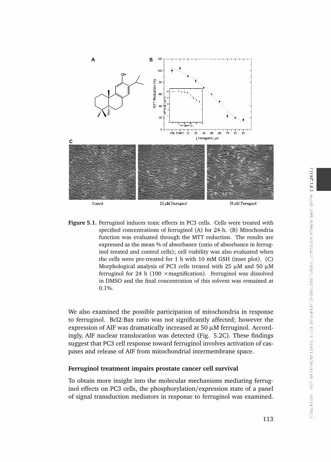
PC3 cells were treated with ferruginol in concentrations up to 100 µM andthe effect of ferruginol on cell viability was determined employing the MTTmethod. As shown in Fig. 5.1B, ferruginol caused a dose-dependent re-duction in the cell number displaying an IC50 value of 55 µM. Importantly,pre-treatment of the cells with 10 mM GSH prevented the toxic action offerruginol (inset plot). In agreement, the microscopy analysis also demon-strated a decrease of the cell number (Fig. 5.1C).
Ferruginol induces apoptosis of PC3 cells via caspases and AIFactivation
In the next series of experiments, it was determined whether treatment ofPC3 cells to ferruginol led to apoptosis. Ferruginol caused around 15%and 30% (in the presence of 25 and 50 µM ferruginol, respectively) ofcell death via apoptosis as detected through phosphatidylserine exposure(Fig. 5.2A). In agreement, we also observed activation of caspases 8, 9 and3 (Fig. 5.2B). Additionally, at the lowest concentration, ferruginol led toan overexpression of TNFR1; however, FADD expression was not affected.
112
thesis March 11, 2015 7:27 Page 113 ☛✡✟✠
☛✡✟✠ ☛✡✟✠
☛✡✟✠
CHAPTER 5 FERRU
GINOL SUPRESS SU
RVIVAL SIG
NALING PATH
WAYS IN
ANDROGEN-INDEPEN
DENT HUMAN PRO
TASTE C
ANCER C
ELLS
Figure 5.1. Ferruginol induces toxic effects in PC3 cells. Cells were treated withspecified concentrations of ferruginol (A) for 24 h. (B) Mitochondriafunction was evaluated through the MTT reduction. The results areexpressed as the mean % of absorbance (ratio of absorbance in ferrug-inol treated and control cells); cell viability was also evaluated whenthe cells were pre-treated for 1 h with 10 mM GSH (inset plot). (C)Morphological analysis of PC3 cells treated with 25 µM and 50 µMferruginol for 24 h (100 ×magnification). Ferruginol was dissolvedin DMSO and the final concentration of this solvent was remained at0.1%.
We also examined the possible participation of mitochondria in responseto ferruginol. Bcl2:Bax ratio was not significantly affected; however theexpression of AIF was dramatically increased at 50 µM ferruginol. Accord-ingly, AIF nuclear translocation was detected (Fig. 5.2C). These findingssuggest that PC3 cell response toward ferruginol involves activation of cas-pases and release of AIF from mitochondrial intermembrane space.
Ferruginol treatment impairs prostate cancer cell survival
To obtain more insight into the molecular mechanisms mediating ferrug-inol effects on PC3 cells, the phosphorylation/expression state of a panelof signal transduction mediators in response to ferruginol was examined.
113

thesis March 11, 2015 7:27 Page 114 ☛✡✟✠
☛✡✟✠ ☛✡✟✠
☛✡✟✠
CHAPTER 5 FERRU
GINOL SUPRESS SU
RVIVAL SIG
NALING PATH
WAYS IN
ANDROGEN-INDEPEN
DENT HUMAN PRO
TASTE C
ANCER C
ELLS
Figure 5.2. Apoptosis induction of PC3 cells by ferruginol. (A) Cell samples wereprepared as described in Section 2 and Annexin V-positive, 7-AAD-positive and Annexin V/7-AAD-positive populations were analyzed byflow cytometry. (B) Caspases 3, 8 and 9 activities were determined byusing colorimetric assay. (C) The expression of pro- and anti-apoptoticproteins was determined by Western blot analysis. Soluble lysates werematched for protein content and analyzed on Western blot and im-munoblots were probed with actin antibody to ensure equal loading.Nuclear translocation of AIF was also determined.
As shown in Fig. 5.3, PC3 cell treatment with 50 µM ferruginol provokeddownregulation of p85 subunit of PI3K and inhibition of AKT. The resultspresented above indicate that ferruginol should produce a decrease of thesurvival and anti-apoptotic relevant kinase activities. Accordingly, cells
114

thesis March 11, 2015 7:27 Page 115 ☛✡✟✠
☛✡✟✠ ☛✡✟✠
☛✡✟✠
CHAPTER 5 FERRU
GINOL SUPRESS SU
RVIVAL SIG
NALING PATH
WAYS IN
ANDROGEN-INDEPEN
DENT HUMAN PRO
TASTE C
ANCER C
ELLS
treated with 50 µM ferruginol displayed activation of MAPK p38, an im-portant apoptosis inductor, and a slight inhibition of ERK2. However, theupstream activator of ERK, MEK, was not affected. Apparently, ferruginolimpairs prostate cancer proliferation by modulating survival and prolifera-tion signaling cascades.
Figure 5.3. Effect of ferruginol treatment on the function of key mediators involvedin the PC3 cells proliferation/survival. Cells were treated with specifiedconcentrations of ferruginol for 24 h and the expression or phosphory-lation of the proteins determined by Western blot. Equal loading wasconfirmed by reprobing them for β-actin.
Inhibition of cell cycle progression by Ferruginol
By plotting the ratio of the cells in G0 plus G1 against cells in G2/M plusS-phase it is apparent that PC3 cells undergo G0/ G1-phase cell cycle arrestafter ferruginol treatment (Fig. 5.4A). Approximately 2.5-fold of the PC3population was at phase G0/G1 after exposure to ferruginol for 24 h. Wetherefore turned to characterize the effect of ferruginol treatment on di-rect regulators of cell cycle progression. As shown by immunoblot analysis(Fig. 5.4B), the expression of p21 was increased even at the lowest con-centration of ferruginol. On the other hand, the phosphorylated Rb proteinand the expression of PCNA were not affected. The level of phosphor-cdc2,the key protein of the cell cycle progression from G2 to M phase, remainedunchanged. Ferruginol decreased the level of CDK4, CDK6, cyclin D1 andcyclin D3. This response directly mirrored the ability of ferruginol to inducecell cycle arrest in PC3 cells. We conclude that the combination of reduced
115

thesis March 11, 2015 7:27 Page 116 ☛✡✟✠
☛✡✟✠ ☛✡✟✠
☛✡✟✠
CHAPTER 5 FERRU
GINOL SUPRESS SU
RVIVAL SIG
NALING PATH
WAYS IN
ANDROGEN-INDEPEN
DENT HUMAN PRO
TASTE C
ANCER C
ELLS
mitotic activity and induction of apoptosis accounts for the observed cyto-toxic effect of ferruginol on PC3 cells.
Figure 5.4. Ferruginol impedes prostate cancer cell proliferation by targeting keycell cycle mediators. After treating PC3 cells with ferruginol for 24 h,cells were harvested, stained with PI and analyzed by flow cytometry(A) or lysed for Western blotting analysis (B). The number of cells ineach phase of the profile and ratios of cells in resting phase (G0/G1)versus those undergoing mitosis (S, G2/M) was determined. The ex-pression or phosphorylation of the proteins was determined by Westernblot. Equal loading was confirmed by reprobing them for β-actin.
116

thesis March 11, 2015 7:27 Page 117 ☛✡✟✠
☛✡✟✠ ☛✡✟✠
☛✡✟✠
CHAPTER 5 FERRU
GINOL SUPRESS SU
RVIVAL SIG
NALING PATH
WAYS IN
ANDROGEN-INDEPEN
DENT HUMAN PRO
TASTE C
ANCER C
ELLS
Ferruginol causes downregulation of IKKa and hypophosphorylationof STATs
The immunoblot analysis data showed that the expression of IKKα wassignificantly decreased after treatment with ferruginol; however, the to-tal level of NFκB remained unchanged (Fig. 5.5). In addition, ferruginolcaused a decrease of this transcription factor into the nucleus. The phos-phorylation status of STAT 3 and 5, key signaling molecules for many cy-tokines and growth-factor receptor response, was also evaluated by West-ern blot. The treatment of PC3 with 50 µM ferruginol was able to decreasethe phosphorylation levels of STAT3 (Tyr705) and STAT5 (Tyr694). Inter-estingly, 50 µM ferruginol caused a strong decline in Hsp27 expression,which can be associated with the increase in the number of apoptotic cells.
Figure 5.5. Ferruginol modulates molecules that play a crucial role in the prostatecancer aggressiveness. Cells were treated with specified concentrationsof Ferruginol for 24 h, harvested and total cell lysates were prepared.The expression of IκB kinase, cytosolic and nuclear NFκB, and phos-phorylation status of Hsp27 and STAT 3 and 5 were determined byWestern blot analysis. Equal loading was probed with actin antibodyto ensure equal loading.
Redox status on PC3 cells treated with ferruginol
Based on the diterpene chemical properties, which can lead to antioxidantand/or oxidant action depending on its concentration, and the importanceof reducing equivalents for PC3 cells survival [23], we investigated the ef-
117

thesis March 11, 2015 7:27 Page 118 ☛✡✟✠
☛✡✟✠ ☛✡✟✠
☛✡✟✠
CHAPTER 5 FERRU
GINOL SUPRESS SU
RVIVAL SIG
NALING PATH
WAYS IN
ANDROGEN-INDEPEN
DENT HUMAN PRO
TASTE C
ANCER C
ELLS
fect of ferruginol on PC3 cell GSH metabolism. PC3 cells treated with fer-ruginol displayed a more oxidizing environment as defined by a decreaseof GSH and an increase of GSSG levels (Fig. 5.6). Both effects were dosedependent and the highest concentration of ferruginol employed caused a2-fold change. It is important to note that even at the highest concentrationof ferruginol, GSH:GSSG ratio remained almost in equilibrium (GSH:GSSG= 4.4, 2.7 and 1.1 at 0, 25 and 50 µM ferruginol, respectively).
Figure 5.6. Influence of ferruginol on the level of intracellular GSH and GSSG. Cellswere treated with ferruginol for 24 h and the concentration of GSH andGSSG determined as described in Section 2.
Low molecular weight protein tyrosine phosphatase is modulated byferruginol
Besides the fact that protein tyrosine phosphatases are highly sensitive tocell redox status, there is some evidence that this class of phosphatasescan contribute to tumor cell progression and aggressiveness. EspeciallyLMWPTP has been recognized as a positive regulator of tumor growth [24].Therefore, to address the possible modulation of LMWPTP by ferruginol,we examined the LMWPTP activity as well as expression. LMWPTP im-munoprecipitated from PC3 cells was inhibited around 30 % by 50 mMferruginol (Fig. 5.7). On the other hand, when the LMWPTP activity waschecked after treating the cells for 24 h, this enzyme displayed only 20 % ofresidual activity. Reduced LMWPTP activity is consistent with the changein redox status in response to ferruginol. In addition, treated cells demon-strated downregulation of LMWPTP expression.
118

thesis March 11, 2015 7:27 Page 119 ☛✡✟✠
☛✡✟✠ ☛✡✟✠
☛✡✟✠
CHAPTER 5 FERRU
GINOL SUPRESS SU
RVIVAL SIG
NALING PATH
WAYS IN
ANDROGEN-INDEPEN
DENT HUMAN PRO
TASTE C
ANCER C
ELLS
Figure 5.7. Ferruginol affects low molecular weight protein phosphatase activityand expression. (A) The effect of ferruginol was examined directly inthe LMWPTP immunoprecipitated from non-treated PC3 cells and alsoafter PC3 treatment with ferruginol (inset plot). The LMWPTP specificactivity was used to determine the relative activity. (B) Expression ofLMWPTP was checked by Western blotting.
5.4 Discussion
Prostate cancer is commonly malignant and it is the second leading cause ofcancer-related deaths (after lung cancer) of males in Brazil, with a similartrend in many Western countries (data from Instituto Nacional do Câncer-INCA). Since prostate cancer usually occurs in men aged 50 years and olderand because of the increasing life expectancy, its incidence is expected tofurther rise in the years to come [25]. Chemoprevention and interventionstrategies using anticancer agents are considered as promising therapeuticoptions. The search for new chemopreventive and/or chemotherapeuticagents that are more effective without toxic side-effects has generated greatinterest in phytochemicals with potential activity in this respect [26]. Sup-pression of tumorigenesis often involves modulation of signal transductionpathways, leading to alterations in gene expression, cell cycle progressionor apoptosis. Apoptosis is considered as an ideal way for destroying dam-aged cells and also a potential target for chemopreventive elimination ofcancer cells [27] and as a consequence targeting signaling elements con-
119

thesis March 11, 2015 7:27 Page 120 ☛✡✟✠
☛✡✟✠ ☛✡✟✠
☛✡✟✠
CHAPTER 5 FERRU
GINOL SUPRESS SU
RVIVAL SIG
NALING PATH
WAYS IN
ANDROGEN-INDEPEN
DENT HUMAN PRO
TASTE C
ANCER C
ELLS
Figure 5.8. Schematic representation of the molecular mechanism of ferruginol-induced PC3 cell death. Data presented in this report revealed that fer-ruginol exhibits multi-activities, which culminate with apoptosis induc-tion of prostate cancer. Ferruginol caused inhibition of two importantsignaling cascade pathways involved in the cell survival/proliferation(Ras/PI3K and Jak/STAT). Additionally treated prostate cancer cellsdisplayed a decrease in the phosphorylated form of Hsp27. Ferruginol-induced apoptosis was accompanied by activation of caspase 3, an in-crease of AIF expression and maintenance of Bax and Bcl-2 levels. AIFis released from mitochondria and translocated to the nucleus, andparticipates in peripheral chromatin condensation. In accordance withcell survival diminishing, Ferruginol caused cell cycle arrest. Ferrugi-nol caused an overexpression of protein p21 (a member of the cyclin-dependent kinase inhibitors), and downward expression of cyclin D1,cyclin D3, CDK4 and CDK6, which indicates cell cycle arrest at G0/G1.Importantly, LMWPTP was directly and indirectly modulated by ferrug-inol, which indicates that this enzyme can be a target for this naturalcompound.
trolling apoptosis may open novel therapeutic avenues [28,29]. Severalplant-derived bioactive agents may have such action, at least as judgedfrom model systems [4,9,30-33]; the present study may add ferruginol tothis growing list.
120

thesis March 11, 2015 7:27 Page 121 ☛✡✟✠
☛✡✟✠ ☛✡✟✠
☛✡✟✠
CHAPTER 5 FERRU
GINOL SUPRESS SU
RVIVAL SIG
NALING PATH
WAYS IN
ANDROGEN-INDEPEN
DENT HUMAN PRO
TASTE C
ANCER C
ELLS
Our results suggest that ferruginol is a negative regulator of cancer cellproliferation. Androgen-independent human prostate cancer cells (PC3cells) a model that exhibits extreme therapy resistance exhibited, upontreatment with this phytocompound, remarkable induction of apoptosisvia extrinsic and intrinsic pathways, as demonstrated by the observationof overexpression of TNFR1 and activation of caspases 8, 9 and 3. The ex-trinsic pathway for cell death involves plasma membrane death receptors[34]. These receptors trimerize and recruit the adaptor molecule FADDwhich, in turn, activates caspase 8 and also leads to the activation of down-stream execution caspases [35-38]. In both pathways, activation of effectorcaspases leads to a series of morphological changes that are characteristicfor apoptosis [28].
Ferruginol-induced apoptosis and cell growth inhibition were also ac-companied by an increase of apoptosis-inducing factor (AIF) expressionand maintenance of Bax and Bcl2 levels. AIF was identified as a mitochon-drial intermembrane space protein, which is released from mitochondriaand translocated to the nucleus, in response to apoptotic stimuli, and par-ticipates in peripheral chromatin condensation and the exposure of phos-phatidylserine in the outer leaf of the plasma membrane. Increasing ev-idence supports the notion that AIF plays an important role in caspase-independent apoptosis [39,40].
Ferruginol, even at lower concentration, caused inhibition of Ras/PI3Kcascade and suppression of downstream mitogenic targets such as cyclinD1. Additionally, this diterpene also induced activation of MAPK p38. Thephosphatase and tensin homologue (PTEN) gene is deleted in PC3 cells.This phosphatase is defined as a tumor suppressor, since it is the majornegative modulator of AKT protein, an important mediator of cell survival.It is therefore important to identify agents that can overcome the thera-peutic resistant properties of PTEN deficient tumor cells. Importantly, inaccordance with cell survival diminishing, ferruginol caused cell cycle ar-rest. Eukaryotic cell cycle progression is regulated by sequential activa-tion and subsequent inactivation of a series of CDKs at different phases[41]. Ferruginol caused an overexpression of protein p21 a member ofthe cyclin-dependent kinase inhibitors, and downward expression of cyclinD1, cyclin D3, CDK4 and CDK6. These data showed the involvement ofp21 in ferruginol- induced G1 phase arrest, through binding to and sub-sequently inhibiting the cyclin-CDK activity. The active complex of cyclinD/CDK4 targets the Rb protein for phosphorylation, allowing the releaseof E2F transcription factors that activate G1/S-phase gene expression. Im-portantly cdc-2, a key protein responsible for the entry of the cell from G2to M phase, remained unchanged. Cell cycle regulation and its modulationby various plant-derived agents are gaining widespread attention in recentyears. A large number of phytochemicals has been shown to inhibit cellcycle progression of various cancer cells [42].
121
thesis March 11, 2015 7:27 Page 122 ☛✡✟✠
☛✡✟✠ ☛✡✟✠
☛✡✟✠
CHAPTER 5 FERRU
GINOL SUPRESS SU
RVIVAL SIG
NALING PATH
WAYS IN
ANDROGEN-INDEPEN
DENT HUMAN PRO
TASTE C
ANCER C
ELLS
Ferruginol decreased the phosphorylation level of STAT3, STAT5 andHsp27. STATs are latent cytoplasmic transcription factors consisting ofseven mammalian members. They become phosphorylated on Tyr residuesupon activation, a post-translational modification that is critical for dimer-ization, nuclear import, DNA binding, and transcriptional activation [43].The activation of STATs is mediated by the action of an upstream Januskinase (JAK), usually JAK1 or JAK2, showing that the JAK cascade mightitself be a target for therapy in prostate cancer. Ahohen and coworkers[44] demonstrated that STAT5 is activated in a significant number of hu-man prostate cancer specimens. Additionally, these authors also reportedinduction of apoptosis via caspases 9 and 3 activation dependent on inhi-bition of STAT5 phosphorylation. Activated STAT3 was reported in manytypes of malignancies, such as myeloma, head and neck cancer, breast can-cer, and prostate cancer [45]. Recently, it has been demonstrated that in-hibition of STAT3 in tumors impeded vascular endothelial growth factorproduction [46]. Data from the literature have identified Hsp27 as a mod-ulator of STAT3-regulated apoptosis after androgen ablation. Hsp27 is a 27kDa protein of which expression is seen to be correlated with an increaseof survival in response to a wide variety of physiological and environmen-tal insults including heat, reactive oxygen species and anticancer drugs.Indeed analysis by co-immunoprecipitation and immunofluorescence con-firmed that Hsp27 is able to interact with STAT3 and that STAT3 levelscorrelate directly with Hsp27 levels. There are some reports in the liter-ature demonstrating that the prostate cancer Hsp27 level increases afterandrogen ablation and that this protein is highly expressed in androgen-independent tumors, and inhibition of Hsp27 in prostate cancer cells canincrease the number of apoptotic cells (G0/G1), an event that seems tobe associated with the decrease in the STAT3 levels [47]. These findingsindicate that the anti-apoptotic effects of Hsp27 are associated with its abil-ity to interact and stabilize the STAT3 molecule, leading to more resistantprostate cancer cells. In accordance with this notion, our results show adecrease in the phosphorylated forms of Hsp27 and STAT3, when PC3 cellswere treated with ferruginol at the concentration of 50 mM, suggesting thatthe pro-apoptotic and anti-proliferative actions of ferruginol might be as-sociated with diminished function of STAT3 through the decrease of Hsp27levels.
Another important finding is that ferruginol did not induce the NFκBtranslocation to the nucleus; this result is in agreement with the findingsof Rodriguez and coworkers [17] related to the anti-inflammatory effect offerruginol.
Recently, Chaiswing and collaborators [23] reported in a very well de-signed paper the effect of cellular redox state on prostate cancer cell growthin vitro. These authors demonstrated that during PC3 cells growth, thesecells require higher ratio of reduced glutathione (GSH)/glutathione disul-
122

thesis March 11, 2015 7:27 Page 123 ☛✡✟✠
☛✡✟✠ ☛✡✟✠
☛✡✟✠
CHAPTER 5 FERRU
GINOL SUPRESS SU
RVIVAL SIG
NALING PATH
WAYS IN
ANDROGEN-INDEPEN
DENT HUMAN PRO
TASTE C
ANCER C
ELLS
fide (GSSG). Based on this observation, we evaluated the redox status to-ward PC3 cell treatment with ferruginol. This diterpene caused a decreaseof GSH and increase of GSSG, indicating a dominant effect in favor of oxi-dizing equivalent. Several signaling mediators can be modulated by redoxmodifiers, including protein tyrosine phosphatases [48, 9]. To investigatethe effect of ferruginol on PTPs we chose LMWPTP. The rational reason forthis was based on the following aspects: (a) we have observed a high levelof this phosphatase in PC3 cells and (b) Chiarugi and collaborators [24]have reported the importance of this enzyme for cancer cell growth. In-terestingly, we observed a direct effect of ferruginol on LMWPTP but alsothere was a correlation between cellular oxidizing equivalents and inhibi-tion of this enzyme. The oxidation of catalytic site cysteine of PTPs, such asLMWPTP, leads to the transformation of the sulfhydrylic residue in sulfenicacid and the consequent inactivation of the enzyme due to its inability toform cysteinyl-phosphate intermediate during the first step of the catalysis[48]. Altogether, our results demonstrate that ferruginol can act as a chem-ical and genetic modulator of LMWPTP. These data indicate that at least inpart, the anti-proliferative ction of ferruginol is dependent on changing cel-lular redox, which is in agreement with the protective effect of GSH. Thisobservation also confirms the importance of reducing equivalents for PC3cell survival, as recently reported [23]. Further experiments to clarify therole of LMWPTP on prostate cancer progression are currently in progressin our laboratory.
5.5 Conclusion
One of the challenges of cancer therapy is to combine efficacy with fewside effects and consequently improve the quality of life of the patient.Prostate cancer represents a spectrum of diseases in which the cost of curemay be substantial, with short- and long-term side effects. Therefore, newagents are needed to extend survival, improve cure rates, and avoid un-desired treatment-related toxicities. In this scenario, there are at leasttwo aims: (a) to provide therapeutic agents with a very specific targetand (b) to discover agents which present differential action mechanismsin comparison with the traditional chemotherapy. In this context, ferrugi-nol appears as an interesting bioactive compound, since it exhibits multi-activities in the signal transduction/biochemical aspects in prostate can-cer cells. In summary, ferruginol negatively modulates signaling cascades,which are known to be defective in some types of prostate cancers, namelyRas/PI3K and Jak/STAT, as well as cell cycle regulators (Fig. 5.8). Im-portantly, we demonstrated for the first time that LMWPTP is directly andindirectly modulated by ferruginol, which indicates that this enzyme canbe a target for this natural compound. Besides affecting signal transduction
123
thesis March 11, 2015 7:27 Page 124 ☛✡✟✠
☛✡✟✠ ☛✡✟✠
☛✡✟✠
CHAPTER 5 FERRU
GINOL SUPRESS SU
RVIVAL SIG
NALING PATH
WAYS IN
ANDROGEN-INDEPEN
DENT HUMAN PRO
TASTE C
ANCER C
ELLS
triggered by TNFR1, ferruginol also affected mitochondria permeability asdemonstrated by the presence of nuclear AIF (protein involved with chro-matin condensation and DNA fragmentation); however, the Bax:Bcl2 ratioremained unchanged. In general, this study provides an overview of bio-chemical aspects which were affected by ferruginol and in turn confirms itsanti-tumor activity. This type of investigation can contribute to the devel-opment of “smart” drugs.
5.6 Acknowledgments
Financial support by FONDECYT (Grant No. 1060841) and the Programade Productos Bioactivos, University of Talca is gratefully acknowledged.C.A. thanks the Universidad de Talca for a doctoral grant. The authors ac-knowledge financial support by the Brazilian Agencies: Fundação de Am-paro à Pesquisa do Estado de São Paulo, Conselho Nacional de Desenvolvi-mento Científico e Tecnológico and Fundo de Apoio ao Ensino, à Pesquisae à Extensão.
5.7 References
[1] J. Moul, Report from Durham, Prostate Cancer Prostatic Dis. 8 (2005)1.[2] N. Dawson, New molecular targets in advanced prostate cancer, ExpertRev. Anticancer Ther. 6 (2006) 993e1002.[3] M.A. Miranda, A.K. Okamoto, C.V. Ferreira, T.L. Silva, J.M. Granjeiro,H. Aoyama, Differential effects of flavonoids on bovine kidney low molec-ular mass protein tyrosine phosphatase, J. Enzyme Inhib. Med. Chem.21(2006) 419e425.[4] A.G. Freire, P.S. Melo, M. Haun, N. Duran, H. Aoyama, C.V. Ferreira,Cytotoxic effect of the diterpene lactone dehydrocrotonin from Crotoncajucara on human promyelocytic leukaemia cells, Planta Med. 69 (2003)67e69.[5] C.V. Ferreira, C.L. Bos, H.H. Versteeg, G.Z. Justo, N. Duran, M.P. Pep-pelenbosch, Molecular mechanism of violacein-mediated human leukemiacell death, Blood 104 (2004) 1459e1464.[6] A.D. Martins Cavagis, C.V. Ferreira, H.H. Versteeg, C.F. Assis, C.L. Bos,S.A. Bleuming, S.H. Diks, H. Aoyama, M.P. Peppelenbosch, Tetrahydrox-yquinone induces apoptosis of leukemia cells through diminished survivalsignaling, Exp. Hematol. 34 (2006) 188e196.[7] L.L. Kodach, C.L. Bos, N. Duran, M.P. Peppelenbosch, C.V. Ferreira, J.C.Hardwick, Violacein synergistically increases 5-fluorouracil cytotoxicity,induces apoptosis and inhibits Akt-mediated signal transduction in humancolorectal cancer cells, Carcinogenesis 27 (2006) 508e516.
124
thesis March 11, 2015 7:27 Page 125 ☛✡✟✠
☛✡✟✠ ☛✡✟✠
☛✡✟✠
CHAPTER 5 FERRU
GINOL SUPRESS SU
RVIVAL SIG
NALING PATH
WAYS IN
ANDROGEN-INDEPEN
DENT HUMAN PRO
TASTE C
ANCER C
ELLS
[8] A.C.S. de Souza, L. Kodach, F.R. Gadelha, C.L. Bos, A.D. Cavagis, H.Aoyama, M.P. Peppelenbosch, C.V. Ferreira, A promising action of riboflavinas a mediator of leukaemia cell death, Apoptosis 11 (2006) 1761e1771.[9] R.R.R. Sousa, A.C.S. Souza, K.C. Queiroz, M.P. Peppelenbosch, C.V.Ferreira, H. Aoyama, Phosphoprotein levels, MAPK activities and NFκBexpression are affected by fisetin, J. Enzyme Inhib. Med. Chem. 22 (2007)439e444.[10] K.C. de Souza Queiroz, W.F. Zambuzzi, A.C. Santos de Souza, R.A.da Silva, D. Machado, G.Z. Justo, H.F. Carvalho, M.P. Peppelenbosch,C.V. Ferreira, A possible anti-proliferative and anti-metastatic effect ofirradiated riboflavin in solid tumours, Cancer Lett. 258 (2007) 126e134.[11] M. Iwamoto, T. Minami, H. Tokuda, H. Ohtsu, R. Tanaka, Potentialantitumor promoting diterpenoids from the stem bark of Thuja standishii,Planta Med. 69 (2003) 69e72.[12] J. Becerra, C. Flores, J. Mena, P. Aqueveque, J. Alarcon, M. Bittner,V. Hernández, M. Hoeneisen, E. Ruiz, M. Silva, Antifungal and antibac-terial activity of diterpenes isolated from wood extractables of ChileanPodocarpaceae, Boletín de la Sociedad Chilena de Química 47 (2002)151e157.[13] S. Chang, P. Chen, S. Wang, H. Wu, Antimite activity of essential oilsand their constituents from Taiwania cryptomerioides, J. Med. Entomol.38 (2001) 455e457.[14] A. Ulubelen, H. Birman, S. Oksuz, G. Topcu, U. Kolak, A. Barla, W.Voelter, Cardioactive diterpenes from the roots of Salvia eriophora, PlantaMed. 68 (2002) 818e821.[15] M. Ono, M. Yamamoto, C. Masuoka, Y. Ito, M. Yamashita, T. Nohara,Diterpenes from the fruits of Vitex rotundifolia, J. Nat. Prod. 62 (1999)1532e1537.[16. C. Clarkson, W.E. Campbell, P. Smith, In vitro antiplasmodial activ-ity of abietane and totarane diterpenes isolated from Harpagophytumprocumbens (devil’s claw), Planta Med. 69 (2003) 720e724.[17] J.A. Rodriguez, C. Theoduloz, T. Yanez, J. Becerra, G. Schmeda-Hirschmann, Gastroprotective and ulcer healing effect of ferruginol inmice and rats: assessment of its mechanism of action using in vitro models,Life Sci. 78 (2006) 2503e2509.[18] C. Areche, J.A. Rodríguez, I. Razmilic, T. Yañez, C. Theoduloz, G.Schmeda-Hirschmann, Gastroprotective and cytotoxic effect of semisyn-thetic ferruginol derivatives, J. Pharm. Phamacol. 59 (2007) 289e300.[19] T. Mosmann, Rapid colorimetric assay for cellular growth andsurvival: application to proliferation and cytotoxicity assay, J. Immunol.Methods 65 (1983) 55e63.[20] F. Denizot, R. Lang, Rapid colorimetric assay for cell growth andsurvival: modifications to the tetrazolium dye procedure giving improvedsensitivity and reliability, J. Immunol. Methods 89 (1986) 71e77.
125

thesis March 11, 2015 7:27 Page 126 ☛✡✟✠
☛✡✟✠ ☛✡✟✠
☛✡✟✠
CHAPTER 5 FERRU
GINOL SUPRESS SU
RVIVAL SIG
NALING PATH
WAYS IN
ANDROGEN-INDEPEN
DENT HUMAN PRO
TASTE C
ANCER C
ELLS
[21] E.F.Hartree, Determination of proteins: a modification ofLowrymethod that give a linear photometric response, Anal. Biochem. 48(1972) 422e427.[22] N. Li, T.D. Oberley, Modulation of antioxidant enzymes, reac-tive oxygen species, and glutathione levels in manganese superoxidedismutase-overexpressing NIH/3T3 fibroblasts during the cell cycle, J. CellPhysiol. 177 (1998) 148e160.[23] L. Chaiswing, J.M. Bourdeau-Heller, W. Zhong, T.D.Oberley, Charac-terization of redox state of two human prostate carcinoma cell lines withdifferent degrees of aggressiveness, Free Radic. Biol. Med. 43 (2007)202e215.[24] P. Chiarugi, M.L. Taddei, N. Schiavone, L. Papucci, E. Giannoni, T.Fiaschi, S. Capaccioli, G. Raugei, G. Ramponi, LMW-PTP is a positiveregulator of tumor onset and growth, Oncogene 23 (2004) 3905e3914.[25] A. Jemal, R.C. Tiwari, T. Murray, A. Ghafoor, A. Samuels, E. Ward,E.J. Feuer, M.J. Thun, American Cancer Society Cancer statistics, Cancer J.Clin. 54 (2004) 8e29.[26] A.S. Tsao, E.S. Kim, W.K. Hong, Chemoprevention of cancer, CACancer J, Clin 54 (2004) 150e180.[27] R.P. Singh, S. Dhanakakshmi, R. Agarwal, Phytochemicals as cell cyclemodulators: a less toxic approach in halting human cancer, Cell Cycle 1(2002) 156e161.[28] W. Hu, J.J. Kavanagh, Anticancer therapy targeting the apoptoticpathway, Lancet Oncol. 4 (2003) 721e729.[29] S. Fulda, K.M. Debatin, Extrinsic versus intrinsic apoptosis pathwaysin anticancer chemotherapy, Oncogene 25 (2006) 4798e4811.[30] M. D-Huang, J.-H. Guh, Y.-T. Huang, S.-C. Chueh, P.-C. Chian, C.-M.Teng, Induction of mitotic arrest and apoptosis in human prostate cancerpc-3 cells by evodiamine, J. Urol. 173 (2005) 256e261.[31] G.Z. Justo, C.V. Ferreira, Coagulation and cancer therapy: the poten-tial of natural compounds, Curr. Genomics. 6 (2005) 461e469.[32] J.C. Hsu, A. Dev, A. Wing, C.T. Brew, L.F. Bjeldanes, G.L. Firestone,Indole-3-carbinol mediated cell cycle arrest of LnCap human prostatecancer cells requires the induced production of activated p53 tumorsuppressor protein, Biochem. Pharmacol. 72 (2006) 1714e1723.[33] L. Gapter, Z. Wang, J. Glinski, K. -Y. Ng, Induction of apoptosis inprostate cancer cells by pachymic acid from Poria cocos, Biochem. Biophys.Res. Commun. 332 (2005) 1153e1161.[34] E. Solary, N. Droin, O. Sordet, Cell death pathways as targets foranticancer drugs, in: B.C. Baguley, D.J. Kerr (Eds.), Anticancer DrugDevelopment, Academic Press, San Diego 2002, pp. 55e76.[35] N. Thornberry, Y. Lazebnik, Caspases: enemies within, Science281(1998) 1312e1316.[36] A. Ashkenazi, V.M. Dixit, Death receptors: signaling and modulation,
126
thesis March 11, 2015 7:27 Page 127 ☛✡✟✠
☛✡✟✠ ☛✡✟✠
☛✡✟✠
CHAPTER 5 FERRU
GINOL SUPRESS SU
RVIVAL SIG
NALING PATH
WAYS IN
ANDROGEN-INDEPEN
DENT HUMAN PRO
TASTE C
ANCER C
ELLS
Science 281 (1998) 1305e1308.[37] A.M. Chinnaiyan, K. O’Rourke, M. Tewari, V.M. Dixit, FADD, a noveldeath domain-containing protein, interacts with the death domain of Fasand initiates apoptosis, Cell 81 (1995) 505e512.[38] M.E. Peter, P.H. Krammer, Mechanisms of CD95 (APO-1/ Fas)-mediated apoptosis, Curr. Opin. Immunol. 10 (1998) 545e551.[39] C. Pepper, T. Hoy, D.P. Bently, Bcl-2/Bax ratios in chronic lymphocyticleukaemia and their correlation with in vitro apoptosis and clinical resis-tance, Br. J. Cancer 76 (1997) 935e938.[40] C. Cande, I. Cohen, E. Daugas, L. Ravagnan, N. Larochette, N.Zamzami, G. Kroemer, Apoptosis-inducing factor (AIF): a novel caspase-independent death effector released from mitochondria, Biochimie. 84(2002) 215e222.[41] C.J. Sherr, G1 phase progression: Cycling on cue, Cell 79 (1994)551e 555.[42] J.-M. Yun, M.-H. Kweon, H. Kwon, J.-K. Hwang, H. Mukhtar, Induc-tion of apoptosis and cell cycle arrest by a chalcone panduratin A isolatedfrom Kaempferia pandurata in androgen-independent human prostatecancer cells PC3 and DU145, Carcinogenesis 27 (2006) 1454e1464.[43] C.P. Lim, X. Cao, Structure, function, and regulation of STAT proteins,Mol. Biosyst. 2 (2006) 536e550.[44] T.J. Ahonen, J. Xie, M.J. LeBaron, J. Zhu, M. Nurmi, K. Alanen, H. Rui,M.T. Nevalainen, Inhibition of transcription factor Stat5 induces cell deathof human prostate cancer cells, J. Biol. Chem. 278 (2003) 27287e27292.[45] B.E. Barton, J.G. Karras, T.F. Murphy, A. Barton, H.F. Huang, Signaltransducer and activator of transcription 3 (STAT3) activation in prostatecancer: Direct STAT3 inhibition induces apoptosis in prostate cancer lines,Mol. Cancer Ther. 3 (2004) 11e20.[46] G. Niu, K.L. Wright, M. Huang, L. Song, E. Haura, J. Turkson, S.Zhang, T. Wang, D. Sinibaldi, D. Coppola, R. Heller, L.M. Ellis, J. Karras,J. Bromberg, D. Pardoll, R. Jove, H. Yu, Constitutive Stat3 activity up-regulates VEGF expression and tumor angiogenesis, Oncogene 21 (2002)2000e2008.[47] P. Rocchi, E. Beraldi, S. Ettinger, L. Fazli, R.L. Vessella, C. Nelson,M. Gleave, Increased Hsp27 after androgen ablation facilitates androgen-independent progression in prostate cancer via signal transducers andactivators of transcription 3-mediated suppression of apoptosis, CancerRes. 65 (2005) 11083e11093.[48] J. den Hertog, A. Groen, T. van der Wijk, Redox regulation of protein-tyrosine phosphatases, Arch. Biochem. Biophys. 434 (2005) 11e15.[49] C.V. Ferreira, G.Z. Justo, A.C.S. Souza, K.C. Queiroz, W.F. Zambuzzi,H. Aoyama, M.P. Peppelenbosch, Natural compounds as a source of proteintyrosine phosphatase inhibitors: application to the rational design ofsmall-molecule derivatives, Biochimie 88 (2006) 1859e1873.
127

thesis March 11, 2015 7:27 Page 128 ☛✡✟✠
☛✡✟✠ ☛✡✟✠
☛✡✟✠