Embed Size (px)
Citation preview

University of Groningen
Immunologic aspect of ovarian cancer and p53 as tumor antigenNijman, HW; Lambeck, A; van der Burg, SH; van der Zee, AGJ; Daemen, T
Published in:Journal of translational medicine
DOI:10.1186/1479-5876-3-34
IMPORTANT NOTE: You are advised to consult the publisher's version (publisher's PDF) if you wish to cite fromit. Please check the document version below.
Document VersionPublisher's PDF, also known as Version of record
Publication date:2005
Link to publication in University of Groningen/UMCG research database
Citation for published version (APA):Nijman, HW., Lambeck, A., van der Burg, SH., van der Zee, AGJ., & Daemen, T. (2005). Immunologicaspect of ovarian cancer and p53 as tumor antigen. Journal of translational medicine, 3, 1-12. [34].https://doi.org/10.1186/1479-5876-3-34
CopyrightOther than for strictly personal use, it is not permitted to download or to forward/distribute the text or part of it without the consent of theauthor(s) and/or copyright holder(s), unless the work is under an open content license (like Creative Commons).
Take-down policyIf you believe that this document breaches copyright please contact us providing details, and we will remove access to the work immediatelyand investigate your claim.
Downloaded from the University of Groningen/UMCG research database (Pure): http://www.rug.nl/research/portal. For technical reasons thenumber of authors shown on this cover page is limited to 10 maximum.
Download date: 05-01-2020
BioMed CentralJournal of Translational Medicine
ss
Open AcceReviewImmunologic aspect of ovarian cancer and p53 as tumor antigenHW Nijman*1, A Lambeck1,2, SH van der Burg3, AGJ van der Zee1 and T Daemen2Address: 1Dept. of Gynaecologic Oncology, Groningen University Medical Center, 2Dept. of Medical Microbiology, Molecular Virology Section, Groningen University Medical Center and 3Dept. of Immunohematology and Blood Transfusion, Leiden University Medical Center
Email: HW Nijman* - [email protected]; A Lambeck - [email protected]; SH van der Burg - [email protected]; AGJ van der Zee - [email protected]; T Daemen - [email protected]
* Corresponding author
AbstractOvarian cancer represents the fifth leading cause of death from all cancers for women. During thelast decades overall survival has improved due to the use of new chemotherapy schedules. Still, themajority of patients die of this disease. Research reveals that ovarian cancer patients exhibitsignificant immune responses against their tumor. In this review the knowledge obtained thus faron the interaction of ovarian cancer tumor cells and the immune system is discussed. Furthermorethe role of p53 as tumor antigen and its potential role as target antigen in ovarian cancer issummarized. Based on the increased knowledge on the role of the immune system in ovariancancer major improvements are to be expected of immunotherapy based treatment of this disease.
IntroductionOvarian cancer is the most common cause of death fromgynecological malignancies. Its nonspecific clinical pres-entation and the absence of effective screening methodsare responsible for the 70% of patients who present withan advanced stage of disease at the time of diagnosis. Pri-mary treatment for advanced stage ovarian cancer iscytoreductive surgery followed by platinum/paclitaxelbased chemotherapy. An aggressive surgical approach hasbeen advocated with the intent to remove all macroscopicdisease which should yield better survival than leavingresidual disease [1-3]. Response rates to primary chemo-therapy are 65–80%. When residual or recurrent diseasemanifests itself, resistance to chemotherapy will prohibitfurther curative therapy, resulting in an overall survival forpatients with advanced stage ovarian disease of only 10–20%[4,5].
Research during the last decades has revealed that ovariancancer patients exhibit significant immune responsesagainst their tumor (reviewed in this paper). In designingalternative treatments to successfully eradicate ovariancancer it is important to consider both the positive effectsof immune responses to ovarian cancer and the con-founding negative effects on the immune system causedby the tumor cells. As the main target for a potential vac-cine is the (overexpressed / mutated) p53 protein we willfocus on studies aimed at the induction of humoral andcellular responses against this antigen. However, beforereviewing these studies we will briefly introduce somegeneral aspects of the cellular immune system includingantigen encounter, antigen processing and presentationand factors influencing the outcome of the immuneresponse in ovarian cancer.
Published: 15 September 2005
Journal of Translational Medicine 2005, 3:34 doi:10.1186/1479-5876-3-34
Received: 21 July 2005Accepted: 15 September 2005
This article is available from: http://www.translational-medicine.com/content/3/1/34
© 2005 Nijman et al; licensee BioMed Central Ltd. This is an Open Access article distributed under the terms of the Creative Commons Attribution License (http://creativecommons.org/licenses/by/2.0), which permits unrestricted use, distribution, and reproduction in any medium, provided the original work is properly cited.
Page 1 of 12(page number not for citation purposes)

Journal of Translational Medicine 2005, 3:34 http://www.translational-medicine.com/content/3/1/34
General introduction on the cellular immune systemAntigen presenting cells, most likely dendritic cells, cancapture tumor antigens that are secreted or shed by tumorcells or by taking up dying tumor cells. The tumor anti-gens are processed and presented as peptides by major his-tocompatability complex (MHC) I and II molecules onthe cell surface, and recognized by the T-cell receptor onT-cells. This phenomenon is often referred to as the firstsignal of activation. After cleavage of proteins into pep-tides by the proteasome complex and loading of peptidesinto the class I molecules in the endoplasmatic reticulum,these MHC class I – peptide complexes, recognized bycytotoxic T lymphocytes, are transported to the cell sur-face. MHC class II molecules mainly present exogenousendocytosed proteins. Antigen (peptide) loading of MHCclass II molecules occurs within the endocytic pathway(MHC class II compartments). MHC class II – peptidecomplexes expressed on the cell surface are recognized bythe CD4+ T helper cells. Next to this first antigen specificsignal there is a need for a second signal. This signalinvolves the ligation of CD28 or CTLA-4 on lymphocytesby co-stimulatory molecules CD80 (B7.1) or CD86 (B7.2)respectively on antigen presenting cells or target cells.Binding of the CD28 receptor results in proliferation andactivation of T cells, in contrast to binding of CTLA-4which results in T cell anergy. Another important co-acti-vation signal is mediated by interaction of CD40 ligandon T cells and CD40 on the antigen presenting cell. Fullyactivated CD8+ T cells differentiate into cytotoxic T lym-phocytes and can lyse tumor cells. Memory CD4+ andCD8+ T cells play a critical role in maintaining protectiveimmunity. Apart from their role in expanding CD8+ T-cells, CD4+ T-cells are also involved in the activation ofCD8+ independent tumoricidal mechanisms which mayplay a role in the eradication of tumor cells that have lostMHC class I expression [6]. The CD4+ T cells can bedivided into at least two subsets of T helper cells (Th), des-ignated Th1 and Th2. Whereas a Th1 type immuneresponse generally stimulates the generation of cellularimmunity, a Th2 type response stimulates humoralimmunity next to growth and differentiation of mast cellsand eosinophils. Th1 cells secrete cytokines like IFN-γ, Il-2 and TNF-α, Th2 type cells mainly produce IL-4 and IL-10. Regulatory or suppressor T cells, represent potentiallya major barrier to successful anti-tumor immuneresponses. These include Natural Killer T cells[7],CD25+CD4+ T cells [8,9] and Th3 cells[10]. The balanceof signals processed by regulatory T cells can determinevastly different scenarios in tumor surveillance [11]. In themouse system, CD25+CD4+ regulatory T cells suppressthe activation and proliferation of other CD4+ and CD8+T cells specific for auto antigens which of course is impor-tant to prevent autoimmunity but on the other hand pre-
vents the effective generation of immunity to tumorantigens.
The rules that govern the balance between immunity andtolerance is controlled by the conditions of antigenencounter and activation status of the antigen presentingcell [10,12]. In general, systemic and persistent exposureof T cells to antigen in the absence of costimulation tendsto result in T cell tolerization. The type and level of cos-timulation received during the first encounter with anti-gen are key determinants in the outcome of an immuneresponse. This depends largely on the activation status ofthe professional antigen presenting cell that presents theantigenic peptide to naive T cells, in most cases the den-dritic cell. The costimulatory state of professional antigenpresenting cell is promoted by activated CD4+ T cells, inparticular by interaction between CD40L on Th cells andCD40 on the APC [13-16]. This type of T cell help is essen-tial for CTL induction under noninflammatory condi-tions, whereas lack of CD4+ T cell help can lead to CTLtolerization[17]. Direct demonstration that the activationstatus of antigen presenting cells influences the outcomeof antigen recognition by CD8+ T cells was obtained instudies in which vaccination with mature dendritic cellinduced cytotoxic T lymphocyte immunity, whereas infu-sion of immature dendritic cells failed to do so [15,18].The conditions involved in setting the balance betweentolerance and immunity seem to be different for activatedT cells, because circumstances that tolerize naive T cellsmay not be tolerogenic for memory T cells. More detailson the cellular immune system are to be found in recentreviews [19-22])
Ovarian cancer and the immune systemWhile the interaction between the host immune systemand ovarian cancer tumor cells is still not completelyunderstood, several observations suggest that cell-medi-ated immune responses could be important in controllingovarian cancer.
As already stated, the presence of antigen presenting cells,most favorable dendritic cells, is crucial in activating theimmune system. In cancer patients the number of den-dritic cells is decreased and functionally suppressed by thetumor microenvironment, inhibiting immune responsesand thereby causing an impaired tumor immunity [23-27]. For several tumor types it was shown that the numberof infiltrating dendritic cells correlated with good progno-sis. In a retrospective study using immunohistochemistrythe same phenomenon was observed in ovarian cancer[28]. The potential role of dendritic cells in ovarian cancerwas demonstrated by Schlienger et al[29]. In 50% of ovar-ian cancer patients dendritic cells derived from peripheralblood mononuclear cells could, in vitro, induce tumorspecific T cells upon loading the dendritic cells with tumor
Page 2 of 12(page number not for citation purposes)

Journal of Translational Medicine 2005, 3:34 http://www.translational-medicine.com/content/3/1/34
antigen derived from autologous tumor. The antigen(s)recognized by these T cells were not defined. Dendriticcells derived from peripheral blood mononuclear cellsand tumor associated macrophages obtained from ascitesfrom the same ovarian cancer patients, cultured with IL-4,GM-CSF and TNF-α, comparably stimulated T celllines[30]. In contrast to the beneficial effects of macro-phages and dendritic cells on the tumor specific immuneresponses, tumor associated macrophages have beenshown to secrete the immunosuppressive cytokine IL-10[27,31]. One of the effects of IL-10 is that it induces B7-H1 expression on myeloid derived dendritic cells [32]. B7-H1, belonging to the B7 family of costimulatory mole-cules, is thought to be involved in the regulation of cellu-lar immune responses through its receptors on activated Tand B cells [33,34]. B7-H1 was first described to beexpressed by ovarian cancer cells. Later it has been shownto be also present in other human carcinomas [33].Tumor associated B7-H1 induces apoptosis of activatedantigen specific T cells, contributing to the immune eva-sion of tumor cells [35]. Not only the ovarian cancertumor cells but also myeloid derived dendritic cellsobtained from ovarian tumor tissue and their draininglymph nodes express B7-H1, and are capable to downreg-ulate T cell responses[32]. INF-γ upregulates B7-H1 on thesurface of tumor cell lines [35], which might have impli-cations for IFN-γ based cancer immunotherapy. To dealwith this issue one could consider blockade of the B7-H1pathway by e.g. neutralizing mAb. The efficacy of thisapproach has been shown very nicely in a mouse modelfor squamous cell carcinoma [36].
In ascites and tumors from patients with ovarian cancermyeloid dendritic cells are outnumbered by plasmacytoiddendritic cells [27,37,38]. The exact role of the plasmacy-toid dendritic cells in priming naive T cells needs to be fur-ther elucidated. It seems that plasmacytoid dendritic cellsproduce high levels of the angiogenic cytokines TNFα andIL-8 in contrast to the myeloid dendritic cells which pro-duce cytokine IL-12, an inhibitor of angiogenesis. Thus,the accumulation of plasmacytoid dendritic cells in ascitesand ovarian cancer tumors is of benefit for the vasculari-zation of the tumor and thereby promotes tumorgrowth[39].
In ovarian cancer tumor infiltrating CD4+ and CD8+ Tcells have been studied extensively. MHC restricted tumorinfiltrating lymphocytes cell lines and clones have beendeveloped from lymphocytes derived from ascites andsolid tumors of patients with ovarian cancer [40-44]. Aclear association between tumor infiltrating lymphocytesand clinical outcome in ovarian cancer patients has beenreported in a landmark paper by Zhang et al[45]. In a largecohort of 186 ovarian cancer patients, the five year sur-vival rate was 38% among patients whose tumors con-
tained T cells and only 4,5% among patients whosetumors contained no T cells. The presence of intratumoralT cells was an independent prognostic factor in a multivar-iate analysis. One of the other remarkable observationsfrom this study was the correlation between high vascularendothelial growth factor expression and low number ofT cells, suggesting that vascular endothelial growth factorreduces the number of T cells. T cells from patients withlate-stage ovarian cancer contained increased proportionsof regulatory CD25+CD4+ T cells, that secreted the immu-nosuppressive cytokine TGF-β[9]. In a very elegant studyby Curiel et al it was shown that ovarian cancer tumorcells and associated macrophages produce the chemokineCCL22, which mediates trafficking of regulatory T cells intumors and ascites but not to draining lymph nodes[46].It was shown that these regulatory T cells suppressedtumor specific T cells and were associated with worseprognosis[46]. The regulatory T cells expressed high levelsof CCR4, a receptor for CCL22. By blocking regulatory Tcell attracting factors, like CCL22, patients might benefitto a higher extent of immunotherapeutic approaches. Alsoin the same paper by Curiel it was shown that HER-2/neuspecific T cells were blocked by the regulatory T cells intheir proliferative function, cytokine production and cyto-lytic activity. The papers of Zhang et al [45] and Curiel etal [46] seem to have conflicting results with Zhang et alshowing a positive correlation between the presence ofintratumoral T cells and survival and Curiel et al showingan inverse correlation. However in the first study the totalnumber of T cells was taken into account and in the latterpaper only the number of regulatory T cells. One canimagine that ovarian cancer patients with intratumoral Tcells have a favorable prognosis as long as regulatory Tcells are absent. Nevertheless, it will be important that thedata from Zhang et al will be confirmed by others to elu-cidate the role of intratumoral T cells in ovarian cancer. Ithas been proposed by Conejo-Garcia et al that the ligand"Letal" (lymphocyte effector cell toxicity-activating lig-and), expressed by ovarian cancer tumor cells has a role insurvival and expansion of tumor infiltrating lymphocytes[47]. Higher levels of tumor derived "Letal" correlatedwith stronger lymphocyte infiltration. The same grouprecently published on a new mechanism of tumor vascu-logenesis involving vascular endothelial growth factor incooperation with antimicrobial inflammatory peptidescalled β-defensins mediated by a new population ofCD11c positive leucocytes (DC precursors) named bythese group "vascular leucocytes"' [48,49]. These observa-tions provide a role for the immune system in tumor ang-iogenesis and need further research to assess what theimplications for the clinic could be.
Cytokines and their role in the normal ovary and in ovar-ian cancer is nicely reviewed by Nash et al[50] and will notbe discussed extensively in this review. Ovarian cancer
Page 3 of 12(page number not for citation purposes)

Journal of Translational Medicine 2005, 3:34 http://www.translational-medicine.com/content/3/1/34
cells probably only partially retain the ability to producecytokines with important immunostimulatory functions,that are expressed by normal ovarian epithelial cells butlost during neoplastic transformation e.g. the pro-inflam-matory cytokine IL-18 [51]. Stat3, a mediator in inflam-matory responses and overexpressed in ovarian cancer[52,53], might play an important role in this change incytokine production by tumor cells suppressing proin-flammatory cytokine production[54].
MHC class I down regulation, an often observed immuneescape mechanism in different types of cancer, has notbeen described frequently for ovarian cancer [55-57].However recently, Vitale et al showed that MHC class Idown regulation was associated with higher stage of dis-ease, yet in a multivariate analysis not with survival [58].
The influence of cytoreductive surgery and platinum/pacl-itaxel based chemotherapy on the immune system inovarian cancer has not been elucidated up to now.Whether the anti-tumor reactivity in ovarian cancerpatients is influenced by surgery and / or chemotherapyremains to be determined. The immunogenicity of dyingtumor cells upon chemotherapeutical treatment, doesdepend on the nature of the cell death (apoptosis ornecrosis), but probably as important are local environ-ment and the activation state of the dendritic cells. Plati-num based chemotherapy induces apoptosis of ovarian
cancer tumor cells. It is therefore encouraging that den-dritic cells loaded with autologous apoptotic tumor cellsare capable to induce strong tumor specific T cellresponses[29]. T cells themselves are susceptible to chem-otherapy [59], but high expression of "Letal" by tumorcells protects lymphocytes from cisplatinum induced celldeath [47]. For tumor associated antigens like Mov18,OV-TL3 and OC125 only limited differences in expressionon the cell surface of ovarian cancer cells were observedbefore and after chemotherapy[57].
p53 as tumor antigenGeneral introduction on p53Specific T cell-mediated immunotherapy requires theidentification of tumor-specific antigens carrying T cellepitopes presented in the context of MHC class I and/orMHC class II molecules (reviewed by[19,20,60,61]) Anattractive tumor specific antigen in ovarian cancer is thefrequently overexpressed and mutated p53 protein. Otherpossible target antigens like HER-2/neu and MUC-1 areless frequently expressed by ovarian tumor cells. P53 is atumor suppressor protein. The role of p53 and other can-cer genes has been reviewed by Vogelstein and Vousden[62-64]. P53 acts as a transcription factor, playing a keyrole in coordinating cell cycle arrest, DNA repair andapoptosis following DNA damage to promote genomicstability. P53, as a transcription factor, mediates apoptosisby pathways involving the upregulation of pro-apoptotic
Table 1: Serum p53 antibodies in patients with epithelial ovarian cancer.
Reference Total no of patients No of patients with p53 serum antibodies (%) Correlation with overall survival
In all patients In patients with stage I/II disease
In patients with stage III/IV disease
[146] 86 18 (21) 3 (10) 15 (27) no1
[131] 113 21 (19) 3 (8) 18 (23) yes1,2
[147] 83 38 (46) 5 (26) 33 (52) no2
[148] 193 24 (12) 4 (6) 20 (15) no1,2
[149] 33 12 (36) 3 (21) 9 (47) yes1
[150] 30 10 (33) 2 (22) 8 (38) -[151] 174 41 (24) 8 (21) 29 (28) no1,2
[133] 113 28 (25) - - no1
[152] 99 25 (25) - - -[127] 46 4 (9) - - -[130] 30 8 (27) - - yes1
[153] 30 8 (27) - - -[129] 40 15 (38) - - -[126] 46 4 (9) - - -[154] 38 11(29) - - -
1154 267 (23) 28 (13) 132 (28)
1: tested in an univariate analyses. 2: tested in a multivariate analyses.
Page 4 of 12(page number not for citation purposes)
Journal of Translational Medicine 2005, 3:34 http://www.translational-medicine.com/content/3/1/34
genes as well as downregulation of anti-apoptotic genes[65]. P53 also has the capacity to induce apoptosis directlyfrom the cytoplasm via direct activation of Bax to perme-abilize mitochondria which will release cytochrome cleading to the induction of apoptosis [66]. In cancer cellsloss of wild-type p53 function may lead to more aggres-sive tumor growth and failure to respond to standard ther-apy. The most common way of loss of function is throughmutation. P53 is one of the most commonly mutatedtumor suppressor proteins in human tumors [67], andalready more than 4000 different mutations have beendescribed. The majority are point mutations, resulting insingle amino-acid substitutions, generally occurring in thecentral region of the protein (amino acid 100–300).Other tumor suppressor genes often lose their expressionafter mutation, but the point mutated p53 protein is oftenmore stable and therefore overexpressed in tumor cells.The loss of function of p53 might be due to binding of themutated protein to the wild type protein (non-functionaltetramers) or to loss of the wild type allele (loss of heter-ozygosity) [67,68]. P53 mutations are associated withpoor prognosis. Other ways of inactivation include bind-ing to overexpressed MDM2 or E6 protein of human pap-illomavirus, both causing rapid p53 protein degradationvia the ubiquitin pathway[62,63]. Increased resistance tochemotherapy by mutant p53 has been linked to loss ofthe presumed triggering role of wild-type p53 in the proc-ess of apoptosis.
P53 as tumor antigen (preclinical studies)P53 protein is overexpressed in 50–60% of ovarian can-cers [69-73]. Restoration of the function of p53 in tumorcells is one therapeutic approach. Important progress hasbeen made recently in this field, using viral and non-viralvectors [74], or p53 activating peptides [75]. On the otherhand, p53 seems an attractive target for cancer immuno-therapy. Due to mutation, nuclear and cytoplasmatic lev-els of p53 are strongly increased in tumor cells comparedto normal cells, thereby providing an immunological win-dow for p53 wild-type specific immune effector cells
[76,77]. Still, tolerance against an autoantigen as wildtype p53 needs to be overcome, without development ofautoreactive T cells. Mutant and wild-type p53 specificCTL have been described in mice [78-85] In mice, eradica-tion of tumors was achieved with vaccines composed ofp53 wild type and mutant peptides [81-83], as well aswith adoptive transfer of wild type p53 specific T cells[78,85-87]. To immunize with whole p53 proteinexpressed by e.g. viral vectors or long peptides overlap-ping a whole protein has the advantage of multiple MHCclass I and II restricted epitope expression (dominant aswell as cryptic). Mouse dendritic cells transduced with anadenoviral wild type p53 encoding construct generatedwild type p53 specific CTL (after i.v. or s.c. immunization)capable of preventing the outgrowth of sarcomatumors[88,89]. Moreover, the same construct used intra-tumorally, induced a systemic antitumor response againstp53 overexpressing tumors, despite the fact that anti p53T cell responses could not be measured[90]. Intratumoralinjections with recombinant canarypox virus expressingwild type murine p53 (ALVAC-p53) showed antitumoreffects in 66% of the mice, however without detectableanti p53 CTL responses [91]. Using different routes ofALVAC-p53 immunizations only intravenous administra-tion was capable of inducing anti-p53 CTL response [92].More successful than the ALVAC-p53 immunizations inmice was the approach using a recombinant modified vac-cinia virus Ankara, expressing wild-type murine p53(MVAp53). This cell free immunization strategy protectedmice for the outgrowth of a syngeneic murine sarcoma byintraperitoneal injection of MVAp53[93]. Mice immu-nized s.c. with a recombinant vaccinia virus constructexpressing wild type p53 were protected against challengewith a p53 overexpressing glioblastome cell line (GL261).Achieving successful p53 based immunization in the pres-ence of well established tumors probably requires activeadjuvants. CTLA-4 plays an important role in (negative)regulation of T cell responses [94]. The p53 specific CTLand Th responses can be enhanced by using anti-CTLA-4at the time of antigenic stimulation, thereby even more
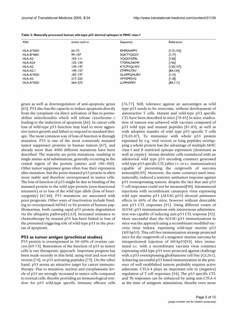
Table 2: Naturally processed human wilt-type p53 derived epitopes in MHC class I
Allel amino acid nr. Sequence Reference
HLA-A*0201 65–73 RMPEAAPPV [115,155]HLA-B*4601 99–107 SQKTYQGSY [117]HLA-A2 103–111 YQGSYGFRL [120]HLA-A24 125–134 TYSPALNKMF [156]HLA-A2 139–147 KTCPVQLWV [120,157]HLA-A2.1 149–157 STPPPGTRV [84,124]HLA-A*0201 187–197 GLAPPQHLIRV [115]HLA-A2 217–225 VPYEPPEVG [118]HLA-A*0201 264–272 LLFRNSFEV [84,111]
Page 5 of 12(page number not for citation purposes)

Journal of Translational Medicine 2005, 3:34 http://www.translational-medicine.com/content/3/1/34
effectively breaking tolerance [93,95]. Anti-CTLA-4 block-ade in combination with a vaccine adjuvant, CpG ODN(synthetic oligodeoxynucleotide containing unmethyl-ated cytosine-phosphate-guanine motifs) had a synergis-tic effect on the improvement of MVAp53 inducedantitumor immunity[96]. Using MVAp53 based immuni-zation Dafterian et al showed eradication of large, wellestablished tumors in three different tumor models in twodifferent strains of mice[96]. The immune responseagainst p53 can also be enhanced by the activation ofCD40 [89,97]. Triggering of the CD40 receptor on den-dritic cells is vital for their adequate activation and matu-ration. Both compounds, anti-CTLA4 and activators ofCD40, will become available to test on a wide-based scalein clinical studies within the near future. Another route ofenhancement of p53 specific immune response afterimmunization was obtained by administration of Flt3Ligand, a strong DC stimulating adjuvant[98]. Highsteady state levels of p53 are not a pre-requisite for tumoreradication by p53 specific CTL as mentioned in onestudy[99]. Instead, p53 turnover is an important factor indetermining the sensitivity of tumor cells to these CTL[87,100]. CD4+ T helper cells are crucial in the recruit-ment and regulation of the innate and adaptive immuneeffector cells[101]. We have demonstrated that CD4+ p53specific T-helper cells are able to help tumor-specific CTLin controlling p53 overexpressing tumors [102]. UsingMHC-transgenic mice has shown to be very efficient inobtaining MHC class I restricted CTL against p53 withhigh avidity capable of lysing p53 overexpressing tumorcells without lysis of normal cells expressing normal levelsof p53 [77]. Very elegantly Kuball et al showed that aCD8-independent p53 specific T cell receptor, generatedin HLA A2.1 transgenic mice, could be expressed inhuman CD8+ and CD4+ T cells with p53 specific tumorrecognition[103]. This is at least a very efficient way toobtain p53 specific class I restricted T cells with very highaffinity. These model systems might help to answer ques-tions on self tolerance for tumor antigens like p53 andintriguing aspects like cross presentation, cross primingand different aspects of immunotherapy in cancer. So farneither clinical nor immunopathological damage to nor-mal tissue has been observed in different mouse models,despite the fact that wild type p53 is expressed in normal
tissue. This indicates that p53 specific T cells are trulytumor-specific. Data available so far support the view thatp53 specific immunotherapy may offer a wide therapeuticmargin in cancer patients. Proof of the pudding is still inthe eating, knowing that their might be important differ-ences in the immune system between preclinical modelsand men as nicely reviewed by Mestas et al [104].
Cicinnati et al studied the potential of prophylactic vacci-nation with p53 epitopes using DNA and /or peptidepulsed dendritic cell vaccination in the tumor model giv-ing rise to sarcomas[105]. Compared to control mice ahigher incidence of epitope loss tumors were detected inthe prophylactic vaccinated group resulting in an increasein tumor growth. Vaccine induced tumor escape thereforecould be an important risk in p53 based prophylacticvaccines.
P53 as tumor antigen (clinical studies)In humans MHC class I restricted p53 specific CTL [106-121], MHC class II restricted p53 specific proliferating Thcells [122-125], and p53 antibody responses (summa-rized in Table 1) have been observed [123,126-133]. Thefirst phase I/II immunization trials using p53 as an anti-gen have just finished and new trials are being initiated. Ina phase I study, six advanced stage cancer patients wereimmunized with an adenoviral vector encoding wild typep53[134]. Neither tumor responses nor anti p53responses were observed, however all patients showed anadenoviral immune response. This strong anti adenoviralspecific response may limit a p53 specific response. Basedon the results in the mouse system[91,92,135] and rhesusmacaques [136], a phase I/II clinical study involving vac-cination of end-stage colorectal cancer patients with arecombinant canarypox virus (ALVAC) encoding wildtype p53 was performed[137]. Patients were immunizedintravenously with an increasing dosage of ALVAC-p53.From this study it appeared that this modality is safe andcapable of stimulating p53-specific Th1 (IFNγ) responsesin several of these patients. One out of 16 patients showedstable disease for a short period of time after immuniza-tion with the highest dose. Fever was the only vaccinerelated adverse effect. The authors conclude from this trialthat repeated immunizations are probably necessary to
Table 3: Naturally processed human wilt-type p53 derived epitopes in MHC class II
Allel amino acid nr. Sequence Reference
HLA-DR1/HLA-DR4 108–122 GFRLGFLHSGTAKSV [158]HLA-DRB1*0401 110–124 RLGFLHSGTAKSVTC [124]HLA-DP5 153–165 PGTRVRAMAIYKQ [125]HLA-DRB1*1401 193–204 HLIRVEGNLRVE [125]
Page 6 of 12(page number not for citation purposes)

Journal of Translational Medicine 2005, 3:34 http://www.translational-medicine.com/content/3/1/34
obtain good clinical responses. Again, anti-vectorresponses were observed in all patients after vaccinationwhich might have impaired the anti-p53 immuneresponses. Preclinical data have shown the superiority ofprime and boost vaccine strategies using different viralvectors [138,139]. Whether or not the route of administra-tion plays a role is under debate[140]. Clinical studieshave shown the safety and effectiveness of prime andboost vaccination protocols using different viral vectors todeliver the antigen of interest[141,142]. An analysis of thep53 specific Th response before and after surgery for color-ectal cancer showed that the majority of the Th responsesdetected were not associated with the immunostimulatorycytokine IFNγ, whereas a number of Th responses eveninvolved secretion of the immunomodulatory cytokineIL-10, pointing at the activity of T-regulatory cells that areknown to suppress T cell immunity[143]. These resultsmore or less resemble the cytokine profiles of tumor asso-ciated T cells derived from ovarian tumors, which werealso associated with a lower zeta chain expression[144]. Itis important to further investigate the character of the p53specific T cell responses, because p53-based vaccination ofpatients should be aimed at boosting only the desiredTh1-type immunity, while stimulation of T-regulatorycells should be avoided. This finding would argue in favorof application of a p53-specific vaccination using a deliv-ery mode specifically stimulating the anti p53 (cytotoxic Tcell and) Th1 responses. Autologous dendritic cellsexpressing the antigen of interest is one of these ways.Svane et al reported on their phase I immunization studyin breast cancer patients with p53 peptide pulsedDC[145]. Dendritic cells were pulsed with three wild-typeand three modified HLA-A2 restricted p53 peptides com-bined with a MHC class II binding peptide (PADRE).Patients received ten subcutaneous immunizations withat least 5 × 10⁶ peptide pulsed dendritic cells combinedwith 6 mIU/m2 IL2. Two out of six patients had a clinicalresponse and three out of six had p53 specific T cellresponses (including the two patients with a clinicalresponse), without inducing significant toxicity. Anothervaccination strategy would be the use of long peptidesencoding the whole protein of interest. The advantage ofusing long peptides is that, if delivered in the appropriateadjuvant (with dendritic cell stimulatory capacity), allpotential MHC class I and class II epitopes within thedelivered peptides will be processed and presented to hostT cells. Table 2 and 3 summarize the naturally processedwild-type p53 epitopes in MHC class I and II known sofar. These vaccines will thus become independent of MHCbinding motif prediction or processing algorithms andcan be administered to subjects independent of theirMHC type. A phase I – II trial using wild- type p53 derivedlong peptides in ovarian cancer patients will be initiatedat the University Medical Center Groningen in 2005.
ConclusionProgress in the fight against ovarian cancer has been ham-pered by the lack of highly effective therapy to perma-nently eradicate disseminated intraperitoneal metastases,which are present in most patients at the time of diagno-sis. In order to improve the poor outcome for ovarian can-cer patients standard and new treatment modalities, suchas targeted or biologic agents and immunotherapy shouldbe combined. In this review we pointed out that ovariancancer tumor cells may (over)express immunoregulatorymolecules such as ligand "Letal", CD40 and Stat-3 whichstimulate immune response. On the other hand mole-cules are expressed which downregulate MHC class I mol-ecules and / or simultaneously produce ligands such asCCL22 attracking regulatory T cells as immune-escapemechanism. Recent data showing the importance of theimmune response in the course of ovarian cancer and theavailability of new potent immunization strategies urgefurther exploration of immunotherapy as adjuvant treat-ment modality in ovarian cancer patients. The immuneresponse against p53 can be enhanced by the activation ofCD40, anti CTLA-4 blockade, coadministration of Flt3Ligand and CpG ODN. Compounds capable of activatingor blocking these molecules will become available withinthe near future to be tested on a wide-based scale in clini-cal studies. The role of p53 as tumor antigen in ovariancancer in immunotherapy based trials will be unravledwithin the near future as well. Next to important issues assafety and immunogenicity of vaccination strategies, clin-ical effectiveness should be one of the major aims offuture trials.
HW Nijman is supported by the Dutch Cancer Society(Grant nr. 2002-2768)
References1. Eisenkop SM, Spirtos NM: Procedures required to accomplish
complete cytoreduction of ovarian cancer: is there a corre-lation with "biological aggressiveness" and survival? GynecolOncol 2001, 82:435-441.
2. Hoskins WJ, McGuire WP, Brady MF, Homesley HD, Creasman WT,Berman M, Ball H, Berek JS: The effect of diameter of largestresidual disease on survival after primary cytoreductive sur-gery in patients with suboptimal residual epithelial ovariancarcinoma. Am J Obstet Gynecol 1994, 170:974-979.
3. Eisenkop SM, Friedman RL, Wang HJ: Complete cytoreductivesurgery is feasible and maximizes survival in patients withadvanced epithelial ovarian cancer: a prospective study.Gynecol Oncol 1998, 69:103-108.
4. McGuire WP, Ozols RF: Chemotherapy of advanced ovariancancer. Semin Oncol 1998, 25:340-348.
5. Thigpen JT: Chemotherapy for advanced ovarian cancer: over-view of randomized trials. Semin Oncol 2000, 27:11-16.
6. Hung K, Hayashi R, Lafond-Walker A, Lowenstein C, Pardoll D, Lev-itsky H: The central role of CD4(+) T cells in the antitumorimmune response. J Exp Med 1998, 188:2357-2368.
7. Godfrey DI, Hammond KJ, Poulton LD, Smyth MJ, Baxter AG: NKTcells: facts, functions and fallacies. Immunol Today 2000,21:573-583.
8. Sakaguchi S: Regulatory T cells: key controllers of immuno-logic self-tolerance. Cell 2000, 101:455-458.
9. Woo EY, Chu CS, Goletz TJ, Schlienger K, Yeh H, Coukos G, RubinSC, Kaiser LR, June CH: Regulatory CD4(+)CD25(+) T cells in
Page 7 of 12(page number not for citation purposes)

Journal of Translational Medicine 2005, 3:34 http://www.translational-medicine.com/content/3/1/34
tumors from patients with early- stage non-small cell lungcancer and late-stage ovarian cancer. Cancer Res 2001,61:4766-4772.
10. Garza KM, Chan SM, Suri R, Nguyen LT, Odermatt B, SchoenbergerSP, Ohashi PS: Role of antigen-presenting cells in mediatingtolerance and autoimmunity. J Exp Med 2000, 191:2021-2027.
11. Smyth MJ, Godfrey DI, Trapani JA: A fresh look at tumor immu-nosurveillance and immunotherapy. Nat Immunol 2001,2:293-299.
12. Zinkernagel RM, Ehl S, Aichele P, Oehen S, Kundig T, Hengartner H:Antigen localisation regulates immune responses in a dose-and time-dependent fashion: a geographical view of immunereactivity. Immunol Rev 1997, 156:199-209.
13. Bennett SR, Carbone FR, Karamalis F, Flavell RA, Miller JF, Heath WR:Help for cytotoxic-T-cell responses is mediated by CD40signalling. Nature 1998, 393:478-480.
14. Cella M, Scheidegger D, Palmer-Lehmann K, Lane P, Lanzavecchia A,Alber G: Ligation of CD40 on dendritic cells triggers produc-tion of high levels of interleukin-12 and enhances T cell stim-ulatory capacity: T-T help via APC activation. J Exp Med 1996,184:747-752.
15. Ridge JP, Di Rosa F, Matzinger P: A conditioned dendritic cell canbe a temporal bridge between a CD4+ T- helper and a T-killer cell. Nature 1998, 393:474-478.
16. Schoenberger SP, Toes RE, van der Voort EI, Offringa R, Melief CJ: T-cell help for cytotoxic T lymphocytes is mediated by CD40-CD40L interactions. Nature 1998, 393:480-483.
17. Guerder S, Matzinger P: A fail-safe mechanism for maintainingself-tolerance. J Exp Med 1992, 176:553-564.
18. Schuurhuis DH, Laban S, Toes RE, Ricciardi-Castagnoli P, KleijmeerMJ, van der Voort EI, Rea D, Offringa R, Geuze HJ, Melief CJ, Ossen-dorp F: Immature dendritic cells acquire CD8(+) cytotoxic Tlymphocyte priming capacity upon activation by T helpercell-independent or -dependent stimuli. J Exp Med 2000,192:145-150.
19. Melief CJ, Toes RE, Medema JP, Van der Burg SH, Ossendorp F,Offringa R: Strategies for immunotherapy of cancer. AdvImmunol 2000, 75:235-282.
20. Rosenberg SA: A new era for cancer immunotherapy based onthe genes that encode cancer antigens. Immunity 1999,10:281-287.
21. Marincola FM, Wang E, Herlyn M, Seliger B, Ferrone S: Tumors aselusive targets of T-cell-based active immunotherapy. TrendsImmunol 2003, 24:335-342.
22. Turtle CJ, Hart DN: Dendritic cells in tumor immunology andimmunotherapy. Curr Drug Targets 2004, 5:17-39.
23. Almand B, Resser JR, Lindman B, Nadaf S, Clark JI, Kwon ED, CarboneDP, Gabrilovich DI: Clinical significance of defective dendriticcell differentiation in cancer. Clin Cancer Res 2000, 6:1755-1766.
24. Almand B, Clark JI, Nikitina E, van Beynen J, English NR, Knight SC,Carbone DP, Gabrilovich DI: Increased production of immaturemyeloid cells in cancer patients: a mechanism of immuno-suppression in cancer. J Immunol 2001, 166:678-689.
25. Gabrilovich DI, Chen HL, Girgis KR, Cunningham HT, Meny GM,Nadaf S, Kavanaugh D, Carbone DP: Production of vascularendothelial growth factor by human tumors inhibits thefunctional maturation of dendritic cells. Nat Med 1996,2:1096-1103.
26. Kusmartsev S, Gabrilovich DI: Immature myeloid cells and can-cer-associated immune suppression. Cancer ImmunolImmunother 2002, 51:293-298.
27. Zou W, Machelon V, Coulomb-L'Hermin A, Borvak J, Nome F, IsaevaT, Wei S, Krzysiek R, Durand-Gasselin I, Gordon A, Pustilnik T,Curiel DT, Galanaud P, Capron F, Emilie D, Curiel TJ: Stromal-derived factor-1 in human tumors recruits and alters thefunction of plasmacytoid precursor dendritic cells. Nat Med2001, 7:1339-1346.
28. Eisenthal A, Polyvkin N, Bramante-Schreiber L, Misonznik F, HassnerA, Lifschitz-Mercer B: Expression of dendritic cells in ovariantumors correlates with clinical outcome in patients withovarian cancer. Hum Pathol 2001, 32:803-807.
29. Schlienger K, Chu CS, Woo EY, Rivers PM, Toll AJ, Hudson B, MausMV, Riley JL, Choi Y, Coukos G, Kaiser LR, Rubin SC, Levine BL, Car-roll RG, June CH: TRANCE- and CD40 Ligand-matured Den-dritic Cells reveal MHC class I restricted T cells specific for
autologous tumor in late stage ovarian cancer patients. ClinCancer Res 2003, 9:1517-1527.
30. Chu CS, Woo EY, Toll AJ, Rubin SC, June CH, Carroll RG, SchliengerK: Tumor-associated macrophages as a source of functionaldendritic cells in ovarian cancer patients. Clin Immunol 2002,102:291-301.
31. Loercher AE, Nash MA, Kavanagh JJ, Platsoucas CD, Freedman RS:Identification of an IL-10-producing HLA-DR-negativemonocyte subset in the malignant ascites of patients withovarian carcinoma that inhibits cytokine protein expressionand proliferation of autologous T cells. J Immunol 1999,163:6251-6260.
32. Curiel TJ, Wei S, Dong H, Alvarez X, Cheng P, Mottram P, KrzysiekR, Knutson KL, Daniel B, Zimmermann MC, David O, Burow M, Gor-don A, Dhurandhar N, Myers L, Berggren R, Hemminki A, AlvarezRD, Emilie D, Curiel DT, Chen L, Zou W: Blockade of B7-H1improves myeloid dendritic cell-mediated antitumorimmunity. Nat Med 2003, 9:562-567.
33. Dong H, Zhu G, Tamada K, Chen L: B7-H1, a third member ofthe B7 family, co-stimulates T-cell proliferation and inter-leukin-10 secretion. Nat Med 1999, 5:1365-1369.
34. Dong H, Chen L: B7-H1 pathway and its role in the evasion oftumor immunity. J Mol Med 2003, 81:281-287.
35. Dong H, Strome SE, Salomao DR, Tamura H, Hirano F, Flies DB,Roche PC, Lu J, Zhu G, Tamada K, Lennon VA, Celis E, Chen L:Tumor-associated B7-H1 promotes T-cell apoptosis: apotential mechanism of immune evasion. Nat Med 2002,8:793-800.
36. Strome SE, Dong H, Tamura H, Voss SG, Flies DB, Tamada K, Salo-mao D, Cheville J, Hirano F, Lin W, Kasperbauer JL, Ballman KV, ChenL: B7-H1 blockade augments adoptive T-cell immuno-therapy for squamous cell carcinoma. Cancer Res 2003,63:6501-6505.
37. Colonna M, Krug A, Cella M: Interferon-producing cells: on thefront line in immune responses against pathogens. Curr OpinImmunol 2002, 14:373-379.
38. Salio M, Cella M, Vermi W, Facchetti F, Palmowski MJ, Smith CL,Shepherd D, Colonna M, Cerundolo V: Plasmacytoid dendriticcells prime IFN-gamma-secreting melanoma-specific CD8lymphocytes and are found in primary melanoma lesions.Eur J Immunol 2003, 33:1052-1062.
39. Curiel TJ, Cheng P, Mottram P, Alvarez X, Moons L, Evdemon-HoganM, Wei S, Zou L, Kryczek I, Hoyle G, Lackner A, Carmeliet P, ZouW: Dendritic cell subsets differentially regulate angiogenesisin human ovarian cancer. Cancer Res 2004, 64:5535-5538.
40. Freedman RS, Tomasovic B, Templin S, Atkinson EN, Kudelka A,Edwards CL, Platsoucas CD: Large-scale expansion ininterleukin-2 of tumor-infiltrating lymphocytes frompatients with ovarian carcinoma for adoptiveimmunotherapy. J Immunol Methods 1994, 167:145-160.
41. Freedman RS, Platsoucas CD: Immunotherapy for peritonealovarian carcinoma metastasis using ex vivo expanded tumorinfiltrating lymphocytes. Cancer Treat Res 1996, 82:115-146.
42. Ioannides CG, Platsoucas CD, Rashed S, Wharton JT, Edwards CL,Freedman RS: Tumor cytolysis by lymphocytes infiltratingovarian malignant ascites. Cancer Res 1991, 51:4257-4265.
43. Ioannides CG, Fisk B, Fan D, Biddison WE, Wharton JT, O'Brian CA:Cytotoxic T cells isolated from ovarian malignant ascitesrecognize a peptide derived from the HER-2/neu proto-oncogene. Cell Immunol 1993, 151:225-234.
44. Ioannides CG, Fisk B, Pollack MS, Frazier ML, Taylor WJ, FreedmanRS: Cytotoxic T-cell clones isolated from ovarian tumourinfiltrating lymphocytes recognize common determinantson non-ovarian tumour clones. Scand J Immunol 1993,37:413-424.
45. Zhang L, Conejo-Garcia JR, Katsaros D, Gimotty PA, Massobrio M,Regnani G, Makrigiannakis A, Gray H, Schlienger K, Liebman MN,Rubin SC, Coukos G: Intratumoral T cells, recurrence, and sur-vival in epithelial ovarian cancer. N Engl J Med 2003,348:203-213.
46. Curiel TJ, Coukos G, Zou L, Alvarez X, Cheng P, Mottram P, Evde-mon-Hogan M, Conejo-Garcia JR, Zhang L, Burow M, Zhu Y, Wei S,Kryczek I, Daniel B, Gordon A, Myers L, Lackner A, Disis ML, Knut-son KL, Chen L, Zou W: Specific recruitment of regulatory Tcells in ovarian carcinoma fosters immune privilege and pre-dicts reduced survival. Nat Med 2004, 10:942-949.
Page 8 of 12(page number not for citation purposes)

Journal of Translational Medicine 2005, 3:34 http://www.translational-medicine.com/content/3/1/34
47. Conejo-Garcia JR, Benencia F, Courreges MC, Gimotty PA, Khang E,Buckanovich RJ, Frauwirth KA, Zhang L, Katsaros D, Thompson CB,Levine B, Coukos G: Ovarian carcinoma expresses the NKG2Dligand Letal and promotes the survival and expansion of. Can-cer Res 2004, 64:2175-2182.
48. Conejo-Garcia JR, Buckanovich RJ, Benencia F, Courreges MC, RubinSC, Carroll RG, Coukos G: Vascular Leukocytes Contribute toTumor Vascularization. Blood 2004.
49. Conejo-Garcia JR, Benencia F, Courreges MC, Kang E, Mohamed-Hadley A, Buckanovich RJ, Holtz DO, Jenkins A, Na H, Zhang L, Wag-ner DS, Katsaros D, Caroll R, Coukos G: Tumor-infiltrating den-dritic cell precursors recruited by a beta-defensin contributeto vasculogenesis under the influence of Vegf-A. Nat Med2004, 10:950-958.
50. Nash MA, Ferrandina G, Gordinier M, Loercher A, Freedman RS:The role of cytokines in both the normal and malignantovary. Endocr Relat Cancer 1999, 6:93-107.
51. Wang ZY, Gaggero A, Rubartelli A, Rosso O, Miotti S, MezzanzanicaD, Canevari S, Ferrini S: Expression of interleukin-18 in humanovarian carcinoma and normal ovarian epithelium: evidencefor defective processing in tumor cells. Int J Cancer 2002,98:873-878.
52. Chen H, Ye D, Xie X, Chen B, Lu W: VEGF, VEGFRs expressionsand activated STATs in ovarian epithelial carcinoma. GynecolOncol 2004, 94:630-635.
53. Huang M, Page C, Reynolds RK, Lin J: Constitutive activation ofstat 3 oncogene product in human ovarian carcinoma cells.Gynecol Oncol 2000, 79:67-73.
54. Wang T, Niu G, Kortylewski M, Burdelya L, Shain K, Zhang S, Bhatta-charya R, Gabrilovich D, Heller R, Coppola D, Dalton W, Jove R, Par-doll D, Yu H: Regulation of the innate and adaptive immuneresponses by Stat-3 signaling in tumor cells. Nat Med 2004,10:48-54.
55. Kooi S, Zhang HZ, Patenia R, Edwards CL, Platsoucas CD, FreedmanRS: HLA class I expression on human ovarian carcinoma cellscorrelates with T-cell infiltration in vivo and T-cell expansionin vitro in low concentrations of recombinant interleukin-2.Cell Immunol 1996, 174:116-128.
56. Nijman HW, van Diest PJ, Poort-Keesom RJ, Mensdorff-Pouilly S,Verstraeten RA, Kummer A, Meijer CJ, Melief CJ, Hilgers J, KenemansP: T cell infiltration and MHC I and II expression in the pres-ence of tumor antigens: An immunohistochemical study inpatients with serous epithelial ovarian cancer. Eur J ObstetGynecol Reprod Biol 2001, 94:114-120.
57. Ravenswaay Claasen HH, Fleuren GJ: The influence of combina-tion chemotherapy on antigen expression in ovarian cancer.Gynecol Oncol 1995, 58:16-23.
58. Vitale M, Pelusi G, Taroni B, Gobbi G, Micheloni C, Rezzani R, DonatoF, Wang X, Ferrone S: HLA class I antigen down-regulation inprimary ovary carcinoma lesions: association with diseasestage. Clin Cancer Res 2005, 11:67-72.
59. Zaks TZ, Chappell DB, Rosenberg SA, Restifo NP: Fas-mediatedsuicide of tumor-reactive T cells following activation by spe-cific tumor: selective rescue by caspase inhibition. J Immunol1999, 162:3273-3279.
60. Offringa R, Van der Burg SH, Ossendorp F, Toes RE, Melief CJ:Design and evaluation of antigen-specific vaccination strate-gies against cancer. Curr Opin Immunol 2000, 12:576-582.
61. Van Den Eynde BJ, van der BP: T cell defined tumor antigens.Curr Opin Immunol 1997, 9:684-693.
62. Vogelstein B, Lane D, Levine AJ: Surfing the p53 network. Nature2000, 408:307-310.
63. Vousden KH, Lu X: Live or let die: the cell's response to p53.Nat Rev Cancer 2002, 2:594-604.
64. Vogelstein B, Kinzler KW: Cancer genes and the pathways theycontrol. Nat Med 2004, 10:789-799.
65. Coukos G, Rubin SC: Chemotherapy resistance in ovarian can-cer: new molecular perspectives. Obstet Gynecol 1998,91:783-792.
66. Chipuk JE, Kuwana T, Bouchier-Hayes L, Droin NM, Newmeyer DD,Schuler M, Green DR: Direct activation of Bax by p53 mediatesmitochondrial membrane permeabilization and apoptosis.Science 2004, 303:1010-1014.
67. Greenblatt MS, Bennett WP, Hollstein M, Harris CC: Mutations inthe p53 tumor suppressor gene: clues to cancer etiology andmolecular pathogenesis. Cancer Res 1994, 54:4855-4878.
68. de Vries A, Flores ER, Miranda B, Hsieh HM, van Oostrom CT, SageJ, Jacks T: Targeted point mutations of p53 lead to dominant-negative inhibition of wild-type p53 function. Proc Natl Acad SciU S A 2002, 99:2948-2953.
69. Nijman HW, Kenemans P, Poort-Keesom RJ, Verstraeten RA,Mensdorff-Pouilly S, Verheijen RH, Melief CJ, Hilgers J, Meijer CJ:Influence of chemotherapy on the expression of p53, HER-2/neu and proliferation markers in ovarian cancer. Eur J ObstetGynecol Reprod Biol 1999, 83:201-206.
70. Herod JJ, Eliopoulos AG, Warwick J, Niedobitek G, Young LS, KerrDJ: The prognostic significance of Bcl-2 and p53 expression inovarian carcinoma. Cancer Res 1996, 56:2178-2184.
71. Klemi PJ, Pylkkanen L, Kiilholma P, Kurvinen K, Joensuu H: p53 pro-tein detected by immunohistochemistry as a prognostic fac-tor in patients with epithelial ovarian carcinoma. Cancer 1995,76:1201-1208.
72. Marks JR, Davidoff AM, Kerns BJ, Humphrey PA, Pence JC, Dodge RK,Clarke-Pearson DL, Iglehart JD, Bast RCJ, Berchuck A: Overexpres-sion and mutation of p53 in epithelial ovarian cancer. CancerRes 1991, 51:2979-2984.
73. van der Zee AG, Hollema H, Suurmeijer AJ, Krans M, Sluiter WJ, Wil-lemse PH, Aalders JG, de Vries EG: Value of P-glycoprotein, glu-tathione S-transferase pi, c-erbB-2, and p53 as prognosticfactors in ovarian carcinomas. J Clin Oncol 1995, 13:70-78.
74. McCormick F: Cancer gene therapy: fringe or cutting edge?Nat Rev Cancer 2001, 1:130-141.
75. Snyder EL, Meade BR, Saenz CC, Dowdy SF: Treatment of termi-nal peritoneal carcinomatosis by a transducible p53-activat-ing peptide. PLoS Biol 2004, 2:E36.
76. Offringa R, Vierboom MP, Van der Burg SH, Erdile L, Melief CJ: p53:a potential target antigen for immunotherapy of cancer. AnnN Y Acad Sci 2000, 910:223-233.
77. Voss RH, Lotz C, Cellary A, Theobald M: Targeting p53, hdm2,and CD19: vaccination and immunologic strategies. BoneMarrow Transplant 2000, 25 Suppl 2:S43-S45.
78. Hilburger RM, Abrams SI: Characterization of CD8+ cytotoxic Tlymphocyte/tumor cell interactions reflecting recognition ofan endogenously expressed murine wild-type p53determinant. Cancer Immunol Immunother 2001, 49:603-612.
79. Yanuck M, Carbone DP, Pendleton CD, Tsukui T, Winter SF, MinnaJD, Berzofsky JA: A mutant p53 tumor suppressor protein is atarget for peptide-induced CD8+ cytotoxic T-cells. Cancer Res1993, 53:3257-3261.
80. Mathiassen S, Lauemoller SL, Ruhwald M, Claesson MH, Buus S:Tumor-associated antigens identified by mRNA expressionprofiling induce protective anti-tumor immunity. Eur JImmunol 2001, 31:1239-1246.
81. Mayordomo JI, Loftus DJ, Sakamoto H, De Cesare CM, AppasamyPM, Lotze MT, Storkus WJ, Appella E, DeLeo AB: Therapy ofmurine tumors with p53 wild-type and mutant sequencepeptide- based vaccines. J Exp Med 1996, 183:1357-1365.
82. Noguchi Y, Chen YT, Old LJ: A mouse mutant p53 product rec-ognized by CD4+ and CD8+ T cells. Proc Natl Acad Sci U S A1994, 91:3171-3175.
83. Noguchi Y, Richards EC, Chen YT, Old LJ: Influence of interleukin12 on p53 peptide vaccination against established Meth Asarcoma. Proc Natl Acad Sci U S A 1995, 92:2219-2223.
84. Theobald M, Biggs J, Dittmer D, Levine AJ, Sherman LA: Targetingp53 as a general tumor antigen. Proc Natl Acad Sci U S A 1995,92:11993-11997.
85. Vierboom MP, Nijman HW, Offringa R, van der Voort EI, van Hall T,van den BL, Fleuren GJ, Kenemans P, Kast WM, Melief CJ: Tumoreradication by wild-type p53-specific cytotoxic Tlymphocytes. J Exp Med 1997, 186:695-704.
86. Peralta EA, Liu X, McCarthy TM, Wilson TG, Diamond DJ, EllenhornJD: Immunotherapy of bladder cancer targeting P53. J Urol1999, 162:1806-1811.
87. Vierboom MP, Zwaveling S, Bos GMJ, Ooms M, Krietemeijer GM,Melief CJ, Offringa R: High steady-state levels of p53 are not aprerequisite for tumor eradication by wild-type p53-specificcytotoxic T lymphocytes. Cancer Res 2000, 60:5508-5513.
88. Ishida T, Chada S, Stipanov M, Nadaf S, Ciernik FI, Gabrilovich DI,Carbone DP: Dendritic cells transduced with wild-type p53gene elicit potent anti-tumour immune responses. Clin ExpImmunol 1999, 117:244-251.
Page 9 of 12(page number not for citation purposes)
Journal of Translational Medicine 2005, 3:34 http://www.translational-medicine.com/content/3/1/34
89. Nikitina EY, Chada S, Muro-Cacho C, Fang B, Zhang R, Roth JA,Gabrilovich DI: An effective immunization and cancer treat-ment with activated dendritic cells transduced with full-length wild-type p53. Gene Ther 2002, 9:345-352.
90. Murakami T, Tokunaga N, Waku T, Gomi S, Kagawa S, Tanaka N, Fuji-wara T: Antitumor effect of intratumoral administration ofbone marrow-derived dendritic cells transduced with wild-type p53 gene. Clin Cancer Res 2004, 10:3871-3880.
91. Odin L, Favrot M, Poujol D, Michot JP, Moingeon P, Tartaglia J, Pui-sieux I: Canarypox virus expressing wild type p53 for genetherapy in murine tumors mutated in p53. Cancer Gene Ther2001, 8:87-98.
92. Hurpin C, Rotarioa C, Bisceglia H, Chevalier M, Tartaglia J, Erdile L:The mode of presentation and route of administration arecritical for the induction of immune responses to p53 andantitumor immunity. Vaccine 1998, 16:208-215.
93. Espenschied J, Lamont J, Longmate J, Pendas S, Wang Z, Diamond DJ,Ellenhorn JD: CTLA-4 blockade enhances the therapeuticeffect of an attenuated poxvirus vaccine targeting p53 in anestablished murine tumor model. J Immunol 2003,170:3401-3407.
94. Krummel MF, Allison JP: CD28 and CTLA-4 have opposingeffects on the response of T cells to stimulation. J Exp Med1995, 182:459-465.
95. Hernandez J, Ko A, Sherman LA: CTLA-4 blockade enhances theCTL responses to the p53 self-tumor antigen. J Immunol 2001,166:3908-3914.
96. Daftarian P, Song GY, Ali S, Faynsod M, Longmate J, Diamond DJ,Ellenhorn JD: Two distinct pathways of immuno-modulationimprove potency of p53 immunization in rejecting estab-lished tumors. Cancer Res 2004, 64:5407-5414.
97. Erdile LF, Smith D: CD40 activation enhances the magnitude ofcellular immune responses against p53 but not the avidity ofthe effectors. Cancer Immunol Immunother 2000, 49:410-416.
98. Parajuli P, Pisarev V, Sublet J, Steffel A, Varney M, Singh R, LaFace D,Talmadge JE: Immunization with wild-type p53 genesequences coadministered with Flt3 ligand induces an anti-gen-specific type 1 T-cell response. Cancer Res 2001,61:8227-8234.
99. Gnjatic S, Cai Z, Viguier M, Chouaib S, Guillet JG, Choppin J: Accu-mulation of the p53 protein allows recognition by humanCTL of a wild-type p53 epitope presented by breast carcino-mas and melanomas. J Immunol 1998, 160:328-333.
100. Sirianni N, Ha PK, Oelke M, Califano J, Gooding W, Westra W,Whiteside TL, Koch WM, Schneck JP, DeLeo A, Ferris RL: Effect ofhuman papillomavirus-16 infection on CD8+ T-cell recogni-tion of a wild-type sequence p53264-272 peptide in patientswith squamous cell carcinoma of the head and neck. Clin Can-cer Res 2004, 10:6929-6937.
101. Mitra R, Singh S, Khar A: Antitumour immune responses. ExpertRev Mol Med 2003, 2003:1-22.
102. Zwaveling S, Vierboom MP, Ferreira Mota SC, Hendriks JA, OomsME, Sutmuller RP, Franken KL, Nijman HW, Ossendorp F, Van derBurg SH, Offringa R, Melief CJ: Antitumor efficacy of wild-typep53-specific CD4(+) T-helper cells. Cancer Res 2002,62:6187-6193.
103. Kuball J, Schmitz FW, Voss RH, Ferreira EA, Engel R, Guillaume P,Strand S, Romero P, Huber C, Sherman LA, Theobald M: Coopera-tion of human tumor-reactive CD4+ and CD8+ T cells afterredirection of their specificity by a high-affinity p53A2.1-spe-cific TCR. Immunity 2005, 22:117-129.
104. Mestas J, Hughes CC: Of mice and not men: differencesbetween mouse and human immunology. J Immunol 2004,172:2731-2738.
105. Cicinnati VR, Dworacki G, Albers A, Beckebaum S, Tuting T, Kacz-marek E, DeLeo AB: Impact of p53-based immunization on pri-mary chemically-induced tumors. Int J Cancer 2005,113:961-970.
106. Hoffmann TK, Nakano K, Elder EM, Dworacki G, Finkelstein SD,Appella E, Whiteside TL, DeLeo AB: Generation of T cells specificfor the wild-type sequence p53(264-272) peptide in cancerpatients: implications for immunoselection of epitope lossvariants. J Immunol 2000, 165:5938-5944.
107. Houbiers JG, Nijman HW, Van der Burg SH, Drijfhout JW, KenemansP, van de Velde CJ, Brand A, Momburg F, Kast WM, Melief CJ: Invitro induction of human cytotoxic T lymphocyte responses
against peptides of mutant and wild-type p53. Eur J Immunol1993, 23:2072-2077.
108. Nijman HW, Houbiers JG, Van der Burg SH, Vierboom MP, Kene-mans P, Kast WM, Melief CJ: Characterization of cytotoxic Tlymphocyte epitopes of a self-protein, p53, and a non-self-protein, influenza matrix: relationship between major histo-compatibility complex peptide binding affinity and immuneresponsiveness to peptides. J Immunother 1993, 14:121-126.
109. Nijman HW, Van der Burg SH, Vierboom MP, Houbiers JG, Kast WM,Melief CJ: p53, a potential target for tumor-directed T cells.Immunol Lett 1994, 40:171-178.
110. Nikitina EY, Clark JI, van Beynen J, Chada S, Virmani AK, Carbone DP,Gabrilovich DI: Dendritic cells transduced with full-lengthwild-type p53 generate antitumor cytotoxic T lymphocytesfrom peripheral blood of cancer patients. Clin Cancer Res 2001,7:127-135.
111. Ropke M, Regner M, Claesson MH: T cell-mediated cytotoxicityagainst p53-protein derived peptides in bulk and limitingdilution cultures of healthy donors. Scand J Immunol 1995,42:98-103.
112. Ropke M, Hald J, Guldberg P, Zeuthen J, Norgaard L, Fugger L, Svej-gaard A, Van der BS, Nijman HW, Melief CJ, Claesson MH: Sponta-neous human squamous cell carcinomas are killed by ahuman cytotoxic T lymphocyte clone recognizing a wild-type p53-derived peptide. Proc Natl Acad Sci U S A 1996,93:14704-14707.
113. Theobald M, Biggs J, Hernandez J, Lustgarten J, Labadie C, ShermanLA: Tolerance to p53 by A2.1-restricted cytotoxic Tlymphocytes. J Exp Med 1997, 185:833-841.
114. Umano Y, Tsunoda T, Tanaka H, Matsuda K, Yamaue H, Tanimura H:Generation of cytotoxic T cell responses to an HLA-A24restricted epitope peptide derived from wild-type p53. Br JCancer 2001, 84:1052-1057.
115. Wurtzen PA, Pedersen LO, Poulsen HS, Claesson MH: Specific kill-ing of P53 mutated tumor cell lines by a cross-reactivehuman HLA-A2-restricted P53-specific CTL line. Int J Cancer2001, 93:855-861.
116. Asai T, Storkus WJ, Mueller-Berghaus J, Knapp W, DeLeo AB, Chika-matsu K, Whiteside TL: In vitro generated cytolytic T lym-phocytes reactive against head and neck cancer recognizemultiple epitopes presented by HLA-A2, including peptidesderived from the p53 and MDM-2 proteins. Cancer Immun2002, 2:3.
117. Azuma K, Shichijo S, Maeda Y, Nakatsura T, Nonaka Y, Fujii T, KoikeK, Itoh K: Mutated p53 gene encodes a nonmutated epitoperecognized by HLA-B*4601-restricted and tumor cell-reac-tive CTLs at tumor site. Cancer Res 2003, 63:854-858.
118. McArdle SE, Rees RC, Mulcahy KA, Saba J, McIntyre CA, Murray AK:Induction of human cytotoxic T lymphocytes that preferen-tially recognise tumour cells bearing a conformational p53mutant. Cancer Immunol Immunother 2000, 49:417-425.
119. Papadopoulos KP, Hesdorffer CS, Suciu-Foca N, Hibshoosh H, HarrisPE: Wild-type p53 epitope naturally processed and presentedby an HLA-B haplotype on human breast carcinoma cells.Clin Cancer Res 1999, 5:2089-2093.
120. Petersen TR, Buus S, Brunak S, Nissen MH, Sherman LA, ClaessonMH: Identification and design of p53-derived HLA-A2-bindingpeptides with increased CTL immunogenicity. Scand J Immunol2001, 53:357-364.
121. Tokunaga N, Murakami T, Endo Y, Nishizaki M, Kagawa S, Tanaka N,Fujiwara T: Human monocyte-derived dendritic cells pulsedwith wild-type p53 protein efficiently induce CTLs againstp53 overexpressing human cancer cells. Clin Cancer Res 2005,11:1312-1318.
122. Tilkin AF, Lubin R, Soussi T, Lazar V, Janin N, Mathieu MC, Lefrere I,Carlu C, Roy M, Kayibanda M, .: Primary proliferative T cellresponse to wild-type p53 protein in patients with breastcancer. Eur J Immunol 1995, 25:1765-1769.
123. Van der Burg SH, de Cock K, Menon AG, Franken KL, Palmen M,Redeker A, Drijfhout J, Kuppen PJ, van V, Erdile L, Tollenaar RA,Melief CJ, Offringa R: Long lasting p53-specific T cell memoryresponses in the absence of anti-p53 antibodies in patientswith resected primary colorectal cancer. Eur J Immunol 2001,31:146-155.
124. Chikamatsu K, Albers A, Stanson J, Kwok WW, Appella E, WhitesideTL, DeLeo AB: P53(110-124)-specific human CD4+ T-helper
Page 10 of 12(page number not for citation purposes)

Journal of Translational Medicine 2005, 3:34 http://www.translational-medicine.com/content/3/1/34
cells enhance in vitro generation and antitumor function oftumor-reactive CD8+ T cells. Cancer Res 2003, 63:3675-3681.
125. Fujita H, Senju S, Yokomizo H, Saya H, Ogawa M, Matsushita S,Nishimura Y: Evidence that HLA class II-restricted humanCD4+ T cells specific to p53 self peptides respond to p53 pro-teins of both wild and mutant forms. Eur J Immunol 1998,28:305-316.
126. Angelopoulou K, Rosen B, Stratis M, Yu H, Solomou M, Diamandis EP:Circulating antibodies against p53 protein in patients withovarian carcinoma. Correlation with clinicopathologic fea-tures and survival. Cancer 1996, 78:2146-2152.
127. Angelopoulou K, Diamandis EP: Detection of the TP53 tumoursuppressor gene product and p53 auto- antibodies in theascites of women with ovarian cancer. Eur J Cancer 1997,33:115-121.
128. Crawford LV, Pim DC, Bulbrook RD: Detection of antibodiesagainst the cellular protein p53 in sera from patients withbreast cancer. Int J Cancer 1982, 30:403-408.
129. Gadducci A, Ferdeghini M, Buttitta F, Fanucchi A, Annicchiarico C,Prontera C, Bevilacqua G, Genazzani AR: Preoperative serumantibodies against the p53 protein in patients with ovarianand endometrial cancer. Anticancer Res 1996, 16:3519-3523.
130. Gadducci A, Ferdeghini M, Buttitta F, Cosio S, Fanucchi A, Annicchi-arico C, Genazzani AR: Serum anti-p53 antibodies in the follow-up of patients with advanced ovarian carcinoma. AnticancerRes 1998, 18:3763-3765.
131. Gadducci A, Ferdeghini M, Buttitta F, Cosio S, Fanucchi A, Annicchi-arico C, Gagetti O, Bevilacqua G, Genazzani AR: Assessment of theprognostic relevance of serum anti-p53 antibodies in epithe-lial ovarian cancer. Gynecol Oncol 1999, 72:76-81.
132. Labrecque S, Naor N, Thomson D, Matlashewski G: Analysis of theanti-p53 antibody response in cancer patients. Cancer Res1993, 53:3468-3471.
133. Vennegoor CJ, Nijman HW, Drijfhout JW, Vernie L, Verstraeten RA,Mensdorff-Pouilly S, Hilgers J, Verheijen RH, Kast WM, Melief CJ,Kenemans P: Autoantibodies to p53 in ovarian cancer patientsand healthy women: a comparison between whole p53 pro-tein and 18-mer peptides for screening purposes. Cancer Lett1997, 116:93-101.
134. Kuball J, Schuler M, Antunes FE, Herr W, Neumann M, Obenauer-Kutner L, Westreich L, Huber C, Wolfel T, Theobald M: Generatingp53-specific cytotoxic T lymphocytes by recombinant aden-oviral vector-based vaccination in mice, but not man. GeneTher 2002, 9:833-843.
135. Roth J, Dittmer D, Rea D, Tartaglia J, Paoletti E, Levine AJ: p53 as atarget for cancer vaccines: recombinant canarypox virusvectors expressing p53 protect mice against lethal tumorcell challenge. Proc Natl Acad Sci U S A 1996, 93:4781-4786.
136. Rosenwirth B, Kuhn EM, Heeney JL, Hurpin C, Tartaglia J, Bonnet MC,Moingeon P, Erdile L: Safety and immunogenicity of ALVACwild-type human p53 (vCP207) by the intravenous route inrhesus macaques. Vaccine 2001, 19:1661-1670.
137. Menon AG, Kuppen PJ, Van der Burg SH, Offringa R, Bonnet MC,Harinck BI, Tollenaar RA, Redeker A, Putter H, Moingeon P, MorreauH, Melief CJ, van de Velde CJ: Safety of intravenous administra-tion of a canarypox virus encoding the human wild-type p53gene in colorectal cancer patients. Cancer Gene Ther 2003,10:509-517.
138. Aarts WM, Schlom J, Hodge JW: Vector-based vaccine/cytokinecombination therapy to enhance induction of immuneresponses to a self-antigen and antitumor activity. Cancer Res2002, 62:5770-5777.
139. Grosenbach DW, Barrientos JC, Schlom J, Hodge JW: Synergy ofvaccine strategies to amplify antigen-specific immuneresponses and antitumor effects. Cancer Res 2001,61:4497-4505.
140. Kudo-Saito C, Schlom J, Hodge JW: Induction of an antigen cas-cade by diversified subcutaneous/intratumoral vaccination isassociated with antitumor responses. Clin Cancer Res 2005,11:2416-2426.
141. Marshall JL, Hoyer RJ, Toomey MA, Faraguna K, Chang P, RichmondE, Pedicano JE, Gehan E, Peck RA, Arlen P, Tsang KY, Schlom J: PhaseI study in advanced cancer patients of a diversified prime-and-boost vaccination protocol using recombinant vacciniavirus and recombinant nonreplicating avipox virus to elicit
anti-carcinoembryonic antigen immune responses. J ClinOncol 2000, 18:3964-3973.
142. Marshall JL, Gulley JL, Arlen PM, Beetham PK, Tsang KY, Slack R,Hodge JW, Doren S, Grosenbach DW, Hwang J, Fox E, Odogwu L,Park S, Panicali D, Schlom J: Phase I study of sequential vaccina-tions with fowlpox-CEA(6D)-TRICOM alone and sequen-tially with vaccinia-CEA(6D)-TRICOM, with and withoutgranulocyte-macrophage colony-stimulating factor, inpatients with carcinoembryonic antigen-expressingcarcinomas. J Clin Oncol 2005, 23:720-731.
143. Van der Burg SH, Menon AG, Redeker A, Franken KL, Drijfhout JW,Tollenaar RA, Hartgrink HH, van de Velde CJ, Kuppen PJ, Melief CJ,Offringa R: Magnitude and polarization of P53-specific T-helper immunity in connection to leukocyte infiltration ofcolorectal tumors. Int J Cancer 2003, 107:425-433.
144. Rabinowich H, Suminami Y, Reichert TE, Crowley-Nowick P, Bell M,Edwards R, Whiteside TL: Expression of cytokine genes or pro-teins and signaling molecules in lymphocytes associated withhuman ovarian carcinoma. Int J Cancer 1996, 68:276-284.
145. Svane IM, Pedersen AE, Johnsen HE, Nielsen D, Kamby C, Gaarsdal E,Nikolajsen K, Buus S, Claesson MH: Vaccination with p53-pep-tide-pulsed dendritic cells, of patients with advanced breastcancer: report from a phase I study. Cancer Immunol Immunother2004, 53:633-641.
146. Abendstein B, Marth C, Muller-Holzner E, Widschwendter M, Daxen-bichler G, Zeimet AG: Clinical significance of serum and asciticp53 autoantibodies in epithelial ovarian carcinoma. Cancer2000, 88:1432-1437.
147. Vogl FD, Frey M, Kreienberg R, Runnebaum IB: Autoimmunityagainst p53 predicts invasive cancer with poor survival inpatients with an ovarian mass. Br J Cancer 2000, 83:1338-1343.
148. Vogl FD, Stickeler E, Weyermann M, Kohler T, Grill HJ, Negri G,Kreienberg R, Runnebaum IB: p53 autoantibodies in patientswith primary ovarian cancer are associated with higher age,advanced stage and a higher proportion of p53-positivetumor cells. Oncology 1999, 57:324-329.
149. Hogdall EV, Hogdall CK, Blaakaer J, Heegaard NH, Glud E, Chris-tensen L, Bock JE, Norgaard-Pedersen B, Wiik A, Kjaer SK: P53autoantibodies in sera from Danish ovarian cancer patientsand their correlation with clinical data and prognosis. APMIS2002, 110:545-553.
150. Mayerhofer K, Tempfer C, Kucera E, Hefler L, Zeisler H, Kainz C,Zeillinger R, Sliutz G: Humoral p53 antibody response is a prog-nostic parameter in ovarian cancer. Anticancer Res 1999,19:875-878.
151. Marx D, Frey M, Zentgraf H, Adelssen G, Schauer A, Kuhn W, MedenH: Detection of serum autoantibodies to tumor suppressorgene p53 with a new enzyme-linked immunosorbent assay inpatients with ovarian cancer. Cancer Detect Prev 2001,25:117-122.
152. Numa F, Umayahara K, Suehiro Y, Hirakawa H, Nawata S, SuminamiY, Oga A, Ito T, Sasaki K, Kato H: Serum anti-p53 antibodies inuterine and ovarian cancer: association with dna sequencecopy number abnormalities. Tumour Biol 2001, 22:162-168.
153. Montenarh M, Harlozinska A, Bar JK, Kartarius S, Gunther J, Sedlac-zek P: p53 autoantibodies in the sera, cyst and ascitic fluids ofpatients with ovarian cancer. Int J Oncol 1998, 13:605-610.
154. Green JA, Robertson LJ, Campbell IR, Jenkins J: Expression of thep53 gene and presence of serum autoantibodies in ovariancancer: correlation with differentiation. Cancer Detect Prev1995, 19:151-155.
155. Barfoed AM, Petersen TR, Kirkin AF, Thor SP, Claesson MH, ZeuthenJ: Cytotoxic T-lymphocyte clones, established by stimulationwith the HLA- A2 binding p5365-73 wild type peptide loadedon dendritic cells In vitro, specifically recognize and lyseHLA-A2 tumour cells overexpressing the p53 protein. ScandJ Immunol 2000, 51:128-133.
156. Eura M, Chikamatsu K, Katsura F, Obata A, Sobao Y, Takiguchi M,Song Y, Appella E, Whiteside TL, DeLeo AB: A wild-type sequencep53 peptide presented by HLA-A24 induces cytotoxic T lym-phocytes that recognize squamous cell carcinomas of thehead and neck. Clin Cancer Res 2000, 6:979-986.
157. Wurtzen PA, Claesson MH: A HLA-A2 restricted human CTLline recognizes a novel tumor cell expressed p53 epitope. IntJ Cancer 2002, 99:568-572.
Page 11 of 12(page number not for citation purposes)

Journal of Translational Medicine 2005, 3:34 http://www.translational-medicine.com/content/3/1/34
Publish with BioMed Central and every scientist can read your work free of charge
"BioMed Central will be the most significant development for disseminating the results of biomedical research in our lifetime."
Sir Paul Nurse, Cancer Research UK
Your research papers will be:
available free of charge to the entire biomedical community
peer reviewed and published immediately upon acceptance
cited in PubMed and archived on PubMed Central
yours — you keep the copyright
Submit your manuscript here:http://www.biomedcentral.com/info/publishing_adv.asp
BioMedcentral
158. Rojas JM, McArdle SE, Horton RB, Bell M, Mian S, Li G, Ali SA, ReesRC: Peptide immunisation of HLA-DR-transgenic mice per-mits the identification of a novel HLA-DRbeta1*. CancerImmunol Immunother 2005, 54:243-253.
Page 12 of 12(page number not for citation purposes)