Embed Size (px)
Citation preview
University of Groningen
Dissolution of Calcium Hydroxylapatite and its Application to Biological DemineralizationCHRISTOFFERSEN, J; CHRISTOFFERSEN, MR; Arends, J
Published in:Croatica Chemica Acta
IMPORTANT NOTE: You are advised to consult the publisher's version (publisher's PDF) if you wish to cite fromit. Please check the document version below.
Document VersionPublisher's PDF, also known as Version of record
Publication date:1983
Link to publication in University of Groningen/UMCG research database
Citation for published version (APA):CHRISTOFFERSEN, J., CHRISTOFFERSEN, MR., & Arends, J. (1983). Dissolution of CalciumHydroxylapatite and its Application to Biological Demineralization. Croatica Chemica Acta, 56(4), 769-777.
CopyrightOther than for strictly personal use, it is not permitted to download or to forward/distribute the text or part of it without the consent of theauthor(s) and/or copyright holder(s), unless the work is under an open content license (like Creative Commons).
The publication may also be distributed here under the terms of Article 25fa of the Dutch Copyright Act, indicated by the “Taverne” license.More information can be found on the University of Groningen website: https://www.rug.nl/library/open-access/self-archiving-pure/taverne-amendment.
Take-down policyIf you believe that this document breaches copyright please contact us providing details, and we will remove access to the work immediatelyand investigate your claim.
Downloaded from the University of Groningen/UMCG research database (Pure): http://www.rug.nl/research/portal. For technical reasons thenumber of authors shown on this cover page is limited to 10 maximum.
Download date: 08-12-2021

CROATICA CHEMICA ACTA CCACAA 56 (4) 769-777 (1983)
CCA-1412 YU ISSN 0011-1643
UDC 541.8 Invited Conference Paper
Dissolution of Calcium Hydroxylapatite and its Application to Biological Demineralization*
Joergen Christoffersen and Margaret Ruth Christoffersen
Dept. of Chemistry, Panum Insti tute University of Copenhagen Blegdamsvej 3, DK-2200 Copenhagen N , Denmark
and
Joop Arends
Laboratory for Materia Technica, University of Groningen, A. Deusinglaan 1, 9713 AV Groningen, The Netherlands
Received December 10, 1982
Several aspects of the study of the rate of dissolution of calcium hydroxylapatite in aqueous solution are discussed. From a chemical point of view, this system provides the possibility to study the chemical processes taking place at the crystal surface during the dissolution process, both in the pure system and in systems containing foreign substances. From a biological point of view, the study has led to a model for the formation of subsurface lesions in tooth enamel. An approach to a model for demineralization of bone tissue comes from the sfudy of the effect of biologically relevant molecules on the dissolution process in vitro. Citrate ions are found to inhibit the dissolution process at low concentration, but accelerate this process at higher concentrations, where considerable complexing with calcium ions occurs.
NUCLEATION-CONTROLLED DISSOLUTION OF CALCIUM HYDROXYLAPATITE (HAP)
When crystals dissolve in an aqueous suspension two consecutive processes take place, a surface process and a bulk transport process. The surface process is the transformation of crystaUine material to dissolved substance situated in the interface region, the solution adjacent to the crystals. The bulk transport (diffusion or convective diffusion) process is the transport of substance from the interface region to the bulk soluti:on. The rate of dissolution of HAP microcrystals is controlled by the surface process, the rate of which is of the order lQ-4-10-5 times the expected rate for diffusion controlled dissolution. The overall rate of dissolution can be expressed1•2 as
J = km0 F (m/m 0 ) g (C) (1)
We refer to the list of symbols for the definiti:ons of these.
* Based on an invited lecture presented at the »Ruder Boskovic« Institute's International Symposium on Precipitation and Interfacial Phenomena in Mineralization in Biological and Biopolymer Matrices, Cavtat/Dubrovnik, Croatia, Yugoslavia, June 1982.

770 J. CHRISTOFFERSEN ET AL.
C/C 5
0.7 0.5 0.3 0.1 -6.0 .-----..---....-----.---.----.---.---,.-"?>
-6.5
l J/mo og---""---mo l / ( s g)
-7.0
-7.5
pH= 6.77
m/m0 = 0.7-p = 3.9
-m/m0 = 0.5
p = 4.5
- 8.0 '-----L-....1..L.--'-------L-----'
-0.6 -0.4 -0.2 0
log(1-C/C5 )
Figure 1. Plots of log (J/m0 ) against log (1- C/C,) for pH 6.77. Closed squares: m /m 0 = 0.7; open squares : m /m 0 = 0.5. The slopes, p , of the straight lines through the points are show n in the
figure.
Figure 1 shows typical plots of log J/m 0. against log (1-C!Cs) for constant values of mlm0 and pH. The slope of such lines is 3-4, which indicates that the rate cannot be described by the unwinding of a spiral. A polynuclear dissolution mechanism can explain the rate of the surface process in the pH range 5-7.2 and in 30-900/o undersaturation. Microscopic holes, dissolution nuclei, are formed in the crystal surface. These holes grow laterally and intergrow. For this mechanism the linear rate of growth perpendicular to the crystal surface can be expressed2- 7 as
drldt = k' v / 13 v 2i3 (1116 exp (-al {J )
The overall rate can similarly be expressed as
J = k" m 0 F (mlm 0 ) v + 113 v2i3 (J116 exp (-al {J)
(2)
(3)

CALCIUM HYDROXYLAPATITE 771
Hydrogen ions may react with P043- groups in the crystal surface, causing
the interaction between Ca2+ and the P043- groups to be weakened. This n;sults
in an increased exchange rate of phosphate groups between the two phases. The lateral growth rate can thus be described as H+-ion catalysed. The overall rate can then be expressed8,9 as
J = kXHP F (m/m 0 ) (1- C/C5) 2i3 fJ116 exp (-al {J) (4)
An example of a plot of ln J!m 0 (1-C/C5) 213 (3 116 against -1 /(J i s, for constant values of m /m 0 and pH, given in Figure 2. From the sl-opes of such lines the values of the Gibbs surface energy (surface tension) is found to be 45 ± 5 mJ/m 2 . From the intercepts of lines as in Figure 2 with the line - 1/ fJ = 0, the
acidity constant K er' for the HP042- surface complex, can be determined
(5)
Around pH 7, where the crystals are not electrically charged, K er is found to
be 10-7 mol/ l. At lower pH = 5, K er is found to be 10-6 mol/ l.
~
"' "" ~ -"' en
"' "' ~ -..... u 0 I - E -
0 .s
-=
-14 .5
-15.0
- 15.5
-16.0
-16.5
pH= 6.77
• • I
-2 .0
•
C/C5
0.5 0.3 0.1
• • •
•• • •
... m/m0 = 0.7
•
•
-1.5 -1.0 -0.5
-1 /fl Figure 2. Plot of !n J / [m0 (1- C/C,)213 /Jl/6) against -1//3 for pH 6.77. This plot shows that the
rate may be controlled by a polynuclear mechanism.

772 J. CHRISTOFFERSEN ET AL.
FORMATION AND REPAIR OF SUBSURFACE LESIONS
In biological systems inhibitors ·of crystal formation, growth and dissolution play an important role in controlling these processes. Many biological solutions, for example, remain supersaturated or undersaturated without the occurrence of pathological mineralization of demineralization. Of the biological mineralization and demineralization processes the easiest processes to study in vivo are formation and repair of subsurface lesions in tooth enamel. Artificial subsurface lesions can easily be formed. In -recent years much work has been reported on the formation of such lesions; 10- 12 less information is available for description of the corresponding repair processes.13,14
Christoffersen and Arends15 have suggested a model for in vitro subsurface lesion formation in tooth enamel in contact to a solution with a high buffer capacity, pH ~ 4-5, (lactic acid is often used) , and containing an inhibitor for the dissolution process of HAP, for example methylene diphosphonate ions, MDP. With this outer solution having a large volume, the concentrations of calcium and phosphate ions can be a'Ssumed constant. The acid, in the form of hydrogen ions or as undissociated lactic acid and the inhibitor penetrate the enamel surface which is assumed to contain small holes, probably in the interprismatic region. As the concentration -of lactic acid and the buffer capacity of the solution is high, hardly any change in pH will occur when the acid -reacts with the tooth mineral. The rate-determining step in the lesion formation will eventually become the transport of di'Ssolved mineral from the inner part of the enamel block to the outer solution. The presence of inhibitors in the system prevents the complete dissolution -of enamel crystallites. When a certain mole fraction of the adsorption sites are covered with inhibitor units, the crystallites do not dissolve with any important rate. The result is the formation of a subsurface zone in which the mineral density is reduced. The model appears to give good agreement with experimental data. According to the model, the cube of the depth of the lesion, r 1
3, should vary linearly with time. In Figure 3, r 1
3 is plotted against time and a Teasonable fit to a straight line is obtained. For very low values of time, depending on the initial state of the enamel, composition of the outer solution etc, the model
80
M
E u .._ ..,_
60 ~
"' ~
40
20
0 0 JO 20 30 40 50
time/days
Figure 3. Third power of lesion depth versus time (ref. [11]).
CALCIUM HYDROXYLAPATITE 773
cannot be used. Accurate experimental data are lacking for descri:ption of this transient period.
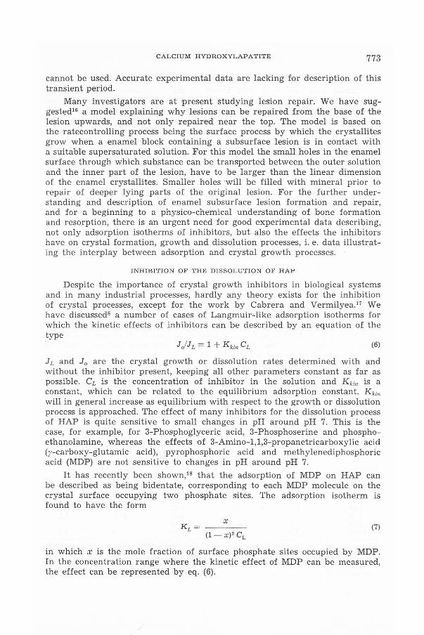
Many investigators are at present studying lesion repair. We have suggested16 a model explaining why lesions can be repaired from the base of the lesion upwards, and not only repaired near the top. The model is based on the ratecontrolling process being the surface process by which the crystallites grow when a enamel block containing a subsurface lesi•on is in contact with a suitable supersaturated solution. For this model the small holes in the enamel surface through which substance can be transported between the outer solution and the inner part of the lesion, have to be larger than the linear dimension of the enamel crystallites. Smaller holes will be filled with mineral prior to repair of deeper lying parts of the original lesion. For the further understanding and descf'iption of enamel subsurface lesion formation and repair, and for a beginning to a physico-chemical understanding of bone formation and resorption, there is an urgent need for good experimental data describing, not only adsorption isotherms of inhibitors, but also the effects the inhibitors have on crystal formation, growth and dissolution processes, i. e. data illustrating the interplay between adsorption and crystal growth processes.
INHIBITION OF THE DISSOLUTION OF HAP
Despite the importance of crystal growth inhibitors in biological systems and in many industrial processes, hardly any theory exists for the inhibition of crystal processes, except for the work by Cabrera and Vermilyea.17 We have discussed8 a number of cases ·of Langmuir-like adsorption isotherms for which the kinetic effects of inhibitors can be described by an equation of the type
(6)
h and J 0 are the crystal growth or dissolution rates determined with and without the inhibitor present, keeping all other parameters constant as far as possible. CL i:s the concentration of inhibitor in the solution and K kin is a constant, which can be related t9 the equilibrium adsorption constant. K kin
will in general increase as equilibrium with respect to t he growth or disso}ution process is approached. The effect of many inhibitors for the dissolution process of HAP is quite sensitive to small changes in pH around pH 7. This is the case, for example, for 3-Phosphoglyceric acid, 3-Phosphoserine and phosphoethanolamine, whereas the effects uf 3-Amino-1,1,3-propanetricarboxylic acid (y-carboxy-glutamic acid) , pyrophosphoric acid and methylenediphosphoric acid (MDP) are not sensitive to changes in pH around pH 7.
It has recently been shown,18 that the adsorption of MDP on HAP can be described as being bidentate, corresponding to each MDP molecule on the crystal surface occupying two phosphate sites. The adsorption isotherm is found to have the form
x (7)
(1-x)2 CL
in which x is the mole fraction of surface phosphate sites occupied by MDP. In the concentration range where the kinetic effect of MDP can be measured, the effect can be represented by eq. (6).

774 J. CHRISTOFFERSEN ET AL.
EFFECT OF CITRATE ION S ON THE RATE OF DISSOLUTION OF HAP
Citrate ions have long been known to affect processes involved in the forma·tion of calcium phosphates. Brecevic and Furedi-Milhofer19 have reported that citrate i·ons adsorb on colloidal calcium phosphate precipitates and slow down the transformation of these particles to octa-calcium phosphate. The effect of citrate ions on the rnte of dissolution of HAP is shown in Figure 4,
0
• ...... _J
•
3.--~~~~~~~~~..---~~~~~~~~~~~~
2
5 105[citrate] /M
Figure 4. J LfJ 0 , the rates of dissolution of HAP with a nd without citrate ions, plotted against the total concentration of citrate, pH 7.15, m e, = 5 mg, V = 0.91 1, total concentration of calcium
1.43 X 10-s M , Ca/P = 1.67.
in which Jd Jo is plotted against the total concentration of citrate ions in the system. Ji and J 0 are the rates of dissolution with and without citrate, all other reaction parameters being constant. From the plot is seen that citrate ions inhibit the dissolution of HAP crystals if the citrate concentration is less than about 3 X 10⁻⁵ M. The largest inhibitory effect of these ions is obtained for a citrate concentration of about 10-5 M. At concentrations larger than 3 X 10⁻⁵ M citrate ions cause an increased rate of dissolution. The solution composition in these experiments is given in Table I. The accelerating effect at citrate concentrations larger than 3 X 10⁻⁵ M can be explained as being due
TABLE I
The Solution Composition and the Relati ve Rate, J LIJ 0
, for the Effect of Citrate on the Rate of Dissolution of HAP*
105 [citrate] 10• [cacn 105 [HCi2-] 105 [Ci3-] 105 [Ca2+] Jl /Jn (M) (M) (M) (M) (M)
0.11 0.03 0.01 0.03 1.40 0.56 0.55 0.22 0.04 0.23 1.21 0.48 1.09 0.42 0.09 0.53 1.01 0.49 3.30 0.88 0.36 2.0 1 0.55 1.1 6.60 1.12 0.82 4.60 0.31 2.0
10.90 1.24 1.46 8.15 0.19 2.8
• [Cal total = 1.43 X 10-s M = 0.25 C,, pH = 7.15, Ca/P = 1.67. The concentrations of the ions, Ca Ci- , HCi•- , Ci•- and Ca•+, are calculated assuming other equilibria than HCi" -:::: Ci•· + H+ a nd Ca•+ + Ci•- -:::: Caci- to be insignificant. The equilibrium constants for these reactions a re taken to be 10-H M and 10"' M- t, respectively . 0.5 µmol /l of the total citrate is assumed to be on the surface of the crystals.

CALCIUM HYDROXYLAPATITE 775
to ion-'pair formation between calcium ions and citrate ions. The specific surface area of the crystals was 32 m 2/g, determined by N2/He gas adsorption using a Quantasorb ® surface area analyzer. From the specific surface area the amount of phosphate i,ons in the surface ·of the crystals was calculated to be about 1,6 X 10⁻⁴ mol/g. This calculation is explained in ref. [8] . For 5 mg crystals as used in the experiments, the amount of citrate ions that can be adsorb on the crystal surface is expected to be of the order of 0.5 µmol. At low citrate concentration only a minor amount ·of the calcium ions will complex with the citrate in S'olution. At low concentrations ·of citrate ions, these ions may adsorb onto the crystal surface and cause a decreased rate of pissolution. As the citrate concentration is raised, the effect of adsorption is counter-acted by the complexing of calcium ions in solution with citrate ions. The latter process will cause an increase in the rate of dissolubon of HAP, corresponding to an increase in the affinity of the dissolution process.
BIOLOGICAL MINERALIZATION
In vertebrates bone resorption is by far the most important form of demineralization and presents many unsolved problems.20 Dominguez and Raisz21 have reported that in vitro acidosis causes 1increased bone resorption. The effect could be explained as mainly due to an increase in the rate of dissolution of devitalized bone, caused by the increase in the hydrogen ion concentration. Cell-mediated bone resorption appears to be practically independent of pH in the pH-range 7.0-7.5 . Neither the enhanced bone resorption during metabolic acidosis, nor the lack of increased bone resorption during respiratory acidosis has been understood in terms of a model.
For future improvement in the description of in vivo bone resorption it is important to obtain information about how in vivo occuring ions and molecules affect in vitro dissolution of calcium phosphates. Ions affecting the rate of dissoluHon of HAP have been shown to behave quite differently. Citrate ions appear to reduce the rate of dissolution of HAP at low concentrations, but, due to their complex formahon with calcium ions in solution, citrate ions increase the rate of dissolution when present in higher concentrations. Fluoride ions have been shown22 to decrease the rate of dissolutiun of HAP, particularly at low values of pH, and to cause an increase in the rate of growth of HAP containing fluoride ions. Diphosphonates and diphosphate ions have been shown to inhibit both the rate of dissolution and the rate of growth of calcium phosphates. The influence on calcium phosphate dissolution of potential candidates for affecting in vivo bone resorption should thus be investigated over a wide range of concentration and solution composition.
Acknowledgements. - We thank Nobuko Christiansen and lben Junghans for technical assistance. JC and MRC acknowledge support of the Danish Medical Research Council, grant no. 12-3617, and of the Carlsberg Foundation, grant. no. 1981/82 35/IV.
a
c
cs
LIST OF SYMBOLS
mean diameter of an ion. concentration of solute. In this paper only dissolution of HAP into a solution with Ca/P04 = 1.67 is discussed. equilibrium value of C at the actual value of pH.

776
CL
dr/dt
J . CHRISTOFFERSEN ET AL.
concentration of an inhibitor.
linear rate of dissolution.
function representing the surface area on which the dissolution takes place. This term may, in the general case, include certain kinetic factors.
g (C) function representing the influence of concentration on the rate.
J overall rate of dissolution of HAP, dnHAP/dt.
J0
overall rate of dissolution for a particular set of values of the rate controlling parameters, no inhibitor being present.
k,k', k"
x
dr/dt
a
a
overall rate of dissolution for the same set of values of the rate controlling parameters as used for definition of J
0, except an inhibitor is present.
rate constants.
Boltzmann constant times the absolute temperature.
Langmuir adsorption constant.
Langmuir adsorption constant, determined from kinetic experiments.
acidity constant for HPOl- ions in the crystal surface.
mass of crystals at time ' .
mass of cristals at time ;.ero.
amount of calcium hydroxylapatite, Ca10(P04)6(0H)2.
linear dimension of crystals.
depth of sub-surface lesion i enamel.
lateral rate of growth of a dissolution nucleus.
lateral rate of growth of a dissolution nucleus if no back reaction takes place
mole fraction of adsorption sites occupied by an inhibitor.
mole fraction of phosphate groups in the crystal surface in the form of P04
3- and HPO/- respectively.
linear rate of dissolution.
n a4 a2/ 3 (kT) 2
dimensionless dissolution affinity for a mean ion.
surface tension of HAP (Gibbs surface energy).
REFERENCES
1. J. Christofferse n and M. R. Christofferse n, J. Crystai Growth 35 (1976) 79.
2. J. Christofferse n , J. Crystai Growth 49 (1980) 29. 3. W. B. Hi 11 i g, Acta Met. 14 (1966) 1868. 4. G. H. G i 1 mer and P. Benne ma, J. AppL Phys. 43 (1972) 1347. 5. G. H. G i 1 mer and P. Benne ma, J . Crystai Growth 13/14 (1972) 148. 6. S. W. H. de Haan, V. J . A. Mee us sen, B. P. Ve 1 t man, P. Benne ma,
C. van Leeuwen, and G. H. G i 1 mer, J. Crystai Growth 24/25 (1974) 491. 7. P . Benne ma and J. P. van de r Ee rd en, J . Crystai Growth 42 (1977) 201. 8. J. Christofferse n and M. R. Cristo ff er sen, J. Crystai Growth 53
(1981) 42.
9. J. Christofferse n and M. R. Christofferse n, J. Crystai Growth 57 (1982) 21.
10. A. Groene v e 1 d and J . Arends, Caries Research 9 (1975) 36.

CALCIUM HYDROXYLAPATITE 777
11. J . D. B. Featherstone, J. F. Duncan, and T .W. Cut res s, Archs. oral Biology 24 (1979) 101.
12. J . Arends and J. Schut ho f, J. Biol Buccale 8 (1980) 175. 13. J . M. ten Cate and J. Arends, Caries Res. 14 (1980) 351. 14. J. Arends and J. M. ten Cate, J. Crystal Growth 53 (1981) 135. 15. J. Christofferse n and J. Arends, Caries Res. 16 (1982) 433. 16. J. Christofferse n, M. R. Christofferse n, and J. Arends, J.
Crystal Growth 60 (1982) 255. 17. N. Cabrera and D. A. Ver mi 1 ye a, Growth and Perfection of Crystals,
eds. R.H. Doremus, B. W. Roberts, and D. Turnbull (N. Y. : Wiley, London: Chapman & Hall, 1958), p. , 393.
18. J. Christoffer s en, M. R. Christofferse n, S. B a ch Chris tens en, and G. H. Nan co 11 as, J . Crystal Growth, in press.
19. Lj. Br e c e vi c and H. Fure di - Mi 1 ho fer, Israel J. Med. Sci. 7 (1971) 423. 20. J. J. Reyno 1 d s et al., Biological Mineralization and Demineralization, ed.
G. H. Nan co 11 as, Dahlem Konferenzen (Springer Verlag, 1982), p. 389. 21. J. H. Dominguez and L. G. Rais z, Cale. Tiss. Int. 29 (1979) 7. 22. M. R. Christofferse n, J . Christofferse n , and J. Are n d s, to
be published.
SAZETAK
Studij otapanja kalcij-hidroksiapatita i njegova primjena na biolos.ku demineralizaciju
J. Christoffersen, M. R. Christoffersen i J. Arends
U ovom radu razmotreni su razni aspekti ispitivanja brzine otapanja kalcij-hidroksilapatita u vodenim otopinama. S kemijskog stajalista sistem je pogodan za proucavanje kemijskog procesa koji se zbiva na povrsini kristala za vrijeme njihova otapanja u Cistim sistemima, kao i u nazocnosti inhibitora. Sa stajalista biologije ta su istrazivanja rezultirala modelom stvaranja subpovrsinskih lezija u zubnoj caklini. Proucavanje in vitro efekta bioloski relevantnih molekula na proces otapanja upucuje i na model demineralizacije kostanog tkiva. Nadeno je da niske koncentracije citrat-iona inhibiraju proces otapanja, a viSe koncentracije ubrzavaju taj proces zbog znatnijeg kompleksiranja kalcij-iona.


![University of Groningen Combining the incompatible Drooge, Dirk … · interface. Nernst and Brunner [76] introduced the diffusion layer model. They assumed that dissolution at the](https://img.dokumen.tips/doc/110x75/5ebe387a39462d4be06c02a3/university-of-groningen-combining-the-incompatible-drooge-dirk-interface-nernst.jpg)