Embed Size (px)
Citation preview

University of Birmingham
Influence of ultrasonic agitation on bond strength,marginal adaptation, and tooth discolorationprovided by three coronary barrier endodonticmaterialsAguiar, Bernardo A; Frota, Luciana M A; Taguatinga, Daniela T; Vivan, Rodrigo R; Camilleri,Josette; Duarte, Marco A H; de Vasconcelos, Bruno CarvalhoDOI:10.1007/s00784-019-02850-y
License:Other (please specify with Rights Statement)
Document VersionPeer reviewed version
Citation for published version (Harvard):Aguiar, BA, Frota, LMA, Taguatinga, DT, Vivan, RR, Camilleri, J, Duarte, MAH & de Vasconcelos, BC 2019,'Influence of ultrasonic agitation on bond strength, marginal adaptation, and tooth discoloration provided by threecoronary barrier endodontic materials', Clinical Oral Investigations, vol. 23, no. 11, pp. 4113-4122.https://doi.org/10.1007/s00784-019-02850-y
Link to publication on Research at Birmingham portal
Publisher Rights Statement:Checked for eligibility: 19/03/2019
This is a post-peer-review, pre-copyedit version of an article published in Clinical Oral Investigations. The final authenticated version isavailable online at: http://dx.doi.org/10.1007/s00784-019-02850-y
This document is subject to Springer Nature re-use terms: https://www.springer.com/gp/open-access/authors-rights/aam-terms-v1
General rightsUnless a licence is specified above, all rights (including copyright and moral rights) in this document are retained by the authors and/or thecopyright holders. The express permission of the copyright holder must be obtained for any use of this material other than for purposespermitted by law.
•Users may freely distribute the URL that is used to identify this publication.•Users may download and/or print one copy of the publication from the University of Birmingham research portal for the purpose of privatestudy or non-commercial research.•User may use extracts from the document in line with the concept of ‘fair dealing’ under the Copyright, Designs and Patents Act 1988 (?)•Users may not further distribute the material nor use it for the purposes of commercial gain.
Where a licence is displayed above, please note the terms and conditions of the licence govern your use of this document.
When citing, please reference the published version.
Take down policyWhile the University of Birmingham exercises care and attention in making items available there are rare occasions when an item has beenuploaded in error or has been deemed to be commercially or otherwise sensitive.
If you believe that this is the case for this document, please contact [email protected] providing details and we will remove access tothe work immediately and investigate.
Download date: 22. Oct. 2021
1
Title Page
Authors:
Bernardo A Aguiar DDS1,2, Luciana MA Frota DDS1, Daniela T Taguatinga DDS, MSc3; Rodrigo R Vivan
DDS, MSc, PhD4; Josette Camilleri BChD, MPhil, PhD, FICD, FADM, FIMMM, FHEA5; Marco AH
Duarte DDS, MSc, PhD4; Bruno C Vasconcelos DDS, MSc, PhD1,6
Title:
Influence of Ultrasonic Agitation on Bond Strength, Marginal Adaptation and Tooth Discoloration
Provided by Three Coronary Barrier Endodontic Materials.
Afflitations:
1 Post-Graduate Program in Dentistry, School of Pharmacy, Dentistry and Nursing, Federal University of
Ceará, Fortaleza, Ceará, Brazil. 2 Department of Endodontics, Faculty of Dentistry, University of Fortaleza, Fortaleza, Ceará, Brazil. 3 Endodontist Private Practice, Goiania, GO, Brazil. 4 Department of Dentistry, Endodontics and Dental Materials, Bauru Dental School, University of São
Paulo, Bauru, SP, Brazil 5 School of Dentistry, College of Medical and Dental Sciences, Institute of Clinical Sciences, University of
Birmingham, Birmingham, United Kingdom. 6 School of Dentistry of Sobral, Federal University of Ceará, Campus Sobral, Sobral, CE, Brazil.
Acknowledgement
This study was partially financed by CAPES - Brazilian Federal Agency for Support and Evaluation of
Graduate Education within the Ministry of Education of Brazil.
The authors wish to thank the Analytical Central - UFC / CT - INFRA / MCTI - SISNANO / CAPES Pró-
Equipment for making the analyzes feasible under microscopy.
Corresponding author
Bruno Carvalho de Vasconcelos, DDS, MSc, PhD
Post-Graduate Program in Dentistry, School of Pharmacy, Dentistry and Nursing, Federal University of
Ceará.
1253 Monsenhor Furtado St.
CEP: 60430-355, Fortaleza, CE, Brazil
E-mail: [email protected]
Manuscript Click here toaccess/download;Manuscript;Manuscript_revised.docx
Click here to view linked References
1 2 3 4 5 6 7 8 9 10 11 12 13 14 15 16 17 18 19 20 21 22 23 24 25 26 27 28 29 30 31 32 33 34 35 36 37 38 39 40 41 42 43 44 45 46 47 48 49 50 51 52 53 54 55 56 57 58 59 60 61 62 63 64 65

2
ABSTRACT
Objectives: The effect of ultrasonic agitation (UA) on bond strength and adaptation of cervical plugs
prepared with MTA Angelus (MTA), MTA Repair HP (MTAHP) and Biodentine (BIO) was evaluated.
Dentin discoloration caused by the materials/treatment was also assessed. Materials and Methods: Seventy-
two single rooted teeth were divided into six groups depending on the materials/treatment. After cervical
plug preparation, dentin discs were excised for the push-out test; additional discs were analysed under the
confocal microscope to determine adaptation (gaps occurrence). For dentin discoloration analysis (ΔE),
blocks of bovine incisors had cavities prepared and filled with the materials/treatment (from 7 to 180 days).
Results: Both bond strength and adaptation were positively influenced by UA (P<0.05). Comparison
between materials showed an advantage for BIO when compared to MTAHP (P<0.05). The best and worst
results were provided by BIO/UA (12.66 MPa and 1.87%) and MTAHP (2.54 MPa and 28.58%),
respectively. For ΔE, significant differences were observed throughout the periods. Just the MTA without
UA exhibited noticeable discoloration at 180 days (P<0.05). Conclusions: UA favoured a better adaptation
of the materials to the dentin root, resulting in higher bond strength and adaptation of the materials to the
root canal walls. Moreover, UA reduced MTA discoloration, keeping it imperceptible over the period
evaluated. Clinical Relevance: The better adaptation and higher bond strength provided by UA can be
considered clinically relevant due to the importance of maintaining blood clot integrity and the possible
aesthetic compromise provided by reparative materials when used as coronary barrier in regenerative
procedures
Key words: Endodontics, hydraulic calcium silicate cement, marginal adaptation, tooth discoloration,
ultrasonic agitation.
INTRODUCTION
The technique of pulpal revascularization has been considered a promising alternative for
endodontic treatment of teeth with incomplete rhizogenesis and pulp necrosis, since it allows the continuity
of root formation and thickening of the dentin walls [1, 2]. In this technique, a preparation of a coronal
barrier with repairing material is necessary to preserve the integrity of the blood clot and to stimulate the
production of a new tissue capable of re-establishing root formation [3-7].
The mineral trioxide aggregate (MTA) was initially developed to be used as root perforation repair
material [8, 9], however, it has been commonly used as a coronal barrier due to its physico-chemical and
biocompatibility characteristics [6, 9]. However, it presents limitations such as difficulty of insertion and
induction of tooth discoloration [10].
In order to overcome these limitations new materials were developed. Biodentine (Septodont, Saint
Maur des Fosse, Cedex, France) was introduced as a bioactive material. It was based on tricalcium and
dicalcium silicate, main components of Portland cement; commercial MTA materials presents 70-90% of
1 2 3 4 5 6 7 8 9 10 11 12 13 14 15 16 17 18 19 20 21 22 23 24 25 26 27 28 29 30 31 32 33 34 35 36 37 38 39 40 41 42 43 44 45 46 47 48 49 50 51 52 53 54 55 56 57 58 59 60 61 62 63 64 65

3
Portland cement. Biodentine also presents calcium carbonate and oxides of iron and zirconia, the latter
added as a radiopacifier [10]. This material liquid was composed of water, calcium chloride, added as a
settling accelerator and a water-soluble polymer to improve its plasticity. This cement has adequate
radiopacity, resistance to displacement, short curing time [11, 12], color stability [10], high dimensional
stability and good sealing capacity [13, 14]. Another material developed to overcome the limitations of
conventional MTA is the MTA Repair HP (Angelus, Londrina, PR, Brazil). It is a repair cement based on
tricalcium silicate which also contains additives. According to the manufacturer, these changes provide
better handling, insertion and color stability. The liquid includes an unidentified plasticizer [12, 15]. So far,
the information on the MTA Repair HP is limited; until now its biocompatibility, biomineralization and
resistance to displacement were evaluated showing promising results [12, 15, 16].
In addition to changes in materials composition aiming to improve clinical performance, the
protocols of material insertion were also investigated. The use of ultrasonic indirect agitation was suggested
to improve the sealing ability provided by MTA when used as root-end filling material [17, 18]. The
ultrasonic agitation was also tested directly on filling materials and provided better sealer the penetration
into dentinal tubules [19, 20]. The changes brought by the manufacturers caused a lack of information on
its physical and chemical properties since there is no study under confocal microscopy for the proper
evaluation of the marginal adaptation.
Moreover, considering the use of these materials in the aesthetic area at the cervical level and
considering that the MTA presents notably the capacity to induce the dentin discoloration, it would be
relevant to evaluate the influence of this event by the radiopacifiers of the new cements. As well as, the use
of the ultrasonic agitation in the evaluation of the adaptation would be interesting to evaluate its effect in
the darkening.
Considering the novel materials disposable for coronal barrier preparation, the possible influence
of ultrasonic agitation on its adaptation and the parallel risk of tooth discoloration, the current study
evaluated the influence of ultrasonic agitation on push-out bond strength and marginal adaptation to root
canal walls in the cervical region of MTA Angelus (Angelus), MTA Repair HP and Biodentine; also
evaluated the tooth discoloration promoted by these materials. It will be considered as null hypotheses the
absence of differences in bond strength, marginal adaptation, and tooth discoloration provided by the
materials in function of the use or not of the ultrasonic agitation.
MATERIALS AND METHODS
Bond strength tests and marginal adaptation
The sample calculation was performed using the G*Power v3.1 for Mac (Heinrich Heine,
Universität Düsseldorf) by selecting the Anova One Way fixed test. Data from an earlier study were
considered and the effect of sample size in the present study was established [20]. The effect size in the
present study was established (=1.36) and the alpha type error of 0.05 in the beta power of 0.80. A total of
9 samples were indicated as the ideal size to observe significant differences; the sample was increased by
30% for safety if specimens were to be lost (n = 12).
1 2 3 4 5 6 7 8 9 10 11 12 13 14 15 16 17 18 19 20 21 22 23 24 25 26 27 28 29 30 31 32 33 34 35 36 37 38 39 40 41 42 43 44 45 46 47 48 49 50 51 52 53 54 55 56 57 58 59 60 61 62 63 64 65

4
Selection and preparation of the sample
After approval by the Local Ethics Committee (#1.900.129/2017), 72 maxillary uni-radicular
human teeth extracted for unknown reasons were collected for the studies of bond resistance and marginal
adaptation. Coronary access with diamond tips (#1014 and #3081; KG Sorensen, Cotia, SP, Brazil) was
carried out, followed by preparation of the cervical and middle thirds with Largo Reamer #5 (Dentsply-
Sirona, Ballaigues, Switzerland). After the preparation specimens were evaluated for circularity and
standardization of the cavity diameter. Where the Largo Reamer #5 did not enter just the same has been
replaced. The teeth were then cross-sectioned 5.0 mm below the cemento-enamel junction by means of a
precision cut double-face diamond disk (Isomet 1000; Buhler Ltda., Lake Bluff, NY, USA). After being
sectioned, the teeth were submitted to ultrasonic baths with 2.5% sodium hypochlorite solution (Asfer
Indústria Química Ltda, São Caetano do Sul, SP, Brazil) for 15 minutes, with distilled water (Asfer Indústria
Química Ltda.) for 1 minute, with 17% ethylenediaminetetraacetic acid (EDTA) (Biodinâmica Química e
Farmacêutica Ltda., Ibiporã, PR, Brazil) for 3 minutes and finally with distilled water for 3 minutes.
After specimen preparation, collagen sponge plugs (Technew, Rio de Janeiro, RJ, Brazil) were
inserted into the canal with the aid of a Schilder condenser (Odous de Deus, Belo Horizonte, MG, Brazil)
obliterating the apical 2 mm. Regardless of the group in which they were to be allocated, the specimens
were irrigated with 1 mL of distilled water. Afterwards, gentle aspiration was performed with the aid of the
endodontic aspirator for 4 seconds and additional drying with absorbent paper cones (Dentsply-Sirona).
Manipulation and insertion of materials
The manipulation of the repair cements was carried out by a single operator previously calibrated,
following the manufacturer's instructions; Fluo-3 fluorophore (Sigma-Aldrich, Warrington, PA, USA) at a
concentration of approximately 0.1% (w/w) was incorporated into the materials to determine marginal
adaptation [21].
The specimens were initially randomly assigned to three experimental groups, depending on the
material (n = 24). For insertion of the materials a 1.2 mm diameter MTA microapplicator (Angelus) was
used followed by condensation with the Schilder condenser. The procedure was repeated until cervical
plugs reached 3.0 mm thickness. Then, the specimens were randomly divided again between the subgroups
with and without ultrasonic agitation (UA) (n = 12). In the groups which received ultrasonic agitation
(MTA/UA, MTAHP/UA and BIO/UA), piezoelectric ultrasound equipment (Piezon Master 200; EMS,
Nyon, Geneva, Switzerland) was set to 10% of power. A specific ultrasonic tip (E5; Helse Ultrassonic,
Santa Rosa do Viterbo, SP, Brazil) was inserted into the material at a depth of 2.0 mm. Two 20-second
activation cycles were performed, one in the mesio-distal direction and one in the vestibular-palatine
direction [19]. Then, a final vertical condensation was performed to accommodate the materials; similarly,
the specimens allocated in groups without UA also received an additional vertical condensation.
After concluding the cervical plugs, temporary restorations with glass ionomer cement (GIC; FGM
Produtos Odontológicos Ltda, Joinville, SC, Brazil) were performed in order to simulate clinical
1 2 3 4 5 6 7 8 9 10 11 12 13 14 15 16 17 18 19 20 21 22 23 24 25 26 27 28 29 30 31 32 33 34 35 36 37 38 39 40 41 42 43 44 45 46 47 48 49 50 51 52 53 54 55 56 57 58 59 60 61 62 63 64 65

5
procedures. The specimens were then stored with the apical portions immersed in phosphate-buffered saline
solution (PBS; Quimlab, Jacareí, SP, Brazil) in an oven at 37 °C for 7 days.
After the storage period, two 1.0 mm thickness dentin discs were extracted from each plug on its
central portion using a cutting machine (Isomet 1000; Buhler Ltda., Lake Bluff, NY, USA) totalling 144
discs; each disk obtained were allocated to one of the tests proposed - the cervical one for the bond strength
test and the apical one for the marginal adaptation test - the portion directly in contact with GIC was
discharged in order to allow any interference.
Evaluation of the bond strength (push-out)
The 72 dentin disks allocated for the bond strength test were tested using a Universal Testing
Machine (#443; Instron, Norwood, MA, USA) handled by a blind operator; for performing the push-out
test using a constant velocity of 0.5 mm/min. For this, punches with 1.2 mm diameter were positioned in
the center of the upper face of the plugs; on the underside, a steel plate with a 3.0 mm diameter central bore
was adapted to serve as a support for the specimen and enable material to escape from the plugs. The
maximum force required for cement displacement was measured in Newtons (N) and converted to MPa by
the division of force (N) by the area in mm2 (1MPa = 1N/mm2).
For analysis of fracture types after the push-out test, all slices were examined in a scanning electron
microscope (Aspex Express; FEI Europe, Eindhoven, Netherlands) at a magnification of 30x. The failures
were classified as: adhesive - rupture of the union at the interface dentin/material, cohesive - rupture on the
material and, mixed – rupture cohesive of the material and adhesive in the dentin.
Assessment of marginal adaptation
The remaining 72 discs were adapted to glass slides and fixed with wax with the coronary face
facing upwards. The sets were taken to the politriz (Arotec, Cotia, SP, Brazil) where, using 600, 900 and
1200 granulometry water sands, were polished. Then the samples were taken to Confocal Laser Scanning
Microscope (LSM 710; Carl Zeiss Microscopy GmbH, Baden-Württemberg, Germany) handled by a blind
operator and examined 10 μm below the surface under 20x magnification; 10 section scans of 1 μm were
recorded at a resolution of 1024 x 1024 pixels using the ZEN 2012 program (Carl Zeiss Microscopy GmbH)
and saved in TIFF format.
To measure the adaptation (i.e. presence of gaps), the images were evaluated in the Image J
software (National Institute of Health, Bethesda, MD, USA), using as a calibration tool the scale offered
by confocal microscopy images. The total perimeter of the canals and the length of gaps were determined
in millimetres using the software polygonal and linear tools, respectively; once these measurements were
obtained, the percentages of gaps were calculated according to the total perimeter of the root canals.
Study of tooth discoloration
1 2 3 4 5 6 7 8 9 10 11 12 13 14 15 16 17 18 19 20 21 22 23 24 25 26 27 28 29 30 31 32 33 34 35 36 37 38 39 40 41 42 43 44 45 46 47 48 49 50 51 52 53 54 55 56 57 58 59 60 61 62 63 64 65

6
Before performing this evaluation, a sample size calculation was performed following the same
parameters used in the previous test; based on the preliminary study results the test pointed to a sample of
10 specimens as a sufficient sample to determine possible statistical differences [22]. An experimental
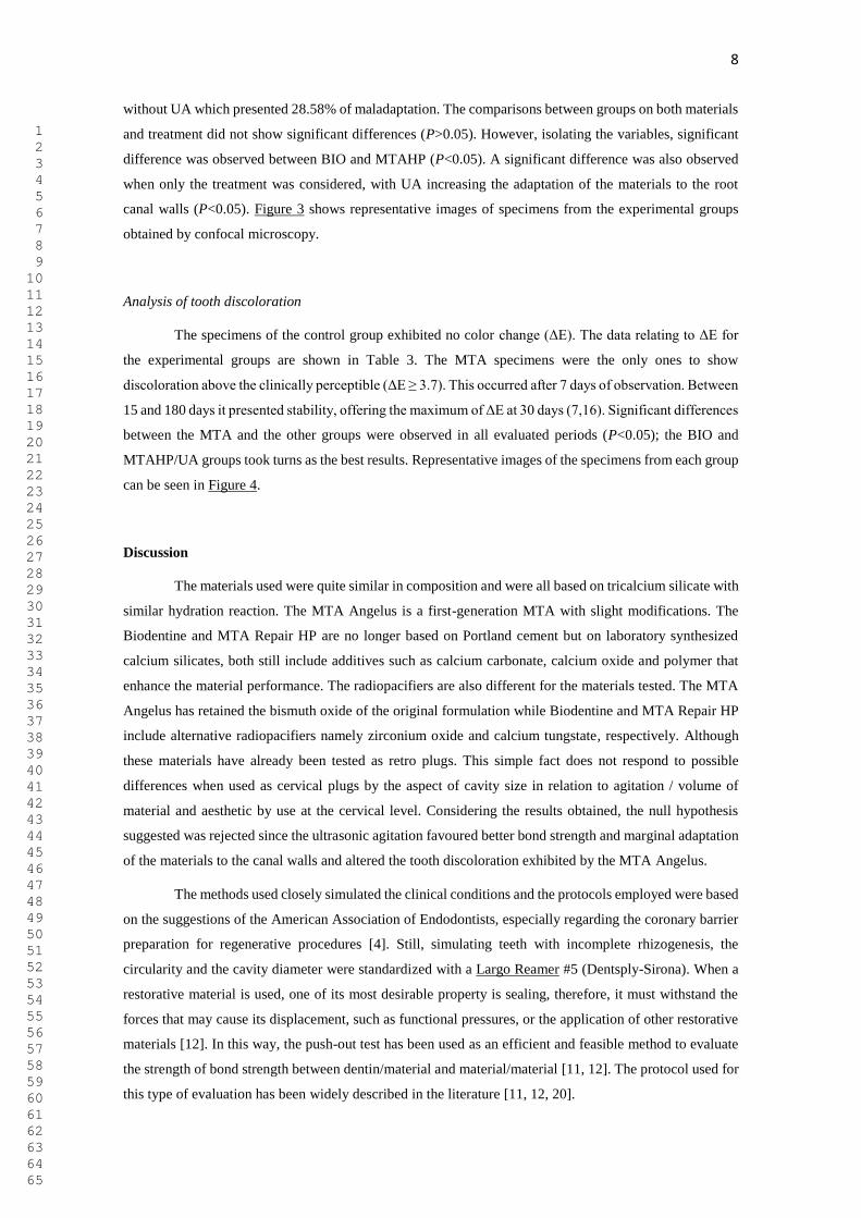
design of this study methods was show in Figure 1.
Selection and preparation of the sample
For the analysis of dentinal discoloration, bovine incisor blocks prepared according to the model
proposed by Marciano et al. [22]. Sixty-five blocks (10 mm x 10 mm) were prepared with the aid of a
diamond blade driven by a cutting machine. After being prepared, they had their lingual faces sectioned in
manner to expose their pulp chambers. Cavities 5 mm in diameter and approximately 1.5 mm deep were
prepared in the center of the lingual face of each specimen using diamond tips #4054 (KG Sorensen);
specimens where a remainder of 2 ± 0.2 mm of dentin and enamel thickness were not obtained at the bottom
of the cavity were replaced.
Subsequently, the specimens were submitted to ultrasonic baths following the previously used
protocol. Afterwards, after being dried with filter paper, the wells had their external boundary conditioned
with 37% phosphoric acid for 15 seconds, washed with distilled water for 1 minute and gently air-dried for
15 seconds. An adhesive layer (Adper Single Bond 2; 3M ESPE, Sumaré, SP, Brazil) was applied in the
conditioned and polymerized area (Optilight LD Max; Gnatus, Ribeirão Preto, SP, Brazil) for 20 seconds
to allow interface resin.
Materials manipulation and insertion
After specimen preparation, they were randomly divided among the six experimental groups (n =
10). The cements, after being manipulated, were inserted into the cavities. For those who underwent
ultrasonic agitation (MTA/UA, MTAHP/UA and BIO/UA), it was accomplished by means of the use of
piezoelectric ultrasound programmed for power 3 (approximately 30%), with a smooth tapered tip (E5;
Helse Ultrasonic). After gentle condensation of the cements in the cavities, the tip was inserted at a depth
of 1 mm. Then, two cycles of agitation of 20 seconds, one in the mesio-distal and one in the buccal-palatal
direction were performed [19]; independent of the group to which they took part, the materials were once
again condensed to fill the entire cavity.
After completion, restorations were performed with flow resin (New DFL, Rio de Janeiro, RJ,
Brazil), which was polymerized with LED light (Optilight LD Max, Gnatus) for 60 seconds. The remaining
5 blocks comprised the negative control group in which the cavities were completely filled with restorative
material. Regardless of the group, the specimens were immersed in individual containers containing 2 mL
of distilled water, where they remained throughout the experimental period at 37ºC and 100% humidity.
Evaluation of tooth discoloration
1 2 3 4 5 6 7 8 9 10 11 12 13 14 15 16 17 18 19 20 21 22 23 24 25 26 27 28 29 30 31 32 33 34 35 36 37 38 39 40 41 42 43 44 45 46 47 48 49 50 51 52 53 54 55 56 57 58 59 60 61 62 63 64 65

7
Color determination was performed using a digital spectrophotometer (VITA Easyshade compact;
VITA Zahnfabrik AG, Bad Sachington, Germany). The equipment was calibrated prior to the measurement
of each specimen. Measurements were obtained immediately after placement of the materials (reference
color; 0) after 7, 14, 30 and 180 days. Color parameters were recorded as determined by the International
Commission On Illumination (CIE, 1978), considering "L", "a" and "b", where "L" represents the values of
color luminosity, "a" corresponds measurement along the red-green axis and "b" is the measurement along
the yellow-blue axis. The change in color (ΔE), in relation to time intervals, was calculated based on the
initial values using the following formula:
ΔE = [(L1-L0)2 + (a1-a0)2 + (b1-b0)2]1/2
Statistical analysis
The data of the evaluations were tabulated and tested for their normality by the Shapiro-Wilk test,
which indicated a parametric nature for the bond strength and dentin discoloration and non-parametric data
for the analysis of the adaptation to the canal walls. Thus, ANOVA and Tukey tests were used for the bond
strength and discoloration tests, and Kruskal-Wallis and Dunn tests were used for analysis of adaptation.
In addition, the influence of ultrasonic agitation was statistically tested in all evaluations, regardless of the
material used, and Student's t-test and Mann-Whitney tests, respectively. For all comparisons, the
significance levels were established at 5%.
Results
Bond strength
Table 1 presents the results of the push-out test. Premature failures were not observed in any
groups/subgroups. The BIO/UA group exhibited the best bond strengths while the MTAHP group without
agitation had the worst. Considering each material with and without treatment, statistically significant
differences were observed between the BIO groups; the UA positively influenced their bond strength
(P<0.05). Comparing the materials submitted or not to UA, significant differences were observed in both
conditions, with MTAHP offering significantly lower results than the others (P<0.05); no differences were
observed between BIO and MTA. Considering only the use or not of UA, significant differences were
observed, where the UA proportioned superior results (P<0.05). The MTA/UA and BIO groups, with and
without UA, presented a predominance of the cohesive failures, and the MTAHP group presented
predominance of the cohesive type as could be observed in Table 1; Figure 2 presents the failure types
illustration.
Analysis of the interface of adaptation to the dentin wall
Percentage of gaps observed in the sections of the cervical plugs prepared with the repair materials
with or without UA are shown in Table 2. Once more the BIO/UA exhibited the optimal interface with
adequate marginal adaptation, giving only 1,87% of gaps. The worst results were exhibited by MTAHP
1 2 3 4 5 6 7 8 9 10 11 12 13 14 15 16 17 18 19 20 21 22 23 24 25 26 27 28 29 30 31 32 33 34 35 36 37 38 39 40 41 42 43 44 45 46 47 48 49 50 51 52 53 54 55 56 57 58 59 60 61 62 63 64 65
8
without UA which presented 28.58% of maladaptation. The comparisons between groups on both materials
and treatment did not show significant differences (P>0.05). However, isolating the variables, significant
difference was observed between BIO and MTAHP (P<0.05). A significant difference was also observed
when only the treatment was considered, with UA increasing the adaptation of the materials to the root
canal walls (P<0.05). Figure 3 shows representative images of specimens from the experimental groups
obtained by confocal microscopy.
Analysis of tooth discoloration
The specimens of the control group exhibited no color change (ΔE). The data relating to ΔE for
the experimental groups are shown in Table 3. The MTA specimens were the only ones to show
discoloration above the clinically perceptible (ΔE ≥ 3.7). This occurred after 7 days of observation. Between
15 and 180 days it presented stability, offering the maximum of ΔE at 30 days (7,16). Significant differences
between the MTA and the other groups were observed in all evaluated periods (P<0.05); the BIO and
MTAHP/UA groups took turns as the best results. Representative images of the specimens from each group
can be seen in Figure 4.
Discussion
The materials used were quite similar in composition and were all based on tricalcium silicate with
similar hydration reaction. The MTA Angelus is a first-generation MTA with slight modifications. The
Biodentine and MTA Repair HP are no longer based on Portland cement but on laboratory synthesized
calcium silicates, both still include additives such as calcium carbonate, calcium oxide and polymer that
enhance the material performance. The radiopacifiers are also different for the materials tested. The MTA
Angelus has retained the bismuth oxide of the original formulation while Biodentine and MTA Repair HP
include alternative radiopacifiers namely zirconium oxide and calcium tungstate, respectively. Although
these materials have already been tested as retro plugs. This simple fact does not respond to possible
differences when used as cervical plugs by the aspect of cavity size in relation to agitation / volume of
material and aesthetic by use at the cervical level. Considering the results obtained, the null hypothesis
suggested was rejected since the ultrasonic agitation favoured better bond strength and marginal adaptation
of the materials to the canal walls and altered the tooth discoloration exhibited by the MTA Angelus.
The methods used closely simulated the clinical conditions and the protocols employed were based
on the suggestions of the American Association of Endodontists, especially regarding the coronary barrier
preparation for regenerative procedures [4]. Still, simulating teeth with incomplete rhizogenesis, the
circularity and the cavity diameter were standardized with a Largo Reamer #5 (Dentsply-Sirona). When a
restorative material is used, one of its most desirable property is sealing, therefore, it must withstand the
forces that may cause its displacement, such as functional pressures, or the application of other restorative
materials [12]. In this way, the push-out test has been used as an efficient and feasible method to evaluate
the strength of bond strength between dentin/material and material/material [11, 12]. The protocol used for
this type of evaluation has been widely described in the literature [11, 12, 20].
1 2 3 4 5 6 7 8 9 10 11 12 13 14 15 16 17 18 19 20 21 22 23 24 25 26 27 28 29 30 31 32 33 34 35 36 37 38 39 40 41 42 43 44 45 46 47 48 49 50 51 52 53 54 55 56 57 58 59 60 61 62 63 64 65

9
The marginal adaptation is a characteristic which is closely related to the bond strength. For this
evaluation, a confocal laser scanning microscope was used as employed in large number of previous studies
[19, 23, 24]. Previously, a pilot study was carried out not finding differences in the characteristics of the
cements when using the flurophore, in agreement with Wiesse et al. [20]. The fluorophore Fluo3 was the
chosen fluorescence indicator. Fluo-3 take the place of the commonly employed Rhodamine B once it binds
to the calcium ions, which is an element present in the great quantity in the materials based on tricalcium
silicate, as pointed out by Jeong et al. [24].
The tooth discoloration was investigated using methods previously described in the literature [25-
27]. Even recognizing that the dental substrate employed is not exactly the same used in bond strength and
adaptation analyzes, it should be considered that it presents advantages in function of, as opposed to human
teeth that have a smaller surface and usually have restorations or caries that can interfere in the color
analysis, the bovine teeth provide a larger flat surface area, allowing adequate and standardized color
evaluation [26]. In addition, bovine teeth are similar to human teeth in the composition of type I collagen
of the organic matrix, which accredits the method for microstructural issues considering that the interaction
between radiopacifiers and this protein are blamed for the darkening process [22, 26-28].
Ultrasonic agitation is based on the transmission of micrometric acoustic energy. Such mechanical
vibration energy is expended from the insert and transmitted to the material, favouring a greater penetration
into the dentinal tubules and a better adaptation of cement/dentin interface [19, 20, 29]. In the present study,
bond strength and improvement in dentin adaptation values were observed when ultrasonic agitation was
used. It is postulated that ultrasonic agitation produced an increase of the cement pressure against the canal
walls favouring a more effective filling of the irregularities, improving adaptation to root canal walls and,
consequently, their bonding resistance [19]. Considering this observation, it could be suggested that this
better adaptation increases the materials adhesive strength making it superior than its cohesive strength.
Additionally, evaluating the role of the materials independently of the treatment, Biodentine
presented results of bond strength and adaptation to the dentine significantly superior to those provided by
the MTA Repair HP. It is suggested that the results obtained by Biodentine can be attributed to the more
plastic consistency of the material [20, 30].
Regarding dentinal discoloration, the results found in the present study confirms the color stability
of the teeth treated with Biodentine and MTA Repair HP, corroborating with findings from previous studies
that pointed the color stability of the first material [27, 31]. To date there was no information on the
discoloration if any caused by MTA Repair HP. Knowledge of the discoloration mechanisms and it being
attributed to the presence of bismuth oxide in materials, the findings in this study were as anticipated. The
materials that were bismuth oxide free did not discolour. The novelty lies with the effect of ultrasonic
agitation on the discoloration where use of ultrasonic agitation mitigated the discoloration caused by MTA
Angelus. Thus, ultrasonic agitation can be suggested as another way of reducing the bismuth oxide induced
discoloration. The dentin discoloration caused by the bismuth oxide present in MTA Angelus and its
interaction with the dental structures had already been reported [22, 27, 28]. This finding was also observed
in the present study, being this the only material to produce a color change higher than 3.7, an index that
was perceived as being perceptible to the human eye [32]. However, results of the present study indicate
1 2 3 4 5 6 7 8 9 10 11 12 13 14 15 16 17 18 19 20 21 22 23 24 25 26 27 28 29 30 31 32 33 34 35 36 37 38 39 40 41 42 43 44 45 46 47 48 49 50 51 52 53 54 55 56 57 58 59 60 61 62 63 64 65

10
that ultrasonic agitation is capable of positively influencing the color stability of bismuth oxide,
consequently of MTA Angelus, an aspect highlighted by the statistically significant difference observed at
15 and 180 days between the MTA groups with and without agitation. Yet, in the observed periods the
ultrasonically agitated MTA Angelus did not show color alteration higher than the perceptible values. It is
known that discoloration caused by bismuth oxide results during the hydration process in its interaction
with strong oxidants such as the amino acids present in the dentin collagen [22]. It is believed that this
finding may be related to the reduction of water available for hydration - the result of the warming generated
by the ultrasound action, reducing the number of particles reacted with the collagen matrix. However, other
investigations are necessary to prove these hypotheses.
Considering the importance of maintaining blood clot integrity and the possible aesthetic
compromise provided by reparative materials when used as coronary barrier in regenerative procedures,
the findings of the present study suggest that ultrasonic agitation can improve the resistance to
displacement of materials and their marginal adaptation; yet, positively influence the color stability of
the MTA Angelus. Thus, although more studies are necessary to confirm the hypotheses raised and to
evaluate other possible interactions, it can be indicated that the use of ultrasonic agitation during the
revascularization procedures presents itself as an accessible and feasible tool for clinical use, capable of
increasing the quality of the procedures performed.
Conclusion
It was concluded that the repair materials evaluated were unable to provide total adaptation to
the root canal walls, however, the use of ultrasonic agitation provided better bond strength and marginal
adaptation of these. Furthermore, the Biodentine and MTA Repair HP cements did not cause dentine
discoloration, however the white MTA Angelus promoted it, having its effect reduced by the use of
ultrasonic agitation.
Compliance with Ethical Standards
Conflict of Interest: The authors deny any conflicts of interest related to this study.
Funding: This study was partially financed by CAPES - Brazilian Federal Agency for Support and
Evaluation of Graduate Education within the Ministry of Education of Brazil.
Ethical approval: This study was previously approved by Local Ethics Committee (#1.900.129/2017) and
were in accordance with the ethical standards of the institutional and/or national research committee and
with the 1964 Helsinki declaration and its later amendments or comparable ethical standards.
Informed consent: Informed consent was obtained from all individual participants included in the study.
REFERENCES
1 2 3 4 5 6 7 8 9 10 11 12 13 14 15 16 17 18 19 20 21 22 23 24 25 26 27 28 29 30 31 32 33 34 35 36 37 38 39 40 41 42 43 44 45 46 47 48 49 50 51 52 53 54 55 56 57 58 59 60 61 62 63 64 65
11
1. Nosrat A, Seifi A, Asgary S (2011) Regenerative endodontic treatment (revascularization) for
necrotic immature permanent molars: a review and report of two cases with a new biomaterial. J
Endod 37:562-567. https://doi:10.1016/j.joen.2011.01.011
2. Nagy MM, Tawfik HE, Hashem AA, Abu-Seida AM (2014) Regenerative potential of immature
permanent teeth with necrotic pulps after different regenerative protocols. J Endod 40:192-198.
https://doi:10.1016/j.joen.2013.10.027
3. Shah N, Logani A, Bhaskar U, Aggarwal V (2008) Efficacy of revascularization to induce
apexification/apexogensis in infected, nonvital, immature teeth: a pilot clinical study. J Endod
34:919-925. https://doi:10.1016/j.joen.2008.05.001
4. American Association of Endodontists (AAE) published on your web site (<http://www.aae.org>
Access in: Apr of 2018).
5. Bortoluzzi EA, Niu LN, Palani CD et al. (2015) Cytotoxicity and osteogenic potential of silicate
calcium cements as potential protective materials for pulpal revascularization. Dent Mater J
31:1510-1522. https://doi:10.1016/j.dental.2015.09.020
6. Torabinejad M, Parirokh M, Dummer PMH (2018) Mineral trioxide aggregate and other bioactive
endodontic cements: an updated overview - part II: other clinical applications and complications.
Int Endod J 51:284-317. https://doi:10.1111/iej.12843
7. Miller AA, Takimoto K, Wealleans J, Diogenes A (2018) Effect of 3 Bioceramic Materials on
Stem Cells of the Apical Papilla Proliferation and Differentiation Using a Dentin Disk Model. J
Endod 44:599-603. https://doi:10.1016/j.joen.2017.12.018
8. Lee SJ, Monsef M, Torabinejad M (1993) Sealing ability of a mineral trioxide aggregate for repair
of lateral root perforations. J Endod 19:541-544. https://doi.org/10.1016/S0099-2399(06)81282-3
9. Torabinejad M, Chivian N (1999) Clinical applications of mineral trioxide aggregate. J Endod
25:197-205. https://doi.org/10.1016/S0099-2399(99)80142-3
10. Camilleri J (2015) Staining Potential of Neo MTA Plus, MTA Plus, and Biodentine used for
pulpotomy procedures. J Endod 41:1139-1145. https://doi.org/10.1016/j.joen.2015.02.032
11. Guneser MB, Akabulit MB, Eldeniz AU (2013) Effect of various endodontic irrigants on the push-
out bond strength of Biodentine and conventional root perforation repair materials. J Endod
39:380-384. https://doi.org/10.1016/j.joen.2012.11.033
12. Silva EJ, Carvalho NK, Zanon M et al. (2016) Push-out bond strength of MTA HP, a new high-
plasticity calcium silicate-based cement. Brazi Oral Res 30, e84. https://doi.org/10.1590/1807-
3107BOR-2016.vol30.0084
13. Butt N, Talwar S, Chaudhry S, Nawal RR, Yadav S, Bali A (2014) Comparision of physical and
mechanical properties of mineral trioxide aggregate and biodentine. Indian J Dent Res 25:692-
697. https://doi.org/10.4103/0970-9290.152163
14. SEPTODONT.BIODENTINE™ BioactiveDentinSubstitute. Available in:
<http://www.septodontusa.com/products/biodentine>. Access in: Oct 2018.
15. Cintra LTA, Benetti F, de Azevedo Queiroz ÍO et al. (2017) Cytotoxicity, biocompatibility and
biomineralization of the new high-plasticity MTA material. J Endod 43:774-778.
https://doi.org/10.1016/j.joen.2016.12.018
1 2 3 4 5 6 7 8 9 10 11 12 13 14 15 16 17 18 19 20 21 22 23 24 25 26 27 28 29 30 31 32 33 34 35 36 37 38 39 40 41 42 43 44 45 46 47 48 49 50 51 52 53 54 55 56 57 58 59 60 61 62 63 64 65

12
16. Tomás-Catalá CJ, Collado-González M, García-Bernal D et al (2017) Comparative analysis of the
biological effects of the endodontic bioactive cements MTA Angelus, MTA Repair HP and
NeoMTA Plus on human dental pulp stem cells. Int Endod J 50:63-72.
https://doi.org/10.1111/iej.12859
17. Bernabé PF, Gomes-Filho JE, Bernabé DG et al. (2013) Sealing ability of MTA used as a root end
filling material: effect of the sonic and ultrasonic condensation. Braz Dent J 24:107-110.
https://doi.org/10.1590/0103-6440201301973
18. Escribano-Escrivá B, Micó-Muñoz P, Manzano-Saiz A, Giner-Lluesma T, Collado-Castellanos N,
Muwaquet-Rodríguez S (2016) MTA apical barrier: In vitro study of the use of ultrasonic
vibration. J Clin Exp Dent 8:318-321. https://doi.org/10.4317/jced.53085
19. Guimarães BM, Amoroso-Silva P, Alcalde MP, Marciano MA, Bombarda FA, Duarte MAH
(2014) Influence of ultrasonic activation of 4 root canal sealer on filling quality. J Endod 40:964-
968. https://doi.org/10.1016/j.joen.2013.11.016
20. Wiesse PEB, Silva-Sousa YT, Pereira RD et al (2018) Effect of ultrasonic and sonic activation of
root canal sealers on the push-out bond strength and interfacial adaptation to root canal dentine.
Int Endod J 51:102-111. https://doi.org/10.1111/iej.12794
21. D’Alpino P, Pereira J, Svizero N, Rueggeberg F, Pashley D (2006) Use of flourescent compounds
in assessing donded resin-based restorations: a literature review. J Dent 34:623-634.
https://doi.org/10.1016/j.jdent.2005.12.004
22. Marciano MA, Camilleri J, Costa RM, Matsumoto MA, Guimarães BM, Duarte MAH (2017) Zinc
Oxide Inhibits Dental Discoloration Caused by White Mineral Trioxide Aggregate Angelus. J
Endod 43:1001-1007. https://doi.org/10.1016/j.joen.2017.01.029
23. Ravichandra P V, Vemisetty H, Deepthi K et al. (2014) Comparative evaluation of marginal
adaptation of Biodentine (TM) and other commonly used root end filling materials-an in vitro
study. J Clin Diagn Res 8:243-245. https://doi.org/10.7860/JCDR/2014/7834.4174
24. Jeong JW, DeGraft-Johnson A, Dorn SO, Di Fiore PM (2017) Dentinal Tubule Penetration of a
Calcium Silicate-based Root Canal Sealer with Different Obturation Methods. J Endod 43:633-
637. https://doi.org/10.1016/j.joen.2016.11.023
25. Lenherr P, Allgayer N, Weiger R, Filippi A, Attin T, Krastl G (2012) Tooth discoloration induced
by endodontic materials: a laboratory study. Int Endod J 45:942-949.
https://doi.org/10.1111/j.1365-2591.2012.02053.x
26. Marciano MA, Costa RM, Camilleri J, Mondelli RF, Guimarães BM, Duarte MA (2014)
Assessment of color stability of white mineral trioxide aggregate angelus and bismuth oxide in
contact with tooth structure. J Endod 40:1235-1240. https://doi.org/10.1016/j.joen.2014.01.044
27. Dettwiler CA, Walter M, Zaugg LK, Lenherr P, Weiger R, Krastl G (2016) In vitro assessment of
the tooth staining potential of endodontic materials in a bovine tooth model. Dent Traumatol
32:480-487. https://doi.org/10.1111/edt.12285
28. Marciano MA, Duarte MA, Camilleri J (2015) Dental discoloration caused by oxide in MTA in
the presence of sodium hypochlorite. Clin Oral Investig 19:2201-2209.
https://doi.org/10.1007/s00784-015-1466-8
1 2 3 4 5 6 7 8 9 10 11 12 13 14 15 16 17 18 19 20 21 22 23 24 25 26 27 28 29 30 31 32 33 34 35 36 37 38 39 40 41 42 43 44 45 46 47 48 49 50 51 52 53 54 55 56 57 58 59 60 61 62 63 64 65

13
29. Duarte MA, Balan NV, Zeferino MA et al (2012) Effect of ultrasonic activation on pH and calcium
released by calcium hydroxide pastes in simulated external root resorption. J Endod 38:834-837.
https://doi.org/10.1016/j.joen.2012.03.005
30. Camilleri J, Sorrentino F, Damidot D (2013) Investigation of the hydration and bioactivity of
radiopacified tricalcium silicate cement, Biodentine and MTA Angelus. Dent Mater 29:580-593.
https://doi.org/10.1016/j.dental.2013.03.007
31. Yoldaş SE, Bani M, Atabek D, Bodur H (2016) Comparison of the Potential Discoloration Effect
of Bioaggregate, Biodentine, and White Mineral Trioxide Aggregate on Bovine Teeth: In Vitro
Research. J Endod 42:1815-1818. https://doi.org/10.1016/j.joen.2016.08.020
32. Ioannidis K, Mistakidis I, Beltes P, Karagiannis V (2013) Spectrophotometric analysis of crown
discoloration induced by MTA- and ZnOE-based sealers. J Appl Oral Sci 21:138-144.
https://doi.org/10.1590/1678-7757201302254
Figures and legends
Figure 1. Experimental design of discoloration analysis methods.
Figure 2. Representative images of failure types observed; A. Cohesive failure (Biodentine with ultrasonic
agitation), B. Adhesive failure (MTA Repair HP without ultrasonic agitation), and C. Mixed failure (MTA
Angelus withou ultrasonic agitation).
Figure 3. Confocal microscopy of the perimeter of gaps at the interface of the cervical dentin with the
repairing material using Fluo-3 with wavelength of 506 nm of excitation and 526 nm of emission. Images
made at 10 μm below the surface of the sample with a magnification of 20x. 10 sections of 1μm were
obtained. Images recorded in CZI with resolution of 1024 x 1024 pixels using the program ZEN 2012 (Carl
Zeiss Microscopy GmbH, Baden-Württemberg, Germany) and saved in TIFF format. Measurement of
adaptation defects at the cement/dentin interface (gaps) in the Image J program (National Institute of Health,
Bethesda, MD, USA) with 100 μm scale.
Figure 4. Representation of bovine teeth samples filled with MTA Angelus, MTA Repair HP and
Biodentine as a function of ultrasonic agitation. The discoloration is evident in the MTA Angelus group
without using the ultrasound with color change of the peripheral dentine to the material. The other groups
do not show any color changes.
1 2 3 4 5 6 7 8 9 10 11 12 13 14 15 16 17 18 19 20 21 22 23 24 25 26 27 28 29 30 31 32 33 34 35 36 37 38 39 40 41 42 43 44 45 46 47 48 49 50 51 52 53 54 55 56 57 58 59 60 61 62 63 64 65
Figure 1 Click here to access/download;Figure;Figure_1.jpeg
Table 1. Bonding strength (MPa) of restorative materials to root dentin as a function of ultrasonic
agitation and distribution of failure types (%).
Material Agitation Bond strength Failure type
Mean ± sd Adhesive Coesive Mixed
MTA-Angelus No 6.83 ± 1.99b,c 31.25 12.5 56.25
Yes 9.54 ± 3.35a,b 12.5 68.75 18.75
MTA Repair HP No 2.54 ± 1.26d 56.25 37.5 6.25
Yes 4.13 ± 2.43c,d 25 25 50
Biodentine No 5.86 ± 2.55c 6.25 68.75 25
Yes 12.66 ± 5.92a 6.25 68.75 25
a,bDifferent lowercase letters overlapped represent significant differences between the groups of material
according to the ANOVA and Tukey tests (P <0.05).
Table 1

Figure 2 Click here to access/download;Figure;Figure_2.jpg

Figure 3 Click here to access/download;Figure;Figure_3.jpg
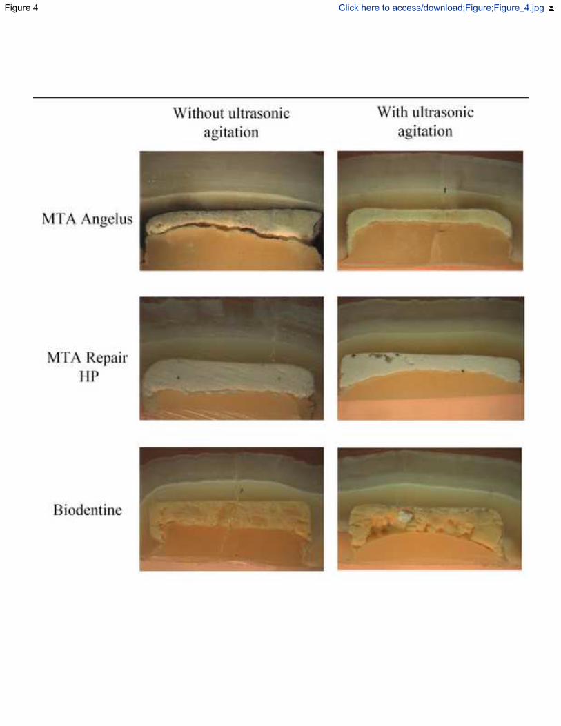
Figure 4 Click here to access/download;Figure;Figure_4.jpg

Table 2. Percentage of gaps in relation to the perimeter provided
by the repair materials as a function of the ultrasonic agitation.
Material Agitation Percentage of gaps
Median (min. - max.)
MTA-Angelus No 11.92 (0.0 – 48.65)a,A
Yes 5.34 (0.0 – 65.47)a,A
MTA Repair HP No 28.58 (8.01 – 63.73)a,A
Yes 17.87 (0.0 – 43.26)a,A
Biodentine No 10.14 (0.0 – 27.56)a,A
Yes 1.87 (0.0 – 16.33)a,A
a,bDifferent lowercase letters overlapped represent significant
differences between the groups according to the materials according to
the Kruskal-Wallis and Dunn tests (P <0.05).
Table 2
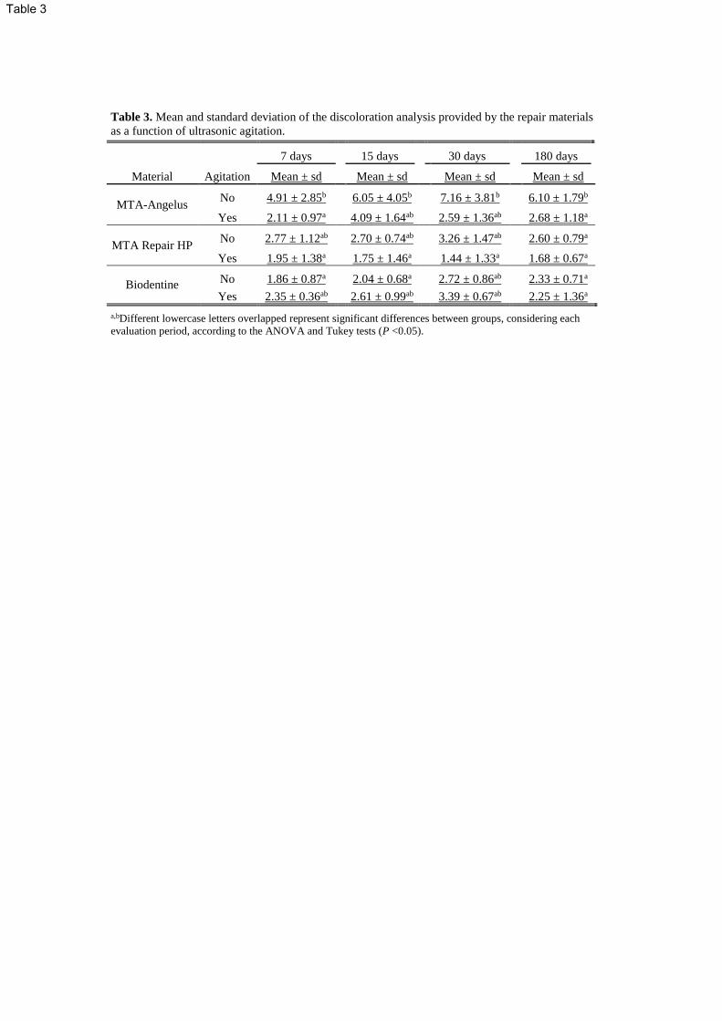
Table 3. Mean and standard deviation of the discoloration analysis provided by the repair materials
as a function of ultrasonic agitation.
Material Agitation
7 days 15 days 30 days 180 days
Mean ± sd Mean ± sd Mean ± sd Mean ± sd
MTA-Angelus No 4.91 ± 2.85b 6.05 ± 4.05b 7.16 ± 3.81b 6.10 ± 1.79b
Yes 2.11 ± 0.97a 4.09 ± 1.64ab 2.59 ± 1.36ab 2.68 ± 1.18a
MTA Repair HP No 2.77 ± 1.12ab 2.70 ± 0.74ab 3.26 ± 1.47ab 2.60 ± 0.79a
Yes 1.95 ± 1.38a 1.75 ± 1.46a 1.44 ± 1.33a 1.68 ± 0.67a
Biodentine No 1.86 ± 0.87a 2.04 ± 0.68a 2.72 ± 0.86ab 2.33 ± 0.71a
Yes 2.35 ± 0.36ab 2.61 ± 0.99ab 3.39 ± 0.67ab 2.25 ± 1.36a
a,bDifferent lowercase letters overlapped represent significant differences between groups, considering each
evaluation period, according to the ANOVA and Tukey tests (P <0.05).
Table 3
AUTHOR’S RESPONSE TO EDITOR/REVIEWERS
Dear Prof. Imad About, Ph.D.
Associate Editor of the Clinical Oral Investigations.
Thank you for allowing us to make the changes suggested by the Reviewers in order to qualify
this paper for possible publication in the Clinical Oral Investigations.
Below are some answers to the Reviewers comments:
Reply to Reviewers # 1
Initially thanks for your considerations and your commendation. Our response for your
concerns are listed below:
1. Concern of the Reviewer: Methods: What did the authors use for root canal preparation? diamond
bur, gates glidden or lentulo?? Please see page 8-line 46 and page 4-line 8. Is not it difficult to insert
the diamond bur up to the middle third of the root? I think, this part is a bit confusing.
Our response: For cervical and middle thirds preparation a Largo Peeso Reamer #5 (Dentsply-Sirona,
Ballaigues, Switzerland) was used in long axis of the teeth. Please, apologies for this typing error.
Revised text: Page 4, Lines 8 and 11: …was carried out, followed by preparation of the cervical and middle thirds
with Largo Reamer #5 (Dentsply-Sirona, Ballaigues, Switzerland). After the preparation specimens
were evaluated for circularity and standardization of the cavity diameter. Where the Largo Reamer #5
did not enter…
Page 8, Line 52: … and the cavity diameter were standardized with a Largo Reamer #5 (Dentsply-
Sirona).
2. Concern of the Reviewer: Table 1-Table 3: I suggest giving mean and standard deviation values in
one column in order to make table simple. Please see following examples;
mean ± sd
6.83± 1.99bc
For Table 2:
median (min - max)
11.92 (0 - 48.65) a,A
Our response: Following the guidance of the Reviewers the alterations was performed in Tables 1-3.
Revised text:
Revisions were highlighted in Tables 1-3.
3. Concern of the Reviewer: Could you please insert a scale bar on confocal images? Additionally,
the way to be outlined the region of interest (ROI) using the polygonal tool in one of the sections could
be stated if possible.
Our response: Regards of the scale bar, it was inserted in Confocal images at Figure 3 (Figure 1 in
original submission). With respect to the inclusion of measurement procedures performed in Image J
we do not understand that an illustration of it in Figure 3 was indicated; we think that it could turn
difficult images comprehension and group comparisons. Whereas, a text better explaining which were
Image J tools used was included in the text.
Revised text:
Authors' Response to Reviewers' Comments Click here to access/download;Authors' Response toReviewers' Comments;Point-to-

Page 5, Line 53: The total perimeter of the canals and the length of gaps were determined in
millimetres using the software polygonal and linear tools, respectively.
4. Concern of the Reviewer: Did you observe premature failure during push-out bond strength test?
Our response: No premature failures were observed during sample preparation for the push-out bond
strength test or during it. Considering this an important information a sentence stating it was inserted in
Results chapter.
Revised text:
Page 7, Line 36 and 37: Table 1 presents the results of the push-out test. Premature failures were not
observed in any groups/subgroups. The BIO/UA…
5. Concern of the Reviewer: I suggest adding a photograph of representative dentin discs showing
cohesive failure or other failure types, if it is available. Furthermore, authors could state the percentage
of failure types for each group (page 7-line 45).
Our response: Considering the asked revision, the percentages of failures types were inserted in Table
1 for better comprehension. Related to the failure types illustration, images were included (Figure 2).
Revised text: Following the guidance of the Reviewers Table 1 was improved.
Page 7, Line 49 to 51: The MTA/UA and BIO groups, with and without UA, presented a predominance
of the cohesive failures, and the MTAHP group presented predominance of the cohesive type as could
be observed in Table 1. Figure 2 presents the failure types illustration.
6. Concern of the Reviewer: Discussion: Please improve discussion with the explanation of why most
of the groups exhibited cohesive type of failure.
Our response: Related to this point, observed in UA and Biodentine groups, we understand that the
cohesive failures observed predominantly in the groups under ultrasonic agitation could be provided by
material better adaptation to the root canal walls. This adaptation increases the adhesive strength
making it superior than the cohesive strength of the materials.
Revised text: Page 9, Line 29 to 35: It is postulated that ultrasonic agitation produced an increase of the cement
pressure against the canal walls favouring a more effective filling of the irregularities, improving
adaptation to root canal walls and, consequently, their bonding resistance [19]. Considering this
observation, it could be suggested that this better adaptation increases the materials adhesive strength
making it superior than its cohesive strength.
7. Concern of the Reviewer: Are doi addresses necessary? Please check the journal reference style.
Following is an example of the reference of journal article;
Smith J, Jones M Jr, Houghton L et al (1999) Future of health insurance. N Engl J Med 965:325-329.
Our response: Taking account the Instruction for Authors accessed in December 26th the most
complete example available is:
Journal article:
Gamelin FX, Baquet G, Berthoin S, Thevenet D, Nourry C, Nottin S, Bosquet L (2009) Effect of high
intensity intermittent training on heart rate variability in prepubescent children. Eur J Appl Physiol
105:731-738. https://doi.org/10.1007/s00421-008-0955-8
Thus, considering the Instructions we think that was indicated to state the DOI number for all listed
References.
Revised text:

Not applicable.
Reply to Reviewer # 2
Thanks for considering this study clinically relevant and to permit us to explain our typing and
methodological options.
1. Concern of the Reviewer: My main major comment is: why didn't researchers consider this study
in two separate parts (adhesive/interface analysis and discoloration). Too many parameters in one paper
made the text confusing.
Our response: Honestly, at first, we thought of writing two papers considering the differences between
the methodologies employed. However, when we started writing the first of them, we understood that,
in clinical terms, one analysis could reinforce or compromise the other. We believe that it would be
ambiguous to conclude that the ultrasonic agitation would be favorable in terms of adaptation and bond
strength if it would compromise the color stability of the materials. Based on the above, we chose to
write a single paper, even more complex, but more complete and with greater clinical impact and
citation potential. We hope that this explanation could make sense, in any case, we are open to
suggestions.
Revised text:
Not applicable.
2. Concern of the Reviewer: Furthermore, one part used extracted teeth, the second part used bovine
teeth. The first part: the method is understandable; the second part (discoloration part) needs a
schematic drawing to be able to make the method understandable.
Our response: We agree the comprehension improvement that an Experimental Design could produce
in method description of Discoloration Evaluation. Thus, an image presenting it sequence was included
(Figure 1).
Revised text:
Not applicable.
3. Concern of the Reviewer: If the authors could divide the paper into two separate parts, they could
add SEM pictures from the failed samples.
Our response: This point was also commented by the Reviewer #1; representative images of the
failure types were now present in paper as Figure 2.
Revised text: Not applicable.
4. Concern of the Reviewers: …could make a more specific discussion and could give more
knowledge.
Our response: We understand the point of view highlighted by the Reviewer and tried to better
separate the Discussion related to each studied subject. Some information was really mixed in the
original submission. We think that after corrections and inclusion asked during this review that
confusion was mitigated.
Revised text:
Not applicable, spread throughout the discussion chapter.

5. Concern of the Reviewers: Abstract: needs revision. Method does not include the details of the
study groups;
Our response: During this paper preparation, the most difficult part to write was the Abstract, mainly
considering the recommendation in Authors Instructions to respect the number of 250 words. Thus,
considering the word limitation we opted to describe the groups in the Objective section, it was stated
as: “Objectives: The effect of ultrasonic agitation (UA) on bond strength and adaptation of cervical
plugs prepared with MTA-Angelus (MTA), MTA Repair HP (MTAHP) and Biodentine (BIO) was
evaluated.” – it intends to presents MTA, MTA/UA, MTAHP, MTAHP/UA, BIO, and BIO/UA. We
wish to better describe this group division; however, the word limitation makes such inclusion almost
impossible.
Revised text: Not applicable.
6. Concern of the Reviewer: Discussion: needs discussion of the differences between the test samples
(bovine and human teeth).
Our response: As requested by the Reviewer an extended Discussion about the method option for
bovine blocks and its advantages was inserted in the text.
Revised text:
Page 9, Line 12 to 22: The tooth discoloration was investigated using methods previously described in
the literature [25-27]. Even recognizing that the dental substrate employed is not exactly the same used
in bond strength and adaptation analyzes, it should be considered that it presents advantages in function
of, as opposed to human teeth that have a smaller surface and usually have restorations or caries that
can interfere in the color analysis, the bovine teeth provide a larger flat surface area, allowing adequate
and standardized color evaluation [26]. In addition, bovine teeth are similar to human teeth in the
composition of type I collagen of the organic matrix, which accredits the method for microstructural
issues considering that the interaction between radiopacifiers and this protein are blamed for the
darkening process [22, 26-28].