Embed Size (px)
Citation preview

Universidade Federal de Pernambuco
Laboratório de Imunopatologia Keizo Asami
Pós-Graduação em Biologia Aplicada à Saúde
DISSERTAÇÃO DE MESTRADO
JOSÉ ERITON GOMES DA CUNHA
Análise da frequência das variações M129V (rs1799990) e E200K (rs28933385)
do gene PRNP em pacientes brasileiros com Doença de Alzheimer
Recife, 2012

JOSÉ ERITON GOMES DA CUNHA
Análise da frequência das variações M129V (rs1799990) e E200K (rs28933385)
do gene PRNP em pacientes brasileiros com Doença de Alzheimer
Dissertação apresentada ao programa de
Mestrado em Biologia Aplicada à Saúde da
Universidade Federal de Pernambuco, como
parte dos requisitos para obtenção do grau de
mestre em Ciências Aplicada à Saúde.
Orientador: João Ricardo Mendes de Oliveira
RECIFE
2012

Catalogação na Fonte:
Bibliotecário Bruno Márcio Gouveia, CRB-4/1788 C972a
Cunha, José Eriton Gomes da
Análise da frequência das variações M121V (rs1799990) e E200K (rs2893385) e do gene PRNP em pacientes brasileiros com doença de Alzheimer / José Eriton Gomes da Cunha. – Recife: O Autor, 2012. 60 folhas: il., fig., tab.
Orientador: João Ricardo Mendes de Oliveira Dissertação (mestrado) – Universidade Federal de Pernambuco.
Centro de Ciências Biológicas. Pós-graduação em Biologia Aplicada à Saude, 2012.
Inclui bibliografia e anexos
1. Alzheimer, Doença de - 2. Variação (Biologia) 3. Polimorfismos (genética) I. Oliveira, José Eriton Gomes (Orientador) II. Título.
616.831 CDD (22.ed.) UFPE/CCB-2012-146

Análise da frequência das variações M129V (rs1799990) e E200K (rs28933385)
do gene PRNP em pacientes brasileiros com Doença de Alzheimer
José Eriton Gomes da Cunha
Banca Examinadora
Prof. Dr. João Ricardo Mendes de Oliveira
Orientador – UFPE / Dept. Neuropsiquiatria/LIKA
Prof.ª. Drª. Clélia Maria Franco
FIR / Dept. Neuropsiquiatria
Prof. Dr. Lucas André Cavalcanti Brandão
UFPE / Dept. Patologia
RECIFE
2012

Prefácio
“All my days I've waited for the sign
The one that brings me closer to isle of Avalon
I can feel the power flowing through my veins
My heart is beating louder, close to Avalon”
“Todos os meus dias eu espero o sinal
Aquele que me trará mais perto da Ilha de Avalon
Eu posso sentir o poder fluindo pelas minhas veias
Meu coração está batendo mais forte, perto de Avalon”
(Iron Maiden – Isle of Avalon)

Agradecimentos
Gostaria de deixar meus sinceros agradecimentos a todos aqueles que me
deram suporte, criticas, sugestões, forças e muito mais nesses dois anos de
mestrado. Tenho a maior dificuldade de demonstrar o quanto fico agradecido por
tudo, Filipe Santos que o diga!
Agradeço em especial à minha amada família por ter me dado educação,
carinho, amor, suporte, criticas e por me fazer quem eu sou como pessoa. Se
gostam de como eu sou, culpem eles! Obrigado por tudo seu Severino, dona Maria
Tereza, Elder, Elaine (minha mentora dessa língua que não sei muito bem, mesmo
após vinte e nove anos). Às minhas tias, de sangue, Elza, Biuzinha (chamem aí de
Severina, se tiverem coragem), Edileuza, Graça, Teresinha (minha pequena
bióloga), a guerreira Marli. Aos meus tios Roberto, Paulo, Manoel, Toninho, Zé e
aos primos e primas: Celina, Natália, William, Alcione, Lidiane (a única planta que
eu gosto), Henrique e a todos os meus primos e familiares de São Paulo.
Agradeço os amados e queridos professores. A todos que me deram tanto
apoio e vão me “aturar” por mais 4 anos! Como já disse a meu orientador, João
Ricardo M. de Oliveira, não há palavras para agradecer por tudo que ele me fez
desde 2007, muito obrigado mesmo. Aos professores José Luiz e Luiz Carvalho,
obrigado pelo conhecimento, conversas, ensinamentos não acadêmicos e
orientações. Às Professoras Dany e Rosângela pelo suporte.
Aos amigos, que são outra espécie de família, obrigado por terem me dado
tantos momentos fantásticos e com toda certeza aos próximos que virão. Espero
não esquecer nenhum nome, mas me perdoem, pois tenho MUITOS bons amigos.
Vou tentar dividir em grupos, para não me esquecer de ninguém!
Aos amigos da faculdade nos quais tive o prazer de cursar e chatear
SEMPRE com meus assuntos de príon (fazer o quê se ela é espetacular?).
Agradeço a todas as “tias” e amigos. Em especial, por ter mais afinidades, gostos e
saídas juntos, fora o grande carinho e amor: Caio e Filipe (Blood Brothers), Lívia,
Morgana, Sarinha e Cassinha.

Obrigado a todos do RPG. A primeira “turma”: Pedro, Damásio, Ogeid, Luiz
Carlos, Vicente (forças cara), Renatinho (infantaria, sempre), Biiinho, Meu e a
segunda “turma”: Berg, Rubão (sim, ele está vivo), KV, Trop, Piauí, Beça, Meu (de
novo).
Aos grandes amigos do amado LIKA (minha primeira ou segunda casa, não
faço ideia). A todos os que pertencem aos Laboratórios de Biotecnologia,
Biossensores, Virologia/Microscopia Eletrônica, Patologia, Bioquímica e
Microbiologia. E claro, ao meu adorável laboratório de Biologia Molecular
representado por Aíla, Jéssica Fragoso, Erik, Juan, Eliane, Marcela, Monique,
Marek, Juliana Nunes e aos demais.
Aos amados parceiros do grupo Psiquiatria Biológica: Danyllo, Lylyan,
Manuela, Roberta, Regina, Samy e Thalita, obrigado pelo carinho e pela amizade.
Obrigado aos amigos da ppg-LIKA que fizeram esses dois anos
maravilhosos e que contribuíram para esse produto final. Em especial a Sérgio e a
André pela ajuda nas “continhas”. Um agradecimento especial também para a
nossa mais do que secretária e grande amiga da ppg-LIKA, Eliete. Obrigado por
me aturar e me ajudar nesses anos.
Deixo meus agradecimentos às agências de fomento: Capes e CNPq pelos
financiamentos.
RESUMO
A Doença de Alzheimer (DA) e a Doença de Creutzfeldt-Jakob (DCJ) apresentam caráter neurodegenerativo, ocasionadas pelo acúmulo extracelular de proteínas insolúveis. Dentre alguns fatores de risco, as mutações nos códons M129V e E200K do gene PRNP são os mais importantes na DCJ. Alguns estudos foram realizados com o códon 129, sendo bastante controversos. Este trabalho teve como objetivo verificar a influência dos códons 129 e 200 e a presença da mutação E200K na DA. 145 amostras de DNA de pacientes com DA e 198 controles foram genotipadas para os códons 129 e 200 através da técnica da PCR e digestão enzimática com as enzimas NspI para o códon M129V e BsmAI, para o códon 200. Onze pacientes e oito controles foram sequenciados com o MegaBace1000. Quarenta e dois pacientes e quarenta e dois controles foram genotipados para o códon 129 sendo 16 MM, 20 MV e 6 VV nos pacientes e 24 MM, 15 MV e 3 VV nos controles. Não foram encontradas diferenças estatísticas significativas. Para a mutação E200K, 61 pacientes e 75 controles foram genotipados, não ocorrendo mutação em nenhuma amostra. Nossos dados corroboram com outros grupos mostrando que o códon 129 parece não estar associado a DA. A baixa frequência da mutação E200K pode justificar a ausência desta na nossa amostragem, o que precisa ser confirmado com maior número de amostras, com devida atenção pela possível estratificação associada a grupos étnicos e outros fatores como co-segregação de outros genes associados ao PRNP. Este trabalho contribuiu para um melhor conhecimento do perfil neuroepidemiológico da população local. Palavras chaves: DOENÇA DE ALZHEIMER; DOENÇA PRIÔNICA; PRÍON; VARIAÇÃO GENÉTICA; BRASIL.

ABSTRACT
Alzheimer's disease (AD) and Creutzfeldt-Jakob disease (CJD) are neurodegenerative disorders with some similar clinical findings and both are caused by the accumulation of insoluble proteins in the Brain. Some risk factors were already described such as genetic polymorphisms and mutations. The codon 129 and mutation E200K at PRNP gene are the most important CJD risk factor in studies with families and populations. Clinical and Biological similarities between these two neuropsychiatric conditions prompted various studies aiming to correlate the influence of codon 129 and the presence of the E200K mutation in AD. In this study we evaluated the variants frequency for the codon 129 and E200K mutation at PRNP gene in AD patients and controls of a Brazilian population. DNA samples from 145 patients and 198 controls were genotyped for codon 129 and 200 by PCR and enzymatic digestion with enzymes with NSPI and BsmAI, respectively. Eleven patients and eight controls were sequenced with MegaBace1000 for both variations. Forty-two patients and controls were genotyped for the codon 129. In patients, the genotype distribution was 16 homozygous for methionine, 6 homozygous for Valine and 20 heterozygous. The control group presented 24 homozygous for Methionine, 3 homozygous for valine and 15 heterozygous. There was no significant difference between groups. None of the samples from 61 patients and 75 controls presented the E200K mutation. Our data corroborate other groups studies showing that the codon 129 have no significant association with AD. The general low frequency of the E200K mutation may explain the absence of this variant in our sample, which must be confirmed with larger cohorts, with particular attention for possible stratification due to ethnicity and other important factors or even due to cosegregation with other genes linked to PRNP. Whole sequencing for PRNP gene will be necessary to clarify additional involvement of this marker and AD. This work contributed to a better understanding of the local population profile and increase more data for frequency of the polymorphism of codon 129 at PRNP gene in Brazilian population. Key Words: ALZHEIMER; DISEASE PRION; PRION; DISEASE; POLYMORPHISM; BRAZIL.

Sumário
Prefácio 4
Agradecimentos 5
Resumo 7
Abstract 8
1. Introdução 10
1.1 Novo perfil epidemiológico e Demências 10
1.2 Doença de Alzheimer
1.2.1 Genética da Doença de Alzheimer
11
12
1.3 Príon
1.3.1 Doenças Priônicas
1.3.2 Variações genéticas do PRNP
1.3.3 Príon e Doença de Alzheimer
13
16
17
18
2. Justificativas 19
3. Objetivos 19
3.1 Gerais 19
3.2 Específicos 20
4. Metodologia 20
5. Resultados 23
6. Discussão
7. Conclusão
8. Referências Bibliográficas
9. Anexo I
eTabela 4 – Frequência genotípica e alélica do codon 129 do PRNP em diversas populações humanas.
25
27
29
10. Anexo II
Punding as a transient symptom in a patient with an early onset form of Dementia
11. Anexo III
Resumos do 57º Congresso Brasileiro de Genética
12. Anexo IV
Produção acadêmica: Exaggerated blood pressure response during exercise treadmill testing:
functional and hemodynamic features, and risk factors
10
1. Introdução
1.1. Novo perfil epidemiológico e Demências
Ao longo do processo de evolução das populações humanas, vários fatores
contribuíram para o aparecimento e reconhecimento de diversas doenças. A
Revolução Agrícola, no período do Neolítico, permitiu aos agrupamentos tribais
mais complexos e numerosos, o aumento das doenças nutricionais,
infectocontagiosas, zoonoses e parasitárias, caracterizando, assim, a primeira
transição epidemiológica1.
Os aprimoramentos na saúde pública, principalmente durante os séculos
XIX e XXI, proporcionaram declínio de várias doenças infecciosas, especialmente
com o advento das vacinas, melhorias na higiene pessoal e do sanitarismo 1,2.
Esses benefícios se refletiram na queda da mortalidade e no aumento significativo
da expectativa média de vida, passando de 58 anos, entre 1970 e 1975, para 67
anos entre 2005 e 2010, segundo a Organização das Nações Unidas. Em países
mais desenvolvidos esse índice chega, atualmente, a 77 anos3.
De acordo com o Instituto Brasileiro de Geografia e Estatística (IBGE), a
expectativa de vida da população brasileira era de 71.3 anos em 2003, sendo a
86ª maior expectativa de vida no mundo, de acordo com a Organização das
Nações Unidas4.
Esse novo perfil demográfico e melhorias em alguns parâmetros de saúde
pública pavimentaram a segunda transição epidemiológica que se caracteriza,
entre outros fatores, pelo aumento dos casos de câncer, demências diversas e
coronariopatias5.
A idade continua sendo o maior fator de risco, em especial nas doenças
demenciais, como Alzheimer, Parkinson, Huntington, Doenças Priônicas, entre
outras, afetando frequentemente adultos tardiamente. A maior parte dos casos
são esporádicos (sem fonte conhecida de contágio ou mutações genéticas), mas
fatores genéticos e iatrogênicos são reportados para boa parte dessas doenças6,7.
A demência é clinicamente caracterizada pelo declínio de múltiplas funções
cognitivas, tais como memória, praxia e linguagem. Dependendo da etiologia e do
grau de comprometimento, a demência pode ter seu quadro revertido8.
11
As demências podem ser ocasionadas por vários fatores, tais como
síndromes metabólicas (Síndrome de Cushing.); Intoxicação (Demência Alcoólica
e outras drogas psicóticas.); doenças neurodegenerativas (Doença de Alzheimer
(DA), Doença de Pick, Demência por corpos de Lewy); ou doenças infecciosas
(AIDS, Neurosífilis, Doença de Creutzfeldt-Jakob (DCJ))9.
A incidência dessas doenças demenciais é de 1,5% ao ano na população
na faixa etária em torno de 60 anos. Este índice pode chegar a 30% na população
acima dos 80 anos. É menos frequente em homens e em populações africanas e
asiáticas. As causas dessas doenças também variam conforme as populações,
sendo a Doença de Alzheimer mais comum na europeia e a Demência Vascular
mais incidente na China10.
1.2. Doença de Alzheimer
Em novembro de 1906, Alois Alzheimer apresentou ao mundo o caso da
paciente Auguste D na 37ª Conferência da South-West German Psychiatrists. A
paciente em questão, com 51 anos, apresentava disfunção cognitiva progressiva,
alucinações, entre outros sintomas. Apenas no ano seguinte, com o trabalho
intitulado “A characteristc serious disease of the cerebral cortex” ele descreveu
hispotatologicamente o caso de Auguste D11.
A DA corresponde a pelo menos metade dos casos de demência no
mundo, com incidência estimada em 4.4% em pessoas com 65 anos de idade e
de 22% aos 90 anos. A idade e histórico familiar positivos são os dois maiores
fatores de risco12.
Atualmente, de acordo com o Código Internacional de Doenças - 10 (CID-
10), a DA é classificada como doença neurodegenetariva primária, com etiologia
ainda desconhecida. Tem caráter progressivo, com curso clínico que pode variar
de poucos anos (2 a 4) até décadas. De acordo com a idade de início da doença,
pode ser subdividida em forma precoce (Early Onset Alzheimer’s Disease –
EOAD), antes dos 65 anos, ou em forma tardia (Late Onset Alzheimer’s Disease –
LOAD), mais incidente que acomete pessoas acima dos 65 anos13.

12
Para o diagnóstico definitivo para a DA, o paciente deve preencher alguns
critérios tais como ter demência, desde que não se tenha evidência de ser
causada por deficiência em vitamina B12, Niacina, presença de neurosílis ou
outras fontes conhecidas; início insidioso de lenta progressão; e ausência de
início súbito, provocado por lesões focais, por exemplo. Para o diagnóstico
diferencial, devem-se considerar transtornos depressivos; delirium; demência
vascular; outras demências primárias como a frontotemporal (doença de Pick) e
DCJ13.
Na década de 80 do século passado, foi criado um grupo com o intuito de
estabelecer critério universal e assim caracterizar clinicamente a DA. Em
princípio, a essa doença se caracterizava pela presença de demência, avaliada
com testes padronizados, como o Mini Mental State Examination (MMSE);
prejuízo da memória e outras funções cognitivas como linguagem, percepção,
aparecer em idade entre 40 a 90 anos; entre outros fatores14.
Vale ressaltar que a DA é heterogênea, o que dificulta uma avaliação no
início da doença. Além disso, podemos encontrar sintomas pontuais em alguns
pacientes, o que foi debatido no artigo gerado a partir da análise clínica de um
dos nossos pacientes do HC-UFPE. Este trabalho encontra-se no anexo II.
Entretanto, como ressalta o neurologista Ricardo Nitrini, algumas
peculiaridades do nosso país fazem com que alguns exames e testes para avaliar
o quadro suspeito de DA devem ser analisados de forma adaptada de acordo com
nossa realidade, principalmente por conta das diferenças na escolaridade15.
1.2.1. Genética da Doença de Alzheimer
Durante os últimos 15 anos, diversos trabalhos foram feitos sobre a DA,
principalmente em busca de marcadores genéticos. O banco de dados Alzgene
(www.alzgene.org) foi criado para facilitar a análise desse montante de dados,
disponibilizando em seu site mais de 300 polimorfismos em diversos genes, mas
apenas alguns confirmados de forma independente16.
A forma EOAD corresponde a cerca de 10% dos casos de DA, possui
herança autossômica dominante e algumas mutações se destacam em três

13
genes: proteína precursora amilóide (APP), presenilina-1 (PSEN-1) e presenilina-
2 (PSEN-2), localizados nos cromossomos 21, 14 e 1 respectivamente17,.
A forma tardia é mais complexa, com herança multifatorial sendo
influenciada por componentes genéticos e ambientais. O principal fator de risco é
o alelo 4 da Apolipoproteína E (APOE), localizado no cromossomo 1918. O gene
da APOE possui três isoformas: 2; 3 e 4. Enquanto que a isoforma 2 agiria
como um fator de proteção, a 4 estaria envolvida na formação das placas senis
desencadeando o processo neurodegenerativo GEORGE-HYSLOP18.
Oliveira et al. reportou os principais fatores de risco na população
brasileira, reunindo dados de estudos de ligação e caso-controle. Além dos
polimorfismos já descritos, outros foram estudados e estão ilustrados na tabela
abaixo16.
Tabela1. Resumo dos estudos de associação caso-controle na população Brasileira
Referência Polimorfismos Nível de significância
Tamanho da amostra
Oliveira et al. (1997)
ApoE-ε4
P < 0,05
57
Oliveira et al. (1998)
5-HTTLPR
P < 0,05
81
Nishimura et al. (2000)
Nishimura et al. (2001)
DXS1047
D10S1423
P > 0,05
P > 0,05
130
130
Nishimura et al. (2004)
BDNF (C-270T) BDNF (C-270T) + ApoE-ε4
P > 0,05 P < 0,05
188
Nishimura et al. (2005)
MAOA MAOA + ApoE-ε4+
5-HTTLPR
P > 0,05
P < 0,05
128
Dados não publicados IL-1α and IL-1β
ApoE-ε4 P >0,05 P <0,05
195 195
Fernandez et al. (2005)
MTHFR (C-677T)
ApoE-ε4
P > 0,05 P < 0,05
30
da Silva et al. (2006)
M HFR (C-677T)/(A-1298C)
P > 0,05
43
1.3. Príon
Comumente se refere à proteína príon para designar o “agente etiológico”
das Encefalopatias Espongiformes Transmissíveis (EET). Entretanto, a príon
possui duas isoformas, sendo apenas uma delas patogênica e denominada príon

14
scrapie (PrPsc). A outra, denominada príon celular (PrPc) é encontrada
normalmente em vertebrados e algumas “prions like” em leveduras19,20,21.
A PrPc possui 3 α-hélices na região globular (correspondente aos resíduos
144-154; 173-194; 200-228) e duas folhas ß-antiparalelas (128-131 e 161-164).
Pode ser encontrada de forma não glicosilada, monoglicosilada ou diglicosilada,
ocorrendo nos aminoácidos de asparagina (181 e 197); Possui uma ponte de
disulfeto entre os aminoácidos de cisteína (179 e 214). A figura 1 abaixo
representa a estrutura terciária da PrPC e as principais mutações associadas às
doenças priônicas.
Figura 1 – Estrutura tridimencial da PrPc mostrando as principais mutações associadas às doenças priônicas. HC- Porção hidrofóbica. HÁ - HC – formação em α – hélice. S1 e S2 – formação em folhas β-pregueadas. S-S – Ponte de disulfeto. (figura retirada de Capellari et al22. Uso da imagem autorizado pela revista: License Number – 2833820137731).
Apesar de ainda não ser totalmente conhecida, sabe-se que a PrPc
participa de diversos eventos celulares que são desde regulação, direta ou
indireta, da apoptose, sinalização celular, proteção neuronal e tendo uma forte
evidência do controle do estresse oxidativo23.
A PrPc é sintetizada a partir do gene PRNP, que em humanos está
localizado no braço curto do cromossomo 20, com 16 kb e 2 éxons. A ORF (open
reading frame) está localizada no segundo éxon e codifica uma proteína de 253
aminoácidos. Na figura 2 abaixo, vemos uma representação esquemática do gene
PRNP evidenciando as principais mutações associadas às doenças priônicas22.

15
Figura 2 – Esquema do gene PRNP evidenciando as principais mutações associadas às doenças priônicas. R1 a R4 representa mutações do tipo inserção deleção OPR. HC- Porção hidrofóbica. HÁ - HC – formação em α – hélice. S1 e S2 – formação em folhas β-pregueadas. S-S – Ponte de disulfeto. Círculos cinzas – sítios de glicosilação. (figura retirada de Capellari et al22. Uso da imagem autorizado pela revista: License Number – 2833820137731).
Diferente da PrPc, a PrPsc é insolúvel, resistente a proteases e diversos
detergentes, além das altas temperaturas, tendendo a formar fibrilas
naturalmente, sendo ricas em folhas β-pregueadas. Apesar de quase não haver
dúvidas sobre a relação da PrPsc com as doenças priônicas, a forma de
“conversão” da forma fisiológica para a patogênica ainda é o grande desafio na
biologia da príon24.
Para Stanley B. Prusiner, essa conversão se daria através de evento da
habilidade da PrPsc induzir a mudança estrutural da PrPc através de interações
bioquímicas. Outros autores sugerem que a interação da PrPc com outra proteína,
“proteína x” resultaria em um composto instável que acabaria transformando a
PrPc em PrPsc, desfazendo a interação com a proteína x25,26.
Outros grupos, inclusive da Universidade Federal do Rio de Janeiro
(UFRJ), liderada pelo pesquisador Jerson L. Silva postula que essa mudança se
daria da interação da PrPc com ácidos nucléicos, possivelmente com o RNA e
essa interação, através de mudanças na entropia da PrPc ocasionaria na
alteração estrutural. Apesar desses estudos, ainda não há um consenso de como
se dá essa mudança, sendo um dos principais desafios na biologia da príon27,26.

16
1.3.1. Doenças Priônicas
As Encefalopatias Espongiformes Transmissíveis (EET) ou doenças
priônicas, formam um grupo único de doenças que podem ser esporádicas,
genéticas ou adquiridas, e possuem um amplo espectro fenotípico, guardando em
comum o aspecto espongiforme do tecido nervoso e depósito extracelular da
PrPsc observado na figura 3 abaixo28.
Figura 3 – A: Corte histológico do córtex frontal de um paciente com Doença de Creuttzfeldt-Jakob (DCJ) evidenciado o aspecto espongiforme do tecido nervoso. A seta mais clara aponta para um neurônio atrofiado enquanto que a seta preta mostra uma célula da glia. B: Imunohistoquímica com anticorpo anti-PrPsc mostrando um intenso depósito protéico.
Dentre as principais EET podemos citar: Doença Crônica Debilitante do
Cervídeo (CWD); Encefalopatia Espongiforme Bovina – “mal da vaca louca”, em
bovinos (BSE); Scrapie, em ovelhas e cabras; Doença de Creutzfeldt-Jakob
(DCJ), Doença de Gerstmann-Straüssler-Scheinker (GSS), Insônia Fatal (IF) e
Kuru em humanos29.
A doença de Creutzfeldt-Jakob (DCJ) apesar de rara, é a doença priônica
mais frequente, sendo descrita em diversos países, inclusive no Brasil. No
entanto, são escassas as informações no país sobre esta doença e, em
publicação recente, foi contestada a atual política de vigilância epidemiológica
para doença priônica excessivamente voltada para poucos centros que realizam
as técnicas necessárias para a adequada confirmação dos casos suspeitos30.
A DCJ possuí incidência de 1 caso a cada 1 milhão de habitantes, afeta
homens e mulheres na mesma proporção e ocorre na faixa dos 55 a 65 anos. É
causada pelo acúmulo da príon scrapie nos neurônios. Pode ser adquirida,
genética ou esporádica, sendo a última a forma mais prevalente,
aproximadamente 85% dos casos31.

17
Clinicamente, a DCJ é caracterizada por demência progressiva com
duração menor que dois anos, mioclonias, ataxia e outros sintomas. O diagnóstico
é feito associando o quadro clínico seguido de exames de ressonância magnética
(RM), eletroecenfalograma (EEG) e pesquisa no líquor da proteína 14-3-3.
Entretanto, na prática clínica reconhecer pacientes com DCJ requer muito
cuidado e habilidade do médico, visto que os sinais e achados de exames para a
DCJ se confundem com outras doenças neurológicas. Por esta razão, Michael
Geschwind da Universidade de São Francisco, Califórnia, a chama de “great
mimicker” o que pode ser ilustrado no exemplo do próprio Creuztfeldt que,
segundo o mesmo, seus casos nada tinham em comum com os casos de Jakob,
e até mesmo alguns dos primeiros casos de Jakob não seriam hoje classificados
como DCJ32.
Acredita-se que essa semelhança com outras doenças não seja apenas
coincidência e até se levanta a hipótese de que a príon participe direta ou
indiretamente da patogenia de outras doenças neurodegenerativas como
Alzheimer e Parkinson33.
1.3.2. Variações genéticas da PRNP
Mais de 20 mutações no PRNP já foram descritas em diversas populações,
sendo P102L, D178N, E200K, e V210I as mais comuns, além do polimorfismo do
códon 129 (M129V), sendo este último o mais importante no estudo das EET,
modulando diversos aspectos, tais como tempo de incubação e outros aspectos
clínicos 34.
O polimorfismo do códon 129 resulta no resíduo de aminoácido metionina
(selvagem) ou valina (mutante). A frequência alélica, na população saudável,
varia entre as diversas populações humanas, podendo ser de 0.653 na população
britânica a 0, 958 na Japonesa para o alelo M. A tabela 2 abaixo mostra as
frequências deste polimorfismo em vários grupos étnicos. No anexo 1, encontra-
se a tabela completa do NCBI35.
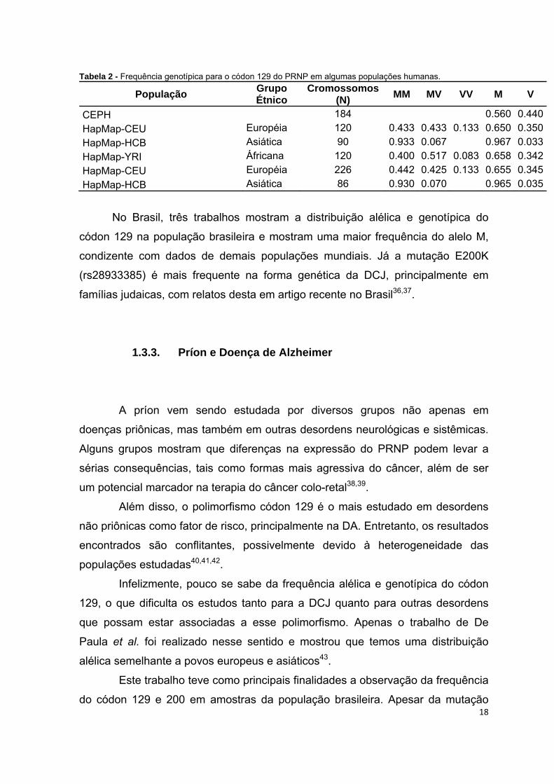
18
Tabela 2 - Frequência genotípica para o códon 129 do PRNP em algumas populações humanas.
População Grupo Étnico
Cromossomos (N)
MM MV VV M V
CEPH 184 0.560 0.440
HapMap-CEU Européia 120 0.433 0.433 0.133 0.650 0.350
HapMap-HCB Asiática 90 0.933 0.067 0.967 0.033
HapMap-YRI Áfricana 120 0.400 0.517 0.083 0.658 0.342
HapMap-CEU Européia 226 0.442 0.425 0.133 0.655 0.345
HapMap-HCB Asiática 86 0.930 0.070 0.965 0.035
No Brasil, três trabalhos mostram a distribuição alélica e genotípica do
códon 129 na população brasileira e mostram uma maior frequência do alelo M,
condizente com dados de demais populações mundiais. Já a mutação E200K
(rs28933385) é mais frequente na forma genética da DCJ, principalmente em
famílias judaicas, com relatos desta em artigo recente no Brasil36,37.
1.3.3. Príon e Doença de Alzheimer
A príon vem sendo estudada por diversos grupos não apenas em
doenças priônicas, mas também em outras desordens neurológicas e sistêmicas.
Alguns grupos mostram que diferenças na expressão do PRNP podem levar a
sérias consequências, tais como formas mais agressiva do câncer, além de ser
um potencial marcador na terapia do câncer colo-retal38,39.
Além disso, o polimorfismo códon 129 é o mais estudado em desordens
não priônicas como fator de risco, principalmente na DA. Entretanto, os resultados
encontrados são conflitantes, possivelmente devido à heterogeneidade das
populações estudadas40,41,42.
Infelizmente, pouco se sabe da frequência alélica e genotípica do códon
129, o que dificulta os estudos tanto para a DCJ quanto para outras desordens
que possam estar associadas a esse polimorfismo. Apenas o trabalho de De
Paula et al. foi realizado nesse sentido e mostrou que temos uma distribuição
alélica semelhante a povos europeus e asiáticos43.
Este trabalho teve como principais finalidades a observação da frequência
do códon 129 e 200 em amostras da população brasileira. Apesar da mutação

19
E200K ser a mais frequente na DCJ, principalmente em famílias judaicas, essa
nunca foi descrita em pacientes com doença de Alzheimer em estudos prévios.
Entretanto, poucos estudos no Brasil foram feitos com o PRNP, mesmo para as
doenças priônicas.
2. Justificativa
Este tipo de estudo é de grande importância, principalmente se
considerarmos o aumento progressivo e contínuo da incidência deste transtorno
neuropsiquiátrico em todo o mundo, inclusive no Brasil, onde coincidentemente
são realizadas relativamente poucas pesquisas sobre o tema. Além disso, a
análise desenvolvida neste projeto poderá auxiliar tanto na definição de métodos
diagnósticos mais rápidos e eficazes como na melhor compreensão da DA,
abrindo novas perspectivas para o diagnóstico e tratamento, principalmente se
considerando as atuais evidências que sugerem a sobreposição da DA e a DCJ.
3. Objetivos
3.1.1. Geral
• Mostrar a distribuição do códon 129 e E200K do gene PRNP na
população estudada e comparar com dados obtidos previamente no
Brasil e em outros países.
3.1.2. Específico
• Investigar a possível associação dos polimorfismos do códon M129V e
E200K do PRNP com a Doença de Alzheimer.
20
4. Metodologia
Foram utilizadas 145 amostras de DNA de pacientes com DA provável ou
possível, diagnosticados clinicamente, e 205 controles, sendo 63 oriundos de
coletas realizadas em Pernambuco (PE) e 82 do Espírito Santo (ES). 20 amostras
controles eram provenientes de PE e as demais do ES14.
Para a genotipagem do PRNP foram utilizados dois pares de primers que
cobriam todo o quadro de leitura do gene. Os pares foram os seguintes: primeiro
par Fw1 - 5'-CTGACGTTCTCCTCTTCATTTTG-3' e Rv1 - 5'-
CTCATGGCACTTCCCAGCATGTA-3' e o segundo par Fw2 - 5'-
AACCAACATGAAGCACATGG-3' e Rv2 - 5'-TCCCTCAAGCTGGAAAAAGA-3'.
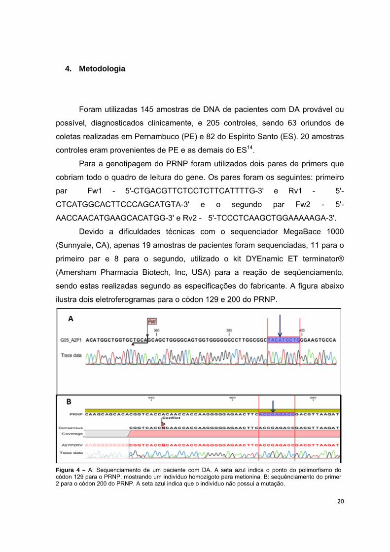
Devido a dificuldades técnicas com o sequenciador MegaBace 1000
(Sunnyale, CA), apenas 19 amostras de pacientes foram sequenciadas, 11 para o
primeiro par e 8 para o segundo, utilizado o kit DYEnamic ET terminator®
(Amersham Pharmacia Biotech, Inc, USA) para a reação de seqüenciamento,
sendo estas realizadas segundo as especificações do fabricante. A figura abaixo
ilustra dois eletroferogramas para o códon 129 e 200 do PRNP.
Figura 4 – A: Sequenciamento de um paciente com DA. A seta azul indica o ponto do polimorfismo docódon 129 para o PRNP, mostrando um indivíduo homozigoto para metionina. B: sequênciamento do primer2 para o códon 200 do PRNP. A seta azul indica que o indivíduo não possui a mutação.

21
Devido a estas importantes dificuldades, mudamos a abordagem para o
uso de duas enzimas de restrição para genotipar o códon 129 e 200. Para tal,
usamos o Primer Fw1 e o Rv2 utilizando a ciclagem: 95º por 3 minutos, seguido
de 30 ciclos de 95º por 30 segundos; 60º por 30 segundos; 72º durante 1 minuto e
extensão final de 72º por 5 minutos. O DNA estava em uma concentração de
30ng, amplificando um produto com 850 pares de base (pb) que contempla
ambos os códons. A figura abaixo mostra um segmento deste produto, mostrando
os sítios de restrição das duas enzimas, mantendo a contagem original das
bases.
Figura 5 – Fragmento do produto de PCR evidenciado os dois sítios de restrição para a enzima NspI (códon 129 do gene PRNP) e um sítio para a BsmAI (códon 200 do gene PRNP)
Para o códon 129, foram utilizados 10µl do produto de PCR com 10
unidades de enzima com tampões e concentrações indicadas pelo fabricante. Em
seguida, as amostras foram separadas por eletroforese em gel de agarose a 1,5%
corados com brometo de etídio. Em indivíduos homozigotos para metionina
observamos os fragmentos de banda de 423, 352 e 75 pb; Indivíduos
heterozigotos apresentavam as bandas de 498, 423, 352 e 75 pb enquanto que
homozigotos valina apresentavam as bandas 498 e 352 pb.

22
O mesmo foi feito para o códon 200, usando a enzima BsmAI (Biolabs),
mas agora utilizando gel de agarose a 1%. A enzima reconhece um sítio de
restrição apenas em indivíduos sem a mutação, resultando em dois fragmentos
de 628 e 222 pb.
Figura 7 - Imagem de uma corrida de eletroforese em gel de agarose a 1% da digestão com BsmAI. 2 a 11 representam amostras de pacientes com DA sem a mutação E200K apresentando as bandas de 628 e 222 pb. M = ladder; F = fragmento sem ser digerido.
De posse das frequêcias alélicas e genotípicas das nossas amostras,
comparamos com dados publicados em trabalho recente de Smid et al (2011) que
fez o mesmo trabalho que o nosso, mas varrendo todo o PRNP e com amostras
unicamente de SP além de dados publicados no Alzgene.
Além disso, comparamos os nossos controles com dados previamente
publicados por de Paula et al (2005) e do artigo de Beck et al. (2010).
Figura 6 - Imagem de uma corrida de eletroforese em gel de agarose a 1,5% da digestão com NspI mostrandoos padrões de bandas para os genótipos MM (6,9 e 10), MV (2,3,4,5,7 e 8) e VV (11). Os retângulos laranja,verde e zul representam as bandas de 498, 423 e 352 pb, respectivamente. M = ladder marcador de pesomolecular.

23
5. Resultados
Das 145 amostras de pacientes e 205 controles, 42 amostras de cada
grupo foram genotipadas para o códon 129 do PRNP. As duas populações
respeitam o equilíbrio de Hardy-Weinberg, com p = 0,95 nos pacientes com DA e
p=0,75 nos controles para os genótipos do códon 129 do PRNP. Já para o códon
200, deste mesmo gene, 61 pacientes e 75 controles foram genotipados, não
sendo encontrada nenhuma amostra com a mutação. Na tabela 3 abaixo vemos a
distribuição genotípica do códon 129 e 200 de pacientes com DA e controles.
Tabela 3 – Distribuição genotípica dos códons 129 e 200 do PRNP nos pacientes com DA e controles. Números entre parênteses representam valores porcentuais aproximados.
Códon Genótipo DA (%) Controles (%) P
129
MM MV VV
16 (38,1)20 (47,7)6 (14,2)
24 (57,1) 15 (35,7)
3 (7,2)
0,187*
200 EE EK KK
61 (100,0)00
75 (100,0) 0 0
-
DA = doença de Alzheimer; M = metionina; V = valina; p = valor de p; * = teste G
A frequência dos alelos M e V também não mostraram diferença
significativa entre os pacientes com DA e controles. No primeiro grupo, a
frequência foi de 0,62 e 0,38 para os alelos M e V, respectivamente e nos
controles de 0,75 e 0,25.
Ao comparar nossos dados com os de Smid, também não observamos
diferença significativa entre pacientes com DA e controles. A tabela abaixo mostra
a distribuição dos genótipos para o códon 129 do PRNP tanto do nosso trabalho
quanto os de Smid44.
As frequências alélicas de M e V deste trabalho comparado com o de Smid
também não tiveram diferença significativa, com p = 0, 57.

24
Tabela 4 - Comparação genotípica do códon 129 do PRNP entre dados deste trabalho com resultados de Smid (2011).
Genótipo DA (%) p Controles (%) P
Gomes da Cunha (2012)
MM MV VV
16 (38,1)20 (47,7)6 (14,2)
0,72*
24 (57,1) 15 (35,7)
3 (7,2)
0,15*
Smid (2011) MM MV VV
45 (45,5)42 (42,4)12 (12,3)
44 (39,6) 56 (50,5) 11 (9,9)
DA = doença de Alzheimer; M = metionina; V = valina; p = valor de p; * = teste G
O trabalho apresentado por Smid também não mostrou a presença da
mutação E200K associada nem com pacientes com DA nem com amostras
controles.
As distribuições genotípica e alélica dos controles deste trabalho
comparado com resultados do estudo de De Paula et al (2005) também não
mostrou diferença significativa com p = 0,34 para os genótipos e p = 0,14 para os
alelos.
6. Discussão
Neste presente estudo, não foi detectada a mutação E200K em amostras
de pacientes nem nos controles. Apesar de ser a mutação mais frequênte na DCJ
familiar, com casos descritos no Brasil, esta não foi relatada em pacientes com
DA até o presente momento, provavelmente devido a sua baixa frequência ou
mesmo por talvez apresentar associação restrita na não DA37.
Não houve diferença significativa entre pacientes com DA e controles para
o códon 129 do PRNP, o que já foi relatado em alguns outros trabalhos. A tabela
abaixo mostra o resumo de alguns trabalhos disponíveis no banco de dado do
Alzgene em que podemos observar que a maior parte deles não aponta influência
deste polimorfismo com a DA.

25
Tabela 5 - Resumo dos estudos publicados que analisaram a influência do códon 129 do PRNP em diversas populações (Modificado do Alzgene, www.alzgene.org).
Estudo População DA (N) Controles (N) Resultado Casadei, 2001
Itália
212
201
Tendência
Combarros, 2000 Espanha 278 268 Negativo
Cousin, 2009 França 428 475 Negativo
Del Bo, 2005 Itália 258 318 Negativo
Dermaut, 2003 Holanda 123 282 Positivo
Gacia, 2006 Polônia 166 194 Positivo
Golanska, 2009 Polônia 213 171 Positivo
Li, 2005 EUA 281 415 Negativo
Li, 2008 Canadá 753 736 Tendência
Poleggi, 2008 Itália 195 124 Negativo
Poleggi, 2008 EUA 109 58 Negativo
Reiman, 2007 EUA, Holanda 861 550 Negativo
Riemenschneider, 2004 Alemanha 482 911 Positivo
Ahn, 2006 Coréia 297 217 Negativo
Jeong, 2007 Coréia 271 236 Negativo
Jeong, 2009 Coréia 152 268 Negativo
Ohkubo, 2003 Japão 548 466 Negativo
Nossos dados mostram frequências alélicas e genotípicas semelhantes as
já descritas para a população brasileira nos trabalhos de Smid, Martins et al.
(2007) e de Paula et al (2005) 36, 44, 43.
Considerando que as amostras de indivíduos saudáveis dos trabalhos de
De Paula et al, Vilma et al e Smid possuem são amostras distintas, pelo que
descrevem suas metodologias, bem como as nossas, temos um pool de 546
amostras com a distribuição genotípica de 49,1% para indivíduos MM; 44,7
indivíduos MV e 6,2% indivíduos VV. O que nos mostra um perfil molecular
semelhante aos descritos em populações européias, mas diferente das asiáticas.
Por conta da falta dos dados epidemiológicos das amostras, tais como
idade média dos primeiros sintomas, proporção de homens e mulheres além de
outros dados genéticos, não efetuamos mais análises para comparar a influência
conjunta do PRNP com outros fatores de risco, como as formas EOAD e LOAD,
bem como o alelo Ɛ4 da APOE45.
Apesar de nossos resultados não apontarem para uma associação do
códon 129 do gene PRNP como fator de risco para a DA, o ampliamento de
estudos mostrando a distribuição genotípica e alélica deste polimorfismo é de
suma importância devido a sua associação como fator de risco para as doenças

26
priônicas ainda pouco conhecida no Brasil por fatores já discutidos por nosso
grupo30.
Além disto, esse mesmo polimorfismo já foi relacionado a outras
desordens, tendo influência no curso clínico de algumas doenças, como a de
Wilson, algumas formas de Parkinson entre outras44.
Entretanto, tanto nossos controles quanto os dos outros artigos possuem
distribuição genotípica e alélica diferente do que é apresentado no aritgo de Beck
et al., que mostra uma frequência do alelo V maior do que do alelo M34.
Esta diferença encontrada em Beck et al. pode estar relacionada com o
perfil das populações usadas neste artigo, que se trata de dois grupos indígenas
(Suruí (Paiter) e Karitiana) um número pequeno das duas populações
estudadas34.
7. Conclusão
Os dados apresentados neste trabalho não mostraram uma diferença
estatística significativa entre as frequências genotípicas e alélicas do códon 129
do PRNP entre os pacientes com DA e controles. Estes dados corroboram com
outros autores, inclusive um recente trabalho em que foi investigado esse
polimorfismo na população brasileira.
Essa ausência na associação de risco do códon 129 do PRNP na população
brasileira pode estar associada a diferenças étnicas da nossa população bem
como outros fatores de risco genético associados à DA.
Entretanto, nossos resultados mostraram que a distribuição alélica e
genotípica da variação em questão se dá de forma semelhante a diversas
populações mundiais, principalmente européias o que é condizente com a história
de colonização do nosso país.
Ao se comparar nossos dados com outros em que foram feitas análises
semelhantes na população brasileira, não observamos diferença significativa, o
que mostra que a nossa amostra é representativa da população brasileira.
Por fim, não houve o relato da mutação E200K nem em indivíduos com DA
nem dos controles o que pode ser explicado pela sua baixa frequência na

27
população mundial além do fato desta poder ter apenas uma relação com as
doenças priônicas.
Para reforçar nossos resultados, iremos dar sequência com as demais
amostras que temos, tanto de pacientes com DA quanto de controles, para ter
uma melhor conclusão desses resultados. Além disso, uma abordagem
epidemiológica será adicionada aos resultados moleculares para ver se há
diferença ao estratificarmos a nossa amostra.
Nosso estudo serviu para delimitar melhor o perfil molecular da população
brasileira para o códon 129 do PRNP, o que é de grande relevância para estudos
de doenças priônicas que ainda é muito negligenciada no Brasil, bem como
contribui para ampliação do debate deste polimorfismo como fator de risco para a
DA.

28
8. Referências Bibliográficas
1. Armelagos, G.J., Barnes, K.C. & Lin, J. Disease in human evolution: the re-emergence of infectious disease in the third epidemiological transition. National Museum of Natural History Bulletin for Teachers 18, 1-6 (1996).
2. Rodrigues, S.P. As Contribuições de Galtier e Pasteur para a Teoria Microbiana Das Doenças The Contributions of Galtier and Pasteur to the Microbian Theory of Disease. IV Simpósio Nacional de Tecnologia e Sociedade (2011).at <http://www.esocite.org.br/eventos/tecsoc2011/cd-anais/arquivos/pdfs/artigos/gt002-ascontribuicoes.pdf>
3. Affairs, D. of E. and S. & Population World Population Prospects: The 2006 Revision, Highlights. ONU (2007).at <http://scholar.google.com/scholar?hl=en&btnG=Search&q=intitle:World+Population+Prospects+The+2006+Revision+Highlights#0>
4. IBGE Tábuas Completas de Mortalidade 2003. (2004).at <http://www.ibge.gov.br/home/presidencia/noticias/noticia_visualiza.php?id_noticia=266>
5. Revolution, T.T. The Third Revolution : The Convergence of the Life Sciences , Physical Sciences , and Engineering. Engineering
6. Tsuji, S. Genetics of neurodegenerative diseases: insights from high-throughput resequencing. Human molecular genetics 19, R65-70 (2010).
7. Johnson, R.T. Prion diseases. Lancet neurology 4, 635-42 (2005).
8. J, Sadock, B. & Sadock, V.A. Compêndio de Psiquiatria Ciência do Comportamento e Psiquiatria Clínica. 750 (Artmed: 2007).
9. Knopman, D.S., Boeve, B.F. & Petersen, R.C. Essentials of the proper diagnoses of mild cognitive impairment, dementia, and major subtypes of dementia. Mayo Clinic proceedings. Mayo Clinic 78, 1290-308 (2003).
10. Ritchie, K. & Lovestone, S. The dementias. Lancet 360, 1759-66 (2002).
11. Maurer, K., Volk, S., Gerbaldo, H. & others Auguste D and Alzheimer’s disease. Lancet 349, 1546–1549 (1997).
12. Bettens, K., Sleegers, K. & Van Broeckhoven, C. Current status on Alzheimer disease molecular genetics: from past, to present, to future. Human molecular genetics 19, R4-R11 (2010).
13. Coord.Mund.da Saúde Classificação de Transtornos Mentais e de Comportamento da CID-10: Descrições Clínicas e Diretrizes Diagnósticas. (1993).
29
14. McKhann, G.M. et al. The diagnosis of dementia due to Alzheimer’s disease: Recommendations from the National Institute on Aging-Alzheimer's Association workgroups on diagnostic guidelines for Alzheimer's disease. Alzheimer’s & dementia : the journal of the Alzheimer's Association 7, 263-9 (2011).
15. Nitrini, R. et al. DIAGNÓSTICO DE DOENÇA DE ALZHEIMER NO BRASIL CRITÉRIOS DIAGNÓSTICOS E EXAMES COMPLEMENTARES Recomendações do Departamento Científico de Neurologia Cognitiva e do Envelhecimento da Academia Brasileira de Neurologia. Current Opinion in Neurology 63, 713-719 (2005).
16. Oliveira, J.R.M., Nishimura, a L., Lemos, R.R. & Zatz, M. The genetics of Alzheimer’s disease in Brazil: 10 years of analysis in a unique population. Journal of molecular neuroscience : MN 37, 74-9 (2009).
17. Dubois, B. et al. Research criteria for the diagnosis of Alzheimer’s disease: revising the NINCDS-ADRDA criteria. Lancet neurology 6, 734-46 (2007).
18. Hirsch-Reinshagen, V., Burgess, B.L. & Wellington, C.L. Why lipids are important for Alzheimer disease? Molecular and cellular biochemistry 326, 121-9 (2009).
19. Horiuchi, M. & Caughey, B. Prion protein interconversions and the transmissible spongiform encephalopathies. Structure (London, England : 1993) 7, R231-40 (1999).
20. Rongyan, Z., Xianglong, L., Lanhui, L., Xiangyun, L. & Fujun, F. Evolution and differentiation of the prion protein gene (PRNP) among species. The Journal of heredity 99, 647-52 (2008).
21. True, H.L. & Lindquist, S.L. A yeast prion provides a mechanism for genetic variation and phenotypic diversity. Nature 407, 477-83 (2000).
22. Capellari, S., Strammiello, R., Saverioni, D., Kretzschmar, H. & Parchi, P. Genetic Creutzfeldt-Jakob disease and fatal familial insomnia: insights into phenotypic variability and disease pathogenesis. Acta neuropathologica 121, 21-37 (2011).
23. Cammarota, N., Linden, R., Martins, V.R., Prado, M.A.M. & Biofı, I.D. Physiology of the Prion Protein. Physiological Reviews 673-728 (2008).doi:10.1152/physrev.00007.2007.
24. Riesner, D. Biochemistry and structure of PrP C and PrP Sc. British Medical Bulletin 21-33 (2003).doi:10.1093/bmb/dg66.021
25. Prusiner, S.B. S b. p. Proc. Natl. Acad 95, 13363-13383 (1998).
26. Hachiya, N.S., Imagawa, M. & Kaneko, K. The possible role of protein X, a putative auxiliary factor in pathological prion replication, in regulating a

30
physiological endoproteolytic cleavage of cellular prion protein. Medical hypotheses 68, 670-3 (2007).
27. Gomes, M.P.B., Cordeiro, Y. & Silva, J.L. The peculiar interaction between mammalian prion protein and RNA nd es io s ci en ce . D o no t di st r ib u te La io s ci en ce o r u. Journal of Molecular Biology 2, 64-66 (2008).
28. Gambetti, P., Kong, Q., Zou, W., Parchi, P. & Chen, S.G. Sporadic and familial CJD : classification and characterisation. British Medical Bulletin 213-239 (2003).doi:10.1093/bmb/dg66.213
29. Johnson, R.T. Prion diseases. Lancet neurology 4, 635-42 (2005).
30. Gomes da Cunha, J.E. & Oliveira, J.R.M. Compulsory notification of prion diseases in Brazil: What has Changed sinCe 2005? to the editor. Neuropsychiatry 2, 155-156 (2008).
31. Fábio, M. et al. DOENÇA DE CREUTZFELDT-JAKOB A propósito de um caso com comprometimento medular. Leonardo 59, 964-967 (2001).
32. Creutzfeldt, S. & Jakob, M. It ’ s Jakob ’ s disease , not Creutzfeldt ’ s. Nature 393, 1998 (1998).
33. Gajdusek, C. Could They All Be Prion Diseases ? Science (2000).
34. Beck, J. a et al. PRNP allelic series from 19 years of prion protein gene sequencing at the MRC Prion Unit. Human mutation 31, E1551-63 (2010).
35. Dyrbye, H., Broholm, H., Dziegiel, M.H. & Laursen, H. The M129V polymorphism of codon 129 in the prion gene (PRNP) in the Danish population. European journal of epidemiology 23, 23-7 (2008).
36. Martins, V.R., Gomes, H.R., Chimelli, L., Rosemberg, S. & Landemberger, M.C. Prion diseases are under compulsory notification in Brazil Surveillance of cases evaluated by biochemical and / or genetic markers from 2005 to 2007. Cancer Research 1, 347-355 (2007).
37. Smid, J. et al. Creutzfeldt-Jakob disease associated with a missense mutation at codon 200 of the prion protein gene in Brazil. Journal of the Neurological Sciences 222-224 (2007).
38. Muras, A.G. et al. Prion protein ablation increases cellular aggregation and embolization contributing to mechanisms of metastasis. International journal of cancer. Journal international du cancer 125, 1523-31 (2009).
39. Antonacopoulou, A.G. et al. Prion protein expression and the M129V polymorphism of the PRNP gene in patients with colorectal cancer. Molecular carcinogenesis 49, 693-9 (2010).

31
40. Combarros, O. et al. Interaction between prion protein and interleukin-1A genes increases early-onset Alzheimer’s disease risk. Journal of neurology 254, 115-7 (2007).
41. Cousin, E. et al. No replication of genetic association between candidate polymorphisms and Alzheimer’s disease. Neurobiology of aging 32, 1443-51 (2011).
42. Del Bo, R. et al. Is M129V of PRNP gene associated with Alzheimer’s disease? A case-control study and a meta-analysis. Neurobiology of aging 27, 770.e1-770.e5 (2006).
43. Paula, E.V.D. et al. Genotype frequencies at codon 129 of the Prion Protein Gene in Brazil : implications in susceptibility to variant Creutzfeldt – Jakob disease compared to European and Asian populations. European Journal of Human Genetics 593-595 (2005).doi:10.1007/s10654-005-7455-5
44. Smid, J. Polimorfismos do gene da proteína príon celular em pacientes com doença de Alzheimer Jerusa Smid Polimorfismos do gene da proteína príon celular em pacientes com doença de Alzheimer. Medicina (2011).
45. Gacia, M. et al. Prion protein gene M129 allele is a risk factor for Alzheimer’s disease. Journal of neural transmission (Vienna, Austria : 1996) 113, 1747-51 (2006).

ANEXO I

Tabela 6 – Frequência genotípica e alélica do codon 129 do PRNP em diversas populações humanas.
População Grupo Étnico Cromossomos (N) MM MV VV M V
CEPH 184 0.560 0.440
ENSEMBL_Venter 2 1.000 0.500 0.500
ENSEMBL_Watson 2 1.000 1.000
BUSHMAN_POP 4 1.000 0.500 0.500
pilot_1_YRI_low_coverage_panel 118 0.661 0.339
pilot_1_CEU_low_coverage_panel 120 0.667 0.333
ESP_Cohort_Populations 4544 0.426 0.467 0.108 0.659 0.341
AoD_African_American 90 0.600 0.400
AoD_Caucasian 92 0.760 0.240
AoD_Chinese 90 0.990 0.010
AoD_Japanese 90 0.990 0.010
HapMap-CEU Européia 120 0.433 0.433 0.133 0.650 0.350
HapMap-HCB Asiática 90 0.933 0.067 0.967 0.033
HapMap-JPT Asiática 90 0.956 0.044 0.978 0.022
HapMap-YRI África Sub-Sa 120 0.400 0.517 0.083 0.658 0.342
HapMap-CEU Euroéia 226 0.442 0.425 0.133 0.655 0.345
HapMap-HCB Asiática 86 0.930 0.070 0.965 0.035
HapMap-JPT Asian 172 0.965 0.035 0.983 0.017
HAPMAP-ASW 98 0.306 0.469 0.224 0.541 0.459
HAPMAP-CHB Asian 82 0.878 0.122 0.939 0.061
HAPMAP-CHD 170 0.976 0.024 0.988 0.012
HAPMAP-GIH 176 0.580 0.375 0.045 0.767 0.233
HAPMAP-LWK 180 0.456 0.500 0.044 0.706 0.294
HAPMAP-MEX 100 0.380 0.460 0.160 0.610 0.390
HAPMAP-MKK 286 0.490 0.322 0.189 0.650 0.350
HAPMAP-TSI 174 0.391 0.448 0.161 0.615 0.385

ANEXO II
Punding as a transient symptom in a patient with an early onset form of Dementia.
Oliveira MF, Gomes da Cunha JE, Oliveira JRM

1- Department of Neurosurgery - Hospital do Servidor Público Estadual de São Paulo, São Paulo-
Brazil
2- Neuropsychiatric Department and Keizo Asami Laboratory - Federal University of Pernambuco
(UFPE), Recife, Pernambuco-Brazil.
*Corresponding author:
Matheus Fernandes de Oliveira
Av. Pedro de Toledo, 1800 – Vila Clementino, São Paulo – São Paulo - Brazil
Introduction:
Punding is characterized by a peculiar stereotyped behavior with complex, excessive, nongoal
oriented, repetitive patterns of engagement in various tasks, firstly described in amphetamine and
cocaine addicts in California and Denmark. Classic examples of Punding are: Manipulation of technical

equipment, handling, examining or sorting through common objects, grooming, hoarding or starting in
extended monologues devoid of content1,2,3.
The behaviors may be linked to increased dopaminergic effect, since amphetamines, cocaine,
and levodopa share a presynaptic mechanism. Selegiline might also plays a role, enhancing the action
of levodopa1,3,4.
The prevalence reported of such finding was 26% among metylphenydate users (Rylander), and
8% among cocaine addicts1,2. Nowadays, it is usually associated with antiparkinsonian drugs, in a
prevalence ranging from 1 to 14% among users4.
Predisposing factors for punding are high-dose levodopa (higher than 800mg/day) and dopamine
agonists as monotherapy or in combination2.
As far as we know, there is no reference in current literature linking Punding with dementia,
neither in presentation nor as complication or evolution.
We describe here what we believe to be a case of Punding being manifested as part of the first
symptoms in an early onset form of dementia.
Such clinical characterization is important to raise the possibility that such cases are under
diagnosed and that pudding might be a much more prevalent clinical finding than previously assumed.
Case Descriptions:
We describe the case of a 60-year-old Brazilian man. He used to be a Sciences teacher in High
School when 6 six years ago presented with progressive memory failure and hypobulia, after retirement.
He was single and used to live with a few siblings.
Relatives refer that he became gradually more quiet and isolated, but initially without depressed
mood.
He was in treatment of blood hypertension with loop diuretics and angiotensin receptor blockers
and he smoked regular cigarettes for thirty years, stopping ten years ago.
The behaviors compatible with Punding were initially noticed when latter on he started to copy his
own Science Books into a White sheet notebook, repeatedly and meaninglessly, occupying sometimes
the whole day in this task, only stopping when strongly persuaded by his relatives. Such behavior was
intermittent and appeared abruptly. Soon after that, aggressive behavior and motor agitation were also
added to the general symptom´s list.
There were no other neurological deficits and no signs of tremor, rigidity or bradychinesia. He
denied seizures, headache or sleep disorders. A difficult interaction with the patient was constant during
the clinical examination and his mood was irritable.

The initial hypothesis was that a Depressive episode was leading to major cognitive impairment
(Pseudodementia) but a therapeutic proof with Paroxetin failed to decrease depressive symptoms,
guiding the clinical investigation toward a more early onset form of neurodegenerative disorder,
especially an early onset Alzheimer´s Disease.
Mini Mental Status revealed a score of 3 out of 30, characterizing severe cognitive impairment.
The laboratory screening for dementia was unremarkable. He was then evaluated by Psychiatry and
Neurology team, being started with drugs to treat a potential EOAD, besides insomnia and psychomotor
agitation (Rivastigmine, Trazodone, Prometazine and Clorpromazine). His follow-up showed partial
control of the symptoms, without resolution. Chlorpromazine and Prometazine were soon after
discontinued. Trazodone and Rivastigmine were maintained.
Initial imaging investigation with computerized tomography in 2007 showed global volumetric
reduction of brain with deep sulci, fissures and lateral ventricles dilation. A nuclear magnetic resonance
(NMR) done in 2010 showed advanced signs of cortical atrophy and microangiopathy in diffuse white
matter, with both hippocampi presenting with quite reduced volumes (See Figure 1).
The nosologic retrospective definition, although hard to achieve in this particular case, was
regarded as a demential profile. The lack of biopsy finding to determine the specific cause leaded us to
consider as differential diagnosis Alzheimer disease, Frontotemporal dementia,Vascular dementia and
associations of different types of dementia.
The patient is now 65 years old and we still follow up on his case in our general Hospital. The
general cognitive decline is progressing.
Discussion:
Punding includes excessive humming or singing, inordinate writing, paper shuffling,
blogging/journaling, doodling, painting, walkabouts, and reciting long meaningless soliloquies without an
audience1,2,3,4,5.
In our report we believe to be facing a new frontier toward the comprehension of the
physiopathology of this symptom, now in Dementia. It was present in an elderly patient with cognitive
deficit characterized especially by language and memory impairment, with adequate metabolic and
nutritional profile and neuroimage studies compatible with primary dementias.
Even often described related to dopaminergic dysregulation neurotransmission, especially in
parkinsonian patients, punding may be more complex than just dopaminergic stimulation, involving the
brain stem serotinergic system and other neurotransmitters, thus explaining its genesis in seemingly
unrelated neuropsychiatric disorders1,4.

Once punding is diagnosed, specific treatment strategies could be adopted. A number of
parkinsonism cases have been treated successfully through a change or reduction in medication,
although this requires careful balance between the control of side effects and worsening motor
functions. Classic neuroleptics must be avoided to preserve motor function5.
Although uncommon, punding provides many challenges for patients and Physicians. Physicians
should be aware of the disorder because the spectrum of normal to abnormal behavior is unclear and
patient insight is impaired, reducing the likelihood of spontaneous self-reporting. Commonly, the
symptom is only triggered at home, and its notification depends on observation and cooperation of close
relatives1,5.
Conclusions:
To our knowledge, our report represents the first association between punding and demential
disorders until the present day, giving light into new insights about physiopathology, clinical
manifestations and treatment of this symptom.
We highlight the need of increasing awareness about Punding for general clinicians, neurologists
and psychiatrists, who deal more often with such patients in a daily basis.
The subtlety of this theme reinforces the need of better concepts, objective scale evaluations and
further studies in the purpose of better understanding of this interesting symptom.
Conflicts of interest:
The authors declare no conflicts of interest.
References:
1. Fasano A, Petrovic I. Insights into pathophysiology of punding reveal possible treatment
strategies. Mol Psychiatry. 2010 Jun;15(6):560-73.
2. Avila A, Cardona X, Bello J, Maho P, Sastre F, Martín-Baranera M. Impulse control disorders and
punding in Parkinson's disease: the need for a structured interview. Neurologia. 2011
Apr;26(3):166-172. Epub 2010 Nov 3.
3. Silveira-Moriyama L, Evans AH, Katzenschlager R, Lees AJ. Punding and dyskinesias. Mov
Disord. 2006 Dec;21(12):2214-7.

4. Evans AH, Katzenschlager R, Paviour D, O'Sullivan JD, Appel S, Lawrence AD, Lees AJ.
Punding in Parkinson's disease: its relation to the dopamine dysregulation syndrome. Mov
Disord. 2004 Apr;19(4):397-405.
5. Fasano A, Ricciardi L, Pettorruso M, Bentivoglio AR. Management of punding in Parkinson's
disease: an open-label prospective study. J Neurol. 2010 Nov 12. [Epub ahead of print]
Figure 1. Imaginological study of the subject in the onset of the symptoms in the year of 2007 (left) and in the follow up, in the year of 2010 (right). Although the modalities of studies are different (tomography at the left and magnetic resonance at the right), there is a notable acentuation of cortical atrophy and dilation of ventricles from one study to the other, suggesting evolution of demential disease.

ANEXO III


ANEXO IV

Full Title:
Exaggerated blood pressure response during exercise treadmill testing: functional and
hemodynamic features, and risk factors
Brief Title:
Exaggerated blood pressure response to exercise treadmill testing
Authors:
Sandro G. LIMAa, Maria de F. P. M. de ALBUQUERQUEa, João R. M. de OLIVEIRAb, Constância F. J.
AYRESa, José E. G. da CUNHAb, Danyllo F. de OLIVEIRAb, Roberta R. de LEMOSb, Manuela B. R. de
SOUZAb, Odwaldo B. e SILVAc.
a. Aggeu Magalhães Research Center, Oswaldo Cruz Foundation, Recife-PE, Brazil.
b. Keizo Asami Laboratory, Federal University of Pernambuco, Recife-PE, Brazil
c. Federal University of Pernambuco, Recife-PE, Brazil.
This work has not been previously published.
Financial Support: FACEPE.
Conflict of interest: None of the authors have declared conflicts of interest.
Address of corresponding author:
Rua Frei Jaboatão, 180/2802 - Torre - Recife-PE, Brazil. CEP: 50710-030.
e-mail: [email protected]
Number of words: 4350
Number of tables: 7
ABSTRACT:
The factors which contribute to an exaggerated blood pressure response (EBPR) during the exercise
treadmill test (ETT) are not wholly understood. The association between the insertion/deletion
polymorphisms of the angiotensin-converting enzyme (ACE) and M235T of the angiotensinogen (AGT)
with EBPR during ETT still remains unstudied. OBJECTIVE: to identify and compare the risk factors for
hypertension between normotensive subjects with EBPR and those who exhibit a normal curve of blood
pressure (BP) during ETT. METHODOLOGY: In a series of EBPR cases from a historical cohort of
normotensive individuals, a univariate analysis was performed to estimate the association of the
studied factors with BP behaviour during ETT. Additionally, logistic multivariate regression was
conducted to analyse the joint effects of the variables. P-values above 0.05 were considered statistically
significant. RESULTS: From a total of 10.027 analysed examinations, only 219 met the criteria
employed to define EBPR, which resulted in a prevalence of 12.6%. For the systolic component of the
BP, hyperreactive subjects displayed a mean age and body mass index (BMI) significantly higher than
the others (p=0.002 and <0.001, respectively). No association was observed between the
polymorphisms cited above and EBPR. An analysis of the joint effect of variables has indicated that only
age (P < 0.001) and BMI (p=0.001) were specifically associated to systolic blood pressure (SBP) during
exercise. CONCLUSION: Age and BMI were the only factors that independently influenced EBPR
during ETT.
Key words: risk factors, hypertension, exaggerated blood pressure response, genes.
INTRODUCTION
Neither the physiopathological mechanisms involved in the way blood pressure (BP) responds to
exercise, nor the factors that contribute to exaggerated blood pressure response (EBPR) during
exercise treadmill testing (ETT) are fully understood. Comparisons between normoreactive and
hyperreactive patients, considering socio-demographic and clinical features, have mostly been carried
out in small cohorts, using nonstandard criteria, which may have significantly influenced the results.
Several authors have compared small normoreactive and hyperreactive cohorts but while these groups

have been assessed with regard to cholesterol, triglycerides and glucose levels, smoking habits and
alcohol consumption, no differences were observed between the groups1-4. However, Jae et al5
observed statistically significant differences regarding all the above-mentioned variables (with the
exception of alcohol) when comparing 8969 normoreactive and 375 hyperreactive patients.
It has been shown that responses to physical exercise vary amongst individuals, suggesting that
the outcome of exercise might be affected by genetic variations6. Similarly, several genes which encode
for proteins of the renin-angiotensin-aldosterone system have been shown to play a role in the
aetiopathogeny of hypertension. However, there are conflicting results within studies that have
assessed the relationship between the polymorphisms in these genes and hypertension. Some
analyses have shown that the D allele of the angiotensin-converting enzyme (ACE) gene increases the
threshold of developing hypertension. On the other hand, other studies have described the I allele as
being responsible for the phenotype or even a lack of association between this polymorphism and
hypertension. Contradictory findings have also been described concerning an association between the
M235T polymorphism of the angiotensinogen gene (AGT) and hypertension7. However, to date, any
association between such polymorphisms and EBPR during ETT, has remained unstudied.
Considering that hyperreactive individuals are four to five times more likely to develop
hypertension 8-10, and that primary healthcare strategies have already been established as an effective
prevention for this disease, understanding and developing forms of combating the risk factors that
influence EBPR are fundamental for preventing hypertension and circulatory system diseases.
Therefore, the objective of this study has been to describe and compare the frequency of risk
factors for hypertension (genetic and environmental), as well as the hemodynamic and functional
variables of ETT among normotensive individuals who display EBPR and those with normal BP during
ETT.
METHODOLOGY
This was a series of EBPR cases from a historical cohort of normotensive subjects who
underwent ETT at the beginning of the study. A group, composed of individuals who presented
abnormal BP behavior during exercise (∆ SBP ≥ 7.5 mmHg/MET and/or SBP at the peak of the exercise
≥ 220 mmHg or ∆ DBP ≥ 15 mmHg, starting from normal BP levels at rest) was compared to another in
which patients displayed normal BP during physical exercise. Any patients taking antihypertensives or
any other drugs which could potentially interfere with BP behavior or heart rate (HR), individuals
undergoing diagnostic investigation for hypertension or those who even after meeting inclusion criteria,
presented any thoracic discomfort or localized pain, cardiac rhythm or conduction disturbances, or any
suspicion of electrocardiographic alterations of miocardic ischemia, patients who presented pulmonary

congestion or broncospasm during ETT and those who did not reach submaximal HR during exercise,
were all excluded from the study. The BP behavior under exercise was analyzed as a dependent
variable. Genetic polymorphism and classic risk factors for hypertension (diabetes mellitus-DM,
hypercholesterolemia, hypertrigliceridemia, patients whose immediate relatives have been affected by
hypertension, skin color and BMI) were analyzed as independent variables. Furthermore, the analysis
was stratified considering the EBPR subgroups: reactive hypertensive individuals through the systolic
component (SRH) and reactive hypertensive individuals through the diastolic component (DRH).
The study protocol was approved by the CPqAM-FIOCRUZ ethics committee and all
participants signed the terms of consent.
The ETT analysis was conducted by one single physician, using the ramp protocol.11 Blood
pressure was measured, both at rest and during exercise, using a mercury sphingomanometer.
Measurements were taken at regular three-minute intervals during the exercise phase and during the
first, second, fourth and sixth minutes of the recovery phase. For SBP, phase “1” of the Korotkoff
sounds was considered and for DBP phase “5”.
All patients enrolled on this study were sent to a clinical analysis laboratory, where blood samples
were collected and DNA was extracted with standard salting out protocols. The genomic region of the
polymorphisms considered for investigation during this study were amplified by polymerase chain
reaction (PCR) with the following primers for AGT FW 5’- GGA AGG ACA AGA ACT GCA CCT C – 3’
and RV 5’ - CAG GGT GCT GTC CAC ACT GGA CCC C – 3’12 and for ACE FW 5’ – CYG GAG ACC
ACT CCC ATC CTT TCT - 3’ and RV 5’ – GAT GTG GCC ATC ACA TTC GTC AGA T - 3’13. To verify
the ACE allelic distribution throughout the cohort, 1.5% agarose was employed. After amplifying the
AGT region of interest, PCR products were purified enzymatically, using exoquinase and shrimp
alkaline phosphatasis. Following this, the purified products were sequenced in a MegaBACE 1000.
Univariate analysis was conducted in order to evaluate the association of each independent variable
with BP behaviour during ETT. Multivariate logistic regression was carried out to evaluate the joint effect
of the variables. Results which presented descriptive levels (P-value) below 0.05 were considered
statistically significant. Hardy-Weinberg equilibrium was also tested and the chi-square test was
employed to verify if the allelic frequencies were in accordance with those predicted by the equation. A
possible occurrence of selection bias in this study could be minimized as long as the normoreactive and
hyperreactive individuals were selected from the same population. Memory and information bias may
have occurred during data collection concerning lifestyle and morbid antecedents, particularly self-
reported hypercholesterolemia, hypertrigliceridemia and DM, which were not measured by biochemical
tests.
RESULTS

From the 10.027 analyzed examinations, only 219 were identified as hyperreactive, which
therefore yielded a prevalence of 12.6%.
Systolic blood pressure (SBP) and diastolic blood pressure (DBP) at rest, at the height of
exercise and the pressure variation between rest and exercise, are presented in Table 1.
DBP variation from dorsal decubitus position to orthostatic position was greater in hyperreactive
individuals than normoreactive (5.7 vs 2.3 mmHg, p<0,001). However, the same behavior was not
observed with regard to SBP (5.7 vs 3.9 mmHg, p=0.140). With respect to HR, it was observed in the
hyperreactive group that the variation associated to posture change was 11.2 bpm, whilst in the
normorreactive group this variation was 9.9 bpm (p=0.266).
The HR at rest for the hyperreactive individuals both in the orthostatic or dorsal decubitus
positions was similar to the normorreactive group (p=0.992 and 0.663 respectively). On the other hand,
the HR measured at maximum exercise was significantly lower in the group that displayed EBPR
throughout the systolic component of the BP (p<0,001). Both groups displayed a decrease in HR during
the first minute of recovery time above 12 bpm. However, in SRH, the decrease was significantly
greater (p<0.001).
The ergometric treadmill ramp inclinations, speeds, maximum oxygen consumption (VO2) and
equivalent metabolic rates achieved by SRH individuals were significantly lower than the
normorreactive individuals, as shown in Table 2.
Information concerning the risk factors for hypertension, SRH and DRH among the normoreactive
individuals, collected on entering the cohort is presented in Table 3.
The I/D polymorphisms of the ACE gene and M235T of the AGT were also investigated as risk
factors for EBPR. Neither the allelic or genotypic distribution revealed any statistically significant
difference between the groups (Tables 4 and 5).
The joint effect of the AGT and ACE polymorphisms on the BP during ETT was also assessed in
this population. No statistically significant difference in the haplotypic distribution between the
normorreactive and hyperreactive groups was observed (Table 6).
The analysis of the joint effect of variables, a family history of hypertension, BMI and age upon
the BP response to exercise, was conducted through a multinomial logistic model, where the reference
category was the normorreactive group. This analysis has shown that only age (P<0.001) and BMI
(p=0.001) were associated to BP behavior during physical exercise. Table 7 shows that the association
between age and BMI occurred specifically with the systolic component of the BP during exercise. It
was observed no association between the I/D polymorphisms of the ACE gene and M235T AGT with
EBPR (table 8), even after having controlled the variables age and BMI.
DISCUSSION

Although the physiopathological effects involved in EBPR are not completely understood, the
attributed risk for this condition to develop hypertension may be based on findings, which suggest that
cardiovascular reactivity, by itself, brings negative outcomes, even in the absence of the clinically
manifested disease 14,15. Studies that have assessed the endothelial function of patients with EBPR
have shown a significant reduction of endothelium-dependent vasodilatation capacity3,16,17. These
studies corroborate the hypothesis that the cardiovascular system of hyperreactive individuals worked
with normal cardiac output (CO) and an increase in peripheral vascular resistance (PVR), whilst the
normorreactive individuals responded to physical exercise with an increase of CO and and a reduction
in PVR. Other hypotheses have been suggested, among other factors: inflammatory mechanisms18,
neurohumoral 3,15 and poor physical condition 15,19,20 .
The higher prevalence of EBPR encountered in the present study (12.6%) in relation to that
reported in the literature may be partly justified by the very stringent criteria adopted by the study in
order to define EBPR, which caused the denominator to suffer an expressive reduction. Many
patients were excluded in spite of having presented EBPR, because they also had other conditions that
did not meet the criteria for defining EBPR, such as: abnormal blood pressure at rest, baseline
hypertension, were taking medications that could interfere with the BP behaviour during exercise, felt
pain or discomfort during ETT that could trigger high BP, among others. The exclusion of these patients
enabled us to select only the patients who presented EBPR without any association with other non-
related conditions. Jae et al18 reported a prevalence of 4.1%, however EBPR was defined as a higher
SBP ≥ 210 mmHg, and did not take into account either the ammount of physical exercise performed or
the DBP. Sharabi et al21 considered EBPR with higher SBP and DBP levels of above 200 mmHg and
100 mmHg, respectively. These authors found a prevalence of 6.3%. The lack of homogeneity to define
EBPR may have been one of the factors influencing the prevalence variation reported in other studies.
The present study observed that SBP levels at rest, and BP and HR variations between the lying
position to orthostatic, were higher in hyperreactive individuals than in normorreactive individuals. It has
been demonstrated that BP levels of hyperreactive individuals, in random measures, at rest, are
significantly higher than those of normorreactive individuals2,21. Rostrup et al22 reported that
normotensive subjects with higher tension levels at rest, show an increase in vascular reactivity to
mental stress and a consistent correlation with cardiovascular risk factors. Diwan et al23, also verified
that the BP of hypereactive individuals at rest were in the pre-hypertension category of the VII Joint
National Committee.
It is possible that sympathetic tonus, assessed by HR at rest, is similar between the groups, since
there was no difference in the HR at rest between the hyperreactive and normoreactive subjects. The
fact that hyperreactive individuals presented a lower HR at the peak of physical exercise when

compared to the normoreactive individuals, may be explained by an early interruption of the
examination due to EBPR, or indeed a shorter exercise period (no statistically significant difference).
Both factors cited above might also be influenced by physical characteristics such as age and BMI,
which were significantly higher in the hyperreactive group. It has been described that limitrophe
hypertensive subjects present EBPR and a faster HR as a response to several stress factors, in
comparison to normotensive subjects14. This could permit us to suggest that if the exercise had not
been interrupted, either because of EBPR or functional limitations, the hyperreactive group would have
been expected to present a higher peak HR than the NR.
The slow decrease of the HR from the peak of the exercise to the first minute of recovery has
been associated to a more increased risk of cardiovascular events 24,25. It was observed, in both groups,
that the decreasing HR in this period of the ETT was over 12 bpm, however, in the SRH group, this
decrease was significantly more accentuated. A possible explanation for this finding might be
encountered in the BP regulation mechanisms. In individuals with a normal BP response to exercise, an
elevated SBP during exercises such as swimming, walking, cycling, among others, may occur
proportionally to the amount of effort. A decreased PVR is balanced by an increase in CO as a way of
preventing BP reductions. EBPR individuals present higher PVR during exercise, thus, it is possible that
the mechanism of BP self-regulation occurs exactly opposite to that which takes place in normorreactive
individuals. As a form of compensating for the higher PVR, these individuals would show a more
accentuated decrease in HR, which in its turn, would directly influence the CO and consequently, the
BP. This hypothesis is enhanced by the finding of lower maximum VO2 in the SRH group as compared
to the normorreactive group. Among the factors which restrain a greater VO2 more expressively, it is
possible to highlight the CO which in its turn, is influenced by HR and systolic volume 26.
The evaluation of the metabolic and functional variables of the ETT allows us to conclude that
SRH individuals present a lower metabolic and functional performance to that of normorreactive
individuals. It is likely that age and BMI, significantly higher in the former group, may explain the ETT
performance-related differences. Takamura et al27 evaluated the ventricular function by echocardiogram
in patients with and without EBPR during exercise, and concluded that, even in the absence of
hypertension, hyperreactive individuals displayed diastolic dysfunction and, as a consequence, an
intolerance to exercise. Sharabi et al21 reported that the time hyperreactive individuals spent
undergoing ETT was significantly shorter than in the control group.
With regard to the classic risk factors for hypertension, our results do not reveal any difference
between the normorreactive and hyperreactive individuals in relation to gender and skin color. Other
studies have also shown no differences in these variables, even between the normotense and
hypertense groups28-32. Knowing that that alterations provoked in the blood vessels by DM and
deslipidemia gradually settle down, associated to the fact that there is no available information

concerning the time of diagnosis of these risk factors in each of the individuals; it is possible to justify
the lack of association in our results. Sharabi et al21 found no difference in blood glucose levels, total
cholesterol and triglyceride levels between the normorreactive and the hyperreactive groups.
The higher prevalence of isolated systolic hypertension observed in the elderly is well described
in the literature 33. Although the mean age of the SRH subjects, at the time of undertaking ETT, did not
allow us to classify them as elderly, this was significantly higher in this group than in the DRH and
normoreactive groups. Thus, the effect of the age variable on the SBP during exercise is in accordance
with the currently available information in the literature regarding the relationship between age and
hypertension. The gradual loss of vessel elasticity and, the consequent reduction in vasodilatation
capacity might explain our results. In this sense, Chang et al3 showed that the vasodilatation extension
was proportionally opposite to age and SBP variation.
Excess weight is related to an increase in PVR, thus, some research has shown a directly
proportional relationship between weight gain and high BP 34. In the present study it has been shown
that the SRH group presented a BMI significantly higher than the normoreactive and DRH groups, which
may justify per se the association between BMI and SRH. Widgren et al35 reported that individuals with
a family history of hypertension have a greater reactivity to physical and mental stress than those with
none at all. Diwan et al23 and Sharabi et al21 reported that the number of individuals with EBPR was
significantly greater in those who had hypertensive relatives compared to that those who did not. It is
possible that our results were biased, since information was collected directly with the participants of the
cohort, therefore, relatives were not directly examined.
No studies have been found in the literature which have analysed the association between
genetic polymorphisms and EBPR. Some have reported the influence of determined genes
duringphysical exercise6, however they have not specifically evaluated BP during exercise. It was
decided to study the I/D ACE and M235T AGT polymorphisms due to the current controversy in the
literature with respect to the real relation between such polymorphisms and hypertension7. In this study,
no association was identified between the I/D polymorphisms of ACE and M235T of AGT with EBPR.
However it is possible that this association exists in populations where there is an association between
these polymorphisms with hypertension, considering that EBPR is a pre-clinical stage of hypertension.
There is a need to investigate this hypothesis in further studies.
In summary, only age and BMI independently influenced EBPR during ETT. If therefore age and
BMI are risk factors for EBPR and if this is a risk marker for hypertension, then preventive strategies
may be applied with greater emphasis to patients who present EBPR rather than those with normal BP
during excercise, even though they present above-normal BMI and are more advanced in years. In
other words, identifying the risk factors in individuals who have markers of the disease (pre-clinical

stages) is of even greater importance, since they are individuals who have no overt signs of the
disease, although they are actually in the process of developing the disease.
REFERENCES
1. Miyai N, Arita M, Morioka I, Takeda S, Miyashita K. Ambulatory blood pressure, sympathetic
activity, and left ventricular structure and function in middle-aged normotensive men with
exaggerated blood pressure response to exercise. Med Sci Monit 2005, 11 (10): 478-484.
2. Oliveira LB, Cunha AB, Martins WA, Abreu RFS, Barros LSN, Cunha DM, Nóbrega ACL, Martins-
Filho LR. Monitorização ambulatorial da pressão arterial e pressão casual em hiperreatores ao
esforço. Arq Bras Cardiol 2007; 88 (5): 565-572.
3. Chang HJ, Chung J, Choi SY, Yoon MH, Hwang GS, Shin JH, Tahk SJ, Choi BI. Endothelial
dysfunction in patients with exaggerated blood pressure response during treadmill test. Clin
Cardiol 2004; 27(7): 421-425.
4. Shim CY, Ha JW, Park S, Choi EY, Choi D, Rim SJ, Chung N. Exaggerated blood pressure
response to exercise is associated with augmented rise of angiotensin II during exercise. J Am
Coll Cardiol 2008; 52(4): 287-292.
5. Jae SY, Fernhall B, Heffernan KS, Kang M, Lee MK, Choi YH,Hong KP, Ahn ES, Park WH.
Exaggerated blood pressure response to exercise is associated with carotid atherosclerosis in
apparently healthy men. J Hypertens 2006; 24 (5): 881-887.
6. Oliveira EM, Alves GB, Barauna VG. Sistema renina-angiotensina: interação gene-exercício. Rev
Bras Hipertens 2003; 10: 125-129.
7. Lima SG, Hatagima A, Silva NLCL. Sistema Renina-Angiotensina: é possível identificar genes de
suscetibilidade à Hipertensão? Arq Bras Cardiol 2007; 89 (6): 427-433.
8. Miyai N, Arita M, Morioka I, Miyashita K, Nishio I, Takeda S. Exercise BP response in subjects
with high-normal BP: exaggerated blood pressure response to exercise and risk of future
hypertension in subjects with high-normal blood pressure. J Am Coll Cardiol 2000; 36(5): 1626-
1631.

9. Miyai N, Arita M, Miyashita K, Morioka I, Shiraishi T, Nishio I. Blood pressure response to heart
rate during exercise test and risk of future hypertension. Hypertension 2002; 39(3): 761-766.
10. II DIRETRIZES da Sociedade Brasileira de Cardiologia sobre teste Ergométrico. Arq Bras
Cardiol 2002; 78 Suppl 2.
11. Myres J, Froelicher VF. Exercice testing: procedures and implementation. Cardiol Clin 1993;
11(2): 199-213.
12. Araújo MS, Menezes BS, Lourenço C, Cordeiro ER, Gatti RR, Goulart LR. O gene do
angiotensinogênio (M235T) e o infarto agudo do miocárdio. Rev Assoc Med Bras 2005; 51(3):
164-169.
13. Alvarez R, Reguero JR, Batalla A, Iglesias-Cubero G, Cortina A, Alvarez V, Coto E. Angiotensin-
converting enzyme and angiotensin II receptor 1 polymorphism: association with early coronary
disease. Cardiovascular Res 1998; 40(2): 375-379.
14. Everson SA, Kaplan GA, Goldberg DE, Salonen JT. Anticipatory blood pressure response to
exercise predicts future high blood pressure in middle-aged men. Hypertension 1996; 27: 1059-
1064.
15. Marsaro EA, Vasquez EC, Lima EG. Avaliação da pressão arterial em indivíduos normais e
hiperreatores. Um estudo comparativo dos métodos de medidas casual e da MAPA. Arq Bras
Cardiol 1996: 67(5): 319-324.
16. Stewart KJ, Sung J, Silber HA, Fleg JL, Kelemen MD, Turner KL,Bacher AC, Dobrosielski DA,
DeRegis JR, Shapiro EP, Ouyanq P. Exaggerated exercise blood pressure is related to impaired
endothelial vasodilator function. Am J Hypertens 2004; 17(4): 314-320.
17. Tzemos N, Lim PO, MacDonald TM. Exercise blood pressure and endothelial dysfunction in
hypertension. Int J Clin Pract 2009; 63(2): 202-206.
18. Jae SY, Fernhall B, Lee M, Heffeman KS, Lee MK, Choi YH, Hong KP, Park WH. Exaggerated
blood pressure response to exercise is associated with inflammatory markers. Journal of
Cardiopulmonary Rehabilitation 2006; 26(3): 145-149.

19. Miyai N, Arita M, Miyashita K, Morioka I, Shiraishi T, Nishio I, Takeda S. Antihypertensive effects
of aerobic exercise in middle-aged normotensive men with exaggerated blood pressure response
to exercise. Hypertension Research 2002; 25(4): 507-514.
20. Bond V, Millis RM, Adams RG, Oke LM, Enweze L, Blakely R, Banks M, Thompson T, Obisesan
T, Sween JC. Attenuation of exaggerated exercise blood pressure response in African-American
women by regular aerobic physical activity. Ethnicity and Disease 2005; 15 Suppl 5: S5-13.
21. Sharabi Y, Ben-Cnaan R, Hanin A, Martonovitch G, Grossman E. The significance of
hypertensive response to exercise as a predictor of hypertension and cardiovascular disease.
Journal of Human Hypertension 2001; 15(5): 353-356.
22. Rostrup M, Westheim A, Kjeldsen SE, Eide, I. Cardiovascular reactivity coronary risk factors and
sympathetic activity in Young men. Hypertension 1993; 22:.891-9.
23. Diwan SK, Jaiswal N, Wanjari AK, Mahajan SN. Blood pressure response to treadmill testing
among medical graduates: the right time to intervene. Indian Heart J 2005; 57(3): 237-240.
24. Morise AP. Heart rate recovery. Predictor of risk today and target of therapy tomorrow?
Circulation 2004; 110: 2778-2780.
25. Vivekamamthan DP, Blackstone EH, Pothier CE, Lauer MS. Heart rate recovery after exercise is
a predictor of mortality, independent of the angiographic severity of coronary disease. J Am Coll
Cardiol 2003; 42(5): 831-838.
26. Hespanha R. Função Respiratória. In: Hespanha R. Ergometria. Bases fisiológicas e
metodologia para a prescrição de exercício. 1.ed. Rio de Janeiro: Rubio; 2004. 53-70.
27. Takamura T, Onishi K, Sugimoto T, Kurita T, Fujimoto N, Dohi K, Tanigawa T, Isaka N, Nobori T,
Ito M. Patients with a hypertensive response to exercise have impaired left ventricular diastolic
function. Hypertension Research 2008; 31(2): 257-263.
28. Cipullo JP, Martin JFV, Ciorlia LAS, Godoy MRP, Cação JC, Loureiro AAC, Cesarino CB,
Carvalho AC, Cordeiro JAC,Burdmann B. Prevalência e fatores de risco para hipertensão em
uma população urbana brasileira. Arq Bras Cardiol 2010; 94(4): 519-526.

29. Duncan BB, Schmidt MI, Polanczyk CA, Homrich CS, Rosa RS, Achutti AC. Fatores de risco
para doenças não transmissíveis em área metropolitana na região sul do Brasil. Prevalências e
simultaneidades. Rev Saúde Pública 1993; 27(1): 143-8.
30. Fuchs FD, Moreira LB, Moraes RS, Bredemeier M, Cardozo SC. Prevalência da hipertensão
arterial sistêmica e fatores associados na região urbana de Porto Alegre. Estudo de base
populacional. Arq Bras Cardiol 1994; 63(6): 473-479.
31. Gillum RF. Pathophysiology of hypertension in blacks and whites. A review of the basis of racial
blood pressure differences. Hypertension 1979; 1: 468-475.
32. Klein CH, Silva NAS, Nogueira AR, Bloch KV, Campos LHS. Hipertensão arterial na Ilha do
Governador, Rio de Janeiro, Brasil. II. Prevalência. Cad Saúde Públ 1995; 11(3): 389-394.
33. Franklin SS. Hypertension in older people: Part 1. The Journal of Clinical Hypertension 2006;
8(6): 444-449.
34. Nadruz Jr W, Coelho OR. A inter-relação da hipertensão arterial com os outros fatores de risco
cardiovascular. In: Brandão A. Hipertensão. 1.ed. Rio de Janeiro: Elsevier; 2006. 159-167.
35. Widgren BR, Wikstrand J, Berglund G, Andersson OK. Increased response to physical and
mental stress in men with hypertensive parents. Hypertension 1992; 20: 606-611.